the regulations of cytokines and chemokines in dengue
TRANSCRIPT

THE REGULATIONS OF CYTOKINES AND CHEMOKINES IN
DENGUE VIRUS-INFECTED PATIENTS
By
CHAN LI CHING
A thesis submitted to the
Faculty of Engineering and Science,
Universiti Tunku Abdul Rahman,
in partial fulfillment of the requirements for the degree of
Master of Science
August 2011

2
ABSTRACT
THE REGULATIONS OF CYTOKINES AND CHEMOKINES IN
DENGUE VIRUS-INFECTED PATIENTS
Chan Li Ching
Dengue virus (DV) infection affects millions of people and it is considered a
major human arbovirosis. Although the clinical manifestations of dengue
fever (DF), dengue hemorrhagic fever (DHF) and dengue shock syndrome
(DSS) have already been described, the immunopathogenesis of these diseases
is still not completely understood. The key pathological feature of DV
infection is the increased levels of vasoactive cytokines that increase vascular
permeability leading to plasma leakage into the interstitial space. In this study,
the serum levels of eight vasoactive cytokines, namely interleukin (IL)-6, IL-8,
IL-15, IL-16, interferon-gamma (IFN-γ), monocytes chemoattractant 1 (MCP-
1), monokine induced by gamma interferon (MIG) and RANTES (regulated
upon activation, normal T-cell expressed and secreted) in the DV-infected
patients were measured by using enzyme-linked immunosorbent assay
(ELISA). The results showed that the DV-infected patients had significant
elevated serum levels of IL-6, IL-8, IL-15, IL-16, IFN-γ, MCP-1 and MIG as
compared to the healthy controls (p<0.05). However, the serum level of
RANTES was found to be significantly down-regulated as compared to the
healthy controls (P<0.05). Therefore, these cytokines, except RANTES, play

3
a vital role in the immunopathogenesis of DV infection. Besides, a proposed
cytokine network and mechanism was also suggested to represent the possible
immunopathogenesis of DV infection.

4
ACKNOWLEDGEMENT
First of all, I would like to express my deepest gratitude to Dr. Chye Soi Moi,
Dr. Soon Siew Choo and Dr. Alan Ong Han Kiat for their guidance, advices
and supports through out this study. Their generosity in sharing their
experiences and knowledge is very much appreciated.
I would also like to thank Dr. Ngau Yen Yew, Dr. Saravanan, Dr. Joseph,
doctors and nurses from Hospital Besar Kuala Lumpur for their assistance and
co-operation in blood specimen collection. Besides, I would like to express
my appreciation to Miss Sung Suet Yee, Miss Yong Lee Mei, Mr. Tony
Chong, my master project team-mate Cheong Pei Fen and fellow friends for
their help and supports in the lab as well as in completing this thesis
Last but not least, I would like to express my greatest love and thanks to my
parents, sister and brothers for their love and encouragement through out my
life.

5
APPROVAL SHEET
This thesis entitled ―THE REGULATIONS OF CYTOKINES AND
CHEMOKINES IN DENGUE VIRUS-INFECTED PATIENTS” was
prepared by CHAN LI CHING and submitted as partial fulfillment of the
requirements for the degree of Master of Science at Universiti Tunku Abdul
Rahman.
Approved by:
___________________________
(Dr. Alan Ong Han Kiat) Date:…………………..
Associate Professor/Supervisor
Faculty of Medicine and Health Sciences
Universiti Tunku Abdul Rahman
___________________________
(DR. CHYE SOI MOI) Date:...............................
Assistant Professor/External Co-supervisor
Faculty of Pharmacy and Health Sciences
International Medical University

6
FACULTY OF ENGINEERING AND SCIENCE
UNIVERSITI TUNKU ABDUL RAHMAN
Date: 17th
August 2011
SUBMISSION OF THESIS
It is hereby certified that CHAN LI CHING (07UEM08592) has completed
this thesis entitled ―THE REGULATIONS OF CYTOKINES AND
CHEMOKINES IN DENGUE VIRUS-INFECTED PATIENTS under the
supervision of Dr Alan Ong Han Kiat (Supervisor) from the Faculty of
Medicine and Health Sciences, Universiti Tunku Abdul Rahman, and Dr Chye
Soi Moi (Co-Supervisor) from the Faculty of Pharmacy and Health Sciences,
International Medical University.
I understand that University will upload softcopy of my thesis in pdf format
into UTAR Institutional Repository, which may be made accessible to UTAR
community and public.
Yours truly,
____________________
(CHAN LI CHING)

7
DECLARATION
I hereby declare that the thesis is based on my original work except for
quotations and citations which have been duly acknowledged. I also declare
that it has not been previously or concurrently submitted for any other degree
at UTAR or other institutions.
CHAN LI CHING
17th
August 2011

8
TABLE OF CONTENTS
Page
ABSTRACT ii
ACKNOWLEDGEMENT iv
APPROVAL SHEET v
SUBMISSION OF THESIS vi
DECLARATION vii
LIST OF TABLES xi
LIST OF FIGURES xiii
LIST OF ABBREVIATIONS xv
CHAPTER
1.0 INTRODUCTION 1
2.0 LITERATURE REVIEW 8
2.1 Epidemiology of Dengue 8
2.2 Pathophysiology of Dengue 10
2.3 Interleukin-6 (IL-6) 12
2.4 Interleukin-8 (IL-8) 15
2.5 Interleukin-15 (IL-15) 17
2.6 Interleukin-16 (IL-16) 19
2.7 Interferon-Gamma (IFN-γ) 21
2.8 Monocyte Chemoattractant Protein-1 (MCP-1) 24
2.9 Monokine Induced by Interferon-Gamma (MIG) 26
2.10 Regulated upon Activation, Normal T-Cell Expressed
and Secreted (RANTES) 27
2.11 Interrelation between the Cytokines and Chemokines 29
3.0 MATERIALS AND METHODS 31
3.1 Ethics Statement 31
3.2 Clinical Samples 31
3.3 Serum Preparation 32
3.4 Laboratory Diagnosis 32
3.4.1 Detection of Dengue NS1 Antigen by Panbio
Dengue Early ELISA 32
3.4.1.1 Samples and Reagents Preparation 32
3.4.1.2 Assay Procedures 33
3.4.2 Detection of DV IgM by Dengue Virus IgM
ELISA 34
3.4.2.1 Samples and Reagents Preparation 34
3.4.2.2 Assay Procedures 34
3.5 Detection of Cytokines and Chemokines in Serum of
DV-infected Patients and Healthy Controls 35
3.5.1 Detection of IL-6 by Human IL-6 ELISA Kit II
(BD OptEIATM
, U.S.A.) 36

9
3.5.1.1 IL-6 Standard Preparation 36
3.5.1.2 Working Detector Preparation 36
3.5.1.3 Wash Buffer Preparation 37
3.5.1.4 Assay Procedures 37
3.5.2 Detection of IL-8 by Human IL-8 ELISA Kit II
(BD OptEIATM
, U.S.A.) 38
3.5.2.1 IL-8 Standard Preparation 38
3.5.2.2 Working Detector Preparation 38
3.5.2.3 Wash Buffer Preparation 39
3.5.2.4 Assay Procedures 39
3.5.3 Detection of IL-15 by Human IL-15 Immunoassay
(R&D Systems, Minneapolis) 40
3.5.3.1 IL-15 Standard Preparation 40
3.5.3.2 Substrate Solution Preparation 40
3.5.3.3 Wash Buffer Preparation 41
3.5.3.4 Assay Procedures 41
3.5.4 Detection of IL-16 by Endogen® Human IL-16
ELISA Kit (Thermo Scientific, U.S.A.) 42
3.5.4.1 IL-16 Standard Preparation 42
3.5.4.2 Streptavidin-HRP Solution Preparation 42
3.5.4.3 Wash Buffer Preparation 42
3.5.4.4 Assay Procedures 43
3.5.5 Detection of IFN-γ by Human IFN-γ ELISA Kit II
(BD OptEIATM
, U.S.A.) 44
3.5.5.1 IFN-γ Standard Preparation 44
3.5.5.2 Working Detector Preparation 44
3.5.5.3 Wash Buffer Preparation 45
3.5.5.4 Assay Procedures 45
3.5.6 Detection of MCP-1 by Human MCP-1/CCL2
Immunoassay (R&D Systems, Minneapolis) 46
3.5.6.1 MCP-1 Standard Preparation 46
3.5.6.2 Substrate Solution Preparation 46
3.5.6.3 Wash Buffer Preparation 46
3.5.6.4 Assay Procedures 47
3.5.7 Detection of MIG by Human CXCL9/MIG
Immunoassay (R&D Systems, Minneapolis) 47
3.5.7.1 MIG Standard Preparation 47
3.5.7.2 Substrate Solution Preparation 48
3.5.7.3 Wash Buffer Preparation 48
3.5.7.4 Assay Procedures 48
3.5.8 Detection of RANTES by Endogen®
Human
RANTES ELISA Kit (Thermo Scientific, U.S.A.) 49
3.5.8.1 RANTES Standard Preparation 49
3.5.8.2 Streptavidin-HRP Solution Preparation 50
3.5.8.3 Wash Buffer Preparation 50
3.5.8.4 Assay Procedures 50
3.6 Statistical Analysis 51

10
4.0 RESULTS 52
4.1 Demographic Data of DV-Infected Patients 52
4.2 Clinical Signs and Symptoms of DV-Infected Patients 53
4.3 Laboratory Findings in DV-Infected Patients 54
4.4 Laboratory Diagnosis for Dengue 55
4.5 Median Levels of IL-6 in Serum of DV-Infected Patients
and Healthy Controls 57
4.6 Median Levels of IL-8 in Serum of DV-Infected Patients
and Healthy Controls 59
4.7 Median Levels of IL-15 in Serum of DV-Infected Patients
and Healthy Controls 61
4.8 Median Levels of IL-16 in Serum of DV-Infected Patients
and Healthy Controls 63
4.9 Median Levels of IFN-γ in Serum of DV-Infected Patients
and Healthy Controls 65
4.10 Median Levels of MCP-1 in Serum of DV-Infected
Patients and Healthy Controls 67
4.11 Median Levels of MIG in Serum of DV-Infected Patients
and Healthy Controls 69
4.12 Median Levels of RANTES in Serum of DV-Infected
Patients and Healthy Controls 71
4.13 Differences and Relationships in Cytokines and
Chemokines Levels between Gender, Age, Day of
Infection and Clinical Signs of Disease 73
4.14 Correlations among the Cytokines and Chemokines Levels
and Clinical Findings 79
5.0 DISCUSSION 82
5.1 IL-6 83
5.2 IL-8 85
5.3 IL-15 88
5.4 IL-16 91
5.5 IFN-γ 92
5.6 MCP-1 96
5.7 MIG 98
5.8 RANTES 99
5.9 Cytokine Network and Mechanism 102
6.0 CONCLUSION 108
REFERENCES 110
APPENDICES 127

11
LIST OF TABLES
Table Page
4.1 Demographic data of DV-infected patients 52
4.2 Clinical signs and symptoms of DV-infected patients 53
4.3 Laboratory findings in DF 54
4.4 The median concentrations of IL-6 (pg/mL) in the serum
of DV-infected patients and healthy controls
57
4.5 The median concentrations of IL-8 (pg/mL) in the serum
of DV-infected patients and healthy controls
59
4.6 The median concentrations of IL-15 (pg/mL) in the
serum of DV-infected patients and healthy controls
61
4.7 The median concentrations of IL-16 (pg/mL) in the
serum of DV-infected patients and healthy controls
63
4.8 The median concentrations of IFN-γ (pg/mL) in the
serum of DV-infected patients and healthy controls
65
4.9 The median concentrations of MCP-1 (pg/mL) in the
serum of DV-infected patients and healthy controls
67
4.10 The median concentrations of MIG (pg/mL) in the serum
of DV-infected patients and healthy controls
69
4.11 The median concentrations of RANTES (pg/mL) in the
serum of DV-infected patients and healthy controls
71
4.12 Median concentration (pg/mL) of cytokines and
chemokines in DV infected patients for different gender
74
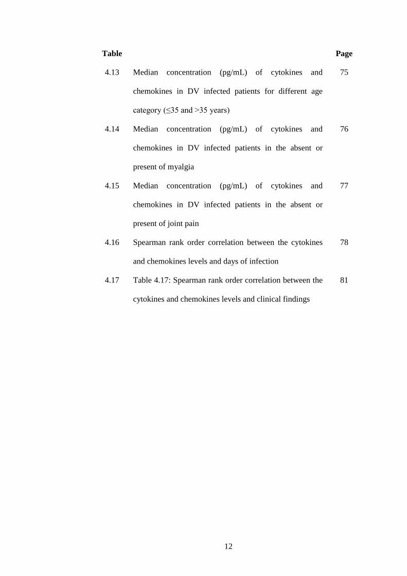
12
Table Page
4.13 Median concentration (pg/mL) of cytokines and
chemokines in DV infected patients for different age
category (≤35 and >35 years)
75
4.14 Median concentration (pg/mL) of cytokines and
chemokines in DV infected patients in the absent or
present of myalgia
76
4.15 Median concentration (pg/mL) of cytokines and
chemokines in DV infected patients in the absent or
present of joint pain
77
4.16 Spearman rank order correlation between the cytokines
and chemokines levels and days of infection
78
4.17 Table 4.17: Spearman rank order correlation between the
cytokines and chemokines levels and clinical findings
81

13
LIST OF FIGURES
Figure Page
2.1 Countries or areas at risk of dengue transmission in 2010
(WHO, 2010)
9
2.2 Incidence rate of dengue fever in Malaysia (per 100,000)
population from 2004 to 2008 (MOH, 2009)
10
2.3 Interrelation among the cytokines and chemokines 29
4.1 The results for NS1 antigen and DV IgM test for DV-
infected patients
56
4.2 The concentrations of IL-6 (pg/mL) in the serum of DV-
infected patients and healthy controls
58
4.3 The concentrations of IL-8 (pg/mL) in the serum of DV-
infected patients and healthy controls
60
4.4 The concentrations of IL-15 (pg/mL) in the serum of
DV-infected patients and healthy controls
62
4.5 The concentrations of IL-16 (pg/mL) in the serum of
DV-infected patients and healthy controls
64
4.6 The concentrations of IFN-γ (pg/mL) in the serum of
DV-infected patients and healthy controls
66
4.7 The concentrations of MCP-1 (pg/mL) in the serum of
DV-infected patients and healthy controls
68
4.8 The concentrations of MIG (pg/mL) in the serum of DV-
infected patients and healthy controls
70

14
Figure Page
4.9 The concentrations of RANTES (pg/mL) in the serum of
DV-infected patients and healthy controls
72
5.1 Proposed cytokine network and mechanism for
immunopathogenesis of dengue
103

15
LIST OF ABBREVIATIONS
AIDS Acquired immunodeficiency syndrome
APC Antigen-presenting cells
Bcl-2 B-cell lymphoma-2
C Nucleocapsid
CCL Chemokine (C-C motif) ligand
DC Dendritic cells
DF Dengue fever
DHF Dengue haemorrhagic fever
DV Dengue virus
DSS Dengue shock syndrome
E Envelope
ENA-78 Epithelial neutrophil activating peptide 78
G-CSF Granulocyte colony-stimulating factor
GM-CSF Granulocyte macrophage colony stimulating factor
HCV Hepatitis C virus
HepG2 Human liver carcinoma cell line
HIV Human immunodeficiency virus
HMC-1 Human mast cells line-1
HMEC-1 Human dermal microvascular endothelial cells-1
HRP Horseradish peroxidase
HUVEC Human umbilical cord veins endothelial cells
H2O2 Hydrogen peroxide
IFN Interferon

16
IL Interleukin
IP-10 IFN-gamma inducible protein-10
I-TAC Interferon-inducible T-cell alpha chemoattractant
JE Japanese encephalitis
kb kilo-base pair
kDa kilo-Dalton
KU812 Human basophil cells line
LCF Lymphocyte chemoattractant factor
M Membrane-associated
MAb Monoclonal antibody
MCP Monocyte chemoattractant protein
MHC Major histocompatibility complex class
MIG Monokine induced by interferon-gamma
MIP Macrophage inflammatory protein
NK Natural killer
NKT Natural killer T
NS Nonstructural
O.D. Optical density
PBMC Peripheral blood mononuclear cells
PDGF Platelet-derived growth factor
RANTES Regulated upon activation, normal T-cell expressed and
secreted
SARS Severe acute respiratory syndrome
TGF Transforming growth factor
Th1 Type 1 helper T

17
Th2 Type 2 helper T
TMB Tetramethylbenzidine
TNF Tumor necrosis factor

18
CHAPTER 1
INTRODUCTION
Dengue is the most important mosquito-borne disease in the world in terms of
morbidity, mortality, and economic costs, (Sang, Cuzzubbo, & Devine, 1998)
with an estimation of up to 100 million of individuals infected annually (King,
Anderson, & Marshall, 2002). About 0.5% of these infections result in
potentially fatal dengue haemorrhagic fever (DHF) and dengue shock
syndrome (DSS) (Khanam, Khanna, & Swaminathan, 2006). Besides, this
disease is also affecting more than 100 countries in the tropical and subtropical
regions which include Africa, Americas, India, Southeast Asia and Western
Pacific.
Dengue is transmitted from human to human mainly by the mosquito Aedes
aegypti (McBride & Bielefeldt-Ohmann, 2000). This mosquito is a highly
domesticated mosquito that lives in urban environments, breeding in small
collections of clean water in and around human habitats (Jacobs, 2005). Only
the females seek blood meals and they feed principally during the day, and
feed repeatedly on different hosts, enhancing their role as vectors (Solomon &
Mallewa, 2001). Besides, Aedes albopictus also plays an important role as a
vector in the dengue virus (DV) transmission (Clyde, Kyle, & Harris, 2006).
This vector prefers common forested habitats in suburban and rural areas. The
microhabitats of A. albopictus larvae are mainly found in tree holes and a wide
variety of containers including natural and artificial containers (Cecilio,

19
Campanelli, Souza, Figueiredo, & Resende, 2009). In addition, Aedes
polynesiensis was found to be another Aedes mosquito that amplifies the
spreading of dengue (Cao-Lormeau, 2009).
In the transmission of the DV, the female Aedes mosquito must bite an
infected human during the viraemic phase of the illness. This viraemic phase
of DF generally last 4 to 5 days but may also last up to 12 days (McBride &
Bielefeldt-Ohmann, 2000). Then, this female Aedes mosquito becomes
infective after an extrinsic incubation period of 7–14 days (Teo, Ng, & Lam,
2009). During this time, viral replication occurs in different mosquito tissues
and the virus finally infects the salivary glands of the mosquitoes (Cao-
Lormeau, 2009). DV is then transmitted back to humans by Aedes mosquito
bites. The incubation period of 4-6 days occurred and is followed by viraemic
phase of illness (Simasathien & Watanaveeradej, 2005). The cycle is the
repeated and caused the spreading of this viral infection.
DV is a lipid-enveloped RNA virus that belongs to the family Flaviviridae
(Chareonsirisuthigul, Kalayanarooj, & Ubol, 2007). The genomic RNA is
approximately 11 kb in length and is composed of three structural protein
genes, which includes nucleocapsid (C) protein, a membrane-associated (M)
protein, an envelope (E) protein and seven nonstructural (NS) protein genes
(NS1, NS2a, NS2b, NS3, NS4a, NS4b, and NS5) (Kumarasamy et al., 2007).
The largest structural protein is the E protein, which consists of 500 amino
acids in three antigenic domains. It is thought to be important in viral entry
into host cells, as well as being the major target of the humoral immune

20
response (Guzmán & Kourí, 2001) which involves the induction of
neutralizing antibodies and development of protective immune response in the
host (Pang, Cardosa, & Guzman, 2007). M protein is thought to be involved
in virion release (Clyde et al., 2006) and the C protein is a structural protein
involved in virion assembly (Solomon & Mallewa, 2001).
There are four antigenically distinct serotypes (dengue serotype-1, 2, 3 and 4)
(King, Anderson, & Marshall, 2002) for DV. Any of the four DV can cause
dengue infection (Azizan et al., 2006) and patients infected by DV may
display DF, or a more severe form of dengue called DHF or DSS (Lee et al.,
2007). DF is a self-limiting illness characterized by fever, headache, myalgia,
arthralgia, nausea, mild thrombocytopenia, fatigue (V´azquez et al., 2005) and
sometimes with the presence of rash (K. Huang et al., 2000). The higher viral
level is DHF, and it is the potentially life-threatening form of DV infection
(Pichyangkul et al., 2003). World Health Organization (WHO) categorizes
DHF into four grades (WHO, 1997), from less severe form of illness in grade I
to more severe form of illness in grade IV. Grade I is characterized by fluid
leakage where there is an increment of haematocrit by 20%, presence of
thrombocytopenia (<100 000/mL) and haemorrhage manifestation or positive
tourniquet test. Frank bleeding (e.g. gum, nose or gastrointestinal bleeding)
defines the occurrence of grade II. Plasma leakage in DHF grades III and IV,
also known as DSS, can be so profound that shock (undetectable blood
pressure) can occur (Lee et al., 2007). Cases of DHF/DSS can be fatal and
have a case fatality rate as high as 44%, unless plasma leakage is corrected
early (Azizan et al., 2006). Although infection with one serotype of DV

21
confers life-long protective immunity to that serotype, it does not protect the
host from infection with other serotypes (Fink et al., 2007). Hence, infection
with one serotype does not induce solid immunity to the others, and
individuals may be infected with dengue more than once (Jacobs, 2005).
The immune system contributes to the maintenance of physiological integrity
of the body mainly by eliminating foreign material and infectious pathogen
(Chaturvedi, Shrivastava, & Upreti, 2004). As soon as viruses try to establish
a site of infection, the host launches a complex defense system. Innate
immunity is a non-specific response and serves as the first-line of defense
where phagocytes, such as neutrophils and macrophages, and natural killer
(NK) cells play central roles in neutralizing and clearing of pathogen
(Matsukawa, Hogaboam, Lukacs, & Kunkel, 2000). Whilst the adaptive
immune response, which provides long-lasting protection, takes days to
develop and requires somatic mutations leading to the development of antigen-
specific T cell receptor (cell-mediated immunity) and immunoglobulins
(humoral immunity). Members of the cytokine and chemokine superfamily
are crucially involved in both innate and adaptive immune responses (Esche,
Stellato, & Beck, 2005).
Cytokines are proteins of low molecular weight and are locally acting tissue
hormones that normally function as autocrine and paracrine regulators (Berczi
& Szentivany, 2003). They comprise of interleukins (IL), tumor necrosis
factors (TNF), chemokines, interferons (IFN), mesenchymal growth factors,
transforming growth factor β and others (Sommer, 2006). Cytokines regulate

22
growth, differentiation, function, inhibition and apoptosis of cells in various
tissues (Berczi & Szentivany, 2003). The subclass of cytokine are named
chemokines, which comprised of a large family of small proteins consisting of
80-130 amino acids with molecular masses ranging from 6–14 kDa (Seet &
McFadden, 2002). They have a homologous structure consisting of anti-
parallel -strands with connecting loops held together by disulfide bonds
between cysteine residues (Melchjorsen, Sørensen, & Paludan, 2003).
Chemokines are further subclassified on the basis of the arrangement of the
cysteine residues located in the N-terminal region, as designated C, CC, CXC,
and CX3C members, in which C represents the number of cysteine residues in
the N-terminal region and X denotes the number of intervening amino acids in
between the first two cysteines (Esche et al., 2005). The CXC subfamily is
sometimes further classified into ELR+ and ELR- types based on the presence
or absence of a triplet amino acid motif (Glutamic acid-Leucine-Arginine) that
precedes the first cysteine residue in the primary structure of these chemokines
(Chensue, 2001). The CXC ELR+ chemokines are predominantly neutrophil
attractants and activators, while the CXC ELR- chemokines are mainly
chemotactic for T cells (Le, Zhou, Iribarren, & Wang, 2004). Upon infection,
viral products trigger the release of cytokines, which in turn are potent
inducers of chemokines. Primary cytokines act as endogenous activators of
the immune response, whereas inducible chemokines act as secondary
mediators to attract leukocytes. Interaction between cytokines and
chemokines will then enhance the antiviral response in the host (Gouwy,
Struyf, Proost, & Damme, 2005).

23
There are a number of research that demonstrated the elevation levels of
certain cytokines during DV infection (Medin, Fitzgerald, & Rothman, 2005)
and thus, cytokines and chemokines have been thought to play an important
role in the immunopathogenesis of DV infection (Chen & Wang, 2002). In
this study, a number of cytokines and chemokines namely IL-6, IL-8, IL-15,
IL-16, IFN-γ, MCP-1, MIG and RANTES, were chosen to study the
immunopathogenesis of DV infection. IL-6, IL-8, IFN-γ and MCP-1 were
chosen in this research since these cytokines and chemokines are associated
with thrombocytopenia (Bozza et al., 2008; Rachman & Rinaldi, 2006) and
plasma leakage (Lee et al., 2007; Medin et al., 2005; Schroder, Hertzog,
Ravasi, & Hume, 2004; Yamada et al., 2003), where both are seen present in
dengue patients. Thus, these cytokines and chemokines might have
contributions towards the immunopathogensis of DV infection. In addition,
IL-6 and IL-8 are endogenous pyrogen (Pela´ et al., 2000; Restrepo et al.,
2008b), which might cause fever to dengue patients. Hence, these cytokine
and chemokine were chosen in this research. Furthermore, cytokine rarely, if
ever, act alone in an immune system. A combined effect of two or more
cytokines is sometimes greater that the additive effects of the individual
cytokines. This combined effect of the cytokines ensures the outcome where
the magnitude of defense system is sufficient to clear invading virus.
Hence, the objective of this study is to highlight the point of which cytokines
and chemokines contribute to the immune response of DV-infected patients.
Also, this research was aimed to propose the possible correlation of the named
cytokines and chemokines and the possible immunological pathways involved

24
by these cytokines and chemokines that leads to the pathogenesis of this
disease.

25
CHAPTER 2
LITERATURE REVIEW
2.1 Epidemiology of Dengue
Dengue epidemics that are clinically compatible with dengue fever (DF)
occurred as early as 1635 and 1699 in the West Indies and Central America
respectively. Another literature also revealed an epidemic of knee fever both
in Cairo, Egypt and Jakarta, Indonesia in 1979 (Simasathien &
Watanaveeradej, 2005). However, the first accurate clinical description of DF
was disclosed by Benjamin Rush from Philadelphia, USA in 1780 where he
named this disease as ―break bone fever‖ (Solomon & Mallewa, 2001).
During the Second World War South-East Asia experienced the co-circulation
of multiple DV serotypes and epidemic activity increased (Rigau-Pérez et al.,
1998). Co-circulation of multiple serotypes resulted in the emergence of
epidemic DHF in 1950s. The first outbreak of DHF in Asia was recognized in
Manila, Philippines in 1953-1954 (Chaturvedi & Nagar, 2008) followed by an
outbreak in Bangkok, Thailand, in 1958. Singapore, Malaysia and Vietnam
experienced this DHF epidemic in the 1960s (Teo et al., 2009). Furthermore,
the situation had worsened where there was a dramatic increase in frequency
and in geographic extension of DF in Latin America and Brazil in the last two
decades (Bozza et al., 2008). It is now the most widely distributed mosquito-
borne virus disease and occurring in mostly every tropical and subtropical
country (Figure 2.1).

26
Figure 2.1: Countries or areas at risk of dengue transmission in 2010 (WHO,
2010)
In Malaysia, dengue continues to be a major health threat a century after its
first reported outbreak in 1902 (Chua et al., 2006). Examination of the
available outbreak data suggested that the major DF/DHF outbreak occurred in
Malaysia in a cyclical pattern of approximately every 8 years (Abubakar &
Shafee, 2002). However, the outbreak of disease had now changed to occur
yearly (Senior, 2007). According to the data from Ministry of Health (MOH),
Malaysia (2009), the incidence rate (per 100,000 population) of dengue in
Malaysia had increased from 50.92 in the year 2004 to 167.76 in the year 2008.
Also the mortality rate (per 100,000 population) of dengue was within 0.02

27
through out these 5 years (Figure 2.2). Thus, this disease had brought a big
impact to health, social and even economy for this country.
20042005
20062007
2008
167.76
85.78
64.3760.71
50.92
0
20
40
60
80
100
120
140
160
180
Incidence Rate
(per 100,000
population)
Year
Incidence Rate of Dengue Fever in Malaysia (per 100,000 Population)
From 2004 to 2008
Figure 2.2: Incidence rate of dengue fever in Malaysia (per 100,000)
population from 2004 to 2008 (MOH, 2009)
2.2 Pathophysiology of Dengue
The immune system plays an important role in the maintenance of
physiological integrity of the body. It eliminates foreign materials and
infectious pathogens that invade the host. This process is mediated through
innate and adaptive immunity, which is a complicated process involving
coordinated efforts of several types of cells and their secretory products
(Chaturvedi et al., 2004). Cytokines are proteins secreted during innate and
adaptive immunological responses, acting as inflammatory mediators or
modulator molecules. During DV infections, cytokines are involved in the

28
disease onset and homeostatic regulation (Bozza et al., 2008). Also, they play
an important role in the pathogenesis of DV infection (Chen & Wang, 2002).
There are numbers of research that investigated the regulations of cytokines in
the virus infected cells (Cardier et al., 2006). King et al. (2002) showed that
elevated levels of secreted macrophage inflammatory protein (MIP)-1 α , MIP-
1 β, and regulated upon activation, normal T-Cell expressed and secreted
(RANTES), but not IL-8 or epithelial neutrophil activating peptide (ENA)-78,
were observed following infection of human basophil cell line (KU812) or
human mast cells line (HMC-1) with DV. Chen and Wang (2002) further
demonstrated the release of other cytokines and chemokines in DV-infected
monocyte and macrophage, including TNF-α, IFN-α, IL-1β and IL-12, but not
IL-6, IL-15, or nitric oxide. The up-regulation of TNF- α in DV-infected cell
was confirmed by Lin et al. (2002) where they showed that infection by DV
on B cells induced the production of TNF-α. However, Lin et al. (2002) found
the infected B cells induced the production of IL-6, which was contradictory
to the results obtained by Chen and Wang (2002). These contradictory results
might be due to the difference in the cell line used in the respective research.
On the other hand, a number of different cytokines was studied by Azizan et al.
(2006). They reported that there was an increased level in IL-4, IL-10, INF-γ
and granulocyte macrophage colony stimulating factor (GM-CSF), within DV-
infected pulmonary endothelial cell line HPMEC-ST1.6R. Also, higher levels
of IL-6 and RANTES were released from DV-infected human primary lung
epithelial cells (Lee et al., 2007)

29
Serum levels of certain cytokines were also reported to be elevated during DV
infection (Chen & Wang, 2002). Elevation of IL-12 level in dengue patients
was demonstrated by Pacsa et al. in 2000. Mustafa, Elbishbishi, Agarwal, &
Chaturvedi (2001) further demonstrated that IL-13 and IL-18 were up-
regulated in DF patients and also have a higher magnitude in DHF patients.
Increased levels of other cytokines and chemokines, including IL-2, IL-6, IL-8,
IL-10, IFN-γ, TNF-α and monocyte chemoattractant protein (MCP)-1, were
also observed in patients with DV infection (Lin et al., 2005). This result was
confirmed by Chen et al. (2007) where they showed the presence of IFN- α,
IFN- γ, and IL-10 in the plasma of dengue patients. However, IL-11 was not
detected in DF and DHF patients (Cardier et al., 2006). Also, high
concentrations of two chemokines named IFN-gamma inducible protein 10
(IP-10) and interferon-inducible T-cell alpha chemoattractant (I-TAC) were
present in the serum of dengue patients (Fink et al., 2007).
However, the physiological roles of cytokines might be different (Salazar-
Mather, Hamilton, & Biron, 2000); and thus it is important to investigate the
regulation of different cytokines that are released from the DV-infected
patients.
2.3 Interleukin-6 (IL-6)
In 1986, Kishimoto and collaborators first cloned a DNA encoding new
human IL, T cell-derived B-cell differentiation factor (Streetz, Luedde, Manns,
& Trautwein, 2000), which was later named IL-6 (Maggio, Guralnik, Longo,

30
& Ferrucci, 2006). IL-6 belongs to the IL-6 family of cytokines, including IL-
11, oncostatin M, leukemia inhibitory factor, ciliary neurotrophic factor,
cardiotrophin-1 and cardiotrophin-like cytokine. These cytokines are
characterized by their common use of the gp130 receptor as a signaling
subunit (Cronstein, 2007). The human IL-6 protein comprises 212 amino
acids with a signal peptide of 27 amino acids and two potential NH2-linked
glycosylation sites. The molecular weight ranges from 21 to 28 kDa
(Kristiansen & Mandrup-Poulsen, 2005).
IL-6 is produced mainly by monocytes and macrophages (Cronstein, 2007)
and in a smaller percentage by fibroblasts, endothelial cells, T cells, B cells,
chondrocytes and keratinocytes (Łukaszewicz, Mroczko, & Szmitkowski,
2007). However, IL-6 is not constitutively expressed, but is readily induced
by multiple stimuli, including DNA and RNA virus infection (Streetz et al.,
2000). The production of IL-6 is also in response to many cytokines like
TNF-, IL-1, platelet-derived growth factor, IFN-γ (Yap & Lai, 2010) and IL-
16 (Qi et al., 2002) but dampened by IL-4, IL-10, and IL-13 (Yap & Lai,
2010).
Because of its multiple activities, it has been suggested that IL-6 is the main
factor involved in host response to a foreign pathogen. Its importance lies in
the stimulation of B cells differentiation and induction of permanent
differentiation of B cells into plasma cells which produce different classes of
immunoglobulin (Łukaszewicz et al., 2007). Besides, IL-6 promotes
inflammatory events through the T cells and NK proliferation, differentiation
31
and survival (Jones, 2005). It can also act as an endogenous pyrogen and
interact with nervous and endocrine systems to modify host defense responses
(Luheshi & Rothwell, 1996) as well as affect the permeability of the
endothelium (Restrepo et al., 2008b).
IL-6 was shown to play important role in several virus infections. In the study
done by Huang et al. (1999), they demonstrated that the serum level of IL-6
was significantly elevated in the patients with Hepatitis C, a disease infected
from virus of Flaviviridae family, as compared with the controls. The level of
IL-6 was also significantly increased in the microgial following Japanese
Encephalitis (JE) Virus infection (Ghoshal et al., 2007). On the other hand,
human umbilical cord veins endothelial cells (HUVEC) produced large
amounts of IL-6 after DV infection (Y. Huang et al., 2000). Also, Pinto,
Oliveira, Braga, Nogueira and Kubelka (1999) proved that IL-6 level was
significantly higher in dengue patients than in the controls. In addition, Chen
et al. (2006) confirmed that serum level of IL-6 was significantly increased in
adult dengue patients. Similar results was obtained by Restrepo et al. (2008a)
where they reported that there was significantly higher serum levels of IL-6 in
children with dengue than without dengue. However, Chen and Wang (2002)
failed to detect the presence of IL-6 in the DV-infected
monocytes/macrophages. Moreover, the level of IL-6 in all dengue cases was
not significantly higher than healthy controls (Priyadarshini et al., 2010).
These opposing results had therefore brought about the aim of this cytokine to
be studied in this research.

32
2.4 Interleukin-8 (IL-8)
Despite the tremendous interest in chemokines as a whole, the only chemokine
with an IL designation is IL-8. According to the new chemokine
nomenclature, IL-8 is now referred to as CXCL8 (Lin et al., 2005). In 1987,
IL-8 was discovered as a neutrophil chemotactic factor (Yoshimura et al.,
1987) and it was purified from supernatants of lipopolysaccharide stimulated
human monocyte cultures (Payne & Cornelius, 2002). IL-8 is a member of
ELR+ CXC chemokine family which contains the three amino acid sequence
of glutamic acid-leucine-arginine that immediately precedes the first cysteine
amino acid residue in the primary structure of the protein. This ELR+ CXC
chemokine has its primary biological effect in promoting neutrophil
recruitment and angiogenesis (Lin et al., 2005). Transcription of the IL-8 gene
encodes for a protein of 99 amino acids that is subsequently processed to yield
a 72 amino acids with molecular weight of 8400 Dalton (Waugh & Wilson,
2008), which is the major form secreted by monocytes and macrophages (Brat,
Bellail, & Meir, 2005)
Normally, IL-8 protein is barely secreted from non induced cells, but its
production is rapidly induced by a wide range of stimuli encompassing pro-
inflammatory cytokines such as TNF, IL-1 (Taub, Anver, Oppenheim, Longo,
& Murphy, 1996), IL-15 (Jabłońska et al., 2003), IL-6 and IFN-γ (Brat et al.,
2005). The production of IL-8 is also in response to the stimuli like bacterial
and viral products (Medin et al., 2005). IL-8 production had been observed in
vitro in a wide variety of cells including monocytes, macrophages, neutrophils,

33
NK cells (Mukaida, 2003) and even non-leukocytic somatic cells like
endothelial cells, fibroblasts and epithelial cells (Juffrie et al., 2000). The
secreted IL-8 has numerous roles, including inflammation, cell recruitment,
lymphoid trafficking, wound healing and angiogenesis. The diverse biological
functions of IL-8 are vital in the recruitment of basophils, eosinophils,
neutrophils (Y. Huang et al., 2000) and naïve T cells to the site of infection
(Bosch et al., 2002). Also, IL-8 induces a respiratory burst of neutrophils,
release of lytic enzymes, platelet-activating factor and leukotrienes (Medin et
al., 2005), which are all inflammatory reactions to rid the host of invading
pathogen.
Studies have shown that IL-8 is crucial in the defense system in several viral
infections. In the study done by Juffrie et al. (2000) whey they illustrated that
the level of IL-8 in dengue patients was increased as compared to the controls.
In vitro studies were carried out to study the regulation of IL-8 in DV infection
by a number of researchers to show the importance of this chemokine in
pathogenesis of DV infection. Chen and Wang (2002) revealed that DV-
infected monocytes/macrophages induced the production of IL-8. Also, a
significant increased in IL-8 levels in the culture supernatant of primary
human monocytes infected with DV was found in the study done by Bosch et
al. (2002). Two yeara later, a similar result was obtained by Talavera, Castillo,
Dominguez, Gutierrez and Meza (2004) where they showed that DV-infected
human dermal microvascular endothelial cells (HMEC-1) induced the
production of IL-8. Moreover, in vitro infection of human myeloid or
endothelial cells with DV has been reported to induce secretion of IL-8

34
(Medin et al., 2005). All these results suggested the involvement of this
chemokine in the host defense against the DV infection. However, a
dissimilar result was obtained where the level of IL-8 was not significantly
elevated in DF patients as compared to normal individuals (Y. Huang et al.,
2000). King et al. (2002) also showed that level of IL-8 was not elevated in
DV-infected human mast cell/basophil line.
2.5 Interleukin-15 (IL-15)
IL-15 is a 14–15 kDa protein with 114 amino acids (Alpdogan & Brink, 2005)
in its structure. It is a member of the four-helix bundle cytokine family, which
includes IL-2, IL-3, IL-6 and IL-7 and granulocyte colony-stimulating factor
(G-CSF) (Ahmad, Ennaciri, Cordeiro, Bassam, & Menezes, 2007). It was first
isolated from a simian kidney epithelial cell line CV-1/EBNA in 1994
(Budagian, Bulanova, Paus, & Bulfone-Paus, 2006). IL-15 is a novel cytokine
related to type 1 helper T (Th1) response (Ueda et al., 2007) and was
originally discovered due to its ability to promote T cell proliferation (Wang et
al., 2005), a biological activity that is similar to IL-2 (Alpdogan & Brink,
2005). It has low secretion potential and soluble IL-15 is hardly detected in
normal physiological conditions (Hocke et al., 2007). This pro-inflammatory
cytokine is produced by macrophages (Ahmad et al., 2007), monocytes,
dendritic cells (DC) (Hu, Wang, Wang, Huang, & Dong, 2007), neutrophils
(Jabłońska et al., 2003) and epithelial cells (Hocke et al., 2007), but not by T
cells (Ramsborg & Papoutsakis, 2007), in response to the environmental
stimuli and infectious agent (Ueda et al., 2007).

35
IL-15 has been shown to play a vital role in the homeostasis and activation of
natural killer T (NKT) cells (Alpdogan & Brink, 2005), neutrophils and
eosinophils (Budagian et al., 2006). It activates NK cells proliferation and
regulates its cytotoxicity (D’Ettorre et al., 2006). Moreover, IL-15 increases
the production of IFN-γ from NK cells and T cells in enhancing Th1 response
(Azeredo et al., 2005). It is also needed for the maintenance and renewal of
viral-specific memory CD8+ T cells (Berard, Brandt, Paus, & Tough, 2003).
Besides, it is important for the growth and differentiation in T cells (Ramsborg
& Papoutsakis, 2007), B cells (Hu et al., 2007) and monocytes/macrophage
(Ueda et al., 2007).
According to Fawaz, Sharif-Askari, and Menezes (1999), higher concentration
of IL-15 was obtained after infection with influenza virus, a RNA virus, in
cultured peripheral blood mononuclear cells (PBMC). Besides, D’Ettorre et al.
(2005) also showed an enhancement in the production of IFN-γ by IL-15-
stimulated NK cells, which was isolated from the human immunodeficiency
virus (HIV)-infected patients. They also proved that IL-15 stimulated NK
cells to produce CC chemokines which are able to inhibit HIV infection and
replication. These results suggested that IL-15 is important in the immune
response against various viral infections.
On the other hand, Chen and Wang (2002) infected blood monocytes and
tissue macrophages with DV. They failed to detect the presence of IL-15 at
any time after DV infection. However, there was another research done by
Azeredo et al. in 2005 where they found that IL-15 was significantly elevated

36
in the plasma of most dengue patients during the early acute phase as
compared to healthy donors. These contrary results brought the aim of this
study to compare the level of IL-15 in the DV infected patients and healthy
controls.
2.6 Interleukin-16 (IL-16)
IL-16 was initially identified in 1982 by Cruikshank & Center as a T cell
chemoattractant factor that was secreted from mitogen- or antigen-stimulated
human PBMC. It was one of the first characterized cytokines with
chemoattractant activity for human T cells and therefore was originally
designated as lymphocyte chemoattractant factor (LCF) (Cruikshank, Kornfeld,
& Center, 2000).
This cytokine is synthesized by CD4+ T cells and CD8+ T cells and is
released in response to antigen, mitogen and histamine (Reich et al., 2001).
IL-16 is also produced in the circulation by activated monocytes and DC (Hu
et al., 2007). Production of IL-16 was also demonstrated in eosinophils
(Gianoukakis, Martino, Horst, Cruikshank, & Smith, 2003), mast cells
(Desnues, Raoult, & Mege, 2005) and B cells (Tsai et al., 2005). Besides,
non-immune cells like epithelial cells (Ferland, Flamand, Davoine, Chakir, &
Laviolette, 2004), fibroblasts (Gianoukakis et al., 2003), neuronal cells
(Cruikshank et al., 2000), microglial cells (Suzuki, Ishiguro, & Shimbo, 2003)
have been found capable of IL-16 production. IL-16 is synthesized as a non-
bioactive precursor protein (pro-IL-16) (Reich et al., 2001) which has 631

37
amino acids (Center, Kornfeld, Ryan, & Cruikshank, 2000). To generate
active IL-16, the 631 amino acid pro-protein is cleaved by caspase-3 to
generate a 121 amino acid protein that has biological activity after stimulation
with specific antigen or other cytokines like IL-4, GM-CSF, IL-1β or
transforming growth factor (TGF)- β (Liu et al., 2007).
Interleukin-16 is a T cell chemoattractant factor (Skundric, Zhou, Cruikshank,
& Dai, 2005) that contributes to the regulatory process of T cell recruitment
(Guo, Mittelbronn, Brabeck, Mueller, & Schluesener, 2004). The recruited T
cells then synthesize and secrete more IL-16 that may serve to function as a
positive feedback mechanism for further cell recruitment and activation (Wu
et al., 1999). Besides induction of T cells chemotaxis, IL-16 also induces the
migratory response in monocytes, eosinophils (Koike et al., 2002) and DC
(Lynch, Heijens, Horst, Center, & Cruikshank, 2003). Furthermore, IL-16
stimulate monocytes to produce pro-inflammatory cytokines like IL-6, TNFα,
IL-1α and IL-15 (Qi et al., 2002) and induce eosinophils to generate RANTES
(Bandeira-Melo et al., 2002), which then further evokes the immune response
towards the antigen. Besides, IL-16 up-regulates IL-2 receptor alpha on T
cells (Hu et al., 2007) which in turn expanding the activated T cells (Reich et
al., 2001). Although IL-16 has chemotactic properties, it is not a chemokine
as it lacks the structural motifs of this family of proteins (Liu et al., 2007). On
the other hand, IL-16 is also known to be preferentially induces a migratory
response in Th1 cells (Jin et al., 2009). It inhibits the production of IL-4 and
IL-5 from T cells and thus, impairs the type 2 helper T (Th2) immunity while
induces a greater migratory response in the Th1 subset (Hu et al., 2007).

38
There was one research done by Message and Johnston in 2004 where they
used the Rhinovirus as the model. Rhinovirus carries a similarity with DV as
both of the viruses are single stranded RNA. They demonstrated that
Rhinoviruses infection in bronchial epithelial cell lines and primary human
epithelial cells increased the production of IL-16. Pinto et al. (2000) also
found that the supernatant of influenza A virus-stimulated PBMC isolated
from HIV patients, contained higher level of IL-16 as compared to the control.
To our knowledge however, no findings exist concerning IL-16 levels in DV
infected patients. Therefore, the levels of IL-16 between the DV-infected
patients and healthy controls was determined and compared.
2.7 Interferon-Gamma (IFN-γ)
IFN was discovered as an antiviral agent during studies on virus interference
by Isaacs and Lindenmann in 1957. They reported that influenza virus-
infected chick cells produced a secreted factor, which later named as IFN,
mediated the transfer of a virus-resistant state against viruses. Although the
IFN were first identified and named for their potent ability to interfere with
and inhibit viral infections, it soon became apparent that they could be divided
into two categories (Ellis & Beaman, 2004). The first category, named type I
IFN is consisting of the IFN- and IFN- molecules. They are the classical
IFN induced in response to viral infections (Samuel, 2001). The second class
is solely composed of IFN-γ, which is also termed type II or immune IFN.
IFN-γ is not related to the type I IFN at both the genetic and the protein levels.

39
to the other IFN, it has a lower specific antiviral activity, but presents more
immuno-modulatory properties than the type I IFN (Teixeira, Fonseca,
Barboza, & Viola, 2005). Both types of IFN are crucial in the immediate
cellular response to viral infection, but immuno-modulatory activities of IFN-γ,
however, become important later in coordinating the immune response and
establishing an antiviral state for longer term of control (Schroder et al., 2004).
IFN-γ with the molecular weight of 34 kDa (Teixeira et al., 2005) is mainly
produced by Th1 cells, activated NK cells (Restrepo et al., 2008b) and
macrophages (Antoniou, Ferdoutsis, & Bouros, 2003). Additionally, CD8+ T
cells also produce IFN-γ after being activated (Yang, Wang, Asavaroengchai1,
& Dey, 2005). There is now evidence that other cells, such as B cells, NKT
cells, DC, and antigen-presenting cells (APC) secrete IFN-γ upon activation
(Schroder et al., 2004). IFN-γ secretion by NK cells and possibly APC is
likely to be important in early host defense against infection, whereas T cells
become the major source of IFN-γ in the adaptive immune response (Yang et
al., 2005). The production of IFN-γ is also controlled by other cytokine. For
example, macrophage recognition of many pathogens induces secretion of
chemokines which then in turn to attract NK cells to the site of inflammation.
This chemotaxis promotes IFN-γ synthesis in these cells. Macrophages and
NK cells in combination of chemokine stimulation will then further increases
the IFN-γ production (Schroder et al., 2004).

40
IFN-γ is a major product of Th1 cells (Chen et al., 2005) and it stimulates the
differentiation of naive T cells toward Th1 phenotype and inhibits Th2 cell
differentiation by suppressing the release of Th2 cytokine from activated T
cells (Teixeira et al., 2005). The inhibition of Th2 response further skews the
immune response toward a Th1 phenotype (Gattoni, Parlato, Vangieri,
Bresciani, & Derna, 2006). It also enhances the antigen presentation and
stimulates the development of CD8+ T cells (Antoniou et al., 2003). In
addition, it is crucial in the activation and differentiation of B cells, NK cells,
macrophages, and others (Gattoni et al., 2006). Exposure to IFN-γ greatly
enhances the activity of macrophages and induces them to secrete reactive
oxidants like nitric oxide and cytokines such as IL-1, IL-6, IL-8, and TNF- in
the killing of intracellular and some extracellular infections. On the other
hand, there has been increasing evidence that IFN-γ also down-regulates or
restricts immune responses. IFN-γ plays an important role in the maintenance
of T cell homeostasis and eliminates activated CD4+ T cells and CD8+ T cells
by inducing apoptosis (Yang et al., 2005).
There are number of research showing the importance of IFN-γ in DV
infection. Chen et al. (2005) found that IFN-γ was higher in the DV-infected
patients than those healthy controls. Similar results were obtained by
Chakravarti and Kumaria (2006) and Chen et al. (2006) where they found that
the serum levels of IFN-γ was significantly increased in DF patients than in
healthy controls. This result was proven again two years later by Bozza et al.
(2008) where IFN-γ was significantly increased in dengue patients as
compared to control samples. While in the research done by Restrepo and co-

41
workers (2008a), they showed a higher cytokine level in children with dengue
than without dengue, with statistically significant difference for IFN-γ.
Additionally, a parallel result was obtained by Priyadarshini et al. (2010)
where level of IFN-γ was higher in dengue patients than in the controls. The
involvement of IFN-γ suggested that this cytokine is critical for the immune
response against DV infection and that IFN-γ -mediated immune responses are
necessary for both the early and late clearance of the virus (Hsieh et al., 2006).
However, the exact role of IFN-γ in DV infection is still controversial.
Although some studies suggested that it protects the host against DV infection,
others argue that it contributes to the pathogenesis of DHF, the more severe
form of the illness (Diamond et al., 2000).
2.8 Monocyte Chemoattractant Protein-1 (MCP-1)
MCP-1, which is known as chemokine (C-C motif) ligand 2 (CCL2) (Dhillon
et al., 2009), consists of 76 amino acid residues (Coillie, Damme, &
Opdenakker, 1999). It is a member of the C-C chemokine family (Mori et al.,
2000). MCP-1 can be produced by several cells like monocytes, macrophages,
epithelial cells, endothelial cells, fibroblasts (Han, Han, Grenn, &
Quehenberger, 1999) and smooth muscle cells (Cleanthis, Bhattacharya,
Smout, Ashour, & Standby, 2007) but not T cells (Mori et al., 2000). It is
secreted in response to lipopolysaccharide, or cytokines including IL-1, IL-4,
IFN-γ, TNF-α (Zhou et al., 2001) and platelet-derived growth factor (PDGF)
(Yamamoto et al., 1999).
42
MCP-1 plays a pivotal role in anti-viral immune responses, such as the
recruitment of immune cells (Deauvieau et al., 2007). It recruits and activates
monocytes to the site of inflammation (Cai et al., 2009) after exposed to
activation stimuli, such as virus infection. MCP-1 then increases the
endothelial permeability by disrupting tight junctions among cells (Bozza et
al., 2008) to cause vascular leakage (Yamada et al., 2003) which is the
necessary action for leukocytes to leave the circulation and infiltrate tissues
(Mori et al., 2000) in clearing the invading virus. Also, MCP-1 actively takes
part in the T-cell-mediated immune response against intracellular pathogens
by controlling leukocyte recruitment to the site of infection and clearance of
the pathogen (Dessing, Sluijs, Florquin, & Poll, 2007). Besides, MCP-1 also
recruits NK cells (Han et al., 1999), macrophages, basophils, mast cells, DC
(Dhillon et al., 2009) and neutrophils (Dessing et al., 2007) to the sites of
inflammation.
Jin et al. (2009) showed that the plasma level of MCP-1 in adult acquired
immunodeficiency syndrome (AIDS) patients were significantly higher than in
their healthy controls. This showed that MCP-1 plays an important role in the
immune response of the patients infected with single stranded RNA virus.
The level of MCP-1 was significantly elevated in the JE virus-infected
microglial (Ghoshal et al., 2007). Again it showed that MCP-1 is not only
playing a vital role in the immune response of single stranded RNA virus-
infected patients, but also the immune response of the patients who are
infected with flavivirus. Lin et al. (2005) also showed that an increased of
MCP-1 was observed in patients with DV infections. These results lead the
43
objective in this study to compare the level of MCP-1 in DV-infected patients
and healthy controls in Malaysia.
2.9 Monokine Induced by Interferon-Gamma (MIG)
MIG is one of the members of ELR- (lacking a Glutamic acid-Leucine-
Arginine motif in the N-terminal region) CXC chemokines that are associated
with inflammatory reactions (Park et al., 2002). Monocytes and macrophages
are thought to comprise the majority of MIG secreting cells (Brice, Graber,
Hoffman, & Doolan, 2001). Besides, MIG is also secreted by eosinophils
(Dajotoy et al., 2004), neutrophils, APC, B cells (Berthoud, Dunachie, Todryk,
Hill, & Fletcher, 2009). The production of this chemokine is induced by IFN-
γ and thus, has the potential to amplify the IFN-γ signal. In consequence,
MIG is an important regulator in Th1 responses (Colvin, Campanella, Sun, &
Luster, 2004). In mediating leukocyte migration to sites of infection (Hsieh et
al., 2006), MIG mainly recruits T cells and NK cells in the immune response
upon activation (Dajotoy et al., 2004). MIG also influences the behavior of
non-immune cells where it inhibits endothelial cell proliferation resulting in
the inhibition of angiogenesis (Gorbachev et al., 2007).
Elevation in MIG was observed in Hepatitis C virus (HCV)-infected patients
in the study done by Bièche et al. (2005). On the other hand, Brainard et al.
(2007) also demonstrated that MIG level was significantly increased in HIV-
infected patients as compared to the levels in uninfected controls.
Furthermore, production of MIG was induced from DV-infected DC in the

44
study done by Dejnirattisai et al. (2008). These results suggest that MIG has
an essential role in the host defense system against viral infection.
MIG was selected for examination because its induction is reported to have a
uniquely stringent and specific requirement for IFN-γ (Salazar-Mather et al.,
2000). This allows it to provide a functional measure of IFN-γ activity in
addition of detecting the presence of IFN-γ in DV-infected patients directly.
In addition, the production of MIG is followed by the amplification of the
IFN-γ signal. This may be a more sensitive measure and an easier way to
detect the presence of bio-active IFN-γ (Berthoud et al., 2009) in DV-infected
patients.
2.10 Regulated upon Activation, Normal T-Cell Expressed and Secreted
(RANTES)
CCL5 or RANTES (regulated upon activation, normal T-cell expressed and
secreted) belongs to a family of immuno-regulatory cytokines called
chemokines and it is classified as C-C chemokines (Gianoukakis et al., 2003).
This chemokine is secreted by many different cell types, such as endothelial
cells, smooth muscle cells, macrophages (Cavusoglu et al., 2007), monocytes
(Gianoukakis et al., 2003), lympohcytes (Gleissner, Hundelshausen, & Ley,
2008) and platelets (Tang, Yeaman, & Selsted, 2002).
RANTES is a chemoattractant factor for monocytes and T cells (Gleissner et
al., 2008). It is found preferentially attracts Th1 cells rather than Th2 cells
45
(Kanda & Watanabe, 2003), which is important in the control of intracellular
pathogen. Also, it is crucial in the induction of NK cell activation, chemotaxis,
adhesion, and trans-endothelial migration (Chen & Wang, 2002).
Several studies proved that RANTES plays an important role in the immune
system during viral infection. Jin et al. (2009) showed that the levels of
RANTES in the adult AIDS patients were significantly higher than in their
healthy controls, as RANTES was found to be able to suppress HIV
replication (Appay & Rowland-Jones, 2001). In the study done by Sládková
and Kostolanský (2006) where they found that RANTES was produced after
epithelial cells and leukocytes infected by influenza A virus. Also, the level of
RANTES was being measured in the JE patients. Results showed that there
was an up-regulation of RANTES level in these flavivirus infected patients as
compared to the healthy controls (Winter et al., 2004).
In addition, there were a number of studies done to investigate the level of
RANTES in the supernatant of DV-infected cell line. An elevated level of
secreted RANTES was observed following DV infection of KU812 or HMC-1
(King et al., 2002). Medin et al. (2005) infected both of the human liver
carcinoma cell line (HepG2) and primary DC with DV. They also showed that
infection of both cells induced RANTES production. Interestingly, there was
a contrast result obtained by Pérez et al. (2004) where they reported that serum
levels of RANTES detected in Cuban patients hospitalized with dengue
infection were lower than those observed in the controls. Also, this is the only
research that detects the presence of RANTES in the human biological sample.

46
Thus, the levels of RANTES in the serum of DV-infected patients and healthy
controls were chosen to be studied.
2.11 Interrelation between the Cytokines and Chemokines
The cytokines and chemokines studied in this research are interrelated among
each other. This interrelation among cytokines and chemokines might enable
the host to have a desired magnitude of defense system to clear the invading
virus. Thus, interrelation involving IL-6, IL-8, IL-15, IL-16, IFN-γ, MCP-1,
MIG and RANTES, was revealed to figure out the immunopathogenesis of
DV infection (Figure 2.3).
Figure 2.3: Interrelation among the cytokines and chemokines

47
Upon DV infection, IL-6, IL-8, IL-15, IL-16, IFN-γ, MCP-1, MIG and
RANTES will be produced by a number of different leucocytes in magnifying
the immune response. Besides the induction of viral particles, the production
of these cytokines also takes place in response to other stimuli like cytokines.
In response to viral particles, IL-16 is synthesized by T cells (Reich et al.,
2001) and this cytokines might then stimulate the production of RANTES
from eosinophils (Bandeira-Melo et al., 2002), IL-6 and IL-15 from
monocytes (Qi et al., 2002). Production of IL-6 and IL-15 from monocytes
might then further stimulate the synthesis of IL-8 (Jabłońska et al., 2003; Brat
et al., 2005) from monocytes, macrophages, neutrophils and NK cells.
Besides, IL-15 will enhance the Th1 response by increasing the production of
IFN-γ from NK cells and T cells (Azeredo et al., 2005). Interferon-gamma
will in turn act as the inducer for the additional production of IL-6 and IL-8
(Gattoni et al., 2006). In addition, the production of MCP-1 (Zhou et al., 2001)
and MIG (Colvin et al., 2004) from monocytes or macrophages could be
induced by IFN-γ upon DV infection in order to recruit more leucocytes to the
site of infection for viral clearance.

48
CHAPTER 3
MATERIALS AND METHODS
3.1 Ethics Statement
This study was approved by the Medical Research and Ethics Committee,
Ministry of Health Malaysia. Informed consent was obtained from each
individual before inclusion in the study. The privacy and the confidentiality of
the patients were taken care by using indirect identifiers and anonymous
specimens without making any reference to identifying information of the
participants.
3.2 Clinical Samples
In this research, patients presenting dengue-like illness and were admitted in
Hospital Besar Kuala Lumpur, Malaysia from Sept 2007 to June 2009 were
included. Blood of the patients (n=25) who showed clinical symptoms like
fever, headache, myalgia, joint pain, and thrombocytopenia were collected.
For each patient, the day of onset of fever was designated as first day of illness.
The blood from the patients was collected after 6 days in average of onset of
fever. Patients with other parasitic infection, respiratory infection and/or other
diseases were excluded by the clinicians. As controls, bloods from healthy
volunteers (n=25) with no signs of infection, and who were not taking any
medications at the time of this study, were included.

49
Clinical symptoms presented by the patients were recorded and estimations of
leucocytes, lymphocytes, neutrophils, monocytes, eosinophils and basophils
counts for each patient were carried out at Hospital Besar Kuala Lumpur,
Malaysia. Data were collected from patients’ profiles.
3.3 Serum Preparation
Blood of the DV-infected patients and the healthy donors were clotted in the
serum collection tube (BD Vacutainer plus, U.S.A.). After that, the serum
collection tube was centrifugated (Hettich ZENTRIGUGEN, U.K.) at
9000rpm for 5 minutes. Serum was then collected and stored at -80°C
(SANYO, Japan) for future assay.
3.4 Laboratory Diagnosis
In all cases, dengue infection was confirmed by the serological test with the
detection of dengue NS1 antigen (Panbio Dengue Early ELISA) (Panbio,
Australia) and detection of DV IgM (CALBIOTECH, U.S.A.).
3.4.1 Detection of Dengue NS1 Antigen by Panbio Dengue Early ELISA
3.4.1.1 Samples and Reagents Preparation
Serum of DV-infected patients and healthy controls, Positive Controls,
Negative Controls and Calibrator were diluted 10X by mixing the sample with

50
Serum Diluent. Also Wash Buffer (1X) was prepared by adding Wash Buffer
Concentrate to ultra pure water (ELGA, UK).
3.4.1.2 Assay Procedures
Diluted serum, Positive Controls, Negative Controls and Calibrators were
pipetted (RAININ, U.S.A.) into the Anti-NS1 Antibody pre-coated microwells
and was then covered and incubated at 37°C ± 1°C for 1 hour. After that, the
wells were washed 6 times with 1X Wash Buffer and followed by the addition
of Horseradish Peroxidase (HRP) Conjugated Anti-NS1 Monoclonal Antibody
(MAb) into each well. Again, the wells were covered and incubated at 37°C ±
1°C for 1 hour. Then, the wells were washed 6 times with 1X Wash Buffer.
Tetramethylbenzidine (TMB) was then added into each well and was
subsequently incubated at room temperature for 10 minutes. The optical
density (O.D.) (TECAN, Switzerland) of each well at wavelength of 450nm
with a reference filter of 650nm was taken after the addition of Stop Solution.
Calculation was done as follows to obtain the index value in determining the
presence of dengue specific NS1.
Cut-off value = Calibrator mean O.D. X Calibrator Factor (in
this case, 0.51)
Index Value = (Sample O.D. / Cut-off value)
A positive result with more than 1.1 Index Value indicated the presence of
dengue NS1 antigen while sample with Index Value less than 0.9 showed the

51
negative results where there was absence of dengue NS1 antigen. Samples
with Index Value within 0.9-1.1 were repeated with the test.
3.4.2 Detection of DV IgM by Dengue Virus IgM ELISA
3.4.2.1 Samples and Reagents Preparation
Dilution factor of 21X were prepared for the serum of DV-infected patients
and healthy controls, Negative Controls, Positive Controls, and Calibrators by
mixing the samples with Sample Diluent. Wash Buffer (1X) was also
prepared by adding Wash Concentrate 20X with ultra pure water prior to assay
procedure.
3.4.2.2 Assay Procedures
Diluted serum, Positive Controls, Negative Controls and Calibrators were
dispensed into the Dengue Antigen pre-coated microwells and the plate was
incubated for 20 minutes at room temperature. After incubation, the wells
were washed 3 times with 1X Wash Buffer. Next, Enzyme Conjugated Anti-
IgM MAb was added into each well and the plate was incubated at room
temperature for 20 minutes. After that, the wells were washed 3 times with
1X Wash Buffer and TMB Substrate was added into each well. Wells were
then incubated for 10 minutes at room temperature and followed by the
addition of Stop Solution into each well. The O.D. of each well at wavelength

52
of 450nm with reference filter of 650nm was finally taken. Calculation was
done as follows in order to obtain the Index Value of each sample.
Cut-off value = Calibrator mean O.D. X Calibrator Factor (in
this case, 0.4)
Index Value = Sample O.D. / Cut-off value
Sample with Index Value more than 1.1 indicated the positive result where
there was presence of DV IgM while sample with Index Value less than 0.9
showed that there was no detectable DV IgM. This test was repeated if the
sample’s Index Value fell within the range of 0.9 to 1.1.
3.5 Detection of Cytokines and Chemokines in Serum of DV-Infected
Patients and Healthy Controls
Concentrations of IL-6, IL-8, IL-15, IL-16, IFN-γ, MCP-1, MIG and RANTES
were measured in the serum of 25 confirmed DV-infected patients and 25
apparent healthy controls. Standards were included in each assay and the
standard curves were used for the estimation of cytokine and chemokines
concentrations (in pg/ml).

53
3.5.1 Detection of IL-6 by Human IL-6 ELISA Kit II (BD OptEIATM
,
U.S.A.)
3.5.1.1 IL-6 Standard Preparation
In the preparation of standard, lyophilized recombinant human IL-6 was
reconstituted with required volume (noted on vial label) of standard/sample
diluent to prepare a 300pg/mL stock standard. The stock standard was
allowed to equilibrate for 15 minutes before making dilution. After that,
standard/sample diluent was added into six tubes labeled with 150 pg/mL, 75
pg/mL, 37.5 pg/mL, 18.8 pg/mL, 9.4 pg/mL and 4.7 pg/mL. Serial dilution
was then performed by adding fixed volume of each standard to the next tube
and vortexing between each transfer. The undiluted standard served as the
high standard (300pg/mL) while the standard/sample diluent served as the zero
Standard (0 pg/mL).
3.5.1.2 Working Detector Preparation
Working detector with dilution factor of 250X was prepared within 15 minutes
prior to use. In this preparation, required volume of biotinylated anti-human
IL-6 MAb was mixed well with required quantity of concentrated streptavidin-
HRP conjugate (250X) in a clean tube.
54
3.5.1.3 Wash Buffer Preparation
Wash buffer with dilution factor of 20X was prepared by mixing the wash
concentrate (20X) with ultra pure water.
3.5.1.4 Assay Procedures
First, ELISA diluent was added into anti-human IL-6 MAb pre-coated
microwells. Prepared standards and serum were then pipetted into each well.
The plate was gently tapped to ensure complete mixing. After that, plate was
covered with a plate sealer and was incubated for 2 hours at room temperature.
Next, the plate was washed 5 times with diluted wash buffer and followed by
the addition of working detector (was prepared within 15 minutes prior to use)
into each well. Again, the plate was covered with a plate sealer and was
incubated for 1 hour at room temperature. The plate was washed 7 times with
diluted wash buffer after the incubation. TMB one-step substrate reagent was
subsequently dispensed into each well and the plate was incubated without a
plate sealer for 30 minutes at room temperature in the dark. Stop solution was
then added into each well and the absorbance of each well at wavelength of
450nm with reference filter of 570nm was taken. Standard curve of IL-6 was
finally plotted and the concentrations of IL-6 in the serum of DV-infected
patients and healthy controls were determined (Appendix A).

55
3.5.2 Detection of IL-8 by Human IL-8 ELISA Kit II (BD OptEIATM
,
U.S.A.)
3.5.2.1 IL-8 Standard Preparation
Lyophilized recombinant human IL-8 was reconstituted with required volume
(noted on vial label) of standard/sample diluent to prepare a 200pg/mL stock
standard. The stock standard was then allowed to equilibrate for 15 minutes
before making dilution. Standard/sample diluent was subsequently added into
six tubes labeled with 100 pg/mL, 50 pg/mL, 25 pg/mL, 12.5 pg/mL, 6.3
pg/mL and 3.1 pg/mL. Serial dilution was then performed by adding fixed
volume of each standard to the next tube and vortexing between each transfer.
The undiluted standard served as the high standard (200pg/mL) while the
standard/sample diluent served as the zero standard (0 pg/mL).
3.5.2.2 Working Detector Preparation
Working detector with dilution factor of 250X was prepared within 15 minutes
prior to use. In this preparation, required volume of biotinylated anti-human
IL-8 MAb was mixed well with required quantity of concentrated streptavidin-
HRP conjugate (250X) in a clean tube.

56
3.5.2.3 Wash Buffer Preparation
Wash buffer with dilution factor of 20X was prepared by mixing the wash
concentrate (20X) with ultra pure water.
3.5.2.4 Assay Procedures
ELISA diluent was first added into anti-human IL-8 MAb pre-coated
microwells. Prepared standards and serum were then dispensed into each well.
This plate was covered with a plate sealer and was incubated at room
temperature for 2 hours. After that, the plate was washed 5 times with diluted
wash buffer. Plate was covered with a plate sealers and was incubated at room
temperature for 1 hour following the addition of working detector (was
prepared within 15 minutes prior to use) into each well. The plate was then
washed 7 times with diluted wash buffer and followed by the addition of TMB
one-step substrate reagent into each well. Again, the plate (without a plate
sealer) was incubated for 30 minutes at room temperature in the dark. After
that, stop solution was added into each well and absorbance of each well at
wavelength of 450nm with reference filter of 570nm was taken. Standard
curve of IL-8 was finally plotted and the concentrations of IL-8 in the serum
of DV-infected patients and healthy controls were calculated (Appendix B).

57
3.5.3 Detection of IL-15 by Human IL-15 Immunoassay (R&D Systems,
Minneapolis)
3.5.3.1 IL-15 Standard Preparation
Lyophilized recombinant human IL-15 was reconstituted with 1.0mL of ultra
pure water producing a stock standard of 2500 pg/mL. The stock standard was
mixed to ensure complete reconstitution and was agitated gently for 15
minutes prior to making dilution. Nine hundred micro liter of calibrator
diluent RD6-10 was pipetted into the tube labeled 250 pg/mL. Also, 500 μL
of calibrator diluent RD6-10 was pipetted into the remaining tubes which
labeled with 125 pg/mL, 62.5 pg/mL, 31.2 pg/mL, 15.6 pg/mL, 7.8 pg/mL and
3.9 pg/mL. Stock standard was then used to produce a dilution series and each
tube was mixed thoroughly before the next transfer. The 250 pg/mL standard
served as the high standard. The calibrator diluent RD6-10 served as the zero
standard (0 pg/mL).
3.5.3.2 Substrate Solution Preparation
Stabilized hydrogen peroxide (H2O2) (Colour Reagents A) and stabilized
chromogen (TMB) (Colour Reagent B) were mixed together in equal volumes
within 15 minutes of use and the resultant mixture was protected from light.

58
3.5.3.3 Wash Buffer Preparation
Wash buffer with dilution factor of 25X was prepared by mixing the wash
buffer concentrate with ultra pure water.
3.5.3.4 Assay Procedures
Assay diluent RD1-19 was added into mircowells pre-coated with mouse MAb
against IL-15. Prepared standards and serum were then added into each well.
The plate was covered with the adhesive strip provided and was incubated for
3 hours at room temperature. After that, the plate was washed 4 times with
diluted wash buffer and followed by the addition of anti-IL-15 conjugate
(mouse MAb against IL-15 conjugated to HRP). Then, the plate was covered
with a new adhesive strip and was incubated for 1 hour at room temperature.
Washing steps were then repeated as previous. After washing, substrate
solution (prepared within 15 minutes of use) was added into each well and the
plate was incubated for 30 minutes at room temperature in the dark.
Subsequently, stop solution was added into each well and O.D. of each well at
wavelength of 450nm with reference filter of 570nm was taken. Finally, the
standard curve of IL-15 was constructed and the concentrations of IL-15 in the
serum of DV-infected patients and healthy controls were calculated (Appendix
C).

59
3.5.4 Detection of IL-16 by Endogen® Human IL-16 ELISA Kit
(Thermo Scientific, U.S.A.)
3.5.4.1 IL-16 Standard Preparation
Lyophilized Escherichia coli (E.coli)-derived recombinant human IL-16
standard was reconstituted with ultra pure water. Tubes were labeled with
2000 pg/mL, 800 pg/mL, 320 pg/mL, 128 pg/mL, 51 pg/mL, and 0 pg/mL.
Serial dilution of 1:2.5 was carried out to prepare the above said standard
concentration in constructing the standard curve of IL-16.
3.5.4.2 Streptavidin-HRP Solution Preparation
Streptavidin-HRP concentrate was mixed with streptavidin-HRP dilution
buffer to obtain a diluted solution with 400X dilution factor. Streptavidin-
HRP Solution was prepared immediately before use.
3.5.4.3 Wash Buffer Preparation
Wash buffer (1X) was prepared by diluting 30X wash buffer with ultra pure
water.

60
3.5.4.4 Assay Procedures
Prepared standards and five-fold diluted serum were added into anti-human
IL-16 pre-coated microwells. The plate was covered with an adhesive plate
cover and incubated for 1 hour at room temperature. After that, without
washing, biotinylated antibody reagent was added into each well. The plate
was then covered with adhesive plate cover and incubated for 1 hour at room
temperature. Next, the plate was washed 3 times with 1X wash buffer and
streptavidin-HRP solution (was prepared immediately before use) was
subsequently added into each well. The plate was covered with a new
adhesive plate cover and incubated for 30 minutes at room temperature. Then,
the plate was washed 3 times with 1X wash buffer and followed by the
addition of TMB substrate solution into each well. After that, the plate
(without plate cover) was incubated at room temperature in the dark for 30
minutes. Stop solution was subsequently added into each well. The
absorbance of each well at wavelength of 450nm with reference filter of
550nm was taken. Standard curve for IL-16 was finally plotted and the
concentrations of IL-16 in the serum of DV-infected patients and healthy
controls were calculated (Appendix D).

61
3.5.5 Detection of IFN-γ by Human IFN-γ ELISA Kit II (BD OptEIATM
,
U.S.A.)
3.5.5.1 IFN-γ Standard Preparation
Lyophilized recombinant human IFN-γ was reconstituted with
standard/sample diluent to prepare a 300pg/mL stock standard. The stock
standard was allowed to equilibrate for 15 minutes before making dilutions.
Standard/sample diluent was then added into six tubes labeled with 150 pg/mL,
75 pg/mL, 37.5 pg/mL, 18.8 pg/mL, 9.4 pg/mL and 4.7 pg/mL. After that,
serial dilutions were performed by adding fixed volume of each standard to the
next tube and vortexing between each transfer. The undiluted standard served
as the high standard (300 pg/mL) whilst the standard/sample diluent served as
the zero standard (0 pg/mL).
3.5.5.2 Working Detector Preparation
Working detector with dilution factor of 250X was prepared within 15 minutes
prior to use. In this preparation, required volume of biotinylated anti-human
IFN-γ MAb was mixed well with required quantity of concentrated
streptavidin-HRP conjugate (250X) in a clean tube.

62
3.5.5.3 Wash Buffer Preparation
Wash buffer with dilution factor of 20X was prepared by mixing the wash
concentrate (20X) with ultra pure water.
3.5.5.4 Assay Procedures
ELISA diluent was pipetted into each microwell pre-coated with anti-human
IFN-γ MAb. Prepared standards and serum were then pipetted into each well.
Next, the plate was covered with a plate sealer and incubated for 2 hours at
room temperature. After incubation, the plate was washed 5 times with
diluted wash buffer, followed by addition of working detector (was prepared
within 15 minutes prior to use) into each well. The plate was again covered
with a plate sealer and incubated for 1 hour at room temperature. After that,
the plate was washed 7 times with diluted wash buffer and TMB one-step
substrate reagent was subsequently added into each well. Then, the plate
(without a plate sealer) was incubated for 30 minutes at room temperature in
the dark. Stop solution was added into each well after the incubation.
Absorbance of each well at wavelength of 450nm with reference filter of
570nm was taken. Standard curve of IFN-γ was plotted and the concentrations
of IFN-γ in the serum of DV-infected patients and healthy controls were
determined (Appendix E).

63
3.5.6 Detection of MCP-1 by Human MCP-1/CCL2 Immunoassay (R&D
Systems, Minneapolis)
3.5.6.1 MCP-1 Standard Preparation
Standard of MCP-1 (lyophilized recombinant human MCP-1) was
reconstituted with calibrator diluent RD6Q to produce a stock standard of
2000 pg/mL. The stock standard was allowed to sit for 15 minutes with gentle
agitation prior to making dilutions. Calibrator diluent RD6Q was then added
into tubes labeled with 1000 pg/mL, 500 pg/mL, 250 pg/mL, 125 pg/mL, 62.5
pg/mL and 31.2 pg/mL. Each tube was mixed thoroughly before the next
transfer. The 1000pg/mL standard served as the high standard while the
calibrator diluent RD6Q served as the zero standard (0 pg/mL).
3.5.6.2 Substrate Solution Preparation
Stabilized H2O2 (Colour Reagents A) and Stabilized chromogen (TMB)
(Colour Reagent B) were mixed together in equal volumes within 15 minutes
of use and the resultant mixture was protected from light.
3.5.6.3 Wash Buffer Preparation
Wash buffer with dilution factor of 25X was prepared by mixing the wash
buffer concentrate with ultra pure water.

64
3.5.6.4 Assay Procedures
Assay diluent RD1-83 was added to mouse MAb against MCP-1 pre-coated
microwells. Prepared standards and two-fold diluted serum was then added
into each wells. After that, the plate was covered with the adhesive strip
provided and was incubated for 2 hours at room temperature. Next, the plate
was washed 3 times with diluted wash buffer. Then, anti-MCP-1 conjugate
(polyclonal antibody against MCP-1 conjugated to HRP) was dispensed into
each well. The plate was covered with a new adhesive strip and was incubated
for 2 hours at room temperature. Washing steps was then repeated as previous.
Substrate solution (was prepared within 15 minutes of use) was subsequently
added into each well and the plate was incubated for 30 minutes at room
temperature in the dark. After incubation, stop solution was added into each
well. Finally, the O. D. of each well at wavelength of 450nm with reference
filter 570nm was determined. The standard curve for MCP-1 was plotted and
the concentration of MCP-1 in the serum of DV-infected patients and healthy
controls was calculated (Appendix F).
3.5.7 Detection of MIG by Human CXCL9/MIG Immunoassay (R&D
Systems, Minneapolis)
3.5.7.1 MIG Standard Preparation
MIG standard (lyophilized recombinant human MIG) was reconstituted with
ultra pure water producing a stock standard of 20 000 pg/mL. The Stock

65
Standard was mixed to ensure complete reconstitution and allowed to sit for
15 minutes with gentle agitation prior to making dilutions. Nine hundred
micro liter of calibrator diluent RD5P was pipetted into the tube labeled with
2000 pg/mL. Then, 500μL of calibrator diluent RD5P was pipetted into the
remaining tubes which labeled with 1000 pg/mL, 500 pg/mL, 250 pg/mL, 125
pg/mL, 62.5 pg/mL and 31.2 pg/mL. After that, the stock standard was used
to produce the dilution series. Each tube was mixed thoroughly before the
next transfer. The 2000 pg/mL standard served as the high standard while the
calibrator diluent RD5P served as the zero standard (0 pg/mL).
3.5.7.2 Substrate Solution Preparation
Stabilized H2O2 (Colour Reagents A) and Stabilized chromogen (TMB)
(Colour Reagent B) were mixed together in equal volumes within 15 minutes
of use and the resultant mixture was protected from light.
3.5.7.3 Wash Buffer Preparation
Wash buffer with dilution factor of 25X was prepared by mixing the wash
buffer concentrate with ultra pure water.
3.5.7.4 Assay Procedures
Assay diluent RD1W was pipetted into the microwells pre-coated with mouse
MAb against MIG. Prepared standards and serum was then added into each

66
well. Plate was covered with adhesive strip provided and was incubated for 2
hours at room temperature. After that, the plate was washed 4 times with
diluted wash buffer, followed by the addition of anti-MIG conjugate
(polyclonal antibody against MIG conjugated to HRP) into each well. Again,
the plate was covered with a new adhesive strip and was incubated for 2 hours
at room temperature. Washing steps was then repeated as previous. Substrate
solution (was prepared within 15 minutes of use) was subsequently added into
each wells and the plate was incubated for 30 minutes at room temperature in
the dark. Stop solution was later added into each well and O.D. of each well
at wavelength of 450nm with reference filter of 570nm was taken. Standard
curve for MIG was finally plotted and the concentrations of MIG in the serum
of DV-infected patients and healthy controls were determined (Appendix G).
3.5.8 Detection of RANTES by Endogen® Human RANTES ELISA Kit
(Thermo Scientific, U.S.A.)
3.5.8.1 RANTES Standard Preparation
Lyophilized E.coli-derived recombinant human RANTES standard was
reconstituted with ultra pure water. Tubes were then labeled with 2000 pg/mL,
800 pg/mL, 320 pg/mL, 128 pg/mL, 51.2 pg/mL, and 0 pg/mL. Serial dilution
of 1:2.5 was carried out to prepare the above said standard concentration in
constructing the standard curve of RANTES.

67
3.5.8.2 Streptavidin-HRP Solution Preparation
Streptavidin-HRP concentrate was mixed with Streptavidin-HRP dilution
buffer to obtain a diluted solution with 400X dilution factor. Streptavidin-
HRP solution was prepared immediately before use.
3.5.8.3 Wash Buffer Preparation
Wash buffer (1X) was prepared by diluting 30X wash buffer with ultra pure
water.
3.5.8.4 Assay Procedures
Prepared standards and 100-fold diluted serum were added into the well pre-
coated with anti-human RANTES. The plate was then covered with a new
adhesive plate cover and was incubated for 1 hour at room temperature. After
that, the plate was washed 3 times with 1X wash buffer. Following this,
biotinylated antibody reagent was added into each well. Again, the plate was
covered with a new adhesive plate cover and was incubated for 1 hour at room
temperature. Washing steps was repeated as previous after the incubation.
Streptavidin-HRP solution (was prepared immediately before use) was
dispended into each well. Next, the plate was cover with adhesive plate cover
and was incubated for 30 minutes at room temperature. The plate was washed
3 times again with 1X wash buffer and followed by the addition of TMB
substrate solution into each well. The plate (without the adhesive plate cover)

68
was then incubated for 30 minutes at room temperature in the dark. Stop
solution was subsequently added into each well. Absorbance of each well at
wavelength of 450nm with reference filter at 550nm was taken. Standard
curve of RANTES was finally constructed and the concentration of RANTES
in the serum of DV-infected patients and healthy controls was determined
(Appendix H).
3.6 Statistical Analysis
Differences between the groups were analyzed using the Mann-Whitney U test.
Spearman rank order correlation was used to study the correlation between the
cytokines and chemokines levels and clinical findings. Significance was set at
p < 0.05. All the statistical analyses of the data were done using SPSS
Statistics 19.0.

69
CHAPTER 4
RESULTS
4.1 Demographic Data of DV-Infected Patients
The demographic data of DV-infected patients was collected and summarized
in Table 4.1.
Table 4.1: Demographic data of DV-infected patients
Category Subcategory
Total number of dengue cases
25
Dengue (number of cases) Dengue Fever 24
Dengue haemorrhagic fever 1
Mean age (years ± SD) 28 ± 9
Average day of infection 6
Gender Males 17
Females 8

70
4.2 Clinical Signs and Symptoms of DV-Infected Patients
Clinical signs and symptoms of DV-infected patients are tabulated in Table
4.2.
Table 4.2: Clinical signs and symptoms of DV-infected patients
Clinical sign/symptom Number of cases in DF (%)
Fever 100
Myalgia 87.8
Joint pain 95.9
Headache 91.9
Nausea/vomiting 39.2
Rash 59.5
Thrombocytopenia 100

71
4.3 Laboratory Findings in DV-Infected Patients
Laboratory findings in DV-infected patients are shown in Table 4.3.
Table 4.3: Laboratory findings in DF
Variable
Normal
range
DV-infected patients
Leukocyte count, mean cells
(X 103 ± SD)
4-11 3.419 ± 1.442
No. of cases with leucopenia (%) - 73.5
Neutrophils count, mean cells
(X 103 ± SD)
2-7
1.325 ± 0.727
No. of cases with neutropenia (%) - 85.4
Lymphocytes count, mean cells
(X 103 ± SD)
1.5-4
1.370 ± 0.864
Lymphocytopenia (%) - 60.8
Monocytes count, mean cells
(X 103 ± SD)
0.2-0.8
0.902 ± 2.155
Monocytopenia (%) - 9.1
Eosinophils counts, mean cells
(X 103 ± SD)
0.04-0.4
0.0580 ± 0.1
Eosinopenia (%) - 65.5
Basophils counts, mean cells
(X 103 ± SD)
0.02-0.1
0.0833 ± 0.1
Basopenia (%) - 43.1

72
4.4 Laboratory Diagnosis for Dengue
Blood taken from DV-infected patients were diagnosed as ―confirmed and/or
positive dengue cases‖ by using NS1 antigen and DV IgM serological test
detection. All DV-infected patients included in this study had index value
more than 1.1, which is an indication of positive results for both NS1 antigen
and DV IgM tests and hence confirmed and/or positive for dengue infection
(Figure 4.1).
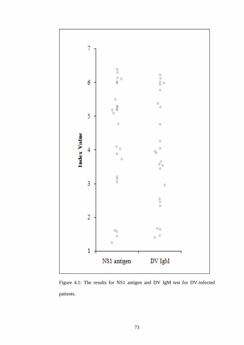
73
Figure 4.1: The results for NS1 antigen and DV IgM test for DV-infected
patients.

74
4.5 Median Levels of IL-6 in Serum of DV-Infected Patients and
Healthy Controls
Mann-Whitney U test reveals that there is a significant difference (p<0.05) in
the levels of IL-6 in serum of DV-infected patients and healthy controls. The
concentrations of IL-6 in serum of DV-infected patients and healthy controls
are illustrated in Figure 4.2. The median level of IL-6 in healthy controls was
approximately 3 times lower than the median level of IL-6 in DV-infected
patients as tabulated in Table 4.4.
Table 4.4: The median concentrations of IL-6 (pg/mL) in the serum of DV-
infected patients and healthy controls.
Cytokine Groups
Median
concentration
(pg/mL)
Concentration
range (pg/mL)
Mann-
Whitney U
test
P value
IL-6
Healthy
controls
5.03 1.31-10.13
p<0.001
DV-infected
patients
15.36 0.13-53.81

75
Figure 4.2: The concentrations of IL-6 (pg/mL) in the serum of DV-infected
patients and healthy controls.
Mean (pg/mL)
Controls: Patients:
5.38 17.13
----- Standard deviation
Controls: Patients:
2.17 12.61

76
4.6 Median Levels of IL-8 in Serum of DV-Infected Patients and
Healthy Controls
A significant difference (p<0.05) is found in levels of IL-8 in the serum of
DV-infected patients and healthy controls using Mann-Whitney U test and IL-
8 concentrations in serum of both groups are shown in Figure 4.3. The
increment of median IL-8 level in the serum of DV-infected patients was
about three times as compared to the median levels of IL-8 in serum of healthy
controls (Table 4.5).
Table 4.5: The median concentrations of IL-8 (pg/mL) in the serum of DV-
infected patients and healthy controls.
Chemokine Groups
Median
concentration
(pg/mL)
Concentration
range (pg/mL)
Mann-
Whitney U
test
P value
IL-8
Healthy
controls
30.92 15.41-60.58
p<0.001
DV-
infected
patients
86.78 3.14-269.65
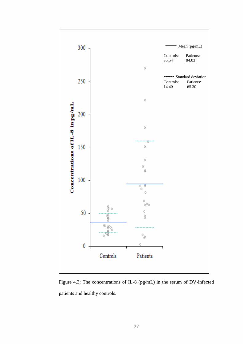
77
Figure 4.3: The concentrations of IL-8 (pg/mL) in the serum of DV-infected
patients and healthy controls.
Mean (pg/mL)
Controls: Patients:
35.54 94.03
----- Standard deviation
Controls: Patients:
14.40 65.30

78
4.7 Median Levels of IL-15 in Serum of DV-Infected Patients and
Healthy Controls
There is significant (p < 0.05) upregulated level of IL-15 in serum of DV-
infected patients as compared to healthy controls. Figure 4.4 shows the
concentrations of IL-15 in serum of DV-infected patients and healthy controls.
Table 4.6 tabulates the approximately two times increment in the median level
of IL-15 in serum of DV-infected patients as compared to the median level of
IL-15 in serum of healthy controls.
Table 4.6: The median concentrations of IL-15 (pg/mL) in the serum of DV-
infected patients and healthy controls.
Cytokine Groups
Median
concentration
(pg/mL)
Concentration
range (pg/mL)
Mann-
Whitney U
test
P value
IL-15
Healthy
controls
1.97 0.36-4.25
p<0.001
DV-
infected
patients
4.58 1.76-17.42

79
Figure 4.4: The concentrations of IL-15 (pg/mL) in the serum of DV-infected
patients and healthy controls.
Mean (pg/mL)
Controls: Patients:
2.09 5.25
----- Standard deviation
Controls: Patients:
1.03 3.62
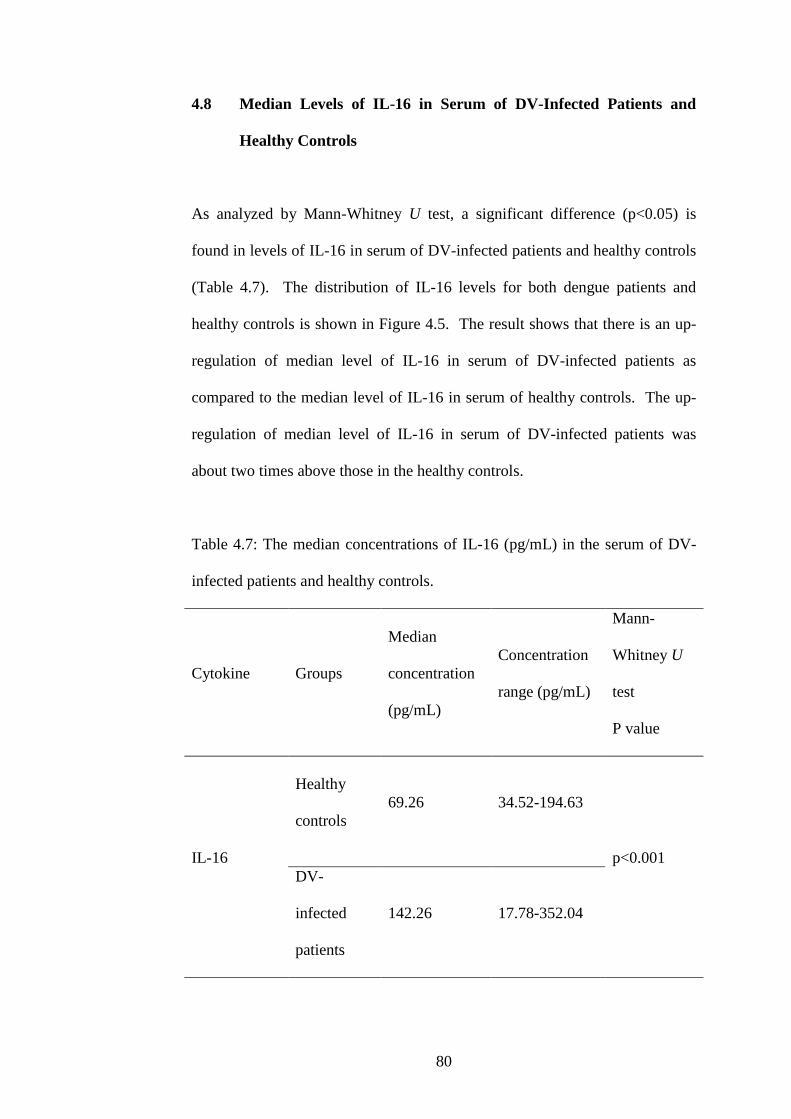
80
4.8 Median Levels of IL-16 in Serum of DV-Infected Patients and
Healthy Controls
As analyzed by Mann-Whitney U test, a significant difference (p<0.05) is
found in levels of IL-16 in serum of DV-infected patients and healthy controls
(Table 4.7). The distribution of IL-16 levels for both dengue patients and
healthy controls is shown in Figure 4.5. The result shows that there is an up-
regulation of median level of IL-16 in serum of DV-infected patients as
compared to the median level of IL-16 in serum of healthy controls. The up-
regulation of median level of IL-16 in serum of DV-infected patients was
about two times above those in the healthy controls.
Table 4.7: The median concentrations of IL-16 (pg/mL) in the serum of DV-
infected patients and healthy controls.
Cytokine Groups
Median
concentration
(pg/mL)
Concentration
range (pg/mL)
Mann-
Whitney U
test
P value
IL-16
Healthy
controls
69.26 34.52-194.63
p<0.001
DV-
infected
patients
142.26 17.78-352.04

81
Figure 4.5: The concentrations of IL-16 (pg/mL) in the serum of DV-infected
patients and healthy controls.
Mean (pg/mL)
Controls: Patients:
78.06 162.79
----- Standard deviation
Controls: Patients:
38.90 74.26

82
4.9 Median Levels of IFN-γ in Serum of DV-Infected Patients and
Healthy Controls
Mann-Whitney U test demonstrates a significant difference (p<0.05) in levels
of IFN-γ in serum of both DV-infected patients and healthy controls.
Concentrations of IFN-γ in serum of patients with DV infection and healthy
controls are plotted in Figure 4.6. As shown in Table 4.8, median level of
IFN-γ in serum of DV-infected patients was around two times higher than
those in the healthy controls.
Table 4.8: The median concentrations of IFN-γ (pg/mL) in the serum of DV-
infected patients and healthy controls.
Cytokine Groups
Median
concentration
(pg/mL)
Concentration
range (pg/mL)
Mann-
Whitney U
test
P value
IFN-γ
Healthy
controls
7.82 4.20-69.44
p<0.01
DV-
infected
patients
13.70 4.90-71.76

83
Figure 4.6: The concentrations of IFN-γ (pg/mL) in the serum of DV-infected
patients and healthy controls.
Mean (pg/mL)
Controls: Patients:
11.17 23.01
----- Standard deviation
Controls: Patients:
12.76 19.04

84
4.10 Median Levels of MCP-1 in Serum of DV-Infected Patients and
Healthy Controls
A significant difference (p<0.05) in levels of MCP-1 in serum of DV-infected
patients and healthy controls is obtained by using Mann-Whitney U test.
Figure 4.7 shows the MCP-1 levels in serum of both dengue patients and
healthy controls. The results indicate that the median level of MCP-1 in serum
of DV-infected patients was approximately two fold higher than those levels
in healthy controls (Table 4.9).
Table 4.9: The median concentrations of MCP-1 (pg/mL) in the serum of DV-
infected patients and healthy controls.
Chemokine Groups
Median
concentration
(pg/mL)
Concentration
range (pg/mL)
Mann-
Whitney U
test
P value
MCP-1
Healthy
controls
140.59 80.96-212.82
p<0.001
DV-
infected
patients
248.29 117.82-625.73

85
Figure 4.7: The concentrations of MCP-1 (pg/mL) in the serum of DV-
infected patients and healthy controls.
Mean (pg/mL)
Controls: Patients:
137.54 283.64
----- Standard deviation
Controls: Patients:
39.47 121.72

86
4.11 Median Levels of MIG in Serum of DV-Infected Patients and
Healthy Controls
The levels of MIG is seen to be significantly higher (p<0.05) in the serum of
DV-infected patients as compared to healthy controls (Figure 4.8). There was
a large increase in the median level of MIG in serum of DV-infected patients,
where the median level of MIG in serum of DV-infected patients was around
twelve times higher than those in the healthy controls (Table 4.10).
Table 4.10: The median concentrations of MIG (pg/mL) in the serum of DV-
infected patients and healthy controls.
Chemokine Groups
Median
concentration
(pg/mL)
Concentration
range (pg/mL)
Mann-
Whitney U
test
P value
MIG
Healthy
controls
53.36 25.75-547.58
p<0.001
DV-
infected
patients
631.92 119.50-3685.61

87
Figure 4.8: The concentrations of MIG (pg/mL) in the serum of DV-infected
patients and healthy controls.
Mean (pg/mL)
Controls: Patients:
81.19 868.22
----- Standard deviation
Controls: Patients:
102.34 771.77

88
4.12 Median Levels of RANTES in Serum of DV-Infected Patients and
Healthy Controls
The different concentrations of RANTES in serum of dengue patients and
healthy controls are plotted in Figure 4.9. As illustrated by Mann-Whitney U
test, a significant difference (p<0.05) was found in the levels of RANTES in
serum of DV-infected patients and healthy controls. As shown in Table 4.11,
the median level of RANTES in serum of DV-infected patients was down-
regulated around eight folds as compared to the median level of RANTES in
serum of healthy controls.
Table 4.11: The median concentrations of RANTES (pg/mL) in the serum of
DV-infected patients and healthy controls.
Chemokine Groups
Median
concentration
(pg/mL)
Concentration
range (pg/mL)
Mann-
Whitney U
test
P value
RANTES
Healthy
controls
457.93 57.47-1030.87
p<0.001
DV-
infected
patients
54.93 5.00-644.27
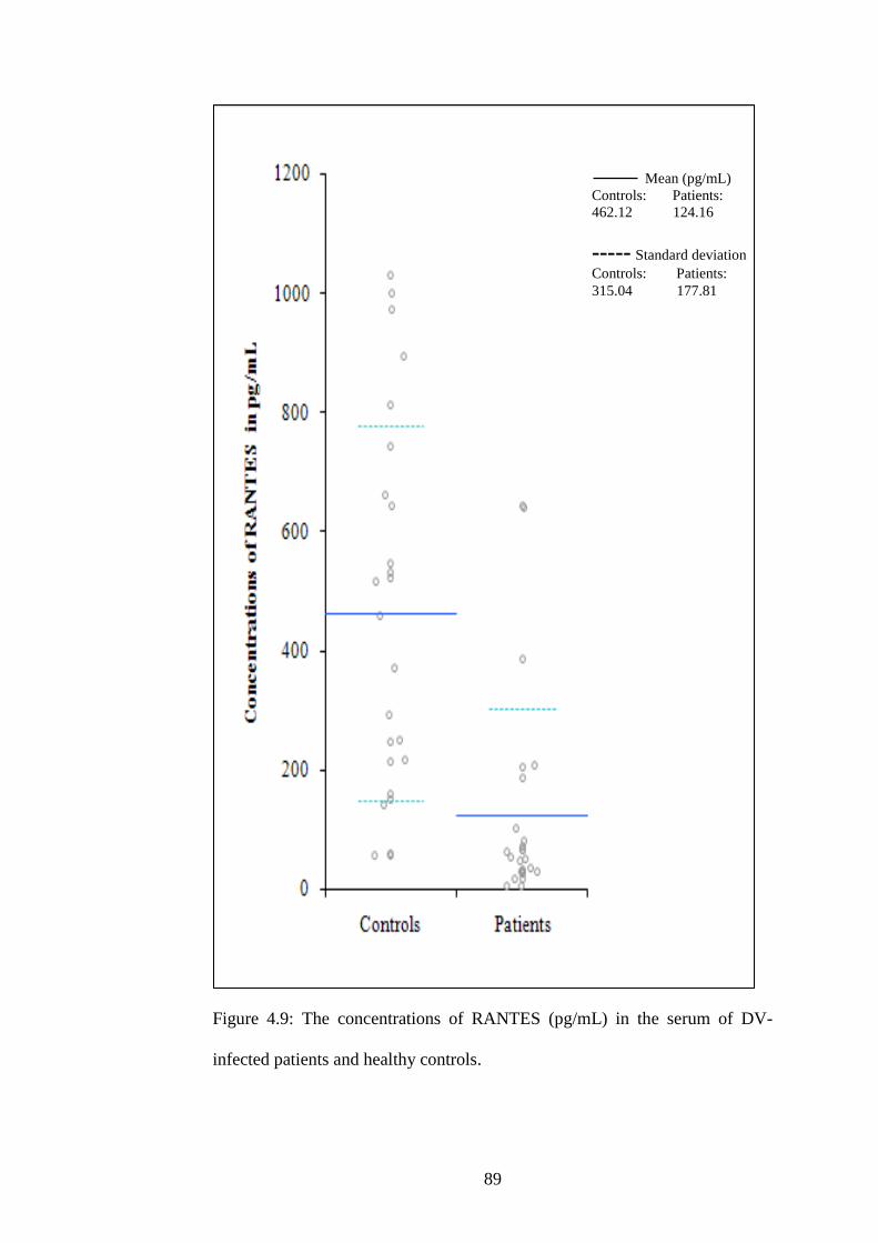
89
Figure 4.9: The concentrations of RANTES (pg/mL) in the serum of DV-
infected patients and healthy controls.
Mean (pg/mL)
Controls: Patients:
462.12 124.16
----- Standard deviation
Controls: Patients:
315.04 177.81

90
4.13 Differences and Relationships in Cytokines and Chemokines
Levels between Gender, Age, Day of Infection and Clinical Signs of
Disease.
As shown by Mann-Whitney U test, there is no significant difference in
cytokines and chemokines levels between female and male (p>0.05) (Table
4.12). Besides, Mann-Whitney U test also disclosed that there was no
significant difference (p>0.05) between cytokines and chemokines levels as
compared to different age groups (age ≤ 35 and >35) (Table 4.13), presence or
absence of myalgia (Table 4.14) and presence or absence of joint pain (Table
4.15). On the other hand, in Table 4.16, the Spearman rank order correlation
shows that there was no significant relationship (p>0.05) between cytokines
and chemokines levels with days of infection.

91
Table 4.12: Median concentration (pg/mL) of cytokines and chemokines in
DV infected patients for different gender.
Category
Median concentration (pg/mL) of cytokine or
chemokine in DV-infected patients
Mann-
Whitney
U test
Gender and standard deviation
P value
Male
Standard
deviation
Female
Standard
deviation
IL-6 17.66 10.77 12.99 16.09 0.61
IL-8 91.24 68.47 75.58 58.60 0.45
IL-15 5.19 4.02 3.02 1.12 0.12
IL-16 158.07 78.10 142.26 65.39 0.72
IFN-γ 13.79 18.02 11.41 21.98 0.32
MCP-1 278.56 143.72 223.94 56.56 0.34
MIG 612.07 507.05 631.92 1107.65 0.78
RANTES 33.97 197.18 63.73 127.58 0.20

92
Table 4.13: Median concentration (pg/mL) of cytokines and chemokines in
DV infected patients for different age category (≤35 and >35 years).
Category
Median concentration (pg/mL) of cytokine or
chemokine in DV-infected patients
Mann-
Whitney U
test
Age (years) and standard deviation
P value
≤35
Standard
deviation
>35
Standard
deviation
IL-6 17.92 14.07 15.11 8.48 0.45
IL-8 81.71 70.19 125.71 52.87 0.22
IL-15 5.32 3.91 3.01 0.84 0.06
IL-16 177.52 74.03 132.35 72.58 0.31
IFN-γ 15.23 20.44 18.28 6.71 0.12
MCP-1 236.99 126.30 328.94 93.11 0.40
MIG 738.61 867.56 542.50 303.82 0.30
RANTES 66.23 163.67 50.87 224.68 0.90

93
Table 4.14: Median concentration (pg/mL) of cytokines and chemokines in
DV infected patients in the absent or present of myalgia.
Category
Median concentration (pg/mL) of cytokine or
chemokine in DV-infected patients
Mann-
Whitney U
test
Myalgia and standard deviation
P value
Absent
Standard
deviation
Present
Standard
deviation
IL-6 17.92 8.64 15.11 13.55 0.89
IL-8 114.78 30.96 66.13 70.95 0.15
IL-15 3.24 0.87 4.93 3.85 0.46
IL-16 130.76 67.18 177.52 77.56 0.61
IFN-γ 12.72 12.67 13.74 20.37 0.73
MCP-1 183.05 92.26 252.39 121.55 0.23
MIG 639.49 404.04 577.08 851.94 0.66
RANTES 65.27 265.95 50.90 156.39 0.79

94
Table 4.15: Median concentration (pg/mL) of cytokines and chemokines in
DV infected patients in the absent or present of joint pain.
Caterogy
Median concentration (pg/mL) of cytokine or
chemokine in DV-infected patients
Mann-
Whitney U
test
Joint pain and standard deviation
P value
Absent
Standard
deviation
Present
Standard
deviation
IL-6 15.46 18.33 16.39 12.38 0.13
IL-8 76.51 15.46 89.00 64.41 0.13
IL-15 2.24 3.63 3.98 3.64 0.27
IL-16 172.41 61.49 142.26 76.38 0.76
IFN-γ 13.79 20.84 13.21 19.35 0.89
MCP-1 174.14 153.67 250.34 122.13 0.27
MIG 576.00 403.57 631.92 771.77 0.37
RANTES 54.17 39.55 54.93 184.21 0.76
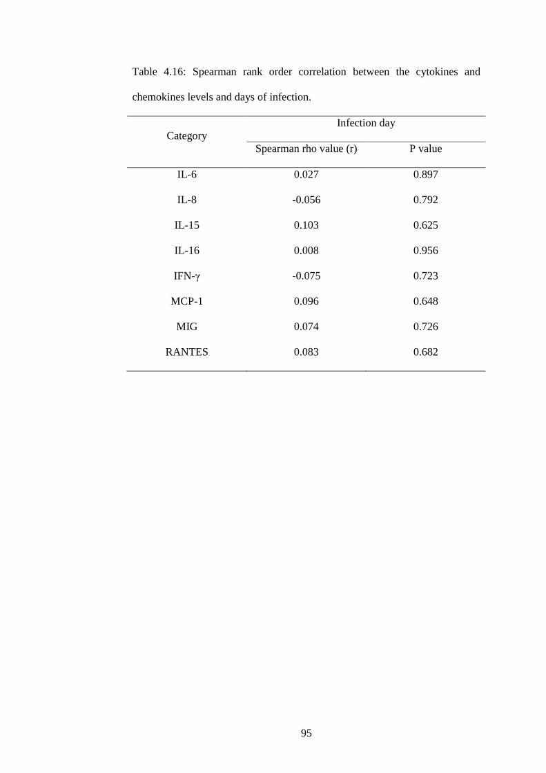
95
Table 4.16: Spearman rank order correlation between the cytokines and
chemokines levels and days of infection.
Category
Infection day
Spearman rho value (r) P value
IL-6 0.027 0.897
IL-8 -0.056 0.792
IL-15 0.103 0.625
IL-16 0.008 0.956
IFN-γ -0.075 0.723
MCP-1 0.096 0.648
MIG 0.074 0.726
RANTES 0.083 0.682

96
4.14 Correlations among the Cytokines and Chemokines Levels and
Clinical Findings.
As shown in Table 4.17, Spearman rank order correlation reveals that the
levels of IL-15 were positively and moderately correlated with the levels of
IFN-γ (r = 0.343, p<0.05) in DV-infected patients, where high levels of IFN-γ
were associated with high levels of IL-15. Also, levels of IL-15 were
negatively and moderately correlated with the levels of basophils (r = -0.395,
p<0.05) in dengue patients. In addition, IL-15 levels showed a strong,
negative associations with white blood cells count (r = -0.557, p<0.01),
lymphocytes count (r = -0.755, p<0.01), monocytes count (r = -0.757, p<0.01)
and eosinophils count (r = -0.640, p<0.01) in DV-infected patients. These
results indicated that high levels of IL-15 were correlated with low levels of
white blood cells like lymphocytes, monocytes, eosinophils and basophils. On
the other hand, levels of IL-16 were positively and averagely correlated with
MIG levels (r = 0.482, p<0.05) in DV-infected patients, where high MIG
levels were associated with high levels of IL-16. Moreover, there was a strong,
positive association between levels of MCP-1 and IL-8 (r = 0.531, p<0.01) in
DV-infected patients, with high levels of MCP-1 associated with high levels of
IL-8 in the patients. In contrast, a strong, negative association between MCP-
1 levels and monocytes counts (r = -0.575, p<0.01) was obtained in DV-
infected patients. Additionally, levels of MCP-1 was negatively and
moderately associated with basophils count (r = -0.485, p<0.01) in patients
with DV infection. These results suggested that low levels of monocytes and
basophils were correlated with high levels of MCP-1. Furthermore, high

97
levels of IL-15 were associated with low levels of RANTES since a medium,
negative relationship was found between RANTES and IL-15 levels (r = -
0.388, p<0.05) in DV-infected patients. Besides, a large, positive relationship
was obtained between levels of RANTES and lymphocytes counts (r = 0.548,
p<0.01) in dengue patients whilst the correlation between levels of RANTES
and eosinophils count in dengue patients was positively, medium associated (r
= 0.385, p<0.05). Also, a positive, medium association was demonstrated
between RANTES levels and platelets counts (r = 0.474, p<0.01) in patients
with DV infection. These results again proved that low levels of RANTES
were associated with low levels of lymphocytes, eosinophils and platelets.

98
Table 4.17: Spearman rank order correlation between the cytokines and
chemokines levels and clinical findings.
Scale
Spearman rho value (r)
IL-15 IL-16 MCP-1 RANTES
IL-8 -0.261 0.073 0.531** 0.219
IFN-γ 0.343* 0.301 0.081 -0.256
MIG -0.226 0.482* 0.205 0.030
RANTES -0.388* 0.130 0.067 -
WBC -0.557** -0.085 -0.240 0.215
Neutrophils -0.114 -0.109 0.261 0.076
Lymphocytes -0.755** -0.066 -0.305 0.548**
Monocytes -0.757** 0.115 -0.575** 0.222
Eosinophils -0.640** 0.038 -0.293 0.385*
Basophils -0.395* 0.084 -0.485** 0.052
Platelets 0.059 -0.049 0.303 0.474**
*. Correlation is significant at the 0.05 level
**. Correlation is significant at the 0.01 level

99
CHAPTER 5
DISCUSSION
DV infection is an acute infectious viral disease that presents a broad clinical
spectrum from asymptomatic to more severe manifestations (McBride &
Bielefeldt-Ohmann, 2000). It is believed that host immune response plays a
significant role in pathogenesis of DV infection (Pichyangkul et al., 2003).
Studies have shown the elevation of certain cytokines and chemokines in the
serum, blood or pleural fluid of patients with DV infection (Chen & Wang,
2002; Medin et al., 2005), as cytokines and chemokines are involved in the
disease onset and homeostatic regulation (Bozza et al., 2008). Although the
clinical manifestations of dengue had already been described, the
immunopathogenesis of this disease is still not completely understood. Thus,
a number of cytokines and chemokines that contributed to the immune
response of DV-infected patients were highlighted and a possible cytokine
network and mechanism was proposed in this study in order to figure out the
immunopathogenesis of DV infection.
In this study, regulations of IL-6, IL-8, IL-15, IL-16, IFN-γ, MCP-1, MIG and
RANTES showed no significant relationship with both age and gender. These
results suggested that age and gender had no influence in the production of the
mentioned cytokines and chemokines. Also, the days of DV infection had no
effect in regulating the cytokines and chemokines levels in DV-infected
patients as no significant correlation was observed between these two groups.

100
Besides, the levels of these cytokines and chemokines did not contribute to
myalgia or joint pain that were observed in dengue patients since no
significant relationship was obtained between this two groups.
5.1 IL-6
Levels of IL-6 in serum of DV-infected patients were significantly higher than
the healthy controls. The up-regulation of IL-6 in dengue patients in this study
was supported by Chen et al. (2006). They demonstrated that serum levels of
IL-6 were significantly increased in dengue patients than in healthy controls.
Similar result was obtained years later by Restrepo et al. (2008a) where they
found that serum levels of IL-6 in children with dengue was higher than those
without dengue. In addition, the degree of increment of IL-6 in DV-infected
patients in this present research was approximately the same as the research
done by Chen et al. (2006). In contrast, the mean levels of IL-6 in dengue
patients in this present study were about three times higher than the mean
levels of IL-6 in dengue patients in a study done by Restrepo et al. (2008a)
(5.65 pg/mL). This might be due to a difference in the age group as the levels
of IL-6 in this research were obtained from the adult patients whilst the levels
of IL-6 done by Restrepo et al. (2008a) was obtained from the children
patients.
Increased levels of IL-6 in serum of dengue patients were induced by DV
infection. This is because IL-6 is not a cytokine that is constitutively
expressed, but it is readily induced by multiple stimuli, which includes viral

101
infection (Streetz et al., 2007). Interleukin-6 is synthesized predominantly by
monocytes and macrophages (Yap & Lai, 2010) upon viral infection. Besides,
IL-6 is also produced by T cells and B cells in response to stimuli such as
RNA virus (Yap & Lai, 2010), which lead to the up-regulation of IL-6 in DV-
infected patients. Interleukin-16 also plays an important role in stimulating
the production of IL-6 from monocytes (Qi et al., 2002). However it was
found to have no or limited effect on IL-6 production in DV-infected patients
in this present study as levels of IL-6 were not significantly associated with
levels of IL-16 in DV-infected patients.
The elevation of IL-6 levels in the DV-infected patients showed that this
cytokine played an essential role in the host defense against DV infection. IL-
6 is thought to be involved in the transition from an innate immune response to
an adaptive immune response (Ellis & Beaman, 2004) as it is essential in the
normal development and function of both T cells and B cells (Yap & Lai,
2010). IL-6 activates T cells, which recognizes antigens and stimulates the
proliferation and differentiation of CD8+ T cells (Łukaszewicz et al., 2007),
which subsequently take part in the viral clearance. Also, it stimulates B cells
differentiation and induces the maturation of B cells into plasma cells, which
produces different classes of immunoglobulin (Yap & Lai, 2010) that might
play an important role in the clearance of DV and viral products.
The up-regulation of IL-6 levels in the DV-infected patients might also
contribute to several manifestations of this disease. IL-6 is an endogenous
pyrogen (Restrepo et al., 2008b) where it increases the body temperature by

102
inducing the synthesis of prostaglandins (Łukaszewicz et al., 2007) and thus
caused fever in the DV-infected patients. Elevated body temperature is
beneficial to host defense as viruses grow less efficiently at raised temperature
(Coico, Sunshine, & Benjamini, 2003). This might control the virus
proliferation in the host system. Besides, up-regulation of IL-6 levels
probably also cause anorexia and lethargy (Maggio et al., 2006) in DV-
infected patients, as these manifestations are normally seen to occur in patients
with DV infection. In addition, high levels of IL-6 in DV-infected patients
might increase vessel permeability of the endothelial cells (Restrepo et al.,
2008b) through induction of gap formation between adjacent endothelial cells,
which subsequently caused the pleural effusion in the DV-infected patients
(Lee et al., 2007). Priyadarshini et al. (2010) supported this idea where they
found that there was an increased levels of IL-6 in 65% of the DV-infected
patients who showed pleural effusion. This cytokine was also reported to play
a crucial role in the enhancement of production of anti-platelet auto antibodies
during DV infection (Rachman & Rinaldi, 2006) that possibly caused
thrombocytopenia in the DV-infected patients. Therefore, IL-6 is a main
factor involved in the immune response of DV-infected patients due to its
multiple activities against the viral infection.
5.2 IL-8
Normally, IL-8 protein is barely secreted from non-induced cells (Hoffmann,
Dittrich-Breiholz, Holtmann, & Kracht, 2002). However, levels of IL-8 in the
serum of DV-infected patients, in this study, were significantly elevated as

103
compared to the serum levels of IL-8 in the healthy controls. This might be
due to the production of IL-8 that was rapidly induced by DV or viral products
(Medin et al., 2005) from monocytes. Macrophages and neutrophils might
also produce IL-8 (Mukaida, 2003) upon DV infection. Interleukin-15 which
was also found to act as an inducer in the production of IL-8 (Jabłońska et al.,
2003), had no or limited effect in the production of IL-8 from DV-infected
patients in this present study as levels of IL-8 were not in significant
correlation with the levels of IL-15 in DV-infected patients.
Increased levels of IL-8 might subsequently chemoattracted neutrophils to the
site of infection (Bosch et al., 2002) and induced a respiratory burst of
neutrophils which consequently caused the release of lytic enzymes to rid the
host of invading virus (Medin et al., 2005). Surprisingly, there was no
significant relationship between levels of IL-8 and neutrophils counts. This
result suggested that IL-8 had no or limited effect in chemoattracting
neutrophils to infiltrate the tissue to reach the site of infection. However, this
chemokine might have a role in degranulation of neutrophils to release the
lytic enzyme to clear the viral particles.
Additionally, high concentrations of IL-8 could possibly alter the cytoskeleton
and tight junctions of microvascular endothelium and changed the
permeability of the endothelial monolayer (Medin et al., 2005). This leads to
the occurrence of plasma leakage, which is normally seen in the more severe
form of dengue illness. Priyadarshini et al. (2010) also showed that the
increased levels of IL-8 were associated with plasma leakage.

104
Besides, high levels of IL-8 in the serum of DV-infected patients may also
induce the synthesis and release of platelet-activating factor in neutrophils
(Mukaida, 2003). Platelet-activating factor might then participate in platelet
activation, which led to platelet consumption (Bozza et al., 2008) in DV-
infected patients. This later could cause the platelet number to decrease and
hence, thrombocytopenia was seen in patients with DV infection.
Priyadarshini et al. (2010) supported the fact that IL-8 were associated with
thrombocytopenia where they reported that the levels of IL-8 in dengue
patients with thrombocytopenia were significantly higher than in the patients
that showed normal platelet count. Furthermore, high levels of IL-8 may also
contribute to fever in DV-infected patients since IL-8 is an endogenous
pyrogen (Pela´ et al., 2000).
Moreover, elevation of IL-8 levels in DV-infected patients might indirectly
cause T cell chemotaxis by inducing neutrophils to release several factors
from their granules with chemotactic activities for T cells (Mukaida, 2003).
Chemoattracted T cells might then produce more IL-8 as T cells are producers
of this chemokine. Again, a positive feedback mechanism took place and this
process amplified the T cells pool at the site of infection in helping the
clearance of invading virus. As a result, this positive feedback mechanism led
to high concentrations of IL-8 in the serum of DV-infected patients. This is
supported by the research done by Y. Huang et al. (2000) where they found
that IL-8 were up-regulated in DV-infected patients as compared to the
healthy controls. The mean IL-8 concentrations in dengue patients of their
study were also found to not deviate much from the mean IL-8 concentrations

105
in dengue patients conducted in this study. On the other hand, although Juffrie
et al., (2000) found that the levels of IL-8 in DV-infected children were up-
regulated as compared to the healthy controls, but the median IL-8 level
obtained in that research was about three times lower than the median IL-8
level obtained in this study. This deviation may be due to the different age
group that was included in their study, where they used children as their target
group as compared to adults. Concentrations of IL-8 were determined in DV-
infected children by Juffrie et al. (2000) while the concentrations of this
chemokine were determined in DV-infected adults in this present research.
5.3 IL-15
Serum levels of IL-15 in DV-infected patients were significantly elevated as
compared to the healthy controls. Azeredo et al. (2005) also demonstrated a
similar result where they found that IL-15 was significantly elevated in the
plasma of most dengue patients during the early acute phase as compared to
healthy donors. The mean IL-15 concentrations shown by Azeredo et al.
(2005) was approximately two times higher than mean IL-15 concentrations in
this study. The measurement of IL-15 was done during the early acute phase
(1-5 days after disease onset) by Azeredo et al. (2005) whilst the measurement
of IL-15 was done 6 days in average after disease onset in this present study.
The difference in the day of infection probably led to the difference seen in the
levels of IL-15 in both studies.

106
The production of IL-15 in dengue patient was in response to DV infection.
This is because IL-15 has low secretion potential (Hocke et al., 2007), and
soluble IL-15 is hardly detected in normal physiological condition (Alpdogan
& Brink, 2005). In fact, the production of IL-15 is in response to the danger
signal such as RNA virus infection (Meier et al., 2005), in this case, DV
infection. During DV infection, IL-15 might be produced by macrophages,
monocytes and DC (Jabłońska et al., 2003) where these cells are critical in the
first line of defense against viral infection. Moreover, the up-regulation of
serum levels of IL-15 in DV-infected patients was not due to the release of IL-
15 from monocytes by the stimulation of IL-16 as there was no significant
correlation between these two cytokines.
The increased levels of IL-15 in serum of DV-infected patients suggested that
this cytokine played an important role in the viral clearance during DV
infection. IL-15 is involved in the activation and homeostatic maintenance of
cells in the innate immune systems (Wang et al., 2005) as it is a NK cell
regulator. It probably regulated NK-cell development, homeostasis and
activity (Alpdogan & Brink, 2005) during the DV infection in this present
study. This regulation is vital in the host defense against the viral infection
since numerous studies had demonstrated that NK cells can selectively lyse
virus-infected target cells while sparing uninfected cells (Fawaz et al., 1999).
Azeredo et al. (2005) also proved the above statements where it was shown
that IL-15 was seen to have an NK cell-dependent antiviral activity against
HIV. In addition, high levels of IL-15 could possibly drive T-cell proliferation
and induced effector T cells along with the stimulation of development of

107
memory T cells (Ramsborg & Papoutsakis, 2007) in the immune system of
DV-infected patients. Also, the levels of IL-15 were significantly moderately
correlated with the levels of IFN-γ in dengue patients. This proved that the
levels of IFN-γ were in response to the levels of IL-15 in dengue patients. The
high levels of IL-15 in DV-infected patients caused the up-regulation of IFN-γ
as IL-15 appeared to enhance Th1 responses by increasing IFN-γ production
from NK cells and T cells (D’Ettorre et al., 2006; Milano et al., 2002), which
in turn contributed to an antiviral state in DV-infected patients.
High levels of IL-15 were also significantly correlated with low levels of
white blood cells, which include lymphocytes, monocytes eosinophils and
basophils in the circulation of dengue patients. This is because, IL-15 served
as an important cytokine for the differentiation of lymphocytes (Hu et al.,
2007; Ramsborg & Papoutsakis, 2007) and monocytes (Ueda et al., 2007) and
this cytokine caused differentiation of these leukocytes and subsequently
infiltrate through tissues to reach the site of infection in the defense system.
Thus, lower levels of leukocytes were found in the circulation of dengue
patients in this present study. Also, this cytokines could be regulating the
activities for both eosinophils and basophils in establishing the barrier against
the viral attack.
Besides the above, IL-15 is important in expansion, survival, homeostasis and
functional maturation of NKT cells (Alpdogan & Brink, 2005). The
functional status of NKT cells fall somewhere between innate and the adaptive
immune system suggesting that the up-regulation of IL-15 in DV-infected

108
patients may aid the transition of host immune response in between both
innate and the adaptive immune system. Also, increased level of IL-15 might
enhance the activity of NK cells, T cells and NKT cells by delaying their
apoptosis (Jabłońska et al., 2003) via induction of anti-apoptotic molecules
like B-cell lymphoma-2 (Bcl-2) (Budagian et al., 2006).
5.4 IL-16
In this study, the levels of IL-16 in the serum of DV-infected patients were
significantly higher as compared to the serum levels of IL-16 in healthy
controls. A similar result was obtained where HIV infection up-regulated the
production of microglial IL-16 (Zhao et al., 2004). Also, the mean
concentrations of IL-16 in severe acute respiratory syndrome (SARS) patients
also showed similar results where there were higher concentrations of IL-16 in
SARS patients than that of the control group (Wang et al., 2003). On the other
hand, serum levels of IL-16 in patients with systemic lupus erythematosus,
which has the similar symptoms as dengue (Koike et al., 2002), were also up-
regulated. These results suggested that IL-16 might play a crucial role in
defense system of viral infection, in this case, the DV infection.
The induction of high levels of IL-16 in the DV-infected patients during the
immunological response, might be due to the stimulation of cytokines
production by the released viral products, in this case DV, as viruses are
known to be the potent stimulators of cytokine expression in vitro and in vivo
(Shen et al., 2004). Upon infection, APC internalize the viruses or viral

109
products and present the immunogenic viral peptides with major
histocompatibility complex class (MHC)-I or class II (MHC-II) molecules for
activation of virus-specific CD8+ or CD4+T cells, respectively (Abdel-Motal,
Guay, Wigglesworth, Welsh, & Galili, 2007). IL-16 is then secreted by CD4+
or CD8+ T cells and response to antigen (Reich et al., 2001). Subsequently,
IL-16 contributes to the recruitment of CD4+ T cells (McFadden et al., 2007)
to the site of infection. The recruited T cells then synthesize and secrete more
IL-16 that may serve to function as a positive feedback mechanism for further
cell recruitment and activation (Wu et al., 1999). All this possibly brought
about the up-regulation of IL-16 levels in DV-infected patients.
Besides induction of T cells chemotaxis, IL-16 also induces the migratory
response in DC (Lynch et al., 2003). The chemotaxis of this APC to the site
of infection might enhance their ability as APC and therefore, more IL-16 is
being produced by CD4+ T cells and CD8+ T cells. Furthermore, DC are also
the source of production of IL-16 (Hu et al., 2007), which might further up-
regulated IL-16 levels in DV-infected patients in this present study.
5.5 IFN-γ
During viral infection, NK cells, which play an important role in the innate
defense mechanisms of the host, has an aim to eliminate virus-infected cells
(Fawaz et al., 1999). Upon infection, NK cells produce IFN-γ (Yang et al.,
2005), which play a role in mediating protection against viral infection,
especially long-term control of viral infections (Samuel, 2001). IFN-γ then
110
activates the NK cell effector functions (Schroder et al., 2004), where various
cytotoxic molecules are released by NK cells to eliminate the invading virus.
Since NK cells are the source of IFN-γ production, the up-regulation of IFN-γ
in this present study might be produced by NK cells as IFN-γ is vital in
enhancing the viral clearance during the innate immune response (Diamond et
al., 2000) in DV-infected patients.
Besides, IFN-γ also mediates the differentiation of naive T cells toward Th1
phenotype (Teixeira et al., 2005) as the phenotype adopted by a naive T cell
during T cell activation is heavily influenced by the cytokine (Schroder et al.,
2004). Th1 cells then secrete large amount of IFN-γ (Chen et al., 2005) as
Th1 cells are the main producers of this cytokine (Restrepo et al., 2008b).
This process further skews the immune response toward a Th1 phenotype, and
this process probably caused the up-regulation of IFN-γ levels in the serum of
DV-infected patients. Th1 cells are important in the viral elimination as they
are responsible for cell-mediated immunity with the activation of NK cells,
macrophages and CD8+ T cells (Coico et al., 2003). Th1 cells are also
essential in providing phagocyte-dependent protective responses (Romagnani,
2000) in the host immune response. In downstream process of Th1 response,
cytokine and chemokine are produced in order to chemoattract specific
immunocytes to the site of infection. As discussed earlier, IFN-γ secretion is
the hallmark of Th1 response (Gattoni et al., 2006). The IFN-γ might amplify
the production of chemokines that participate in a wide range of bioactivities.
One of the most important roles of chemokine is to direct the recruitment or
chemotaxis of immune cells to the site of infection, which ultimately lead to

111
destruction of virally infected cells (Le et al., 2004). In addition, IFN-γ may
also stimulate the development of CD8+ T cells (Yang et al., 2005), which is
important as the downstream effectors of Th1 cells. At the same time, CD8+
T cells synthesize IFN-γ (Samuel, 2001) and this might further caused the
higher serum levels of IFN-γ in DV-infected patients than that in the healthy
controls.
Apart from that, IFN-γ is an activator of macrophages (Ellis & Beaman, 2004)
and it converts macrophages from a resting state to an active state and induces
the synthesis of an array of receptors for binding to pathogens (Antoniou et al.,
2003). Macrophage activation is very important in the viral clearance as they
act as APC. They ingest the virus, digest them and present their antigens with
MHC class molecules on their cell membranes to B cells or T cells to generate
antigen-specific immune response (Chaturvedi, Nagar, & Shrivastava, 2006).
This process is probably enhanced by IFN-γ as it is able to up-regulate antigen
processing and presentation pathways (Diamond et al., 2000). Furthermore,
activated macrophages acquire antiviral effector functions and secrete pro-
inflammatory cytokines like IL-6 and IL-8 (Gattoni et al., 2006). All these
events might cause further increase of the IL-6 and IL-8 concentrations in the
DV-infected patients in this present study. As a result, different types of
immune cells might be recruited to the site of infection followed by the
elimination of the virus by phagocytosis or the release of toxic metabolites
(Schroder et al., 2004). Besides, IFN-γ is shown to be produced by
macrophages (Ellis & Beaman, 2004). The cycle of macrophage activation by
IFN-γ and the production of IFN-γ from macrophages might again cause the

112
elevation of serum IFN-γ levels in the DV-infected patients in this present
study.
In addition to NK cells, Th1 cells, CD8+ T cells and macrophages, IFN-γ is
also produced by B cells (Teixeira et al., 2005). IFN-γ activates B cells and
regulates its functions such as immunoglobulin production and class switching
(Rosloniec, Latham, & Guedez, 2002). Positive feedback mechanism takes
place again to ensure higher production of IFN-γ in order to activate more B
cells in enhancing the elimination of invading virus. As a result of the above,
a high concentration of IFN-γ was obtained in the serum of DV-infected
patients in this present study. Similar results were reported where the
productions of IFN-γ were induced in DV-infected patients and a similar mean
IFN-γ concentrations were obtained between this present study and the other
research (Chen et al., 2005; Chen et al., 2006; Hsieh et al., 2006; Priyadarshini
et al., 2010; Restrepo et al., 2008b). This suggested that IFN-γ is critical for
the early immune response to DV infection and IFN-γ-mediated immune
responses are necessary for both the early and late clearance of the virus
(Hsieh et al., 2006).
High levels of IFN-γ in serum of DV-infected patients might cause local
dilation of the blood vessels and thereby decreasing the local blood flow rate
which caused the gathering of blood in leaky vessels (Schroder et al., 2004).
This cytokine may also regulate other chemokines like MIG (Dajotoy et al.,
2004) and MCP-1 (Zhou et al., 2001) to chemoattract the specific leukocytes
to extravasate into the tissue for viral clearance, and subsequently caused

113
plasma leakage in the DV-infected patients. Also, IFN-γ in DV-infected
patient could have produced platelet activating factors, which participated in
platelet and endothelial cell activation, leading to platelet consumption (Bozza
et al., 2008) that led to thrombocytopenia in the DV-infected patients in the
present study.
5.6 MCP-1
MCP-1 is produced by monoctyes (Mori et al., 2000) upon viral infection.
The high levels of MCP-1 were significantly highly correlated to high levels
of IL-8 in dengue patients as both of these cytokines are produced by
monocytes after stimulation of virus infection (Bozza et al., 2008). Similar to
IL-8, elevated MCP-1 in the DV-infected patients might cause the increase of
endothelial permeability by disrupting tight junctions among cells (Lee et al.,
2006), leading to plasma leakage and haemoconcentration (Yamada et al.,
2003) in the DV-infected patients in the present study. Lee et al. (2006) found
that the increased permeability and disrupted tight junctions of human vascular
endothelium cells were affected through a mechanism partially dependent on
MCP-1. Besides, high levels of MCP-1 might contribute to thrombocytopenia
in DV-infected patients as they are able to cause platelet activation and platelet
consumption (Bozza et al., 2008).
As the concentration of MCP-1 increased, the number of monocytes and
basophils in serum of DV-infected patients decreased since the levels of MCP-
1 was significantly negatively associated with monocytes and basophils count

114
in dengue patients. This is because MCP-1 is considered an important
mediator that specifically stimulates directional migration of monocytes
(Cleanthis et al., 2007) and basophils (Dhillon et al., 2009) to extravasate into
the tissue for viral clearance and hence the lower amount of both leukocytes in
the blood circulation. Monoctyes might then differentiate into macrophage,
which is then responsible for the engulfment of the viral particles, in this case,
DV, since they possess the phagocytic ability. Also, chemoattracted
monocytes and macrophages amplify the antigen presenting pathway since
they are APC (Glew et al., 2003). Additional virus will then be internalized
and presented to T cells. This might further increase the production of IFN-γ
in the host system and this cytokines might in turn stimulate the secretion of
MCP-1 from monocytes in controlling the magnitude of cellular accumulation
at the site of infection (Rahangdale et al., 2006). Hence, significant elevated
levels of MCP-1 in the serum of DV-infected patients were observed in this
study. High concentrations of MCP-1 in the DV-infected patients might also
evoke T cells and NK cells chemotaxis (Han et al., 1999), which enhanced the
host immune system in removing of viral product during the infection.
The up-regulation of MCP-1 in DV-infected patients in this study was parallel
with the research done by Dejnirattisai et al., (2008) and Lin et al., (2005)
where levels of MCP-1 in DV-infected patients were higher than that in the
normal controls. Mean MCP-1 concentrations obtained in their research was
higher than the mean MCP-1 concentrations obtained in this study. This
might be due to the inclusive of the DHF group patients in the research done
by Dejnirattisai et al., (2008) and Lin et al., (2005) whilst the patients included

115
in this present research were mostly from DF group. However, the increased
levels of MCP-1 observed in dengue patients in this present study showed the
importance of this chemokine in the host defense against DV infection.
5.7 MIG
Levels of MIG in the serum of patients with DV infection was found to be
significantly elevated as compared to the serum levels of MIG in the healthy
controls. Dejnirattisai et al. (2008) also demonstrated a similar result where it
was shown that MIG was induced in the patients with DV infection. However,
the mean MIG levels in dengue patients shown by Dejnirattisai et al. (2008)
were three times lower than that in this present study. This was because
different category of patients was included in the study. Dejnirattisai et al.
(2008) determined the MIG levels in serum of DHF patients whereas MIG
levels were determined in mostly DF patients in this present study.
MIG is secreted by monocytes, macrophages, APC, B cells (Whiting et al.,
2004) and eosinophils (Dajotoy et al., 2004) upon viral infection. MIG was
thought to attract NK cells to the site of infection (Hsieh et al., 2006) and
subsequently mediate their protective effect by releasing the cytotoxic
molecules in clearing the virus-infected cells. Also, NK cells may produce
large amount of cytokine such as IFN-γ in the protective action during the
viral infection (Makrigiannis & Anderson, 2003). Interferon-gamma, which is
the inducer for MIG production (Colvin et al., 2004) would then induce extra
secretion of MIG from different immune cells that consequently contributed to

116
the up-regulation of MIG concentration in the serum of DV-infected patients
in this present study.
Additionally, MIG also promote the migratory response of activated T cells,
especially Th1 cells to the site of infection (Takeuchi et al., 2006) in the host
defense system. The induction could have enhanced the production of IL-16
from activated T cells. Since IL-16 levels were significantly correlated with
MIG levels in DV-infected patients, the up-regulation of MIG could have
caused an increased in the amount of IL-16 in DV-infected patients in this
present study.
Furthermore, the determination of the MIG levels in the DV-infected patients
is very important. This is because production of MIG is uniquely stringent
and it is specifically induced by IFN-γ (Salazar-Mather et al., 2000), which
may in turn allows a functional measure of IFN-γ activity. Also, MIG is
produced following an amplification of the IFN-γ signal and detecting the
concentration of MIG is a more sensitive measure of bio-active IFN-γ than
detecting IFN-γ directly (Esche et al., 2005). Hence, the high concentration of
MIG in the serum of DV-infected patients suggested the presence of bio-active
IFN-γ in the immune system of DV-infected patients.
5.8 RANTES
RANTES is a chemokine secreted by activated T cells (Cavusoglu et al., 2007).
It is also produced by monocytes and macrophages (Sun, Wang, Zhang, Zeng,

117
& Wang, 2005). NK cell is also another source of production of this
chemokine. RANTES induces the migration of a variety of leukocyte types,
but found to preferentially attract Th1 cells (Kanda & Watanabe, 2003). It is
also a chemoattractant for monocytes (Gianoukakis et al., 2003), which play
essential role in the host defense system. In addition, this chemokine also has
a crucial role in trafficking and activating other immune cells such as DC
(Carr, Ash, Lane, & Kuziel, 2006), mast cells (Fryer et al., 2000) and NK cells
(Dorner et al., 2002) towards site of infection. The involvement of RANTES
in viral infection is shown in various in vitro studies, where they showed that
it is secreted in the supernatant of DV-infected cell lines (Avirutnan, Malasit,
Seliger, Bhakdi, & Husmann, 1998; Chen, Ng, & Chu, 2008; Chen & Wang,
2002; King et al., 2002; Medin et al., 2005).
Interestingly, in contrast with other in vitro studies, the RANTES levels in the
serum of DV-infected patients in this present study were significantly down-
regulated as compared to healthy controls. This may be because the RANTES
levels were determined from the serum of DV-infected patients in this present
study. In other studies, the determination of RANTES levels was obtained
from the supernatant of DV-infected cell lines. Other soluble factors that are
naturally present in DV-infected patients but not in DV-infected cell line may
influence the production of RANTES and hence the contrast results obtained.
Given that the activated immune cells are among the stimuli that elicit the
production of RANTES, it would have been expected that there would be an
elevated levels of RANTES in the serum of patients with DV infection in this

118
study. However, the observed reduction in the serum RANTES levels might
be due to the decreased amount of platelets in DV-infected patients since there
was a significant correlation between the levels of RANTES and platelet count
in dengue patients. This is because in addition to T cells, monocytes,
macrophages and NK cells, platelets are also considered one of the major
source of RANTES (Eissa, Zaki, El-maghraby, & Kadry, 2005; Gleissner et al.,
2008; Niwa et al., 2001). Since the DV-infected patients involved in this
study all suffered from thrombocytopenia, the low amount of platelets in these
DV-infected patients might contribute to the low levels of RANTES in these
patients. Ellis and co-workers (2005) also supported this result where they
found that platelet count changed in a parallel fashion to the RANTES
concentrations and the correlation between platelets and RANTES was
significant. Down-regulation of RANTES in the individual with severe
malaria was also found to correspond to the thrombocytopenic in the malaria
patients (John, Opika-Opoka, Byarugaba, Idro, & Boivin, 2006). Moreover,
one single report from Cuba supported the result obtained for DV infection,
where they found that serum RANTES levels detected in patients with DV
infection were also lower than those observed in the healthy controls (Pérez et
al., 2004). Also, lymphocytes are one of the producers of RANTES (Gleissner
et al., 2008). Low levels of lymphocytes contributed to the low levels of
RANTES in patients with DV infection as there was a strong significant
correlation between the levels of RANTES and lymphocytes counts. Low
RANTES concentrations then caused the decreased in the eosinophils count in
DV-infected patients as RANTES levels had medium, significant relationship
with eosinophils count. Moreover, a negative, medium significant correlation

119
between levels of RANTES and IL-15 showed that the low RANTES levels
contributed to high levels of IL-15 in DV-infected patients. This might be
because both RANTES and IL-15 have important role in regulating the
activities of NK cells (Alpdogan & Brink, 2005; Chen & Wang, 2002) and T
cells (Appay et al., 2000; Ramsborg & Papoutsakis, 2007). The low levels of
RANTES were compensated with high levels of IL-15 in order to regulate the
activities of both NK cells and T cells to ensure the efficiency of viral
clearance.
5.9 Cytokine Network and Mechanism
As mentioned earlier, cytokines rarely, if ever, act alone in an immune system.
A combined effect of two or more cytokines is sometimes greater that the
additive effects of the individual cytokines. The combined effects of the
cytokines ensure the outcome where the magnitude of defense system is
sufficient to clear the invading virus. Hence, a proposed cytokine network and
mechanism is suggested to represent the immunopathogenesis of DV infection
(Figure 5.1).

120
Figure 5.1: Proposed cytokine network and mechanisam for
immunopathogenesis of dengue
Upon DV infection, productions of IL-6, IL-8, IL-15, IL-16, IFN-γ and MCP-
1 from monocytes might be induced by the viral particles. Occurrence of
anorexia and lethargy in DV-infected patients might be due to the up-
regulation of IL-6. Also, as endogenous pyrogens, IL-6 and IL-8 would then
induce fever in dengue patients by raising the temperature in the host defense
system to reduce the activities of viral particles in the host. At the same time,
IL-6, IL-8, MCP-1 and IFN-γ then induce the change in endothelial
permeability by disrupting tight junctions among cells to cause vascular
leakage, which is deemed to be a necessary action for leukocytes to leave the
circulation and infiltrate tissue to site of infection in clearing the invading
virus. If the vascular leakage is too serious and is not corrected early, a more
severe form of illness named plasma leakage might take place in severe
dengue patient.
Secreted IL-8 might recruit naïve T cells to the site of infection (Bosch et al.,
2002) to expose this naïve T cells to encounter the viral particles in DV

121
infection. The naïve T cells bounded with viral particles are necessary to
initiate the immune response. IFN-γ might stimulate the differentiation of
naïve T cells toward Th1 phenotype (Teixeira et al., 2005) as the phenotype
adopted by a naïve T cell during T cell activation is heavily influenced by the
cytokine (Schroder et al., 2004). Th1 cells might then take part in cell-
mediated immunity and also provides phagocytes-dependent protective
response in the host immune system.
Besides inducing the development of Th1 cells, IFN-γ also activated
macrophages (Ellis & Beaman, 2004), which act as APC to present the viral
peptides to T cells for further degradation (Abdel-Motal et al., 2007).
Interferon-gamma might then indirectly enhance the antigen processing and
presentation pathways (Diamond et al., 2000). Also, as phagocytes,
macrophages play this crucial role in engulfing those viral particles in clearing
foreign pathogens from the host. Furthermore, activated macrophages might
secrete pro-inflammatory cytokines like IL-6 and IL-8 (Gattoni et al., 2006)
which led to further increase of the IL-6 and IL-8 concentrations in the DV-
infected patients.
A raise in the IL-6 levels in DV-infected patients might then stimulate the
proliferation and differentiation of CD8+ T cells (Łukaszewicz et al., 2007),
which are the important effector cells in the downstream of Th1 immune
response as CD8+ T cells (Łukaszewicz et al., 2007) take part in the viral
clearance by releasing the cytotoxic substances from their granule and caused
apoptosis of the viral infected cells. Also, high levels of IL-6 in dengue
122
patients might stimulate differentiation and maturations of B cells to produce
different classes of immunoglobulin (Yap & Lai, 2010) that subsequently take
part in the clearance of DV products. On the other hand, increased levels of
IL-8 might induce a respiratory burst of neutrophils which consequently
caused the release of lytic enzymes to rid the host of invading virus (Medin et
al., 2005) in enhancing the immune status in the dengue patients.
High levels of MCP-1 were significantly highly correlated with high levels of
IL-8 in dengue patients as both of these cytokines are produced by monocytes
after stimulation of virus infection (Bozza et al., 2008). Also, the significant
negative correlations between MCP-1 levels and both monocytes and
basophils counts suggested that MCP-1 stimulated directional migration of
monocytes (Cleanthis et al., 2007) and basophils (Dhillon et al., 2009) to
extravasate into the tissue through the disturbed tight junctions among cells
that caused by high levels of IL-6, IL-8, MCP-1 and IFN-γ. Therefore, this
phenomenon caused the significant lower amount of both moncotyes and
basophils in the blood circulation. Accordingly, monoctyes were then
differentiated into macrophages in the tissue, which engulfed the DV, since
they possess the phagocytic ability. Also, chemoattracted macrophages
amplified the antigen presenting pathway since they are APC (Glew et al.,
2003). Additional virus will then be internalized and presented to T cells and
this increased the efficacy in viral clearance. High concentrations of MCP-1
in the DV-infected patients might also evoke T cells and NK cells chemotaxis
(Han et al., 1999) to the site of DV infection, which further enhanced the host
immune system.

123
MIG, on the other hand, is specially induced by IFN-γ (Salazar-Mather et al.,
2000). The presence of high concentrations of MIG demonstrated that IFN-γ
were bioactively present in the DV-infected patients. High levels of MIG
were significantly associated with high levels of IL-16. This showed that high
levels of MIG in dengue patients might chemoattract extra NK cells and
activated T cells to the site of infection (Takeuchi et al., 2006) by infiltrating
themselves through the disrupted tight junctions among cells. The
chemoattracted T cells at the site of infection might enhance the production of
IL-16 from activated T cells (Reich et al., 2001) that subsequently
significantly up-regulated IL-16 levels in dengue patients and also formed a T
cells pool at the site of infection. Interleukin-16 might then induce the
migratory response in DC where chemotaxis of this APC to the site of
infection might cause extra viral antigens to be presented to T cells for
degradation.
On the other hand, IL-15 might regulate cytotoxicity of the chemoattrated NK
cells (D’Ettorre et al., 2006). This regulation is important in host defense
system as NK cells are found to be selectively able to lyse virus-infected target
cells but at the same time sparing the uninfected cells. This is done by
destroying the viral infected cells not by phagocytosis but by releasing
biologically potent molecules that, within a very short time, kill the viral
infected cells (Coico et al., 2003). In addition, high levels of IL-15 might
stimulate the development of memory T cells (Ramsborg & Papoutsakis,
2007), which later play a vital role in secondary infection in order to boost up
the immune response in a faster pace within a short time against the virus.

124
Furthermore, the high concentrations of IL-15 were significantly correlated
with high concentrations of IFN-γ in dengue patients. This showed that IL-15
might increase the production of IFN-γ from NK cells and T cells in
enhancing Th1 response (Azeredo et al., 2005). Additionally, significant
negative relationships between IL-15 and white blood cells were demonstrated.
This is because IL-15 is important for the differentiation of lymphocytes (Hu
et al., 2007; Ramsborg & Papoutsakis, 2007) and monocytes (Ueda et al.,
2007). The differentiation of these leukocytes and the subsequent infiltration
of these leukocytes through disrupted tight junctions between cells to reach the
site of infection would then cause significant lower levels of these leukocytes
found in the blood circulation of dengue patients. Also, these cytokines
regulate the activities for eosinophils in establishing the barrier against the
viral attack.
Moreover, IL-6 might enhance the production of anti-platelets antibodies
which then contributed to low amount of platelet in the dengue patients. Also,
IL-8, MCP-1 and IFN-γ might produce platelet-activating factor that
participated in platelet activation. This led to platelet consumption and caused
the platelet number to decrease in circulation and hence the occurrence of
thrombocytopenia in DV-infected patients. Since the RANTES levels was
parallel with the amount of platelets presence in the host, less RANTES is
produced upon DV infection due to the presence of thrombocytopenia.
125
CHAPTER 6
CONCLUSION
As compared to the serum levels of IL-6, IL-8, IL-15, IL-16, IFN-γ, MCP-1
and MIG in healthy controls, serum levels of all mentioned cytokines and
chemokines were up-regulated in the patients with DV infection. However,
levels of RANTES in the serum of DV-infected patients were down-regulated
as compared to those in the healthy controls. This suggested that IL-6, IL-8,
IL-15, IL-16, IFN-γ, MCP-1 and MIG but not RANTES played a crucial role
in the immunopathogenesis of DV infection. However, age, gender and day of
DV infection had no significant effect in the regulations of the mentioned
cytokines and chemokines. Productions of IL-6, IL-8, IL-15, IL-16, IFN-γ and
MCP-1 might be induced by the viral particles upon infection. Besides
induction by viral particles, synthesis of IFN-γ was found to be induced by IL-
15 in enhancing the host defense system. Also, bioactive IFN-γ was proven
by the presence of MIG because the production of MIG is uniquely stringent
and it is specifically induced by IFN-γ. Induced MIG might then recruit more
T cells to the site of infection and subsequently enhanced the production of IL-
16 from activated T cells. The up-regulation and inter-relationship of these
cytokines and chemokines led to the manifestations of this disease, which
might evoke a more severe form of illness in these DV-infected patients. High
concentrations of IL-6 possibly induce the occurrence of anorexia and lethargy
in DV-infected patients. As an endogenous pyrogen, high levels of both IL-8
and IL-6 were probably playing a role in inducing fever in dengue patients by

126
raising the temperature in the host defense system. Also, elevated IL-6, IL-8,
MCP-1 and IFN-γ then possibly induced a change in endothelial permeability
to cause vascular leakage, which sometimes may subsequently cause the
occurrence of plasma leakage in some of the severe form of dengue patients.
Besides, the up-regulated IL-6, IL-8, MCP-1 and IFN-γ could possibly cause
thrombocytopenia in dengue patients, which then caused the presence of low
amount of RANTES in dengue patients. Last but not least, all these
mentioned cytokines and chemokines did not contribute significantly to the
occurrence of myalgia and joint pain in the dengue patients.
Future study should be carried out to study the precise inter-relationship and
roles of these cytokines and chemokines. Also, other cytokines and
chemokines shall be studied in future in order to map out the whole immune
mechanism which is involved in patients with DV-infection. In addition,
comparison of levels of cytokines and chemokines between DF patients and
DHF patients should be studied in identifying the possible biological marker
that leads to the development of more severe disease illness, and thus, a
possible drug development to prevent the occurrence of DHF.

127
REFERENCES
Abdel-Motal, U. M., Guay, H. M., Wigglesworth, K., Welsh, R. M., & Galili,
U. (2007). Immunogenicity of influenza virus vaccine is increased by
anti-gal-mediated targeting to antigen-presenting cells. Journal of
Virology, 81, 9131–9141.
Abubakar, S., & Shafee, N. (2002). Outlook of dengue in Malaysia: a century
later. Malaysian Journal of Pathology, 24, 23-27.
Ahmad, R., Ennaciri, J., Cordeiro, P., Bassam, S. E., & Menezes, J. (2007).
Herpes simplex virus-1 up-regulates IL-15 gene expression in
monocytic cells through the activation of protein tyrosine kinase and
PKC zeta/lambda signaling pathways. Journal of Molecular Biology,
367, 25-35.
Alpdogan, O¨., & Brink, M. R. M. (2005). IL-7 and IL-15: therapeutic
cytokines for immunodeficiency. Trends in Immunology, 26, 56-64.
Antoniou, K. M., Ferdoutsis, E., & Bouros, D. (2003). Interferons and their
application in the diseases of the lung. CHEST, 123, 209 –216.
Appay, V., Dunbar, P. R., Cerundolo, V., McMichael, A., Czaplewski, L., &
Rowland-Jones, S. (2000). RANTES activates antigen-specific
cytotoxic T lymphocyte in a mitogen-like manner through cell surface
aggregation. International immunology, 12, 1173-1182.
Appay, V., & Rowland-Jones S. (2001). RANTES: a versatile and
controversial chemokine. Trends in Immunology, 22, 83-87.
Avirutnan, P., Malasit, P., Seliger, B., Bhakdi, S., & Husmann, M. (1998).
Dengue virus infection of human endothelial cells leads to chemokine
production, complement activation, and apoptosis. The Journal of
Immunology, 16, 6338–6346.
Azeredo, E. L., Oliveira-Pinto, L. M., Zagne, S. M., Cerqueira, D. I. S.,
Nogueira, R. M. R., & Kubelka, C. F. (2005). NK cells, displaying
early activation, cytotoxicity and adhesion molecules, are associated
with mild dengue disease. Clinical and Experimental Immunology, 143,
345–356.
Azizan, A., Sweat, J., Espino, C., Gemmmer, J., Stark, L., & Kazanis, D.
(2006). Differential proinflammatory and angiogenesis-specific
cytokine production in human pulmonary endothelial cells, HPMEC-
ST1.6R infected with dengue-2 and dengue-3 virus. Journal of
Virological Methods, 138, 211–217.

128
Bandeira-Melo, C., Sugiyama, K., Woods, L. J., Phoofolo, M., Center, D. M.,
Cruikshank, W. W., & Weller, P. F. (2002). IL-16 promotes
leukotriene C4 and IL-4 release from human eosinophils via CD4- and
autocrine CCR3-chemokine-mediated signaling. The Journal of
Immunology, 168, 4756-4763.
Berard, M., Brandt, K., Paus, S. B., & Tough, D. F. (2003). IL-15 promotes
the survival of naive and memory phenotype CD8+ T Cells. The
Journal of Immunology, 170, 5018–5026.
Berczi, I., & Szentivany, A. (2003). Cytokines and chemokines. NeuroImmune
Biology, 3, 191-220.
Berthoud, T. K., Dunachie, S. J., Todryk, S., Hill, A. V. S., & Fletcher, H. A.
(2009). MIG (CXCL9) is a more sensitive measure than IFN-γ of
vaccine induced T-cell responses in volunteers receiving investigated
malaria vaccines. Journal of Immunological Methods, 340, 33–41.
Bièche, I., Asselah, T., Laurendeau, I., Vidaud, D., Degot, C., Paradis,
V., . . .Vidaud, M. (2005). Molecular profiling of early stage liver
fibrosis in patients with chronic hepatitis C virus infection. Virology,
332, 130-144.
Bosch, I., Xhaja, K., Estevez, L., Raines, G., Melichar, H., Warke, R. V., . . .
Rothman, A. L. (2002). Increased production of interleukin-8 in
primary human monocytes and in human epithelial and endothelial cell
lines after dengue virus challenge. Journal of Virology, 76, 5588–5597.
Bozza, F. A., Cruz, O. G., Zagne, S. M., Azeredo, E. L., Nogueira, R. M.,
Assis, E. F., . . . Kubelka, C. F. (2008). Multiplex cytokine profile from
dengue patients: MIP-1beta and IFN-gamma as predictive factors for
severity. BioMed Central Infectious Diseases, 8. Retrieved from
http://www.biomedcentral.com/1471-2334/8/86.
Brainard, D. M., Tager, A. M., Misdraji, J., Frahm, N., Lichterfeld, M.,
Draenert, R., . . . Luster, A. D. (2007). Decreased CXCR3+ CD8 T
cells in advanced human immunodeficiency virus infection suggest
that a homing defect contributes to cytotoxic T-lymphocyte
dysfunction. Journal of Virology, 81, 8439–8450.
Brat, D. J., Bellail, A. C., & Meir, E. G. V. (2005). The role of interleukin-8
and its receptors in gliomagenesis and tumoral angiogenesis. Neuro-
Oncology. 7, 122–133.
Brice, G. T., Graber, N. L., Hoffman, S. L., & Doolan, D. L. (2001).
Expression of the chemokine MIG is a sensitive and predictive marker
for antigen-specific, genetically restricted IFN-gamma production and
IFN-gamma-secreting cells. Journal of Immunological Methods, 257,
55-69.

129
Budagian, V., Bulanova, E., Paus, R., & Bulfone-Paus, S. (2006). IL-15/IL-15
receptor biology: A guided tour through an expanding universe.
Cytokine & Growth Factor Reviews, 17, 259–280.
Cai, Z., Chen, Q., Chen, J., Lu, Y., Xiao, G., Wu, Z., . . . Zhang, J. (2009).
Monocyte chemotactic protein 1 promotes lung cancer–induced bone
resorptive lesions in vivo. Neoplasia, 11, 228–236.
Cao-Lormeau, V. M. (2009). Dengue viruses binding proteins from Aedes
aegypti and Aedes polynesiensis salivary glands. Virology Journal,6-35
Carr, D. J. J., Ash, J., Lane, T. E., & Kuziel, W. A. (2006). Abnormal immune
response of CCR5-deficient mice to ocular infection with herpes
simplex virus type 1. Journal of General Virology, 87, 489–499.
Cardier, J. E., Balogh, V., Perez-Silva, C., Romano, E., Rivas, B., Bosch, N.,
& Rothman, A. L. (2006). Relationship of thrombopoietin and
interleukin-11 levels to thrombocytopenia associated with dengue
disease. Cytokine, 34, 155–160.
Cavusoglu, E., Eng, C., Chopra, V., Clark, L. T., Pinsky, D. J., & Marmur, J.
D. (2007). Low plasma RANTES levels are an independent predictor
of cardiac mortality in patients referred for coronary angiography.
Arteriosclerosis, Thrombosis and Vascular Biology, 27, 929-935.
Cecilio, A. B., Campanelli, E. S., Souza, K. P. R., Figueiredo, L. B., &
Resende, M. C. (2009). Natural vertical transmission by Stegomyia
albopicta as dengue vector in Brazil. Brazilian Journal of Biology, 69,
123-127.
Center, D. M., Kornfeld, H., Ryan, T. C., & Cruikshank, W. W. (2000).
Interleukin 16: implications for CD4 functions and HIV-1 progression.
Immunology Today, 21, 273-280.
Chakravarti, A., & Kumaria, R. (2006). Circulating levels of tumour necrosis
factor- -γ in patients with dengue & dengue
haemorrhagic fever during an outbreak. The Indian Journal of Medical
Research, 123, 25-30.
Chareonsirisuthigul, T., Kalayanarooj, S., & Ubol, S. (2007). Dengue virus
(DENV) antibody-dependent enhancement of infection upregulates the
production of anti-inflammatory cytokines, but suppresses anti-DENV
free radical and pro-inflammatory cytokine production, in THP-1 cells.
Journal of General Virology, 88, 365–375.
Chaturvedi, U. C., & Nagar, R. (2008). Dengue and dengue haemorrhagic
fever: Indian perspective. Journal of Biosciences, 33, 429–441.

130
Chaturvedi, U. C., Nagar, R., & Shrivastava, R. (2006). Macrophage &
dengue virus: Friend or foe? Indian Journal of Medical Research, 124,
23-40.
Chaturvedi, U. C., Shrivastava, R., & Upreti, R. K. (2004). Viral infections
and trace elements: A complex interaction. Current Science, 87, 1536-
1554.
Chen, J., Ng, M. M. L., & Chu, J. J. H. (2008). Molecular profiling of T-helper
immune genes during dengue virus infection. Virology Journal, 5, 165.
Chen, L. C., Lei, H. Y., Liu, C. C., Shiesh, S. C., Chen, S. H., Liu, H. S., . . .
Yeh, T. M. (2006). Correlation of serum levels of macrophage
migration inhibitory Factor with disease severity and clinical outcome
in Dengue patients. The American Journal of Tropical Medical and
Hygiene, 74, 142–147.
Chen, R. F., Liu, J. W., Yeh, W. T., Wang, L., Chang, J. C., Yu, H. R., . . .
Yan, K. D. (2005). Altered T helper 1 reaction but not increase of virus
load in patients with dengue hemorrhagic fever. FEMS Immunology
and Medical Microbiology, 44, 43–50.
Chen, R. F., Yang, K. D., Wang, L., Liu, J. W., Chiu, C. C., & Cheng, J. T.
(2007). Different clinical and laboratory manifestations between
dengue haemorrhagic fever and dengue fever with bleeding tendency.
Transactions of the Royal Society of Tropical Medicine and Hygiene,
101, 1106—1113.
Chen, Y. C., & Wang, S. Y. (2002). Activation of terminally differentiated
human monocytes/macrophages by dengue virus: productive infection,
hierarchical production of innate cytokines and chemokines, and the
synergistic effect of lipopolysaccharide. Journal of Virology, 76,
9877–9887.
Chensue, S. W. (2001). Molecular machinations: chemokine signals in host-
pathogen interactions. Clinical Microbiology Reviews, 14, 821–835.
Chua, S. K., Selvanesan, S., Sivalingam, B., Chem, Y. K., Norizah, I., Zuridah,
H., . . . Chua, K. B. (2006). Isolation of monoclonal antibodies escape
variant of dengue virus serotype 1. Singapore Medical Journal, 47,
940-946.
Cleanthis, M., Bhattacharya, V., Smout, J., Ashour, H., & Standby, G. (2007).
Platelet monocyte aggregates and monocyte chemoattractant protein-1
are not inhibited by aspirin in critical limb ischaemia. European
Journal of vascular and Endovascular Surgery, 33, 725-730.
Clyde, K., Kyle, J. L., & Harris, E. (2006). Recent advances in deciphering
viral and host determinants of dengue virus replication and
pathogenesis. Journal of Virology, 80, 11418–11431.

131
Coico, R., Sunshine, G., & Benjamini, E. (2003). Immunology a short course
(5th ed.). New Jersey, NJ: Wiley-Liss.
Coillie, E. V., Damme, J. V., & Opdenakker, G. (1999). The MCP/eotaxin
subfamily of CC chemokines. Cytokines & Growth Factor Reviews, 10,
61-86.
Colvin, R. A., Campanella, G. S. V., Sun, J., & Luster, A. D. (2004).
Intracellular domains of CXCR3 that mediate CXCL9, CXCL10, and
CXCL11 function. The Journal of Biological Chemistry, 279, 30219–
30227.
Cronstein, B. N. (2007). Interleukin-6 a key mediator of systemic and local
symptoms in rheumatoid arthritis. Bulletin of the NYU Hospital for
Joint Diseases, 65, S11-5.
Cruikshank, W., & Center, D. M. (1982). Modulation of lymphocyte
migration by human lymphokines. II. Purification of a lymphotactic
factor (LCF). The Journal of Immunology, 6, 2569-2574.
Cruikshank, W., Kornfeld, H., & Center, D. M. (2000). Interleukin-16.
Journal of Leukocyte Biology, 67, 757–766.
Dajotoy, T., Andersson, P., Bjartell, A., Löfdahl, C. G., Tapper, H., & Egesten,
A. (2004). Human eosinophils produce the T cell-attracting
chemokines MIG and IP-10 upon stimulation with IFN-γ. Journal of
Leukocyte Biology, 76, 685–691.
Deauvieau, F., Sanchez, V., Balas, C., Kennel, A., Montfort, A. D., Lang, J.,
& Guy, B. (2007). Innate immune responses in human dendritic cells
upon infection by chimeric yellow-fever dengue vaccine serotypes 1–4.
The American Journal of Tropical Medical and Hygiene, 76, 144–154.
Dejnirattisai, W., Duangchinda, T., Lin, C. L., Vasanawathana, S., Jones, M.,
Jacobs, M., . . . Mongkolsapaya, J. (2008). A complex interplay among
virus, dendritic cells, T cells, and cytokines in dengue virus infections.
The Journal of Immunology, 181, 5865-5874.
Desnues, B., Raoult, D., & Mege, J. (2005). IL-16 is critical for Tropheryma
whipplei replication in Whipple’s disease. The Journal of Immunology,
175, 4575–4582.
Dessing, M. C., Sluijs, K. F., Florquin, S., & Poll, T. (2007). Monocyte
chemoattractant protein 1 contributes to an adequate immune response
in influenza pneumonia. Clinical. Immunology, 125, 328-336.
D’Ettorre, G., Andreotti, M., Carnevalini, M., Andreoni, C., Zaffiri, L., Vullo,
V., . . . Mastroianni, C. M. (2006). Interleukin-15 enhances the
secretion of IFN-γ and CC chemokines by natural killer cells from HIV
viremic and aviremic patients. Immunology Letters, 103, 192–195.

132
Dhillon, N. K., Williams, R., Callen, S., Zien, C., Narayan, O., & Buch, S.
(2009). Roles of MCP-1 in development of HIV-dementia. Frontiers in
Bioscience, 13, 3913–3918.
Diamond, M. S., Roberts, T. G., Edgil, D., Lu, B., Ernst, J., & Harris, E.
(2000). Modulation of dengue virus infection in human cells by alpha,
beta, and gamma interferons. Journal of Virology, 74, 4957–4966.
Dorner, B. G., Scheffold, A., Rolph, M. S., Hu, M. B., Kaufmann, S. H. E.,
Radbruch, A., . . . Kroczek, R. A. (2002). MIP-1, MIP-1β, RANTES,
and ATAC/lymphotactin function together with IFN-γ as type 1
cytokines. Proceedings of the National Academy of Science of the
United States of America, 99, 6181-6186.
Eissa, S. A. L.; Zaki, S. A., El-maghraby, S. M., & Kadry, D. Y. (2005).
Importance of serum IL-18 and RANTES as markers for breast
carcinoma progression. Journal of the Egyptian National Cancer
Institute, 17, 51-55.
Ellis, M., Al-Ramadi, B., Hedstro¨m, U., Alizadeh, H., Shammas, V., &
Kristensen., J. (2005). Invasive fungal infections are associated with
severe depletion of circulating RANTES. Journal of Medical
Microbiology, 54, 1017–1022.
Ellis, T., & Beaman, B. L. (2004). Interferon-γ activation of
polymorphonuclear neutrophil function. Immunology, 112, 2–12.
Esche, C., Stellato, C., & Beck, L. A. (2005). Chemokines: key players in
innate and adaptive immunity. Journal of Investigative Dermatology,
125, 615–628.
Fawaz, L. M., Sharif-Askari, E., & Menezes, J. (1999). Up-Regulation of NK
cytotoxic activity via IL-15 induction by different viruses: A
comparative study. The Journal of Immunology, 163, 4473–4480.
Ferland, C., Flamand, N., Davoine, F., Chakir, J., & Laviolette, M. (2004). IL-
16 activates plasminogen-plasmin system and promotes human
eosinophil migration into extracellular matrix via CCR3-chemokine-
mediated signaling and by modulating CD4 eosinophil expression. The
Journal of Immunology, 173, 4417–4424.
Fink, J., Gu, F., Ling, L., Tolfvenstam, T., Olfat, F., Chin, K. C., . . . Hibberd,
M. L. (2007). Host gene expression profiling of dengue virus infection
in cell lines and patients. PLoS Neglected Tropical Diseases, 1, e86.
Fryer, A. A., Spiteri, M. A., Bianco, A., Hepple, M., Jones, P. W., Strange, R.
C., . . . Hajeer, A. H. (2000). The -403 G→A promoter polymorphism
in the RANTES gene is associated with atopy and asthma. Genes and
Immunity, 1, 509–514.

133
Gattoni, A., Parlato, A., Vangieri, B., Bresciani, M., & Derna, R. (2006).
Interferon-gamma: biologic functions and HCV therapy (type I/II) (1
of 2 parts). La Clinica Terapeutica, 157, 377-386.
Ghoshal, A., Das, S., Ghosh, S., Mishra, M. K., Sharma, V., Koli, P, . . . Basu,
A. (2007). Proinflammatory mediators released by activated microglia
induces neuronal death in Japanese Encephalitis. Glia; 55, 483-496.
Gianoukakis, A. G., Martino, L. J., Horst, N., Cruikshank, W. W., & Smith, T.
J. (2003). Cytokine-induced lymphocyte chemoattraction from cultured
human thyrocytes: Evidence for interleukin-16 and regulated upon
activation, normal T cell expressed, and secreted expression.
Endocrinology, 144, 2856–2864.
Gleissner, C. A., Hundelshausen, P., & Ley, K. (2008). Platelet chemokines in
vascular disease. Arteriosclerosis, Thrombosis, and Vascular Biology,
28, 1920–1927.
Glew, E. J., Carr, B. V., Brackenbury, L. S., Hope, J. C., Charleston, B., &
Howard, C. J. (2003). Differential effects of bovine viral diarrhoea
virus on monocytes and dendritic cells. Journal of General Virology,
84, 1771–1780.
Gorbachev, A. V., Kobayashi, H., Kudo, D., Tannenbaum, C. S., Finke, J. H.,
Shu, S., . . . Fairchild, R. L. (2007). CXC chemokine ligand
9/monokine induced by IFN-γ Production by tumor cells is critical for
T cell-mediated suppression of cutaneous tumors. The Journal of
Immunology, 178, 2278 –2286.
Gouwy, M., Struyf, S., Proost, P., & Damme, J. V. (2005). Synergy in
cytokine and chemokine networks amplifies the inflammatory response.
Cytokine & Growth Factor Reviews, 16, 561–580.
Guo, L. H., Mittelbronn, M., Brabeck, C., Mueller, C. A., & Schluesener, H. J.
(2004). Expression of interleukin-16 by microglial cells in
inflammatory, autoimmune, and degenerative lesions of the rat brain.
Journal of Neuroimmunology, 146, 39– 45.
Guzmán, M. G., & Kourí, G. (2001). Dengue: An update. Lancet Infectious
Diseases, 2, 33–42.
Han, K. H., Han, K. O., Grenn, S. R., & Quehenberger, O. (1999). Expression
of the monocyte chemoattractant protein-1 receptor CCR2 is increased
in hypercholesterolemia: differential effects of plasma lipoproteins on
monocyte function. Journal of Lipid Research, 40, 1053–1063.

134
Hocke, A. C., Lampe, M. P., Witzenrath, M., Mollenkopf, H., Zerrahn, J.,
Schmeck, B., . . . Rosseau , S. (2007). Cell-specific interleukin-15 and
interleukin-15 receptor subunit expression and regulation in
pneumococcal pneumonia—Comparison to chlamydial lung infection.
Cytokine, 38, 61–73.
Hoffmann, E., Dittrich-Breiholz, O., Holtmann, H., & Kracht, M. (2002).
Multiple control of interleukin-8 gene expression. Journal of
Leukocyte Biology, 72, 847–855.
Hsieh, M. F., Lai, S. L., Chen, J. P., Sung, J. M., Lin, Y. L., Wu-Hsieh, B.
A., . . . Liao, F. (2006). Both CXCR3 and CXCL10/IFN-Inducible
protein 10 are required for resistance to primary infection by dengue
virus. The Journal of Immunology, 177, 1855–1863.
Hu, W., Wang, H., Wang, Z., Huang, H., & Dong, M. (2007). Elevated serum
levels of interleukin-15 and interleukin-16 in preeclampsia. Journal of
Reproductive Immunology, 73, 166–171.
Huang, K. J., Li, S. Y. J., Chen, S. C., Liu, H. S., Lin, Y. S., Yeh, T. M., . . .
Lei, H. Y. (2000). Manifestation of thrombocytopenia in dengue-2-
virus-infected mice. Journal of General Virology, 81, 2177–2182.
Huang, Y. H., Lei, H. Y., Liu, H. S., Lin, Y. S., Liu, C. C., & Yeh, T. M.
(2000). Dengue virus infects human endothelial cells and induces IL-6
and IL-8 production. The American Journal of Tropical Medicine and
Hygiene, 63, 71–75.
Huang, Y. S., Hwang, S. J., Chan, C. Y., Wu, J. C., Chao, Y., Chang, F. Y., &
Lee, S. D. (1999). Serum levels of cytokines in Hepatitis C-related
liver disease: a longitudinal study. Zhonghua Yi Xue Za Zhi (Taipei),
62, 327-333.
Isaacs, A., & Lindenmann, J. (1957). Virus interference. I. The interferon.
Proceedings of the Royal Society of London. Series B, Biological
sciences, 147, 258–267.
Jabłońska, E., Marcińczyk, M., Talarek, Ł., Pancewicz, S., Hermanowska-
Szpakowicz, T., & Jabłoński, J. (2003). IL-15 in the culture
supernatants of PMN and PBMC and the serum of patients with Lyme
disease. Roczniki Akademii Medycznej w Białymstoku, 48, 78-81.
Jacobs, M. (2005). Dengue. Medicine, 33, 18-20.
Jin, C. Z., Zhao, Y., Zhang, F. J., Yao, H. P., Wu, L. J., Zhao, H. X., . . . Wu,
N. P. (2009). Different plasma levels of interleukins and chemokines:
comparison between children and adults with AIDS in China. Chinese
Medical Journal, 122, 530-535.

135
John, C. C., Opika-Opoka, R., Byarugaba, J., Idro, R., & Boivin, M. J. (2006).
Low levels of RANTES are associated with mortality in children with
cerebral malaria. The Journal of Infectious Diseases, 194, 837-845.
Jones, S. A. (2005). Directing transition from innate to acquired immunity:
defining a role for IL-6. The Journal of Immunology,175, 3463-3468.
Juffrie, M., van Der Meer, G. M., Hack, C. E., Haasnoot, K., Sutaryo,
Veerman, A. J. P., & Thijs, L. G. (2000). Inflammatory mediators in
dengue virus infection in children: interleukin-8 and its relationship to
neutrophil degranulation. Infection and Immunity, 68, 702–707.
Kanda, N., & Watanabe, S. (2003). 17b-estradiol inhibits the production of
RANTES in human keratinocytes. Journal of Investigative
Dermatology, 120, 420-427.
Khanam, S., Khanna, N., & Swaminathan, S. (2006). Induction of neutralizing
antibodies and T cell responses by dengue virus type 2 envelope
domain III encoded by plasmid and adenoviral vectors. Vaccine, 24,
6513–6525.
King, C. A., Anderson, R., & Marshall, J. S. (2002). Dengue virus selectively
induces human mast cell chemokine production. Journal of Virology,
76, 8408–8419.
Koike, M., Sekigawa, I., Okada, M., Matsumoto, M., Iida, N., Hashimoto, H.,
& Oshimi , K. (2002). Relationship between CD4+/CD8+ T cell ratio
and T cell activation in multiple myeloma: reference to IL-16.
Leukemia Research, 26, 705–711.
Kristiansen, O. P., & Mandrup-Poulsen, T. (2005). Interleukin-6 and diabetes:
the good, the bad, or the indifferent? Diabetes, 54, S114–S124.
Kumarasamy, V., Abdul Wahab, A. H., Chua, S. K., Hassan., Z., Chem, Y. K.,
Mohamad, M., & Chua, K. B. (2007). Evaluation of a commercial
dengue NS1 antigen-capture ELISA for laboratory diagnosis of acute
dengue virus infection. Journal of Virological Methods, 140, 75–79.
Le, Y., Zhou, Y., Iribarren, P., & Wang, J. M. (2004). Chemokines and
Chemokine receptors: their manifold roles in homeostasis and disease.
Cellular & Molecular Immunology, 1, 95-104.
Lee, Y. R., Liu, M. T., Lei, H. Y., Liu, C. C., Wu, J. M., Tung, Y. C., . . . Liu,
H. S. (2006). MCP-1, a highly expressed chemokine in dengue
haemorrhagic fever/dengue shock syndrome patients, may cause
permeability change, possibly through reduced tight junctions of
vascular endothelium cells. Journal of General Virology, 87, 3623–
3630.

136
Lee, Y. R., Su, C. Y., Chow, N. H., Lai, W. W., Lei, H. Y., Chang, C. L., . . .
Liu, H. S. (2007). Dengue viruses can infect human primary lung
epithelia as well as lung carcinoma cells, and can also induce the
secretion of IL-6 and RANTES. Virus Research, 126, 216–225.
Lin, C. F., Chiu, S. C., Hsiao, Y. L., Wan, S. W., Lei, H. Y., Shiau, A. L., . . .
Lin, Y. S. (2005). Expression of cytokine, chemokine, and adhesion
molecules during endothelial cell activation induced by antibodies
against dengue virus nonstructural protein 1. The Journal of
Immunology, 174, 395–403.
Lin, Y. W., Wang, K. J., Lei, H. Y., Lin, Y. S., Yeh, T. M., Liu, H. S., . . .
Chen, S. H. (2002). Virus replication and cytokine production in
dengue virus-infected human B lymphocytes. Journal of Virology, 76,
12242–12249.
Liu, C., Mills, J., Dixon, K., Vennarini, J., Cunningham, M., Vecchio, A.
D., . . . Glass, W. (2007). IL-16 signaling specifically induces STAT6
activation through CD4. Cytokine, 38, 145–150.
Luheshi, G., & Rothwell, N. (1996). Cytokines and fever. International
Archives of Allergy and Immunology, 109, 301-307.
Łukaszewicz, M., Mroczko, B., & Szmitkowski, M. (2007). Clinical
significance of interleukin-6 (IL-6) as a prognostic factor of cancer
disease. Polskie Archiwum Medycyny Wewnętrznej, 117, 247-251.
Lynch, E. A., Heijens, C. A. W., Horst, N. F., Center, D. M., & Cruikshank,
W. W. (2003). Cutting edge: IL-16/CD4 preferentially induces Th1
cell migration: requirement of CCR51. The Journal of Immunology,
171, 4965–4968.
Maggio, M., Guralnik, J. M., Longo, D. L., & Ferrucci, L. (2006). Interleukin-
6 in aging and chronic disease: a magnificent pathway. The Journals of
Gerontology. Series A, Biological Sciences and Medical Sciences; 61,
575–584.
Makrigiannis, A. P., & Anderson, S. K. (2003). Regulation of natural killer
cell function. Cancer Biology & Therapy, 2, 610-616.
Matsukawa, A., Hogaboam, C. M., Lukacs, N. W., & Kunkel, S. L. (2000).
Chemokines and innate immunity. Reviews in Immunogenetics, 2, 339-
358.
McBride, W. J. H., & Bielefeldt-Ohmann, H. (2000). Dengue viral infections;
pathogenesis and epidemiology. Microbes and Infection, 2, 1041−1050.
McDonald, P. P., Russo, M. P., Ferrini, S., & Cassatella, M. A. (1998).
Interleukin-15 (IL-15) induces NF-kB activation and IL-8 production
in human neutrophils. Blood, 92, 4828-4835.

137
McFadden, C., Morgan, R., Rahangdale, S., Green, D., Yamasaki, H., Center,
D., & Cruikshank, W. (2007). Preferential migration of T regulatory
cells induced by IL-16. The Journal of Immunology, 179, 6439–6445.
Medin, C. L., Fitzgerald, K. A., & Rothman, A. L. (2005). Dengue virus
nonstructural protein NS5 induces interleukin-8 transcription and
secretion. Journal of Virology, 79, 11053–11061.
Meier, U. C., Owen, R. E., Taylor, E., Worth, A., Naoumov, N., Willberg,
C., . . . Borrow, P. (2005). Shared alterations in NK cell frequency,
phenotype, and function in chronic human immunodeficiency virus
and hepatitis c virus infections. Journal of Virology, 79, 12365–12374.
Melchjorsen, J., Sørensen, L. N., & Paludan, S. R. (2003). Expression and
function of chemokines during viral infections: from molecular
mechanisms to in vivo function. Journal of Leukocyte Biology, 74,
331–343.
Message, S. D., & Johnston, S. L. (2004). Host defense function of the airway
epithelium in health and disease: clinical background. Journal of
Leukocyte Biology, 75, 5–17.
Ministry of Health of Malaysia. (2009). Statistic-Health Facts. Retrieved from
http://www.moh.gov.my/v/stats_si
Milano, S., Di Bella, G., D’Agostino, P., Barbera, C., Caruso, R., La Rosa,
M., . . . Cillari, E. (2002). IL-15 in human visceral leishmaniasis
caused by Leishmania infantum. Clinical and Experimental
Immunology, 127, 360–365.
Mori, N., Ueda, A., Ikeda, S., Yamasaki, Y., Yamada, Y., Tomonaga, M., . . .
Yamamoto, N. (2000). Human T-Cell leukemia virus type I Tax
activates transcription of the human monocyte chemoattractant protein-
1 gene through two nuclear factor-κB sites. Cancer Research, 60,
4939–4945.
Mukaida, N. (2003). Pathophysiological roles of interleukin-8/CXCL8 in
pulmonary diseases. American Journal of Physiology. Lung Cellular
and Molecular Physiology, 284, L566–L577.
Mustafa, A. S., Elbishbishi, E. A., Agarwal, R., & Chaturvedi, U. C. (2001).
Elevated levels of interleukin-13 and IL-18 in patients with dengue
hemorrhagic fever. FEMS Immunology and Medical Microbiology, 30,
229-33.
Niwa, Y., Akamatsu, H., Niwa, H., Sumi, H., Ozaki, Y., Abe, A. (2001).
Correlation of tissue and plasma RANTES levels with disease course
in patients with breast or cervical cancer. Clinical Cancer Research, 7,
285–289.

138
Pacsa, A. S., Agarwal, R., Elbishbishi, E. A., Chaturvedik, U. C., Nagar, R., &
Mustafa, A. S. (2000). Role of interleukin-12 in patients with dengue
hemorrhagic fever. FEMS Immunology and Medical Microbiology, 28,
151-155.
Pang, T., Cardosa, M. J., & Guzman, M. G. (2007). Of cascades and perfect
storms: the immunopathogenesis of dengue haemorrhagic fever-
dengue shock syndrome (DHF/DSS). Immunology and Cell Biology,
85, 43–45.
Park, M. K., Amichay, D., Love, P., Wick, E., Liao, F., Grinberg, A., . . .
Farber, J. M. (2002). The CXC Chemokine murine monokine induced
by IFN-γ (CXC chemokine ligand 9) is made by APCs, Targets
lymphocytes including activated B cells, and supports antibody
responses to a bacterial pathogen in vivo. The Journal of Immunology,
169, 1433–1443.
Payne, A. S., & Cornelius, L. A. (2002). The Role of chemokines in
melanoma tumor growth and metastasis. The Journal of Investigative
Dermatology, 118, 915-922.
Pela´, I. R., Ferreira, M. E. S., Melo, M. C. C., Silva, C. A. A., Coelho, M. M.,
& Valenzuela, C. F. (2000). Evidence that platelet-derived growth
factor may be a novel endogenous pyrogen in the central nervous
system. American Journal of Physiology. Regulatory, Integrative and
Comparative Physiology, 278, R1275–R1281.
Pérez, A. B., García, G., Sierra, B., Alvarez, M., Vázquez, S., Cabrera, M.
V., . . . Guzmán, M. G. (2004). IL-10 levels in dengue patients: some
findings from the exceptional epidemiological conditions in Cuba.
Journal of Medical Virology,73, 230-234.
Pichyangkul, S., Endy, T. P., Kalayanarooj, S., Nisalak, A., Yongvanitchit, K.,
Green, S., . . . Libraty, D. H. (2003). A blunted blood plasmacytoid
dendritic cell response to an acute systemic viral infection is associated
with increased disease severity. The Journal of Immunology, 171,
5571–5578.
Pinto, L. A., Blazevic, V., Patterson, B. K., Trubey, M., Dolan, M. J., &
Shearer, G. M. (2000). Inhibition of human immunodeficiency virus
type 1 replication prior to reverse transcription by influenza virus
stimulation. Journal of Virology, 74, 4505–4511.
Pinto, L. M., Oliveira, S. A., Braga, E. L., Nogueira, R. M., & Kubelka, C. F.
(1999). Increased pro-inflammatory cytokines (TNF-alpha and IL-6)
and anti-inflammatory compounds (sTNFRp55 and sTNFRp75) in
Brazillian patients during exanthematic dengue fever. The Memórias
do Instituto Oswaldo Cruz, 94, 387-394.

139
Priyadarshini, D., Gadia, R. R., Tripathy, A., Gurukumar, K. R., Bhagat, A.,
Patwardhan, S., . . . Cecilia, D. (2010). Clinical findings and pro-
inflammatory cytokines in dengue patients in Western India: a facility-
based study. PLoS ONE, 5, e8709.
Qi, J. C., Stevens, R. L., Wadley, R., Collins, A., Cooley, M., Naif, H. M., . . .
Krilis, S. A. (2002). IL-16 regulation of human mast cells/basophils
and their susceptibility to HIV-1. The Journal of Immunology, 168,
4127–4134.
Rachman, A., & Rinaldi, I. (2006). Coagulopathy in dengue infection and the
role of interleukin-6. Acta Medica Indonesiana, 38, 105-108.
Rahangdale, S., Morgan, R., Heijens, C., Ryan, T. C., Yamasaki, H., Bentley,
E., . . . Cruikshank, W. W. (2006). Chemokine receptor CXCR3
desensitization by IL-16/CD4 signaling is dependent on CCR5 and
intact membrane cholesterol. The Journal of Immunology, 176, 2337–
2345.
Ramsborg, C. G., & Papoutsakis, E. T. (2007). Global transcriptional analysis
delineates the differential inflammatory response interleukin-15 elicits
from cultured human T cells. Experimental Hematology, 35, 454–464.
Reich, K., Heine, A., Hugo, S., Blaschke, V., Middel, P., Kaser, A.,. . .
Neumann, C. (2001). Engagement of the FcєRI stimulates the
production of IL-16 in Langerhans cell-like dendritic cells. The
Journal of Immunology, 167, 6321–6329.
Restrepo, B. N., Isaza, D. M., Salazar, C. L., Ramírez, R., Ospina, M., &
Alvarez, L. G. (2008a). Serum levels of interleukin-6, tumor necrosis
factor-alpha and interferon-gama in infants with and without dengue.
Revista da Sociedade Brasileira de Medicina Tropical, 41, 6-10.
Restrepo, B. N., Ramirez, R. E., Arboleda, M., Alvarez, G., Ospina, M., &
Diaz, F. J. (2008b). Serum levels of cytokines in two ethnic groups
with dengue virus infection. The American Journal of Tropical
Medicine and Hygiene, 79, 673–677.
Rigau-Pérez, J. G., Clark, G. G., Gubler, D. J., Reiter, P., Sanders, E. J., &
Vorndam, A. V. (1998). Dengue and dengue haemorrhagic fever.
Lancet, 352, 971–977.
Romagnani, S. (2000). T-cell subsets (Th1 versus Th2). Annals of Allergy,
Asthma & Immunology : Official Publication of the American College
of Allergy, Asthma, & Immunology, 85, 9-18.
Rosloniec, E., Latham, K., & Guedez, Y. B. (2002). Paradoxical roles of IFN-
γ in models of Th1-mediated autoimmunity. Arthritis Research, 4, 333-
336.

140
Salazar-Mather, T. P., Hamilton, T. A., & Biron, C. A. (2000). A chemokine-
to-cytokine-to-chemokine cascade critical in antiviral defense. The
Journal of Clinical Investigation, 105, 985–993.
Samuel, C. E. (2001). Antiviral actions of interferons. Clinical Microbiology
Reviews, 14, 778–809.
Sang, C. T., Cuzzubbo, A. J., & Devine, P. L. (1998). Evaluation of a
commercial capture enzyme-linked immunosorbent assay for detection
of immunoglobulin M and G antibodies produced during dengue
infection. Clinical and Diagnostic Laboratory Immunology, 5, 7–10.
Schroder, K., Hertzog, P. J., Ravasi, T., & Hume, D. A. (2004). Interferon-γ:
an overview of signals, mechanisms and functions. Journal of
Leukocyte Biology, 75, 163–189.
Seet, B. T., & McFadden, G. (2002). Viral chemokine-binding proteins.
Journal of Leukocyte Biology, 72, 24–34.
Senior, K. (2007). Dengue fever: what hope for control? URL:
http://infection.thelancet.com. Accessed on 31st March 2009.
Shen, Y., Xu, W., Chu, Y. W., Wang, Y., Liu, Q. S., & Xiong, S. D. (2004).
Coxsackievirus group b type 3 infection upregulates expression of
monocyte chemoattractant protein 1 in cardiac myocytes, which leads
to enhanced migration of mononuclear cells in viral myocarditis.
Journal of Virology, 78, 12548–12556.
Simasathien, S., & Watanaveeradej, V. (2005). Dengue Vaccine. Journal of
the Medical Association of Thailand = Chotmaihet Thangphaet, 88,
363-377.
Skundric, D. S., Zhou, W., Cruikshank, W. W., & Dai, R. (2005). Increased
levels of bioactive IL-16 correlate with disease activity during
relapsing experimental autoimmune encephalomyelitis (EAE). Journal
of Autoimmunity, 25, 206-214.
Sládková, T., & Kostolanský, F. (2006). The role of cytokines in the immune
response to influenza A virus infection. Acta Virologica, 50, 151-162.
Solomon, T., & Mallewa, M. (2001). Dengue and other emerging flaviviruses.
Journal of Infection, 42, 104–115.
Sommer, C. (2006). Cytokines and pain. Handbook of Clinical Neurology, 81,
231-248.
Streetz, K. L., Luedde, T., Manns, M. P., & Trautwein, C. (2000). Interleukin
6 and liver regeneration. Gut, 47, 309-312.

141
Sun, W., Wang, G., Zhang, Z. M., Zeng, X. K., & Wang, X. (2005).
Chemokine RANTES is upregulated in monocytes from patients with
hyperhomocysteinemia. Acta Pharmacologica Sinica, 26, 1317–1321.
Suzuki, T., Ishiguro, A., & Shimbo, T. (2003). Transient elevation of
interleukin-16 levels at the initial stage of meningitis in children.
Clinical and Experimental Immunology, 131, 484–489.
Takeuchi, M., Oh-i, K., Suzuki, J., Hattori, T., Takeuchi, A., Okunuki, Y., . . .
Usui, M. (2006). Elevated serum levels of CXCL9/monokine induced
by Interferon-γ and CXCL10/Interferon-γ-inducible protein-10 in
ocular sarcoidosis. Investigative Ophthalmology & Visual Science, 47,
1063–1068.
Talavera, D., Castillo, A. M., Dominguez, M. C., Gutierrez, A. E., & Meza, I.
(2004). IL8 release, tight junction and cytoskeleton dynamic
reorganization conducive to permeability increase are induced by
dengue virus infection of microvascular endothelial monolayers.
Journal of General Virology, 85, 1801–1813.
Tang, Y. Q., Yeaman, M. R., & Selsted, M. E. (2002). Antimicrobial peptides
from human platelets. Infection and Immunity, 70, 6524–6533.
Taub, D. D., Anver, M., Oppenheim, J. J., Longo, D. L., & Murphy, W. J.
(1996). T Lymphocyte recruitment by interleukin-8 (IL-8) IL-8–
induced degranulation of neutrophils releases potent chemoattractants
for human T lymphocytes both in vitro and in vivo. The Journal of
Clinical Investigation, 97, 1931–1941.
Teixeira, L. K., Fonseca, B. P .F., Barboza, B. A., & Viola, J. P. B. (2005).
The role of interferon-γ on immune and allergic responses. The
Memórias do Instituto Oswaldo Cruz, 100, 137-144.
Teo, D., Ng, L. C., & Lam S. (2009). Is dengue a threat to the blood supply?
Transfusion Medicine, 19, 66–77.
Tsai, I. S., Tsai, C. C., Ho, Y. P., Ho, K. Y., Wu, Y. M., & Hung, C. C. (2005).
Interleukin-12 and interleukin-16 in periodontal disease. Cytokine, 31,
34-40.
Ueda, T., Takeyama, Y., Yasuda, T., Shinzeki, M., Sawa, H., Nakajima, T., . . .
Kuroda, Y. (2007). Serum interleukin-15 level is a useful predictor of
the complications and mortality in severe acute pancreatitis. Surgery,
142, 319-326.
V´azquez, S., P´erez, A. B., Ruiz, D., Rodr´ıguez, R., Pupo, M., Calzada,
N., . . . Guzm´an, M. G. (2005). Serological markers during dengue 3
primary and secondary infections. Journal of Clinical Virology, 33,
132–137.
142
Wang, B., Lohrengel, B., Lu, Y., Meng, Z., Xu, Y., Yang, D., . . . Lu, M.
(2005). Molecular characterization of woodchuck interleukin 15 (wIL-
15) and detection of its expression in liver samples of woodchucks
infected with woodchuck hepatitis virus (WHV). Cytokine, 32, 296-
303.
Wang, C., & Pang, B. S. (2003). Dynamic changes and the meanings of blood
cytokines in severe acute respiratory syndrome. Zhonghua Jie He He
Hu Xi Za Zhi, 26, 586-589.
Waugh, D. J. J., & Wilson, C. (2008). The interleukin-8 pathway in cancer.
Clinical Cancer Research : An Official Journal of The American
Association for Cancer Research, 14, 6735-6741.
Whiting, D., Hsieh, G., Yun, J. J., Banerji, A., Yao, W., Fishbein, M. C., . . .
Ardehali A. (2004). Chemokine monokine induced by IFN-
gamma/CXC chemokine ligand 9 stimulates T lymphocyte
proliferation and effector cytokine production. The Journal of
Immunology, 172, 7417-7424.
Winter, P. M., Dung, N. M., Loan, H. T., Kneen, R., Wills, B., Thu, T. H.
D., . . . Solomon, T. (2004). Proinflammatory cytokines and
chemokines in humans with Japanese encephalitis. The Journal of
Infectious Diseases, 190, 1618-1626.
Wu, D. M. H., Zhang, Y., Parada, N. A., Kornfeld, H., Nicoll, J., Center, D.
M., & Cruikshank, W. W. (1999). Processing and release of IL-16
from CD41 but not CD81 T Cells is activation dependent. The Journal
of Immunology, 162, 1287–1293.
World Health Organization. (1997). Dengue Haemorrhagic Fever – Diagnosis,
Treatment, Prevention and Control. 2nd
Edition. Geneva: WHO
World Health Organization. (2010). International Travel and Health.
Retrieved from
http://www.wpro.who.int/health_topics/dengue/overview.htm.
Yamada, M., Kim, S., Egashira, K, Takeya, M., Ikeda, T., Mimura, O., &
Iwao , H. (2003). Molecular mechanism and role of endothelial
monocyte chemoattractant protein-1 induction by vascular endothelial
growth factor. Arteriosclerosis, Thrombosis, and Vascular Biology, 23,
1996-2001.
Yamamoto, K., Takeshima, H., Hamada, K., Nakao, M., Kino, T., Nishi,
T., . . . Ushio , Y. (1999). Cloning and functional characterization of
the 5-flanking region of the human monocyte chemoattractant protein-
1 receptor (CCR2) gene. The Journal of Biological Chemistry, 274,
4646–4654.

143
Yang, Y. G., Wang, H., Asavaroengchai1, W., & Dey, B. R. (2005). Role of
interferon-γ in GVHD and GVL. Cellular & Molecular Immunology, 2,
323-329.
Yap, D. Y. H., & Lai, K. N. (2010). Cytokines and their roles in the
pathogenesis of systemic lupus erythematosus: from basics to recent
advances. Journal of Biomedicine and Biotechnology, 2010. Retrieved
from
http://www.ncbi.nlm.nih.gov/pmc/articles/PMC2866250/pdf/JBB2010-
365083.pdf
Yoshimura, T., Matsushima, K., Tanaka, S., Robinson, E. A., Appella, E.,
Oppenheim, J. J., & Leonard, E. J. (1987). Purification of a human
monocyte-derived neutrophil chemotactic factor that has peptide
sequence similarity to other host defense cytokines. Proceedings of
The National Academy of Sciences of The United States of America, 84,
9233–9237.
Zhao, M. L., Si, Q., & Lee, S. C. (2004). IL-16 expression in lyphocytes and
microglia in HIV-1 encephalitis. Neuropathology and Applied
Neurobiology, 30, 233-242.
Zhou, Z. H. L., Han, Y., Wei, T., Aras, S., Chaturvedi, P., Tyler, S., . . .
Ransohoff, R. M. (2001). Regulation of monocyte chemoattractant
protein (MCP)-1 transcription by interferon-gamma (IFN-γ) in human
astrocytoma cells: postinduction refractory state of the gene, governed
by its upstream elements. The FASEB journal : Official Publication of
the Federation of American Societies for Experimental Biology, 15,
383–392.

144
APPENDIX A
Standard Curve for IL-6:
Standard Curve for IL-6
y = 0.0088x
0.00
0.50
1.00
1.50
2.00
2.50
3.00
0 50 100 150 200 250 300 350
Concentration of IL-6 (pg/mL)
Ab
sorb
an
ce
Calculation of serum IL-6 concentration in a patient or healthy control:
The relationship between absorbance and concentration of IL-6 was obtained
from the standard curve, which is
y = 0.0088x , where
y is the absorbance
x is the concentration of IL-6
The serum IL-6 concentration in a patient or healthy control
= (absorbance / 0.0088) X dilution factor of the serum

145
APPENDIX B
Standard Curve for IL-8:
Standard Curve for IL-8
y = 0.0086x
0.00
0.20
0.40
0.60
0.80
1.00
1.20
1.40
1.60
1.80
2.00
0 50 100 150 200 250
Concentration of IL-8 (pg/mL)
Ab
sorb
an
ce
Calculation of serum IL-8 concentration in a patient or healthy control:
The relationship between absorbance and concentration of IL-8 was obtained
from the standard curve, which is
y = 0.0086x , where
y is the absorbance
x is the concentration of IL-8
The serum IL-8 concentration in a patient or healthy control
= (absorbance / 0.0086) X dilution factor of the serum

146
APPENDIX C
Standard Curve for IL-15:
Standard Curve for IL-15
y = 0.0083x
0.00
0.50
1.00
1.50
2.00
2.50
0 50 100 150 200 250 300
Concentration of IL-15 (pg/mL)
Ab
sorb
an
ce
Calculation of serum IL-15 concentration in a patient or healthy control:
The relationship between absorbance and concentration of IL-15 was obtained
from the standard curve, which is
y = 0.0083x , where
y is the absorbance
x is the concentration of IL-15
The serum IL-15 concentration in a patient or healthy control
= (absorbance / 0.0083) X dilution factor of the serum
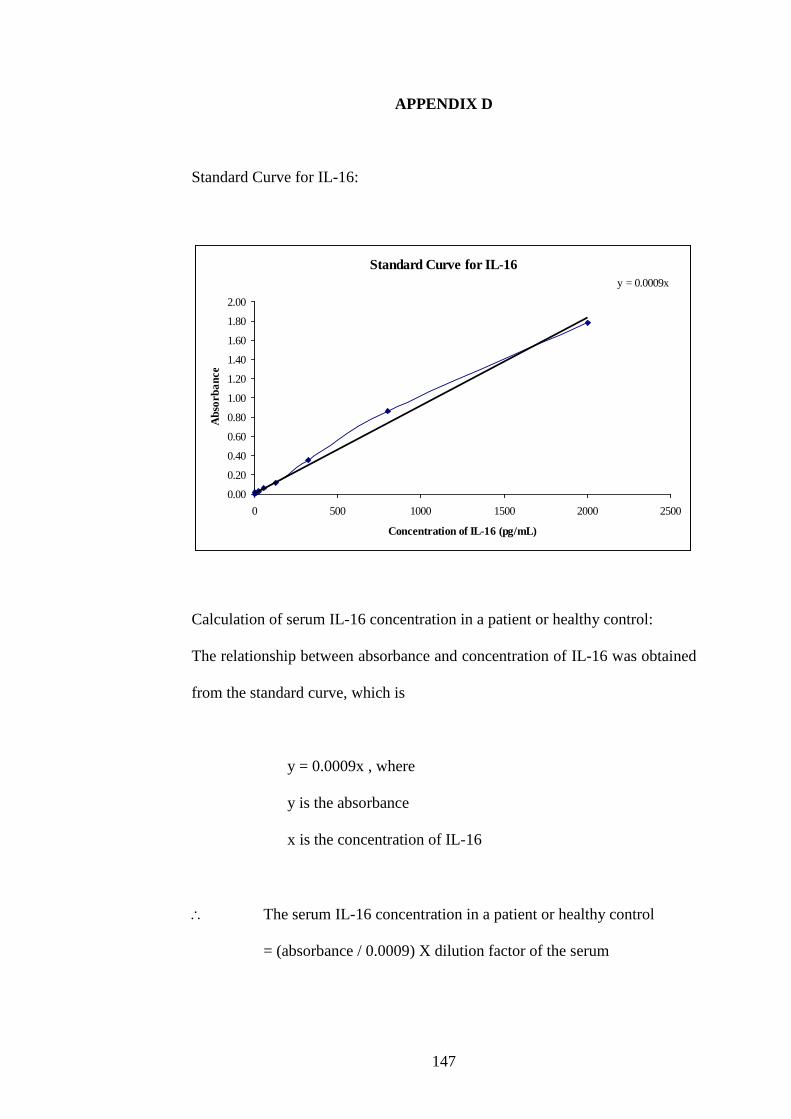
147
APPENDIX D
Standard Curve for IL-16:
Standard Curve for IL-16
y = 0.0009x
0.00
0.20
0.40
0.60
0.80
1.00
1.20
1.40
1.60
1.80
2.00
0 500 1000 1500 2000 2500
Concentration of IL-16 (pg/mL)
Ab
sorb
an
ce
Calculation of serum IL-16 concentration in a patient or healthy control:
The relationship between absorbance and concentration of IL-16 was obtained
from the standard curve, which is
y = 0.0009x , where
y is the absorbance
x is the concentration of IL-16
The serum IL-16 concentration in a patient or healthy control
= (absorbance / 0.0009) X dilution factor of the serum

148
APPENDIX E
Standard Curve for IFN-γ:
Standard Curve for IFN-γy = 0.0051x
0.00
0.20
0.40
0.60
0.80
1.00
1.20
1.40
1.60
1.80
0 50 100 150 200 250 300 350
Concentration of IFN-γ (pg/mL)
Ab
sorb
an
ce
Calculation of serum IFN-γ concentration in a patient or healthy control:
The relationship between absorbance and concentration of IFN-γ was obtained
from the standard curve, which is
y = 0.0051x , where
y is the absorbance
x is the concentration of IFN-γ
The serum IFN-γ concentration in a patient or healthy control
= (absorbance / 0.0051) X dilution factor of the serum

149
APPENDIX F
Standard Curve for MCP-1:
Standard Curve for MCP-1y = 0.0017x
0.00
0.20
0.40
0.60
0.80
1.00
1.20
1.40
1.60
1.80
0 200 400 600 800 1000 1200
Concentration of MCP-1 (pg/mL)
Ab
sorb
an
ce
Calculation of serum MCP-1concentration in a patient or healthy control:
The relationship between absorbance and concentration of MCP-1was
obtained from the standard curve, which is
y = 0.0017x , where
y is the absorbance
x is the concentration of MCP-1
The serum MCP-1 concentration in a patient or healthy control
= (absorbance / 0.0017) X dilution factor of the serum

150
APPENDIX G
Standard Curve for MIG:
Standard Curve for MIGy = 0.0006x
0.00
0.20
0.40
0.60
0.80
1.00
1.20
1.40
0 500 1000 1500 2000 2500
Concentration of MIG (pg/mL)
Ab
sorb
an
ce
Calculation of serum MIG concentration in a patient or healthy control:
The relationship between absorbance and concentration of MIG was obtained
from the standard curve, which is
y = 0.0006x , where
y is the absorbance
x is the concentration of MIG
The serum MIG concentration in a patient or healthy control
= (absorbance / 0.0006) X dilution factor of the serum

151
APPENDIX H
Standard Curve for RANTES:
Standard Curve for RANTESy = 0.0005x
0.00
0.20
0.40
0.60
0.80
1.00
1.20
0 500 1000 1500 2000 2500
Concentration of RANTES (pg/mL)
Ab
sorb
an
ce
Calculation of serum RANTES concentration in a patient or healthy control:
The relationship between absorbance and concentration of RANTES was
obtained from the standard curve, which is
y = 0.0005x , where
y is the absorbance
x is the concentration of RANTES
The serum RANTES concentration in a patient or healthy control
= (absorbance / 0.0005) X dilution factor of the serum