reversible posterior leukoencephalopathy in a patient with wegener granulomatosis
TRANSCRIPT

Pediatr Nephrol (2004) 19:442–444DOI 10.1007/s00467-003-1286-y
B R I E F R E P O R T
Toshiyuki Ohta · Takashi Sakano · Mami Shiotsu ·Takeki Furue · Hideyuki Ohtani ·Yoshihisa Kinoshita · Tatsuya Mizoue · Katsuzo Kiya ·Issei Tanaka
Reversible posterior leukoencephalopathy in a patientwith Wegener granulomatosisReceived: 28 February 2003 / Revised: 8 July 2003 / Accepted: 14 July 2003 / Published online: 16 December 2003� IPNA 2003
Abstract A 14-year-old girl with rapidly progressiveglomerulonephritis was transferred to our hospital be-cause of acute renal failure. A diagnosis of Wegenergranulomatosis was made according to the symptom triadof a renal biopsy demonstrating crescentic glomerulone-phritis, severe sinusitis, and serological findings of raisedproteinase 3 anti-neutrophil cytoplasmic antibody level.In spite of combination therapy with methylprednisolone,cyclophosphamide, and plasma exchange, her renalfunction gradually deteriorated. Thereafter, she suffereda severe headache and generalized seizures. Brain com-puted tomography (CT) scan revealed bilateral low-density areas in the parieto-occipital lobes. Magneticresonance imaging (MRI) disclosed a high-intensity areaon T2-weighted images and a low-signal intensity area onT1-weighted images in the same lesion. Follow-up brainCT scan 3 weeks and MRI 2 months after the first studiesshowed complete resolution of the abnormal lesions,which indicated reversible posterior leukoencephalopathysyndrome. In addition to renal failure, hypertension, andcyclophoshamide, the primary disease may have played a
role in the development of this uncommon syndrome inour patient.
Keywords Hypertension · Chronic renal failure ·Reversible posterior leukoencephalopathy syndrome ·Wegener granulomatosis · Anti-neutrophil cytoplasmicantibody
Introduction
Reversible posterior leukoencephalopathy syndrome(RPLS) is a rare neurological syndrome characterizedby headache, altered mental status, seizures, and visualdisturbance, associated with reversible white matterchanges [1]. It has been commonly reported in patientswith severe hypertension and pre-eclampsia [1, 2, 3]. Todate, there are only a few cases of RPLS with systemicvasculitis. We report a patient with Wegener granuloma-tosis (WG) complicated by RPLS.
Case report
Recurrent polyarthritis involving the knees and ankles developed ina 14-year-old girl without a previous history of neurologicalmanifestations. Two weeks after the first manifestation, she wasadmitted to a local hospital with a tentative diagnosis of post-streptococcal acute glomerulonephritis. She had no fevers and norespiratory symptoms or signs. On admission to the hospital,laboratory studies revealed a blood urea nitrogen (BUN) of60.9 mg/dl and serum creatinine 3.0 mg/dl. Eleven days later, shewas referred to our hospital for further evaluation and treatment.
Her clinical course in our hospital is shown in Fig. 1. Onadmission to our hospital, physical examination showed severalerythematous lesions in the second and third proximal interpha-langeal joints and her blood pressure was 90/40 mmHg. Laboratorystudies revealed a serum sodium of 139 mEq/l, serum potassium4.1 mEq/l, serum chloride 103 mEq/l, BUN 80.1 mg/dl, serumcreatinine 6.0 mg/dl, bicarbonate 24.1 mmol/l, serum creatinekinase 63 U/l, and C-reactive protein 1.9 mg/dl. The levels ofproteinase 3 anti-neutrophil cytoplasmic antibody (ANCA), rheu-matoid factor, and anti-nuclear antibody were more than 500 U/ml(normal, less than 3.5 U/ml), 576 IU/ml, and 1:320, respectively.Serum anti-glomerular basement membrane antibody, anti-DNA
T. Ohta · T. Sakano · M. Shiotsu · T. Furue · H. Ohtani ·Y. KinoshitaDepartment of Pediatrics,Hiroshima Prefectural Hospital,Hiroshima, Japan
T. Mizoue · K. KiyaDepartment of Neurosurgery,Hiroshima Prefectural Hospital,Hiroshima, Japan
I. TanakaDialysis Center,Hiroshima Prefectural Hospital,Hiroshima, Japan
T. Ohta ())Department of Pediatrics,Hiroshima Prefectural Hospital,1–5-54 Ujinakanda, Minami-ku, Hiroshima 734–8530, Japane-mail: [email protected].: +81-82-2541818Fax: +81-82-2538274
antibody, anti-Sm antibody, anti-SS-A antibody, anti-SS-B anti-body, myeloperoxidase ANCA, and cryoglobulin were all negative.The urine gave a 2+ test for protein; the sediment contained 50–100red blood cells per high-power field and red blood cell casts. Sheunderwent a renal biopsy, which demonstrated diffuse and globalfibrous or fibrocellular crescents. Although chest computed tomo-graphy (CT) failed to show any abnormality, severe chronicsinusitis was confirmed clinically and radiographically. Therefore,a diagnosis of WG was made. She received intravenously six pulsesof methylprednisolone at a dose of 1,000 mg, followed by oralprednisolone (1 mg/kg body weight per day) and oral cyclophos-phamide (2 mg/kg body weight per day), with little effect.Cyclophosphamide was withdrawn at 6 weeks because of leuko-penia and diffuse alopecia. Thereafter, her renal function deterio-rated gradually in spite of six sessions of single plasma exchange.
On the 94th hospital day, she developed a severe headache andher blood pressure was 180/92 mmHg. She was treated successfullywith nifedipine. The following day she experienced suddengeneralized tonic-clonic seizures seven times and became lethargic.The seizures were treated successfully with intravenous diazepam.At that time, she was afebrile and her blood pressure was 158/100 mmHg. Laboratory studies revealed a serum creatinine of7.8 mg/dl, BUN 126.1 mg/dl, and hemoglobin 7.2 g/dl. Bloodglucose, electrolytes, and bicarbonate were in the normal range.Cerebrospinal fluid examination showed no abnormality, withnegative testing for myelin basic protein. Thus, she underwentemergency hemodialysis with a tentative diagnosis of uremicencephalopathy. Although emergency brain CT scan failed toreveal any abnormal findings, brain CT scan 2 days later disclosedbilateral low-density areas in the parieto-occipital lobes. Magneticresonance imaging (MRI) showed a high-signal intensity area onT2-weighted fluid-attenuated inversion-recovery images and a low-signal intensity on T1-weighted images in the same lesions(Fig. 2A). Electroencephalography conducted 1 month after thefirst seizure showed diffuse slow activity without any sharp wave.Phenytoin was given intravenously for 7 days after the first seizure,followed by per os valproate. Follow-up CT scan at 3 weeks andMRI at 2 months (Fig. 2B) after the first studies showed completeresolution of the abnormal lesions in the bilateral parieto-occipitallobes, which was compatible with the diagnosis of RPLS. Shegradually recovered consciousness and had no additional episodesof convulsions. No neurological abnormality was present and herblood pressure was well controlled with nifedipine and an
angiotensin-converting enzyme inhibitor when she left our hospitalon peritoneal dialysis 3 months after the first seizure.
Discussion
RPLS has been characterized by headache, altered mentalfunction, seizures, and visual disturbance, associated withreversible white matter lesions, predominantly in theposterior region of the cerebral hemispheres on brain CTscan or MRI [1]. The cause of this syndrome remainsunknown. However, the rapid resolution of clinical andneuroradiological abnormalities suggests that cerebraledema, caused by impaired cerebrovascular autoregula-tion and endothelial injury, is the main pathophysiologicalmechanism. Mukherjee and McKinstry [2] reported 12cases with RPLS, including 2 cases of lupus nephritis, 2of thrombotic thrombocytopenic purpura, 1 of acute renalfailure, 1 of end-stage renal disease, 1 of eclampsia, 1 offocal segmental glomerulosclerosis, and 3 of ciclosporineadministration. Of the 12 cases, 11 manifested with mild-to-severe hypertension. It is likely that hypertensionplayed a major role in the pathogenesis in these patients.
Patients with end-stage renal disease have a dysfunc-tion in vasopressor homeostasis and endothelial functionrelated to elevations in lipoproteins, blood pressure,uremia, and as a result of drug therapy. Therefore, end-stage renal disease is also an important factor in RPLS.RPLS has also been reported after administration ofciclosporine, tacrolimus, and cytotoxic agents, which mayhave a direct toxic effect on the cerebral vasculature [4,5]. Thus, our patient also had several known causativefactors, i.e., renal failure and increased blood pressure,together with recent chemotherapy.
To our best knowledge, the patient we present here isthe first clinically and radiographically demonstrated case
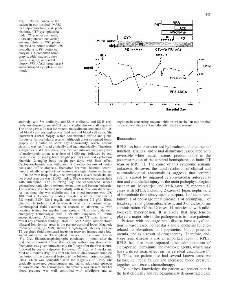
Fig. 1 Clinical course of thepatient in our hospital. (mPSLmethylprednisolone, PSL pred-nisolone, CYP cyclophospha-mide, PE plasma exchange,ACEI angiotensin-convertingenzyme inhibitor, PHT phenyt-oin, VPA valporate sodium, HDhemodialysis, PD peritonealdialysis, CT computed tomo-graphy, MRI magnetic reso-nance imaging, RBx renalbiopsy, PR3-ANCA proteinase 3anti-neutrophil cytoplasmic an-tibody)
443

of RPLS associated with WG. The central nervous systemhas rarely been affected in patients with WG in theliterature. In two large pediatric series of WG, seizure wasnoted in 2 of 40 patients [6, 7]. In the largest series ofWG, central nervous system abnormalities, which includ-ed stroke, cranial nerve abnormalities, and diabetes
insipidus, occurred in 8% of patients [8]. However, it isunclear whether thorough neuroimaging studies wereconducted in these patients. Recently, with the wide-spread use of MRI, RPLS is becoming more common.Some of the above patients might have been misdiag-nosed because RPLS may have been overlooked withonly an emergency CT evaluation. It is likely that WGplayed a role in the progression to RPLS in our patientbecause the serum from patients with WG containsANCA, which can activate neutrophils and cause endo-thelial cell injury [9]. In general, the important points ofsuccessful treatment for WG are early diagnosis and earlyintense immunosuppressive therapy, including severalcytotoxic agents [9]. Moreover, Ellis et al. [10] suggestedthat pediatric patients with WG had rapid progression toend-stage renal disease. Therefore, RPLS should beconsidered in patients, especially pediatric patients, withend-stage renal disease due to systemic vasculitis,including WG.
References
1. Hinchey J, Chaves C, Appignani BA, Breen J, Pao L, Wang A,Pessin MS, Lamy C, Mas JL, Caplan LR (1996) A reversibleposterior leukoencephalopathy syndrome. N Engl J Med234:494–500
2. Mukherjee P, McKinstry RC (2001) Reversible posteriorleukoencephalopathy syndrome: evaluation with diffusion-ten-sor MR imaging. Radiology 219:756–765
3. Saatci I, Topaloglu (1994) Cranial computed tomographicfindings in a patient with hypertensive encephalopathy in acutepoststreptococcal glomerulonephritis. Turk J Pediatr 36:325–328
4. Singh N, Bonham A, Fukui M (2000) Immunosuppressive-associated leukoencephalopathy in organ transplant recipients.Transplantation 69:467–472
5. Edwards MJJ, Walker R, Vinnicombe S, Barlow C, MacCallumP, Foran JM (2001) Reversible posterior leukoencephalopathysyndrome following CHOP chemotherapy for diffuse B-celllymphoma. Ann Oncol 12:1327–1329
6. Belostotsky VM, Shah V, Dillon MJ (2002) Clinical features in17 paediatric patients with Wegener granulomatosis. PediatrNephrol 17:754–761
7. Rottem M, Fauci AS, Hallahan CW, Kerr GS, Lebovics R,Leavitt RY, Hoffman GS (1993) Wegener granulomatosis inchildren and adolescents: clinical presentation and outcome. JPediatr 122:26–31
8. Hoffman GS, Kerr GS, Leavitt RY, Hallahan CW, LebovicsRS, Travis WD, Rottem M, Fauci AS (1992) Wegenergranulomatosis: an analysis of 158 patients. Ann Intern Med116:488–498
9. Savage COS (2001) ANCA-associated renal vasculitis. KidneyInt 60:1614–1627
10. Ellis EN, Wood EG, Berry P (1995) Spectrum of diseaseassociated with anti-neutrophil cytoplasmic auto antibodies inpediatric patients. J Pediatr 126:40–43
Fig. 2 T2-weighted MRI scans. A Areas of hyper-intense signalson T2-weighted fluid-attenuated inversion-recovery images involv-ing parieto-occipital lobes bilaterally. The follow-up MRI (B)reveals the complete resolution of abnormal signals
444