research report literature review
TRANSCRIPT
RESEARCH REPORT Click or tap here to enter text.Literature review regarding the colour vision requirements for aircrew An Agency of the European Union
Disclaimer This study has been carried out for the European Union Aviation Safety Agency by an external organization and expresses the opinion of the organization undertaking the study. It is provided for information purposes only and the views expressed in the study have not been adopted, endorsed or in any way approved by the European Union Aviation Safety Agency. Consequently it should not be relied upon as a statement, as any form of warranty, representation, undertaking, contractual, or other commitment binding in law upon the European Union Aviation Safety Agency. Ownership of all copyright and other intellectual property rights in this material including any documentation, data and technical information, remains vested to the European Union Aviation Safety Agency. All logo, copyrights, trademarks, and registered trademarks that may be contained within are the property of their respective owners. Reproduction of this study, in whole or in part, is permitted under the condition that the full body of this Disclaimer remains clearly and visibly affixed at all times with such reproduced part. No part of this report may be reproduced and/or disclosed, in any form or by any means without the prior written permission of the owner. REPORT NUMBER: EASA_REP_RESEA_2019_1 REPORT CLASSIFICATION: UNCLASSIFIED DATE: 20 February 2019 KNOWLEDGE AREA(S): Human vision DESCRIPTOR(S): color vision, aeromedical standards CUSTOMER: CONTRACT NUMBER: N/A OWNER: European Union Aviation Safety Agency DISTRIBUTION: Limited CLASSIFICATION OF TITLE: UNCLASSIFIED Author(s): Dr. Constantin MIHAI, MD REVIEWED BY: AUTHOR REVIEWER MANAGING DEPARTMENT EASA Dr. C. Panait Flight Standards
1
Dr. Constantin MIHAI, MD Ophthalmology 20/02/2019
Literature review study regarding the colour vision requirements for aircrew
2
Contents 1 Introduction ..................................................................................................................................................... 3 1.1 Abbreviations ........................................................................................................................................... 3 1.2 Terminology ............................................................................................................................................. 4 1.3 Background .............................................................................................................................................. 4 1.4 Objective of the study ............................................................................................................................. 5 1.5 Colour vision deficiencies as a contributing factor in aviation accidents ................................................ 5 2 Analysis of literature data on colour vision assessment of pilots ................................................................... 6 2.1 Method .................................................................................................................................................... 6 2.2 Colour vision physiology .......................................................................................................................... 6 2.3 Colour vision deficiencies ........................................................................................................................ 7 2.3.1 Congenital colour vision deficiencies .............................................................................................. 7 2.3.2 Acquired colour vision deficiencies ................................................................................................. 8 2.4 Operational consideration ..................................................................................................................... 12 2.5 Colour vision testing in aviation ............................................................................................................ 17 2.5.1 Pseudo-isochromatic plates (PIP) .................................................................................................. 18 2.5.2 Anomaloscopes ............................................................................................................................. 19 2.5.3 Holmes-Wright lantern test (type A and B) ................................................................................... 20 2.5.4 The Colour Assessment and Diagnosis (CAD) Test ........................................................................ 20 2.5.5 Computerized Colour Vision Test (CCVT) ...................................................................................... 21 2.5.6 The Cambridge Colour Test (Cambridge CT) ................................................................................. 22 2.5.7 Cone Contrast Test (CCT) ............................................................................................................... 22 2.5.8 Farnsworth-Munsell 100 hue test ................................................................................................. 23 3 Discussion and conclusion ............................................................................................................................. 23 4 Current European colour vision requirements for flight crew ...................................................................... 34 MED.B.075 ..................................................................................................................................................... 34 AMC1 MED.B.075 Colour vision ................................................................................................................... 35 AMC2 MED.B.075 Colour vision ................................................................................................................... 35 AMC14 MED.B.095 Medical examination and assessment of applicants for LAPL medical certificates ..... 35 5 Recommendations ......................................................................................................................................... 36 5.1 Recommendations for colour vision testing for pilots .......................................................................... 36 5.2 Further research .................................................................................................................................... 37 6 References ..................................................................................................................................................... 37
3
1 Introduction Vision is one of the most fascinating abilities of human beings. It is the most powerful sense and at the same time the most difficult to describe. A ray of light caught by the human eye enters a chain of highly complex processing steps that ends in the upper levels of the visual cortex. Colour vision is the ability of the eye to distinguish objects based on the wavelengths (or frequencies) of the light they reflect, emit, or transmit. Colours can be measured and quantified in various ways; indeed, a person's perception of colours is a subjective process whereby the brain responds to the stimuli that are produced when incoming light reacts with the several types of cone cells in the eye. There is the possibility that different people see the same illuminated object or light source in different ways. 1.1 Abbreviations CVD – colour vision deficiency CVN – Normal colour vision CCT – Cone contrast test CAD – Colour Assessment and Diagnosis test CCVT – Computerized Colour Vision Test Cambridge CT – Cambridge Colour Test PIP – Pseudo-isochromatic plates EASA – European Union Aviation Safety Agency ICAO – International Civil Aviation Organisation JAA – Joint Aviation Authorities RG- Red green YB – Yellow Blue RE – Right eye LE – Left eye NTs - Normal trichromats IT – Ishihara test ON – Optic nerve
4
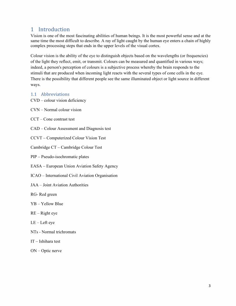
1.2 Terminology Sensitivity measures the proportion of actual positives that are correctly identified as such (e.g., the percentage of sick people who are correctly identified as having the condition). Sensitivity refers to the test's ability to correctly detect ill patients who do have the condition. It can also be expressed in a mathematical equation as: Sensitivity = No of true positives/ Total No of sick individuals in population Specificity measures the proportion of actual negatives that are correctly identified as such (e.g., the percentage of healthy people who are correctly identified as not having the condition). Specificity of a test is the proportion of healthy patients known not to have the disease, who will test negative for it. Specificity relates to the test's ability to correctly reject healthy patients without a condition. Specificity= No of true negative / No of colour normal individuals in population. (Wikipedia s.d.) Chroma is an attribute of perceived color relating to chromatic intensity that enables an observer to judge how much chromatic colour it contains irrespective of achromatic colour present. Can be a synonym of the saturation of a color. Intraobserver implies describing variation in the scores, responses etc. obtained by the same observer on different occasions 1.3 Background Colour contrast aids in detection and identification of objects in the visual scene. Colour is a quality of the mind given to light of a certain spectral composition in a certain state of ocular adaptation. Physiologically, colour can be described by the three qualities: hue, saturation and lightness. These have psychophysical counterparts which can be given colorimetric figures in order to characterise the colour in question. The early use of colour in sea and land traffic was limited by the techniques available to produce light of sufficient saturation and brightness. Therefore only red, yellow (white) and green signals were adopted originally, and their significance is today so deeply rooted in us all that they cannot be exchanged. This is unfortunate, since all people do not perceive colour in the same way and exactly these hues give rise to separation difficulties. Although attempts have been made to minimise the use of colour contrast as the sole characteristic of a stimulus, colours are still used to such an extent that some applicants have to be rejected for safety reasons.
Condition positive Condition negative Test outcome positive True positive (TP) False positive (FP) Positive predictive value = TP / (TP + FP) Test outcome negative False negative (FN) True negative (TN) Negative predictive value = TN / (FN + TN) Sensitivity = TP / (TP + FN) Specificity = TN / (FP + TN) Figure 1. Terminology
5
The first reported use of colour vision selection was during the First World War when the Royal Flying Corps introduced vision testing for applicants, including CVD testing, because of the importance of picking out the colour or markings of hostile aircraft, recognizing signal lights, and judging the nature of landing grounds (Watson 2014). Occupational colour vision standards were introduced in aviation in 1919 by The Aeronautical Commission of the International Civil Air Navigation Authority (Watson 2014). These standards reflected both the needs and the methods available for colour vision assessment at the time. The standards were taken over by International Civil Aviation Organization. In Europe, the Joint Aviation Authorities (JAA) included similar requirements in their medical requirements (JAR-FCL 3). These were transposed in the current requirements in Commission Regulation (EU) No 1178/2011 with small changes in regard to the testing methods. 1.4 Objective of the study The general objective of this study is to review the relevance and effectiveness of colour vision testing for pilots. This literature review study aims to assess the possible need to mitigate the risk to flight safety resulting from colour deficient pilots by means of colour vision testing. The literature review aims to look at the need of colour vision in aviation environment as well as at the testing methods and their suitability for use in pilots’ testing. 1.5 Colour vision deficiencies as a contributing factor in aviation accidents Although there have been some accidents in military aviation where colour vision deficiencies were listed e.g. a series of F-4 Phantom accidents linked with special disorientations involved pilots with a degree of colour vision deficiencies, no causal link was demonstrated. For this reason the link to colour vision deficiency is seen as unsubstantiated, due to the fact that the USAF selection policy in regard to colour vision in the ‘70s and ‘80s was virtually inefficient. However, there is at least one accident where the colour vision deficiency of the pilot flying was demonstrated to be a causing factor. This is the Federal Express flight 1478 accident. Fed Ex 1478 – On July 26, 2002, Federal Express flight 1478, a Boeing 727-232F, struck trees on short final approach and crashed short of runway 9 at the Tallahassee Regional Airport (TLH), Tallahassee, Florida as mentioned in the NTSB accident investigation report. The investigations concluded that the probable cause of the accident was the captain’s and first officer’s failure to establish and maintain a proper glidepath during the night visual approach to landing. Contributing to the accident was a combination of the captain’s and first officer’s fatigue, the captain’s and first officer’s failure to adhere to company flight procedures, the captain’s and flight engineer’s failure to monitor the approach, and the first officer’s colour vision deficiency. The first officer suffered from a severe colour vision deficiency that made it difficult for him to correctly identify the colour of the precision approach path indicator signal during the below-glidepath, night-time, visual approach to runway 9 at Tallahassee Regional Airport. NTSB also concluded that existing aviation medical certification standards for colour vision and use of related screening tests may not ensure detection of colour vision deficiencies that can be detrimental to safety; it is possible that in some emergency situations, the speed of colour recognition may assume an importance that is not currently reflected in the standards.
6
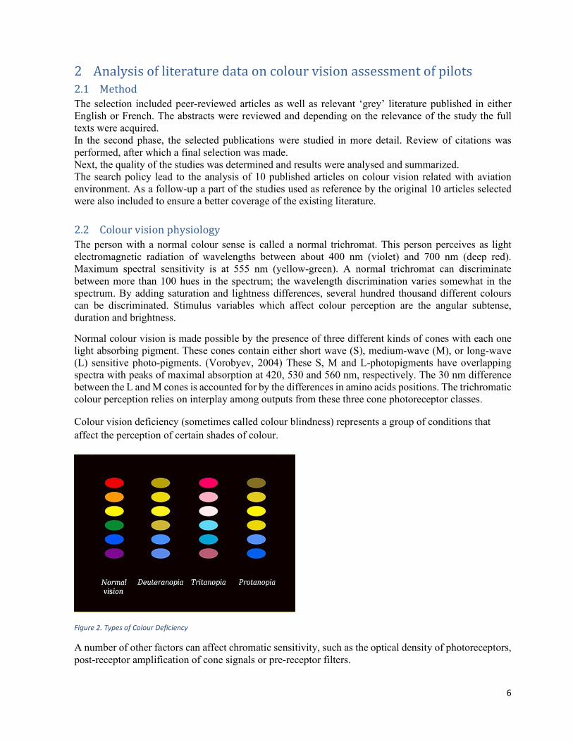
2 Analysis of literature data on colour vision assessment of pilots 2.1 Method The selection included peer-reviewed articles as well as relevant ‘grey’ literature published in either English or French. The abstracts were reviewed and depending on the relevance of the study the full texts were acquired. In the second phase, the selected publications were studied in more detail. Review of citations was performed, after which a final selection was made. Next, the quality of the studies was determined and results were analysed and summarized. The search policy lead to the analysis of 10 published articles on colour vision related with aviation environment. As a follow-up a part of the studies used as reference by the original 10 articles selected were also included to ensure a better coverage of the existing literature. 2.2 Colour vision physiology The person with a normal colour sense is called a normal trichromat. This person perceives as light electromagnetic radiation of wavelengths between about 400 nm (violet) and 700 nm (deep red). Maximum spectral sensitivity is at 555 nm (yellow-green). A normal trichromat can discriminate between more than 100 hues in the spectrum; the wavelength discrimination varies somewhat in the spectrum. By adding saturation and lightness differences, several hundred thousand different colours can be discriminated. Stimulus variables which affect colour perception are the angular subtense, duration and brightness. Normal colour vision is made possible by the presence of three different kinds of cones with each one light absorbing pigment. These cones contain either short wave (S), medium-wave (M), or long-wave (L) sensitive photo-pigments. (Vorobyev, 2004) These S, M and L-photopigments have overlapping spectra with peaks of maximal absorption at 420, 530 and 560 nm, respectively. The 30 nm difference between the L and M cones is accounted for by the differences in amino acids positions. The trichromatic colour perception relies on interplay among outputs from these three cone photoreceptor classes. Colour vision deficiency (sometimes called colour blindness) represents a group of conditions that affect the perception of certain shades of colour. Figure 2. Types of Colour Deficiency A number of other factors can affect chromatic sensitivity, such as the optical density of photoreceptors, post-receptor amplification of cone signals or pre-receptor filters.
7
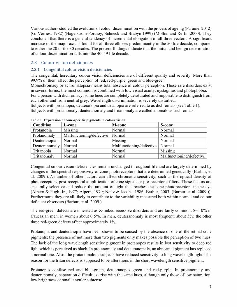
Various authors studied the evolution of colour discrimination with the process of ageing (Paramei 2012) (G. Verriest 1982) (Hagerstrom-Portnoy, Schneck and Brabyn 1999) (Mollon and Reffin 2000). They concluded that there is a general tendency of incremental elongation of all three vectors. A significant increase of the major axis is found for all three ellipses predominantly in the 50 life decade, compared to either the 20 or the 30 decades. The present findings indicate that the initial and benign deterioration of colour discrimination falls into the 40–49 life decade. 2.3 Colour vision deficiencies 2.3.1 Congenital colour vision deficiencies The congenital, hereditary colour vision deficiencies are of different quality and severity. More than 99.9% of them affect the perception of red, red-purple, green and blue-green. Monochromacy or achromatopsia means total absence of colour perception. These rare disorders exist in several forms; the most common is combined with low visual acuity, nystagmus and photophobia. For a person with dichromacy, some hues are completely desaturated and impossible to distinguish from each other and from neutral grey. Wavelength discrimination is severely disturbed. Subjects with protanopia, deuteranopia and tritanopia are referred to as dichromats (see Table 1). Subjects with protanomaly, deuteranomaly and tritanomaly are called anomalous trichromats. Table 1. Expression of cone-specific pigments in colour vision Condition L-cone M-cone S-cone Protanopia Missing Normal Normal Protanomaly Malfunctioning/defective Normal Normal Deuteranopia Normal Missing Normal Deuteranomaly Normal Malfunctioning/defective Normal Tritanopia Normal Normal Missing Tritanomaly Normal Normal Malfunctioning/defective Congenital colour vision deficiencies remain unchanged throughout life and are largely determined by changes in the spectral responsivity of cone photoreceptors that are determined genetically (Barbur, et al. 2009.). A number of other factors can affect chromatic sensitivity, such as the optical density of photoreceptors, post-receptoral amplification of cone signals or pre-receptoral filters. These factors are spectrally selective and reduce the amount of light that reaches the cone photoreceptors in the eye (Alpern & Pugh, Jr., 1977; Alpern, 1979; Neitz & Jacobs, 1986; Barbur, 2003; (Barbur, et al. 2009.)). Furthermore, they are all likely to contribute to the variability measured both within normal and colour deficient observers (Barbur, et al. 2009.) The red-green defects are inherited as X-linked recessive disorders and are fairly common: 8– 10% in Caucasian men, in women about 0·5%. In men, deuteranomaly is most frequent: about 5%; the other three red-green defects affect approximately 1%. Protanopia and deuteranopia have been shown to be caused by the absence of one of the retinal cone pigments; the presence of not more than two pigments only makes possible the perception of two hues. The lack of the long wavelength sensitive pigment in protanopes results in lost sensitivity to deep red light which is perceived as black. In protanomaly and deuteranomaly, an abnormal pigment has replaced a normal one. Also, the protanomalous subjects have reduced sensitivity to long wavelength light. The reason for the tritan defects is supposed to be alterations in the short wavelength sensitive pigment. Protanopes confuse red and blue-green, deuteranopes green and red-purple. In protanomaly and deuteranomaly, separation difficulties arise with the same hues, although only those of low saturation, low brightness or small angular subtense.

8
Anomalous trichromacy is a less pronounced defect. Subjects with such an anomaly show, compared to normal, increased thresholds for saturation and wavelength discrimination in certain spectral regions. The anomalous trichromacies vary in severity and some are almost as pronounced as the dichromacies: extreme anomalous trichromacy. Borderline cases between normal trichromacy and anomalous trichromacy are pigment amblyopia and colour asthenopia. The former confuse pigment colours, e.g. those on pseudo-isochromatic charts but pass other colour vision tests. Colour asthenopia is essentially an increased ‘fatigue’ to spectral lights. These and other borderline cases are usually considered as normal in practice. Some people have blue colour blindness-> which disguises the colours blue and yellow (tritanopia). The yellow-blue (YB) defects are rare, about 1 in 50 000. This affects both men and women equally because there is no X-chromosome. Table 2: Prevalence of different types of inherited red-green colour deficiency Type of colour deficiency Prevalence in men Prevalence in women Protanopia 1% 0.01% Protanomalous trichromatism 1% 0.03% Deuteranopia 1% 0.01% Deuteranomalous trichromatism 5% 0.35% Total 8% 0.40% 2.3.2 Acquired colour vision deficiencies Acquired colour vision disorders are characterized by unilateral or bilateral alterations in the function of the retina or the conduction pathways leading from the retina to the cerebral cortex. In most cases, acquired colour vision disorders have a concomitant deterioration in visual acuity, exhibit visual field defects and may show other functional disorders of the sensory end organ. Acquired colour vision deficiencies arise from diseases in the eye or the visual pathways. An ocular disorder most often gives rise to a yellow-blue defect. It is generally combined with other visual disturbances like reduced visual acuity or visual field defects and the ocular damage is thus overt. Of greater practical interest is the red-green deficiency caused by an optic nerve lesion. Such a problem invariably accompanies an optic neuritis and may result in difficulties in identifying colour signals although the visual acuity is normal. With increasing age and density of the yellow lens pigment, a slight degree of tritanomaly follows. Table 3: Characteristics of congenital vs. acquired colour vision (CV) defects CONGENITAL ACQUIRED Colour loss in specific spectral region Often no clear cut area of discrimination loss Less marked dependence of CV on target Marked dependence of CV on size and illuminance Characteristic results obtained on various clinical CV tests Conflicting or variable results on clinical CV tests

9
Many object colours are named correctly or predictable errors are made Some object colours are named incorrectly Both eyes are equally affected Eyes are affected asymmetrically Usually no other visual complaint May have decreased acuity and field loss Defect is stable Defect may go towards progression or regression Type I Acquired Red-Green deficiencies: Red-green chromatic discrimination progressively deteriorates. There is an accompanying loss of visual acuity. The photopic luminosity function (curve) becomes more and more scotopic. In advanced stages, there is total colour blindness that resembles congenital achromatopsia. This condition is associated with retinal diseases, especially those involving photoreceptors of the posterior pole (macular cones are destroyed). The photopic luminosity function is normal. Type II Acquired Red-Green deficiencies: There is a red-green discrimination loss which is moderate or severe with a concomitant. Blue-yellow loss that is mild. The apparent saturation of colours is decreased and in advanced cases, the 500 nm region of the spectrum looks grey to the patient. In late stages, there is eccentric fixation with nearly complete achromatopsia. Seen in medical conditions such as optic neuritis, retrobulbar neuritis, optic atrophies, O.N. intoxication, malformed disc, and O.N. or chiasm tumours. Type III Acquired Blue-Yellow deficiencies: This is the most frequent acquired deficit. There is a mild to moderate blue-yellow sensitivity loss. In the early stages there is a blue deficit. However in the later stages the disease proceeds to dichromacy with a neutral zone (that appears grey) around 500 nm. This type of colour vision deficiencies may be seen in medical conditions such as nuclear cataract chorioretinal, inflammation, vascular disorders chorioretinal, degeneration, papilledema autosomal dominant, optic atrophy, senile macular glaucoma, degeneration. Normal ageing of the retinal pigment epithelium and retina may give similar symptoms to type III deficiencies. Testing for acquired defects (Tritan) is becoming increasingly important in occupational settings as the population ages. Up to the age of 60 years there is little correlation between the subject’s age and chromatic sensitivity. A small effect can be observed when examining YB thresholds (but the correlation with age remains very poor) and virtually absent in the case of RG thresholds. The age range examined is representative of the typical working life of pilots. Colour vision is usually assessed in demanding occupational environments. Loss of colour vision later in life is described as acquired colour deficiency and can be caused by a number of factors including both systemic diseases and specific diseases of the eye (such as diabetes, glaucoma, age-related macular degeneration, etc.). Since loss of chromatic sensitivity usually precedes the reliable detection of any structural changes using fundus imaging, regular screening for acquired colour vision loss may be of great clinical value. In view of these findings, it makes sense to recommend that in addition to assessing colour vision at the start of the working career, periodic re-assessments should also be done, simply as a way of testing for acquired deficiencies. In addition to permanent or transient colour vision disorders, and those acquired through illness, disorders may also be caused by the different environmental conditions encountered in flying operations. Hypoxia comes first to mind in this connection. It is caused by insufficient oxygen saturation of the respired air (hypoxic hypoxia). Hypoxia can, however, also be caused by a reduced oxygen supply due
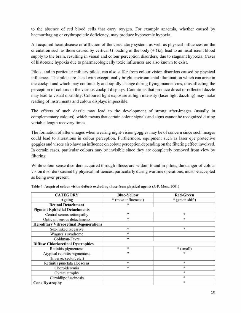
10
to the absence of red blood cells that carry oxygen. For example anaemia, whether caused by haemorrhaging or erythropoietic deficiency, may produce hypoxemic hypoxia. An acquired heart disease or affliction of the circulatory system, as well as physical influences on the circulation such as those caused by vertical G loading of the body (+ Gz), lead to an insufficient blood supply to the brain, resulting in visual and colour perception disorders, due to stagnant hypoxia. Cases of histotoxic hypoxia due to pharmacologically toxic influences are also known to exist. Pilots, and in particular military pilots, can also suffer from colour vision disorders caused by physical influences. The pilots are faced with exceptionally bright environmental illumination which can arise in the cockpit and which may continually and rapidly change during flying manoeuvres, thus affecting the perception of colours in the various cockpit displays. Conditions that produce direct or reflected dazzle may lead to visual disability. Coloured light exposure at high intensity (laser light dazzling) may make reading of instruments and colour displays impossible. The effects of such dazzle may lead to the development of strong after-images (usually in complementary colours), which means that certain colour signals and signs cannot be recognized during variable length recovery times. The formation of after-images when wearing night-vision goggles may be of concern since such images could lead to alterations in colour perception. Furthermore, equipment such as laser eye protective goggles and visors also have an influence on colour perception depending on the filtering effect involved. In certain cases, particular colours may be invisible since they are completely removed from view by filtering. While colour sense disorders acquired through illness are seldom found in pilots, the danger of colour vision disorders caused by physical influences, particularly during wartime operations, must be accepted as being ever present. Table 4: Acquired colour vision defects excluding those from physical agents (J.-P. Menu 2001) CATEGORY Blue-Yellow Red-Green Ageing * (most influenced) * (green shift) Retinal Detachment * Pigment Epithelial Detachments Central serous retinopathy * * Optic pit serous detachments * * Hereditary Vitreoretinal Degenerations Sex-linked recessive * * Wagner’s syndrome * Goldman-Favre * Diffuse Chlorioretinal Dystrophies Retinitis pigmentosa * * (small) Atypical retinitis pigmentosa (Inverse, sector, etc.) * * Retinitis punctata albescens * * Choroideremia * * Gyrate atrophy * Ceroidlipofuscinosis * Cone Dystrophy *
11
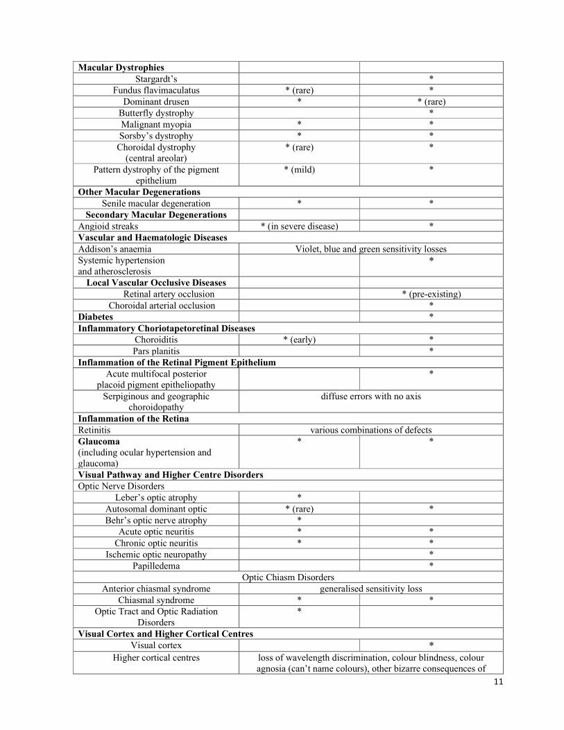
Macular Dystrophies Stargardt’s * Fundus flavimaculatus * (rare) * Dominant drusen * * (rare) Butterfly dystrophy * Malignant myopia * * Sorsby’s dystrophy * * Choroidal dystrophy (central areolar) * (rare) * Pattern dystrophy of the pigment epithelium * (mild) * Other Macular Degenerations Senile macular degeneration * * Secondary Macular Degenerations Angioid streaks * (in severe disease) * Vascular and Haematologic Diseases Addison’s anaemia Violet, blue and green sensitivity losses Systemic hypertension and atherosclerosis * Local Vascular Occlusive Diseases Retinal artery occlusion * (pre-existing) Choroidal arterial occlusion * Diabetes * Inflammatory Choriotapetoretinal Diseases Choroiditis * (early) * Pars planitis * Inflammation of the Retinal Pigment Epithelium Acute multifocal posterior placoid pigment epitheliopathy * Serpiginous and geographic choroidopathy diffuse errors with no axis Inflammation of the Retina Retinitis various combinations of defects Glaucoma (including ocular hypertension and glaucoma) * * Visual Pathway and Higher Centre Disorders Optic Nerve Disorders Leber’s optic atrophy * Autosomal dominant optic * (rare) * Behr’s optic nerve atrophy * Acute optic neuritis * * Chronic optic neuritis * * Ischemic optic neuropathy * Papilledema * Optic Chiasm Disorders Anterior chiasmal syndrome generalised sensitivity loss Chiasmal syndrome * * Optic Tract and Optic Radiation Disorders * Visual Cortex and Higher Cortical Centres Visual cortex * Higher cortical centres loss of wavelength discrimination, colour blindness, colour agnosia (can’t name colours), other bizarre consequences of
12
stroke as related to colour perception, fluctuations in sensitivity, hue discrimination range expands resulting in extremely poor performance 2.4 Operational consideration The main visual tasks of the pilot are the following: a. Distance visual tasks b. Intermediate and near vision tasks c. Spatial orientation d. Processing coloured information. Based on the necessity of the pilot to be able to perform these tasks reliably, historically, visual requirements have been established within the following areas: a. Distance visual acuity b. Near vision c. Visual fields d. Binocular function e. Stereoscopic vision f. Colour perception As colour-deficient people can only see a limited number of colours, their ability to differentiate by colour is restricted. This means mistakes can be made in colour identification and some colours are confused. People with significant colour deficiency (dichromats and severe anomalous trichromats) confuse bright colours in all viewing conditions, whereas people with slight colour deficiency typically confuse pale or dark colours. The range of colour confusions increases in low-level illumination and if the coloured areas are small. Colour confusions for acquired defects are unpredictable and the colour perception of such people is often unstable. As very well described in the JAA Manual of Civil Aviation Medicine, Amd. 6 -2009, the effectiveness of the visual system is of highest importance for flight crew to safely discard their duties. Environmental factors specific to aviation may reduce the visual performance of the flight crew members to a degree not ordinarily experienced in normal ground tasks and should be taken into account accordingly. Among these environmental factors the following can be identified: Altitude Speed Acceleration Vibration Ambient light With increasing flight altitude, the normal environmental light distribution reverses; when flying over clouds sunlight is reflected so that the lower part of the visual field is brighter than the upper one. Cockpit illumination may produce visual problems for several reasons. At low illumination levels, the visual acuity is reduced and the depth of focus decreased due to the pupillary dilatation. This way presbyopic problems are enhanced. Also colour discrimination deteriorates making the reading of colour
13
maps more difficult. Red light illumination causes even more problems with colours and may also induce a relative hypermetropia (as long wavelengths are less refracted in the ocular media). It is generally not necessary to reduce cockpit illumination to a level corresponding to a deep mesopic or scotopic adaptation. (Under daylight conditions, only the cones of the retina are in operation and under full dark adaptation especially the rods. Mesopic vision is an adaptation level in-between with both cones and rods functioning. It ranges from weak daylight to moonlight.) Most of the in-flight information in commercial aviation is provided by instruments. Likewise the runway illumination on international aerodromes is of such a standard that signals are seen without dark adaptation. In special situations, however, a certain degree of dark adaptation may be required for the correct identification of objects outside the aircraft. The high speeds of modern aircraft at take-off, cruising and landing put special demands on the visual system. We have good reason to believe that dynamic visual skills, i.e. dynamic visual acuity and the threshold for angular motion is of greater importance than the static skills under these circumstances. The pronounced decrease in dynamic visual ability after the age of 50 to 60 years is of great concern in older pilots. Vibration, especially within the 22–64 Hz range, may cause difficulties in reading instruments or printed material. In practice, problems arise under special operational conditions such as in helicopters. Vibration within the range of 2–10 Hz encountered in turbulence or on rough runways has a significant detrimental effect on visual performance. In aviation, colour is used to identify and process colour coded information displayed on various formats (e.g. signals, instruments, signs or prints). Furthermore, the colour coding is used in aviation to reduce the reaction times for different actions e.g. assessment of engine functions. Among the colour coded information in the literature we found reference to the following: Airfield lighting e.g. the parking lights, taxiways Aircraft navigation lights - regulatory lights of airplanes PAPI lights Hazard marker beacons Signal gun Flight deck displays. Maps Hazardous obstacles such as tall constructions, construction equipment or high terrain may be marked by lights or beacons. This is not universal and the absence of a beacon does not mean the absence of an obstacle. Moreover, the presence of a beacon does not necessarily mean there is an obstacle present as the beacon could, for example, be on another aircraft. The appearance of beacons is not consistent. In some cases a steady red light may be used however in other cases alternative light colours may be used. In some cases, such as the high point at an aerodrome, a flashing or rotating white beacon may be used. In considering the pre-flight planning that aircrew undertake prior to a flight, the presence of high features which may be hazardous to an aircraft will be noted from notices to airmen (NOTAM), plates and maps, whether that feature is marked by a beacon or not and whether that feature is permanent or temporary. Assuming that every hazardous feature will
14
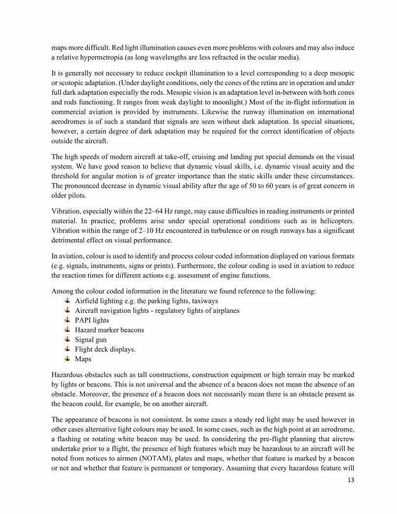
be marked by a beacon and relying on every beacon to be serviceable and visible is far from satisfactory airmanship. Individuals with protan (reduced red sensitivity) type CVD have a reduced ability to detect red beacons (B. L. Cole 1983). Aircraft of all sizes feature navigation lights. An adaptation of maritime technology, aircraft are fitted with three lights – red on the left extremity, green on the right extremity and white on the tail. The intent of these lights was to enable the presence and direction of travel of ships and aircraft to be determined. Early navigation lights were basic but now we see rotating beacons, strobe lights, high intensity landing and taxi lights, flashing landing lights and tail illumination, all of which are visible from much greater distances than navigation lights. Relative motion of another aircraft is very quickly and easily determined at great distances by the relative motion of the lights just as the relative motion of an aircraft during the day provides a quick indication of a collision risk. The airfield lighting also includes colour coded information. McKelvey (1982) found that experienced pilots made operational judgements just as well from achromatic slides of the aviation environment as they did with the chromatic counterpart, except for control tower signal lights. However, this study placed no operational stress on the observers who had 5 seconds to view each scene and were not performing any other task. Furthermore the study did not measure the reaction times between the two groups. With the technical evolution, some colour signals have lost much of their significance because the message they convey has been taken over by other instruments. However, that is not the case for all systems, an essential system for landing which has not been fully replace by other technologies is the Precision approach path indicator (PAPI). The PAPI displays four lights to an aircraft on approach. Each is angled such that aircrew will see a different combination of lights depending on the slope of their approach. If flying the nominal approach path, the crew will see two red and two white lights as depicted in Figure 3. If slightly higher they will see three white and one red. If still higher they will see four white. If slightly low they will see three red and one white. If very low they will see four red lights (Watson 2014) Figure 3. Simulator view of the PAPI lights to the left of the runway lights. Two red and two white lights as pictured indicate the normal approach path. Notice the red lights are of lower intensity in the simulated image. A US study demonstrated that mild colour deficient persons performed within reasonable normal performance limits for correctly and efficiently recognizing PAPI lights and coloured cockpit display symbology. However, moderate and severely colour vision deficient persons fell far outside the range of normal response time and accuracy.
15
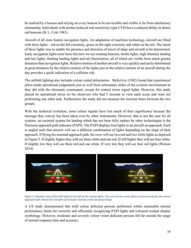
During their career colour deficient flight crew members gather experience in using aeronautical maps by day within the civil aviation community. As already mentioned aircrew do not simply come across a map in flight but study and prepare it before flight. They then track their progress along it in flight. Symbols and lines, whether coloured or not, should come as no surprise. In light of the evidence that colour deficient flight crew member already utilise maps during daylight without issue and may actually be at an advantage in adverse lighting as they are less reliant on chromatic cues, there is no reason to consider a colour deficient flight crew member less capable in map reading. In emergency situations as well as in normal flight conditions pilots are required to focus on multiple tasks, for this reason and following multiple accident investigation reports efforts have been made to reduce reaction times in aviation environment. The prompt and accurate recognition of colours is an integral part of safe aviation, increasingly so as colour-coded Electronic Flight Instruments Systems (EFIS) displays are becoming the norm. The advent of colour radar screens has rendered the interpretation of weather phenomena much easier. The information showing on an EFIS is said to be redundant because it is readable even for someone with no colour perception. However, faultless rapid data processing is important. Without normal colour perception there is potential to confuse information. The Engine indicating and crew-alerting system (EICAS) uses colour coding to support faster interpretation of the data provided and to improve the reaction times in case of an emergency situation. Additional equipment, as the weather radar or navigation maps make use of the colour coding as a way to provide information to the pilot in a manner which is easy to process and does not require time consuming mental processing. Figure 4. Multi-function aircraft display The coloured object can be self-luminous (lamp + filter, LED or colour phosphor) or can be produced with pigment colours. In the latter case, the colour appearance depends on the lighting conditions. In some cases, a luminous contrast to the background is also present, and the identification of the colour is of assistance but not necessary to read the information. In other cases, e.g. navigation lights, the hue is the only clue to the correct identification.
16
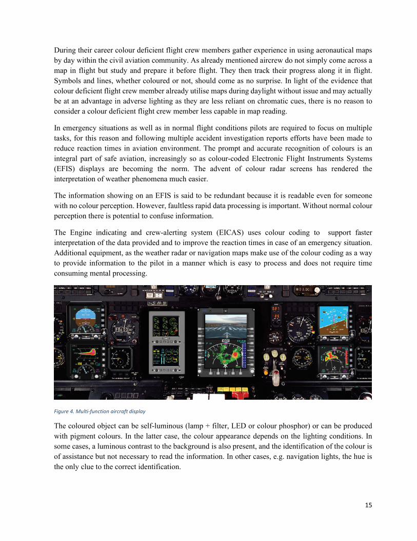
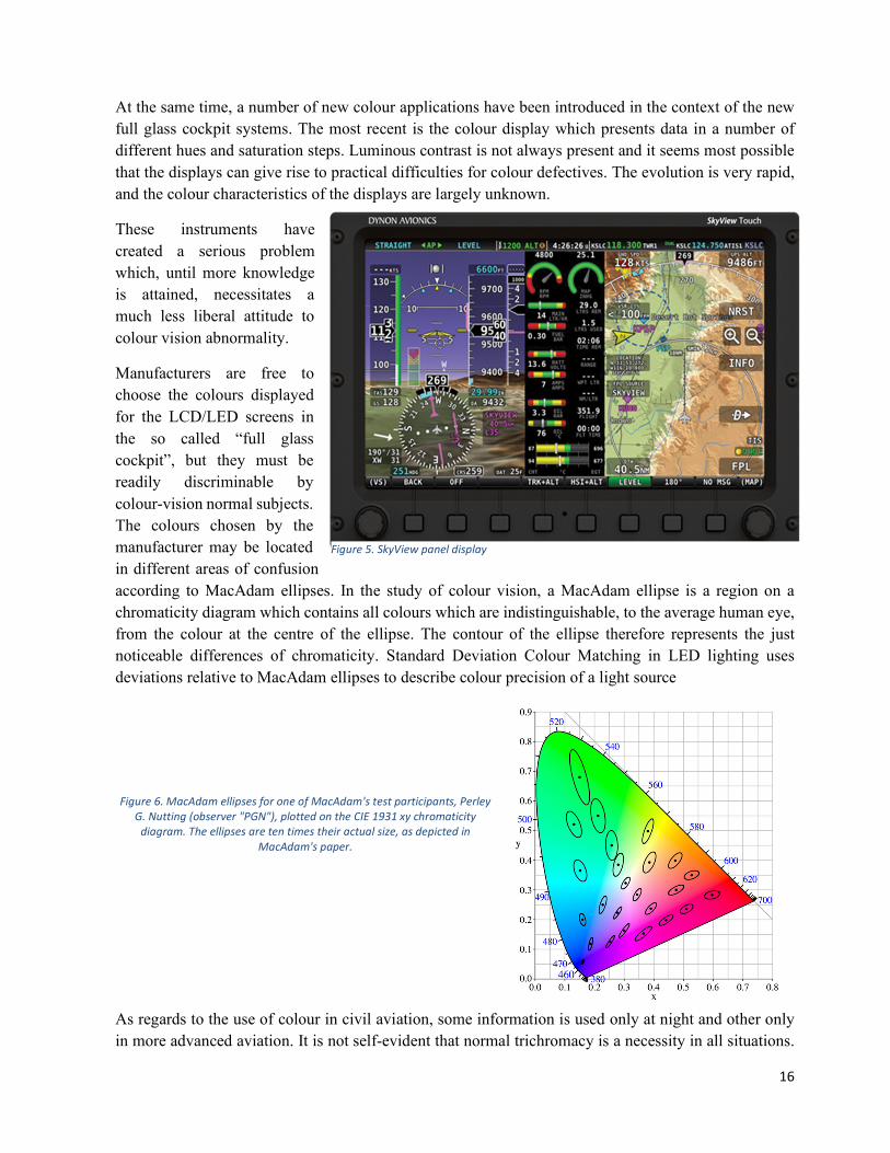
At the same time, a number of new colour applications have been introduced in the context of the new full glass cockpit systems. The most recent is the colour display which presents data in a number of different hues and saturation steps. Luminous contrast is not always present and it seems most possible that the displays can give rise to practical difficulties for colour defectives. The evolution is very rapid, and the colour characteristics of the displays are largely unknown. These instruments have created a serious problem which, until more knowledge is attained, necessitates a much less liberal attitude to colour vision abnormality. Manufacturers are free to choose the colours displayed for the LCD/LED screens in the so called “full glass cockpit”, but they must be readily discriminable by colour-vision normal subjects. The colours chosen by the manufacturer may be located in different areas of confusion according to MacAdam ellipses. In the study of colour vision, a MacAdam ellipse is a region on a chromaticity diagram which contains all colours which are indistinguishable, to the average human eye, from the colour at the centre of the ellipse. The contour of the ellipse therefore represents the just noticeable differences of chromaticity. Standard Deviation Colour Matching in LED lighting uses deviations relative to MacAdam ellipses to describe colour precision of a light source Figure 6. MacAdam ellipses for one of MacAdam's test participants, Perley G. Nutting (observer "PGN"), plotted on the CIE 1931 xy chromaticity diagram. The ellipses are ten times their actual size, as depicted in MacAdam's paper. As regards to the use of colour in civil aviation, some information is used only at night and other only in more advanced aviation. It is not self-evident that normal trichromacy is a necessity in all situations. Figure 5. SkyView panel display
17
By setting standards for the chromaticity of various colours, an attempt has been made to make their identification easier for air personnel with a colour deficiency. The mere qualitative diagnosis of the colour vision deficiency is not sufficient, because the colour discrimination varies considerably between individuals with the same type of defect. A practical colour vision test certainly has the highest validity but only for the conditions present at the test. It has been the practice of some countries to assess as fit applicants with simple deuteranomaly who readily pass lantern tests. In some cases, a practical test with a signal gun has been decisive. Even individuals with rather outspoken defects may pass this test which does not signify whether the applicant normally perceives other less conspicuous signals. In order to assess the fitness of an applicant with a colour vision deficiency with regard to a possible fit assessment, it is necessary to have at hand the results of a battery of colour vision tests. As many different aspects of colour vision as possible should be examined. 2.5 Colour vision testing in aviation Comparison of results from conventional tests has revealed enormous variability and inconsistency. Individuals with mild colour-vision defects often fail normal trichomacy tests as do some normal trichromats (individuals with three classes of cone receptors; considered normal), and are therefore prevented from choosing certain career paths. These individuals with mild colour-vision defects might well be able to perform tasks that are critical to safety in aviation, as might normal trichromats, when presented with the same colour signals (Barbur JL 2012) In an attempt to include these individuals in aviation, some authorities either relaxed the pass limits on colour vision screening tests or used less demanding colour-vision tests. These attempts compromised the required trichromatic performance in the most safety-critical tasks that are colour-coded. Although there is an obvious need to have colour safe pilots, due to the challenging working environment, there is less consensus on standard medical requirements and colour-vision protocols in civil aviation world-wide. Conditions under which colour-vision tests are done and the interpretation thereof vary from country to country. Different International Civil Aviation Organization (ICAO)-affiliated countries have set different requirements and use different tests for colour-vision testing. These inconsistencies make it possible for an applicant to fail colour-vision testing in one country and pass it in another (Werfelman, Aviation Medicine: Colour vision GAP 2019) A question is often asked by aviation personnel as to how severe a colour-vision defect should be before an individual is considered unable to operate safely in the aviation environment. In the text book, Adler’s Physiology of the Eye – Clinical Application, the author cautions healthcare workers to not over-diagnose colour-vision defects and consequently exclude individuals from occupations that they could manage. The need for the development of an internationally accepted, defensible, objective and standardised set of colour-vision tests that can accurately classify and quantify severity of colour-vision defects was addressed by Milburn et al. in their paper entitled “Pilot colour vision – research and recommendations” (Milburn, et al. 2013). Linda Werfelman also addressed the same need in her article that summarised Dougal Watson’s study of colour-vision testing in aviation in 78 countries (Werfelman 2019). In the
18
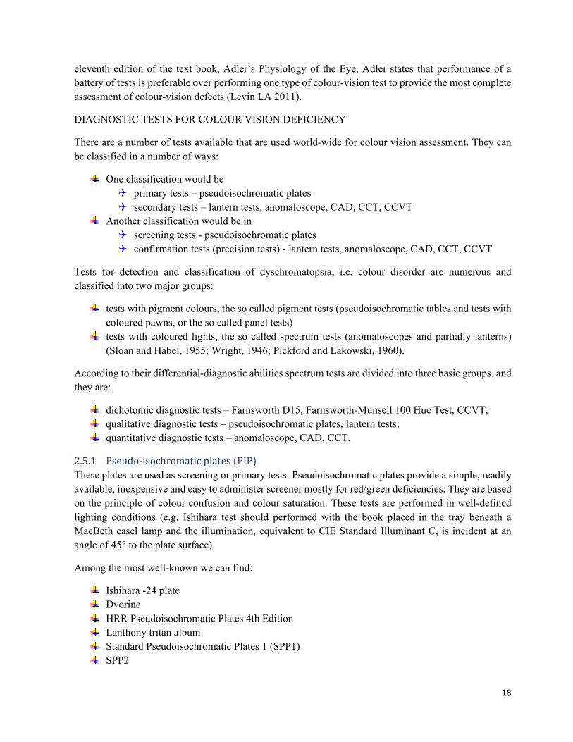
eleventh edition of the text book, Adler’s Physiology of the Eye, Adler states that performance of a battery of tests is preferable over performing one type of colour-vision test to provide the most complete assessment of colour-vision defects (Levin LA 2011). DIAGNOSTIC TESTS FOR COLOUR VISION DEFICIENCY There are a number of tests available that are used world-wide for colour vision assessment. They can be classified in a number of ways: One classification would be primary tests – pseudoisochromatic plates secondary tests – lantern tests, anomaloscope, CAD, CCT, CCVT Another classification would be in screening tests - pseudoisochromatic plates confirmation tests (precision tests) - lantern tests, anomaloscope, CAD, CCT, CCVT Tests for detection and classification of dyschromatopsia, i.e. colour disorder are numerous and classified into two major groups: tests with pigment colours, the so called pigment tests (pseudoisochromatic tables and tests with coloured pawns, or the so called panel tests) tests with coloured lights, the so called spectrum tests (anomaloscopes and partially lanterns) (Sloan and Habel, 1955; Wright, 1946; Pickford and Lakowski, 1960). According to their differential-diagnostic abilities spectrum tests are divided into three basic groups, and they are: dichotomic diagnostic tests – Farnsworth D15, Farnsworth-Munsell 100 Hue Test, CCVT; qualitative diagnostic tests – pseudoisochromatic plates, lantern tests; quantitative diagnostic tests – anomaloscope, CAD, CCT. 2.5.1 Pseudo-isochromatic plates (PIP) These plates are used as screening or primary tests. Pseudoisochromatic plates provide a simple, readily available, inexpensive and easy to administer screener mostly for red/green deficiencies. They are based on the principle of colour confusion and colour saturation. These tests are performed in well-defined lighting conditions (e.g. Ishihara test should performed with the book placed in the tray beneath a MacBeth easel lamp and the illumination, equivalent to CIE Standard Illuminant C, is incident at an angle of 45° to the plate surface). Among the most well-known we can find: Ishihara -24 plate Dvorine HRR Pseudoisochromatic Plates 4th Edition Lanthony tritan album Standard Pseudoisochromatic Plates 1 (SPP1) SPP2
19
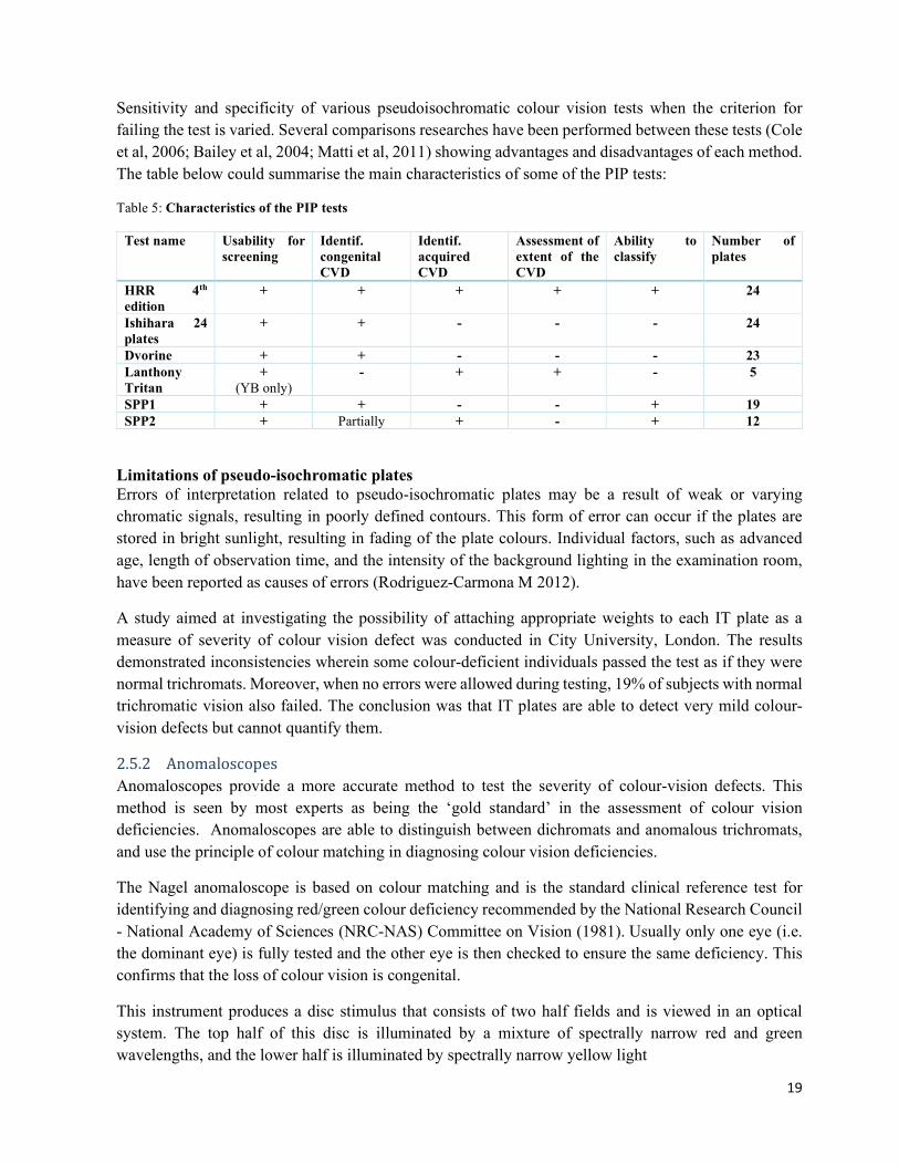
Sensitivity and specificity of various pseudoisochromatic colour vision tests when the criterion for failing the test is varied. Several comparisons researches have been performed between these tests (Cole et al, 2006; Bailey et al, 2004; Matti et al, 2011) showing advantages and disadvantages of each method. The table below could summarise the main characteristics of some of the PIP tests: Table 5: Characteristics of the PIP tests Test name Usability for screening Identif. congenital CVD Identif. acquired CVD Assessment of extent of the CVD Ability to classify Number of plates HRR 4th edition + + + + + 24 Ishihara 24 plates + + - - - 24 Dvorine + + - - - 23 Lanthony Tritan + (YB only) - + + - 5 SPP1 + + - - + 19 SPP2 + Partially + - + 12 Limitations of pseudo-isochromatic plates Errors of interpretation related to pseudo-isochromatic plates may be a result of weak or varying chromatic signals, resulting in poorly defined contours. This form of error can occur if the plates are stored in bright sunlight, resulting in fading of the plate colours. Individual factors, such as advanced age, length of observation time, and the intensity of the background lighting in the examination room, have been reported as causes of errors (Rodriguez-Carmona M 2012). A study aimed at investigating the possibility of attaching appropriate weights to each IT plate as a measure of severity of colour vision defect was conducted in City University, London. The results demonstrated inconsistencies wherein some colour-deficient individuals passed the test as if they were normal trichromats. Moreover, when no errors were allowed during testing, 19% of subjects with normal trichromatic vision also failed. The conclusion was that IT plates are able to detect very mild colour-vision defects but cannot quantify them. 2.5.2 Anomaloscopes Anomaloscopes provide a more accurate method to test the severity of colour-vision defects. This method is seen by most experts as being the ‘gold standard’ in the assessment of colour vision deficiencies. Anomaloscopes are able to distinguish between dichromats and anomalous trichromats, and use the principle of colour matching in diagnosing colour vision deficiencies. The Nagel anomaloscope is based on colour matching and is the standard clinical reference test for identifying and diagnosing red/green colour deficiency recommended by the National Research Council - National Academy of Sciences (NRC-NAS) Committee on Vision (1981). Usually only one eye (i.e. the dominant eye) is fully tested and the other eye is then checked to ensure the same deficiency. This confirms that the loss of colour vision is congenital. This instrument produces a disc stimulus that consists of two half fields and is viewed in an optical system. The top half of this disc is illuminated by a mixture of spectrally narrow red and green wavelengths, and the lower half is illuminated by spectrally narrow yellow light
20
2.5.3 Holmes-Wright lantern test (type A and B) Contain filters allowing the presentation of three colours – red, green and white – the chromaticities of which are within the CIE-approved specifications for signal lights (Vingrys and Cole, 1983). The colours are presented in pairs, arranged vertically in the case of the type A and horizontally in the case of the type B, and the 9 possible combinations of the three colours are presented in each run. As per the protocol for the HW-A, the examiner first shows an example of each of the lights before the test followed by three runs of the 9 combinations in mesopic lighting conditions, followed by a period of 12-15 minutes of dark adaptation and a repeat three runs in scotopic lighting conditions. The pass criteria for the HW-A is ‘no errors on the first run of either lighting condition, or if errors are made, no error on the following two runs of either lighting condition 2.5.3.1 Limitations of lantern tests and anomaloscopes Adler states that accurate diagnosis of colour-vision defects is difficult because tests that are easy to do can give inaccurate results, whereas the more accurate tests require rigorous training for the examiner. (Moses RA 1987) Conventional tests do not assess the nature and severity of colour vision defects. There was poor correlation of outcomes of the different tests and they did not give reliable information about safe minimum colour vision required for flying. (Squire TJ 2005 May;) 2.5.4 The Colour Assessment and Diagnosis (CAD) Test The UK Civil Aviation Authority and other Civil Aviation Authorities have selected the CAD test as the precision test of choice. The CAD test cannot be learnt and has high sensitivity. It provides automatic classification of colour vision deficiencies and separates normal subjects from congenital and acquired colour-deficient subjects, with 100% sensitivity and 100% specificity (Barbur JL 2012). As opposed to the static luminance contrast (LC) noise of traditional pseudoisochromatic tests, the CAD employs dynamic luminance contrast noise in which all checks on the display vary randomly in luminance within a specified percentage of the background luminance throughout the presentation, every 40-80ms. The subject is required to respond to the direction of motion of the coloured stimulus by pressing one of four corresponding buttons on a response pad. A four-alternative, forced-choice procedure in which staircases for each of the 16 colour directions are randomly interleaved is used to determine the minimum CD that a subject requires in order to reliably discriminate stimuli in each of the colour directions. The CAD test has a number of advantages over conventional tests both in terms of isolation of colour signals as well as sensitivity and accuracy: a) Isolation of colour signals b) Measurement of chromatic detection thresholds c) The statistical limits of chromatic sensitivity within “normal” trichromats d) Detection of colour vision loss that falls outside normal range e) Diagnosis of the type of deficiency involved f) Quantifying the severity of colour vision loss g) Effects of light level and stimulus size h) The effects of aging on red-green and yellow-blue loss of chromatic sensitivity
21
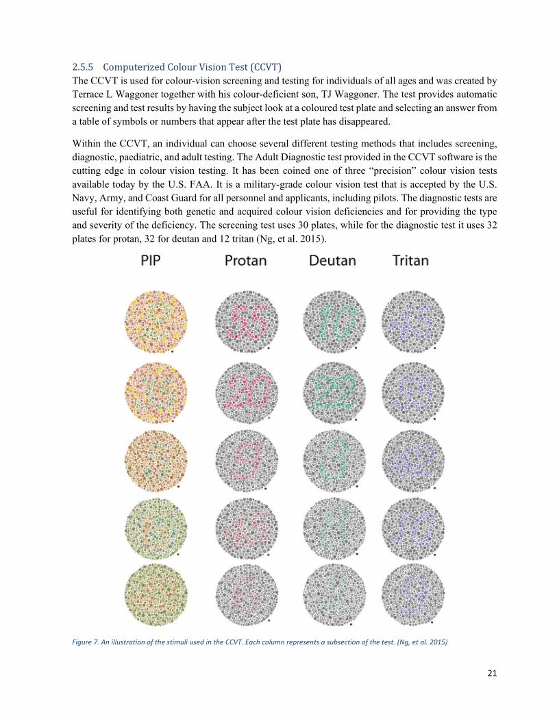
2.5.5 Computerized Colour Vision Test (CCVT) The CCVT is used for colour-vision screening and testing for individuals of all ages and was created by Terrace L Waggoner together with his colour-deficient son, TJ Waggoner. The test provides automatic screening and test results by having the subject look at a coloured test plate and selecting an answer from a table of symbols or numbers that appear after the test plate has disappeared. Within the CCVT, an individual can choose several different testing methods that includes screening, diagnostic, paediatric, and adult testing. The Adult Diagnostic test provided in the CCVT software is the cutting edge in colour vision testing. It has been coined one of three “precision” colour vision tests available today by the U.S. FAA. It is a military-grade colour vision test that is accepted by the U.S. Navy, Army, and Coast Guard for all personnel and applicants, including pilots. The diagnostic tests are useful for identifying both genetic and acquired colour vision deficiencies and for providing the type and severity of the deficiency. The screening test uses 30 plates, while for the diagnostic test it uses 32 plates for protan, 32 for deutan and 12 tritan (Ng, et al. 2015).
Figure 7. An illustration of the stimuli used in the CCVT. Each column represents a subsection of the test. (Ng, et al. 2015)

22
In regard to the diagnostic capabilities for subjects with CVD, the CCVT had a lower unclassifiable rate compared with the Ishihara and HRR tests. The CCVT classified subjects more similarly to the anomaloscope than the HRR or Ishihara tests. In 2016, Makunyane, P. conducted a study “An update on diagnostic tests for colour vision defects in individuals working in the aviation industry : Back to basics” (Makunyane 2016).The purpose of this study was to provide an update on colour-vision tests approved by the International Civil Aviation Organization and to highlight the importance of choosing appropriate colour-vision tests that can be used with confidence to detect colour-vision deficiency, as well as to classify the type of deficiency involved, and to quantify the severity of loss. Based on this thorough review, the authors recommend using either the Waggoner HRR or Waggoner PIP24 as a screening tool and the Waggoner CCVT as a secondary precision test. In the study “Validation of a Computerized Color Vision Test“ (Rings 2014, March), the Waggoner CCVT was compared against the Nagel anomaloscope with 300 participants, 236 has normal colour vision and 64 had a colour vision deficiency. The Waggoner CCVT had a 100% sensitivity and 100% specificity. The Waggoner PIP24 was also used in this study and had a 100% sensitivity and 89% specificity. (Rings 2014, March) 2.5.6 The Cambridge Colour Test (Cambridge CT) The test was developed by John Mollon and colleagues to determine discrimination ellipses in colour-deficient subjects, by probing chromatic signals along colour confusion lines. Ellipses measured in these individuals are characteristically oriented and enlarged. It is a computerised test, and he offers the advantage of being able to adjust the difficulty depending on the patient’s performance, as well as randomising plates to avoid the patient from recalling previously previewed plates. The design of the Cambridge CT uses a computer version of pseudoisochromatic plates and combines the Principles of Chibret and Stilling. (Mollon JD 1989) Chibret’s Principle (1877) states the possibility of varying the chromatic difference of the target and field dynamically and adaptively, along different directions in colour space. (Regan BC 1994) Stilling (1877) achieved non-hue noise by varying the size and luminance of the target and background elements. (Regan BC 1994)Each of the computer pseudo-isochromatic plates in the Cambridge CT contains a Landolt C; that is, each stimulus is a large coloured letter C of specific hue and luminance that is presented in four different orientations; the opening in the letter can be up, down, right or left, which is embedded in a background comprising circles of varying size, colour and luminance. (Hasrod N 2015).The Cambridge CT stimuli backgrounds eliminate luminance or contour cues, and the figures (the test stimuli of Landolt letters) must be identified solely by their hues. (Paramei 2012) As mentioned, this is accompanied by non-hue noise achieved by varying the luminance and size of compositional elements in accordance with Stilling’s Principle. (Regan BC 1994) 2.5.7 Cone Contrast Test (CCT) It is an innovative colour vision testing (simultaneous congenital & acquired). Its sensitivity and specificity comparable to anomaloscope testing and exceeds PIP sensitivity in practiced observers. This test uses coloured letters to stimulate single cone type / pathway, also uses decreasing contrast steps to detects cone function threshold, numeric scores to provide Hereditary vs. Acquired discrimination. It is

23
sensitive enough to detect severity of cone deficiency and it can be used to detect and monitor acquired colour deficiency in AMD, glaucoma, diabetic retinopathy, Parkinson’s disease, etc. The CCT provides a rapid (6 minutes), clinically expedient, measure of colour vision for quantifying normal colour performance, diagnosing type and severity of hereditary deficiency, and detection of acquired sensitivity loss due to ocular, neurologic, and/or systemic disease, as well as injury and physiological stressors, such as altitude and fatigue. As limitation of CCT: No blue cone (tritan) or acquired YB deficiencies included (Rabin, Gooch et Douglas Ivan 2011) 2.5.8 Farnsworth-Munsell 100 hue test The Farnsworth-Munsell 100 Hue Colour Vision test is a test of the human visual system often used to test for colour blindness. (Visual disturbances and blindness s.d.)The system was developed by Dean Farnsworth in the 1940s and it tests the ability to isolate and arrange minute differences in various colour targets with constant value and chroma that cover all the visual hues described by the Munsell colour system. (Farnsworth 1943) There are several variations of the test, one featuring 100 colour hues and one featuring 15 colour hues. Originally taken in an analogue environment with physical hue tiles, the test is now taken from computer consoles. An accurate quantification of colour vision accuracy is particularly important to designers, photographers and colourists, who all rely on accurate colour vision to produce quality content. The test assesses both RG and YB deficiencies and is useful to identify various congenital colour vision deficiencies, for measuring the changes due to neuronal disease or possible side effects in therapeutic management (Kinnear and Sahraie 2002). 3 Discussion and conclusion I will review here the results of some significant studies related to the colour vision requirements and the colour test in aviation In a very recent study ”Colour Vision Tests in Pilots' Medical Assessments“ Marechal, M et All, they have tried to assess the abilities of 8 colour vision tests for screening, qualification and quantification of red / green hereditary deficiency, to improve and to adapt the current colour selection protocols. (Marechal, et al. 2018 Aug ) The aim of this study was to evaluate the ability of 8 colour vision tests to screen for and accurately measure hereditary colour-deficiency in order to improve colour vision assessment methods for aircraft pilots. The inclusion criteria included colour-deficient subjects: addressed for a failure in reading the Ishihara plates by - military selection centre - or Aviation Medical Examiner and colour vision normal subjects: healthy volunteers. The exclusion criteria were: BCVA less than 6/10, sunglasses or tinted contact lenses, ophthalmologic pathology (evolutionary or iatrogenic). Subjects were classified into two groups: colour-vision normal (CVN) and colour-vision deficient (CVD) via all test results. All tests were used because of the risk of false negatives. The anomaloscope
24
is often the reference test and was the most important for group classification, but a patient with a normal anomaloscope and other abnormal tests was classified as CVD. Secondly, CVD subjects were classified into four types (protanomalous, protanope, deuteranomalous, and deuteranope) according the anomaloscope results. All the subjects of the study were assessed using to the following tests: Ishihara Beyne lantern Fletcher lantern Farnsworth D15 Arrangements test Lanthony 15 Hue Munsell100 Hue CAD (Colour Assessment and Diagnosis) test Anomaloscope Nagel type Subjects first performed the tests of the current colour vision assessment protocol: the Ishihara plate and the Beyne lantern tests. They then performed the other tests in a random order to limit the tiredness effect. For the Ishihara 38-plate test, the subject was considered to pass the test if the first 17 plates were read without error. Candidates were then assessed using the Beyne lantern test for aviation (the five different aviation lights (red, green, blue, yellow-orange, and white) were presented in random order), the Fletcher-Evans CAM lantern test for aviation was done; The Farnsworth D15 test was successful in the absence of confusion lines (circular scheme). The Lanthony desaturated 15 Hue test was successful if the scheme provided by the subject contained less than 2 confusion lines (the presence of 2 confusion lines was tolerated in patients over 40 yr. of age). The Farnsworth-Munsell 100 Hue was then performed. The anomaloscope IF2 (All-Colour Anomaloscope, Tomey, Japan) with the Rayleigh equation was used. The test was performed by a trained examiner on the dominant eye in mesopic conditions. For each test, evaluation of: Sensibility, specificity, positive and negative predictive values Application to aviation environment: Total number and success ratio for class 1 ability Severity assessing: ROC curves for dichromatism diagnosis for CAD test and 100 Hue The results where: of the 55 subjects, 32 were colour vision deficient subjects (CVD). 3 were excluded (missing results), and 23 colour vision normal subjects (CVN).

25
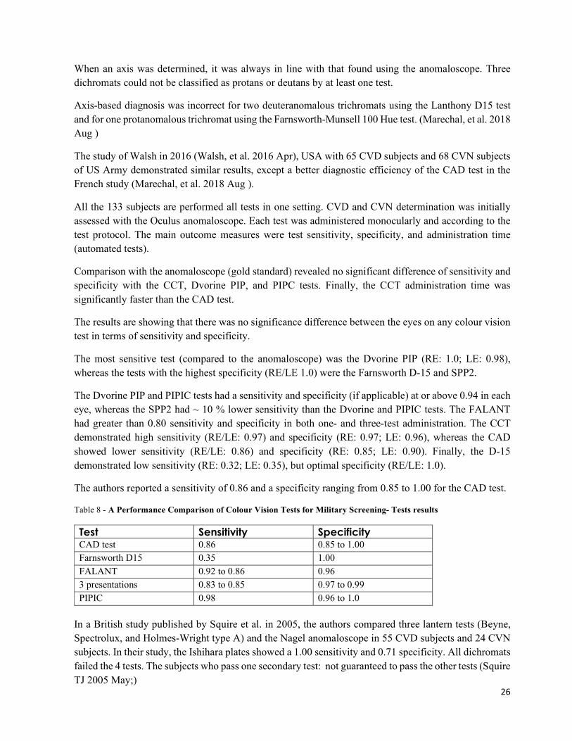
Table 6 - Distribution of the subjects included in the study 29 CVD 23 CVN p 23 years ±6.09 26 ±6.1 years p = 0.075 0 woman 9 women p = 0.015 -11 deuteranomalous trichromats (37.9%) -7 protanomalous trichromats (24.1%) -6 protanopes (20.69%) -5 deuteranopes (17.24%) Table 7 - Tests results Marechal, M. Marechal, Delbarre , J. Tesson, C. , J. Tesson, C. , J. Tesson, C. , J. Tesson, C. Lacambre , F. , F. Froussart Sensitivity Specificity Positive predictive values Negative predictive values ISHIHARA FARNSWORTH D15 LANTHONY HUE desaturated MUNSELL 100 HUE 0.97 1.00 1.00 0.96 0.58 1.00 1.00 0.64 0.79 1.00 1.00 0.79 0.79 0.96 0.96 0.79 BEYNE Lantern 1s / 4' 0.79 0.96 0.96 0.79 1/25th s 2' 0.97 0.57 0.76 0.93 1s / 3' 0.76 0.96 0.96 0.76 1 s/3' x 3series 0.69 1.00 1.00 0.72 FLETCHER Lantern 1 presentation 1.00 0.78 0.85 1.00 2 retests 0.97 1.00 1.00 1.00 CAD test 1.00 1.00 1.00 1.00 ANOMALOSCOPE Automatic 0.97 0.96 0.97 0.96 Manual 1.00 1.00 1.00 1.00 Apart from the lantern tests, all tests provided qualitative diagnostic information on the colour-deficiency axis. The authors compared the axis identified using the Ishihara plates, Farnsworth D15, Lanthony 15 Hue, Farnsworth-Munsell 100 Hue, and CAD tests with the diagnosis obtained using the anomaloscope. Only the CAD test found the expected diagnosis for each subject. Qualitative diagnosis could not be established using Ishihara plates and the Farnsworth D15 test for, respectively, 19% and 41% of CVD subjects (mostly subjects with a low to moderate deficiency, with arrangement tests leading to “low discrimination”).
26
When an axis was determined, it was always in line with that found using the anomaloscope. Three dichromats could not be classified as protans or deutans by at least one test. Axis-based diagnosis was incorrect for two deuteranomalous trichromats using the Lanthony D15 test and for one protanomalous trichromat using the Farnsworth-Munsell 100 Hue test. (Marechal, et al. 2018 Aug ) The study of Walsh in 2016 (Walsh, et al. 2016 Apr), USA with 65 CVD subjects and 68 CVN subjects of US Army demonstrated similar results, except a better diagnostic efficiency of the CAD test in the French study (Marechal, et al. 2018 Aug ). All the 133 subjects are performed all tests in one setting. CVD and CVN determination was initially assessed with the Oculus anomaloscope. Each test was administered monocularly and according to the test protocol. The main outcome measures were test sensitivity, specificity, and administration time (automated tests). Comparison with the anomaloscope (gold standard) revealed no significant difference of sensitivity and specificity with the CCT, Dvorine PIP, and PIPC tests. Finally, the CCT administration time was significantly faster than the CAD test. The results are showing that there was no significance difference between the eyes on any colour vision test in terms of sensitivity and specificity. The most sensitive test (compared to the anomaloscope) was the Dvorine PIP (RE: 1.0; LE: 0.98), whereas the tests with the highest specificity (RE/LE 1.0) were the Farnsworth D-15 and SPP2. The Dvorine PIP and PIPIC tests had a sensitivity and specificity (if applicable) at or above 0.94 in each eye, whereas the SPP2 had ~ 10 % lower sensitivity than the Dvorine and PIPIC tests. The FALANT had greater than 0.80 sensitivity and specificity in both one- and three-test administration. The CCT demonstrated high sensitivity (RE/LE: 0.97) and specificity (RE: 0.97; LE: 0.96), whereas the CAD showed lower sensitivity (RE/LE: 0.86) and specificity (RE: 0.85; LE: 0.90). Finally, the D-15 demonstrated low sensitivity (RE: 0.32; LE: 0.35), but optimal specificity (RE/LE: 1.0). The authors reported a sensitivity of 0.86 and a specificity ranging from 0.85 to 1.00 for the CAD test. Table 8 - A Performance Comparison of Colour Vision Tests for Military Screening- Tests results Test Sensitivity Specificity CAD test 0.86 0.85 to 1.00 Farnsworth D15 0.35 1.00 FALANT 0.92 to 0.86 0.96 3 presentations 0.83 to 0.85 0.97 to 0.99 PIPIC 0.98 0.96 to 1.0 In a British study published by Squire et al. in 2005, the authors compared three lantern tests (Beyne, Spectrolux, and Holmes-Wright type A) and the Nagel anomaloscope in 55 CVD subjects and 24 CVN subjects. In their study, the Ishihara plates showed a 1.00 sensitivity and 0.71 specificity. All dichromats failed the 4 tests. The subjects who pass one secondary test: not guaranteed to pass the other tests (Squire TJ 2005 May;)
27
An American study published by Gaska and Wright in 2016 evaluated the recognition of colours in the cockpit for 45 CVN pilots and 49 colour-deficient pilots. (Gaska JP 2016) They compared CCT results with colour recognition in a “Situation Awareness” simulator and evaluated the accuracy, speed, and throughput of answers. The colour-deficient subjects performed statistically less well than control subjects and a statistically significant relationship was reported between their performance and the CCT score. Because there are numerous studies that demonstrate that colour vision deficient (CVD) individuals perform less well than colour vision normal (CVN) individuals in tasks that require discrimination or identification of colored stimuli, there remains a need to quantify the relationship between the type and severity of CVD and performance on operationally relevant tasks. The participants were classified as CVN (N = 45) or CVD (N = 49) using the Rabin cone contrast test, which is the standard colour vision screening test used by the United States Air Force. In the colour condition, test images that were representative of the size, shape, and colour of symbols and lines used on fifth-generation fighter aircraft displays were used to measure operational performance. In the achromatic condition, all symbols and lines had the same chromaticity but differed in luminance. Subjects were asked to locate and discriminate between friend vs. foe symbols (red vs. green, or brighter vs. dimmer) while speed and accuracy were recorded. The results are showing that the increasing colour deficiency was associated with decreasing speed and accuracy for the colour condition, but not for the achromatic condition. Mean differences between CVN and CVD individuals showed the same pattern. In the “Minimum Colour Vision Requirements for Professional Flight Crew” study (Barbur, et al. 2009.), the purpose was to produce standards and a new test for chromatic sensitivity that can be used to quantify the severity of an individual’s colour deficiency. The research was necessary because of the lack of reliable, standardized tests and the absence of information on the specific colour vision needs of professional flight crew. The goal was to produce the minimum colour vision requirements for modern flight crew, and a new colour assessment and diagnosis test, and “to quantify the severity of colour vision loss and to recommend minimum colour vision requirements by establishing the level of colour vision loss when colour deficient observers can no longer perform the most safety-critical, colour related tasks with the same accuracy as normal trichromats”. In the study where included 182 subjects: 65 normal trichromats and 117 subjects with deutan- and protan-like colour deficiencies. The age of the subjects ranged from 15 to 55 years (mean 30.2 years, median 27 years). The colour vision of each subject was examined using five different colour vision tests (Ishihara plate test, Dvorine plate test, Nagel anomaloscope, CAD test and Aviation Lights Test ), as well as the PAPI and PSL simulator tests. Results from each of the five tests were then examined in relation to the subject’s performance on the PAPI to establish which test yields the best prediction of performance in the PAPI task.
28
The results show that normal trichromats can also make errors, both on the PAPI and the Ishihara tests (i.e. five subjects produce one error, one subject produces two errors and one other subject produces three errors on the PAPI). The rest of the normal subjects score 100% correct on the PAPI test. Results for deutan colour deficient observers reveal that all subjects with scores > 70% (i.e. 16 or more correct plates out of 24 on the Ishihara 24-plate test) pass the PAPI with a score of 100% correct. Results for protan observers show that four subjects with scores greater than 40% pass the PAPI test. Overall the results reveal very poor correlation between the subjects’ performance on the Ishihara and the PAPI test scores. Many of the subjects that pass the PAPI task can score anything from 0 to95% correct on the Ishihara test. Comparisons of data from the Dvorine plate test with the PAPI simulator show similar results to those obtained with the Ishihara test: three normal trichromats obtain less than 100% on the Dvorine test (but pass the PAPI with no errors). Deutan and protan colour deficient subjects need more than 65 and 50%, respectively, on the Dvorine plate test to achieve 100% on PAPI. Since the prediction of the class of deficiency (protan or deutan) involved is poor with both Ishihara and Dvorine tests, it is difficult to know which of the two limits one should apply to any colour deficient subject. Only a few deutan and protan observers pass the PAPI with Nagel sensitivity > 0.6 (deutan) and > 0.4 (protan). The Nagel anomaloscope test is excellent at distinguishing between deutan and protan like deficiencies, but fails to quantify reliably the severity of colour vision loss and does not test for yellow/blue deficiency. Most normal trichromats perform 100% correct in the identification of the red and white lights. However, 7 out of the 65 normal subjects made some errors. This could be due to lack of attention and / or reduced visual acuity at low light levels caused by increased higher order aberrations when the pupil size is large. The errors were found to be distributed with equal probability amongst the five conditions. They have compared the performance on the PAPI test with the corresponding CAD measure of RG sensitivity for subjects with deutan- and protan-like deficiencies. The sensitivity limits beyond which deutans and protans perform the PAPI test as well as normal trichromats are 0.17 (RG threshold ~ 6 SN units) and 0.085 (RG threshold ~ 12 SN units), respectively. The very few subjects that pass the PAPI with CAD sensitivities less than 0.2 (deutan) and 0.1 (protan) have poor overall chromatic sensitivity. The results also show that the pass / fail limits proposed on the basis of the CAD test ensure that the colour deficient subjects that pass have an overall chromatic sensitivity greater than ~ 0.7 (deutans) and greater than ~ 0.5 (protans) In the study of British CAA (Barbur, et al. 2009.), the CAD test was pass in 36.1 % of deuteranomalous trichromats and 29.8% of protanomalous trichromats, compared to the Marechal et al study (Marechal, et al. 2018 Aug ), where only 18.2% deuteranomalous trichromats and 14.3% protanomalous trichromats passed the test. Few studies have reported that individuals with normal colour vision can fail the Ishihara test. A conservative figure of 9.7% of normal trichromats failing the 24-plate edition has been reported, (Rodriguez-Carmona M 2012) ; although other studies report this to be as high as 44.6% (Miyahara 2008), and up to 75.8% in children. (Cosstick M 2005).
29
These types of error are classed as false positives. A number of causes for these errors have been suggested and include digit confusion error where the number is seen but incorrectly interpreted. Digit confusion errors are usually a misinterpretation of 3 and 8, or 7 and 2. The study of Adrian C. Chorley, “A modified protocol for colour vision screening using Ishihara” (Chorley 2015) has two parts: PART 1: MODIFIED PROTOCOL TO REDUCE FALSE POSITIVE RATE The subjects were applicants for initial aeromedical examination at the UK CAA AeMC. Six Ishihara plates from a 38-plate edition were selected using, where possible, plates with different numbers from the 24-plate edition. Each dot within the number was coloured black and the plates were photocopied, laminated, and placed above the Ishihara testing area. Three plates had two-digit numbers and three had single-digit numbers and together represented samples of numbers from 1-9. Where a subject made up to two errors, they were shown these sample typeface numbers with particular attention paid to the numbers which were confused. The subject was then allowed a second attempt at identifying those numbers initially misread. Using this technique, it was found that most subjects then correctly identified the Ishihara plate(s) when shown them for a second time. PART 2: MODIFIED PROTOCOL TO MINIMIZE FALSE NEGATIVES Subjects were applicants for initial Class 1, 2, or 3 medicals at the UK CAA AeMC and were tested, as part of the eye examination, using the 24-plate Ishihara edition with randomized plates and modified protocol as described in Part 1. Only those applicants who had passed Ishihara and did not require CAD testing were included for this part of the study. Six plates (excluding plate numbers 1, 14, and 15) were randomly selected within a second 24-plate Ishihara book and were randomly rotated by 90° either clockwise or anticlockwise. These plates were reinserted into the book and were not changed between each assessment. The Ishihara test is known to be an extremely sensitive test at detecting congenital red-green colour vision deficiencies. (Birch 2010) The principle of allowing zero errors is likely to increase its sensitivity further, but at the detriment of its specificity, with high false positive rates reported. The modified testing protocol described in Part 1 should maintain high sensitivity while reducing false positive results to around 4%. It is recognized that as these data were collected retrospectively, no intraobserver measure of false positives between the standard and modified protocols can be made. Additionally, the number of subjects who made one or two errors and who then correctly identified the Ishihara plates on the second attempt was not documented. However, it has been consistently reported in studies employing large cohorts that a percentage of normal trichromats make errors on the Ishihara plates The lower the number of errors allowed to pass the test, the higher the incidence of a false positive result.
30
The Ishihara test may not be able to detect subjects with colour vision deficiencies who employ other techniques in plate recognition in order to obtain a normal result. This is more likely to occur in environments such as aeromedical examination, where applicants are motivated to gain a favourable medical outcome. It is seen occasionally using the randomized plates where a subject calls the correct numbers in the original order of the book. It has also been shown that subjects with colour vision deficiencies could potentially use dot pattern recognition to ensure they read all plates correctly. There is no means of assessing if colour vision deficient subjects have achieved this to date as they would have been passed as colour normal without further colour vision testing. The rotation of selected plates offers an addition safeguard against this strategy and these results indicate that it would only marginally affect the false positive rate if the plates were rotated. Conclusion: Watson noticed in his study that the secondary tests that are used to diagnose and quantify colour deficiency varies from country to country. Such variability of the testing methods encourages “aeromedical tourism,” with pilot candidates seeking aeromedical examiners in countries where colour assessment standards are less demanding. (Watson 2014). As a conclusion of the study Watson considered that the Ishihara test is an excellent screening test for red /green deficiency. Taking into consideration the statements above Watson recommended to consider any hesitation or mistake during the Ishihara assessment as failure of the test. Furthermore, the testing protocol should be strictly respected and examiners must be trained in order to minimize the number of false negatives as well as the false positives. In September 2015, Ryan E. Brookes said in “Colour Vision Requirements for Aircrew “(Brookes R. E., September 2015): the compatibility of CVD with crewing aircraft is assessed by medical personnel using clinical diagnosis tests. These clinical tests were developed specifically to detect the presence, nature and severity of CVD. No clinical tests yet provide a measure of vocational performance in operating an aircraft. Nevertheless several tests are used for pilot’s ability. The acceptable colour-deficiency for a pilot to be safe is difficult to determine, as well as the risk of not detected blue-yellow deficiency. Despite the lack of relevance to vocational performance, arbitrary pass marks have been assigned to clinical tests such that a failing candidate will either be subject to operational restrictions or excluded completely. The prescribed clinical tests and associated pass marks vary considerably between regulators. While an individual may be subject to no restrictions in one jurisdiction, they may be excluded in another. The following test are the most frequently used on a global scale to assess the colour vision of pilots and ATCOs: Ishihara plates: excellent screening test for red / green deficiency. Farnsworth D15: quick and easy but some dichromats subjects can pass this test. Lanthony 15 Hue test: ability of 20% and seems suitable for use in professional selection. Munsell 100 Hue : too long

31
Fletcher-Evans CAM lantern : too restrictive Beyne lantern: simple and quick to use, but some dichromats pass. Anomaloscope: qualify and quantify the deficiency but requires a strong experience of the examiner. CAD test: quantitative test , yellow/blue and red/green thresholds used by the British CAA seem to be more restrictive Some of the study results are discordant, nevertheless the CAD test and anomaloscope seem to be the most accurate tests in many studies. The measures of chromatic discrimination on the CAD and Nagel anomaloscope reveal poor correlation between the results of these. Colour deficient subjects can produce very different scores on the two tests and that normal trichromats can make more errors on one test, but not on the other. Further, the pseudoisochromatic tests designed for screening are not suitable to either diagnose accurately the class of deficiency, or to quantify the severity of colour vision loss (Belcher et al, 1958; Birch, 1997). In the case of Dvorine and Ishihara tests, less than 1% of the applicants that pass these tests fail the PAPI. All colour deficient applicants that pass the ALT test also pass the PAPI The results also show that one cannot treat as equivalent subjects with deutan- and protan-like deficiencies. Therefore, minimum colour vision requirements must be set separately for each class of deficiency. The use of so many tests that may not correlate well with each other (when used to assess the severity of colour vision loss) increases variability and gives rise to odd outcomes when an applicant passes one test and fails another. Although the number of colour deficient applicants that pass can increase significantly, this increase is test specific and often dependent on the use of the Signal Light Gun test that has not been validated against the PAPI task. The loss of red/green chromatic sensitivity is greater in subjects with protan congenital colour deficiency when compared to the deutan class. Unlike many conventional colour vision tests, the CAD test cannot be learned and hence the outcome depends entirely on the subject’s chromatic sensitivity. The test provides the means to classify protan and deutan subjects and also quantifies the severity of colour vision loss. If these findings were adopted as pass / fail limits for pilots ~ 35% of colour deficient applicants would be certified as safe to fly. In terms of degenerative effects of ageing on colour vision, Hagerstrom-Portnoy (Hagerstrom-Portnoy, Schneck and Brabyn 1999) showed that the degenerative loss of colour vision starts around the age of 40, however, below 60 years of age, normal ageing does not affect significantly either RG or YB thresholds, provided adequate levels of ambient illumination are employed. Congenital defects are unaltered with age and cannot, contrary to what is sometimes claimed, be treated in any way. Tinted filters, e.g. the so-called X-chrom lens, make possible a better discrimination of some confusion colours but do not improve colour perception. Furthermore, such lens diminish the visual acuity in low light environment. For this reason, applicants passing a colour test by the use of such a device are not ‘colour safe’.

32
In many civil and military populations, waivers have been given to CVD subjects who demonstrated competency in an operational environment. In some cases waivers were issued simply to achieve intake quotas. There is no record of such candidates suffering difficulty on operations as a result of their CVD. To the contrary there is much evidence to indicate that candidates with CVD, when aware of their condition, have been able to perform operational tasks to the required levels of competence. The multiplication of coloured signals in new-generation cockpits and the use of a greater number of different colours may call for more stringent colour vision requirements for pilots. The discrepancy in results confirm that the current colour vision test protocols need to be reassessed. Furthermore, the current protocol does not detect yellow-blue deficiency. In terms of colour vision selection of professional flight crew, the acceptable levels of colour-deficiency still need to be defined at both the European and worldwide level to ensure flight safety. The sensitivity and sensibility of the CV tests varies from one study to the other. For a better view of how these figures vary table no 9 below is intended to bring together the sensitivity and sensibility of various tests as they resulted from various studies (Marechal, et al. 2018 Aug ) (Walsh, et al. 2016 Apr) (Squire TJ 2005 May;) (Ng, et al. 2015). Table 9- Sensitivity and sensibility of the colour vision tests Study Test Marechal et al, 2018 Walsh et al, 2016 Squire et al, 2005 Jason et al, 2015 sensitivity specificity sensitivity specificity sensitivity specificity sensitivity specificity Ishihara 0.97 1.0 0.95 - 1.0 0.71 0.93 0.99 Farnsworth 0.58 1.0 0.35 1.0 - - - - CAD 1.0 1.0 0.86 0.85-1.0 - - - - CCT 0.97 0.97 - - - - Anomaloscope manual mode 1.0 1.0 - - - - - - Anomaloscope auto mode 0.97 0.96 - - - - - - Lantern 0.69 1.0 0.86 0.96 - - - - HRR 4th ed - - - - - - 0.93 0.98 CCVT - - - - - - 0.95 1.0 This section aims to provide additional literature data related to strong points and weak points of the most frequently used tests. Table 10: SWOT analysis of the existing colour vision testing in aviation Test name Strengths Weaknesses Opportunities Threats Ishihara 24 plates Quick and reliable in identifying with red/green deficiencies (congenital). Cannot quantify the deficiency It is widely spread and readily available. Due to low cost, good availability and reduced number of plates makes it easy to cheat. By not identifying blue/yellow deficiencies allows
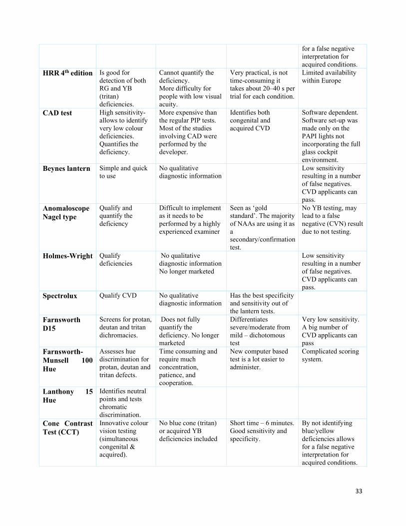
33
for a false negative interpretation for acquired conditions. HRR 4th edition Is good for detection of both RG and YB (tritan) deficiencies. Cannot quantify the deficiency. More difficulty for people with low visual acuity. Very practical, is not time-consuming it takes about 20–40 s per trial for each condition. Limited availability within Europe CAD test High sensitivity-allows to identify very low colour deficiencies. Quantifies the deficiency. More expensive than the regular PIP tests. Most of the studies involving CAD were performed by the developer. Identifies both congenital and acquired CVD Software dependent. Software set-up was made only on the PAPI lights not incorporating the full glass cockpit environment. Beynes lantern Simple and quick to use No qualitative diagnostic information Low sensitivity resulting in a number of false negatives. CVD applicants can pass. Anomaloscope Nagel type Qualify and quantify the deficiency Difficult to implement as it needs to be performed by a highly experienced examiner Seen as ‘gold standard’. The majority of NAAs are using it as a secondary/confirmation test. No YB testing, may lead to a false negative (CVN) result due to not testing. Holmes-Wright Qualify deficiencies No qualitative diagnostic information No longer marketed Low sensitivity resulting in a number of false negatives. CVD applicants can pass. Spectrolux Qualify CVD No qualitative diagnostic information Has the best specificity and sensitivity out of the lantern tests. Farnsworth D15 Screens for protan, deutan and tritan dichromacies. Does not fully quantify the deficiency. No longer marketed Differentiates severe/moderate from mild – dichotomous test Very low sensitivity. A big number of CVD applicants can pass Farnsworth-Munsell 100 Hue Assesses hue discrimination for protan, deutan and tritan defects. Time consuming and require much concentration, patience, and cooperation. New computer based test is a lot easier to administer. Complicated scoring system. Lanthony 15 Hue Identifies neutral points and tests chromatic discrimination. Cone Contrast Test (CCT) Innovative colour vision testing (simultaneous congenital & acquired). No blue cone (tritan) or acquired YB deficiencies included Short time – 6 minutes. Good sensitivity and specificity. By not identifying blue/yellow deficiencies allows for a false negative interpretation for acquired conditions.

34
Cambridge Colour Test (Cambridge CT) Is able to adjust the difficulty depending on the patient’s performance No blue cone (tritan) or acquired deficiencies included By not identifying blue/yellow deficiencies allows for a false negative interpretation for acquired conditions. Computerized Colour Vision Test (CCVT) The test uses 30 screening plates as the first phase and diagnostic plates (32 for protan, 32 for deutan and 12 tritan) for the second phase. Assesses the severity of the CVD in 3 categories: mild, moderate, severe (dichotomous) The on-line availability and limited number of plates allow cheating strategies. 4 Current European colour vision requirements for flight crew In accordance with the requirements listed in Annex IV – Part-MED of the Commission Regulation (EU) 1178/2011 point MED.B.075 Colour vision, the: MED.B.075 (a) Applicants shall be assessed as unfit, where they cannot demonstrate their ability to readily perceive the colours that are necessary for the safe exercise of the privileges of the licence. (b) Examination and assessment (1) Applicants shall be subjected to the Ishihara test for the initial issue of a medical certificate. Applicants who pass that test may be assessed as fit. (2) For a class 1 medical certificate: (i) Applicants who do not pass the Ishihara test shall be referred to the medical assessor of the licensing authority and shall undergo further colour perception testing to establish whether they are colour safe. (ii) Applicants shall be normal trichromats or shall be colour safe. (iii) Applicants who fail further colour perception testing shall be assessed as unfit. (3) For a class 2 medical certificate: (i) Applicants who do not pass the Ishihara test shall undergo further colour perception testing to establish whether they are colour safe. (ii) Applicants who do not have satisfactory perception of colours shall be limited to exercising the privileges of the applicable licence in daytime only. Additional details are provided in the acceptable means of compliance (AMC) applicable for the appropriate class of medical certification:
35
For commercial pilots: AMC1 MED.B.075 Colour vision (a) At revalidation and renewal examinations, colour vision should be tested on clinical indication. (b) The Ishihara test (24 plate version) is considered passed if the first 15 plates, presented in a random order, are identified without error. (c) Those failing the Ishihara test should be examined either by: (1) anomaloscopy (Nagel or equivalent). This test is considered passed if the colour match is trichromatic and the matching range is 4 scale units or less, or if the anomalous quotient is acceptable; or by (2) lantern testing with a Spectrolux, Beynes or Holmes-Wright lantern. This test is considered passed if the applicant passes without error a test with accepted lanterns. (3) Colour Assessment and Diagnosis (CAD) test. This test is considered passed if the threshold is less than 6 standard normal (SN) units for deutan deficiency, or less than 12 SN units for protan deficiency. A threshold greater than 2 SN units for tritan deficiency indicates an acquired cause which should be investigated. For private pilots: AMC2 MED.B.075 Colour vision (a) Colour vision should be tested on clinical indication at revalidation and renewal examinations. (b) The Ishihara test (24 plate version) is considered passed if the first 15 plates, presented in a random order, are identified without error. (c) Those failing the Ishihara test should be examined either by: (1) anomaloscopy (Nagel or equivalent). This test is considered passed if the colour match is trichromatic and the matching range is 4 scale units or less, or if the anomalous quotient is acceptable; or by (2) lantern testing with a Spectrolux, Beynes or Holmes-Wright lantern. This test is considered passed if the applicant passes without error a test with accepted lanterns. (3) Colour Assessment and Diagnosis (CAD) test. This test is considered passed if the threshold is less than 6 standard normal (SN) units for deutan deficiency, or less than 12 SN units for protan deficiency. A threshold greater than 2 SN units for tritan deficiency indicates an acquired cause which should be investigated. For light aircraft pilot licence holders: AMC14 MED.B.095 Medical examination and assessment of applicants for LAPL medical certificates COLOUR VISION Applicants for a night rating should correctly identify 9 of the first 15 plates of the 24-plate edition of Ishihara pseudoisochromatic plates or should be colour safe.
36
5 Recommendations Colour vision is of great importance in visually-demanding work environments. This is especially applicable in aviation where in critical situations a high amount of data, the majority obtained via the visual system, needs to be processed in a very short time. Challenges of inconsistency and variability of results have been demonstrated by most of the reviewed studies. In recent years, automated colour vision tests have been a step forward from the traditional PIP, arrangement, and naming tests. It is recommended that a standardized list of internationally accepted tests be compiled in order to properly diagnose and quantify colour vision. New techniques of colour vision assessment, called by some authors ‘precision tests’, have been designed for this purpose. Further studies are, however, needed to analyse each of these tests for each class of colour vision. 5.1 Recommendations for colour vision testing for pilots Most of the authors recommended for initial screening PIP tests. Out of this the most recommended are Ishihara 38 and 24 plates and HRR 4th edition. If Ishihara is used then the HRR YB plates should be added to assess acquired YB CVD. There should not be more than 1 testing session during an examination. Cole et al., found that 10% of those that pass the first testing session make more mistakes when subjected to additional testing sessions (Cole, Lian and Lakkis 2006). Nevertheless, in their study (Walsh, et al. 2016 Apr) showed an increase of approx. 5% in passing rates after the 3rd testing session. However, the authors admit that due to the low number of their cohort (65 persons) this 5% increase might not be statistically relevant. Tests with a limited number of trials fixed in position are subject to memorization and loss of sensitivity and specificity. Precision tests, even though more time-consuming, offer the most reliable means of detecting the presence of congenital deficiency and of quantifying the severity of loss. These tests are also useful in healthcare and eye-related clinical use. Some authors recommended the possibility of having a medical flight test in real conditions for the applicants that fail the screening tests. However, taking into consideration the multi factorial influence of colour vision such as illumination conditions (e.g. intensity, angle of light), type of aircraft (cockpit equipment), position of the runway, moment of the day, type of aerodrome navigation lights, and the fact that such assessment is performed before starting the pilot training as well as the fact that the synthetic training devices do not provide an identical environment in terms of illumination and security concerns posed by the flight test in real environment, such a medical flight test may be very difficult and costly to organise. Nevertheless, the precision tests are a lot cheaper compared with a medical flight test, and identifies 83% of the CVD that pass all the job sample tests (Milburn, et al. 2013) As secondary (precision) tests the anomaloscope, the CAD test, the CCT test and the CCVT test have been evaluated as being the most accurate and are recommended. However, the anomaloscope, the CCT and CCVT do not test for yellow/blue deficiency, thus not able to identify acquired YB colour deficiencies. Furthermore, for the use of the anomaloscope the manual mode is preferred, this requires an experienced operator to perform the test to its maximum capabilities.
37
Multiple tests and varied protocols yield inconsistent results which are difficult to interpret. Lantern tests have proven to provide questionable results and should not be used for assessing colour safety in persons in safety relevant positions. In regard to the testing frequency it is recommended to test at the initial examination, on clinical indication, at the first examination after the age of 60 and every 5 years thereafter. All examinations should include methods that can detect both red/green and yellow/blue defects. 5.2 Further research As the development of the equipment in the cockpit as well as the colour navigation signals the pilot colour dependent tasks and needs should be reviewed. Further research is needed to evaluate the colour needs in the ‘full glass cockpit’ and identify which test is more suitable to identify the pilots that fulfil such needs. In addition to the assessment of congenital CVD, a study to assess the effect of ageing on the colour discrimination, its consequences on aviation environment and if there are any preventable risk factors. 6 References Barbur JL, Rodriguez-Carmona M. 2012. “Variability in normal and defective vision: consequences for occupational environments. .” City University: London Woodhead Publishing Limited, pp 24-26. Barbur JL, Rodriguez-Carmona M. 2012. “Variability in normal and defective vision: consequences for occupational environments.” City University: London Woodhead Publishing Limited 24-26. Barbur, J.L., M. Rodriguez-Carmona, S Evans, and N Milburn. 2009. “Minimum Colour Vision Requirementsfor Professionnal Flight Crew. Recommendations for a new colourvision standards. Safety Regulation Group.” Civil Aviation Authority. Birch, J. 2010. “Identifi cation of red-green colour defi ciency: sensitivity of the Ishihara and American Optical Company (Hard, Rand and Rittler) pseudo-isochromatic plates to identify slight anomalous trichromatism.” Ophthalmic Physiol Opt. 30( 5): 667. Brookes, RE. 2015. Colour vision requirements for aircrew. http://www.dta.mil.nz/wp-content/uploads/ Report405.pdf. Brookes, Ryan E. September 2015. “Colour Vision Requirements for Aircrew.” Devonport. Chorley, Adrian C. 2015. “A Modified Protocol for Color Vision Screening Using Ishihara.” Aerospace Medicine and Human Performance 747-751. Cole, B. L. and Vingrys, A. J. 1983. “Do protanomals have difficulty seeing red light? .” Paris. Cole, B.L., K.Y. Lian, and C Lakkis. 2006. “Color vision assessment: fail rates of two versions of the Farnsworth lantern test.” Aviation, space, and environmental medicine. Cosstick M, Robaei D, Rose K, Rochtchina E, Mitchell P . 2005. “Numerical confusion errors in Ishihara testing: fi ndings from a population-based study.” Am J Ophthalmol. 140( 1): 154 – 156.
38
D. de Fez, J. Luque, and V. Viqueira. 2002. “Enhancement of contrast sensitivity and losses of chromatic discrimination with tinted lenses.” Optom. Vis. Sci. 79, 590–597 . Farnsworth, Dean. 1943. “The Farnsworth-Munsell 100-Hue and Dichotomous Tests for Color Vision.” Journal of the Optical Society of America 33: 568–574. G. Verriest, J. Van Laethem, and A. Uvijls,. 1982. “A new assessment of the normal ranges of the Farnsworth-Munsell 100-hue test scores.” Am. J. Ophthalmol. 93, 635–642 . Gaska JP, Wright ST, Winterbottom MD, Hadley SC. 2016. “Color vision and performance on color-coded cockpit displays. .” Aerosp Med Hum Perform 87(11):921–927. Hagerstrom-Portnoy, G., M.E. Schneck, and J.A. Brabyn. 1999. “Seeing into old age: vision function beyond acuity.” Optom. Vis. Sci. 76 141–158. Hasrod N, Rubin A. 2015. “Colour vision: A review of the Cambridge Colour Test and other colour testing methods.” Afr Vision Eye Health. 74(1), Art. #23, 7 pages. J.-P. Menu, D. Ivan, F-J. Daumann, I. Diamantopoulos, J.L. Firth, M-F. Heikens, B. Lebail, A. Leger, J. Walraven, J. Alferdinck, J.T. Yates,. 2001. Operational colour vision in the modern aviation environment, . RTO technical report 16, . Kinnear, P.R., and A Sahraie. 2002. “New Farnsworth-Munsell 100 hue test norms of normal observers for each year of age 5–22 and for age decades 30–70.” Br J Ophthalmol. 2002 Dec; 86(12):1408–1411. Levin LA, Nilsson SFE, Ver Hoeve J, Wu S, Kaufman PL, Alm A,. 2011. Adler’s Physiology of the Eye. 11th ed. , pp 648-651. New York: Elsevier Saunders. Makunyane, P. 2016. “An update on diagnostic tests for colour vision defects in individuals working in the aviation industry : Back to basics. .” Occupational Health Southern 22(3), 12–16. Marechal, Marie, Maxime Delbarre, Joachim Tesson, Carole Lacambre, Hélène Lefebvre, and Françoise Froussart-Maille. 2018 Aug . “Color Vision Tests in Pilots’ Medical Assessments.” Aerosp Med Hum Perform. 1;89(8):737-743. doi: 10.3357/AMHP.5009.2018. Milburn N, Gildea K, Perry D, Roberts C, Peterson L. May 2014. Usability of light-emitting diodes in precision approach path indicator systems by individuals with marginal color vision . Washington, DC: Civil Aerospace Medical Institute, Federal Aviation Administration; . Milburn, N, T Chidester, S Peterson, D Perry, C Roberts, and K Gildea. 2013. “Pilot Color Vision – Research and Recommendations.” 84th Annual AsMA Scientific Meeting 15 May 2013. Miyahara, E. 2008. “Errors reading the Ishihara pseudoisochromatic plates made by observers with normal colour vision.” Clin Exp Optom. 91 ( 2 ): 161 – 165 . Mollon JD, Reffin JP. 1989. “A computer controlled colour vision test that combines the principles of Chibret and Stilling.” Journal of Physiology. 414:20. Mollon, J.D, and J.P Reffin. 2000. Handbook of the Cambridge Colour. Cambridge Research Systems.
39
Moses RA, Hart WM, Jr. 1987. Adler’s Physiology of the Eye (Clinical Application). . Ed. St Kouis: The CV Mosby Company. Ng, Jason S, Eriko Self, John E Vanston, Andrew L Nguyen, and Michael A Crognale. 2015. “Evaluation of the Waggoner Computerized Color Vision Test.” Optometry and Vision Science 92 (4): 480-486. Paramei, GV. 2012. “Colour discrimination across four decades assessed by the Cambridge Colour Test.” J Opt Soc Am A Opt Image Sci Vis. 29:A290–A297. Rabin, Jeff, John Gooch, and D Douglas Ivan. 2011. “Rapid Quantification of Color Vision: The Cone Contrast Test, Visual Psychophysics and Physiological Optics .” Investigative Ophthalmology & Visual Science, February: Vol.52, 816-820. Regan BC, Reffin JD, Mollon JD. 1994. “Luminance noise and the rapid determination of discrimination ellipses in colour deficiency. .” Vision Res. 34:1279–1299. Rings, M., Picken, D., & Waggoner, T. 2014, March. “Validation of a Computerized Color Vision Test.” Aerospace Medical Association. San Diego. Rodriguez-Carmona M, O’ Neill-Biba M, Barbur J.L. 2012. “Assessing the severity of colour vision loss with implications for aviation and other occupational environments. .” Aviat Space Environ Med. 83(1):19-27. Squire TJ, Rodriguez-Carmona M, Evans ADB, BarburJL. 2005 May;. “Color vision tests for aviation: comparison of the anomaloscope and three lantern types.” AviatSpace Environ Med. 76(5):421. n.d. Visual disturbances and blindness. https://en.wikipedia.org/wiki/Farnsworth-Munsell_100_hue_test. Walsh, David, J Robinson , G.M. Jurek, J.E. Capo-Aponte, D.W. Riggs, and L.A. Temme. 2016 Apr. “A Performance Comparison of Color Vision Tests for Military Screening.” AerospMed Hum Perform. 87(4):382–7. Watson, Dougal B. 2014. Lack of Uniformity in Assessing Color Vision Deficiency in Professional Pilots in Aviation Space Environmental Medicine. Vols. 85:148-59. Werfelman, L. 2019. “Aviation Medicine: Colour vision GAP.” Flight Safety Foundation. 02 January. http://flightsafety.org/aerosafety-world-magazine/may-2014/ color-vision-gap. http://flightsafety.org/aerosafety-world-magazine/may-2014/color-vision-gap. May. 2014 Wikipedia. n.d. https://en.wikipedia.org/wiki/Sensitivity_and_specificity. Accessed 2018.
European Union Aviation Safety Agency Konrad-Adenauer-Ufer 3 50668 Cologne Germany
Tel. +49 221 89990- 000
An Agency of the European Union Mail [email protected] Web www.easa.europa.eu