research article propolis is an efficient fungicide and...
TRANSCRIPT
Research ArticlePropolis Is an Efficient Fungicide and Inhibitor ofBiofilm Production by Vaginal Candida albicans
Isis Regina Grenier Capoci,1 Patrícia de Souza Bonfim-Mendonça,1 Glaucia Sayuri Arita,1
Raphaela Regina de Araújo Pereira,2 Marcia Edilaine Lopes Consolaro,3
Marcos Luciano Bruschi,2 Melyssa Negri,1 and Terezinha Inez Estivalet Svidzinski1
1Division of Medical Mycology, Teaching and Research Laboratory in Clinical Analyses, Department of Clinical Analysis ofState University of Maringa, Avenida Colombo 5790, 87020-900 Maringa, PR, Brazil2Laboratory of Research and Development of Drug Delivery Systems, Department of Pharmacy, State University of Maringa,Maringa, PR, Brazil3Clinical Cytology Laboratory, Department of Clinical Analysis and Biomedicine, State University of Maringa, PR, Brazil
Correspondence should be addressed to Terezinha Inez Estivalet Svidzinski; [email protected]
Received 6 June 2014; Accepted 14 September 2014
Academic Editor: Jung-Nien Lai
Copyright © 2015 Isis Regina Grenier Capoci et al. This is an open access article distributed under the Creative CommonsAttribution License, which permits unrestricted use, distribution, and reproduction in any medium, provided the original work isproperly cited.
Vulvovaginal candidiasis (VVC) is one of themost common genital infections inwomen.The therapeutic arsenal remains restricted,and some alternatives to VVC treatment are being studied. The present study evaluated the influence of a propolis extractivesolution (PES) on biofilm production by Candida albicans isolated from patients with VVC. Susceptibility testing was used toverify the minimum inhibitory concentration (MIC) of PES, with fluconazole and nystatin as controls. The biofilm formation of 29vaginal isolates of C. albicans and a reference strain that were exposed to PES was evaluated using crystal violet staining. Colony-forming units were evaluated, proteins and carbohydrates of the matrix biofilm were quantified, and scanning electron microscopywas performed. The MIC of PES ranged from 68.35 to 546.87𝜇g/mL of total phenol content in gallic acid. A concentration of546.87 𝜇g/mL was able to cause the death of 75.8% of the isolates. PES inhibited biofilm formation by C. albicans from VVC.Besides antifungal activity, PES appears to present important antibiofilm activity on abiotic surfaces, indicating that it may have anadditional beneficial effect in the treatment of VVC.
1. Introduction
Vulvovaginal candidiasis (VVC) is a frequently reportedgynecological condition during the lives of healthy women.The literature shows that approximately 5–8% of women willdevelop a recurrent form of VVC [1], which has a significanteffect on quality of life and poses a substantial burden tothe healthcare system. Treatment and management costs forVVC are reported to be approximately USD$1 billion per yearin the United States. Uncomplicated VVC cases are definedas single episodes that usually respond to treatment, whereasidiopathic recurrent VVC (RVVC) episodes are commonlyuntreatable [2].
The therapeutic arsenal that is available for VVC treat-ment is limited, and fluconazole and nystatin are the most
frequently used [3]. However, nystatin has little therapeuticeffect, and fluconazole is associated with the developmentof resistance by non-Candida albicans Candida species [4].Amphotericin B may be an excellent therapeutic resourcebecause of its high efficacy, but it has very high toxicity [5].
Alternatives to the use of commercial antifungal agentsare natural products. Several factors have contributed tothe development of medical practices that include medicinalplants, especially those that are inexpensive and easily han-dled [6].
Natural products, such as propolis, have been describedas a promising option. It is a resin containing a complexmixture of substances, produced by honey bees, that resultsfrom the collection of substances secreted by plants, withthe purpose of sealing and protecting the hive. Its chemical
Hindawi Publishing CorporationEvidence-Based Complementary and Alternative MedicineVolume 2015, Article ID 287693, 9 pageshttp://dx.doi.org/10.1155/2015/287693
2 Evidence-Based Complementary and Alternative Medicine
composition is complex, varying ecological characteristicsof the region where it is collected [7]. Despite its complex-ity and variation, the biological activities are already welldefined in the propolis studied worldwide, which have theirstandardized extracts with chemical composition determined[8]. Therapeutic properties, such as antimicrobial, anti-inflammatory, immunostimulatory, healing, and antisepticeffects, have been described in the literature [7–9].
In addition to factors related to therapy, several virulencefactors appear to be responsible for VVC. Among these is theability to adhere to human cells. Biofilm formation is closelyrelated to the prolonged stay of these microorganisms in thevaginal cavity and is predictive of infection [10]. Evaluatingthe ability of biofilm formation is important in VVC becauseof the possibility of its occurrence in intrauterine devices(IUDs) and contraceptive vaginal rings [11, 12]. Moreover,biofilm is important in VVC and RVVC that are not related tothe IUD because some fungal cells may remain in the vaginalmucosa together with a variety of other microorganisms thatare organized in the form of biofilm.These yeasts have greaterresistance to conventional antifungal therapy and may beresponsible for the noneradication of Candida in the vaginallumen, thus at least partially explaining the occurrence ofRVVC [11].
Thus, knowing the problems associated with VVC, thepresent study evaluated the antifungal activity of propolisagainst C. albicans obtained from VVC and its potential toinhibit biofilm formation as a possible preventive therapeuticstrategy for the treatment of VVC.
2. Materials and Methods
2.1. Propolis. Propolis was collected from hives of Apismellifera L. bees at the apiary located in Cianorte (Parana,Brazil).The samples were frozen, tritured,and stored at−20∘Cuntil use [13].
2.2. Preparation of Propolis Extractive Solution. Propolisextractive solution (PES) was prepared by turbo extraction(3500 rpm) three times for 15min at intervals of 5min, witha propolis/ethanol ratio of 30/70 (w/w). The PES was filteredthrough filter paper, and the final initial weight was adjustedwith ethanol [14].
2.3. Determination of Total Phenol Content. The total phe-nol content (TPC) of PES was determined by the Folin-Ciocalteumethod [15] with somemodifications [16].The PES(2.0 𝜇L) was mixed with 1.0mL Folin-Ciocalteu and 10.0mLof water, with the final volume of 25mL adjusted with 14.06%Na2CO3(w/v). After 15min, absorbance was read with a
Shimadzu UV-1650PC spectrophotometer (Tokyo, Japan) ata wavelength of 760 nm. A calibration curve with solutionsof gallic acid was used as a reference. The TPC is expressedas a percentage of total phenolic substances in PES andcorresponds to the mean of six replicates.
2.4. Candida albicans Isolates and Growth Conditions. To testsusceptibility and determine the total biomass of the biofilms
on abiotic surfaces, we used the ATCC90028 reference strainof C. albicans from the American Type Culture Collectionand 29 C. albicans isolates from patients with VVC thatbelong to the archive collection of the Laboratory of MedicalMycology, Universidade Estadual de Maringa, Brazil.
In each experiment, the isolates were subcultured onSabouraud Dextrose Agar (SDA; Difco) overnight at 37∘Cand then in CHROMagar Candida. The cellular density wasadjusted using a Neubauer chamber before each assay.
2.5. Antifungal Assays. For susceptibility testing, we used thebrothmicrodilutionmethod according to the standards of theClinical and Laboratory Standards Institute (M27-A3) [17],with some modifications for natural products [18]. We usedRPMI 1640 (Roswell Park Memorial Institute, Gibco) withL-glutamine (without sodium bicarbonate) and 0.165M 3-Nmorfolinopropanosulfonico (pH 7.2) as the buffer (Sigma),supplemented with 2% glucose. The final cellular density ofthe yeast was adjusted to 2.5–5 × 10³ colony-forming units(CFU)/mL in RPMI. The test was performed in flat-bottom96-well microtiter plates (Techno Plastic Products, Switzer-land). For the assay with PES, we tested concentrations of34.17, 68.35, 136.71, 273.43, 546.87, 1093.75, 2187.5, 4375, 8750,and 17500 𝜇g/mL of total phenol content expressed in gallicacid. The plates were incubated at 35∘C with shaking (70–80 rpm) for 48 h. Readings were performed with a visualreflection mirror. The minimum inhibitory concentration(MIC) of PES was considered the lowest concentrationat which no fungal growth was evident. The minimumfungicidal concentration (MFC) was also determined byinoculating each concentration from the MIC test into platesthat contained SDA. The plates were then incubated at 35∘Cfor 24 h.TheMFC was defined as the lowest concentration ofPES that prevented yeast growth. For the antibiofilm assays,we used a subinhibitory concentration (0.5 × MIC) of PES(preconcentration MIC). This concentration was previouslyshown to be able to change the phenotypic and genotypiccharacteristics of the yeast, without affecting viability [19].
The antifungals fluconazole (Pfizer, Brazil) and nystatin(Sigma, St. Louis, MO, USA) were used. Serial dilutions weremade with diluents that were appropriate in accordance withtheM27-A3 guidelines of the CLSI, andmicrodilution testingwas performed in accordancewith the samedocument. Read-ings were performed on a microplate reader (Expert Plus,ASYS, UK) at 450 nm after 48 h of incubation. The MIC wasdefined as the lowest concentration of the antifungal agentthat was able to inhibit 50% fluconazole and 90% nystatinrelative to the positive control without drugs. As defined bythe CLSI, negative controls (medium only), positive controls(medium and yeast), and the reference strain (C. albicansATCC90028) were used in each test.
The cut-off levels of susceptibility to fluconazole andnystatin were utilized according to CLSI supplement M27-S3 [20] and Dalben-Dota et al. [18] to identify strainsas susceptible (𝑆), dose-dependent susceptible (DDS), andresistant (𝑅): fluconazole (𝑆 ≤ 8 𝜇g/mL, DDS = 16–32 𝜇g/mL,𝑅 ≥ 64 𝜇g/mL), nystatin (𝑆 ≤ 4 𝜇g/mL, DDS = 8–32 𝜇g/mL,𝑅 ≥ 64 𝜇g/mL).
Evidence-Based Complementary and Alternative Medicine 3
2.6. Cytotoxicity Assay. For the cytotoxicity experiments,HeLa cells (cervix adenocarcinoma cell line) donated byDr. Luısa Lina Villa, ICESP-USP, Sao Paulo, Brazil, werecultured at 37∘C under 5% CO
2in Dulbecco’s modified
Eagle’s medium (DMEM; Gibco) that contained 10% fetalbovine serum (Gibco) and 1% penicillin/streptomycin (P/S;Gibco). After achieving 80% confluence, the cells weredetached using 25% trypsin-ethylenediamine tetra-aceticacid (EDTA) solution (Gibco). The cell concentration wasadjusted to 2 × 10⁵ cell/mL with fresh DMEM without P/S,and the suspension was added to the wells of a 96-wellplate. Prior to the cytotoxicity assays, the wells were washedtwice with phosphate-buffered saline (PBS), and PES at aMIC concentration was added to the cells and incubatedovernight at 37∘C under 5% CO
2. Cells that were treated
with the corresponding percentage of ethanol were used as acontrol. Afterward, cytotoxicity with PES was assessed usingthe Cell Titer 96 assay (Promega, Madison, WI, USA), basedon the reduction of MTS (3-[4,5-dimethylthiazol-2-yl]-5-[3-carboxymethoxyphenyl]-2-[4-sulfophenyl]-2H-tetrazolium)in DMEMwithout phenol red. MTS is bioreduced by humanepithelial cells into a formazan product that is soluble intissue culture medium. After 3 h incubation at 37∘C in thedark, the absorbance of formazan was measured at 490 nmusing ASYS (Biochrom, Holliston, MA, USA). A control wasperformed by measuring the cellular activity of human cellsgrown under the same conditions but in the absence of PES.The cytotoxicity of the compound is presented as the averageof three independent experiments with three replicates [21].The percentage of cell viability (%CV) was calculated by thefollowing equation: %CV = (𝐴 sample/𝐴blank) × 100, whereblank is the medium with cells and MTS.
2.7. Biofilm Biomass Quantification. The cellular density wasadjusted to 1 × 10⁷ yeast/mL in RPMI for the 29 isolatesof C. albicans and the reference strain, and the cells werethen added to a 96-well plate [22]. For biofilm formation, themicrotiter plates were incubated for 24 h at 35∘Cwith shaking(60 rpm). The microtiter plates were then washed once inPBS (0.1M, pH 7) to remove loosely attached cells. Biofilmformation was then assessed by quantifying the total biomassusing crystal violet staining [23]. The optical density (OD)was then determined with a spectrophotometer (Q798DRM,Quimis, Diadema, Brazil) at 570 nm. The experiments wereperformed in triplicate.
2.8. Assessment of Antibiofilm Activity of the Propolis Extrac-tive Solution. The effect of PES on biofilm formation wasevaluated similarly to the biofilm assay, with minor modi-fications. The PES at 0.5 × MIC (273.43 𝜇g/mL) was addedsimultaneously to the addition of the 29 isolates and referencestrain in a 96-well plate. To form biofilms, the microtiterplates were incubated for 24 h at 35∘C with shaking (60 rpm).Themicrotiter plates werewashedwith PBS to remove looselyattached cells. Afterward, biofilm formation was assessed byquantifying the total biomass using crystal violet staining[23], and theODwas read on a spectrophotometer at 570 nm.The experiments were performed in triplicate.
2.8.1. Candida albicans Biofilm Characterization. Biofilmcharacterization was performed using (i) Candida albicansviability assays to determine colony-forming units (CFUs),(ii) protein and carbohydrate quantification of the biofilmmatrix, and (iii) scanning electron microscopy (SEM). TheC. albicans clinical isolates were A2 and 31MC, which werechosen randomly, and the reference strain.The concentrationof PES was 273.43 𝜇g/mL (0.5 ×MIC).
2.8.2. Candida albicans Viability Assays. The number ofcultivable cells is expressed as CFU/mL. Briefly, the sameprocedure as the one for biofilm formation was performedwith and without exposure to PES but before staining withcrystal violet. Phosphate-buffered saline (200𝜇L) was addedto each well. The wells were then scraped. The completeremoval of adhered cells was confirmed by crystal violetstaining. The obtained suspensions were vortexed vigorouslyfor 5min, and then serial dilutions in PBS were subculturedonto SDA and incubated for 24 h at 35∘C to determineCFU/mL. The determination of CFUs was performed intriplicate [24].
2.8.3. Quantification of Proteins and Carbohydrates in BiofilmMatrix. For the analysis of matrix material, biofilms wereformed in 24-well polystyrene microtiter plates (TechnoPlastic Products, Switzerland). For this, 1mL of the yeastcell suspension (1 × 10⁷ cells/mL in RPMI) with or withoutPES was added to each well, and biofilms were formedas described previously. After 24 h, the biofilm matrix wasextracted using a slightmodification of a previously describedprotocol [24]. Briefly, the biofilm samples were scrapedfrom the 24-well plates, resuspended with ultra-pure water,and sonicated (Sonic Dismembrator Ultrasonic Processor,Fisher Scientific) for 45 s at 30W, and then the suspensionwas vortexed for 2min. The suspension was centrifuged at3000×g for 10min at 4∘C, and the supernatant was filteredthrough a 0.2mm nitrocellulose filter and stored at −20∘Cuntil analysis. Proteins and carbohydrates were measuredusing aNanoDrop spectrophotometer (NanoDrop 2000UV-Vis Spectrophotometer, Thermo Scientific, Wilmington, DE,USA). The experiments were performed in triplicate and inthree independent assays.
2.8.4. Scanning Electron Microscopy. Biofilms for SEM wereformed in 24-well polystyrene microtiter plates (TechnoPlastic Products, Switzerland), in which 1mL of the yeastcell suspension (1 × 10⁷ cells/mL in RPMI) with and withoutPES was added to each well. The biofilms were then formedas described previously. The plate wells were washed withsterile PBS. The plate was allowed to air-dry. Glutaraldehyde(2.5%) was then added for fixation for 2 h. After fixation, thecells were dehydrated with a series of ethanol washes (70,80, 90, 95, and 100%). The surface of the well was cut andfixed on supports, critical-point dried in CO
2, coated with
gold-palladium under argon atmosphere using a gold sputtermodule in a high-vacuum evaporator. Samples were thenobserved with Shimazu SS-550 Super scan (SHIMADZU,

4 Evidence-Based Complementary and Alternative Medicine
Table 1: Physicochemical characteristics of propolis extractivesolution (PES).
Parameters Average SD RSD (%)Relative density (g/mL) 0.8718 0.0008 0.09pH value 5.31 0.0115 0.22Dryness residue (%, w/w) 17.11 0.5733 3.35Total phenol content (%, w/v) 4.07 0.0806 1.98SD, standard deviation; RSD (%), relative standard deviation.
Tokyo, Japan) at magnifications of 350x, 1000x, and 4000x[24].
2.9. Statistical Analysis. The data were analyzed using Prism6.0 software (GraphPad, San Diego, CA, USA). One-wayanalysis of variance (ANOVA) with the Bonferroni test wasused. All of the tests were performed with a confidence levelof 95%. Values of 𝑃 ≤ 0.05 were considered statisticallysignificant.
3. Results
3.1. Chemical Composition of the Propolis Extractive Solution.Propolis was collected in North Region of Parana State(Brazil). Even presenting biochemically complicated sub-stances, propolis of this place is well studied and chemicallycharacterized, as well as its ethanolic extractive solutions[13, 14, 18].
Phenolic compounds may be simple or complex struc-tures [25], and they may be isolated from ethanolic extractsfrom different natural sources, such as plants, lichens, andmacroscopic fungi [26]. It is a class of compounds, orig-inates from the secondary metabolism of plants, and hasanti-inflammatory, antimicrobial, and particularly antifungalactivities [27–29]. The flavonoids constitute a very importantclass of polyphenols, widely present in propolis, to whichthe greatest part of propolis biological activities is attributed[8]. The quality control of PES was performed according totechniques approved by Farmacopeia [30] and described inscientific studies [14]. The results obtained with regard todryness residue, relative density, pH, and TPC are displayedin Table 1. These results showed that the quality of PES [16]was adequate for the present study.
3.2. Propolis Extractive Solution Activity against C. albicansIsolated from VVC. The results of MICs for the 29 C. albicansisolated from VVC and the reference strain are presentedin Figure 2. All of the C. albicans strains were inhibited byPES, with MICs that ranged from 68.35 to 546.87𝜇g/mL.The MIC
50(i.e., the MIC that was able to inhibit 50%
of the isolates tested) and MIC90
(i.e., the MIC that wasable to inhibit 90% of the isolates tested) corresponded to546.87𝜇g/mL. Based on these results, the MFC was alsodetermined by fungicidal activity (Figure 3). The MFC testpresented the same value as the one found for the MIC(546.87𝜇g/mL).
24 hours48 hours
PES (𝜇g/mL of total phenol content in gallic acid)
Cel
l via
bilit
y (%
)
0
20
40
60
80
100
546,87 273,43 136,71 68,35 34,17
Figure 1: Cell viability 24 and 48 h after exposure to concentrationsof PES.
PES (𝜇g/mL of total phenol content in gallic acid)
Cand
ida
albi
cans
(%)
0
20
40
60
80
100
546,87 273,43 136,71 68,35
Figure 2: Susceptibility of Candida albicans to PES (𝜇g/mL of totalphenol content in gallic acid) from 29 vaginal isolates and thereference strain.
The MICs of fluconazole and nystatin for the C. albicansisolates and reference strain are presented in Table 2. All29 isolates were susceptible to fluconazole, with MICs thatranged from 0.125 to 8𝜇g/mL; (MIC
50) and (MIC
90) were
0.125 and 1.0 𝜇g/mL, respectively. For nystatin, 75% of theclinical isolates were susceptible, and 25% were DDS. TheMICs ranged from 0.125 to 8 𝜇g/mL, and the MIC
50and
MIC90were 0.125 and 8 𝜇g/mL, respectively.
3.3. Effect of the Propolis Extractive Solution on Human Cer-vical Cell Monolayer Viability. Human cervical cells showed42.24% cell viability after 24 h exposure to PES at con-centration tested in the susceptibility tests (546.87𝜇g/mL),whereas cell viability was 91.72% at the lower concentration(34.17 𝜇g/mL) according to Figure 1. After 48 h exposure toPES, cell viabilities were 40.82% and 87.65% for 546.87 𝜇g/mLand 34.17 𝜇g/mL, respectively. Formost of the concentrations

Evidence-Based Complementary and Alternative Medicine 5
Table 2:Minimum inhibitory concentration (MIC) values (𝜇g/mL) for fluconazole andnystatin against 29 vaginal isolates ofCandida albicansand the reference strain.
C. albicans MIC (𝜇g/mL)a,b MIC50 MIC90𝑛 0,125 0,25 0,5 1,0 2,0 4,0 8,0 16,0 32,0 64,0
Fluconazole 30 18 7 1 3 — — 1 — — — 0,125 1,0Nystatin 30 18 1 — — 2 2 7 — — — 0,125 8,0aMIC of the fluconazole: the lowest concentration of the drug that could inhibit 50% of the growth of each yeast.bMIC of the nystatin: the lowest concentration of the drug that could inhibit 90% of the growth of each yeast.MIC50 and MIC90: MIC of fluconazole/nystatin that could inhibit 50% and 90% of the growth of the isolates, respectively.
A2
31MC
Reference strain
C+ 34.17 68.35 136.71 273.43 546.87
Figure 3: Example of plate bioassay to determine minimum fungi-cidal concentration (MFC) in Candida albicans (clinical isolates)and ATCC90028 (reference strain). Foot note: C+: positive control(Candida albicans without PES); reference strain: ATCC90028;concentrations are in 𝜇g/mL of total phenol content (expressed ingallic acid).
(60%), no statistically significant difference in cell viabilitywas found at 24 or 48 h (𝑃 ≤ 0.05).
3.4. Biofilm Biomass Formation on Abiotic Surface. All 29clinical isolates and the reference strain were able to form abiofilm biomass with 24 h incubation. However, variability inthe biofilms was observed among the clinical isolates, whichranged from 0.53 to 12.11 Abs/cm2. The average OD of thebiofilm was 3.73 Abs/cm2 (Table 3).
3.5. Effect of the Propolis Extractive Solution on Biofilm. Asshown in Table 3, PES was able to decrease biofilm biomassformation inmost of the isolates (93.34%) compared with thecontrol group that was not exposed to PES. This reductionranged from 26.44% to 95.35%. Only 6.66% (2/30) of theisolates exhibited an increase in biofilm formation afterexposure to PES.
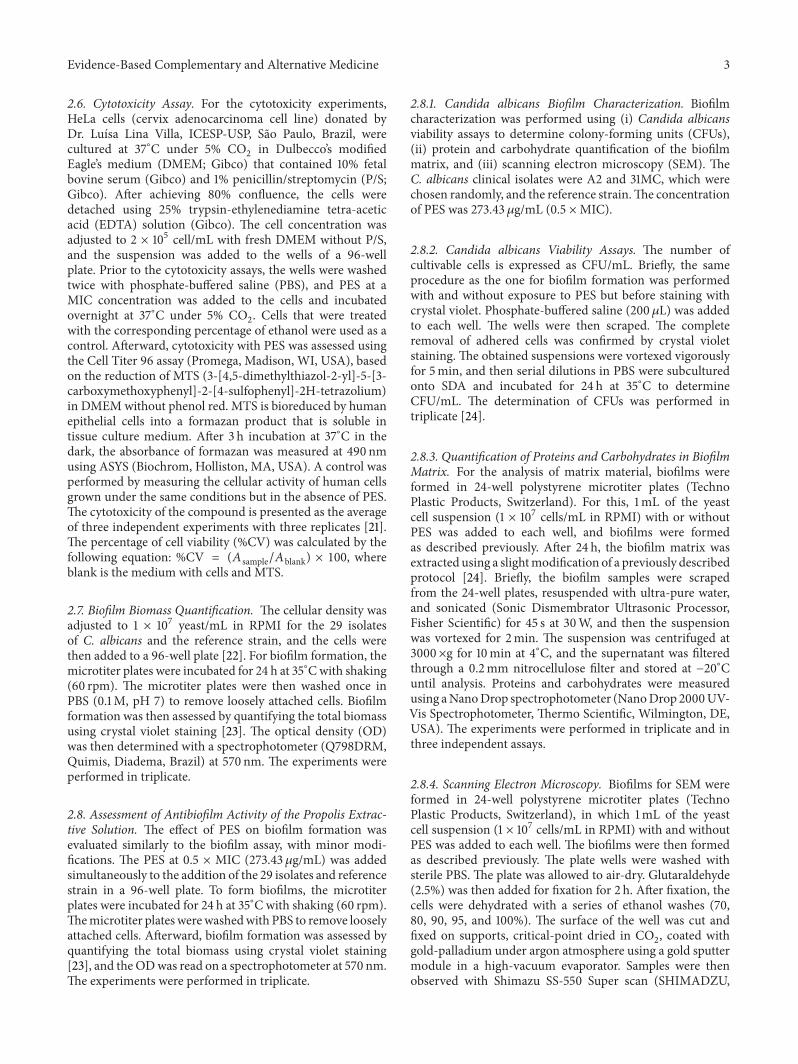
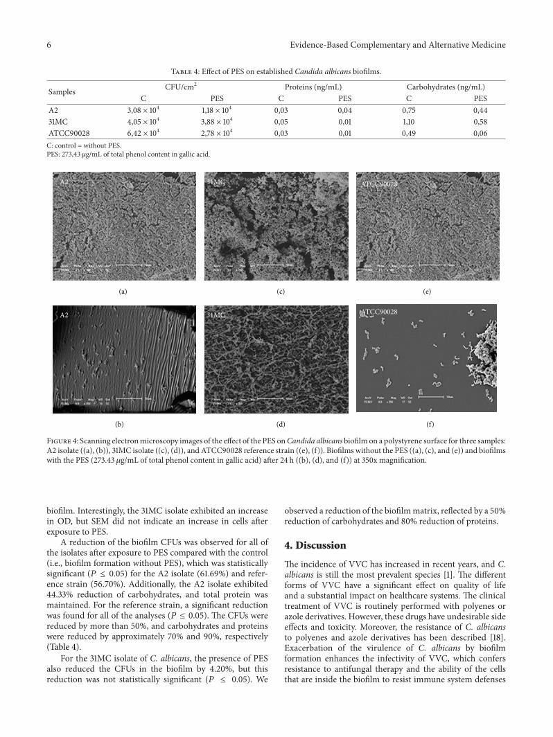
To better understand the action of PES on biofilmformation and matrix composition, two clinical isolates (A2and 31MC) and the reference strain were randomly chosento analyze the characteristics of the biofilm (Table 4). SEMwas used to examine the biofilm structure before and afterexposure to PES; observing themorphological characteristicsof C. albicans was possible (Figure 4). After exposure toPES, the mature biofilms showed a dense network of cellswith various morphologies. The biofilms of the A2 isolateand reference strains were composed of both yeast andpseudohyphae and formed multilayer, compact biofilms thatcovered the entire surface. In contrast, the biofilm of the31MC isolate was devoid of pseudohyphae and consisted of
Table 3: Effect of PES on biofilm biomass for the 29 samples ofCandida albicans and the reference strain isolated from VVC.
Samples Biofilm withoutPES (Abs/cm2)
Biofilm withPES (Abs/cm2) Reduction (%)
A2∗ 2,37 0,11 95,35B11∗ 12,11 0,76 93,72D4 1,91 0,54 71,72F9 2,15 0,24 88,83F10∗ 5,36 0,73 86,38F12 4,07 0,97 76,16G23∗ 2,68 0,23 91,41H1∗ 4,32 0,32 92,59H5 2,21 0,79 64,25I1∗ 8,76 4,07 53,53I10∗ 2,48 0,65 73,79I14 1,08 0,40 62,6961KD∗ 3,28 0,62 81,09109KD 1,00 0,27 73,00110KD∗ 5,19 2,07 60,11111KD 1,21 0,89 26,44112KD 2,02 1,38 31,68117KD 2,91 2,03 30,24119KD 2,56 1,40 45,31126KD∗ 6,74 2,39 64,54132KD∗ 5,84 1,09 81,33134KD∗ 2,79 0,20 92,8373D 2,77 4,14 01MG∗ 5,35 1,97 63,176MG∗ 4,72 1,11 76,4821MG 2,46 1,55 37,003MC∗ 3,06 0,99 67,6431MC∗ 2,90 3,28 0100MC∗ 6,98 1,02 85,38ATCC90028∗ 0,53 0,35 33,96Means 3,73 1,22 63,35The values are means. ∗Significantly different (𝑃 < 0.05) among biofilmwithout PES and with PES.
noncontiguous cell aggregates. After exposure to PES, weobserved a decrease in theOD for theA2 isolate and referencestrain (Table 3). SEM showed a marked reduction of this
6 Evidence-Based Complementary and Alternative Medicine
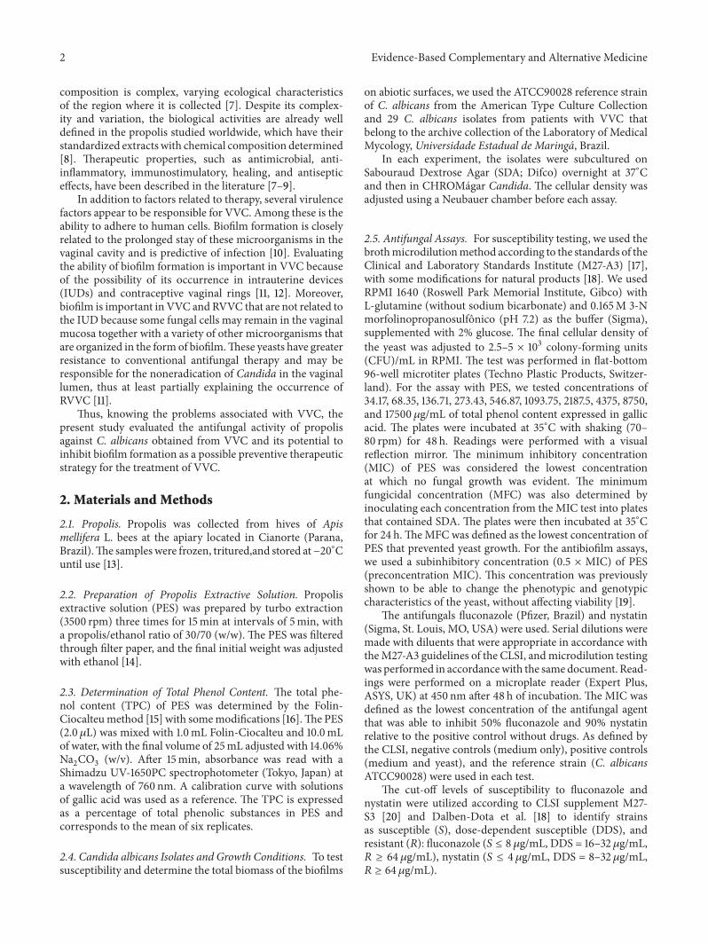
Table 4: Effect of PES on established Candida albicans biofilms.
Samples CFU/cm2 Proteins (ng/mL) Carbohydrates (ng/mL)C PES C PES C PES
A2 3,08 × 10⁴ 1,18 × 10⁴ 0,03 0,04 0,75 0,4431MC 4,05 × 10⁴ 3,88 × 10⁴ 0,05 0,01 1,10 0,58ATCC90028 6,42 × 10⁴ 2,78 × 10⁴ 0,03 0,01 0,49 0,06C: control = without PES.PES: 273,43 𝜇g/mL of total phenol content in gallic acid.
(a)
(d)
(e)
(f)(b)
(c)
A2
A2
31MC
31MC
ATCC90028
ATCC90028
Figure 4: Scanning electronmicroscopy images of the effect of the PES onCandida albicans biofilmon a polystyrene surface for three samples:A2 isolate ((a), (b)), 31MC isolate ((c), (d)), and ATCC90028 reference strain ((e), (f)). Biofilms without the PES ((a), (c), and (e)) and biofilmswith the PES (273.43 𝜇g/mL of total phenol content in gallic acid) after 24 h ((b), (d), and (f)) at 350x magnification.
biofilm. Interestingly, the 31MC isolate exhibited an increasein OD, but SEM did not indicate an increase in cells afterexposure to PES.
A reduction of the biofilm CFUs was observed for all ofthe isolates after exposure to PES compared with the control(i.e., biofilm formation without PES), which was statisticallysignificant (𝑃 ≤ 0.05) for the A2 isolate (61.69%) and refer-ence strain (56.70%). Additionally, the A2 isolate exhibited44.33% reduction of carbohydrates, and total protein wasmaintained. For the reference strain, a significant reductionwas found for all of the analyses (𝑃 ≤ 0.05). The CFUs werereduced by more than 50%, and carbohydrates and proteinswere reduced by approximately 70% and 90%, respectively(Table 4).
For the 31MC isolate of C. albicans, the presence of PESalso reduced the CFUs in the biofilm by 4.20%, but thisreduction was not statistically significant (𝑃 ≤ 0.05). We
observed a reduction of the biofilmmatrix, reflected by a 50%reduction of carbohydrates and 80% reduction of proteins.
4. Discussion
The incidence of VVC has increased in recent years, and C.albicans is still the most prevalent species [1]. The differentforms of VVC have a significant effect on quality of lifeand a substantial impact on healthcare systems. The clinicaltreatment of VVC is routinely performed with polyenes orazole derivatives. However, these drugs have undesirable sideeffects and toxicity. Moreover, the resistance of C. albicansto polyenes and azole derivatives has been described [18].Exacerbation of the virulence of C. albicans by biofilmformation enhances the infectivity of VVC, which confersresistance to antifungal therapy and the ability of the cellsthat are inside the biofilm to resist immune system defenses

Evidence-Based Complementary and Alternative Medicine 7
[31, 32]. The limited number of antifungal drugs that areavailable for treatment combined and the continuous increasein the incidence of C. albicans infection have necessitated thesearch for novel treatment and prevention strategies. Thus,the present study evaluated the in vitro effect of PES as apossible antifungal drug and antibiofilm agent.
Natural products with antifungal activity have been dis-covered [33–36]. Propolis has received the attention of clini-cians and researchers because of its diverse pharmacologicalactivities and low toxicity [37–39].
Our first step was to evaluate the susceptibility of clinicalisolates from VVC to antifungals that are routinely used inclinical practice. As shown in Table 2, C. albicans isolatesfrom VVC were susceptible to fluconazole, but 25% ofthe isolates showed resistance to nystatin. Similar resultswere reported by Dalben-Dota et al. [18]. The PES inhib-ited the growth of all of the strains tested, with a MICof 546.87𝜇g/mL. Importantly, the complete inhibition ofgrowth anddeath occurred even for clinical isolateswithDDSto nystatin, suggesting a better antifungal action than theindependent drugs tested against the isolates tested.
In addition to being effective against microorganisms, adrugmust also show low cytotoxicity for clinical applicability.Propolis varies according to the geographic region where it isextracted [7]. Based on theMIC results for PES, we evaluatedcytotoxicity in HeLa cells at the MIC, 0.5 ×MIC, 0.25 ×MIC,0.125 × MIC, and 0.06 × MIC. The viability of HeLa cellswas satisfactory for more than 80% of these concentrationsat 24 and 48 h (Figure 1). Therefore, PES used in the presentstudy demonstrated low toxicity in human cells, which hasalso been reported by other authors who worked with PESsof different origins [40]. Research indicates that PES canbe a good treatment alternative for chronic vaginitis [9].Moreover, in vitro and in vivo studies have focused on usingPES in pharmaceutical formulations that retain its properties,including mucoadhesive gels [41] and mucoadhesive systemsthat contain thermoresponsive PES [42], for the possibletreatment of VVC.
Biofilm formation in Candida species, in addition topossibly being a key factor in the survival of this species, mayalso be responsible for their being particularly well adapted tothe colonization of tissues and indwelling devices. In VVC,biofilm may be closely related to RVVC and therefore theresistance to antifungal therapy. This could be attributed tobiofilm formation on medical devices, like IUD [11]. There-fore, biofilm formation on surfaces is a key attribute of thepathogenicity of Candida spp. and a major challenge for thetreatment of Candida infections in related biomaterials [43].The possible mechanisms of biofilm resistance to antimi-crobial agents include limited drug penetration throughthe extracellular matrix, phenotypic changes, induction ofthe expression of resistance genes, and a small number of“resistant” cells [44].
Therefore, in the present study, our next step was toevaluate the biofilm formation ability of C. albicans fromVVC (Table 3). All of the isolates studied herein formedbiofilms on polystyrene surfaces under the assayed condi-tions, and this ability was highly strain-dependent. Theseresults reflect inherent differences in the clinical isolates
and may be related to potential pathogenicity. Furthermore,intra- and interspecific variability with regard to the ability ofCandida species to form biofilms has been observed [45]. Infact, SEM revealed structural and morphological differencesin the biofilms between the studied strains.
Based on the biofilm formation that was observed andthe effect of PES on C. albicans from VVC, we evaluatedthe influence of PES at 0.5 ×MIC during biofilm formation.Generally, PES inhibited biofilm formation in 93.34% (28/30)of the strains tested (𝑃 ≤ 0.05) and inhibited the biofilmformation of DDS isolates to nystatin. This reduction ofbiofilm formation by PES has been previously reported,but the previous study analyzed others parameters, such asmetabolic activity, in vitro [46].
One of the most important characteristics of fungalbiofilms is the presence and composition of the extracellularmatrix [47]. Therefore, to better understand the influence ofPES on C. albicans biofilm, we performed SEM and assessedthe cell viability, protein, and carbohydrate characteristics ofthe biofilm (Table 4).The biofilms of A2 isolate and referencestrainwere composed of yeast and pseudohyphae and formedmultilayer, compact biofilms that covered the entire surface.After exposure to PES, SEM revealed a marked reductionof these biofilms. The results demonstrated that both strains(A2 and reference) exhibited a significant decrease in CFUs(𝑃 ≤ 0.05). Furthermore, the biofilm of the reference strainexhibited reductions of the biofilm biomass, carbohydrates,and proteins (𝑃 ≤ 0.05). Another study demonstrated theefficient action of the ethanolic extract of three types ofpropolis on planktonic and biofilm cells of Candida speciesand observed the antibiofilm action of PES, reflected by areduction of the biofilm formed by yeast [48].
Interestingly, the biofilm of the 31MC isolate presenteda multilayer, compact biofilm that covered the entire sur-face. After exposure to PES, the biofilm matrix exhibiteda significant reduction of carbohydrates and proteins. Theincrease inOD revealed by crystal violet stainingwas justifiedby the observation of filamentation, which was visible onlyunder a SEM. The increase in biofilm was justified by yeastfilamentation and possibly occurred as a response of C.albicans to environmental stress [10, 49], which was, in thiscase, exposure to PES.
Notwithstanding the observations that PES reduced thematrix and/or number of cells of C. albicans in the biofilm,PESmay have affected the structure of C. albicans. Accordingto the literature, the deformation of the biofilm impliesgreater permeability of the drug and consequently a reduc-tion of the resistance and infectivity of the clinical isolates[19].
5. Conclusions
Our results support the already described limited effective-ness of nystatin. Despite the susceptibility of the clinicalisolates to fluconazole, the present results demonstrated theincreasing resistance of C. albicans to this azole. The PES hadantifungal activity and may be a useful antibiofilm productthat addresses the problem of drug resistance and RVVC

8 Evidence-Based Complementary and Alternative Medicine
associated with the biofilm growth of C. albicans. Furtherresearch should be extended to biotic surfaces. The presentstudy contributes to a better understanding of the antibiofilmaction of propolis and helps elucidate the development ofRVVC related to the use of IUDs and biofilm formation.
Conflict of Interests
The authors declare that there is no conflict of interestsregarding the publication of this paper.
Acknowledgments
This study was supported by grants from the Fundacao Arau-caria, Conselho Nacional de Desenvolvimento Cientıfico eTecnologico (CNPq), and Coordenacao de Aperfeicoamentode Pessoal de Nıvel Superior (CAPES).
References
[1] J. D. Sobel, “Vulvovaginal candidosis,”The Lancet, vol. 369, no.9577, pp. 1961–1971, 2007.
[2] P. L. Fidel Jr. and J. E. Cutler, “Prospects for development ofa vaccine to prevent and control vaginal candidiasis,” CurrentInfectious Disease Reports, vol. 13, no. 1, pp. 102–107, 2011.
[3] S. R. Fan and X. P. Liu, “In vitro fluconazole and nystatinsusceptibility and clinical outcome in complicated vulvovaginalcandidosis,”Mycoses, vol. 54, no. 6, pp. 501–505, 2011.
[4] X. P. Liu, S. R. Fan, Y. T. Peng, and H. P. Zhang, “Speciesdistribution and susceptibility of Candida isolates from patientwith vulvovaginal candidiasis in Southern China from 2003 to2012,” Journal of Medical Mycology, vol. 24, no. 2, pp. 106–111,2014.
[5] D. P. Kontoyiannis, G. P. Bodey, and C. S. Mantzoros, “Flucona-zole vs. amphotericin B for the management of candidaemia inadults: a meta-analysis,” Mycoses, vol. 44, no. 5-6, pp. 125–135,2001.
[6] E. C. Almeida and H. Menezes, “Anti-inflammatory activity ofpropolis extracts: a review,” Journal of Venomous Animals andToxins, vol. 8, no. 2, pp. 191–212, 2002.
[7] Y. K. Park, S. M. Alencar, and C. L. Aguiar, “Botanical originand chemical composition of Brazilian propolis,” Journal ofAgricultural and Food Chemistry, vol. 50, no. 9, pp. 2502–2506,2002.
[8] G. A. Burdock, “Review of the biological properties and toxicityof bee propolis (propolis),” Food and Chemical Toxicology, vol.36, no. 4, pp. 347–363, 1998.
[9] M. Imhof, M. Lipovac, C. Kurz, J. Barta, H. C. Verhoeven, andJ. C. Huber, “Propolis solution for the treatment of chronicvaginitis,” International Journal of Gynecology and Obstetrics,vol. 89, no. 2, pp. 127–132, 2005.
[10] R. A. Calderone andW. A. Fonzi, “Virulence factors of Candidaalbicans,” Trends in Microbiology, vol. 9, no. 7, pp. 327–335, 2001.
[11] L. J. Douglas, “Candida biofilms and their role in infection,”Trends in Microbiology, vol. 11, no. 1, pp. 30–36, 2003.
[12] D. P. Camacho, M. E. L. Consolaro, E. V. Patussi, L. Donatti, A.Gasparetto, and T. I. E. Svidzinski, “Vaginal yeast adherence tothe combined contraceptive vaginal ring (CCVR),” Contracep-tion, vol. 76, no. 6, pp. 439–443, 2007.
[13] M. L. Bruschi, D. S. Jones, H. Panzeri, M. P. D. Gremiao, O.de Freitas, and E. H. G. Lara, “Semisolid systems containingpropolis for the treatment of periodontal disease: in vitro releasekinetics, syringeability, rheological, textural, andmucoadhesiveproperties,” Journal of Pharmaceutical Sciences, vol. 96, no. 8, pp.2074–2089, 2007.
[14] M. L. Bruschi, T. Klein, R. S. Lopes, S. L. Franco, and M.P. D. Gremiao, “Contribuicao ao protocolo de controle dequalidade da propolis e de seus extratos,” Revista de CienciasFarmaceuticas, vol. 23, no. 2, pp. 289–306, 2002.
[15] Farmacopeia Brasileira, vol. 1-2, Agencia Nacional de VigilanciaSanitaria, Brasılia, Brazil, 5th edition, 2010.
[16] R. R. A. Pereira and M. L. Bruschi, “Evaluation of two spec-trophotometric methods for analysis of green propolis extract,”Latin American Journal of Pharmacy, vol. 32, no. 5, pp. 719–726,2013.
[17] Clinical and Laboratory Standards Institute (CLSI), ReferenceMethod for Broth Dilution Antifungal Susceptibility Testingof Yeasts: Approved Standard, CLSI Document M27-A3, 3rdedition, 2008.
[18] K. F. Dalben-Dota, M. G. I. Faria, M. L. Bruschi, S. M. Pelloso,M. E. Lopes-Consolaro, and T. I. E. Svidzinski, “Antifungalactivity of propolis extract against yeasts isolated from vagi-nal exudates,” The Journal of Alternative and ComplementaryMedicine, vol. 16, no. 3, pp. 285–290, 2010.
[19] M. S. A. Khan and I. Ahmad, “Biofilm inhibition by Cymbo-pogon citratus and Syzygium aromaticum essential oils in thestrains of Candida albicans,” Journal of Ethnopharmacology, vol.140, no. 2, pp. 416–423, 2012.
[20] Clinical and Laboratory Standards Institute (CLSI), “Referencemethod for broth dilution antifungal susceptibility testing ofyeasts,” Approved Standard 3rd Informational Supplement,M27-S3, CLSI, Wayne, Pa, USA, 2008.
[21] G. Malich, B. Markovic, and C. Winder, “The sensitivity andspecificity of theMTS tetrazoliumassay for detecting the in vitrocytotoxicity of 20 chemicals using human cell lines,” Toxicology,vol. 124, no. 3, pp. 179–192, 1997.
[22] J. H. Shin, S. J. Kee, M. G. Shin et al., “Biofilm productionby isolates of Candida species recovered from nonneutropenicpatients: comparison of bloodstream isolates with isolates fromother sources,” Journal of Clinical Microbiology, vol. 40, no. 4,pp. 1244–1248, 2002.
[23] M. Negri, V. Goncalves, S. Silva, M. Henriques, J. Azeredo, andR. Oliveira, “Crystal violet staining to quantity Candida adhe-sion to epithelial cells,” British Journal of Biomedical Science, vol.67, no. 3, pp. 120–125, 2010.
[24] M. Negri, S. Silva, M. Henriques, J. Azeredo, T. Svidzinski, andR.Oliveira, “Candida tropicalis biofilms: artificial urine, urinarycatheters and flow model,”Medical Mycology, vol. 49, no. 7, pp.739–747, 2011.
[25] J. C. P. Mello, L. A. Mentz, and P. R. Petrovick, Farmacognosia:da planta aomedicamento, EFRGS, Porto Alegre Rio Grande doSul, Brazil, 2003.
[26] B. Rankovic, D. Rankovic, and D. Maric, “Antioxidant andantimicrobial activity of some lichen species,”Microbiology, vol.79, no. 6, pp. 809–815, 2010.
[27] I. deOliveira, R. A. G. Lima, E. deOliveira Lima, N.M. P. Farias,and E. L. de Souza, “Atividade antifungica de oleos essenciaissobre especies deCandida,”Brazilian Journal of Pharmacognosy,vol. 16, no. 2, pp. 197–201, 2006.
[28] S. Sonmez, L. Kirilmaz, M. Yucesoy, B. Yucel, and B. Yilmaz,“The effect of bee propolis on oral pathogens and human

Evidence-Based Complementary and Alternative Medicine 9
gingival fibroblasts,” Journal of Ethnopharmacology, vol. 102, no.3, pp. 371–376, 2005.
[29] T. P. Cushnie and A. J. Lamb, “Antimicrobial activity of flavo-noids,” International Journal of Antimicrobial Agents, vol. 26, no.5, pp. 343–356, 2005.
[30] Farmacopeia Brasileira, Atheneu, Sao Paulo, Brazil, 4th edition,1988.
[31] Y. Jin, H. K. Yip, Y. H. Samaranayake, J. Y. Yau, and L. P.Samaranayake, “Biofilm-forming ability of Candida albicans isunlikely to contribute to high levels of oral yeast carriage incases of human immunodeficiency virus infection,” Journal ofClinical Microbiology, vol. 41, no. 7, pp. 2961–2967, 2003.
[32] M. M. Coogan, P. L. Fidel Jr., M. C. Komesu, N. Maeda, andL. P. Samaranayake, “(B1) Candida and mycotic infections,”Advances in Dental Research, vol. 19, no. 1, pp. 130–138, 2006.
[33] A. A. Basma, Z. Zuraini, and S. Sasidharan, “A transmissionelectron microscopy study of the diversity of Candida albicanscells induced by Euphorbia hirta L. leaf extract in vitro,” AsianPacific Journal of Tropical Biomedicine, vol. 1, no. 1, pp. 20–22,2011.
[34] J.-D. Zhang, Z. Xu, Y.-B. Cao et al., “Antifungal activities andaction mechanisms of compounds from Tribulus terrestris L,”Journal of Ethnopharmacology, vol. 103, no. 1, pp. 76–84, 2006.
[35] J. A. Lee and H. Y. Chee, “In vitro antifungal activity of equolagainst Candida albicans,” Mycobiology, vol. 38, no. 4, pp. 328–330, 2010.
[36] A. Khodavandi, F. Alizadeh, F. Aala, Z. Sekawi, and P. P.Chong, “In vitro investigation of antifungal activity of allicinalone and in combination with azoles against Candida species,”Mycopathologia, vol. 169, no. 4, pp. 287–295, 2010.
[37] C. O. D. S. Frozza, C. S. C. Garcia, G. Gambato et al.,“Chemical characterization, antioxidant and cytotoxic activitiesof Brazilian red propolis,” Food andChemical Toxicology, vol. 52,pp. 137–142, 2013.
[38] G. Agarwal, G. G. Vemanaradhya, and D. S. Mehta, “Evaluationof chemical composition and efficacy of Chinese propolisextract on Porphyromonas gingivalis and Aggregatibacter acti-nomycetemcomitans: an in vitro study,” Contemporary ClinicalDentistry, vol. 3, no. 3, p. 256, 2012.
[39] V. Bankova, “Chemical diversity of propolis makes it a valu-able source of new biologically active compounds,” Journal ofApiProduct andApiMedical Science, vol. 1, no. 2, pp. 23–28, 2009.
[40] M. Barbaric, K. Miskovic, M. Bojic et al., “Chemical compo-sition of the ethanolic propolis extracts and its effect on HeLacells,” Journal of Ethnopharmacology, vol. 135, no. 3, pp. 772–778,2011.
[41] A. A. Berretta, P. A. De Castro, A. H. Cavalheiro et al.,“Evaluation of mucoadhesive gels with propolis (EPP-AF) inpreclinical treatment of candidiasis vulvovaginal infection,”Evidence-Based Complementary and Alternative Medicine, vol.2013, Article ID 641480, 18 pages, 2013.
[42] R. R. A. Pereira, J. S. Ribeiro Godoy, T. I. Stivalet Svidzinski, andM. L. Bruschi, “Preparation and characterization of mucoad-hesive thermoresponsive systems containing propolis for thetreatment of vulvovaginal candidiasis,” Journal of Pharmaceu-tical Sciences, vol. 102, no. 4, pp. 1222–1234, 2013.
[43] C. J. Diskin, T. J. Stokes, L. M. Dansby, L. Radcliff, and T. B.Carter, “Is systemic heparin a risk factor for catheter-relatedsepsis in dialysis patients? An evaluation of various biofilm andtraditional risk factors,” Nephron Clinical Practice, vol. 107, no.4, pp. c128–c132, 2008.
[44] L. J. Douglas, “Medical importance of biofilms in Candidainfections,” Revista Iberoamericana de Micologia, vol. 19, no. 3,pp. 139–143, 2002.
[45] A. Valentın, E. Canton, J. Peman, and G. Quindos, “In vitroactivity of amphotericin B and anidulafungin against Candidaspp. biofilms,” Revista Iberoamericana de Micologıa, vol. 24, no.4, pp. 272–277, 2007.
[46] P. A. de Castro, V. L. P. Bom, N. A. Brown et al., “Identificationof the cell targets important for propolis-induced cell death inCandida albicans,” Fungal Genetics and Biology, vol. 60, pp. 74–86, 2013.
[47] M. A. Al-Fattani and L. J. Douglas, “Biofilm matrix of Candidaalbicans andCandida tropicalis: Chemical composition and rolein drug resistance,” Journal of Medical Microbiology, vol. 55, no.8, pp. 999–1008, 2006.
[48] V. C. P. P. Queiroz, Avaliacao do potencial antifungico depropolis de Apis mellifera contra leveduras do genero Candida[dissertacao de mestrado], Universidade Estadual de Campinas,Faculdade de Odontologia de Piracicaba, Sao Paulo, Brazil,2010.
[49] F. C. Odds, “Pathogenesis of Candida infections,” Journal of theAmerican Academy of Dermatology, vol. 31, no. 3, pp. S2–S5,1994.

Submit your manuscripts athttp://www.hindawi.com
Stem CellsInternational
Hindawi Publishing Corporationhttp://www.hindawi.com Volume 2014
Hindawi Publishing Corporationhttp://www.hindawi.com Volume 2014
MEDIATORSINFLAMMATION
of
Hindawi Publishing Corporationhttp://www.hindawi.com Volume 2014
Behavioural Neurology
EndocrinologyInternational Journal of
Hindawi Publishing Corporationhttp://www.hindawi.com Volume 2014
Hindawi Publishing Corporationhttp://www.hindawi.com Volume 2014
Disease Markers
Hindawi Publishing Corporationhttp://www.hindawi.com Volume 2014
BioMed Research International
OncologyJournal of
Hindawi Publishing Corporationhttp://www.hindawi.com Volume 2014
Hindawi Publishing Corporationhttp://www.hindawi.com Volume 2014
Oxidative Medicine and Cellular Longevity
Hindawi Publishing Corporationhttp://www.hindawi.com Volume 2014
PPAR Research
The Scientific World JournalHindawi Publishing Corporation http://www.hindawi.com Volume 2014
Immunology ResearchHindawi Publishing Corporationhttp://www.hindawi.com Volume 2014
Journal of
ObesityJournal of
Hindawi Publishing Corporationhttp://www.hindawi.com Volume 2014
Hindawi Publishing Corporationhttp://www.hindawi.com Volume 2014
Computational and Mathematical Methods in Medicine
OphthalmologyJournal of
Hindawi Publishing Corporationhttp://www.hindawi.com Volume 2014
Diabetes ResearchJournal of
Hindawi Publishing Corporationhttp://www.hindawi.com Volume 2014
Hindawi Publishing Corporationhttp://www.hindawi.com Volume 2014
Research and TreatmentAIDS
Hindawi Publishing Corporationhttp://www.hindawi.com Volume 2014
Gastroenterology Research and Practice
Hindawi Publishing Corporationhttp://www.hindawi.com Volume 2014
Parkinson’s Disease
Evidence-Based Complementary and Alternative Medicine
Volume 2014Hindawi Publishing Corporationhttp://www.hindawi.com