relative statistics of elements concentration in human...
TRANSCRIPT

International Journal of Current Engineering and Technology E-ISSN 2277 – 4106, P-ISSN 2347 – 5161 ©2016 INPRESSCO®, All Rights Reserved Available at http://inpressco.com/category/ijcet
Research Article
2004| International Journal of Current Engineering and Technology, Vol.6, No.6 (Dec 2016)
Relative statistics of elements Concentration in Human teeth using X- Ray Fluorescence Technique Kadhim A. Aadim, Ali A-K. Hussain and Asmaa N. Ahmed*
Department of Physics, College of Science, University of Baghdad, Baghdad, Iraq
Accepted 02 Nov 2016, Available online 05 Nov 2016, Vol.6, No.6 (Dec 2016)
Abstract In this work, (XRF) technique was used to investigate the elements concentration in deciduous and permanent teeth compared with smoking. Many elements have been detected in the deciduous teeth samples, the important once are Ca, K, P, Mg, C and Na. The same elements were detected in permanent teeth with another two elements Fe and Pb. Thus, the concentrations of most toxic elements were significantly in the smoker group. The maximum concentrations of toxic elements such as Pb, Cd and Co were found in older age (above 60 year). The minimum concentrations of trace elements Ca, P and Na were detected in this age group. The relative statistics of calcium concentration in smoker and nonsmoker human teeth were studied according to age. It was concluded that the maximum calcium concentration for nonsmokers was found in age group (20-40 year) compared with other groups. Keywords: Deciduous& permanent teeth, smoking related, XRF 1. Introduction
1 The X-ray fluorescence (XRF) technique can be used for multi-elemental analyses [J. Robinson et al, 2004]. The XRF method has been utilized to determine traced element concentrations in a wide range of samples, for instance biochemistry samples, chemical samples, and archaeological samples. However, the XRF as a portable system frequently lack the ability to analyses large samples (larger than 1 g), and samples with 10 mm in diameter or more [B. Beckhoff et al, 2007]. Therefore, the large samples should be converted to a homogeneous powder. Moreover, the system (XRF) is often unable to detect elements with atomic number larger than 92 [M. Shackley, 2010]. In the present work, using the XRF method for detect of the elements concentration in teeth samples.
2. Types of teeth Teeth are the hardest structures of the human body. The type, number, and arrangement of a set of teeth represent the dentition. Humans have two set of teeth Primary teeth and Permanent teeth. The Primary teeth are also known as deciduous teeth, milk teeth, baby teeth or temporary teeth. Primary teeth start to form during the embryo phase and erupt during infancy (from 6 months to 3 years). The Permanent teeth (or adult teeth) are the second set of teeth and normally consist of 32 teeth. The first permanent teeth appear *Correaponding author: Asmaa N. Ahmed
around the age of 8 and are usually the first molars which erupt right behind the last "milk" molars of the primary dentition [H. Thomas, 1995]. The human tooth consists of four main tissues, enamel is the hardest material found in the human body which protects the other weakly tooth parts from damage, Dentine has a bone like consistency, pulp is found in the tooth center and contains vessels and nerves that keep the tooth alive and the cementum layer covers tooth root [P. Gonçalves et al, 2005]. The crystalline enamel of a tooth is a biological composite containing 4% water, 95% mineral (carbonated hydroxyapatite), and 1% organic matter [L. Geros et al, 2009; F. Brudevold et al, 1967].
3. Smoking Effects Nicotine is the most important constituent among more than 4000 potentially toxic substances in tobacco products. It is the main chemical component responsible for tobacco addiction, appears to mediate the hemodynamic effects of smoking, and has been implicated in the pathogenesis of numerous diseases [O.Ciftci et al, 2013]. Studies have also demonstrated the detrimental effects of smoking on oral health. A clinical study [M. Albandar et al, 2000] observed that smokers had a higher prevalence of moderate and severe periodontitis and higher prevalence and extent of attachment loss and gingival recession than non-smokers, suggesting poorer periodontal health in smokers. In addition, smokers had a higher number of missing teeth than non-smokers. Concerning the bone-

Kadhim A. Aadim et al Relative statistics of elements Concentration in Human teeth using X- Ray Fluorescence Technique
2005| International Journal of Current Engineering and Technology, Vol.6, No.6 (Dec 2016)
implant interface, the deleterious effects of tobacco smoke reflects a series of direct and indirect systemic and local effects on bone metabolism [M.Pereira et al, 2010]. It has been strongly suggested that local exposure of the peri-implant tissues to tobacco products is the main factor leading to an overall increase in implant failure rate in smokers [G.Johnson et al, 2007]. A recent meta-analysis on the subject [H.Chen et al, 2013] observed that smoking was associated with a higher risk of dental implant failure. 4. Experimental part a. Samples preparation The teeth samples were supplied by dental center in Alfurat Hospital (Baghdad, Iraq). They were washed in sodium hypochlorite diluted with distilled water for 10 min to remove all the contamination from the outer surface and dried at room temperature. Then they were preserved in formalin solution. Figure (1) shows some of teeth samples using in this work.
Fig.1 Some of teeth samples using in this work (a) deciduous Teeth (b) permanent teeth age group (20-40 year) (c) permanent teeth age group (40-60 year) (d)
permanent teeth age group (>60 year)
b. X-Ray Fluorescence
Fig.2 Some of teeth samples after grinding and pressing (a) deciduous Teeth (b) permanent teeth age group (20-40 year) (c) permanent teeth age group
(40-60 year) (d) permanent teeth age group (>60 year)
The elemental concentration of teeth samples were studied by X-Ray Fluorescence. The samples were prepared by grinding teeth samples by mechanical mortar, then press 5 grams of the powder using a piston under the pressure of 3.5 tons to make of a disk with 1 cm diameter. Figure (2) illustrates the teeth samples after grinding and pressing. The used X-ray florescence spectrometer is (Spectro Analytical Instruments, Kleve, Germany, Model 2010) using X-ray tube working at a 44.69 kV voltage and 0.55 mA current, with Pd target. Figure (3) shows the typical X-ray fluorescence spectroscopy. Special software was used to analyze the secondary X-rays emitted from samples to identify the elemental content.
Fig.3: Scheme of the experimental setup arrangement for X-ray fluorescence spectroscopy
5. Results and Discussion A. Deciduous Teeth X-Ray fluorescence spectroscopy (XRFS) used to investigate the elements concentration in deciduous teeth.
Table.1: X-Ray Fluorescence (XRF) of deciduous Teeth
Kadhim A. Aadim et al Relative statistics of elements Concentration in Human teeth using X- Ray Fluorescence Technique
2006| International Journal of Current Engineering and Technology, Vol.6, No.6 (Dec 2016)
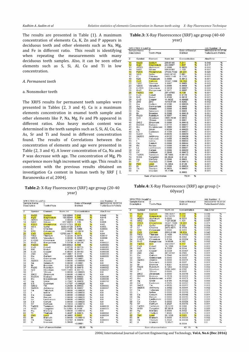
The results are presented in Table (1). A maximum concentration of elements Ca, K, Zn and P appears in deciduous teeth and other elements such as Na, Mg, and Fe in different ratio. This result is identifying when repeating the measurements with many deciduous teeth samples. Also, it can be seen other elements such as S, Si, Al, Cu and Ti in low concentration. A. Permanent teeth a. Nonsmoker teeth The XRFS results for permanent teeth samples were
presented in Tables (2, 3 and 4). Ca is a maximum
elements concentration in enamel teeth sample and
other elements like P, Na, Mg, Fe and Pb appeared in
different ratios. Also heavy metals content was
determined in the teeth samples such as S, Si, Al, Cu, Ga,
As, Sr and Ti and found in different concentration
found. The results of Correlations between the
concentration of elements and age were presented in
Table (2, 3 and 4). A lower concentration of Ca, Na and
P was decrease with age. The concentration of Mg, Pb
experience more high increment with age. This result is
consistent with the previous results obtained on
investigation Ca content in human teeth by XRF [ I.
Baranowska et al, 2004].
Table.2: X-Ray Fluorescence (XRF) age group (20-40
year)
Table.3: X-Ray Fluorescence (XRF) age group (40-60 year)
Table.4: X-Ray Fluorescence (XRF) age group (> 60year)

Kadhim A. Aadim et al Relative statistics of elements Concentration in Human teeth using X- Ray Fluorescence Technique
2007| International Journal of Current Engineering and Technology, Vol.6, No.6 (Dec 2016)
Table.5: X-Ray Fluorescence smoker age group (20-40year)
Table.6: X-Ray Fluorescence smoker age group (40-60years)

Kadhim A. Aadim et al Relative statistics of elements Concentration in Human teeth using X- Ray Fluorescence Technique
2008| International Journal of Current Engineering and Technology, Vol.6, No.6 (Dec 2016)
b. Smoker Teeth X-Ray fluorescence (XRF) was used to analyze the elements contents in smoker teeth. Tables (5) to (7) illustrate the elements that have been identified in the smoker teeth by XRF. It has been identified many elements such as Ca, Na, P, Fe, Mg, Pb, Cd, Cr and Co. The proportion of these elements varies with ages, where the highest concentration of toxic elements Co, Cd and Pb appearance in older smokers compared with younger. While the concentration of trace elements Ca, Na, P and Fe decrease with age. The comparison between smokers and non-smokers tell us that the trace elements decrease in smokers relative to the non-smokers. While the toxic elements Co, Cd and Pb increase in smoker compare with non-smoker. In addition, other elements were appeared at high concentration in smokers teeth compared with non-smokers such as As, Sr, Mo and Nb. These results agree with Abdul [M. Abdul et al, 2015].
Table.7: X-Ray Fluorescence smoker age group (>60year)
c- Relative statistics of elements Concentration This section studies a comparison for trace elements (Ca, P, and Na) with toxic elements concentration in total enamel teeth for group samples which classified
according to age. Figure (4) as shown the relative statistics of elements concentration in nonsmoker group .This figure illustrates that the maximum concentration of (Ca, P, and Na) belong to age group (20-40 year). Also, it can notice from this figure, the presence of lead concentration increase with age. These results are agreement with I. Baranowska [I. Baranowska et al, 2004].
Fig.4 Concentration of elements in nonsmoker group at different ages
Figure (5) shows the concentration of trace elements
and toxic elements cobalt, cadmium and lead in smoker
group at different ages. The maximum concentration of
toxic elements Co and Cd were found in age group
(above 60 year). The present of toxic elements in
smoker due to some habit like smoking or drinking
alcohols are more as compared to normal [M. Abdul et
al, 2015]. Also the trace elements decreasing with age,
furthermore, it is also observed that the trace elements
concentration is almost higher in nonsmoker as
compared to smoker.

Kadhim A. Aadim et al Relative statistics of elements Concentration in Human teeth using X- Ray Fluorescence Technique
2009| International Journal of Current Engineering and Technology, Vol.6, No.6 (Dec 2016)
Fig.5 Concentration of elements in smoker teeth at different ages
Conclusions The elements constituents of deciduous and permanent teeth samples were analyzed by X-ray fluorescence technique (XRF). This method has a high sensitivity and its detection for the concentration of elements in tooth sample in a very short time. Distinguishing between deciduous and permanent teeth was possible by exploiting the change in the concentration ratios of the matrix constituent elements Ca and P, and the non-matrix elements. Also, in results compare between smoker and nonsmoker permanent teeth. The concentration of matrix elements (Ca and P) and non-matrix elements (Na and Fe) increase in nonsmoker teeth while (Mg and Pb) increase in smoker teeth. The concentration of several atomic elements in teeth sample changes with ages.
It probes the presence of the several atomic elements such as Ca, P and Na decrease with age. While a positive correlation for Pb and Mg content in teeth samples with age was noticed. Thus, the concentrations of most toxic elements were significantly in the smoker group. The maximum concentrations of toxic elements such as Pb, Cd and Co were found in older age (above 60 year). Also, the minimum concentrations of trace elements Ca, P and Na in this aged groups. Reference J. Robinson, E. Frame, and G. Frame (2004), Undergraduate
Instrumental Analysis, Sixth Edition, Taylor & Francis. B. Beckhoff, B. Kanngießer, N. Langhoff, R. Wedell and H.
Wolff (2007), Handbook of Practical X-Ray Fluorescence Analysis, Springer.
M. Shackley (2010) X-Ray Fluorescence Spectrometry (XRF) in Geoarchaeology, Springer.
H. Thomas (1995), Root formation, Int J Dev Biol, vol.39, pp. 231-7.
P. Gonçalves, E. Sallum, A. Sallum, M. Casati, S. Toledo and F. Junior (2005), Dental cementum reviewed: development, structure, composition, regeneration and potential functions, Braz. J. Oral Sci., Vol.4, no.12, pp.651-658.
L. Geros, A. Ishikawa, K. Sakae, E. Geros (2009), Fundamentals of hydroxyapatite and related calcium phosphates, Advanced biomaterials: fundamentals, processing and applications, Wiley, New York, pp. 19–52.
F. Brudevold, R. Soremark (1967), Chemistry of the mineral phase of enamel. In: Miles AEW (ed) Structural and chemical organization of teeth, Academic Press, London, pp. 247–277.
O. Ciftci, M. Gunday, M. Caliskan, H. Gullu, A. Guven and H. Muderrisoglu (2013), Light cigarette smoking and vascular function, Acta Cardiologica, vol.68, pp.255–61.
M. Albandar, C. Streckfus, M. Adesanya, D.Winn (2000), Cigar, pipe, and cigarette smoking as risk factors for periodontal disease and tooth loss, Journal of Periodontology, vol.71, pp.1874–81.
M. Pereira, J. Carvalho, F.Peres, M. Fernandes (2010), Simultaneous effects of nicotine, acrolein, and acetaldehyde on estrogenic induced bone marrow cells cultured on plasma-sprayed titanium implants, International Journal of Oral and Maxillofacial Implants, vol.25,pp.112–22.
G. Johnson and J. Guthmiller (2007), The impact of cigarette smoking on periodontal disease and treatment, Periodontology, vol.44, pp.178–94.
H. Chen, N. Liu and E. Lu (2013), Smoking, radiotherapy, diabetes and osteoporosis as risk factors for dental implant failure: a meta-analysis, Plops ONE, vol.8, pp.71955.
I. Baranowska, L. Barchański, M. Bąk, B. Smolec, and Z. Mzyk (2004), X-Ray Fluorescence Spectrometry in Multielemental Analysis of Hair and Teeth, Polish J. Environ. Stud., vol. 13, no. 6, pp. 639–646.
M. Abdul, A. Mohammed, M. Mohamed, S. Sami and Y. Habibullah (2015), Detection of toxic elements using laser-induced breakdown spectroscopy in smokers and nonsmokers teeth and investigation of periodontal parameters, Appl. Opt., vol. 54, no. 24, pp. 7342–7349.