rays were discovered is interesting. in the late 1800’s there · medical x-rays come from a...
TRANSCRIPT
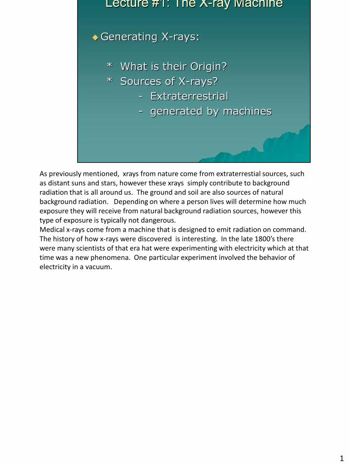
As previously mentioned, xrays from nature come from extraterrestial sources, such as distant suns and stars, however these xrays simply contribute to background radiation that is all around us. The ground and soil are also sources of natural background radiation. Depending on where a person lives will determine how much exposure they will receive from natural background radiation sources, however this type of exposure is typically not dangerous. Medical x-rays come from a machine that is designed to emit radiation on command. The history of how x-rays were discovered is interesting. In the late 1800’s there were many scientists of that era hat were experimenting with electricity which at that time was a new phenomena. One particular experiment involved the behavior of electricity in a vacuum.
1
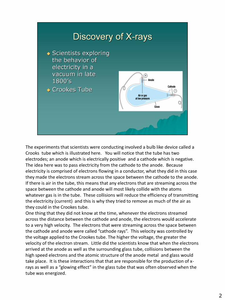
The experiments that scientists were conducting involved a bulb like device called a Crooks tube which is illustrated here. You will notice that the tube has two electrodes; an anode which is electrically positive and a cathode which is negative. The idea here was to pass electricity from the cathode to the anode. Because electricity is comprised of electrons flowing in a conductor, what they did in this case they made the electrons stream across the space between the cathode to the anode. If there is air in the tube, this means that any electrons that are streaming across the space between the cathode and anode will most likely collide with the atoms whatever gas is in the tube. These collisions will reduce the efficiency of transmitting the electricity (current) and this is why they tried to remove as much of the air as they could in the Crookes tube.One thing that they did not know at the time, whenever the electrons streamed across the distance between the cathode and anode, the electrons would accelerate to a very high velocity. The electrons that were streaming across the space between the cathode and anode were called “cathode rays”. This velocity was controlled by the voltage applied to the Crookes tube. The higher the voltage, the greater the velocity of the electron stream. Little did the scientists know that when the electrons arrived at the anode as well as the surrounding glass tube, collisions between the high speed electrons and the atomic structure of the anode metal and glass would take place. It is these interactions that that are responsible for the production of x-rays as well as a “glowing effect” in the glass tube that was often observed when the tube was energized.
2
There were many famous people that were conducting the electrical experiments at the time. Besides Roentgen, there were other scientists that were involved in the early experiments. Later even individuals like Thomas Edison were credited with producing the first fluoroscopic device which uses xrays and can provide “moving x-ray images” We will discuss fluoroscopy in greater detail later in this course. Fluoroscopy is used to do examinations of many parts of the body, but perhaps it is most well known for conducting x-ray examinations of the digestive organs such as the esophagus, stomach, small intestine, and colon.
3

William Conrad Roentgen who is credited for the discovery of x-rays was a professor and physicist at the University of Wurzburg in Germany. In the Fall of November, 1895 he was conducting an experiment where he was discharging high voltage in a Crookes tube which was partially evacuated. The tube he was using happened to have a black cardboard cover and he happened to notice that whenever he energized the Crookes tube, there was a faint glowing of an small screen laying close by. It was this observation of the glowing effect that led to additional investigation of the phenomena.
4
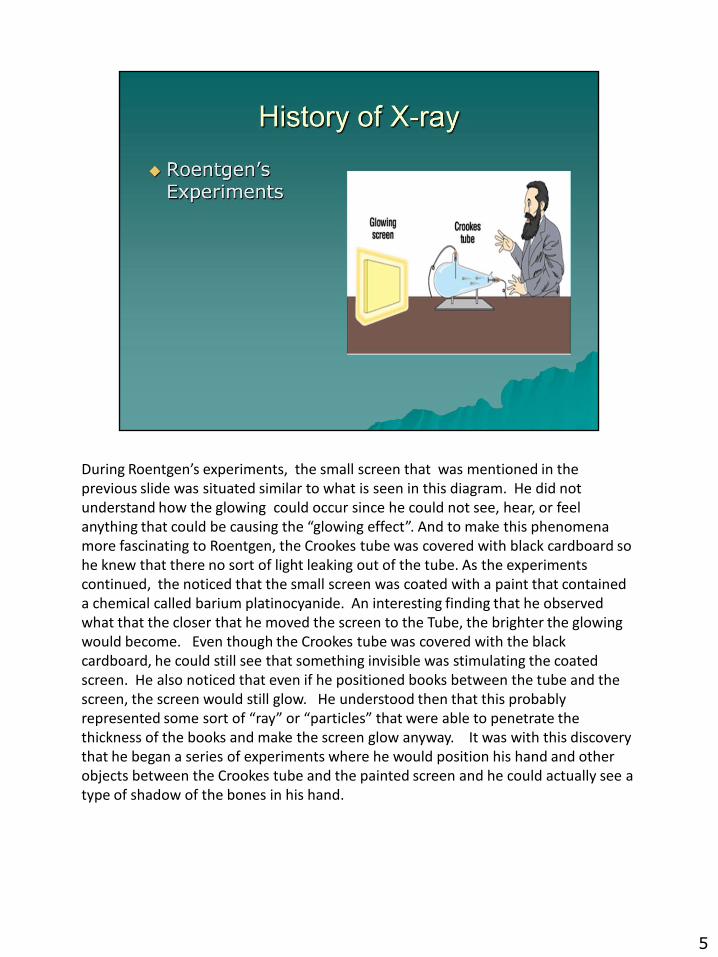
During Roentgen’s experiments, the small screen that was mentioned in the previous slide was situated similar to what is seen in this diagram. He did not understand how the glowing could occur since he could not see, hear, or feel anything that could be causing the “glowing effect”. And to make this phenomena more fascinating to Roentgen, the Crookes tube was covered with black cardboard so he knew that there no sort of light leaking out of the tube. As the experiments continued, the noticed that the small screen was coated with a paint that contained a chemical called barium platinocyanide. An interesting finding that he observed what that the closer that he moved the screen to the Tube, the brighter the glowing would become. Even though the Crookes tube was covered with the black cardboard, he could still see that something invisible was stimulating the coated screen. He also noticed that even if he positioned books between the tube and the screen, the screen would still glow. He understood then that this probably represented some sort of “ray” or “particles” that were able to penetrate the thickness of the books and make the screen glow anyway. It was with this discovery that he began a series of experiments where he would position his hand and other objects between the Crookes tube and the painted screen and he could actually see a type of shadow of the bones in his hand.
5

Roentgen also discovered that when photographic plates were brought close to the energized Crookes tube, it would also cause the photographic emulsion to become fogged or dark without any exposure to light, so this led to him making the first actual x-ray of a human hand. He enlisted the help of his wife and the image is presented here. Notice that there was no great detail with this first image but it was the start of radiography. Note the image of the ring on the finger.
6
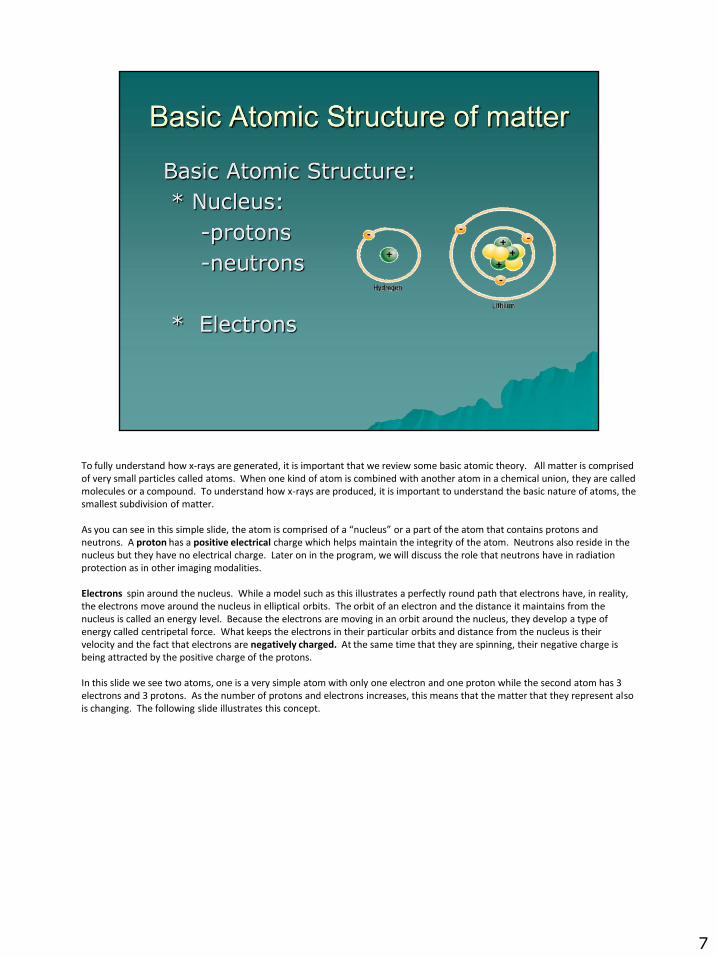
To fully understand how x-rays are generated, it is important that we review some basic atomic theory. All matter is comprisedof very small particles called atoms. When one kind of atom is combined with another atom in a chemical union, they are called molecules or a compound. To understand how x-rays are produced, it is important to understand the basic nature of atoms, the smallest subdivision of matter.
As you can see in this simple slide, the atom is comprised of a “nucleus” or a part of the atom that contains protons and neutrons. A proton has a positive electrical charge which helps maintain the integrity of the atom. Neutrons also reside in the nucleus but they have no electrical charge. Later on in the program, we will discuss the role that neutrons have in radiation protection as in other imaging modalities.
Electrons spin around the nucleus. While a model such as this illustrates a perfectly round path that electrons have, in reality, the electrons move around the nucleus in elliptical orbits. The orbit of an electron and the distance it maintains from the nucleus is called an energy level. Because the electrons are moving in an orbit around the nucleus, they develop a type of energy called centripetal force. What keeps the electrons in their particular orbits and distance from the nucleus is their velocity and the fact that electrons are negatively charged. At the same time that they are spinning, their negative charge is being attracted by the positive charge of the protons.
In this slide we see two atoms, one is a very simple atom with only one electron and one proton while the second atom has 3 electrons and 3 protons. As the number of protons and electrons increases, this means that the matter that they represent also is changing. The following slide illustrates this concept.
7

The classification of the types of matter that make up human beings is important because everything is made of atoms and molecules and depending on the type of matter, the radiation tends to react differently with bones versus soft tissue, etc.
For example, for solid matter such as bone, muscle, and soft tissue, while all of these tissues in the human body are solid, they do vary in their atomic density. This is to say that bone is the hardest to penetrate with the radiation and then muscle, and soft tissue also have variable absorption rates. When radiation is absorbed by matter, this is referred to as attenuation. In general, as the radiation goes through the body, you will find that bone tends to absorb more radiation than does muscle and soft tissue.It can be said that bone, because of its increased atomic density, attenuates the radiation more readily. Because bone is more solid and dense, the number of electrons that make up its atomic structure is more dense than it is for softer tissues thereforethe radiation tends to collide with substances with a greater numbers of electrons.
Fluid tends to vary in viscosity and therfore there is a slight difference in attenuation when you compare the attenuation rate of blood versus clear fluids such as urine. However it should be noted that if there is a significant amount of fluid present as sometimes happens with certain diseases, this results in more difficulty in obtaining diagnostic quality images with x-rays. This is because fluid in great quantities is hard to penetrate.
Gas on the other hand is very similar in its atomic structure to the simple model we say on slide #6. If you go back to that slide, notice that the atom with the single electron (Hydrogen) does not have significant numbers of electrons to attenuate the radiation.
8
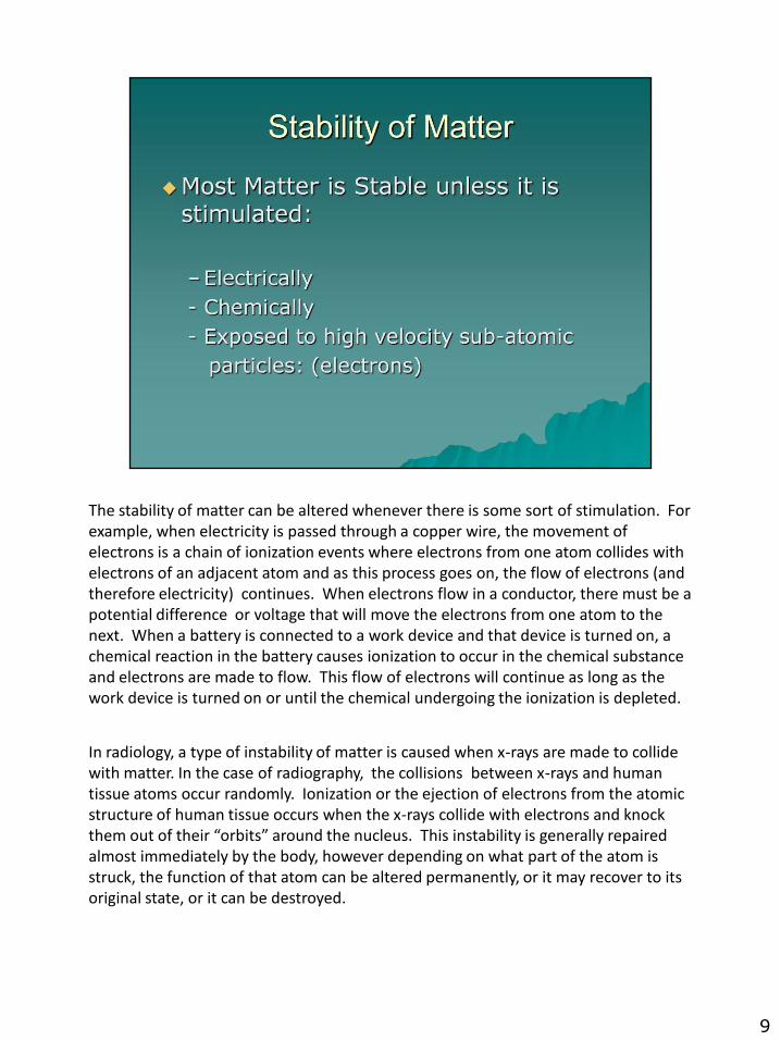
The stability of matter can be altered whenever there is some sort of stimulation. For example, when electricity is passed through a copper wire, the movement of electrons is a chain of ionization events where electrons from one atom collides with electrons of an adjacent atom and as this process goes on, the flow of electrons (and therefore electricity) continues. When electrons flow in a conductor, there must be a potential difference or voltage that will move the electrons from one atom to the next. When a battery is connected to a work device and that device is turned on, a chemical reaction in the battery causes ionization to occur in the chemical substance and electrons are made to flow. This flow of electrons will continue as long as the work device is turned on or until the chemical undergoing the ionization is depleted.
In radiology, a type of instability of matter is caused when x-rays are made to collide with matter. In the case of radiography, the collisions between x-rays and human tissue atoms occur randomly. Ionization or the ejection of electrons from the atomic structure of human tissue occurs when the x-rays collide with electrons and knock them out of their “orbits” around the nucleus. This instability is generally repaired almost immediately by the body, however depending on what part of the atom is struck, the function of that atom can be altered permanently, or it may recover to its original state, or it can be destroyed.
9
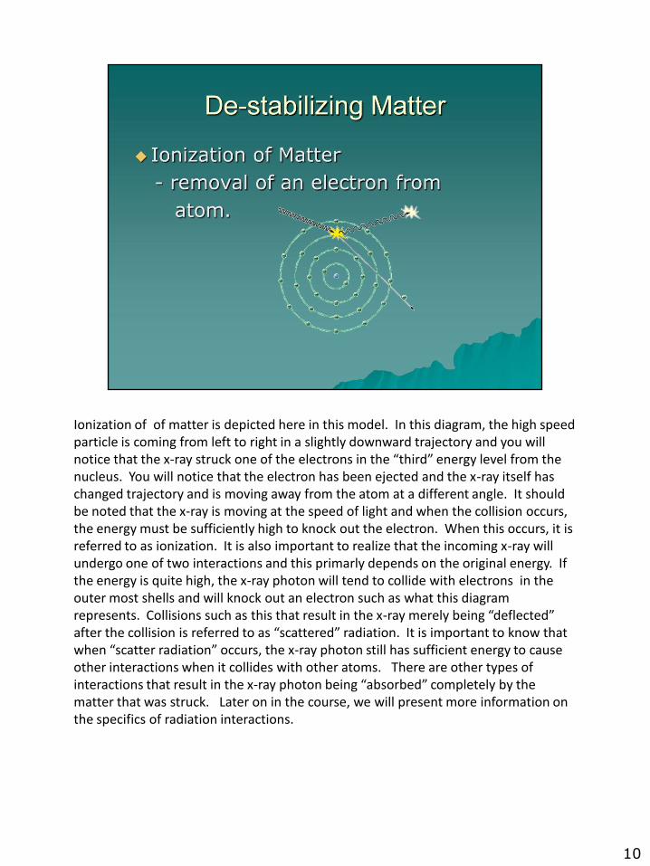
Ionization of of matter is depicted here in this model. In this diagram, the high speed particle is coming from left to right in a slightly downward trajectory and you will notice that the x-ray struck one of the electrons in the “third” energy level from the nucleus. You will notice that the electron has been ejected and the x-ray itself has changed trajectory and is moving away from the atom at a different angle. It should be noted that the x-ray is moving at the speed of light and when the collision occurs, the energy must be sufficiently high to knock out the electron. When this occurs, it is referred to as ionization. It is also important to realize that the incoming x-ray will undergo one of two interactions and this primarly depends on the original energy. If the energy is quite high, the x-ray photon will tend to collide with electrons in the outer most shells and will knock out an electron such as what this diagram represents. Collisions such as this that result in the x-ray merely being “deflected” after the collision is referred to as “scattered” radiation. It is important to know that when “scatter radiation” occurs, the x-ray photon still has sufficient energy to cause other interactions when it collides with other atoms. There are other types of interactions that result in the x-ray photon being “absorbed” completely by the matter that was struck. Later on in the course, we will present more information on the specifics of radiation interactions.
10
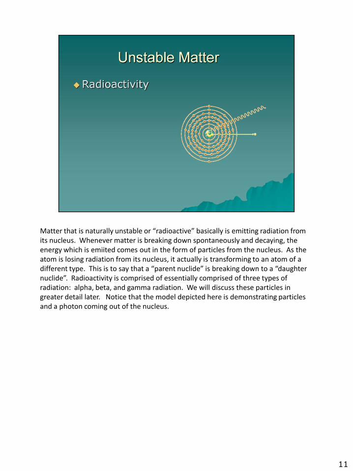
Matter that is naturally unstable or “radioactive” basically is emitting radiation from its nucleus. Whenever matter is breaking down spontaneously and decaying, the energy which is emiited comes out in the form of particles from the nucleus. As the atom is losing radiation from its nucleus, it actually is transforming to an atom of a different type. This is to say that a “parent nuclide” is breaking down to a “daughter nuclide”. Radioactivity is comprised of essentially comprised of three types of radiation: alpha, beta, and gamma radiation. We will discuss these particles in greater detail later. Notice that the model depicted here is demonstrating particles and a photon coming out of the nucleus.
11
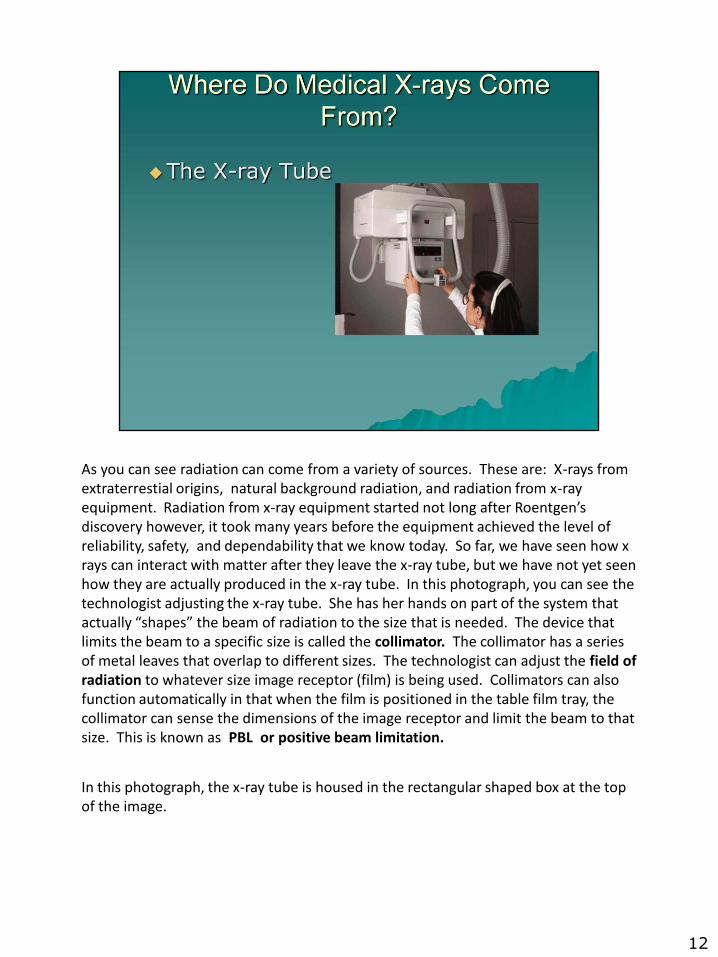
As you can see radiation can come from a variety of sources. These are: X-rays from extraterrestial origins, natural background radiation, and radiation from x-ray equipment. Radiation from x-ray equipment started not long after Roentgen’s discovery however, it took many years before the equipment achieved the level of reliability, safety, and dependability that we know today. So far, we have seen how x rays can interact with matter after they leave the x-ray tube, but we have not yet seen how they are actually produced in the x-ray tube. In this photograph, you can see the technologist adjusting the x-ray tube. She has her hands on part of the system that actually “shapes” the beam of radiation to the size that is needed. The device that limits the beam to a specific size is called the collimator. The collimator has a series of metal leaves that overlap to different sizes. The technologist can adjust the field of radiation to whatever size image receptor (film) is being used. Collimators can also function automatically in that when the film is positioned in the table film tray, the collimator can sense the dimensions of the image receptor and limit the beam to that size. This is known as PBL or positive beam limitation.
In this photograph, the x-ray tube is housed in the rectangular shaped box at the top of the image.
12
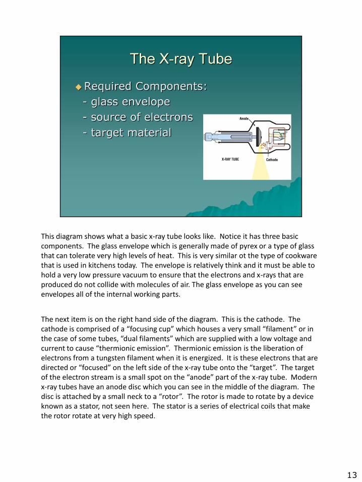
This diagram shows what a basic x-ray tube looks like. Notice it has three basic components. The glass envelope which is generally made of pyrex or a type of glass that can tolerate very high levels of heat. This is very similar ot the type of cookware that is used in kitchens today. The envelope is relatively think and it must be able to hold a very low pressure vacuum to ensure that the electrons and x-rays that are produced do not collide with molecules of air. The glass envelope as you can see envelopes all of the internal working parts.
The next item is on the right hand side of the diagram. This is the cathode. The cathode is comprised of a “focusing cup” which houses a very small “filament” or in the case of some tubes, “dual filaments” which are supplied with a low voltage and current to cause “thermionic emission”. Thermionic emission is the liberation of electrons from a tungsten filament when it is energized. It is these electrons that are directed or “focused” on the left side of the x-ray tube onto the “target”. The target of the electron stream is a small spot on the “anode” part of the x-ray tube. Modern x-ray tubes have an anode disc which you can see in the middle of the diagram. The disc is attached by a small neck to a “rotor”. The rotor is made to rotate by a device known as a stator, not seen here. The stator is a series of electrical coils that make the rotor rotate at very high speed.
13

The x-ray tube housing basically provides mechanical support and holds the x-ray tube firmly and safely in place. The housing is made to tolerate very high levels of heat because when xrays are generated, the vast majority of energy that is expended is converted into heat. In this diagram, you will notice the various components we have discussed that includes the collimator, cathode, and anode. A very important function of the tube housing is to ensure that it cools the x-ray tube. Whenever you generate x-rays , a tremendous amount of heat is generated and it is the job of the housing to help dissipate the heat. The housing does this through a couple of mechanisms that are not visible here. First, the housing is filled with oil. The oil serves to help cool the very hot glass envelope as x-rays are being produced. Because x-rays do travel through this oil as they exit the x-ray tube, the oil also has a very slight “filtering” effect and does provide some electrical insulation as well. Most x ray tube housings also have a cooling fan that circulates cooling air around the housing to help dissipate the heat more quickly and make the x-ray production process more efficient.
14
In order to produce x-rays, three things must be available:
#1. There has to be a source of electrons. This function is provided by the cathode filament. When x-rays are in the process of being generated, the technologists initiates the exposure by depressing the “rotor boost” button or switch and what this does, is it sends electric current surging through the filament and it makes it glow white hot. This is referred to as “incandesence” or thermionic emission. What is happening at the filament is that a large amount of electricity is being forced through the very small diameter wire such that because of the crowding of the large numbers of electrons, they are literally “forced” out of the filament and they form a “space charge” or electron cloud around the filament. Because they are negatively charged, they tend to repel each other and say in a space charge until the moment of exposure begins. Simultaneously as the electrons begin to boil off the filament, current is also sent to the induction motor around the “rotor” and it begins to rotate at very high speed. Once the normal rotational speed is achieved, the system is ready for the exposure and the technologist can press that button.
#2. At the moment of exposure, a very high voltage is applied between the cathode and anode and this is expressed in kVp (kilo volts peak) . The greater the kVp, the greater the attraction between the electrons around the filament and the anode disc surface. Depending on the anatomical part that is being x-rayed, the kVp will be varied. The electrons that are accelerated and travel to the target material are also known as projectile electrons.
#3. The target is actually the circular anode disc. The disc is connected to the rotor and the rotor spins during the exposure.
15
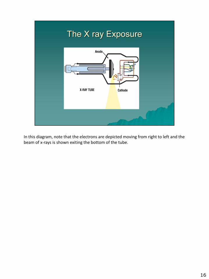
In this diagram, note that the electrons are depicted moving from right to left and the beam of x-rays is shown exiting the bottom of the tube.
16
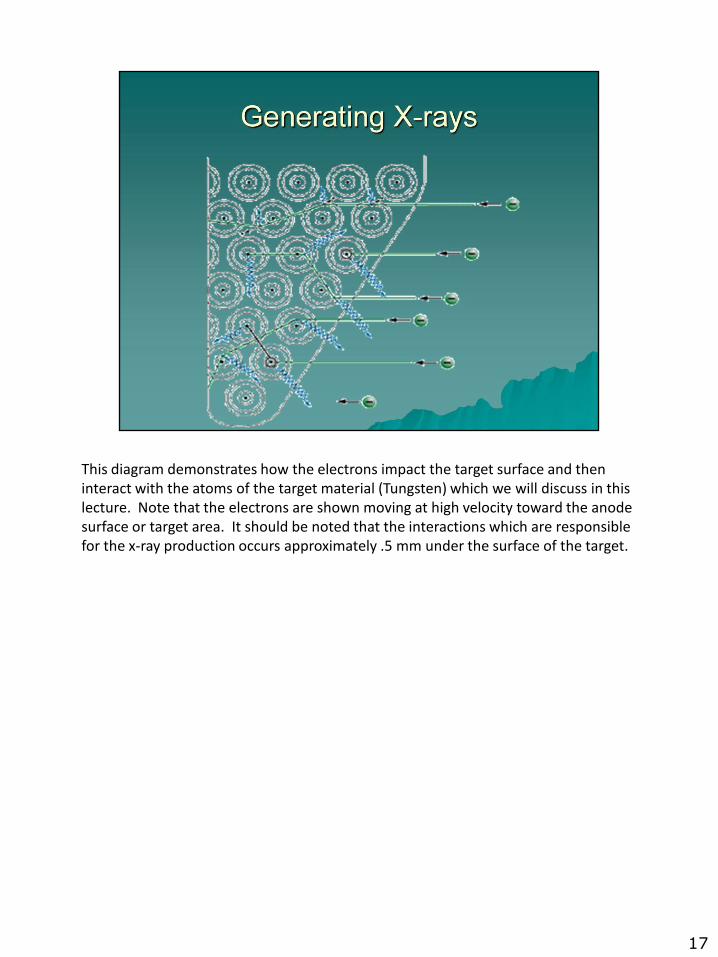
This diagram demonstrates how the electrons impact the target surface and then interact with the atoms of the target material (Tungsten) which we will discuss in this lecture. Note that the electrons are shown moving at high velocity toward the anode surface or target area. It should be noted that the interactions which are responsible for the x-ray production occurs approximately .5 mm under the surface of the target.
17
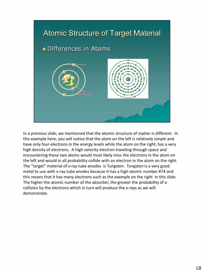
In a previous slide, we mentioned that the atomic structure of matter is different. In this example here, you will notice that the atom on the left is relatively simple and have only four electrons in the energy levels while the atom on the right, has a very high density of electrons. A high velocity electron traveling through space and encountering these two atoms would most likely miss the electrons in the atom on the left and would in all probability collide with an electron in the atom on the right. The “target” material of x-ray tube anodes is Tungsten. Tungsten is a very good metal to use with x-ray tube anodes because it has a high atomic number #74 and this means that it has many electrons such as the example on the right in this slide. The higher the atomic number of the absorber, the greater the probability of a collision by the electrons which in turn will produce the x-rays as we will demonstrate.
18
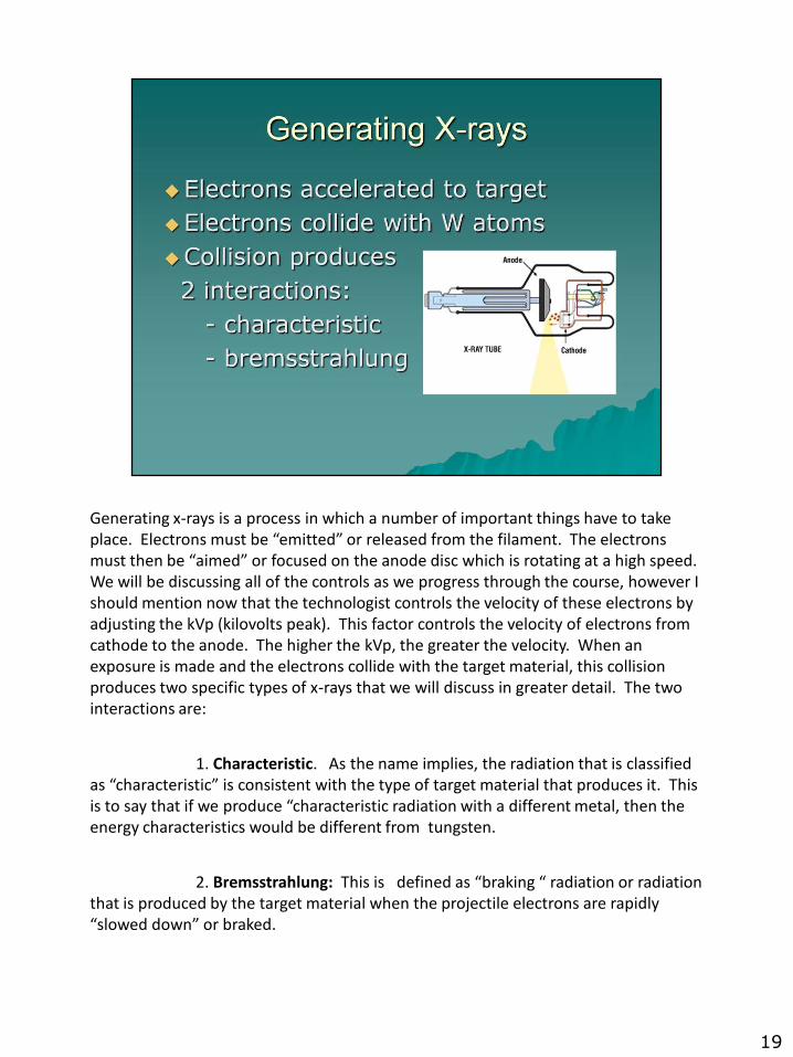
Generating x-rays is a process in which a number of important things have to take place. Electrons must be “emitted” or released from the filament. The electrons must then be “aimed” or focused on the anode disc which is rotating at a high speed. We will be discussing all of the controls as we progress through the course, however I should mention now that the technologist controls the velocity of these electrons by adjusting the kVp (kilovolts peak). This factor controls the velocity of electrons from cathode to the anode. The higher the kVp, the greater the velocity. When an exposure is made and the electrons collide with the target material, this collision produces two specific types of x-rays that we will discuss in greater detail. The two interactions are:
1. Characteristic. As the name implies, the radiation that is classified as “characteristic” is consistent with the type of target material that produces it. This is to say that if we produce “characteristic radiation with a different metal, then the energy characteristics would be different from tungsten.
2. Bremsstrahlung: This is defined as “braking “ radiation or radiation that is produced by the target material when the projectile electrons are rapidly “slowed down” or braked.
19
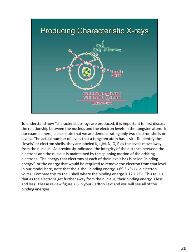
To understand how “characteristic x-rays are produced, it is important to first discuss the relationship between the nucleus and the electron levels in the tungsten atom. In our example here, please note that we are demonstrating only two electron shells or levels. The actual number of levels that a tungsten atom has is six. To identify the “levels” or electron shells, they are labeled K, L,M, N, O, P as the levels move away from the nucleus. As previously indicated, the integrity of the distance between the electrons and the nucleus is maintained by the spinning motion of the orbiting electrons. The energy that electrons at each of their levels has is called “binding energy” or the energy that would be required to remove the electron from that level. In our model here, note that the K shell binding energy is 69.5 kEv (kilo electron volts). Compare this to the L shell where the binding energy is 12.1 kEv. This tell us that as the electrons get further away from the nucleus, their binding energy is less and less. Please review figure 2.6 in your Carlton Text and you will see all of the binding energies
20
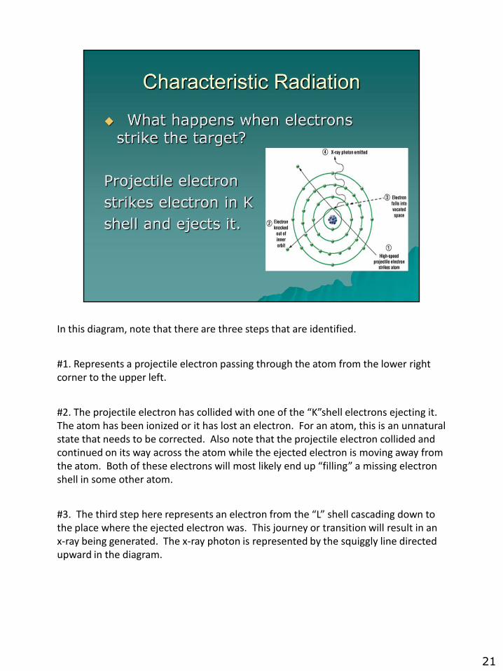
In this diagram, note that there are three steps that are identified.
#1. Represents a projectile electron passing through the atom from the lower right corner to the upper left.
#2. The projectile electron has collided with one of the “K”shell electrons ejecting it. The atom has been ionized or it has lost an electron. For an atom, this is an unnatural state that needs to be corrected. Also note that the projectile electron collided and continued on its way across the atom while the ejected electron is moving away from the atom. Both of these electrons will most likely end up “filling” a missing electron shell in some other atom.
#3. The third step here represents an electron from the “L” shell cascading down to the place where the ejected electron was. This journey or transition will result in an x-ray being generated. The x-ray photon is represented by the squiggly line directed upward in the diagram.
21
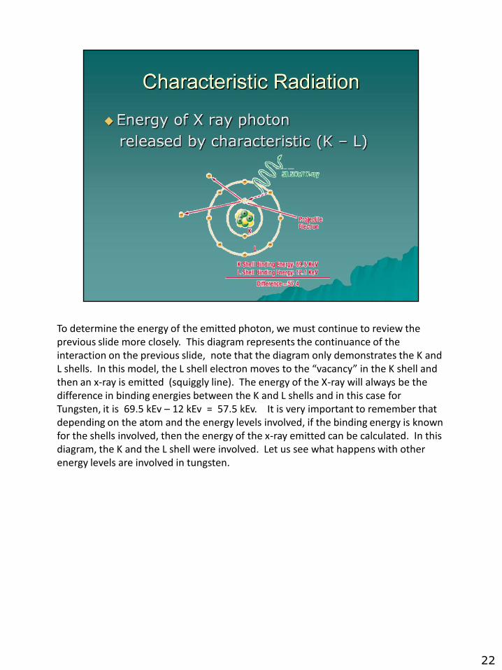
To determine the energy of the emitted photon, we must continue to review the previous slide more closely. This diagram represents the continuance of the interaction on the previous slide, note that the diagram only demonstrates the K and L shells. In this model, the L shell electron moves to the “vacancy” in the K shell and then an x-ray is emitted (squiggly line). The energy of the X-ray will always be the difference in binding energies between the K and L shells and in this case for Tungsten, it is 69.5 kEv – 12 kEv = 57.5 kEv. It is very important to remember that depending on the atom and the energy levels involved, if the binding energy is known for the shells involved, then the energy of the x-ray emitted can be calculated. In this diagram, the K and the L shell were involved. Let us see what happens with other energy levels are involved in tungsten.
22
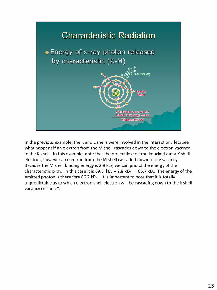
In the previous example, the K and L shells were involved in the interaction, lets see what happens if an electron from the M shell cascades down to the electron vacancy in the K shell. In this example, note that the projectile electron knocked out a K shell electron, however an electron from the M shell cascaded down to the vacancy. Because the M shell binding energy is 2.8 kEv, we can pridict the energy of the characteristic x-ray. In this case it is 69.5 kEv – 2.8 kEv = 66.7 kEv. The energy of the emitted photon is there fore 66.7 kEv. It is important to note that it is totally unpredictable as to which electron shell electron will be cascading down to the k shell vacancy or “hole”.
23
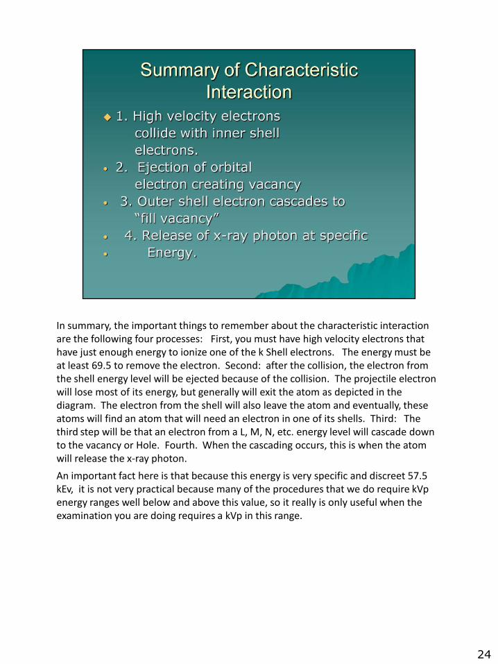
In summary, the important things to remember about the characteristic interaction are the following four processes: First, you must have high velocity electrons that have just enough energy to ionize one of the k Shell electrons. The energy must be at least 69.5 to remove the electron. Second: after the collision, the electron from the shell energy level will be ejected because of the collision. The projectile electron will lose most of its energy, but generally will exit the atom as depicted in the diagram. The electron from the shell will also leave the atom and eventually, these atoms will find an atom that will need an electron in one of its shells. Third: The third step will be that an electron from a L, M, N, etc. energy level will cascade down to the vacancy or Hole. Fourth. When the cascading occurs, this is when the atom will release the x-ray photon.
An important fact here is that because this energy is very specific and discreet 57.5 kEv, it is not very practical because many of the procedures that we do require kVp energy ranges well below and above this value, so it really is only useful when the examination you are doing requires a kVp in this range.
24
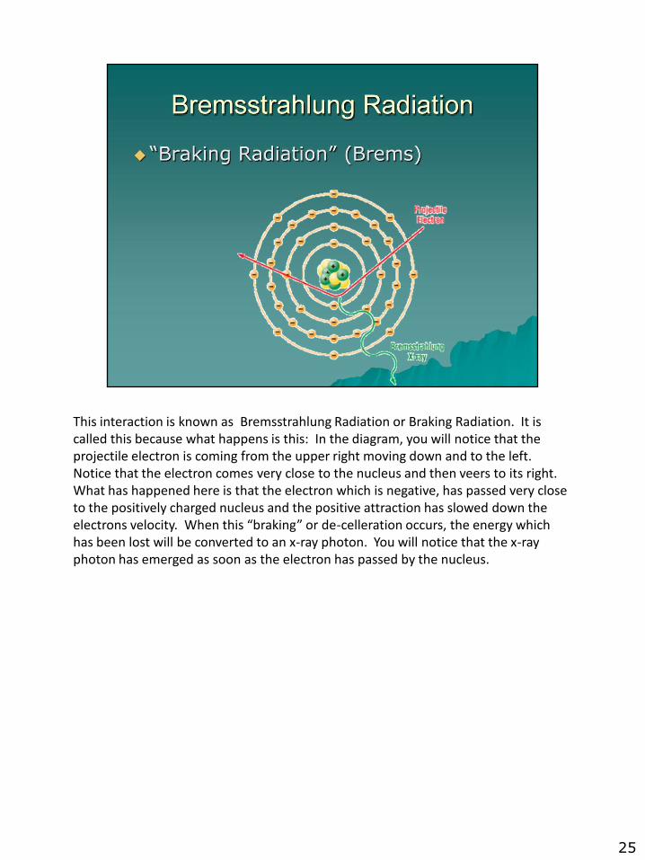
This interaction is known as Bremsstrahlung Radiation or Braking Radiation. It is called this because what happens is this: In the diagram, you will notice that the projectile electron is coming from the upper right moving down and to the left. Notice that the electron comes very close to the nucleus and then veers to its right. What has happened here is that the electron which is negative, has passed very close to the positively charged nucleus and the positive attraction has slowed down the electrons velocity. When this “braking” or de-celleration occurs, the energy which has been lost will be converted to an x-ray photon. You will notice that the x-ray photon has emerged as soon as the electron has passed by the nucleus.
25
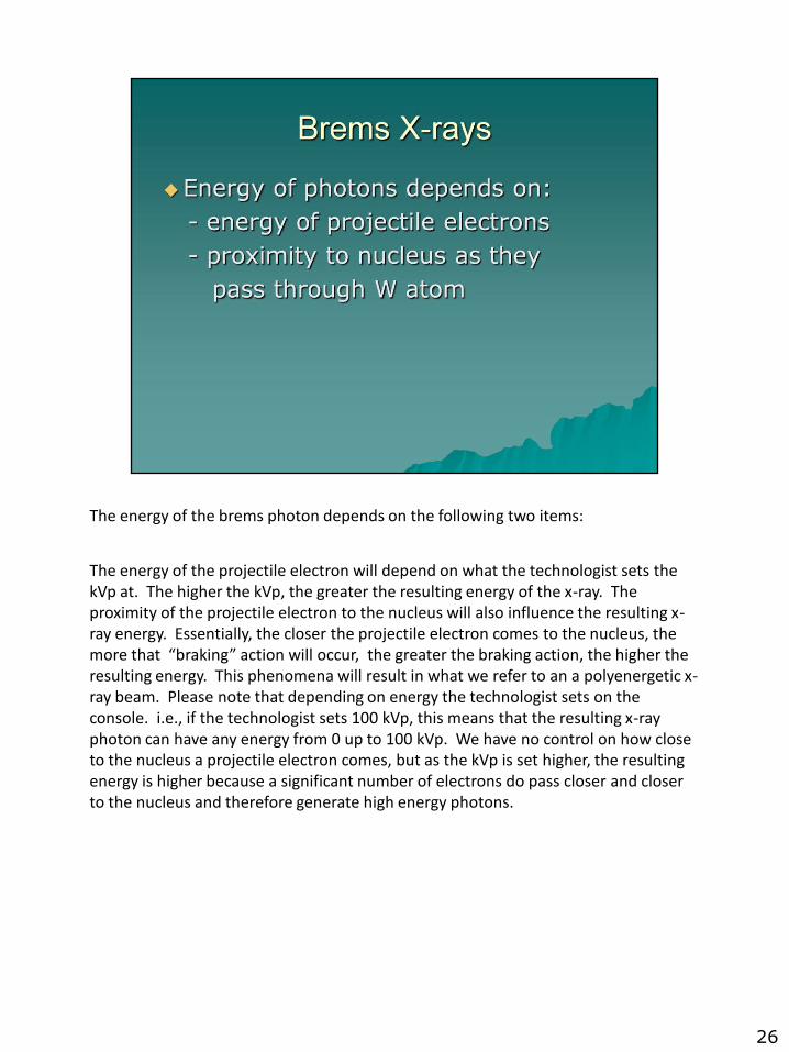
The energy of the brems photon depends on the following two items:
The energy of the projectile electron will depend on what the technologist sets the kVp at. The higher the kVp, the greater the resulting energy of the x-ray. The proximity of the projectile electron to the nucleus will also influence the resulting x-ray energy. Essentially, the closer the projectile electron comes to the nucleus, the more that “braking” action will occur, the greater the braking action, the higher the resulting energy. This phenomena will result in what we refer to an a polyenergetic x-ray beam. Please note that depending on energy the technologist sets on the console. i.e., if the technologist sets 100 kVp, this means that the resulting x-ray photon can have any energy from 0 up to 100 kVp. We have no control on how close to the nucleus a projectile electron comes, but as the kVp is set higher, the resulting energy is higher because a significant number of electrons do pass closer and closer to the nucleus and therefore generate high energy photons.
26
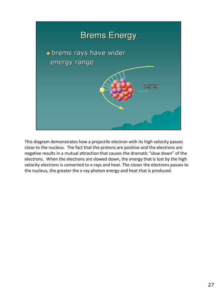
This diagram demonstrates how a projectile electron with its high velocity passes close to the nucleus. The fact that the protons are positive and the electrons are negative results in a mutual attraction that causes the dramatic “slow down” of the electrons. When the electrons are slowed down, the energy that is lost by the high velocity electrons is converted to x-rays and heat. The closer the electrons passes to the nucleus, the greater the x-ray photon energy and heat that is produced.
27

X-rays have some physical characteristics that are important for the technologist to understand. This will help to ensure that x-rays are used safely and efficiently by the technologist in order to achieve the objectives of the examination and protect themselves as well as the patient. The physical characteristics that are essential to understand are as follows:
1. X-ray photons are penetrating. Depending on the type of tissue or matter x-rayed, the technologist will adjust the penetrating ability of the x-rays by adjusting the kVp of the machine. It should be noted that different parts of the body will have different thicknesses and attenuation properties therefore requiring different technical settings. We will perform some simple lab activities to demonstrate how this characteristic works.
2. X-rays are a form of Electromagnetic Radiation. An x-ray is like a bundle of energy moving through the air. It has wavelength which is measured in angstroms, it has frequency, and it is penetrating. X-rays are electrically neutral so they are not attracted by positive or negative polarities and cannot be focused.
3. X-rays are Polyenergetic. This is to say that the x-rays that are emitted by an x-ray tube are poly energetic or of many different energies. We know that with the characteristic interaction, we can predict what the energy of the photon will be (57.5 kEv), however with the Brems interaction, it really depends on how close to the nucleus the projectile electron passes by. Remember, the closer the electron passes to the nucleus, the higher the resulting energy of the emitted x-ray. When you consider the millions of interactions that are occurring during an exposure like this, it is easy to imagine that there are relatively few photons with exactly the same energy. If the technologist sets 100 kVp on the control console, the reality is that the average energy of the beam is probably less than what was set. This is also dependent on the type of xray equipment you are using. There are numerous varieities of machine and in general, state of the art machines are the most powerful units
4. Xrays may be attenuated. This is to say that x-rays can be attenuated or absorbed by matter. Depending on what the x-rays strike, if it is metal, chances are that the x-rays will be absorbed. An example of this is lead. Because lead is very dense atomically, when an x-ray strikes it, the lead will absorb or attenuate the radiation. In the human body, bone tends to absorb a significant amount of the radiation because it is relatively hard compared to other tissues. Soft tissue and air filled organs have the least attenuation because soft tissue has a low atomic density any gas filled structure tends to facilitiate the passage or transmission of x-rays.
5. The angle of trajectory. This refers to the how x-rays emerge from the tube.. If you recall that the anode is angled slightly such that when the projectile electrons strike the surface of the target, x-rays that are produced tend to move downward. For example, if the electrons were hard particles and they simply struck the angled target, they would bounce in a downward trajectory because of the angle. However the reality is that the electrons penetrate the surface of the target and undergo the brems and characteristic interactions just below the surface, usually around 0.5 mm under the surface. X-rays are produced “isotropically” or when they are emitted, they go in all directions. An an example of “isotropic” emission is like having a light bulb suspended in a square room. If the bulb is in the themiddle of the room, when the light is turned on, you would have light in all the surfaces of the room. Basically isotropic means that x-rays are going in all directions, but only the ones that are moving downward will come out of the tube. The rest, are absorbed by the housing.
6. The velocity of x-rays is the speed of light. Irregardless of the kVp that is set, please remember that the projectile electrons do achieve different velocities when they go from filament to the anode, however when the electrons are converted to x-ray energy, the speed of those x-rays is the speed of light.
7. X-rays have ionizing ability. This means that x-rays have the ability to remove electrons from the atoms of the matter that they strike. We will discuss the different interactions that x-rays can produce in matter in a later unit of this course
28

8. X-rays can cause fluorescence of certain types of phosphors. Certain materials interact with x-rays such that when x-rays strike them, the material will glow or give off light. This property is very useful when used in conjunction with film. Over the years, there have been many types of phosphors that have been used. The phosphors are used in cassettes or film holders and when the part of interest is x-rayed, the phosphors glow or fluoroesce and expose the film with the image of whatever the x-rays went through. We will be demonstrating this process in an interesting lab experiment soon.
9. X-rays cannot be focussed. X-rays are not like light that a glass lens can focus. When x-rays pass through matter, they tend to go in a straight line, however if they do strike something such as an atom of bone or some other tissue, the x-ray photon will either be absorbed or it can be deflected and it will go in a different direction. This is very much like a high velocity projectile such as a bullet that strikes something. It can be deflected or it can be stopped. When it is deflected, we refer to it as a “scattered x-ray”.
10. When x-rays interact with film, it will cause electro-chemical changes in the structure of the film so that when the film is developed in chemicals, the film will show “blackness” or “radiographic density”. It is this density that the radiographer must learn to control to ensure that the image is not too dark or too light.
11. X-rays or radiation also has the capability of altering matter chemically. In some cases such as radiation therapy where they use radiation to treat cancer, the radiation can change the chemical environment of the tissue it interacts with such that it can help destroy cancerous cells. Biologically, the atomic structure of living tissue can also be affected in numerous ways. We will be discussing some of these biologic effects in great detail later in this course as well as in a later course in the summer semester.
12. As we have indicated before, when x-rays strike matter, they will either be absorbed or scattered/deflected. Both of these interactions can cause biologic effects through ionization of the atomic structure of biologic tissue. It is important to know also that when x-rays strike human tissue, sometimes they undergo a type of interaction where the x-ray is completely aborbed by the atom it struck. When this occurs, ionization also occurs and the atom that was struck will respond by emitting a “secondary” x-ray photon of its own. This “secondary” photon that is produced is typically very, very low energy. Scatter radiation however is when an x-ray is simply deflected and there is little energy loss. This “scattered’ x-ray can go on and undergo other interactions until it finally gets absorbed somewhere. The scattered x-ray does have the ability of ionizing tissue or matter.
13. As we mentioned before, x-rays are are discreet very small bundles of energy moving through space. They are part of the electromagnetic spectrum. They have wavelength and they have frequency. They have a sinusoidal or “s shaped curve”. The smaller the wavelength, the greater their energy and the higher the frequency. The longer the wavelength, the less penetrating they are.
29
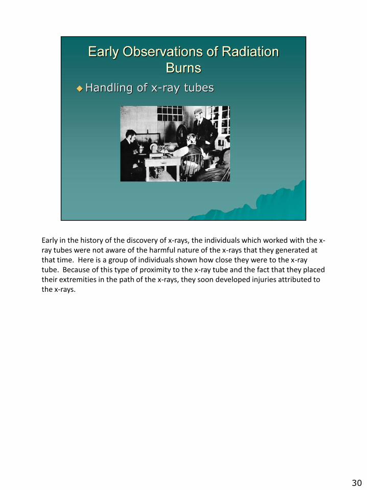
Early in the history of the discovery of x-rays, the individuals which worked with the x-ray tubes were not aware of the harmful nature of the x-rays that they generated at that time. Here is a group of individuals shown how close they were to the x-ray tube. Because of this type of proximity to the x-ray tube and the fact that they placed their extremities in the path of the x-rays, they soon developed injuries attributed to the x-rays.
30
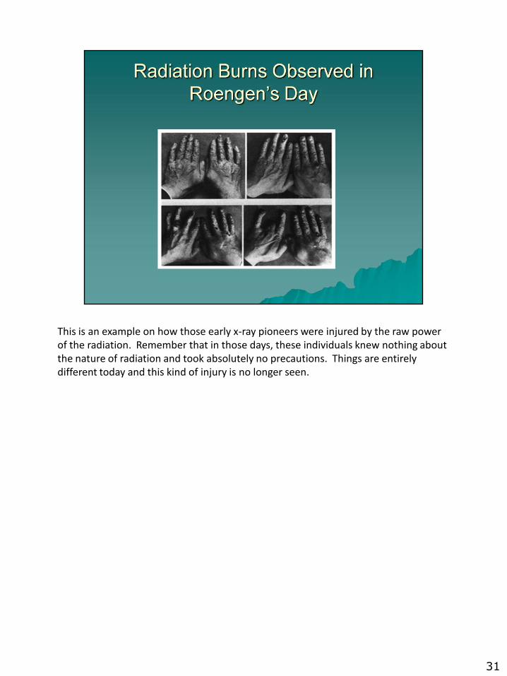
This is an example on how those early x-ray pioneers were injured by the raw power of the radiation. Remember that in those days, these individuals knew nothing about the nature of radiation and took absolutely no precautions. Things are entirely different today and this kind of injury is no longer seen.
31

Here is an example on how x-rays were used early in their development. This of course is a lady’s foot still in her shoe.
32