prenatal diagnosis of limb–body wall complex with craniofacial defects
TRANSCRIPT

Journal of Medical Ultrasound (2011) 19, 112e114
Available online at www.sciencedirect.com
journal homepage: www.jmu-onl ine.com
LETTER TO THE EDITOR
Prenatal Diagnosis of LimbeBody Wall Complex WithCraniofacial Defects
Chih-Ping Chen 1,2,3,4,5,6*, Yi-Yung Chen 1, Jun-Wei Su 1,7,Wayseen Wang 2,8
1Department of Obstetrics and Gynecology, Mackay Memorial Hospital, Taipei, 2Department of Medical Research, MackayMemorial Hospital, Taipei, 3Department of Biotechnology, Asia University, Taichung, 4 School of Chinese Medicine, Collegeof Chinese Medicine, China Medical University, Taichung, 5 Institute of Clinical and Community Health Nursing, NationalYang-Ming University, Taipei, 6Department of Obstetrics and Gynecology, School of Medicine, National Yang-Ming University,Taipei, 7Department of Obstetrics and Gynecology, China Medical University Hospital, Taichung, and 8Department ofBioengineering, Tatung University, Taipei, Taiwan, ROC
A 42-year-old, gravida 4, para 0 woman was referred to thehospital at 17 weeks of gestation to evaluate fetal struc-tural abnormalities. The father was aged 42 years. Themother reported no illness or recent infections. She hadneither a history of prenatal exposure to teratogenic agentsnor any family history of congenital malformations. She hadnot undergone any assisted reproductive technology for thispregnancy.
Prenatal ultrasound at 17 weeks of gestation demon-strated a live fetus with cranioplacental attachment,scoliosis, and abdominal wall defects but no limb deficiency(Fig. 1). The pregnancy was subsequently terminated, anda 180-g male fetus was delivered with exencephaly, acra-nium, abdominoschisis, craniofacial deformity, partialdeficiency of the second and third fingers of the left hand,left club foot, and extracorporeal intestines and liver, butnormal male external genitalia and anus (Fig. 2). A diag-nosis of limbebody wall complex (LBWC) with craniofacial
* Correspondence to: Dr Chih-Ping Chen, Department of Obstetrics andNorth Road, Taipei, Taiwan, ROC.
E-mail address: [email protected] (C.-P. Chen).
0929-6441/$36 ª 2011, Elsevier Taiwan LLC and the Chinese Taipei Socdoi:10.1016/j.jmu.2011.09.001
defects was made. Cytogenetic analysis of the fetusrevealed a karyotype of 46,XY.
LBWC occurs in approximately 1:7000 to 1:42,000 births[1e3]. LBWC is characterized by lateral body-wall defects,limb reduction abnormalities, and/or craniofacial defects[4e12]. The present case is associated with exencephaly,abdominoschisis, club foot, and deficiency of the digits,and belongs to the category of LBWC with craniofacialdefects. It has been suggested that LBWC with craniofacialdefects is caused by early vascular disruption [13].Recently, Hunter et al [14] hypothesized that a primarydefect/deficiency of the ectoderm of the embryonic discmay explain the key malformations seen in LBWC withcraniofacial defects. Prenatal ultrasound diagnosis ofconcomitant neural tube defects and abdominal walldefects in association with cranioplacental attachment andscoliosis should include a differential diagnosis of LBWCwith craniofacial defects.
Gynecology, Mackay Memorial Hospital, 92, Section 2, Chung-Shan
iety of Ultrasound in Medicine. All rights reserved.

Fig. 2 (A) Postnatal illustration of a fetus and the attached placenta associated with limbebody wall complex (LBWC) withcraniofacial defects. (B) Corresponding X-ray illustration of LBWC with craniofacial defects. (C) Craniofacial abnormalities ofacranium, exencephaly, and orbital and nasal deformations. (D) Extracorporeal liver and intestines. (E) Partial deficiency of thesecond and third fingers of the left hand. (F) Left club foot.
Fig. 1 (A) A deformed skull with acranium and attachment of the brain to the placenta. (B) Scoliosis with a curved spine andcraniofacial deformation with acranium, a small skull, and attachment of the brain to the placenta. (C) Extracorporeal intestinesand a deformed spine. (D) Corresponding three-dimensional ultrasound shows a curved spine, extracorporeal liver and intestines,and attachment of the brain to the placenta. bZ brain, iZ intestines, lZ liver, pZ placenta, sZ skull, spZ spine.
LBWC with Craniofacial Defects 113
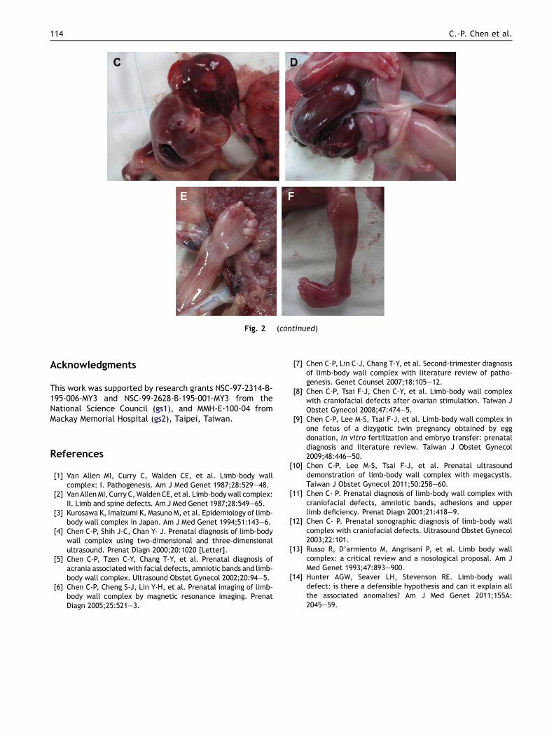
Fig. 2 (continued)
114 C.-P. Chen et al.
Acknowledgments
This work was supported by research grants NSC-97-2314-B-195-006-MY3 and NSC-99-2628-B-195-001-MY3 from theNational Science Council (gs1), and MMH-E-100-04 fromMackay Memorial Hospital (gs2), Taipei, Taiwan.
References
[1] Van Allen MI, Curry C, Walden CE, et al. Limb-body wallcomplex: I. Pathogenesis. Am J Med Genet 1987;28:529e48.
[2] VanAllenMI, Curry C,WaldenCE, et al. Limb-bodywall complex:II. Limb and spine defects. Am J Med Genet 1987;28:549e65.
[3] Kurosawa K, Imaizumi K, Masuno M, et al. Epidemiology of limb-body wall complex in Japan. Am J Med Genet 1994;51:143e6.
[4] Chen C-P, Shih J-C, Chan Y- J. Prenatal diagnosis of limb-bodywall complex using two-dimensional and three-dimensionalultrasound. Prenat Diagn 2000;20:1020 [Letter].
[5] Chen C-P, Tzen C-Y, Chang T-Y, et al. Prenatal diagnosis ofacrania associatedwith facial defects, amniotic bands and limb-body wall complex. Ultrasound Obstet Gynecol 2002;20:94e5.
[6] Chen C-P, Cheng S-J, Lin Y-H, et al. Prenatal imaging of limb-body wall complex by magnetic resonance imaging. PrenatDiagn 2005;25:521e3.
[7] Chen C-P, Lin C-J, Chang T-Y, et al. Second-trimester diagnosisof limb-body wall complex with literature review of patho-genesis. Genet Counsel 2007;18:105e12.
[8] Chen C-P, Tsai F-J, Chen C-Y, et al. Limb-body wall complexwith craniofacial defects after ovarian stimulation. Taiwan JObstet Gynecol 2008;47:474e5.
[9] Chen C-P, Lee M-S, Tsai F-J, et al. Limb-body wall complex inone fetus of a dizygotic twin pregnancy obtained by eggdonation, in vitro fertilization and embryo transfer: prenataldiagnosis and literature review. Taiwan J Obstet Gynecol2009;48:446e50.
[10] Chen C-P, Lee M-S, Tsai F-J, et al. Prenatal ultrasounddemonstration of limb-body wall complex with megacystis.Taiwan J Obstet Gynecol 2011;50:258e60.
[11] Chen C- P. Prenatal diagnosis of limb-body wall complex withcraniofacial defects, amniotic bands, adhesions and upperlimb deficiency. Prenat Diagn 2001;21:418e9.
[12] Chen C- P. Prenatal sonographic diagnosis of limb-body wallcomplex with craniofacial defects. Ultrasound Obstet Gynecol2003;22:101.
[13] Russo R, D’armiento M, Angrisani P, et al. Limb body wallcomplex: a critical review and a nosological proposal. Am JMed Genet 1993;47:893e900.
[14] Hunter AGW, Seaver LH, Stevenson RE. Limb-body walldefect: is there a defensible hypothesis and can it explain allthe associated anomalies? Am J Med Genet 2011;155A:2045e59.