neurofisiopatologia del movimento -...
TRANSCRIPT
Neurofisiopatologia del movimento
Aspetti clinici e terapeutici
Ugo DimanicoNeurofisiologia Riabiliativa - Osp Fossano - ASL CN1 - Cuneo
• cenni anatomici
• vie motorie
• spasticità - UMS
• clinica
• stroke
• PCI
• lesione midollare
• SLA
• Gait Analysis

cenni anatomici

SISTEMA NERVOSO CENTRALE - SNC
Encefalo
emisferi
tronco cerebralemesencefalopontebulbo
Midollo spinale

SISTEMA NERVOSO PERIFERICO
12 nervi craniciorigine dal tronco cerebrale
33 nervi spinaliorigine dal midollo spinale

Gli emisferi cerebrali sono divisi in lobi
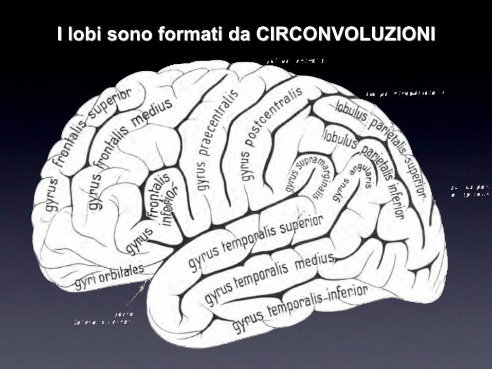
I lobi sono formati da CIRCONVOLUZIONI

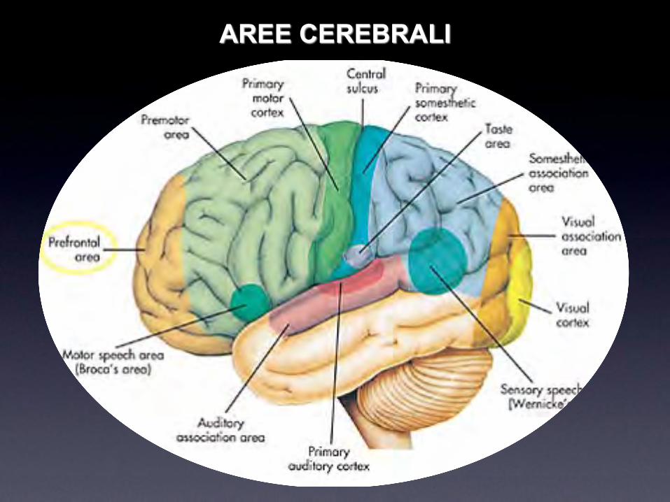
AREE CEREBRALI
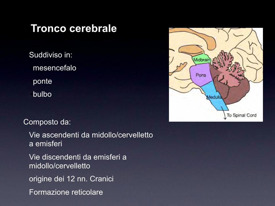
Tronco cerebrale
Suddiviso in:
•mesencefalo
•ponte
•bulbo
Composto da:
Vie ascendenti da midollo/cervelletto a emisferi
Vie discendenti da emisferi a midollo/cervelletto
origine dei 12 nn. Cranici
Formazione reticolare

Cervelletto
Situato posteriormente
al tronco
Comprende:
corteccia
sostanza bianca
nuclei
Connesso con:
emisferi
midollo
orecchio interno (vestibolo)
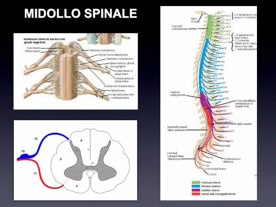
MIDOLLO SPINALE

Neurone - assone - mielina

Emisferi: sostanza grigia e bianca

Emisferi: sostanza grigia e bianca
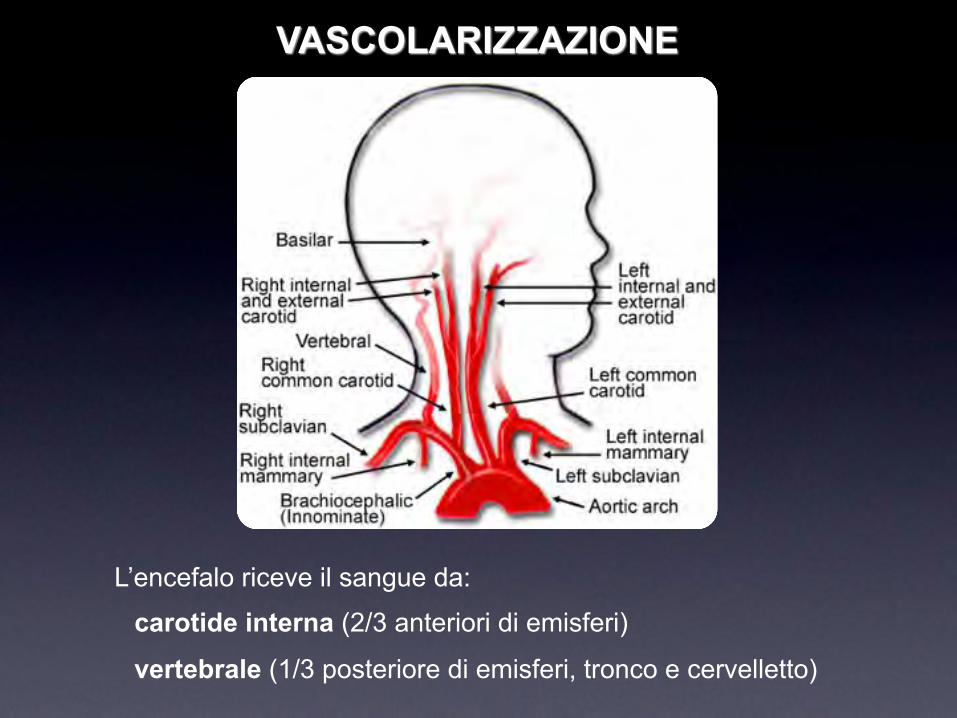
VASCOLARIZZAZIONE
L’encefalo riceve il sangue da:
carotide interna (2/3 anteriori di emisferi)
vertebrale (1/3 posteriore di emisferi, tronco e cervelletto)


Poligono del Willis
Alla base dell’encefalo il
poligono di Willis
“redistribuisce” il sangue
I rami terminali sono:
arteria cerebrale anteriore
arteria cerebrale mediaarteria cerebrale posteriore
vie motorie

corteccia motoria cervellettonuclei della base
Midollo spinale(corna anteriori)
muscolo
Vie Motorie
I motoneurone
II motoneurone

MOTILITA’
riflessa avviene senza il controllo della volontà
automatica avviene con il parziale controllo della volontà
volontaria avviene con il completo controllo della volontà
I tre tipi di M. richiedono livelli di organizzazione progressivamente superiori:
M. riflessa: midollo spinale
M. automatica: strutture sottocorticali
M. volontaria: corteccia cerebrale
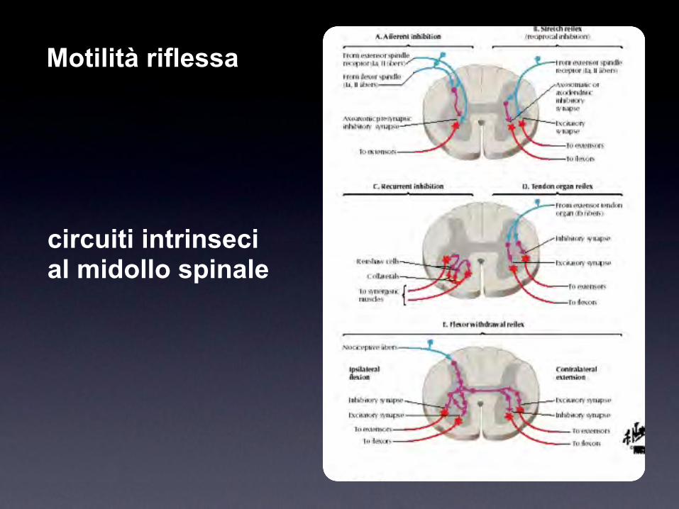
circuiti intrinseci al midollo spinale
Motilità riflessa

Sintomo: assenza/riduzione di coordinazione
atassia
dismetria
disartria
Lesione sistema di coordinazione cerebellare

Sintomo: difficoltà nel controllo e nello svolgimento del movimento
bradicinesiaacinesiadiscinesietremore
Lesione sistema extrapiramidale

Paresi centrale Paresi periferica
Ipertonica Ipotonica
Iperreflessica Iporeflessica
Estensione plantare Flessione plantare
Ipotrofica
Lesione sistema piramidale
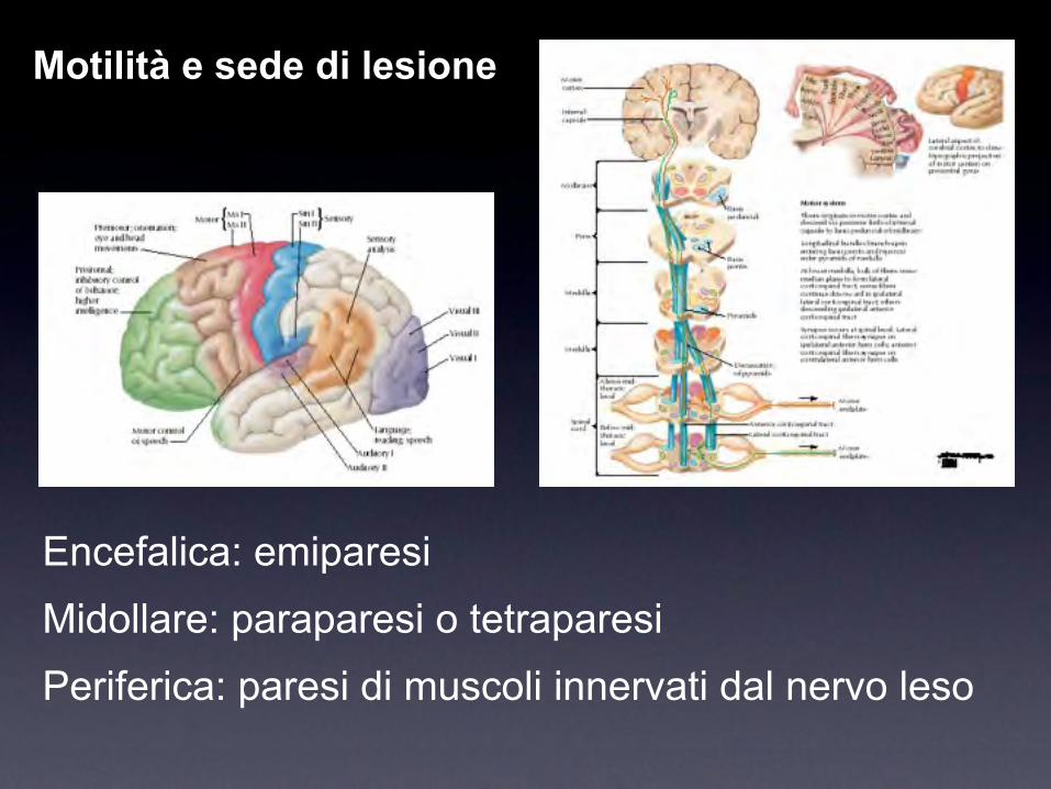
Encefalica: emiparesiMidollare: paraparesi o tetraparesiPeriferica: paresi di muscoli innervati dal nervo leso
Motilità e sede di lesione
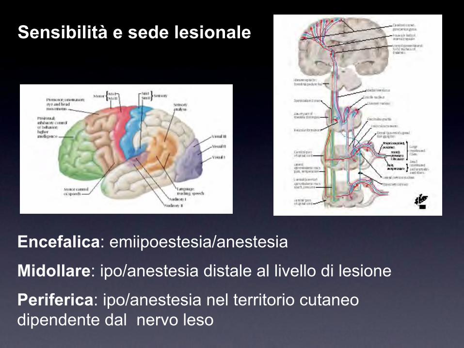
Encefalica: emiipoestesia/anestesia
Midollare: ipo/anestesia distale al livello di lesione
Periferica: ipo/anestesia nel territorio cutaneo dipendente dal nervo leso
Sensibilità e sede lesionale

la spasticità -UMS

aumento velocita’-dipendente del riflesso tonico da stiramento
(tono muscolare)
con aumento dei riflessi osteo-tendinei per la ipereccitabilita’
del riflesso da stiramento,
parte della sindrome del motoneurone superiore (Lance 1980)
incapacità a rilassare adeguatamente il muscolo, che resta
contratto, opponendosi ai movimenti volontari: “congelamento”
Avvertito dal Paz. come “rigidità”, “pesantezza”, ...
La spasticità - il sintomo
La spasticità - la definizione

30
Sindrome del motoneurone superiore UMS
• tossina botulinica
• chirurgia funzionale
• pompe al baclofen
• definizione programma riabilitativo
Analisi del movimentoruolo in clinica

No meaningful plan can be formulated
without first determining the goals of
treatment including:
patient/caregiver goals
functional goals
www.wemove.org
Setting Goals of Treatment
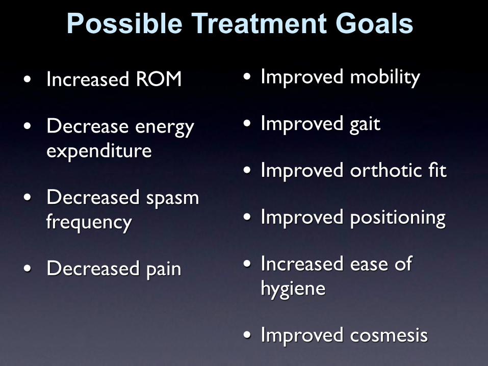
• Increased ROM
• Decrease energy expenditure
• Decreased spasm frequency
• Decreased pain
• Improved mobility
• Improved gait
• Improved orthotic fit
• Improved positioning
• Increased ease of hygiene
• Improved cosmesis
Possible Treatment Goals

34
Equinovarus
equinovalgus
stiff knee
adducted thighs
flexed knee
flexed hip
hyperextension great toe
Pattern nella UMS

35
Common Clinical Patterns: Lower Limbs
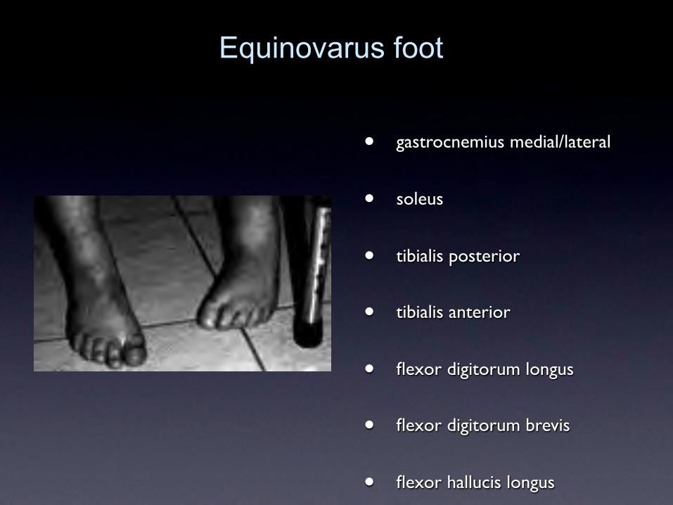
• gastrocnemius medial/lateral
• soleus
• tibialis posterior
• tibialis anterior
• flexor digitorum longus
• flexor digitorum brevis
• flexor hallucis longus
Equinovarus foot

37
• extensor hallucis longus
Striatal toe

38
• adductor magnus
• adductor longus
• adductor brevis
Adducted thighs

39
• medial hamstrings
• lateral hamstrings
• gastrocnemius
Flexed Knee

40
• quadriceps
• retto femore
Extended knee

41
Piede equino - varo - supinato:
TP
in sinergia con
TA
Pattern nella UMS

42
Pattern patologici aa sup.

Gait analysis: esempi clinici

44
Iperattivazione TA sin Post trattamento TA
TA
GM
PL
45
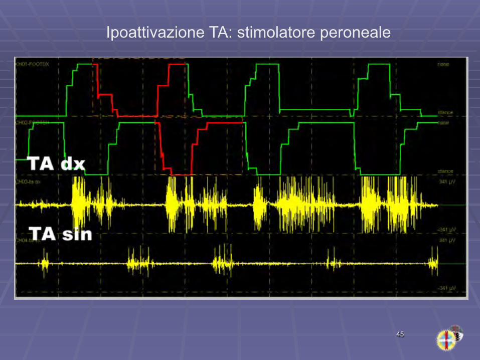
Ipoattivazione TA: stimolatore peroneale

46
Attivazione tonica TA e TP riposo
TA
TA
TP
SOL
SOL

47
Ipoattivazione lato paretico

48
Emi destra: Iperattività loggia posteriore gamba

49
Baclofen IT: 30 microgr 50 microgr
Paraparesi spastica

50
Clono nella fase di stance

51
Emiparesi dx: attivazione tonica TA ipoattivazione soleo (retrazione)
dx
sin
ta
sol
sol
ta

• Neurologist
• Physiatrist
• Neurosurgeon; orthopedic surgeon
• PT and OT
• Family and other caregivers
• Coordinator/administrator
• Wheelchair clinic, gait lab, orthotics clinic, counseling, social work
The Spasticity Management Team

Ictus

ICTUS Cerebrale
• abolizione improvvisa di alcune funzioni cerebrali, di origine vascolare, caratterizzato di solito da paralisi ed emiplegia
• III causa di morte
• 70 % ischemico: rammollimento
• 12% emorragia
• III causa di morte - I causa disabilità

tipi di stroke
• mettere stroke diagram
emorragico ischemico

RAMMOLLIMENTO CEREBRALE (INFARTO)

Metabolismo ossidativo Glucosio (ossigeno)
• Encefalo: – 2% peso ~ 25% del consumo globale 02– 50 cc sangue/100 grammi tessuto/minuto
• 8-10" pdc• 4-8' lesioni irreversibili
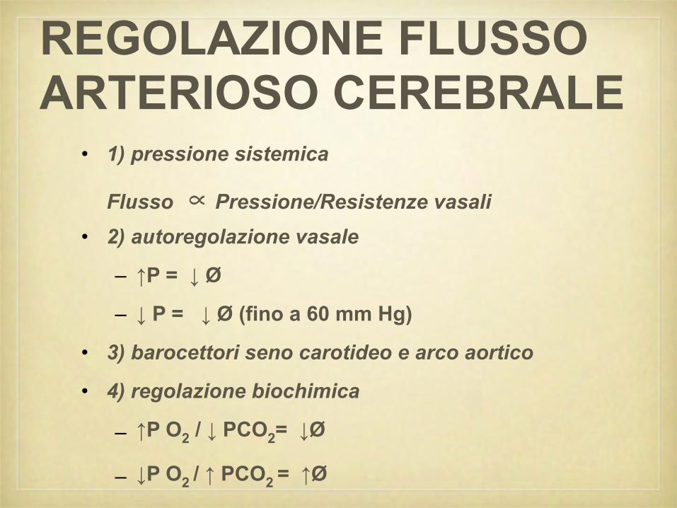
REGOLAZIONE FLUSSO ARTERIOSO CEREBRALE
• 1) pressione sistemica
Flusso ∝ Pressione/Resistenze vasali• 2) autoregolazione vasale
– ↑P = ↓ Ø
– ↓ P = ↓ Ø (fino a 60 mm Hg)
• 3) barocettori seno carotideo e arco aortico
• 4) regolazione biochimica
– ↑P O2 / ↓ PCO2= ↓Ø
– ↓P O2 / ↑ PCO2 = ↑Ø

Autoregolazione vasale
• mettere autoreg vasale

ARTERIOSCLEROSI• aumento resistenze
vascolari
• perdita autoregolazione vasale (rigidità. vasale)
• rigidità barocettori (sbalzi pressori)
• placche fonte di emboli
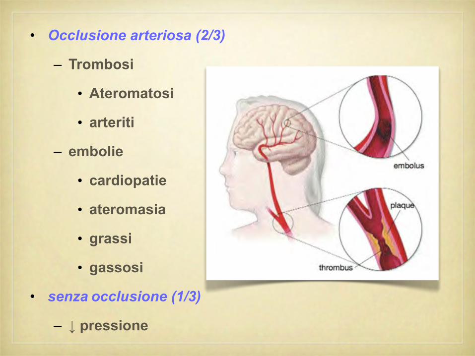
• Occlusione arteriosa (2/3)
– Trombosi
• Ateromatosi
• arteriti
– embolie
• cardiopatie
• ateromasia
• grassi
• gassosi
• senza occlusione (1/3)
– ↓ pressione
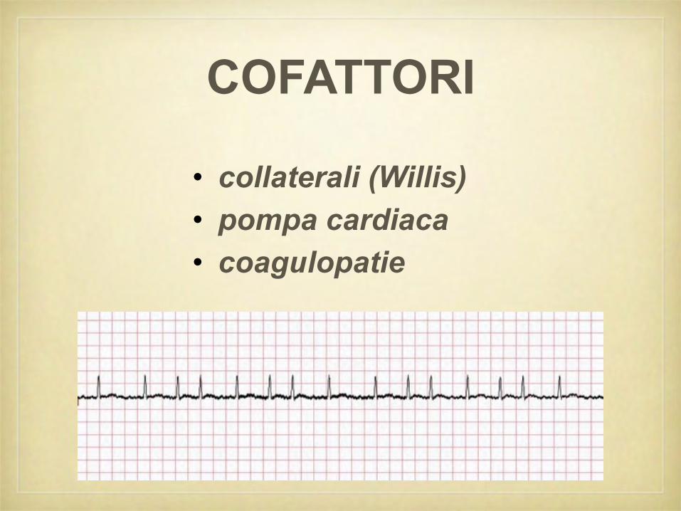
COFATTORI
• collaterali (Willis)• pompa cardiaca• coagulopatie

• regressione in 24 ore
• possibile recidiva e/o evoluzione in stroke
• eziologia
– emboligena (da placche al collo e lisi successiva)
– emodinamica (cofattori)
– spasmo (crisi ipertensiva)
TIAattacco ischemico transitorio, focale

angiografia cerebrale

Placche aterosclerosi (grossi vasi del collo)
• Omogenee
• disomogenee (emboli)


EMORRAGIA CEREBRALE
• capsulare (a sede tipica)
• emorragia cerebro-meningea
• emorragie circoscritte (cervelletto, tronco, talamo)

EMORRAGIA CEREBRALE CAPSULARE
• esordio
– fattori scatenanti (ipertensione, sforzi, sole)
– esordio acuto con pdc → COMA
– talora prodromi: cefalea, vomito, obnubilamento
• alterazioni neurovegetative
– respiratorie
– cardiovascolari temperatura
– trofiche cutanee (anche precoci)
• quadro neurologico
– coma
– emiplegia ("fuma la pipa")
– irrequietudine motoria
– ipertonia generalizzata (decerebrazione)

RAMMOLLIMENTO EMORRAGIA
fattori predisponesti fattori scatenanti
preceduto da TIA
ingravescenza progressiva esordio acuto
non pdc coma
ipertono precoce
negativismo motorio
≠ neurovegetative

Sindromi cerebrali

• carotide interna – comune • cerebrale media
– emiplegia fbc – superficiale
– emianestesia • anteriore
– afasia globale – emiplegia f.b.
– agnosia – afasia motoria Brocà
• cerebrale anteriore • posteriore
– monoplegia crurale – emiaestesia
– ≠ psichiche / sfinteriche – afasia sensitiva Wernicke
– ≠ neurovegetative – astereognosia
• cerebrale posteriore – profondo
– art. occipitale – emianopsia lat. omonima
– art. talamo - genicolatas. algica talamica • emianestesia
• vertebro-basilari
• sindr. alterne tronco

Lesioni del MIDOLLLO SPINALE
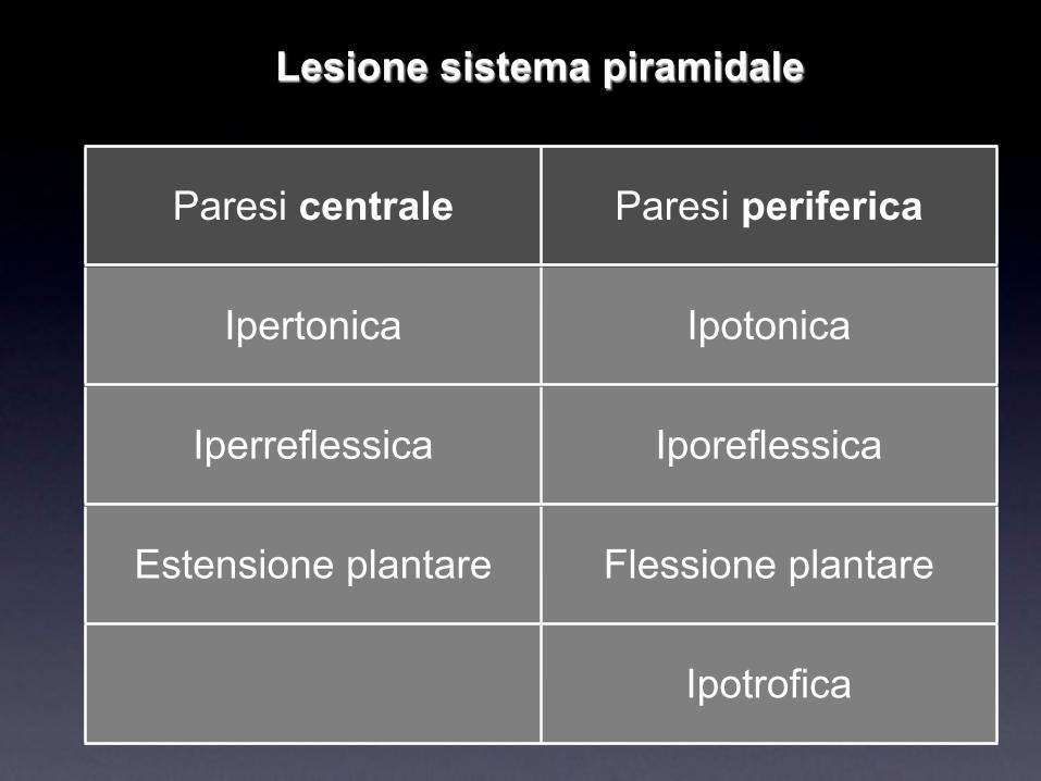
Paresi centrale Paresi periferica
Ipertonica Ipotonica
Iperreflessica Iporeflessica
Estensione plantare Flessione plantare
Ipotrofica
Lesione sistema piramidale

Malattia del MotoneuroneSclerosi laterale amiotrofica - SLA

• most common neurodegenerative disease of the motor neuron system.
• affects motor neurons at 2 or more levels supplying multiple regions of the body.
• lower motor neurons in the anterior horn of the spinal cord and in the brain stem;
• corticospinal upper motor neurons in the precentral gyrus;
• prefrontal motor neurons involved in planning (atrophy).
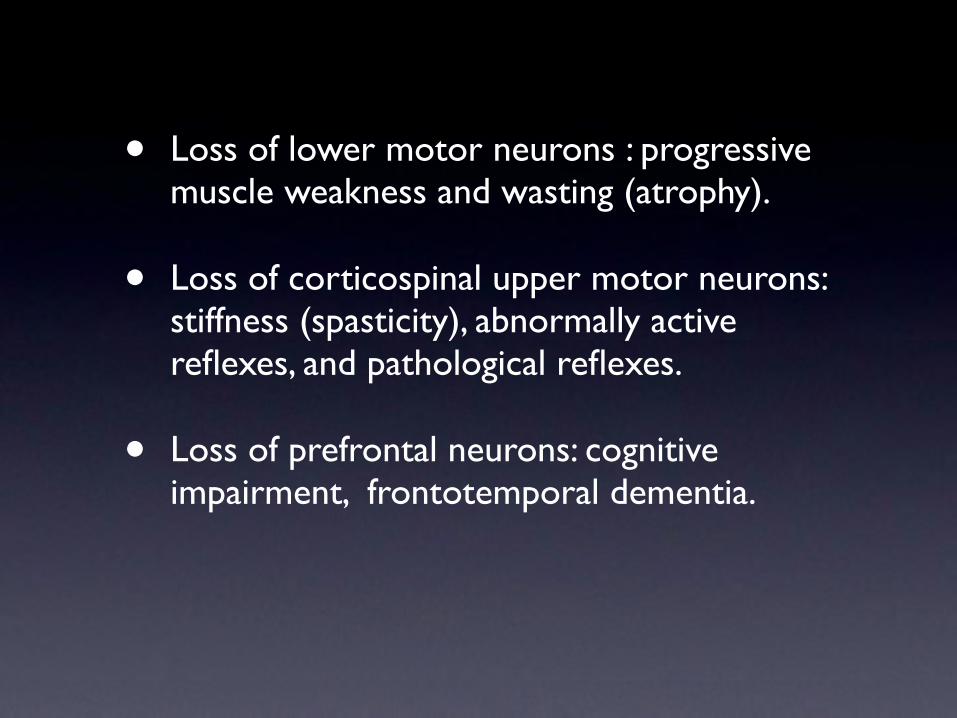
• Loss of lower motor neurons : progressive muscle weakness and wasting (atrophy).
• Loss of corticospinal upper motor neurons: stiffness (spasticity), abnormally active reflexes, and pathological reflexes.
• Loss of prefrontal neurons: cognitive impairment, frontotemporal dementia.

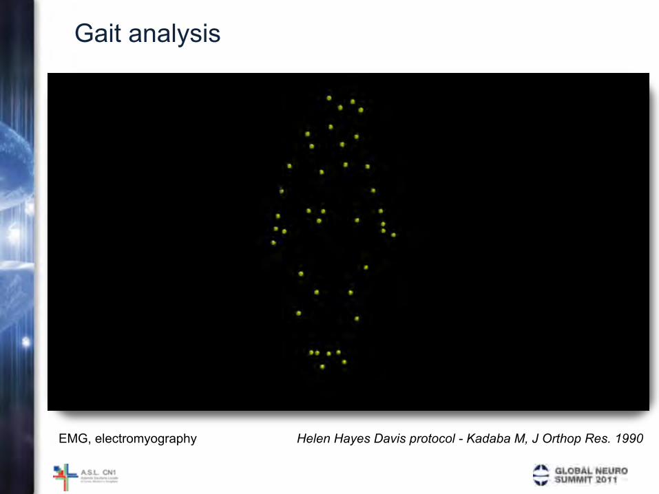
GAIT ANALYSISGeneral remarks

GA as MEASUREMENT TOOL
• GA should be thought as a mesurement tool.
• It provides useful information about the intricacies of the normal gait, as well as about how far the individual’s walking pattern deviates from normal
• However it does not provide a recipe for treatment. That information lies solely within the knowledge base of the investigator who is using the data.
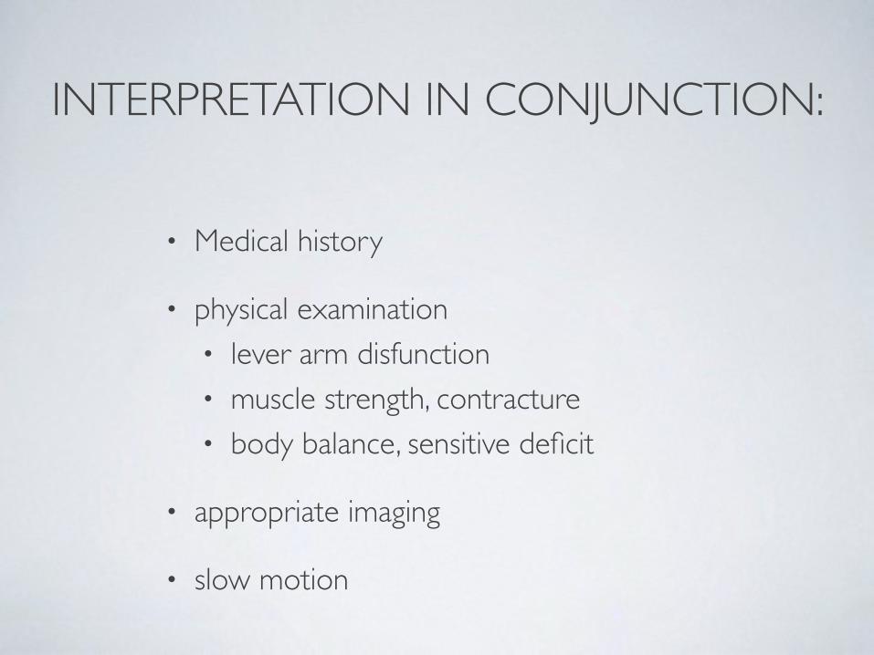
INTERPRETATION IN CONJUNCTION:
• Medical history
• physical examination• lever arm disfunction• muscle strength, contracture• body balance, sensitive deficit
• appropriate imaging
• slow motion

COPING RESPONSE
• Individuals with abnormal cerebral control, muscle contractures and lever-arm dysfunction are forced to introduce other abnormalities into the gait to compensate or “cope”with the problem imposed on them by they condition.
• This coping response maybe sometimes as simple as vaulting on the less involved side to compensate for a drop foot in swing. In severe hemiplegia coping responses may alter gait greatly, furthermore they frequently occur in different planes.
• Sorting this out with gait analysis is difficult, but without gait analysis it is nearly impossible.
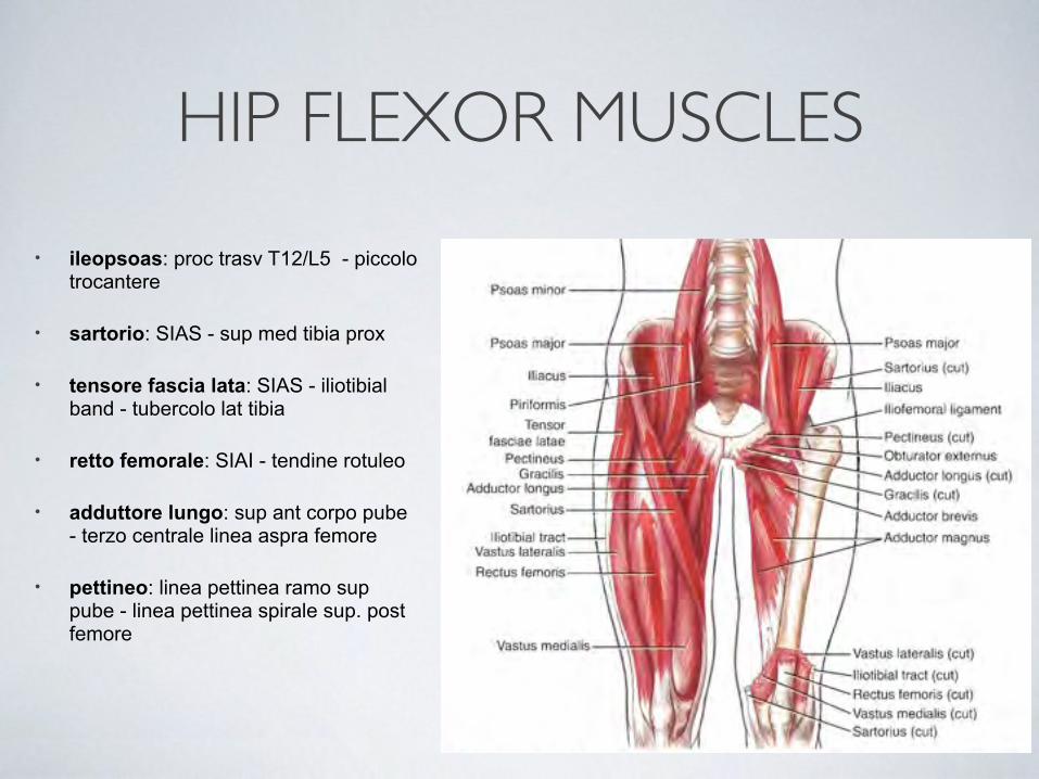
HIP FLEXOR MUSCLES
• ileopsoas: proc trasv T12/L5 - piccolo trocantere
• sartorio: SIAS - sup med tibia prox
• tensore fascia lata: SIAS - iliotibial band - tubercolo lat tibia
• retto femorale: SIAI - tendine rotuleo
• adduttore lungo: sup ant corpo pube - terzo centrale linea aspra femore
• pettineo: linea pettinea ramo sup pube - linea pettinea spirale sup. post femore

HIP ADDUCTORsuperficiale:• pettineo: linea pettinea ramo sup pube - linea pettinea spirale sup. post femore• adduttore lungo: sup ant corpo pube - III centrale linea aspra femore• gracile: corpo/ramo inf pube - sup med prox tibiamedio• adduttore breve: ramo inf pube - III prox linea aspra femoreprofondo• adduttore grande (ant): ramo ischiatico - linea aspra femore (tutta)
MOVIMENTI PURI - ASSOCIATIankle

MOVIMENTI PURI
movimento asse piano del movimento
dorsi / plantiflessione medio - lat sagittale
add / abduzioneverticale
(longit. gamba)orizzontale
pronazione / supinazioneant / post
(longit piede)frontale

MOVIMENTI COMPOSTI
Inversione varismo
adduzione supinazioneplantiflessione
Eversione valgismo
abduzione pronazionedorsiflessione
asse di rotazione obliquo

DORSI - PLANTI FLESSIONE
• asse rotazione
• rif.: 90°
• 15°-25°
• 40°-55°
• acc.: 20°.


Pattern GA

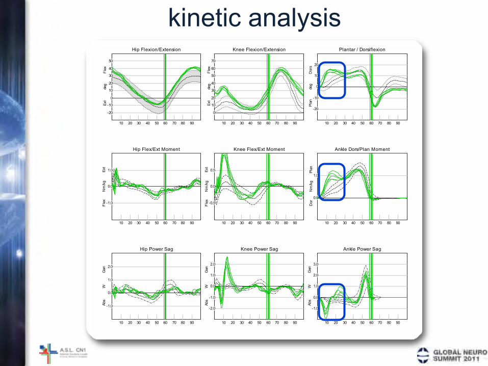
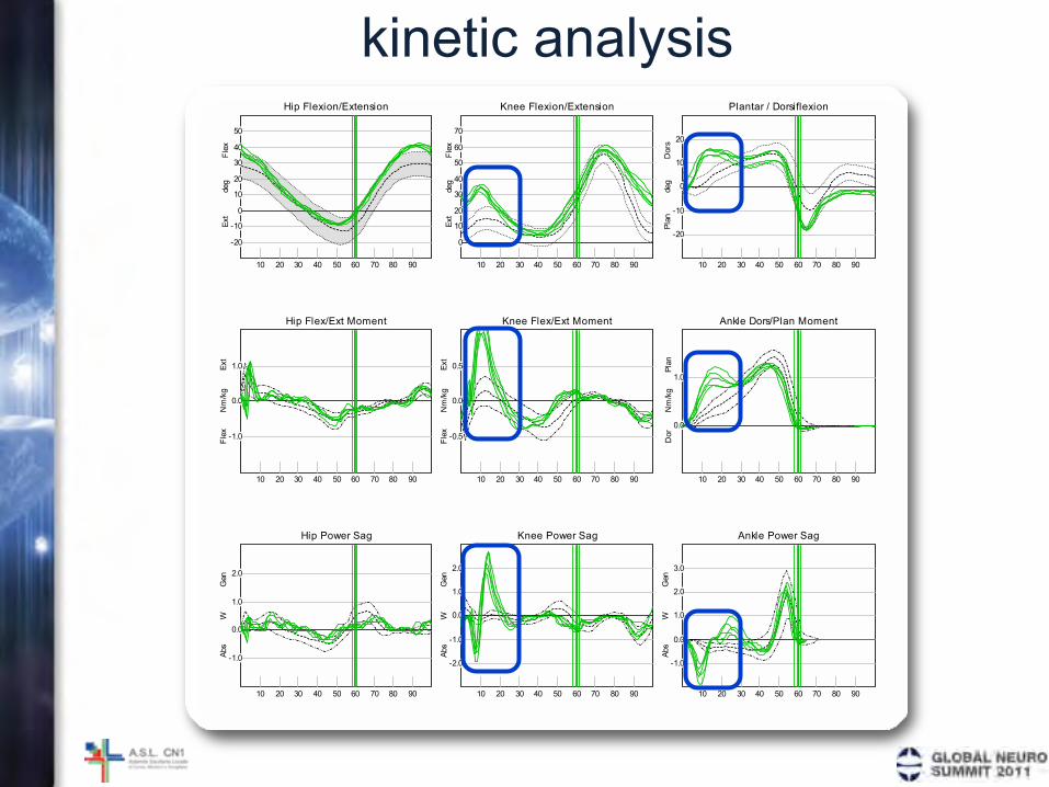
kinetic analysisHip Flexion/Extension
-20
-10
0
10
20
30
40
50
Flex
Ext
deg
10 20 30 40 50 60 70 80 90
Hip Flex/Ext Moment
-1.0
0.0
1.0Ext
Flex
Nm/kg
10 20 30 40 50 60 70 80 90
Hip Power Sag
-1.0
0.0
1.0
2.0
Gen
Abs
W
10 20 30 40 50 60 70 80 90
Knee Flexion/Extension
0
10
20
30
40
50
60
70
Flex
Ext
deg
10 20 30 40 50 60 70 80 90
Knee Flex/Ext Moment
-0.5
0.0
0.5Ext
Flex
Nm/kg
10 20 30 40 50 60 70 80 90
Knee Power Sag
-2.0
-1.0
0.0
1.0
2.0
Gen
Abs
W
10 20 30 40 50 60 70 80 90
Plantar / Dorsiflexion
-20
-10
0
10
20
Dors
Plan
deg
10 20 30 40 50 60 70 80 90
Ankle Dors/Plan Moment
0.0
1.0
Plan
Dor
Nm/kg
10 20 30 40 50 60 70 80 90
Ankle Power Sag
-1.0
0.0
1.0
2.0
3.0
Gen
Abs
W
10 20 30 40 50 60 70 80 90
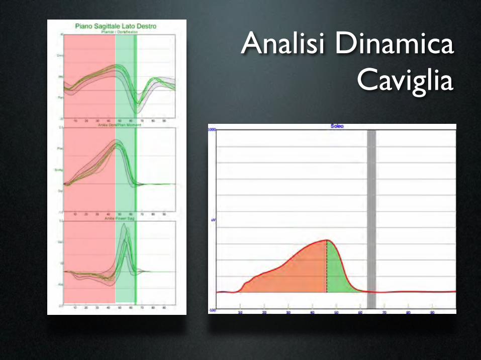
Analisi DinamicaCaviglia

Clinical case 2 - before treatment
• age50
• ischaemic stroke 4/08
• right emiparesis + aphasia

Treatment [UI] 03/31/2010 07/30/2010
rectus femoris
tibialis anterior
soleus
- 50
30 -
80 70
before 3/31/2010 after (US guide) 1/12/2011

Gamba III med
n peroneo prof
a tibiale ant
n tibiale post
a tibiale post
n cut med polpaccio
• tibiale ant
• est lungo dita
• peroneo br
• peroneo lungo
• soleo
• tib post
• gastrocn lat
• gastrocn med
• flex lungo dita
a peronea
n peroneo sup

Clinical case: case history• Age: 28 years
• Cerebral palsy
• Bilateral lengthening of achilleus tendons (3 years)
• Left ankle arthrodesis (10 years)
• Bilateral lengthening of ischiocrural muscles (17 years)

Kinematics
Gait analysis
EMG, electromyography Helen Hayes Davis protocol - Kadaba M, J Orthop Res. 1990

Gait analysis
kinematics
kinetics
dynamic EMG
EMG, electromyography Helen Hayes Davis protocol - Kadaba M, J Orthop Res. 1990
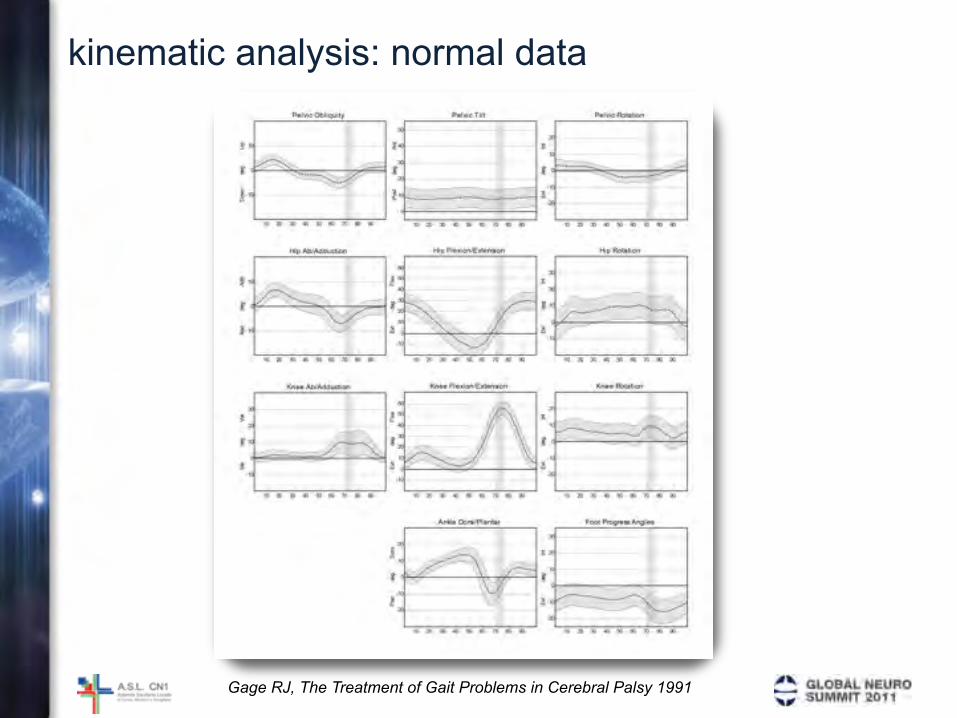
kinematic analysis: normal data
Gage RJ, The Treatment of Gait Problems in Cerebral Palsy 1991

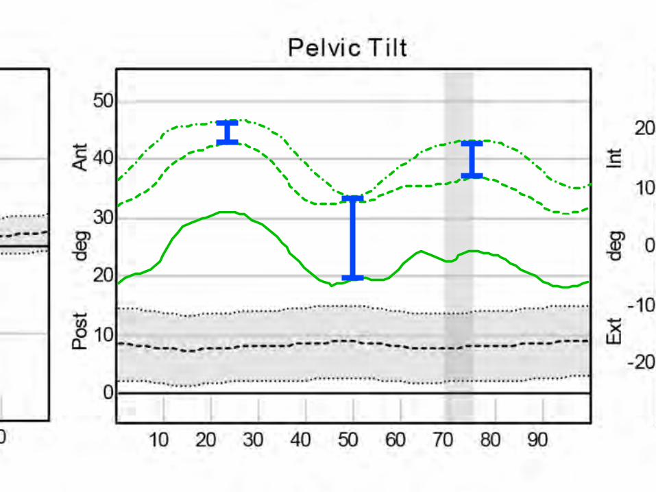
Clinical case: pretreatment kinematic analysis Pelvic Obliquity
-10
0
10Up
Down
deg
10 20 30 40 50 60 70 80 90
Hip Ab/Adduction
-10
0
10Add
Abd
deg
10 20 30 40 50 60 70 80 90
Knee Ab/Adduction
-10
0
10
20
30
Var
Val
deg
10 20 30 40 50 60 70 80 90
Pelvic Tilt
0
10
20
30
40
50
Ant
Post
deg
10 20 30 40 50 60 70 80 90
Hip Flexion/Extension
-10
0
10
20
30
40
50
60
Flex
Ext
deg
10 20 30 40 50 60 70 80 90
Knee Flexion/Extension
-10
0
10
20
30
40
50
60
Flex
Ext
deg
10 20 30 40 50 60 70 80 90
Ankle Dorsi/Plantar
-20
-10
0
10
20
Dors
Plan
deg
10 20 30 40 50 60 70 80 90
Pelvic Rotation
-20
-10
0
10
20
Int
Ext
deg
10 20 30 40 50 60 70 80 90
Hip Rotation
-10
0
10
20
30
Int
Ext
deg
10 20 30 40 50 60 70 80 90
Knee Rotation
-20
-10
0
10
20
Int
Ext
deg
10 20 30 40 50 60 70 80 90
Foot Progress Angles
-20
-10
0
10
20
30Int
Ext
deg
10 20 30 40 50 60 70 80 90
Right
Left

90
Pelvic Tilt
0
10
20
30
40
50
Ant
Post
deg
10 20 30 40 50 60 70 80 90
-20
-10
0
10
20
Int
Ext
deg
Gage RJ, The identification and treatment of gait problems in cerebral palsy 2009

90
Hip Flexion/Extension
-10
0
10
20
30
40
50
60
Flex
Ext
deg
10 20 30 40 50 60 70 80 90
-10
0
10
20
30
Int
Ext
deg
1
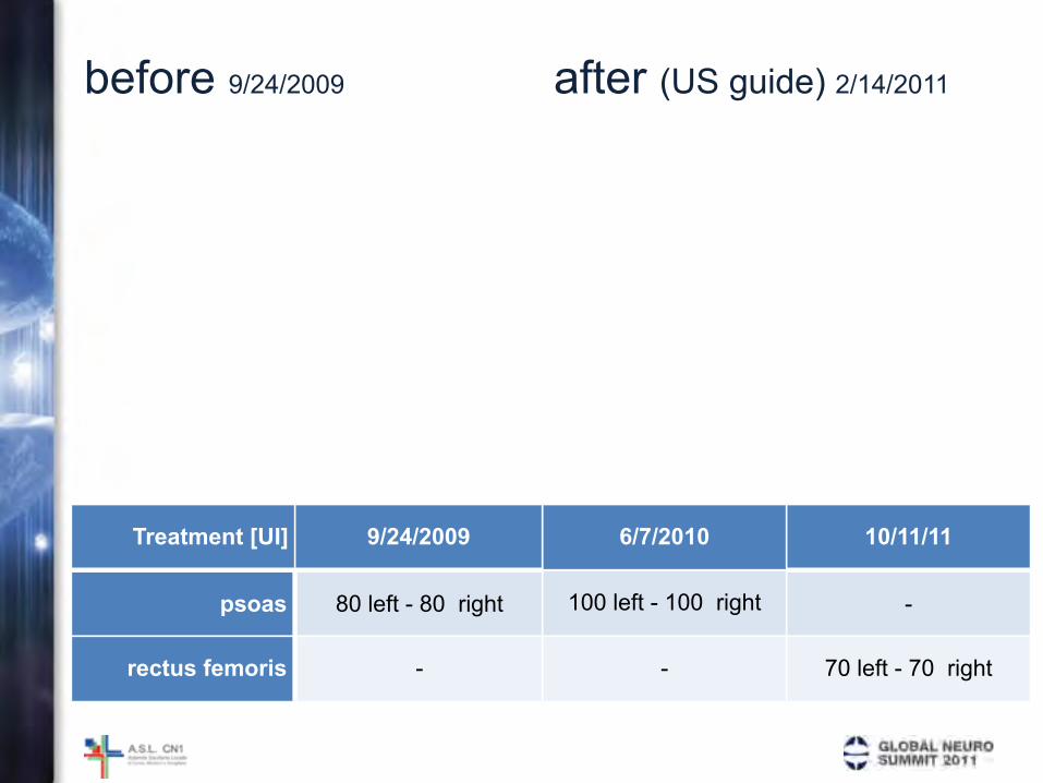
before 9/24/2009 after (US guide) 2/14/2011
Treatment [UI] 9/24/2009 6/7/2010 10/11/11
psoas
rectus femoris
80 left - 80 right 100 left - 100 right -
- - 70 left - 70 right
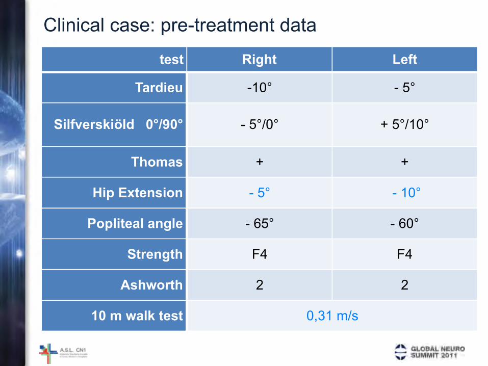
Clinical case: pre-treatment data
test Right Left
Tardieu
Silfverskiöld 0°/90°
Thomas
Hip Extension
Popliteal angle
Strength
Ashworth
10 m walk test
-10° - 5°
- 5°/0° + 5°/10°
+ +
- 5° - 10°
- 65° - 60°
F4 F4
2 2
0,31 m/s0,31 m/s

Clinical case: post-treatment data
test Right Left
Tardieu
Silfverskiöld 0°/90°
Thomas
Hip Extension
Popliteal angle
Strength
Ashworth
10 m walk test
-10° - 5°
- 5°/0° + 5°/10°
+ +
5° 0°
- 65° - 60°
F4 F4
2 2
0,52 m/s0,52 m/s

24 Sep 2009
18 Jan 2010
7 Jun 2010
11 Oct 2010
14 Feb 2011-40%
-20%
0%
20%
40%
60%
80%
Walking Speed Stride Length Cadence Single Support
Comparison of spatio-temporal parameters

kinematic analysis: normal data


Iliopsoas muscle
kidney
psoas
vertebra
Ward AB, Eur J of Neurol 1999 - Kirchmair L et al. Anesth Analg 2001