molecular pathogenesis of prion...
TRANSCRIPT
5
Molecular Pathogenesis of Prion Diseases
Giuseppe Legname1 and Gianluigi Zanusso2
1Scuola Internazionale Superiore di Studi Avanzati, 2Università degli Studi di Verona,
Italy
1. Introduction
Prion diseases or transmissible spongiform encephalopathies (TSEs) are rare, fatal and
incurable neurodegenerative disorders of humans and animals (Prusiner, 1998).
In humans, prion diseases occur with unique aetiology as sporadic, genetic or infectious
disorders. Sporadic cases of prion diseases, which account for the majority of casualties (up
to 85% of all cases), are of unknown origin; the genetic forms are less frequent (up to 15%),
while the infectious cases are extremely rare with an incidence of less than 1% (Prusiner,
2001). Creutzfeldt-Jakob disease (CJD), Gerstmann-Sträussler-Scheinker (GSS) syndrome,
Fatal Familial Insomnia (FFI) are examples of human prion diseases. In animals the disease
is mostly infectious and the mode of transmission is horizontal. Prion diseases include
scrapie in sheep and goats, bovine spongiform encephalopathy (BSE) in cattle, and chronic
wasting disease of deer, elk, and moose (Williams, 2005).
The agents responsible for prion diseases are infectious proteins named prions. The term
‘prion’ was coined when Stanley B. Prusiner introduced the concept of proteinaceous
infectious particles (Prusiner, 1982). Since the introduction of this once heretical notion,
mounting evidence has strengthened its validity.
In the next sections of this chapter we present and discuss the peculiar complexity of the
molecular pathogenesis of prion diseases in humans and animals.
2. Prion protein and prions
2.1 The prion protein
The prion protein (PrP) is one of the most and best-studied models for misfolding diseases.
The cellular form of PrP (PrPC) is a glysosyl-phosphatidylinositol (GPI) anchored
polypeptide present on the outer leaflet of the cellular membrane of most cell types in
mammals. In humans, the PRNP gene, located in the short arm of chromosome 20 (Liao et
al., 1986), features two exons. The second exon contains the entire open reading frame
(ORF), which encodes for the protein. The PrPC is composed of 253 amino acids in humans,
including 22 amino acids of endoplasmic reticulum signal sequence at the N-terminus and
23 amino acids as GPI anchoring signal at the C-terminus (Stahl et al., 1990). The N-terminal
region of PrPC encompasses five characteristic amino acid octarepeats that coordinate
www.intechopen.com

Miscellanea on Encephalopathies
96
copper and, to a lesser extent, other metal ions. The mature 208-residues protein possesses a
single disulphide bridge between Cys179 and Cys214, and two sites for Asn-linked
glycosylation within the carboxy-terminal region at position Asn181 and Asn197.
The protein is the first system where a polypeptide has been shown to exist in at least two significantly different conformations, associated with radically different functions.
The physiological function of PrPC has not been established with certainty yet; nevertheless its evolutionarily conserved sequence suggests that it might play an important role in neuronal development and physiology. Indeed one recent finding indicates a possible involvement of this protein in neuronal differentiation and polarization (Kanaani et al., 2005). On account of additional evidence, it could also contribute to myelin formation and maintenance (Benvegnu et al., 2011a).
A strategy often employed to identify protein function is the development of transgenic mouse lines with a disabled gene. Many lines of knockout (KO) mice have been developed for PrP (Weissmann and Flechsig, 2003). In these models, typically either the entire ORF of exon 3 of Prnp (in mice), or the ORF as well as flanking sequences are deleted (Weissmann and Flechsig, 2003). The Prnp KO mice (Prnp0/0) appear to develop and reproduce normally (Bueler et al., 1992), but their further evaluation found several abnormalities. The mice appear clinically asymptomatic yet they develop peripheral nerve demyelination, have increased susceptibility to ischemic brain injury, altered sleep and circadian rhythm, altered hippocampal neuropathology and physiology, including deficits in hippocampal-dependent spatial learning and hippocampal synaptic plasticity (Tobler et al., 1997, Nishida et al., 1999, Spudich et al., 2005, Criado et al., 2005). Mice with Prnp0/0 are also more susceptible to oxidative stress, and PrPC appears to play a neuroprotective role in cellular response to hypoxic-ischemic injury (Weise et al., 2006). Some Prnp0/0 mouse lines in which the deletion extends beyond the ORF, although developing normally, acquire ataxia and Purkinje cell loss later in life (Moore et al., 1999).
Recent findings show development regional differences of the expression of PrP in mouse central nervous system (CNS), with specific white matter structures showing the earliest and highest expression of PrPC. Indeed, all these regions are part of the thalamo-limbic neurocircuitry, hence suggesting a potential role of PrPC in the development and functioning of this specific brain system (Benvegnu et al., 2010).
Furthermore, the transcriptome during development for the CNS of mice lacking a functional Prnp gene has recently been compared with that of wild-type animals (Benvegnu et al., 2011b). To assess the influence of PrPC on gene expression profile in the mouse brain, a microarray analysis was undertaken using RNA isolated from the hippocampus at two different developmental stages: newborn (4.5-day-old) and adult (3-month-old) mice, both from wild-type and Prnp KO animals. Based on the comparison of these datasets, commonly co-regulated genes and uniquely de-regulated genes during postnatal development were identified. The absence of PrPC affected several biological pathways, the most representative ones being cell signaling, cell-cell communication and transduction processes, calcium homeostasis, nervous system development, and synaptic transmission and cell adhesion. However, there was only a moderate alteration of the gene expression profile in our animal models. PrPC deficiency did not lead to a dramatic alteration of gene expression profile, and produced moderately altered gene expression levels from young to adult animals. Hence,
www.intechopen.com

Molecular Pathogenesis of Prion Diseases
97
these results further support silencing endogenous PrPC as therapeutic approach to prion diseases (Benvegnu et al., 2011b).
Concerning PrPC cellular function, experiments have recently shown that PrPC regulates the cleavage of neuregulin-1 proteins (NRG1). Neuregulins provide key axonal signals, which regulate processes, including glial cells proliferation, survival and myelination. Interestingly, Prnp0/0 mice have recently been reported to have a late-onset demyelinating disease in the peripheral nervous system (PNS), but not in the CNS (Bremer et al., 2010). The comparison of wild-type and Prnp0/0 mice showed that the NRG1 processing is developmentally regulated in the PNS and influenced by PrPC in old but not in young animals. In addition, it has been found that neuregulin-3 processing — another neuregulin family member — is altered in the PNS of Prnp0/0 mice. These differences in neuregulin proteins processing are not paralleled in the CNS, thus suggesting a different cellular function for PrPC between the CNS and the PNS (Benvegnu et al., 2011a).
2.2 Prions and the biology of the conversion mechanism
Prion diseases are caused by changes in the conformation of the endogenous PrPC, which turns into an alternatively folded, disease-causing form called the prion, or PrPSc. The
normal PrPC contains three -helixes and two short -sheet structures in its globular
domain, whereas PrPSc contains fewer –helical and mostly -sheet structures (Prusiner, 1998). PrPC and PrPSc possess the same primary polypeptide sequence, but different secondary and tertiary structures. PrPSc is produced by the conversion of existing PrPC into PrPSc.
The process leading to PrPSc production from PrPC is not completely understood. It is believed that this occurs when PrPC comes into contact with PrPSc and is thus induced to take on the shape of PrPSc (Prusiner, 1998). The fact that mice devoid of PrPC are resistant to infection, as they are unable to replicate prions, provides strong evidence that PrPC is necessary for prion disease (Bueler et al., 1993). Although it is clear that PrPC is necessary for prion disease, it is still debated whether other proteins or molecules are involved in the conformational change in vivo (Telling et al., 1995, Deleault et al., 2003). The conversion of monomeric PrPC into insoluble, protease-resistant PrPSc is a process that seems to occur in structures denominated caveolae-like domains (CLDs) (Gorodinsky and Harris, 1995), and the resulting PrPSc subsequently traffics to other membranous compartments such as endosomes and lysosomes (Marijanovic et al., 2009). The membranes of CLDs seem to be composed of cholesterol-rich rafts and presumably provide the cellular environment for the formation of PrPSc. Two conversion and replication models have been proposed: (i) a nucleation-polymerization reaction, and (ii) a template-assisted conversion process. In the first model, the rate-limiting step is the formation of a critical amount of PrPSc to form a seed for the polymerization of PrPSc. In the template-assisted model, PrPC must first undergo conversion toward a transition state that presumably corresponds to a partially destabilized structure (Aguzzi and Calella, 2009). The structural transition could be mediated by an auxiliary molecule, which facilitates the conversion to a nascent prion (Telling et al., 1995, Deleault et al., 2003). In disease-affected brain homogenates, limited proteolysis completely hydrolyzes PrPC and produces a protease-resistant PrPSc molecule of about 140 amino acids, designated PrP27–30. In the presence of detergent, PrP27–30 polymerizes into amyloid (McKinley et al., 1991). Prion amyloids, or rods, formed by limited proteolysis and detergent
www.intechopen.com
Miscellanea on Encephalopathies
98
extraction, are indistinguishable from the filaments that aggregate to form PrP amyloid plaques in the CNS, exhibiting similar ultrastructural morphology and tinctorial characteristics after staining with Congo red dye (Prusiner et al., 1983). So far, little is known about the structure of prions. Several models have been proposed, attempting to satisfy all available biophysical, biochemical and immunochemical data on infectious prions (Govaerts et al., 2004). The discovery that recombinant PrP, expressed in Escherichia coli, is infectious to mice when polymerized into amyloid fibrils has opened new avenues for research in the prion field (Legname et al., 2004). Characterization of these synthetic prions revealed novel distinctiveness associated with neuropathological changes in mouse models of prion disease (Legname et al., 2005). The conformational changes acquired by the synthetic prions confer increasing stability to PrPSc, as measured by the amount of chaotropic agents necessary to completely unfold PrPSc. Moreover, a linear correlation is established when the measure of stability of any particular isolate is expressed as a function of mouse survival times to the disease (Legname et al., 2006).
One of the strongest arguments for the existence of prions is the link between inherited prion diseases and mutations in the PRNP gene. Currently, almost 60 pathogenic mutations and several polymorphisms have been identified in the PRNP gene (Kovacs et al., 2002). They include missense point mutations, mostly located in the globular part, insertion or deletion mutations involving the N-terminal domain, and non-sense mutations resulting in the premature termination of PrP synthesis. Twelve polymorphisms are silent, while four of them alter the amino acid sequence. The most important one that markedly influences the disease is the M/V polymorphism at codon 129 (Collinge, 2001). The M/V polymorphism at position 129 is common; the homozygous M/M and V/V and the heterozygous M/V subjects account for 43%, 8% and 49%, respectively, in the Caucasian population (Zimmermann et al., 1999). This polymorphism is a key determinant of genetic susceptibility to acquired and sporadic prion diseases, the large majority of which occur in homozygous individuals (Collinge et al., 1991, Palmer et al., 1991, Windl et al., 1996). The PRNP heterozygotes appear to be protected from sporadic CJD (sCJD) compared to the PRNP homozygotes (Kobayashi et al., 2009, Baker et al., 1991, Hsiao et al., 1992). The M/V polymorphism at position 129 affects the disease phenotype when it is located on the mutant allele: D178N-129V causes familial CJD (fCJD), while D178N-129M is responsible for familial FFI. The M/V polymorphism located on the normal allele affects the age onset and duration of the disease. Patients carrying either M or V 129 codon have been observed in all inherited prion diseases. The altered conformation observed in human PrP mutants might lead to a different affinity for extracellular matrix components and cellular membranes and, consequently, to an aberrant localization of PrP in different cellular compartments, favoring formation of altered pathogenic topologies (Hegde et al., 1999). Independent evidence derived from cell culture studies, expressing some of the disease-linked mutants, showed that these mutations may affect folding and maturation of PrPC in the secretory pathway of neuronal cells (Ashok and Hegde, 2009).
How mutations and polymorphisms can structurally modulate the diseases is not clear. In fact, until recently there was no evidence of a pathological point mutation causing substantial structural differences in PrP folding.
To shed new light on the role of pathological point mutations on PrP structure, a high-resolution 3D structure of the truncated recombinant human PrP containing the pathological
www.intechopen.com

Molecular Pathogenesis of Prion Diseases
99
Q212P mutation has recently been determined and examined (Ilc et al., 2010). This mutation is responsible for a GSS syndrome characterized by mild amyloid PrP deposition in patients (Piccardo et al., 1998, Young et al., 1998). The high-resolution NMR structure of Q212P mutant revealed unique conformational features compared to the known structures of either human or other mammalian PrPC (Christen et al., 2009, Christen et al., 2008, Gossert et al., 2005, Lopez Garcia et al., 2000, Riek et al., 1996).
The most remarkable differences involved the C-terminal end of the protein and the β2–α2 loop region. The Q212P mutant is the first known example of PrP structure where the α3 helix between E200 and Y226 is broken into two helices. This breakage brings about dramatic changes in the hydrophobic interactions between the α3 helix and the β2–α2 loop region. In the wild-type protein, long-range interactions between Y225 and M166 define the position of the β2–α2 loop and thus the tertiary structure of the protein. In this protein type, the solvent-exposed surface of the β2–α2 loop and the α3 helix region is smaller, and Y169 is buried inside the hydrophobic cluster (Ilc et al., 2010).
When these structural findings are compared with the already resolved NMR structures of
human PrP, carrying respectively the CJD-related E200K (Zhang et al., 2000) and the
artificial R220K mutation, the α3 helix appears well ordered up to the point mutation
(Calzolai et al., 2000). After this mutation, the α3 helix shows increased flexibility and
significantly less order. At the same time, the R220K mutation does not alter the
hydrophobic interactions between the aromatic residues of the β2–α2 loop and the α3 helix.
Special interest in prion biology is therefore focused on the epitope formed by the β2–α2 loop
and the α3 helix, as this surface has been implicated in interactions with a hypothetical
facilitator of prion conversion involved in the development of TSEs (Kaneko et al., 1997,
Telling et al., 1995). Therefore, the plasticity of the loop may modulate the susceptibility to
prion disease of a given species. While in PrPC from most mammalian species this loop is
flexible, it is well defined in PrPC of elk (Gossert et al., 2005), bank vole (Clethrionomys
glareolus) (Christen et al., 2008), tammar wallaby (Christen et al., 2009) and, as found out
very recently, horse (Perez et al., 2010) and rabbit (Wen et al., 2010). Interestingly, elk and
bank vole are highly susceptible to TSEs, whereas there have been no cases of prion diseases
either in marsupials, horses or rabbits. The structure-function relationship suggested by
these works may provide the molecular basis for understanding the generation of PrPSc in
inherited prion diseases. In fact, the characterization of high-resolution structures of PrP
pathological mutants and their comparison with the wild-type overall folding, highlights
important regions in these proteins that could be involved in early events of PrP misfolding.
This may also provide a molecular explanation for prion formation in the sporadic forms of
prion disease.
3. Molecular pathogenesis of prion diseases
3.1 PrPSc
conformers in human and animal prion disorders
Human and animal TSEs exist as different prion strains characterized by distinct biological
properties. A prion strain is defined using several criteria, such as incubation time and
lesion profile after transmission, as well as by physico-chemical characteristics of
pathological PrPSc conformers (Bruce et al., 1994, Aguzzi et al., 2008).
www.intechopen.com
Miscellanea on Encephalopathies
100
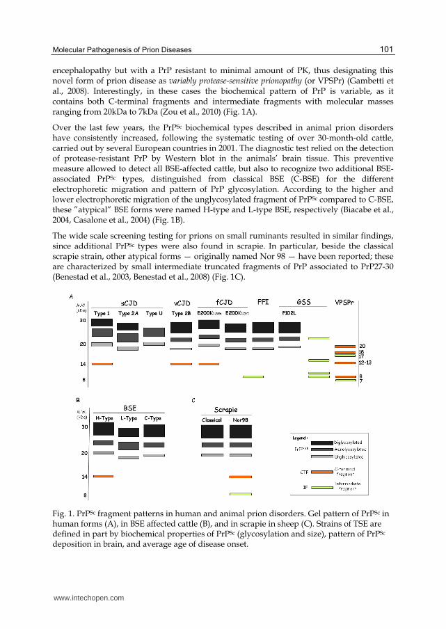
Several studies demonstrated that prion strains can be distinguished based on different biochemical properties of PrPSc, encompassing conformation, glycoform profile, degrees of protease-resistance under different denaturing conditions, thus allowing a molecular strain typing classification of PrPSc (Wadsworth and Collinge, 2011). Treatment of PrPSc with proteinase K (PK) generates a large PK-resistant C-terminal core fragment termed PrP27-30 which is considered the pathogenic and infectious core of PrPSc. Full-length PrPSc and PrP27-30 are associated to the naturally occurring infectious agent causing prion diseases and are thought to be the primary cause of the histological changes in brains of subjects with prion diseases.
In human and animal prion disorders, the remarkable heterogeneity of disease phenotypes is influenced by the combination of either PrPSc type or relevant PRNP polymorphisms (Gambetti et al., 2011).
3.2 The biochemical phenotype of PrPSc
in human and animal TSEs
In human prion disorders, several different types of PrPSc have been recognized. PrPSc types are distinguished based on the electrophoretic migration and the glycosylation profile of PrP27-30. This is composed of a major triplet of bands which represent the differently glycosylated isoforms of PrPSc.
Additional minor C-terminally truncated fragments (CTFs) resistant to proteases have been reported and they contribute to define the biochemical strains of PrPSc. The combination of PrP27-30 and CTFs is representative of specific patterns and correlates to distinct disease phenotypes (Zou et al., 2003, Zanusso et al., 2004).
In sCJD, three distinct PrPSc types have been described: type 1, type 2A and type U (Fig. 1A). Type 1 and type 2 PrPSc are distinguished based on the different electrophoretic mobility of the unglycosylated form of approximately 21kDa in type 1, and 19kDa in type 2, respectively (Parchi et al., 1999, Gambetti et al., 2003). In contrast, type U PrPSc shares apparent gel mobility with type 1, though it lacks the diglycosylated isoforms (Zanusso et al., 2007).
In variant CJD (vCJD), an unglycosylated band migrating at 19kDa and a highly glycosylated-dominant profile characterize PrPSc; wheresas type 2A PrPSc in sCJD is distinguished by the highly glycosylated-dominant profile. Accordingly, the current nomenclature defines type 2A-PrPSc associated to sCJD, and type 2B-PrPSc associated to vCJD, thus identifying BSE agent in humans (Collinge et al. 1996). In familial forms of CJD (E200K-129V) and FFI forms, a type 2B-like pattern is observed (Fig. 1A). The exception to the conventional definition of PrPSc typing is observed in GSS mutations. In GSS, PrP27-30 is absent and pathological PrP is composed of intermediate fragments (IFs) of ~11kDa and ~8kDa spanning residues ~90-150 and ~60-150 (Tagliavini et al., 1991, Tagliavini et al., 1994, Piccardo et al., 1998). According to their sequence, these IFs lack PrP post-translational modifications, including GPI-anchor.
However, in GSS P102L mutation, a hybrid phenotype of PrPSc is observed. In P102L, PrPSc is characterized by the presence of the 8kDa intermediate fragment (PrP8), as in other GSS, but also of PrP27-30 (Parchi et al., 1998) (Fig. 1A).
Although prion diseases are defined based on the presence of a disease-associated protease-resistant PrP that has been proven to retain infectivity, Gambetti et al. shifted this dogmatic definition. They reported on a series of individuals with dementia and spongiform
www.intechopen.com
Molecular Pathogenesis of Prion Diseases
101
encephalopathy but with a PrP resistant to minimal amount of PK, thus designating this novel form of prion disease as variably protease-sensitive prionopathy (or VPSPr) (Gambetti et al., 2008). Interestingly, in these cases the biochemical pattern of PrP is variable, as it contains both C-terminal fragments and intermediate fragments with molecular masses ranging from 20kDa to 7kDa (Zou et al., 2010) (Fig. 1A).
Over the last few years, the PrPSc biochemical types described in animal prion disorders have consistently increased, following the systematic testing of over 30-month-old cattle, carried out by several European countries in 2001. The diagnostic test relied on the detection of protease-resistant PrP by Western blot in the animals’ brain tissue. This preventive measure allowed to detect all BSE-affected cattle, but also to recognize two additional BSE-associated PrPSc types, distinguished from classical BSE (C-BSE) for the different electrophoretic migration and pattern of PrP glycosylation. According to the higher and lower electrophoretic migration of the unglycosylated fragment of PrPSc compared to C-BSE, these ”atypical” BSE forms were named H-type and L-type BSE, respectively (Biacabe et al., 2004, Casalone et al., 2004) (Fig. 1B).
The wide scale screening testing for prions on small ruminants resulted in similar findings, since additional PrPSc types were also found in scrapie. In particular, beside the classical scrapie strain, other atypical forms — originally named Nor 98 — have been reported; these are characterized by small intermediate truncated fragments of PrP associated to PrP27-30 (Benestad et al., 2003, Benestad et al., 2008) (Fig. 1C).
Fig. 1. PrPSc fragment patterns in human and animal prion disorders. Gel pattern of PrPSc in human forms (A), in BSE affected cattle (B), and in scrapie in sheep (C). Strains of TSE are defined in part by biochemical properties of PrPSc (glycosylation and size), pattern of PrPSc deposition in brain, and average age of disease onset.
www.intechopen.com

Miscellanea on Encephalopathies
102
3.3 Correlation between biochemical phenotypes of PrPSc
, disease-phenotypes and prion strain biological properties
Several studies indicate that distinct PrPSc patterns represent the molecular signature of
prion and have relevant biological implications including neuropathological phenotype and
transmissibility. For instance, the occurrence of spongiform changes or amyloid deposits are
strictly dependent on PrPSc species in brain tissue (Table 1).
sCJD, fCJD,
iCJD vCJD FFI
GSS
Classic P102L VPSPr
Clinical
Phenotype
Subacute
dementing illness
with visual,
cerebellar and/or
extrapyramidal
signs, myoclonus
Psychiatric
features,
painful
distal
sensations,
cerebellar
signs
Sleep
disruption,
dysautonomia,
motor
abnormalities
Slow
progressive
dementia and
ataxia,
pyramidal and
extrapyramidal
signs
As sCJD
or classic
GSS
Cognitive
decline,
mood or
behavioural
changes
Disease
Duration
Weeks, months,
less than two
years
Months
or years
15
months
(6-42)
5-6 years
36
months
(3-72)
20
months
(10-60)
Pathological
Phenotype
SD, astrogliosis,
neuronal loss,
amyloid plaques
SD,
astrogliosis,
neuronal
loss,
florid
amyloid
plaques
SD,
astrogliosis,
neuronal loss,
mainly
thalamic
Widespread
amyloid
deposits,
neuronal loss,
astrogliosis,
NFTs
SD,
astrogliosis
or
as classic
GSS
SD,
minimal
astrogliosis
Pattern of PrP
Deposition
Synaptic/punctate,
and/or amyloid
plaques
Synaptic/
punctate,
florid
plaques
Fine
punctuate
staining
Multi-centric
amyloid
plaques
As sCJD
or in
classic
GSS
Intense
staining,
plaque- and
dot-like
PrPSc
Biochemical
Phenotype
Type 1
Type 2A
CTF12-14
Type 2B
CTF12-14
Type 2B-like
CTF12-14
PrP8 and
11kDa
IFs
PrP27-30
and
PrP8
CTFs
IFs
Transmissible Yes Yes Yes No Yes/No Pending
Legend: iCJD: iatrogenic CJD; SD: spongiform degeneration; NFTs: neurofibrillary tangles; CTFs: C-terminal fragments; IFs: intermediate fragments; PsPr: protease-sensitive PrP;
Table 1. Disease characteristics of human prion disorders
www.intechopen.com

Molecular Pathogenesis of Prion Diseases
103
However, this assumption is not fulfilled in GSS, since subjects carrying mutations, which
segregate with GSS, except P102L, show a different disease phenotype, lacking PrP27-30
(Ghetti et al., 2003). As expected, spongiform changes are not observed and the intermediate
fragments promote a PrP amyloidogenesis process widespread to all brain tissue.
Experimental studies in vitro showed the high propensity of these peptides to form amyloid
aggregates (Salmona et al., 2003). Further, GSS does not propagate as a spongiform
encephalopathy. In other words, unlike other prion diseases, GSS shares the disease
characteristics of several other non-transmissible neurodegenerative disorders (Fig. 2).
A first link between PrP pattern and pathological phenotype involves PrP27-30 and the
detection of spongiform degeneration. In general all prion disorders associated with PrP27-
30, either in humans or in animals, are characterized by spongiform degeneration and
astrogliosis. Further, the presence of PrP27-30 is related to transmissibility in susceptible
recipients (Fig. 2).
In contrast, in P102L mutation spongiform changes and diffuse multicentre amyloid plaques are observed, sharing disease characteristics of both CJD and GSS. These findings correlate to the presence of both PrP27-30 and PrP8 (Wadsworth et al., 2006), and only P102L cases with PrP27-30 transmitted the disease, whereas others did not. In particular, a spongiform encephalopathy was observed only in transgenic mice challenged with P102L human cases showing spongiform degeneration and PrP27-30 (Piccardo et al., 2007).
Fig. 2. Correlative analysis between PrPSc fragments and pathological phenotype.
www.intechopen.com

Miscellanea on Encephalopathies
104
In sCJD, the biochemical phenotype of PrPSc comprises both PrP27-30 and CTFs, resulting in
a spongiform encephalopathy and different patterns of PrP deposition.
Conversely, in GSS, where PrP deposits consist of an intermediate fragment (PrP8), which
lacks post-translational modifications including GPI anchor, the pathological phenotype is
characterized by PrP amyloid multicentric plaques (arrows).
VPSPr-affected subjects have a weakly PK-resistant PrP and neuropathologically they show
the distinct feature of a spongiform encephalopathy. As mentioned above, both PrP C-
terminal fragments ― indicating that most of them are GPI-anchored ― consist mainly of
those fragments forming the PrP pattern. Since IFs do not generate spongiform changes,
these findings indicate that GPI-anchored PrP molecules might be associated with
spongiform degeneration. As known from transgenic GPI anchorless mice, GPI anchorless
PrP is able to replicate inducing an amyloidotic disease but not a spongiform
encephalopathy (Chesebro et al., 2005).
3.4 The biological properties of prion strains are enciphered in the biochemical pattern of PrP
Sc: A lesson from two-dimensional analysis
In sCJD, a sextet of subtypes was recognized, characterized by disease phenotypic
heterogeneity, which results from the combination of two PrPSc types and the
polymorphism M/V at codon 129 (Parchi et al., 1999).
A decade ago we performed a 2D analysis ― which separates proteins by molecular weight
and isoelectric point ― aimed at introducing a technique to better define the biochemical
phenotype of PrPSc, beyond the conventional patterns obtained by SDS-PAGE (Zanusso et
al., 2002). In particular, we argued whether additional PrP conformers might be observed
within a given PrP27-30 band. In all sCJD cases associated with type 1 PrPSc, regardless of
the polymorphism at codon 129, the 2D pattern of PrP27-30 and C-terminal fragments
(CTFs) is identical (Fig. 3A). Conversely, we showed that type 2 PrPSc separated as two
distinct patterns, one in MM2 cortical (MM2C) and the other in MV2 and VV2, which
correlated with distinct pathological phenotypes. In particular, MM2 is characterized by a
severe SD in the cerebral cortex with a relative spare of the cerebellum and a coarse pattern
of PrP deposition, while in MV2 and VV2 the distribution of lesions is more diffuse, mostly
concentrated in the cerebellum, with abundant amyloid plaques (Zanusso et al., 2004) (Fig.
3B and 3C).
PrP27-30 core fragment is depicted in black. The different spots composing PrP27-30
represent the N-terminally truncated fragments. The 16-17-kDa and the 12-14 kDa truncated
fragments are seen in sCJD with type 1, and correlate to a synaptic PrP staining seen in the
frontal cortex and cerebellum. MM2C and MV2/VV2 subtypes show distinct PrP 27-30
patterns and CTFs. In MM2C, CTFs are composed of 12-14 kDa species, while in MV2/VV2
these consist of 16-17kDa fragments. These biochemical patterns correlate to distinct
pathological phenotypes.
These results were subsequently confirmed by transmission studies. In transgenic mice
targeting and expressing different forms of PRNP (MM, MV, VV), MM1 and MV1 isolates
showed similar biological properties, while the strain associated with an MV2 patient could
www.intechopen.com
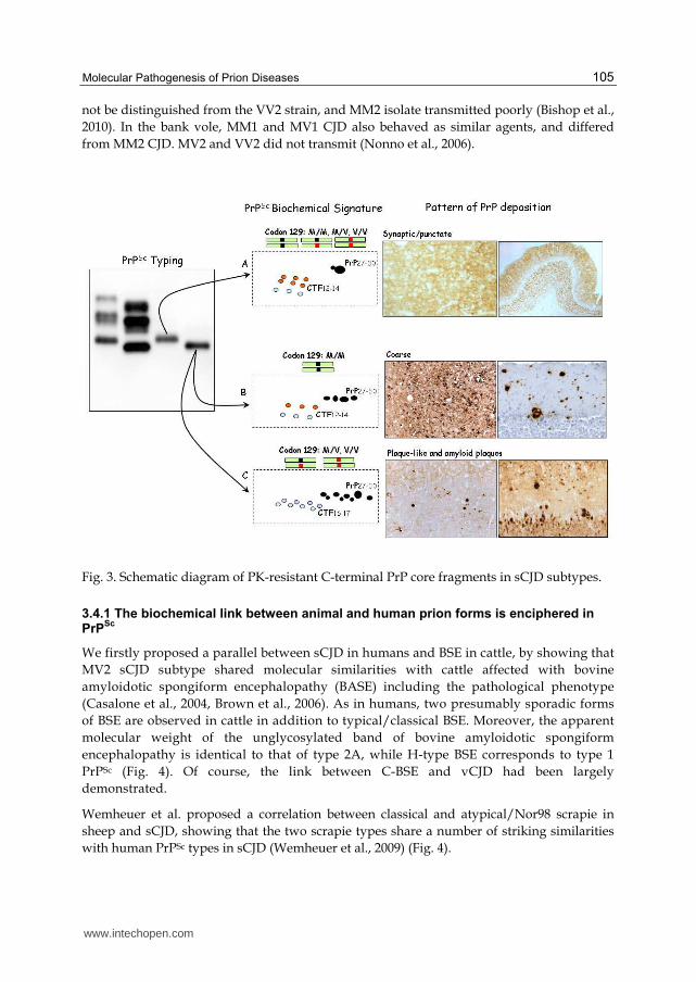
Molecular Pathogenesis of Prion Diseases
105
not be distinguished from the VV2 strain, and MM2 isolate transmitted poorly (Bishop et al.,
2010). In the bank vole, MM1 and MV1 CJD also behaved as similar agents, and differed
from MM2 CJD. MV2 and VV2 did not transmit (Nonno et al., 2006).
Fig. 3. Schematic diagram of PK-resistant C-terminal PrP core fragments in sCJD subtypes.
3.4.1 The biochemical link between animal and human prion forms is enciphered in PrP
Sc
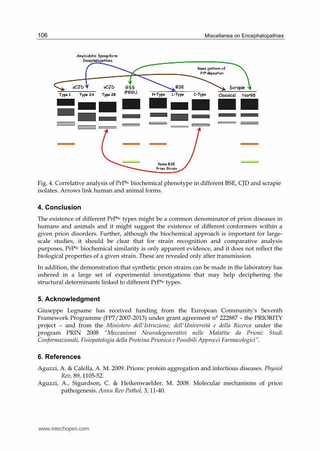
We firstly proposed a parallel between sCJD in humans and BSE in cattle, by showing that
MV2 sCJD subtype shared molecular similarities with cattle affected with bovine
amyloidotic spongiform encephalopathy (BASE) including the pathological phenotype
(Casalone et al., 2004, Brown et al., 2006). As in humans, two presumably sporadic forms
of BSE are observed in cattle in addition to typical/classical BSE. Moreover, the apparent
molecular weight of the unglycosylated band of bovine amyloidotic spongiform
encephalopathy is identical to that of type 2A, while H-type BSE corresponds to type 1
PrPSc (Fig. 4). Of course, the link between C-BSE and vCJD had been largely
demonstrated.
Wemheuer et al. proposed a correlation between classical and atypical/Nor98 scrapie in
sheep and sCJD, showing that the two scrapie types share a number of striking similarities
with human PrPSc types in sCJD (Wemheuer et al., 2009) (Fig. 4).
www.intechopen.com
Miscellanea on Encephalopathies
106
Fig. 4. Correlative analysis of PrPSc biochemical phenotype in different BSE, CJD and scrapie isolates. Arrows link human and animal forms.
4. Conclusion
The existence of different PrPSc types might be a common denominator of prion diseases in humans and animals and it might suggest the existence of different conformers within a given prion disorders. Further, although the biochemical approach is important for large-scale studies, it should be clear that for strain recognition and comparative analysis purposes, PrPSc biochemical similarity is only apparent evidence, and it does not reflect the biological properties of a given strain. These are revealed only after transmission.
In addition, the demonstration that synthetic prion strains can be made in the laboratory has ushered in a large set of experimental investigations that may help deciphering the structural determinants linked to different PrPSc types.
5. Acknowledgment
Giuseppe Legname has received funding from the European Community's Seventh Framework Programme (FP7/2007-2013) under grant agreement n° 222887 – the PRIORITY project – and from the Ministero dell'Istruzione, dell'Università e della Ricerca under the program PRIN 2008 “Meccanismi Neurodegenerativi nelle Malattie da Prioni: Studi Conformazionali, Fisiopatologia della Proteina Prionica e Possibili Approcci Farmacologici”.
6. References
Aguzzi, A. & Calella, A. M. 2009. Prions: protein aggregation and infectious diseases. Physiol Rev, 89, 1105-52.
Aguzzi, A., Sigurdson, C. & Heikenwaelder, M. 2008. Molecular mechanisms of prion pathogenesis. Annu Rev Pathol, 3, 11-40.
www.intechopen.com
Molecular Pathogenesis of Prion Diseases
107
Ashok, A. & Hegde, R. S. 2009. Selective processing and metabolism of disease-causing mutant prion proteins. PLoS Pathog, 5, e1000479.
Baker, H. E., Poulter, M., Crow, T. J., Frith, C. D., Lofthouse, R. & Ridley, R. M. 1991.
Aminoacid polymorphism in human prion protein and age at death in inherited
prion disease. Lancet, 337, 1286.
Benestad, S. L., Arsac, J. N., Goldmann, W. & Noremark, M. 2008. Atypical/Nor98 scrapie:
properties of the agent, genetics, and epidemiology. Vet Res, 39, 19.
Benestad, S. L., Sarradin, P., Thu, B., Schonheit, J., Tranulis, M. A. & Bratberg, B. 2003. Cases
of scrapie with unusual features in Norway and designation of a new type, Nor98.
Vet Rec, 153, 202-8.
Benvegnu, S., Gasperini, L. & Legname, G. 2011a. Aged PrP null mice show defective
processing of neuregulins in the peripheral nervous system. Mol Cell Neurosci, 47,
28-35.
Benvegnu, S., Poggiolini, I. & Legname, G. 2010. Neurodevelopmental expression and
localization of the cellular prion protein in the central nervous system of the mouse.
J Comp Neurol, 518, 1879-91.
Benvegnu, S., Roncaglia, P., Agostini, F., Casalone, C., Corona, C., Gustincich, S. & Legname,
G. 2011b. Developmental influence of the cellular prion protein on the gene
expression profile in mouse hippocampus. Physiol Genomics, 43, 711-25.
Biacabe, A. G., Laplanche, J. L., Ryder, S. & Baron, T. 2004. Distinct molecular phenotypes in
bovine prion diseases. EMBO Rep, 5, 110-5.
Bishop, M. T., Will, R. G. & Manson, J. C. 2010. Defining sporadic Creutzfeldt-Jakob disease
strains and their transmission properties. Proc Natl Acad Sci U S A, 107, 12005-10.
Bremer, J., Baumann, F., Tiberi, C., Wessig, C., Fischer, H., Schwarz, P., Steele, A. D., Toyka,
K. V., Nave, K. A., Weis, J. & Aguzzi, A. 2010. Axonal prion protein is required for
peripheral myelin maintenance. Nat Neurosci, 13, 310-8.
Brown, P., Mcshane, L. M., Zanusso, G. & Detwile, L. 2006. On the question of sporadic or
atypical bovine spongiform encephalopathy and Creutzfeldt-Jakob disease. Emerg
Infect Dis, 12, 1816-21.
Bruce, M., Chree, A., Mcconnell, I., Foster, J., Pearson, G. & Fraser, H. 1994. Transmission of
bovine spongiform encephalopathy and scrapie to mice: strain variation and the
species barrier. Philos Trans R Soc Lond B Biol Sci, 343, 405-11.
Bueler, H., Aguzzi, A., Sailer, A., Greiner, R. A., Autenried, P., Aguet, M. & Weissmann, C.
1993. Mice devoid of PrP are resistant to scrapie. Cell, 73, 1339-47.
Bueler, H., Fischer, M., Lang, Y., Bluethmann, H., Lipp, H. P., Dearmond, S. J., Prusiner, S.
B., Aguet, M. & Weissmann, C. 1992. Normal development and behaviour of mice
lacking the neuronal cell-surface PrP protein. Nature, 356, 577-82.
Calzolai, L., Lysek, D. A., Guntert, P., Von Schroetter, C., Riek, R., Zahn, R. & Wuthrich, K.
2000. NMR structures of three single-residue variants of the human prion protein.
Proc Natl Acad Sci U S A, 97, 8340-5.
Casalone, C., Zanusso, G., Acutis, P., Ferrari, S., Capucci, L., Tagliavini, F., Monaco, S. &
Caramelli, M. 2004. Identification of a second bovine amyloidotic spongiform
encephalopathy: molecular similarities with sporadic Creutzfeldt-Jakob disease.
Proc Natl Acad Sci U S A, 101, 3065-70.
www.intechopen.com
Miscellanea on Encephalopathies
108
Chesebro, B., Trifilo, M., Race, R., Meade-White, K., Teng, C., Lacasse, R., Raymond, L., Favara, C., Baron, G., Priola, S., Caughey, B., Masliah, E. & Oldstone, M. 2005. Anchorless prion protein results in infectious amyloid disease without clinical scrapie. Science, 308, 1435-9.
Christen, B., Hornemann, S., Damberger, F. F. & Wuthrich, K. 2009. Prion protein NMR structure from tammar wallaby (Macropus eugenii) shows that the beta2-alpha2 loop is modulated by long-range sequence effects. J Mol Biol, 389, 833-45.
Christen, B., Perez, D. R., Hornemann, S. & Wuthrich, K. 2008. NMR structure of the bank vole prion protein at 20 degrees C contains a structured loop of residues 165-171. J Mol Biol, 383, 306-12.
Collinge, J. 2001. Prion diseases of humans and animals: their causes and molecular basis. Annu Rev Neurosci, 24, 519-50.
Collinge J, Sidle KC, Meads J, Ironside J, Hill AF. 1996. Molecular analysis of prion strain variation and the aetiology of 'new variant' CJD. Nature. 383:685-90.
Collinge, J., Palmer, M. S. & Dryden, A. J. 1991. Genetic predisposition to iatrogenic Creutzfeldt-Jakob disease. Lancet, 337, 1441-2.
Criado, J. R., Sanchez-Alavez, M., Conti, B., Giacchino, J. L., Wills, D. N., Henriksen, S. J., Race, R., Manson, J. C., Chesebro, B. & Oldstone, M. B. 2005. Mice Devoid Of Prion Protein have cognitive deficits that are rescued by reconstitution of PrP in neurons. Neurobiol Dis, 19, 255-65.
Deleault, N. R., Lucassen, R. W. & Supattapone, S. 2003. RNA molecules stimulate prion protein conversion. Nature, 425, 717-20.
Gambetti, P., Cali, I., Notari, S., Kong, Q., Zou, W. Q. & Surewicz, W. K. 2011. Molecular biology and pathology of prion strains in sporadic human prion diseases. Acta Neuropathol, 121, 79-90.
Gambetti, P., Dong, Z., Yuan, J., Xiao, X., Zheng, M., Alshekhlee, A., Castellani, R., Cohen, M., Barria, M. A., Gonzalez-Romero, D., Belay, E. D., Schonberger, L. B., Marder, K., Harris, C., Burke, J. R., Montine, T., Wisniewski, T., Dickson, D. W., Soto, C., Hulette, C. M., Mastrianni, J. A., Kong, Q. & Zou, W. Q. 2008. A novel human disease with abnormal prion protein sensitive to protease. Ann Neurol, 63, 697-708.
Gambetti, P., Kong, Q., Zou, W., Parchi, P. & Chen, S. G. 2003. Sporadic and familial CJD: classification and characterisation. Br Med Bull, 66, 213-39.
Ghetti, B., Tagliavini, F., Takao, M., Bugiani, O. & Piccardo, P. 2003. Hereditary prion protein amyloidoses. Clin Lab Med, 23, 65-85, viii.
Gorodinsky, A. & Harris, D. A. 1995. Glycolipid-anchored proteins in neuroblastoma cells form detergent-resistant complexes without caveolin. J Cell Biol, 129, 619-27.
Gossert, A. D., Bonjour, S., Lysek, D. A., Fiorito, F. & Wuthrich, K. 2005. Prion protein NMR structures of elk and of mouse/elk hybrids. Proc Natl Acad Sci U S A, 102, 646-50.
Govaerts, C., Wille, H., Prusiner, S. B. & Cohen, F. E. 2004. Evidence for assembly of prions with left-handed beta-helices into trimers. Proc Natl Acad Sci U S A, 101, 8342-7.
Hegde, R. S., Tremblay, P., Groth, D., Dearmond, S. J., Prusiner, S. B. & Lingappa, V. R. 1999. Transmissible and genetic prion diseases share a common pathway of neurodegeneration. Nature, 402, 822-6.
www.intechopen.com
Molecular Pathogenesis of Prion Diseases
109
Hsiao, K., Dlouhy, S. R., Farlow, M. R., Cass, C., Da Costa, M., Conneally, P. M., Hodes, M. E., Ghetti, B. & Prusiner, S. B. 1992. Mutant prion proteins in Gerstmann-Straussler-Scheinker disease with neurofibrillary tangles. Nat Genet, 1, 68-71.
Ilc, G., Giachin, G., Jaremko, M., Jaremko, L., Benetti, F., Plavec, J., Zhukov, I. & Legname, G. 2010. NMR structure of the human prion protein with the pathological Q212P mutation reveals unique structural features. PLoS ONE, 5, e11715.
Kanaani, J., Prusiner, S. B., Diacovo, J., Baekkeskov, S. & Legname, G. 2005. Recombinant prion protein induces rapid polarization and development of synapses in embryonic rat hippocampal neurons in vitro. J Neurochem, 95, 1373-86.
Kaneko, K., Zulianello, L., Scott, M., Cooper, C. M., Wallace, A. C., James, T. L., Cohen, F. E. & Prusiner, S. B. 1997. Evidence for protein X binding to a discontinuous epitope on the cellular prion protein during scrapie prion propagation. Proc Natl Acad Sci U S A, 94, 10069-74.
Kobayashi, A., Hizume, M., Teruya, K., Mohri, S. & Kitamoto, T. 2009. Heterozygous inhibition in prion infection: the stone fence model. Prion, 3, 27-30.
Kovacs, G. G., Trabattoni, G., Hainfellner, J. A., Ironside, J. W., Knight, R. S. & Budka, H. 2002. Mutations of the prion protein gene phenotypic spectrum. J Neurol, 249, 1567-82.
Legname, G., Baskakov, I. V., Nguyen, H. O., Riesner, D., Cohen, F. E., Dearmond, S. J. & Prusiner, S. B. 2004. Synthetic Mammalian Prions. Science, 305, 673-6.
Legname, G., Nguyen, H. O., Baskakov, I. V., Cohen, F. E., Dearmond, S. J. & Prusiner, S. B. 2005. Strain-specified characteristics of mouse synthetic prions. Proc Natl Acad Sci U S A, 102, 2168-73.
Legname, G., Nguyen, H. O., Peretz, D., Cohen, F. E., Dearmond, S. J. & Prusiner, S. B. 2006. Continuum of prion protein structures enciphers a multitude of prion isolate-specified phenotypes. Proc Natl Acad Sci U S A, 103, 19105-10.
Liao, Y. C., Lebo, R. V., Clawson, G. A. & Smuckler, E. A. 1986. Human prion protein cDNA: molecular cloning, chromosomal mapping, and biological implications. Science, 233, 364-7.
Lopez Garcia, F., Zahn, R., Riek, R. & Wuthrich, K. 2000. NMR structure of the bovine prion protein. Proc Natl Acad Sci U S A, 97, 8334-9.
Marijanovic, Z., Caputo, A., Campana, V. & Zurzolo, C. 2009. Identification of an intracellular site of prion conversion. PLoS Pathog, 5, e1000426.
Mckinley, M. P., Meyer, R. K., Kenaga, L., Rahbar, F., Cotter, R., Serban, A. & Prusiner, S. B. 1991. Scrapie prion rod formation in vitro requires both detergent extraction and limited proteolysis. J Virol, 65, 1340-51.
Moore, R. C., Lee, I. Y., Silverman, G. L., Harrison, P. M., Strome, R., Heinrich, C., Karunaratne, A., Pasternak, S. H., Chishti, M. A., Liang, Y., Mastrangelo, P., Wang, K., Smit, A. F., Katamine, S., Carlson, G. A., Cohen, F. E., Prusiner, S. B., Melton, D. W., Tremblay, P., Hood, L. E. & Westaway, D. 1999. Ataxia in prion protein (PrP)-deficient mice is associated with upregulation of the novel PrP-like protein doppel. J Mol Biol, 292, 797-817.
Nishida, N., Tremblay, P., Sugimoto, T., Shigematsu, K., Shirabe, S., Petromilli, C., Erpel, S. P., Nakaoke, R., Atarashi, R., Houtani, T., Torchia, M., Sakaguchi, S., Dearmond, S. J., Prusiner, S. B. & Katamine, S. 1999. A mouse prion protein transgene rescues mice deficient for the prion protein gene from purkinje cell degeneration and demyelination. Lab Invest, 79, 689-97.
www.intechopen.com
Miscellanea on Encephalopathies
110
Nonno, R., Di Bari, M. A., Cardone, F., Vaccari, G., Fazzi, P., Dell'omo, G., Cartoni, C., Ingrosso, L., Boyle, A., Galeno, R., Sbriccoli, M., Lipp, H. P., Bruce, M., Pocchiari, M. & Agrimi, U. 2006. Efficient transmission and characterization of Creutzfeldt-Jakob disease strains in bank voles. PLoS Pathog, 2, e12.
Palmer, M. S., Dryden, A. J., Hughes, J. T. & Collinge, J. 1991. Homozygous prion protein genotype predisposes to sporadic Creutzfeldt-Jakob disease. Nature, 352, 340-2.
Parchi, P., Chen, S. G., Brown, P., Zou, W., Capellari, S., Budka, H., Hainfellner, J., Reyes, P. F., Golden, G. T., Hauw, J. J., Gajdusek, D. C. & Gambetti, P. 1998. Different patterns of truncated prion protein fragments correlate with distinct phenotypes in P102L Gerstmann-Straussler-Scheinker disease. Proc Natl Acad Sci U S A, 95, 8322-7.
Parchi, P., Giese, A., Capellari, S., Brown, P., Schulz-Schaeffer, W., Windl, O., Zerr, I., Budka, H., Kopp, N., Piccardo, P., Poser, S., Rojiani, A., Streichemberger, N., Julien, J., Vital, C., Ghetti, B., Gambetti, P. & Kretzschmar, H. 1999. Classification of sporadic Creutzfeldt-Jakob disease based on molecular and phenotypic analysis of 300 subjects. Ann Neurol, 46, 224-33.
Perez, D. R., Damberger, F. F. & Wuthrich, K. 2010. Horse prion protein NMR structure and comparisons with related variants of the mouse prion protein. J Mol Biol, 400, 121-8.
Piccardo, P., Dlouhy, S. R., Lievens, P. M., Young, K., Bird, T. D., Nochlin, D., Dickson, D. W., Vinters, H. V., Zimmerman, T. R., Mackenzie, I. R., Kish, S. J., Ang, L. C., De Carli, C., Pocchiari, M., Brown, P., Gibbs, C. J., Jr., Gajdusek, D. C., Bugiani, O., Ironside, J., Tagliavini, F. & GhettI, B. 1998. Phenotypic variability of Gerstmann-Straussler-Scheinker disease is associated with prion protein heterogeneity. J Neuropathol Exp Neurol, 57, 979-88.
Piccardo, P., Manson, J. C., King, D., Ghetti, B. & Barron, R. M. 2007. Accumulation of prion protein in the brain that is not associated with transmissible disease. Proc Natl Acad Sci U S A, 104, 4712-7.
Prusiner, S. B. 1982. Novel proteinaceous infectious particles cause scrapie. Science, 216, 136-44.
Prusiner, S. B. 1998. Prions. Proc Natl Acad Sci U S A, 95, 13363-83. Prusiner, S. B. 2001. Shattuck lecture--neurodegenerative diseases and prions. N Engl J Med,
344, 1516-26. Prusiner, S. B., Mckinley, M. P., Bowman, K. A., Bolton, D. C., Bendheim, P. E., Groth, D. F.
& Glenner, G. G. 1983. Scrapie prions aggregate to form amyloid-like birefringent rods. Cell, 35, 349-58.
Riek, R., Hornemann, S., Wider, G., Billeter, M., Glockshuber, R. & Wuthrich, K. 1996. NMR structure of the mouse prion protein domain PrP(121-321). Nature, 382, 180-2.
Salmona, M., Morbin, M., Massignan, T., Colombo, L., Mazzoleni, G., Capobianco, R., Diomede, L., Thaler, F., Mollica, L., Musco, G., Kourie, J. J., Bugiani, O., Sharma, D., Inouye, H., Kirschner, D. A., Forloni, G. & Tagliavini, F. 2003. Structural properties of Gerstmann-Straussler-Scheinker disease amyloid protein. J Biol Chem, 278, 48146-53.
Spudich, A., Frigg, R., Kilic, E., Kilic, U., Oesch, B., Raeber, A., Bassetti, C. L. & Hermann, D. M. 2005. Aggravation of ischemic brain injury by prion protein deficiency: role of ERK-1/-2 and STAT-1. Neurobiol Dis, 20, 442-9.
www.intechopen.com
Molecular Pathogenesis of Prion Diseases
111
Stahl, N., Borchelt, D. R. & Prusiner, S. B. 1990. Differential release of cellular and scrapie prion proteins from cellular membranes by phosphatidylinositol-specific phospholipase C. Biochemistry, 29, 5405-12.
Tagliavini, F., Prelli, F., Ghiso, J., Bugiani, O., Serban, D., Prusiner, S. B., Farlow, M. R., Ghetti, B. & Frangione, B. 1991. Amyloid protein of Gerstmann-Straussler-Scheinker disease (Indiana kindred) is an 11 kd fragment of prion protein with an N-terminal glycine at codon 58. EMBO J, 10, 513-9.
Tagliavini, F., Prelli, F., Porro, M., Rossi, G., Giaccone, G., Farlow, M. R., Dlouhy, S. R., Ghetti, B., Bugiani, O. & Frangione, B. 1994. Amyloid fibrils in Gerstmann-Straussler-Scheinker disease (Indiana and Swedish kindreds) express only PrP peptides encoded by the mutant allele. Cell, 79, 695-703.
Telling, G. C., Scott, M., Mastrianni, J., Gabizon, R., Torchia, M., Cohen, F. E., Dearmond, S. J. & PruSINER, S. B. 1995. Prion propagation in mice expressing human and chimeric PrP transgenes implicates the interaction of cellular PrP with another protein. Cell, 83, 79-90.
Tobler, I., Deboer, T. & Fischer, M. 1997. Sleep and sleep regulation in normal and prion protein-deficient mice. J Neurosci, 17, 1869-79.
Wadsworth, J. D. & Collinge, J. 2011. Molecular pathology of human prion disease. Acta Neuropathol, 121, 69-77.
Wadsworth JD, Joiner S, Linehan JM, Cooper S, Powell C, Mallinson G, Buckell J, Gowland I, Asante EA, Budka H, Brandner S, Collinge J. 2006. Phenotypic heterogeneity in inherited prion disease (P102L) is associated with differential propagation of protease-resistant wild-type and mutant prion protein. Brain 129:1557-69.
Weise, J., Sandau, R., Schwarting, S., Crome, O., Wrede, A., Schulz-Schaeffer, W., Zerr, I. & Bahr, M. 2006. Deletion of cellular prion protein results in reduced Akt activation, enhanced postischemic caspase-3 activation, and exacerbation of ischemic brain injury. Stroke, 37, 1296-300.
Weissmann, C. & FlechsiG, E. 2003. PrP knock-out and PrP transgenic mice in prion research. Br Med Bull, 66, 43-60.
Wemheuer, W. M., Benestad, S. L., Wrede, A., Schulze-Sturm, U., Wemheuer, W. E., Hahmann, U., Gawinecka, J., Schutz, E., Zerr, I., Brenig, B., Bratberg, B., Andreoletti, O. & Schulz-Schaeffer, W. J. 2009. Similarities between forms of sheep scrapie and Creutzfeldt-Jakob disease are encoded by distinct prion types. Am J Pathol, 175, 2566-73.
Wen, Y., Li, J., Yao, W., Xiong, M., Hong, J., Peng, Y., Xiao, G. & Lin, D. 2010. Unique structural characteristics of the rabbit prion protein. J Biol Chem, 285, 31682-93.
Williams, E. S. 2005. Chronic wasting disease. Vet Pathol, 42, 530-49. Windl, O., Dempster, M., Estibeiro, J. P., Lathe, R., De Silva, R., Esmonde, T., Will, R.,
Springbett, A., Campbell, T. A., Sidle, K. C., Palmer, M. S. & Collinge, J. 1996. Genetic basis of Creutzfeldt-Jakob disease in the United Kingdom: a systematic analysis of predisposing mutations and allelic variation in the PRNP gene. Hum Genet, 98, 259-64.
Young, K., Piccardo, P., Kish, S. J., Ang, L. C., Dlouhy, S. & Ghetti, B. 1998. Gerstmann-Sträussler-Scheinker disease (GSS) with a mutation at prion protein (PrP) residue 212. Journal of Neuropathology & Experimental Neurology, 57, 518.
www.intechopen.com

Miscellanea on Encephalopathies
112
Zanusso, G., Farinazzo, A., Prelli, F., Fiorini, M., Gelati, M., Ferrari, S., Righetti, P. G., Rizzuto, N., Frangione, B. & Monaco, S. 2004. Identification of distinct N-terminal truncated forms of prion protein in different Creutzfeldt-Jakob disease subtypes. J Biol Chem, 279, 38936-42.
Zanusso, G., Polo, A., Farinazzo, A., Nonno, R., Cardone, F., Di Bari, M., Ferrari, S., Principe, S., Gelati, M., Fasoli, E., Fiorini, M., Prelli, F., Frangione, B., Tridente, G., Bentivoglio, M., Giorgi, A., Schinina, M. E., Maras, B., Agrimi, U., Rizzuto, N., Pocchiari, M. & Monaco, S. 2007. Novel prion protein conformation and glycotype in Creutzfeldt-Jakob disease. Arch Neurol, 64, 595-9.
Zanusso, G., Righetti, P. G., Ferrari, S., Terrin, L., Farinazzo, A., Cardone, F., Pocchiari, M., Rizzuto, N. & Monaco, S. 2002. Two-dimensional mapping of three phenotype-associated isoforms of the prion protein in sporadic Creutzfeldt-Jakob disease. Electrophoresis, 23, 347-55.
Zhang, Y., Swietnicki, W., Zagorski, M. G., Surewicz, W. K. & Sonnichsen, F. D. 2000. Solution structure of the E200K variant of human prion protein. Implications for the mechanism of pathogenesis in familial prion diseases. J Biol Chem, 275, 33650-4.
Zimmermann, K., Turecek, P. L. & Schwarz, H. P. 1999. Genotyping of the prion protein gene at codon 129. Acta Neuropathol, 97, 355-8.
Zou, W. Q., Capellari, S., Parchi, P., Sy, M. S., Gambetti, P. & Chen, S. G. 2003. Identification of novel proteinase K-resistant C-terminal fragments of PrP in Creutzfeldt-Jakob disease. J Biol Chem, 278, 40429-36.
Zou, W. Q., Puoti, G., Xiao, X., Yuan, J., Qing, L., Cali, I., Shimoji, M., Langeveld, J. P., Castellani, R., Notari, S., Crain, B., Schmidt, R. E., Geschwind, M., Dearmond, S. J., Cairns, N. J., Dickson, D., Honig, L., Torres, J. M., Mastrianni, J., Capellari, S., Giaccone, G., Belay, E. D., Schonberger, L. B., Cohen, M., Perry, G., Kong, Q., Parchi, P., Tagliavini, F. & Gambetti, P. 2010. Variably protease-sensitive prionopathy: a new sporadic disease of the prion protein. Ann Neurol, 68, 162-72.
www.intechopen.com
Miscellanea on EncephalopathiesEdited by Dr. Radu Tanasescu
ISBN 978-953-51-0499-5Hard cover, 202 pagesPublisher InTechPublished online 18, April, 2012Published in print edition April, 2012
InTech EuropeUniversity Campus STeP Ri Slavka Krautzeka 83/A 51000 Rijeka, Croatia Phone: +385 (51) 770 447 Fax: +385 (51) 686 166www.intechopen.com
InTech ChinaUnit 405, Office Block, Hotel Equatorial Shanghai No.65, Yan An Road (West), Shanghai, 200040, China
Phone: +86-21-62489820 Fax: +86-21-62489821
The book project “Miscellanea on Encephalopathies” aims to cover some of the important aspects ofinfectious-related encephalopathies, post-transplantation and drug-induced encephalopathies, by transmittingvaluable information filtered through the real life clinical and research experience of the authors.
How to referenceIn order to correctly reference this scholarly work, feel free to copy and paste the following:
Giuseppe Legname and Gianluigi Zanusso (2012). Molecular Pathogenesis of Prion Diseases, Miscellanea onEncephalopathies, Dr. Radu Tanasescu (Ed.), ISBN: 978-953-51-0499-5, InTech, Available from:http://www.intechopen.com/books/miscellanea-on-encephalopathies/molecular-pathogenesis-of-prion-diseases

© 2012 The Author(s). Licensee IntechOpen. This is an open access articledistributed under the terms of the Creative Commons Attribution 3.0License, which permits unrestricted use, distribution, and reproduction inany medium, provided the original work is properly cited.