modeling molecular mechanisms in the axon
TRANSCRIPT

1 23
Computational MechanicsSolids, Fluids, Structures, Fluid-Structure Interactions, Biomechanics,Micromechanics, Multiscale Mechanics,Materials, Constitutive Modeling,Nonlinear Mechanics, Aerodynamics ISSN 0178-7675Volume 59Number 3 Comput Mech (2017) 59:523-537DOI 10.1007/s00466-016-1359-y
Modeling molecular mechanisms in theaxon
R. de Rooij, K. E. Miller & E. Kuhl

1 23
Your article is protected by copyright and
all rights are held exclusively by Springer-
Verlag Berlin Heidelberg. This e-offprint is
for personal use only and shall not be self-
archived in electronic repositories. If you wish
to self-archive your article, please use the
accepted manuscript version for posting on
your own website. You may further deposit
the accepted manuscript version in any
repository, provided it is only made publicly
available 12 months after official publication
or later and provided acknowledgement is
given to the original source of publication
and a link is inserted to the published article
on Springer's website. The link must be
accompanied by the following text: "The final
publication is available at link.springer.com”.

Comput Mech (2017) 59:523–537DOI 10.1007/s00466-016-1359-y
ORIGINAL PAPER
Modeling molecular mechanisms in the axon
R. de Rooij1 · K. E. Miller2 · E. Kuhl1
Received: 5 September 2016 / Accepted: 8 November 2016 / Published online: 1 December 2016© Springer-Verlag Berlin Heidelberg 2016
Abstract Axons are living systems that display highlydynamic changes in stiffness, viscosity, and internal stress.However, the mechanistic origin of these phenomenologi-cal properties remains elusive. Here we establish a com-putational mechanics model that interprets cellular-levelcharacteristics as emergent properties from molecular-levelevents. We create an axon model of discrete microtubules,which are connected to neighboring microtubules via dis-crete crosslinkingmechanisms that obey a set of simple rules.We explore two types of mechanisms: passive and activecrosslinking. Our passive and active simulations suggestthat the stiffness and viscosity of the axon increase linearlywith the crosslink density, and that both are highly sensi-tive to the crosslink detachment and reattachment times. Ourmodel explains how active crosslinking with dynein motorsgenerates internal stresses and actively drives axon elonga-tion. We anticipate that our model will allow us to probea wide variety of molecular phenomena—both in isolationand in interaction—to explore emergent cellular-level fea-tures under physiological and pathological conditions.
Keywords Finite element · Elasticity · Viscosity · Activeforce · Axon
B E. [email protected]
1 Departments of Mechanical Engineering and Bioengineering,Stanford University, Stanford, CA 94305, USA
2 Department of Integrative Biology, Michigan StateUniversity, East Lansing, MI 48824, USA
1 Introduction
With a diameter of up to 14µm and a length of up to 1m,the neuron is the largest cell in the human body, both insurface area and volume [42,57]. It is arguably also one ofthe most important cell types; it enables all communicationwith and within the brain through conducting electrical sig-nals. The development of a neuron starts with the soma, thecell body, see Fig. 1. Several short dendrites extend fromthe soma into the surroundings, and one of these dendritesdevelops into the axon. In contrast to dendrites, the axon islong, it has a high degree of polarity alignment, and it is typ-ically myelinated [21]. The axonal cytoskeleton consists oflongitudinally aligned microtubules and neurofilaments thatare connected by a variety of different crosslinks [20]. Thecytoskeleton is encapsulated by an actin cortex [28,29] thatis held together by spectrin [27,38,76]. At the tip of the axon,the growth cone is leading axonal growth and is responsiblefor path finding and axonal steering [48]. Extensive researchhas been devoted to neurons since their discovery in the 19thcentury with specific focus on axonal growth [20,33,48], thebiophysics of axonal development [26,71], and the physiol-ogy of axons [23].
Throughout the past decade, several groups have recog-nized the importance of mechanical forces in the axon[7,25,71]. They found that the growth cone applies tensionto the growing axon [11,12,46] and that it tightly regu-lates this tension within the physiological range of 1nN[24,40,60]. Numerous experiments have since been per-formed to quantify how these forces are transmitted along theaxon. These experiments have established several generallyaccepted hypotheses that highlight the role of physical forces[34], for example during axonal development [8,33,71].
Single axon experiments in culture allow us to investigatethe biophysics of individual molecules andmolecular motors
123
Author's personal copy

524 Comput Mech (2017) 59:523–537
Fig. 1 Neuron and cross-sectional view of its axon. The developmentof a neuron starts with the cell body and several short dendrites. One ofthese dendrites develops into the axon, which is led by the growth cone.The axonal cytoskeleton is surrounded by an actin cortex and consistsof longitudinally aligned microtubules that are connected by passivecrosslinks such as tau and by active crosslinks such as dynein motors
within the axon.Wenowknow that dynein is a unipolarmole-cular motor that generates axonal extensile forces bywalkingtowards the minus end of microtubules [2,9,65]. Kinesin,likewise, is a bipolar molecular motor that walks towards theplus end ofmicrotubules and causesmicrotubules to slide outof the neuronal cell body [18,49,77]. Myosin, in constrast,is a molecular motor that is located primarily in the actincortex and in the growth cone where it generates contractileforces [14,53,80]. Force equilibrium and axonal elongationis a competition between the extensile forces of dynein andthe compressive forces of myosin [29,33,65]. In addition tothese actively force-generating motors, molecules like tauthat passively crosslink the axonal cytoskeleton also play acritical role in networkmechanics [81].Although taudoes notgenerate active forces, it stabilizes the axon by cross-linkingneighboring microtubules and preventing them from depoly-merizing. When exposed to strains or strain rates beyondthe physiological limit, the tau-microtubule complex grad-ually weakens, which results in diffuse axonal injury and,ultimately, cell death [5,69,74].
Biological structures are living systems with the abil-ity to adapt to their mechanical environment [61]. It issurprising that cells are often characterized as passive, time-independent, and purely elastic [64]. When constant forcesare applied to an axon over a long period of time, it length-ens at a constant rate and displays a time-dependent behaviorthat is rheologically similar to a viscous fluid [44,54]. Whenconstant forces are applied to the brain, for example duringdevelopment or in response to tumor growth, the brain adaptsgradually over time and displays a time-dependent behavior[35]. In response to bodygrowth or artificial limb lengthening[83], axons can adapt and gradually grow in length [1,68,71].Nonetheless, time-independent, elastic models have success-fully been used to explain prestress in axons and in thebrain [24] and to model the effects of high impact loading[4,58,74].
At present, themost popular approach is tomodel the adultnervous system as time-independent solid and the devel-
oping neuron as time-dependent fluid. Two more recentapproaches suggest to interpret neurons as active fluids orsolids [6,55,62] that are capable of generating active forces,conceptually similar to skeletalmuscle [32,37]. In both cases,internal forces generated at the expenditure of adenosinetriphosphate, ATP, explain internal tensions at the steadystate as proposed by active matter hydrodynamics [51]. Theactive fluid model cleanly explains why axons elongate inresponse to large external forces, retract at low externalforces, and maintain a constant rest length and rest tensionat intermediate forces [55]. However, it does not considerthe elastic behavior of the axon, which limits its use to suffi-ciently long time spans of observation. The active solidmodel[6,10] and the morphoelastic rodmodel [52] excellently cap-ture the elastic properties of axons. The morphoelastic rodmodel characterizes the behavior of axons over long periodsof time using the theory of finite growth [62]. It suggeststhat forces trigger the immediate addition of mass, whereasexperiments indicate that forces first cause axons to stretchand then new mass is added gradually to restore the initialaxonal diameter [39,47]. Further complicating this problemis the well-accepted observation that internal forces in cellschange their viscoelastic properties: The measured mechan-ical properties of cells and tissues can be highly sensitive tothe stresses and strains used to probe them [45,51,59]. Thisis especially important in growing neurons as it is recognizedthat molecular motors such as kinesin, dynein, and myosingenerate forces that modulate axon elongation [3,41,49].Taken together, this active nature of living systems makes itinherently difficult to measure, model, and understand neu-ronal mechanics. Given the complexity of this problem, thedevelopment of analytic equations and simulation tools thatcleanly and simplymodel the problemof neuronalmechanicsacross all time scales and explain the complex dependenceof effective elasticity and viscosity as a function of internaland external forces would be ideal.
The objective of this manuscript is to establish ana-lytic equations and a finite element model that, for thefirst time, fully models the complex time and force depen-dent behavior of active and passive axonal substructures.We present a general framework to model a wide varietyof molecular mechanisms within the an existing finite ele-ment infrastructure. Within this framework, we assign a userdefined molecular mechanism to a standard finite element,whilst preserving the conceptual modularity of the finite ele-ment method. We demonstrate the potential of our methodusing a three-dimensional axonmodel that consists of micro-tubules and crosslinks. We examine two types of crosslinks,passive dissipative crosslinking and active motor crosslink-ing. We use these two mechanisms to interpret cellular-levelcharacteristics such as axon stiffness, viscosity, and internalstress as emergent properties from the subcellular level. Thisallowsus to informconstitutivemodels at the continuum level
123
Author's personal copy

Comput Mech (2017) 59:523–537 525
Fig. 2 General framework for modeling molecular mechanisms as anextension of the finite element method. Orange boxes represent objectsthat are available in a standardfinite element infrastructure.Yellowboxes
represent extensions to this general architecture that enable the model-ing of molecular mechanisms. (Color figure online)
by molecular-level events. We then develop active viscoelas-tic fluid equations which we use to validate and understandthe non-linear behaviors produced by the simulation. Impor-tantly, this conceptual framework can be easily extended toall active systems including other types of cells and tissues.
The remainder of this manuscript is organized as follows:We begin by describing the general challenge of developingan algorithmic framework for dynamic mechanisms in livingcells in Sect. 2. We describe the geometry and mechanicalproperties of our axon model in Sect. 3 and its basic mech-anisms in Sect. 4. We then illustrate the key features of ourmodel by means of two selected mechanisms in Sect. 5, andconclude by discussing our results in Sect. 6.
2 Algorithmic framework
Modeling molecular mechanisms within the context of thefinite element method requires several extensions to the stan-dard finite element infrastructure. Our main objective is todevelop a generic and modular framework that will allowus to implement a wide range of molecular mechanisms. Inpractice, this implies that we only want to add to the existingfinite element method—not change it—with the goal to plugin many different types of molecular mechanisms withoutaffecting the overall algorithmic infrastructure. We provide
an overview of the global implementation in Sect. 2.1 anddiscuss all extensions individually in Sects. 2.2–2.6.
2.1 Overview
Figure 2 illustrates the global architecture of ourmethod. Theorange boxes represent standard objects that are part of everystandard implementation of the finite elementmethod and theyellow boxes indicate the extensions that allow us to modela wide range of molecular mechanisms that are relevant forthe axon.
The architecture consists of twomain branches, the modeland the solver. The first branch, the model, consists of thestandard description of materials, properties, nodes, ele-ments, point loads (LOAD), single point constraints (SPC),and multiple point constraints (MPC). Here we extend thisstructurewith an extended node object (NodeX), an extendedbar element (CBarX), mechanisms, and microtubule (MT)objects. The second branch, the solver, consists of at least animplementation of the Newton–Raphson method (NR) fornonlinear problems. Here we adopt the Newton–Raphsonalgorithm with slight modifications to allow for the applica-tion of molecular mechanisms. We implement this architec-ture into a custom-designed finite element framework.
123
Author's personal copy

526 Comput Mech (2017) 59:523–537
Table 1 Parameter types and names of a standard Node object and anextended NodeX object
Node NodeX
int nid int nid
double x,y,z double x,y,z
int[] dofID int[] dofID
Element elMinus
Element elPlus
Table 2 Parameter types and names of a standard CBar object and anextended CBarX object
CBar CBarX
int eid int eid
Node[] nodes NodeX[] nodes
Property prop Property prop
double restLength
State state
NodeX[] dummyNodes
double timeToNextEvent
Mechanism mechanism
2.2 Node and NodeX objects
Table 1 compares the standard node and the extended nodeobjects. A standard node (Node) is characterized by its nodalindex, its coordinates, and the global indices of its degreesof freedom. An extended node (NodeX) also points to theelements on its plus and minus sides along the microtubule.This allows us to simulate molecular walking. When a mole-cule walks from one node on the microtubule to the next, it isessential for that node to know the elements on its plus andminus sides.
2.3 CBar and CBarX objects
Table 2 compares the standard bar element and the extendedbar element. A standard bar element (CBar) is character-ized by its element index, the two nodes it connects, and theelement property. The extended bar element (CBarX) con-sists of several additional variables. The rest length of theelement allows for active contraction or extension of the ele-ment. The current biological state of the element is a statevariable that is important to identify the next action of anelement. Table 3 summarizes potential element states. Tokeep track of the geometry, the extended bar element storestwo dummy nodes to which the element was previously con-nected and may connect to again in the future. The variabletimeToNextEvent monitors the time until this elementhas to perform its next event that is determined by the mole-cular mechanism that the element is subjected to.
Table 3 Possible states of a CBarX element
State Description
NoState Element has no state
Microtubule Element is part of amicrotubule
CrosslinkAttached Crosslink that is attached tomicrotubules
CrosslinkDetached Crosslink that is notattached to microtubules
Table 4 Parameter types and names of a Microtubule object
MT
int n0,n1
int e0,e1
MTstate state
double timeToNextEvent
MT mtMinus
MT mtPlus
Mechanism mechanism
Table 5 Examples of the parameter types and names of a Mechanismobject, which vary for each mechanism and can be defined by the user
Mechanism for element Mechanism for MT
double tAttach double tPolym
double tDetach double tDepolym
double tStationary
2.4 Microtubule object
Table 4 summarizes the variables that constitute a micro-tubule object (MT). The microtubule object is specific to ourextended architecture of the finite element implementation.Amicrotubule consists of many CBarX elements that collec-tively behave as a single microtubule. It consists of integervariables that contain the indices of the first and last nodesand elements that build up this microtubule. Similar to theCBarX elements,microtubules are characterized by their cur-rent state, the time to their next event, and themechanism theyare subjected to. Potential microtubule mechanisms could bepolymerization and depolymerization. In addition the micro-tubule object has access to the microtubules at its minus andplus ends.
2.5 Mechanism object
Table 5 illustrates the type of variables of element-basedand microtubule-based mechanisms. The mechanism objectis our most important extension to the standard finite ele-
123
Author's personal copy

Comput Mech (2017) 59:523–537 527
ment method as it allows us to simulate the dynamicsinduced by molecular mechanisms. The mechanism objectis a super class and every individual mechanism is a subclass as highlighted in Fig. 2. Because of this generalcharacter, the common denominator among different mech-anisms is not a set of variables, but simply the functionApply(). The Apply() function can be tailored to sim-ulate the mechanism of molecular motors such as dynein,kinesin, or myosin, the detachment and reattachment ofcrosslinking proteins such as tau, or the polymerization anddepolymerization of cytoskeletal filaments such as micro-tubules.
2.6 Solver
Algorithm 1 summarizes the pseudo code of our solverfor a general solution step. The solver is based on astandard Newton–Raphson iteration with adaptive time-stepping, supplemented by modifications that allow for theexecution of all mechanisms. The differences comparedto a standard Newton–Raphson solver are the functionsUpdateModel() and RestoreModel().UpdateModel() is executed at the beginning of each step,and it applies all mechanisms for the duration of that par-ticular step, see Algorithm 2. An action is performed if thetimeToNextEvent variable of the CBarX element orMTobject in Tables 2 or 4 becomes smaller than zero duringthe current step. RestoreModel() is only executed if thesolver did not converge, in which case it restores the begin-ning of the step, see Algorithm 3.
Algorithms 2 and 3 only describe updating and restoringelement-basedmechanisms. The algorithms for updating andrestoring microtubule-based mechanisms are conceptuallysimilar, with the only difference that updating or restoring amicrotubule also involves updating or restoring the elementsthat constitute that microtubule.
Algorithm 1 Pseudo code of modified Newton–Raphsonsolver with adaptive time stepping.Step j
Update model with UpdateModel(), see Algorithm 2.Solve system using iterative Newton–Raphson procedure,with maxIter as maximum number of iterations.if converged then
Proceed to step j + 1.else
Restore model with RestoreModel(), seeAlgorithm 3.Decrease time step: �t ← �t/2.Repeat step j .
end ifend
Algorithm 2 Pseudo code of UpdateModel() function.UpdateModel()
for all Elements el doUpdate time to the next event:el.timeToNextEvent←el.timeToNextEvent-�t.Apply mechanism: el.mechanism.Apply().Note, before an element variable is updated by themechanism, its old value is stored in storage. Itcan be restored by calling RestoreModel(), seeAlgorithm 3.
end forend
Algorithm 3 PseudocodeofRestoreModel() function.RestoreModel()
for Element el in storage doRestore stored variables of el.Restore time to next event:el.timeToNextEvent←el.timeToNextEvent+�t.
end forend
3 Axon model
In this section, we present the geometry and material prop-erties of our axon model. Our axon consists of discretemicrotubules, which are aligned along their longitudinal axisand connected by individual crosslinks. For now,we focus onmodeling molecular mechanisms in the axonal cytoskeletonand neglect the axon cortex and the growth cone.
The first step in generating the geometry is to define thepositions of the microtubules in a given cross section. Fig-ure 3 illustrates 19 potential microtubule positions locatedon a triangular grid. On average, only half of these sites willbe occupied in any given cross section. Each microtubuleis connected to its neighbors by crosslinks which evolvedynamically as a result of different mechanisms.
The second step is to create the full three-dimensionalaxon model by extruding the cross section along the axon’slong axis. Figure 4 shows a three-dimensional view of ouraxon model. At every potential microtubule site, we alter-nate between a microtubule of length lMT and a void spaceof the same length lMT . We distribute the microtubules ran-domly across the axon by starting with a random assignmentof either microtubule or void at a random initial length. Wediscretize each microtubule with 2500 NodeX nodes andCBarX finite elements. We finalize the geometry by addingthe crosslinks to the model. We randomly select one of thepossible crosslinks in Fig. 3 at intervals of length �xCL . Forthe selected crosslink, we check whether microtubules arepresent at both of its ends. If so, we add the crosslink tothe model using a CBarX finite element; if not, we proceedto the next interval. Motivated by electron micrographs ofcrosslinks in axons [38], we add all crosslinks at an angle of±10◦.
123
Author's personal copy

528 Comput Mech (2017) 59:523–537
Fig. 3 Cross section of axon model. Each cross section consists of 19potentialmicrotubule locationswith up to 12 crosslinks permicrotubule.On average, only half of these locations are populatedwithmicrotubules[13,38]. Crosslinks between neighboring microtubules evolve dynam-ically as a result of different mechanisms
Fig. 4 Three-dimensional view of axon model. The axon is created byextruding the cross-section in Fig. 3 into the longitudinal direction. Forvisualization purposes, microtubule dimensions and crosslink densityare not representative of the physiological parameterization
Figure 5 showshowweassign theelMinus andelPlusvariables for eachNodeXand then0,n1,e0,e1,mtMinus,and mtPlus variables for each microtubule object. Allmicrotubules are aligned with the minus end towards the
Table 6 Parameters of the axon model
Parameter Value Unit References
Axon length 40 µm [17]
Axon diameter 540 nm
Microtubulesper cross section 8.5 – [13]
Microtubule length 10 µm [78]
Microtubule stiffness 1200 MPa [31]
Microtubule area 400 nm2 [70]
Crosslink distance 1 nm [38]
Crosslink angle wrt normal 10 deg [38]
Crosslink stiffness 10 MPa [50]
Crosslink area 1 nm2
Max. crosslink stretch 1.5 –
External load 100 pN [36,60,67]
Cytosol viscosity 10−7 MPas
cell body and the plus end towards the growth cone [79]. Inaddition to the nodes and elements that characterize the cur-rent state of the microtubules and crosslinks, two additionalnodes, the storageNodes, are used as nodal connectivi-ties of all detached crosslinks.We submerge the entire axon ina viscous fluid by connecting the first node of every micro-tubule to a fixed point on the left side of the axon using aviscous element. We set the viscosity of this surroundingfluid to η = 10−7MPas, seven orders of magnitudes lowerthan our estimated axonal viscosity. This viscous fluid pre-vents numerical singularities in cases where a microtubulebecomes fully disconnected from the remainder of the axon.
The boundary conditions of the axon include a clamp ofthe storageNodes and all nodes at the left end of themodel representing the cell body. In addition, all nodes atthe right end of the axon representing the growth cone areconstrained tomove together along the axonal direction usinga MPC. For now, all nodes in the model are constrained tohave nomovement in the lateral direction. External forces canbe applied anywhere along the axon, but are most commonlyapplied by the growth cone at the right end of the axon.
Fig. 5 Amicrotubule object consists of multiple nodes and elements. The arrows indicate the assignment of the elMinus and elPlus variablesfor each NodeX object with the minus end oriented towards the cell body, left, and the plus end towards the growth cone, right
123
Author's personal copy

Comput Mech (2017) 59:523–537 529
Table 6 summarizes the geometric parameters and mate-rial properties for the microtubules and crosslinks. Forsimplicity, for now,wemodel all microtubules and crosslinksas linear elastic solids. This implies that all non-linearitiesand time-dependencies in the global axonal response emergecollectively from the mechanisms assigned to the crosslinks.
4 Molecular mechanisms
Our major focus is to explicitly model microscopic mole-cular mechanisms and to trace their macroscopic effects.To demonstrate our generic concept and the general imple-mentation procedure, we highlight two different molecularmechanisms: passive dissipative crosslinking and activemotor crosslinking. Before we turn to their implementationdetails, we illustrate the concept of molecular mechanismsby the simple mechanism of generic dynamic crosslink-ing, a detachment of a crosslink from its microtubulesfollowed by a reattachment to the same nodes at some ran-domly chosen later time. To integrate this mechanism in ourmodel, we first create a mechanism object, see Sect. 2.5,with two variables, tAttach and tDetach. We thenassign this mechanism to the mechanism variable of eachcrosslink element, see Sect. 2.3, and, finally, we complete theApply() function to enable the mechanism, see Sects. 2.5and 2.6.
Algorithm 4 summarizes the pseudo code of the genericdynamic crosslinking mechanism. When the model is firstinitialized, every crosslink is randomly assigned to be eitherattached or detached from its microtubules, both with a 50%chance. The Apply() function then determines for eachelement whether an event has to be executed by checking theel.timeToNextEvent variable.
If the el.timeToNextEvent variable is negativeand the crosslink is currently attached to microtubules,we change the nodal connectivity of this crosslink intothe storageNodes of the model, see Sect. 3, whicheffectively detaches the crosslink. The nodes to which thecrosslink used to be attached are stored in the dummyNodesvariable of the element, the element state is updatedto CrosslinkDetached, and a randomly chosen timebetween zero and tAttach is assigned toel.timeToNextEvent, which determines when thecrosslink will reattach again.
If the el.timeToNextEvent variable is negative andthe crosslink is currently detached, we change the nodalconnectivity to the nodes stored in dummyNodes, whicheffectively reattaches the crosslink to the nodes it was pre-viously attached to. We clear the variable dummyNodes,update state to CrosslinkAttached, and updateel.timeToNextEvent to a random value between zeroand tDetach. In both cases, we update the global element
Algorithm 4 Pseudo code of mechanism.Apply() func-tion. The generic dynamic crosslinking mechanism definescrosslink detachment and reattachment to its original nodes.It is called for each element in each iteration of the modifiedNewton–Raphson solver, see Algorithms 1 and 2.
� Apply mechanism to Element elif el.timeToNextEvent< 0 then
Add element variables to storage.if el.state==CrosslinkAttached then
el.dummyNodes←el.nodesel.nodes←storageNodesel.timeToNextEvent←random()*tAttachel.state←CrosslinkDetached
else if el.state==CrosslinkDetached thenel.nodes←dummyNodesel.dummyNodes←Noneel.timeToNextEvent←random()*tDetachel.state←CrosslinkAttached
end ifUpdate element connectivity matrix.
end if
connectivity matrix of the model to account for changes innodal connectivity.
4.1 Passive dissipative crosslinking
The mechanism of passive dissipative crosslinking involvesthe detachment of crosslinks from their from microtubulesfollowed by a reattachment to different closeby nodes.Instead of reconnecting the crosslink back to its initial nodes,as described in Algorithm 4, the crosslink will now recon-nect to nodes that are near by its initial nodes such that thecrosslink length remains as close as possible to the rest lengthof the element. This mechanism is conceptually similar togeneric dynamic crosslinking, but it additionally introducesthe notion of viscosity at the global axon level.
On one end, one of the two nodes stored inel.dummyNodes, randomly picked with 50% chance, willreattach to the node that the crosslink detached from. On theother end, we search for the node that results in an optimalcrosslink length. We start the search from the other node inel.dummyNodes and use the elMinus and elPlus ofthat node and its neighbors, see Sects. 2.2 and 3, to searchin the plus and minus direction. We keep searching untilwe reach the node that yields the optimal crosslink lengthand attach the crosslink to this new node. This node is usu-ally close to the node where we started the search and thenode search can be efficiently completed in constant timeper crosslink. This implies that the total computation timescales linearly with the total number of crosslinks and withthe values of tAttach and tDetach. Figure 6 illustratesthe mechanism of passive dissipative crosslinking.
123
Author's personal copy
530 Comput Mech (2017) 59:523–537
Fig. 6 Passive dissipative crosslinking. Themechanism of passive dis-sipative crosslinking involves elongation, detachment, elongation, andreattachment. By reattaching to a new nearby node, the crosslink dissi-pates energy as it restores its initial length
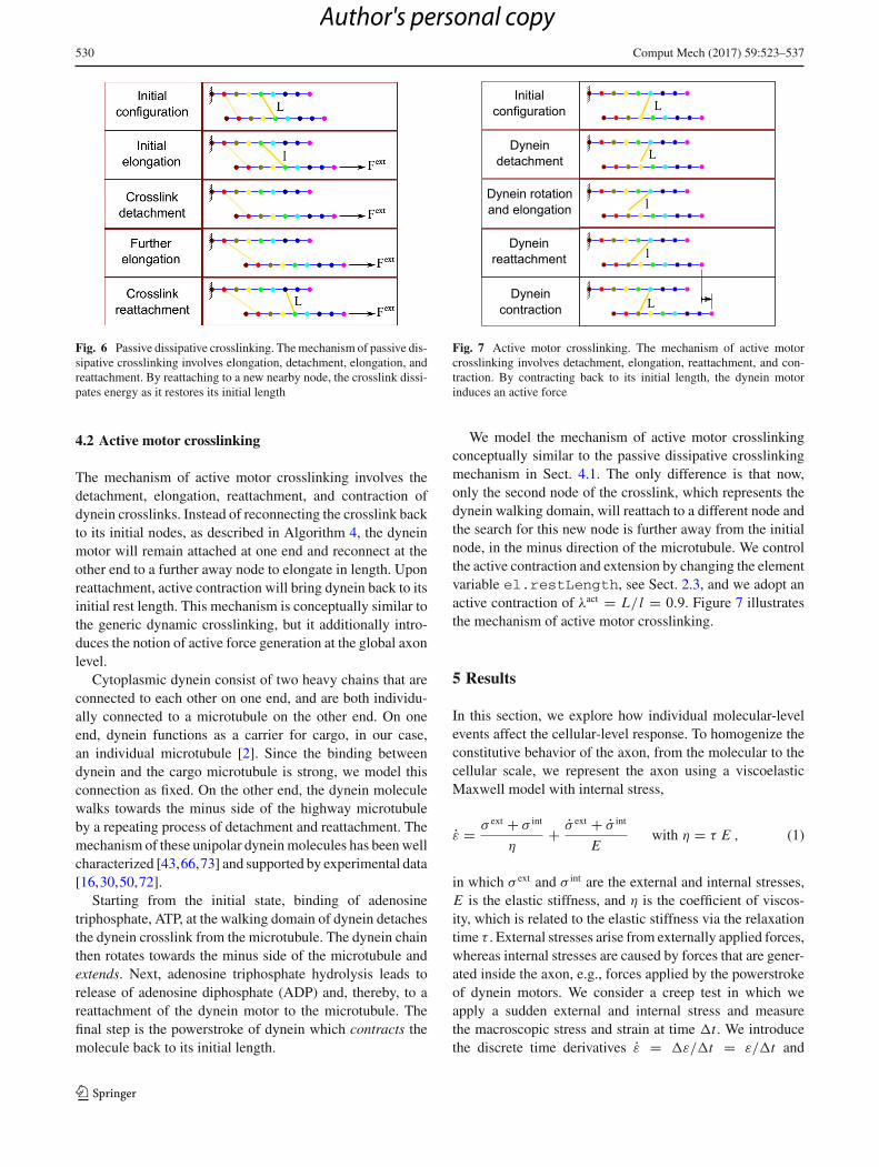
4.2 Active motor crosslinking
The mechanism of active motor crosslinking involves thedetachment, elongation, reattachment, and contraction ofdynein crosslinks. Instead of reconnecting the crosslink backto its initial nodes, as described in Algorithm 4, the dyneinmotor will remain attached at one end and reconnect at theother end to a further away node to elongate in length. Uponreattachment, active contraction will bring dynein back to itsinitial rest length. This mechanism is conceptually similar tothe generic dynamic crosslinking, but it additionally intro-duces the notion of active force generation at the global axonlevel.
Cytoplasmic dynein consist of two heavy chains that areconnected to each other on one end, and are both individu-ally connected to a microtubule on the other end. On oneend, dynein functions as a carrier for cargo, in our case,an individual microtubule [2]. Since the binding betweendynein and the cargo microtubule is strong, we model thisconnection as fixed. On the other end, the dynein moleculewalks towards the minus side of the highway microtubuleby a repeating process of detachment and reattachment. Themechanism of these unipolar dyneinmolecules has beenwellcharacterized [43,66,73] and supported by experimental data[16,30,50,72].
Starting from the initial state, binding of adenosinetriphosphate, ATP, at the walking domain of dynein detachesthe dynein crosslink from the microtubule. The dynein chainthen rotates towards the minus side of the microtubule andextends. Next, adenosine triphosphate hydrolysis leads torelease of adenosine diphosphate (ADP) and, thereby, to areattachment of the dynein motor to the microtubule. Thefinal step is the powerstroke of dynein which contracts themolecule back to its initial length.
Fig. 7 Active motor crosslinking. The mechanism of active motorcrosslinking involves detachment, elongation, reattachment, and con-traction. By contracting back to its initial length, the dynein motorinduces an active force
We model the mechanism of active motor crosslinkingconceptually similar to the passive dissipative crosslinkingmechanism in Sect. 4.1. The only difference is that now,only the second node of the crosslink, which represents thedynein walking domain, will reattach to a different node andthe search for this new node is further away from the initialnode, in the minus direction of the microtubule. We controlthe active contraction and extension by changing the elementvariable el.restLength, see Sect. 2.3, and we adopt anactive contraction of λact = L/ l = 0.9. Figure 7 illustratesthe mechanism of active motor crosslinking.
5 Results
In this section, we explore how individual molecular-levelevents affect the cellular-level response. To homogenize theconstitutive behavior of the axon, from the molecular to thecellular scale, we represent the axon using a viscoelasticMaxwell model with internal stress,
ε̇ = σ ext + σ int
η+ σ̇ ext + σ̇ int
Ewith η = τ E , (1)
in which σ ext and σ int are the external and internal stresses,E is the elastic stiffness, and η is the coefficient of viscos-ity, which is related to the elastic stiffness via the relaxationtime τ . External stresses arise from externally applied forces,whereas internal stresses are caused by forces that are gener-ated inside the axon, e.g., forces applied by the powerstrokeof dynein motors. We consider a creep test in which weapply a sudden external and internal stress and measurethe macroscopic stress and strain at time �t . We introducethe discrete time derivatives ε̇ = �ε/�t = ε/�t and
123
Author's personal copy

Comput Mech (2017) 59:523–537 531
σ̇ = �σ/�t = σ/�t , and obtain the following relationfor the elastic stiffness E ,
E =[1 + �t
τ
]σ ext + σ int
ε. (2)
We further introduce the effective or measured stiffnessEeff = σ ext/ε as the ratio of the externally applied stressσ ext and the measured strain ε, and interpret the actual stiff-ness E as the effective stiffness Eeff scaled by the effects ofviscoelasticity and internal stress,
E =[1 + �t
τ
] [1 + σ int
σ ext
]Eeff with Eeff = σ ext
ε. (3)
Similar to the effective stiffness Eeff, we introduce the effec-tive viscosity ηeff = σ ext/ε̇ as the ratio of the externallyapplied stress σ ext and the strain rate ε̇, and interpret theactual viscosity η as the effective viscosity ηeff scaled by theeffects of internal stress,
η =[1 + σ int
σ ext
]ηeff with ηeff = σ ext
ε̇. (4)
In the following two examples, we investigate how the mole-cular mechanisms of passive dissipative crosslinking andactive motor crosslinking effect the overall axonal viscos-ity through the term 1+�t/τ and the internal stress throughthe term 1 + σ int/σ ext.
5.1 Passive dissipative crosslinking
To explore the effects of the molecular mechanism of passivedissipative crosslinking on whole axon rheology, we use theaxon model from Sect. 3 and assign the dissipative dynamicmechanism of Sect. 4.1 to all crosslinks. On the molecu-lar level, the crosslinks are stretched when they detach andunstretched when they reattach, which inherently inducesenergy dissipation. On the axon level, this behavior collec-tively manifests itself as viscosity, and the axon as a wholebehaves as a rheological Maxwell element.
We perform a series of creep tests and apply an externalforce to the tip of the axon. We increase the external force toF ext = 100pN in the first time step and maintain this forceduring the remainder of the simulation. Unless stated oth-erwise, we use equal detachment and reattachment times oftDetach=tAttach=180s to maintain a constant aver-age crosslink density. Our total simulation time is 2000s andwe use a time step of 10s.
Figure 8 illustrates the typical simulation results of ourmodel with passive dissipative crosslinking. The three dis-cretizations show the axon at three consecutive time points ofthe simulation, top.The correspondingkymographs highlight
µ
Fig. 8 Axon with dissipative dynamic mechanism assigned to eachcrosslink and loaded by an external tip force. The finite element dis-cretization (top) shows the axon three consecutive time points. Thecorresponding numerical kymograph (bottom left) shows good quali-tative agreement with the experimentally obtained kymograph (bottomright) [55]
the longitudinal position of all microtubules as a functionof time, right. The computationally predicted kymograph,bottom left, shows an excellent qualitative resemblancewith the experimentally measured kymograph [55], bottomright.
Figure 9 summarizes the typical readouts for a single sim-ulation. The kymograph, top left, highlights the configurationof individual microtubules. For equal detachment and reat-tachment times tDetach and tAttach, the number ofcrosslinks remains constant during the simulation, bottomleft. The total stretch λ is composed of an initial elasticstretch, caused by the external force, followed by a linearlyincreasing viscous stretch, caused by the viscous crosslink-ing mechanism, top right. The linearly increasing stretch λ
results in a constant stretch rate λ̇, which we compute usinga linear regression of the stretch over an interval of 200saround t , middle right. The resulting macroscopic viscosityηeff = σ/ε̇, which we have calculated using Eq. (1) with thesmall strain assumption ε = λ − 1, increases initially butthen remains constant, bottom right.
To explore the effects of our model parameters on macro-scopic properties, we extract a single value for the axonalstiffness E and viscosity η from Fig. 9. We calculate theaxon stiffness E = σ ext/ε using Eq. (3) for the stiffness ofthe first time step in which the total external force is applied.We define the external stress as σ ext = F ext/Aaxn with anaxonal cross section area of Aaxn = π(r axn)2 and an axonalradius of r axn = 270nm, see Table 6. We calculate the axonviscosity η = σ ext/ε̇ using Eq. (4) and average the viscosityacross the time window of 400 and 2000s to ensure that themodel is in a steady state. Our model consists of several para-meters that may effect the macroscopic axon properties. Forbrevity and to maintain focus, here we only investigate the
123
Author's personal copy

532 Comput Mech (2017) 59:523–537
Fig. 9 Axon with dissipative dynamic mechanism assigned to eachcrosslink and loaded by an external tip force. The typical readout of asingle simulation includes a kymograph (top left), the total number of
crosslinks vs time (bottom left), axon stretch vs time (top right), axonstretch rate vs time (middle right), and axon viscosity vs time (bottomright)
Fig. 10 Axon stiffness Eeff and axon viscosity ηeff as emergent prop-erties in terms of the average crosslink density c. Data points and errorbars indicate the means and standard deviations. For crosslink densi-ties above 0.2nm−1, both stiffness and viscosity increase linearly withincreasing crosslink density; for crosslink densities below 0.2nm−1,stiffness and viscosity decrease rapidly to zero
twomost relevant parameters, the crosslink density, c, and thecharacteristic time constant, τ̃ = tAttach = tDetach.
Figure 10 shows the axon stiffness E = σ ext/ε and theaxon viscosity η = σ ext/ε̇ as functions of the crosslinkdensity c calculated as the number of crosslinks per axonunit length. The data points represents the mean stiffnessesand viscosities of n = 15 simulations; the error bars repre-
sent the standard deviations generated by the randomness inour model. Since the average crosslink density is only con-trolled indirectly by the longitudinal distance between twoconsecutive crosslinks, it is also a randomvariablewith corre-sponding means and standard deviations. Both axon stiffnessand viscosity increase linearly with the density of crosslinksfor an axon density above 0.2nm−1. For smaller crosslinkdensities, the stiffness and viscosity rapidly decrease to zero.Both observations are consistent with a previous report [41]:The rapid decrease of stiffness and viscosity at low crosslinkdensities is a natural result of the reduction of availableload paths within the axon as described by the percola-tion theory. Indeed, microtubules that would be connectedat high crosslinks densities become disconnected when lesscrosslinks are available.
Figure 11 shows the effective axon stiffness Eeff =E/[1+cτ �t/τ̃ ], the axon viscosityη = τ̃ E/cτ , the effectiverelaxation time τ eff = η/Eeff , and the normalized detach-ment and reattachment time τ̃ /τ eff = cτ /[1 + cτ �t/τ̃ ] asfunctions of the characteristic detachment and reattachmenttime τ̃ . Here we have reparameterized the relaxation timeτ = τ̃ /cτ via the characteristic detachment and reattach-ment time τ̃ scaled by the rate constant 1/cτ . The data pointsrepresents the mean values of n=15 simulations; the errorbars represent the standard deviations generated by the ran-domness in our model. The dashed lines represent the bestfit to the viscoelastic Maxwell model as described by theequations above. The two curves in each plot are generatedby the same data, but with different axon stiffnesses using�t = 0 s, shown in blue, and �t = 100 s, shown in orange.
123
Author's personal copy

Comput Mech (2017) 59:523–537 533
Fig. 11 Effective axon stiffness Eeff and axon viscosity η as emergentproperties in terms of the characteristic detachment and reattachmenttime τ̃ .Data points and error bars indicate themeans and standard devi-ations. Dashed lines represent the best fit to the viscoelastic Maxwellmodel
The only difference between these curves is that we mea-sured stiffness as the ratio of stress and strain after the 1stand 11th time step for�t = 0 s and�t = 100 s respectively.We performed individual fits for �t = 0 s and �t = 100 s,and obtained different values for E and cτ for the two curves.The numerical simulations nicely agree with the analyticalpredictions.
5.2 Active motor crosslinking
To explore the effects of the molecular mechanism of activemotor crosslinking on whole axon rheology, we use the axonmodel from Sect. 3 and assign the dynein motor mechanismof Sect. 4.2 to all crosslinks. On the molecular level, thecrosslinks detach, elongate to reattach, and actively contractduring the dynein power stroke as they return to their initiallength. On the axon level, this behavior collectively generatesinternal stresses in the axon.
Figure 12 highlights the three different scenarios that canoccur on the axon level in response to different dynein con-figurations: extension, neutral deformation, and contraction.Dynein is a molecular motor that walks towards the minusend of the microtubule. If the fixed domain of each dyneincrosslink is located closer to the plus end of the microtubule,the powerstroke of dynein induces axonal elongation. If thefixed domain is located closer to the minus end, the pow-erstroke induces axonal contraction. The tendencies towards
Fig. 12 Different dynein configurations and their effects on internalforce generation within the axon.When the fixed domains of the dyneinmotors are predominantly located at the plus ends of the microtubulues,the configuration extends (top); when the fixed domains are locatedat the minus ends, the configuration contracts (bottom); when thefixed domains are evenly distributed, the configuration remains neutral(middle)
Fig. 13 Axon with dynein motor mechanism assigned to eachcrosslink. Axonal stretch λ as a function of time t for n=15 simu-lations. For n=5 simulations with the fixed domains of the dyneinmotors predominantly located at the plus ends of the microtubulues,the axonal stretch is extensile (orange); for n=5 simulations with thefixed domains located at the minus ends, the axonal stretch is contrac-tile (yellow); for n=5 simulations with random orientation, the axonalstretch remains neutral (red). (Color figure online)
elongation and contraction cancel out if all fixed domains arerandomly oriented.
Figure 13 illustrates a simulation of the three differentscenarios, extensile, contractile, and neutral, for a total ofn=15 simulations of which n=5 have the fixed domainsof the dynein motors predominantly located at the plus endsof the microtubulues, n=5 have the fixed domains locatedat the minus ends, and n=5 have a random orientations.We clearly observe the expected difference in axon behaviorfor the three different configurations of the dynein crosslinks:The axonal stretch is extensile for the fixed domains locatednear the plus ends, contractile for the fixed domains locatednear the minus ends, and neutral for random orientations.Recent experiments have shown that the fixed domains ofdynein are predominantly associated with the plus end ofmicrotubules [63,75] and that dynein plays a critical role
123
Author's personal copy

534 Comput Mech (2017) 59:523–537
Fig. 14 Axon stretch rate ε̇ (top) and effective axon viscosityηeff (mid-dle and bottom) as a function of the external forcewith the fixed domainsof all dyneinmotors located at the plus ends of themicrotubulues to gen-erate axonal extension. Data points and error bars indicate the meansand standard deviations; numbers below each data point indicate thenumber of simulations used to create the data point. Dashed lines rep-resent the best fit to the viscoelastic Maxwell model with internal forcegeneration. The internal force that emerges from activemotor crosslink-ing, F int = 210pN, is the negative of the external force at amean stretchrate of zero, Fext = −210pN (top)
in axonal elongation [9,65]. These experiments suggest theextensile configuration in Fig. 12 is the most physiologicallyrelevant. In turn the modeling in Fig. 13 suggests the exper-imentally observed pushing force generated by dynein [65]arises as direct consequence the molecular association of thedynein cargo binding domain with microtubule plus tip pro-teins.
Figure 14 shows the stretch rate ε̇ = [ σ ext + σ int ]/η =[ F ext+F int ]/[ ηAaxn ] and effective viscosity ηeff = η/[ 1+σ int/σ ext ] = η/[ 1 + F int/F ext ] plotted versus the externalforce F ext. For these simulations, we use the same model asbefore, nowwith all dyneinmotors located at the plus ends ofthe microtubulues to generate a net internal force F int, whichwe balance by an external tip force F ext that we vary from−500 to 100pN. The values for the internal force F int and theviscosity η are unknown a priori, both emerge as axon-levelproperties of our axon model. Figure 14, top, provides theexternal force at amean stretch rate of zero, F ext = −210pN,which, by force equilibrium, F ext + F int = 0pN, defines theinternal force that emerges from active motor crosslinking,F int = −F ext = 210pN. We can then calculate the viscos-ity ηeff = ε̇ Aaxn/[ F ext + F int ], which approaches infinityηeff → ±∞ for F int → −F ext. Naturally, the numericalpredictions for viscosity are not able to capture this regionaccurately as a slight deviations in the computed stretch ratelead to enormous variations in the computed effective viscos-ity. However, our model captures the analytical predictionswell for
∣∣F ext − F int∣∣ > 100pN.
6 Discussion
Molecular mechanisms play a critical role in modulatingaxonal physiology, both by transmitting passive forces andby generating active forces. Here we created a generic axonmodel to simulate molecular mechanisms within the contextof the finite element method. Our infrastructure preservesthe inherent modularity of the finite element method and,at the same time, allows us integrate in a wide range ofmolecular mechanisms (Fig. 2). To illustrate the fundamen-tal features of our approach, we created an axon model ofdiscretemicrotubules that are aligned along the axon and con-nected to neighboring microtubules by discrete crosslinkingmechanisms (Figs. 3, 4). Our axon model naturally connectsmolecular-level events to axon-level properties and allows usto explore howcharacteristic axonal features emerge from thecollective interaction of individual molecular mechanisms.What makes our finite element model unique is that a fluid-like behavior (Figs. 10, 11) arises solely from a collection ofsprings, which dynamically make and break connections bya set of simple rules (Figs. 6, 7). The different configurationsof our model naturally capture the idea fundamental to activematter hydrodynamics that the relative orientation of internaland external force vectors profoundly influences the effec-tive viscoelastic properties of soft matter (Figs. 12, 13). Wehighlight these features by means of two examples: passivedissipative crosslinking and active motor crosslinking. Byassigning different mechanisms to standard finite elements,we automatically embed these molecular-level events withinthe solution procedure throughout the entire simulation.
Passive dissipative crosslinking characterizes a passivedetachment and reattachment of a crosslink from and to themicrotubule. A key feature of passive dissipative crosslink-ing is that each crosslink detaches at the elongated state andreattaches at its original rest length, which generates energydissipation and, on the axon level, an emergent rheologythat is conceptually similar to viscosity [15,22]. The majormolecular-level parameters that govern these events are theaverage crosslink density and the characteristic detachmentand reattachment time; the main axon-level parameters thatemerge from these events are the stiffness and the viscosity.To qualitatively compare our axon simulations to experi-ments, we extracted kymographs from our simulation andcompared them to kymographs of axonal stretch experiments[54,55] (Fig. 8). Simulation and experiment showed a goodqualitative agreement. To quantitatively compare molecular-level events to axon-level properties, we performed a seriesof simulations with systematically varying crosslink densi-ties and detachment and reattachment times. Our simulationsreveal that the axon stiffness and viscosity scale linearly withthe average crosslink density. The axon as a whole behaveslike a viscoelastic Maxwell element [22]: The axon stiff-ness is independent of the characteristic time constant of the
123
Author's personal copy

Comput Mech (2017) 59:523–537 535
crosslinks and the viscosity scales linearly with this timeconstant. The strong non-linear stress strain relationshipspredicted by active matter hydrodynamics arise elegantlyas emergent properties from a system of springs that obeysimple rules (Figs. 6, 7). In particular, by allowing springsto make and break connections viscosity arises as an emer-gent property (Fig. 9). This is consistent with experimentalwork that has demonstrated that neurons behave are solidsor fluids depending on the time scale [10,24,54]. In turn,the time-dependent apparent elasticity and viscosity followthe predictions outlined by theMaxwell fluid equations (Fig.11), which may be important for developing a better under-standing of the complex frequency dependence of neuronalrheological parameters in various experimental regimes [19].
Active motor crosslinking characterizes an active detach-ment and reattachment of a crosslink from and to themicrotubule. A key feature of active motor crosslinking isthat each crosslinks detaches at the contracted state andreattaches at its initial length, which generates active con-traction and, on the axon level, an emergent rheology thatis conceptually similar to an active force or internal stress[6,10,55]. The major molecular-level parameters that gov-ern these events are the average crosslink density and thecharacteristic detachment and reattachment time; the mainaxon-level parameters that emerge from these events are thestiffness and the viscosity. Another critical molecular-levelparameter that governs these events is the configuration ofdynein with respect to the plus and minus ends of the micro-tubules. Our simulations reveal that the axon extends whenthe fixed domains of the dynein motors are predominantlylocated at the plus ends of the microtubulues, it contractswhen the fixed domains are located at the minus ends, andit remains neutral when the fixed domains are evenly dis-tributed. In the extensile configuration, the axon as a wholebehaves like a viscoelastic Maxwell element with internalstress generation. Importantly, when assessing the effectiveviscoelastic properties in our active system, effective vis-cosity has the same non-linear pattern predicted by activematter hydrodynamics [51,56] (Fig. 14). While this is anobvious consequence of the kinematic equations, balanceequations, and constitutive equations that govern the system,the model inherently captures the complicated behavior thatarises as an emergent property of a few easily understoodrules [45,51,59]. This is in line with several recent micro-to-macro approaches proposed in the context of populationdynamics [82].
In conclusion, we have created a conceptually novelapproach to characterize axon-level parameters as evolvingproperties from molecular-level events. We have shown thataxon elasticity and axon viscosity increase linearly with thecrosslink density of microtubules, and that they are highlysensitive to the characteristic crosslink detachment and reat-tachment times. We have illustrated these effects for both
passive and active crosslinking mechanisms. Our simula-tions explain how dynein, a molecular motor whose fixeddomains are predominately associated with the plus end ofmicrotubules, generates internal stresses and drives axonelongation. We anticipate that our model will allow us usprobe a wide variety of molecular phenomena—both in iso-lation and in interaction—to explore their role in modulatingcharacteristic cellular features including stiffness, viscosity,and internal stress.
Acknowledgements This study was supported by the Stanford Grad-uate Fellowship to Rijk de Rooij and by the Bio-X IIP seed Grant‘Molecular Mechanisms of Chronic Traumatic Encephalopathy’ toEllen Kuhl.
References
1. Abe I, Tsujino A, Hara Y, Ichimura H, Ochiai N (2002) Paranodaldemyelination by gradual nerve stretch can be repaired by elonga-tion of internodes. Acta Neuropathol 104:505–512
2. Ahmad FJ, Echeverri CJ, Vallee RB, Baas PW (1998) Cytoplasmicdynein and dynactin are required for the transport of microtubulesinto the axon. J Cell Biol 140:391–401
3. Ahmad FJ, Hughey J,Wittmann T, HymanA, GreaserM, Baas PW(2000) Motor proteins regulate force interactions between micro-tubules andmicrofilaments in the axon.NatureCell Biol 2:276–280
4. Ahmadzadeh H, Smith DH, Shenoy VB (2014) Viscoelasticity oftau proteins leads to strain rate-dependent breaking ofmicrotubulesduring axonal stretch injury: predictions from a mathematicalmodel. Biophys J 106:1123–1133
5. Ahmadzadeh H, Smith DH, Shenoy VB (2015) Mechanical effectsof dynamic binding between tau proteins on microtubules duringaxonal injury. Biophys J 109:2328–2337
6. Ahmed WW, Rajagopalan J, Tofangchi A, Saif TA (2012) Neu-romechanics: the role of tension in neuronal growth and memory.Fundamentals and frontiers. Wiley, Chichester
7. Athamneh AIM, Suter DM (2015) Quantifying mechanical forcein axonal growth and guidance. Front Cell Neurosci 9:359
8. Ayali A (2010) The function of mechanical tension in neuronal andnetwork development. Integr Biol 2:178–182
9. Baas P (2001) Force generation by cytoskeletal motor proteins asa regulator of axonal elongation and retraction. Trends Cell Biol11:244–249
10. Bernal R, Pullarkat PA, Melo F (2007) Mechanical properties ofaxons. Phys Rev Lett 99:018301
11. Betz T, Koch D, Lu YB, Franze K, Käs JA (2011) Growth conesas soft and weak force generators. Proc Natl Acad Sci USA108:13420–13425
12. BrayD (1979)Mechanical tension produced by nerve cells in tissueculture. J Cell Sci 37:391–410
13. Bray D, Bunge MB (1981) Serial analysis of microtubules in cul-tured rat sensory axons. J Neurocytol 10:589–605
14. Bridgman PC, Dave S, Asnes CF, Tullio AN, Adelstein RS (2001)Myosin IIB is required for growth cone motility. J Neurosci21:6159–6169
15. Budday S, Nay R, de Rooij R, Steinmann P, Wyrobek T, OvaertTC, Kuhl E (2015) Mechanical properties of gray and white matterbrain tissue by indentation. J Mech Behav Biomed Mater 46:318–330
16. Burgess SA,Walker ML, Sakakibara H, Knight PJ, Oiwa K (2003)Dynein structure and power stroke. Nature 421:715–718
123
Author's personal copy

536 Comput Mech (2017) 59:523–537
17. Caminiti R, Carducci F, Piervincenzi C, Battaglia-Mayer A, Con-falone G, Visco-Comandini F, Pantano P, Innocenti GM (2013)Diameter, length, speed, and conduction delay of callosal axonsin macaque monkeys and humans: comparing data from histologyandmagnetic resonance imaging diffusion tractography. JNeurosci33:14501–14511
18. Carter NJ, Cross RA (2005) Mechanics of the kinesin step. Nature435:308–312
19. Chatelin S, Constantinesco A, Willinger R (2010) Fifty years ofbrain tissue mechanical testing: From in vitro to in vivo investiga-tions. Biorheology 47:255–276
20. Coles CH, Bradke F (2015) Coordinating neuronal actin-microtubule dynamics. Curr Biol 25:R677–R691
21. Conde C, Cáceres A (2009) Microtubule assembly, organizationand dynamics in axons and dendrites. Nat Rev Neurosci 10:319–332
22. de Rooij R, Kuhl E (2016) Constitutive modeling of brain tissue:current perspectives. Appl Mech Rev 68:1–16
23. Debanne D, Campanac E, Bialowas A, Carlier E, Alcaraz G (2011)Axon physiology. Physiol Rev 91(2):555–602
24. Dennerll TJ, Lamoureux P, Buxbaum RE, Heidemann SR (1989)The cytomechanics of axonal elongation and retraction. J Cell Biol109:3073–3083
25. Franze K, Janmey PA, Guck J (2013)Mechanics in neuronal devel-opment and repair. Annu Rev Biomed Eng 15:227–251
26. Franze K, Guck J (2010) The biophysics of neuronal growth. RepProg Phys 73:094601
27. Ganguly A, Tang Y,Wang L, Ladt K, Loi J, Dargent B, Leterrier C,Roy S (2015) A dynamic formin-dependent deep F-actin networkin axons. J Cell Biol 210:401–417
28. Garcia JA, Pena JM, McHugh S, Jerusalem A (2012) A model ofthe spatially dependent mechanical properties of the axon duringits growth. Comput Model Eng Sci 87:411–432
29. Garcia-Grajales JA, Jerusalem A, Goriely A (2016) Continuummechanical modeling of axonal growth. Comput Methods ApplMech Eng. doi:10.1016/j.cma.2016.07.032
30. Gennerich A, Carter AP, Reck-Peterson SL, Vale RD (2007)Force-induced bidirectional stepping of cytoplasmic dynein. Cell131:952–965
31. Gittes F, Mickey B, Nettleton J, Howard J (1993) Flexural rigidityof microtubules and actin filaments measured from thermal fluctu-ations in shape. J Cell Biol 120:923–934
32. Göktepe S, Menzel A, Kuhl E (2014) The generalized Hill model:a kinematic approach towards active muscle contraction. J MechPhys Solids 72:20–39
33. Goldberg JL (2003) How does an axon grow? Genes Dev 17:941–958
34. Goriely A, Budday S, Kuhl E (2015) Neuromechanics: from neu-rons to brain. Adv Appl Mech 48:79–139
35. Goriely A, Geers MGD, Holzapfel GA, Jayamohan J, JérusalemA, Sivaloganathan S, Squier W, van Dommelen JAW, Waters S,Kuhl E (2015) Mechanics of the brain: perspectives, challenges,and opportunities. Biomech Model Mechanobiol 14:931–965
36. Hällström W, Lexholm M, Suyatin DB, Hammarin G, Hess-man D, Samuelson L, Montelius L, Kanje M, Prinz CN (2010)Fifteen-piconewton force detection fromneural growth cones usingnanowire arrays. Nano Lett 10:782–787
37. Hill AV (1938) The heat of shortening and the dynamic constantsof muscle. Proc R Soc B 126:136–195
38. Hirokawa N (1982) Cross-linker system between neurofilaments,microtubules and membranous organelles in frog axons revealedby the quick-freeze, deep-etching method. J Cell Biol 94:129–142
39. HollandMA,Miller KE, Kuhl E (2015) Emerging brain morpholo-gies from axonal elongation. Ann Biomed Eng 43:1640–1653
40. Hyland C, Mertz AF, Forscher P, Dufresne E (2014) Dynamicperipheral traction forces balance stable neurite tension in regen-erating Aplysia bag cell neurons. Sci Rep 4:4961
41. Jakobs M, Franze K, Zemel A (2015) Force generation bymolecular-motor-powered microtubule bundles; implications forneuronal polarization and growth. Front Cell Neurosci 9:2761
42. Kawamura Y, Dyck PJ, Shimono M, Okazaki H, Tateishi J, Doi H(1981) Morphometric comparison of the vulnerability of periph-eral motor and sensory neurons in amyotrophic lateral sclerosis. JNeuropathol Exp Neurol 40:667–675
43. KingSM(2011)Dyneins: structure, biology anddisease.AcademicPress, Boston
44. KochD, RosoffWJ, Jiang J, Geller HM,Urbach JS (2012) Strengthin the periphery: growth cone biomechanics and substrate rigidityresponse in peripheral and central nervous systemneurons.BiophysJ 102:452–460
45. Kollmannsberger P, Fabry B (2011) Linear and nonlinear rheologyof living cells. Annu Rev Mater Res 41:75–97
46. Lamoureux P, Buxbaum RE, Heidemann SR (1989) Direct evi-dence that growth cones pull. Nature 340:159–162
47. Lamoureux P, Heidemann SR, Martzke NR, Miller KE (2010)Growth and elongation within and along the axon. Dev Neurobiol70:135–149
48. Lowery LA, Vactor DV (2009) The trip of the tip: understandingthe growth cone machinery. Nat Rev Mol Cell Biol 10:332–343
49. Lu W, Fox P, Lakonishok M, Davidson MW, Gelfand VI (2013)Initial neurite outgrowth in drosophila neurons is driven by kinesin-powered microtubule sliding. Curr Biol 23:1018–1023
50. Mallik R, Carter BC, Lex SA, King SJ, Gross SP (2004) Cyto-plasmic dynein functions as a gear in response to load. Nature427:649–652
51. Marchetti MC, Joanny JF, Ramaswamy S, Liverpool TB, Prost J,Rao M, Simha RA (2013) Hydrodynamics of soft active matter.Rev Model Phys 85:1143–1189
52. Moulton DE, Lessinnes T, Goriely A (2013) Morphoelastic rods.Part I: a single growing elastic rod. JMech Phys Solids 61:398–427
53. Nagy S, Ricca BL, Norstrom MF, Courson DS, Brawley CM,Smithback PA, Rock RS (2008) A myosin motor that selects bun-dled actin for motility. Proc Natl Acad Sci USA 105:9616–9620
54. O’Toole M, Lamoureux P, Miller KE (2008) A physical model ofaxonal elongation: force, viscosity, and adhesions govern the modeof outgrowth. Biophys J 94:2610–2620
55. O’Toole M, Lamoureux P, Miller KE (2015) Measurement of sub-cellular force generation in neurons. Biophys J 108:1027–1037
56. O’Toole M, Lamoureux P, Miller KE (2016) Forces control effec-tive viscosity in neurons (submitted for publication)
57. Pannese E (2015) Neurocytology: fine structure of neurons, nerveprocesses, and neuroglial cells. Springer, Berlin
58. Peter SJ, Mofrad MRK (2012) Computational modeling of axonalmicrotubule bundles under tension. Biophys J 102:749–757
59. Pritchard RH, Huang YYS, Terentjev EM (2014) Mechanics ofbiological networks: from the cell cytoskeleton to connective tissue.Soft Matter 10:1864–1884
60. Rajagopalan J, Tofangchi A, Saif T (2010) Drosophila neuronsactively regulate axonal tension in vivo. Biophys J 99:3208–3215
61. Rausch MK, Kuhl E (2013) On the effect of prestrain and residualstress in thin biological membranes. J Mech Phys Solids 61:1955–1969
62. Recho P, Jérusalem A, Goriely A (2016) Growth, collapse, andstalling in a mechanical model for neurite motility. Phys Rev E93:032410
63. Roberts AJ, Goodman BS, Reck-Peterson SL, BalasubramanianM(2014) Reconstitution of dynein transport to the microtubule plusend by kinesin. eLife 3:e02641
123
Author's personal copy

Comput Mech (2017) 59:523–537 537
64. Rodriguez ML, McGarry PJ, Sniadecki NJ (2013) Review on cellmechanics: experimental and modeling approaches. Appl MechRev 65:060801
65. RoossienDH, Lamoureux P,Miller KE (2014) Cytoplasmic dyneinpushes the cytoskeletal meshwork forward during axonal elonga-tion. J Cell Sci 127:3593–3602
66. Roossien D, Miller K, Gallo G (2015) Ciliobrevins as tools forstudying dynein motor function. Front Cell Neurosci 9:252
67. Siechen S, Yang S, Chiba A, Saif T (2009) Mechanical tensioncontributes to clustering of neurotransmitter vesicles at presynapticterminals. Proc Natl Acad Sci USA 106:12611–12616
68. SmithDH (2009) Stretch growth of integrated axon tracts: extremesand exploitations. Prog Neurobiol 89:231–239
69. Soheilypour M, Peyro M, Peter SJ, Mofrad MRK (2015) Buck-ling behavior of individual and bundled microtubules. Biophys J108:1718–1726
70. Suresh S (2006) Biomechanics and biophysics of cancer cells. ActaBiomater 3:413–438
71. Suter DM,Miller KE (2011) The emerging role of forces in axonalelongation. Prog Neurobiol 94:91–101
72. Toba S, Watanabe TM, Yamaguchi-Okimoto L, Toyoshima YY,Higuchi H (2006) Overlapping hand-over-hand mechanism of sin-gle molecular motility of cytoplasmic dynein. Proc Natl Acad SciUSA 103:5741–5745
73. Vale RD (2003) The molecular motor toolbox for intracellulartransport. Cell 112:467–480
74. van den Bedem H, Kuhl E (2015) Tau-ism: the yin and yang ofmicrotubule sliding, detachment, and rupture. Biophys J 109:2215–2217
75. Vaughan KT, Tynan SH, Faulkner NE, Echeverri CJ, Vallee RB(1999) Colocalization of cytoplasmic dynein with dynactin andCLIP-170 at microtubule distal ends. J Cell Sci 112:1437–1447
76. Xu K, Zhong G, Zhuang X (2013) Actin, spectrin, and associatedproteins form a periodic cytoskeletal structure in axons. Science339:452–456
77. Yildiz A, Selvin P (2005) Kinesin: walking, crawling or slidingalong? Trends Cell Biol 15:112–120
78. YuW, Baas PW (1994) Changes in microtubule number and lengthduring axon differentiation. J Neurosci 14:2818–2829
79. ZemelA,MogilnerA (2009)Motor-induced sliding ofmicrotubuleand actin bundles. Phys Chem Chem Phys 11:4821–4833
80. Zhang H, Berg JS, Li Z, Wang Y, Lång P, Sousa AD, BhaskarA, Cheney RE, Strömblad S (2004) Myosin-X provides a motor-based link between integrins and the cytoskeleton. Nat Cell Bio6:523–531
81. Zohdi TI (2007) A computational framework for network model-ing of fibrous biological tissue deformation and rupture. ComputMethods Appl Mech Eng 196:2972–2980
82. Zohdi TI (2016) An agent-based computational framework forsimulation of competing hostile planet-wide populations. ComputMethods Appl Mech Eng. doi:10.1016/j.cma.2016.04.032
83. Zöllner AM, Abilez OJ, Böl M, Kuhl E (2012) Stretching skele-tal muscle: chronic muscle lengthening through sarcomerogenesis.PLoS ONE 7:e45661
123
Author's personal copy