molecular mechanisms of acquired gemcitabine …
TRANSCRIPT

MOLECULAR MECHANISMS OF ACQUIRED GEMCITABINE
RESISTANCE IN PANCREATIC CANCER
Li Qin
Submitted to the faculty of the University Graduate School in partial fulfillment of the requirements
for the degree Doctor of Philosophy
in the Department of Pharmacology and Toxicology, Indiana University
November 2014

ii
Accepted by the Graduate Faculty, of Indiana University, in partial fulfillment of the requirements for the degree of Doctor of Philosophy.
________________________________ Jian-Ting Zhang, Ph.D., Chair
________________________________ Nickolay Brustovetsky, Ph.D. Doctoral Committee
________________________________ Karen E. Pollok, Ph.D. September 23, 2014
________________________________ Ahmad R. Safa, Ph.D.
________________________________ William J. Sullivan, Ph.D.
________________________________ Jingwu Xie, Ph.D.

iii
ACKNOWLEDGEMENTS
My love for science was given to me by my sister, my parents and grandparents
and it is to them that I owe my deepest debt of gratitude. They gave me encouragement
of endless efforts for pursuing my goals, and the determination and confidence to reach
them. Their many years of support have been a tremendous source of encouragement.
I would also like to express my deepest appreciation to the many fine individuals
who make up this department, especially the members of my committee, Nickolay
Brustovetsky, Karen E. Pollok, Ahmad R. Safa, William J. Sullivan, and Jingwu Xie.
Together they have guided me through this precious journey, and helped me more than
they can know. A very special thanks goes to Zi-zheng Dong and Jing Qi who have a way
of making things go right and are always there patiently instructing me and guiding me
through my projects, and my research would not be so smooth without their help.
Finally, my gratitude also goes to the many fine scientists, present and passed, who have
made the Zhang lab such a great place to be.
For my advisor I cannot begin to express the depth of my appreciation. JT Zhang
has been more than my mentor; he has been my benefactor and patron saint. Whatever
contribution I have made or will make to science I truly owe to his patient instruction
and counsel. Last but not the least, thanks to the entire faculty, graduate students, and
staff in Department of Pharmacology and Toxicology for their help and support
throughout these years. I truly appreciate everyone encountered in my life in United
States to make my adventure here colorful and unregretful.

iv
Li Qin
MOLECULAR MECHANISMS OF ACQUIRED GEMCITABINE RESISTANCE IN PANCREATIC
CANCER
Most pancreatic cancer patients receiving gemcitabine chemotherapy eventually
develop resistance to gemcitabine. To improve survival and prognosis of pancreatic
cancer patients, better understanding the mechanisms of gemcitabine resistance and
discovery of new therapeutic targets are required. In this study, I investigated the
molecular mechanisms of acquired gemcitabine resistance using a stepwise
gemcitabine-selected pancreatic cancer cell line in comparison to the parental cell line. I
found that 14-3-3σ is up-regulated in the drug resistant cell line due to demethylation in
its first exon, and the up-regulation of 14-3-3σ gene expression, in turn, contributes to
gemcitabine resistance. Intriguingly, I found that demethylation of the 14-3-3σ gene in
gemcitabine resistant cells is reversibly regulated by DNMT1 and UHRF1. Furthermore, I
found that 14-3-3σ over-expression causes gemcitabine resistance by inhibiting
gemcitabine-induced apoptosis and caspase-8 activation possibly via binding to YAP1.
The finding of demethylation of the 14-3-3σ gene in gemcitabine resistant cells led to a
hypothesis that other genes may also be changed epigenetically following gemcitabine
selection. By RRBS (Reduced Representation Bisulfite Sequencing) analysis, 845 genes
were found to have altered methylation. One of these genes, PDGFD, was further

v
investigated and found to have reversible demethylation at its promoter region in the
drug resistant cells and contribute to gemcitabine resistance possibly via autocrine
activation of the STAT3 signaling pathway. Together, these findings not only provide
evidence that 14-3-3σ and PDGFD over-expression contribute to acquired gemcitabine
resistance and that reversible epigenetic changes may play an important role in
acquired gemcitabine resistance, but also demonstrate that the molecular mechanisms
of acquired gemcitabine resistance in pancreatic cancer cells are complex and
multifaceted.
Jian-Ting Zhang, Ph.D., Chair

vi
Table of Contents
List of Tables ........................................................................................................................ x
List of Figures ...................................................................................................................... xi
List of Abbreviations ......................................................................................................... xiv
Introduction ........................................................................................................................ 1
A. Pancreatic ductal adenocarcinoma (PDAC) ...................................................... 1
B. Gemcitabine in pancreatic cancer treatment ................................................... 2
C. Gemcitabine metabolism and known resistance mechanisms ........................ 3
D. 14-3-3 sigma and 14-3-3 family ........................................................................ 8
E. Association of 14-3-3σ expression with drug resistance ................................ 10
F. YAP1, a potential binding partner of 14-3-3σ ................................................. 13
G. 14-3-3σ gene methylation in cancers ............................................................. 15
H. Uhrf1/DNMT1 complex, an emerging regulator of gene methylation ........... 18
I. Platelet-derived growth factor (PDGF) family ................................................ 22
J. PDGFD over-expression in human cancers ..................................................... 24
K. Specific aims of the present work ................................................................... 25
Materials and Methods .................................................................................................... 29
A. Materials ......................................................................................................... 29
B. Cell lines, cell cultures, and transfections ....................................................... 30
C. Cell lysate preparation, TCA protein precipitation and Western blot ............ 31
D. Membrane preparation .................................................................................. 32

vii
E. Survival and apoptosis assay........................................................................... 33
F. Quantitative real-time RT-PCR ........................................................................ 34
G. Immunofluorescence and confocal microscope imaging ............................... 35
H. Immunoprecipitation assay ............................................................................ 36
I. Chromatin-immunoprecipitation (ChIP) Assay ............................................... 36
J. Genomic DNA isolation, bisulfite modification, methylation-specific PCR
and sequencing ............................................................................................... 37
K. Reduced representation bisulfite sequencing (RRBS) and data analysis ....... 38
Results ............................................................................................................................... 40
Section I: Detailed mechanism of reversible epigenetic regulation of 14-3-3σ
during gemcitabine selection
A. A gemcitabine-selected pancreatic cancer cell line is cross-resistant to Ara-C
but not to other anticancer drugs .................................................................. 40
B. Ribonucleotide reductase and 14-3-3σ are up-regulated in G3K cells ........... 43
C. 14-3-3σ over-expression contributes to acquired gemcitabine resistance .... 46
D. Differential methylation of 14-3-3σ gene in MiaPaca-2 and G3K cells .......... 50
E. Demethylation of the 14-3-3σ gene is reversible ........................................... 54
F. Uhrf1 and DNMT1 play important roles in regulating 14-3-3σ expression .... 57
G. Uhrf1 binds and helps recruit DNMT1 to methylated region of 14-3-3σ gene62
H. Gemcitabine treatment does not affect Uhrf1 and DNMT1 expression ........ 65

viii
Section II: Identification of PDGFD as a potential contributor to acquired
gemcitabine resistance by Reduced Representation Bisulfite Sequencing (RRBS)
A. Identification of reversibly-methylated genes using Reduced Representation
Bisulfite Sequencing (RRBS) ............................................................................ 68
B. Reversible up-regulation of PDGFD ................................................................ 76
C. Validation of reversible methylation change of PDGFD gene ........................ 79
D. PDGFD up-regulation plays an important role in gemcitabine resistance ..... 82
E. PDGFD over-expression contributes to gemcitabine resistance possibly by
regulating STAT3 signaling pathway ............................................................... 86
F. Stat3 potentially contributes to gemcitabine resistance ............................... 89
Section III: Molecular mechanism of 14-3-3σ-mediated gemcitabine resistance
in pancreatic cancer
A. YAP1 over-expression in G3K cells and its contribution to gemcitabine
resistance ........................................................................................................ 92
B. Gemcitabine resistance requires both 14-3-3σ and YAP1 .............................. 98
C. 14-3-3σ and YAP1 form a complex .............................................................. 103
D. Both 14-3-3σ and YAP1 protect against gemcitabine-induced caspase-8
activation and apoptosis ............................................................................... 105
Discussion ....................................................................................................................... 112
Summary and Conclusion .............................................................................................. 124
Future Plans .................................................................................................................... 127
Appendices ..................................................................................................................... 132

ix
Appendix 1. List of 230 genes the methylations of which are increased in G3K
cells compared to MiaPaca-2 cells ...................................................................... 132
Appendix 2. List of 615 genes the methylations of which are decreased in G3K
cells compared to MiaPaca-2 cells ...................................................................... 134
Appendix 3. List of 140 genes of which the methylations are increased in
G3KRev cells compared to G3K cells ................................................................... 137
Appendix 4. List of 142 genes of which the methylations are decreased in
G3KRev cells compared to G3K cells ................................................................... 138
Appendix 5. List of 309 genes of which the methylations are increased in
G3KRev cells comparing with MiaPaca2 cells ..................................................... 139
Appendix 6. List of 645 genes of which the methylations are decreased in
G3KRev cells comparing with MiaPaca2 cells ..................................................... 141
References ...................................................................................................................... 145
Curriculum Vitae

x
LIST OF TABLES
Table 1. Frequency of 14-3-3σ gene methylation in different types of cancers .............. 17
Table 2. List of reversibly methylated genes .................................................................... 72
Table 3. Identification and characterization of candidate genes ..................................... 75

xi
LIST OF FIGURES
Figure 1. Gemcitabine metabolic schema and proposed pharmacological mechanisms
of gemcitabine and its metabolites .................................................................................... 6
Figure 2. A successive DNA transfer model for maintenance DNA methylation by Uhrf1
and DNMT1 ....................................................................................................................... 21
Figure 3. PDGF proteins and PDGF-PDGFR interactions ................................................... 23
Figure 4. Drug response profiles of MiaPaca-2 and its derivative G3K cells .................... 42
Figure 5. Ribonucleotide reductase and 14-3-3σ are up-regulated in G3K cells .............. 45
Figure 6. 14-3-3σ up-regulation contributes to gemcitabine and Ara-C resistance ......... 48
Figure 7. Methylation status of 14-3-3σ gene in MiaPaca-2 and G3K cells ...................... 52
Figure 8. 14-3-3σ up-regulation in Adriamycin-selected MCF7/AdVp3000 cells is not
due to gene demethylation .............................................................................................. 56
Figure 9. Role of Uhrf1 and DNMT1 in 14-3-3σ expression .............................................. 60
Figure 10. Binding of Uhrf1 and DNMT1 to the CpG islands of the 14-3-3σ gene ........... 64
Figure 11. Effect of gemcitabine treatment on Uhrf1 and DNMT1 expression ............... 66
Figure 12. Schematic model of epigenetic regulation of 14-3-3σ gene following
gemcitabine selection and drug removal ......................................................................... 67
Figure 13. RRBS sequencing results comparison and characterization of genomic
coverage ............................................................................................................................ 69
Figure 14. Flowchart of DMR filteration of genes ............................................................ 71
Figure 15. Identification of transcriptional changes in candidate genes ......................... 74

xii
Figure 16. Identification of TRPC3 and PDGFD expression in MiaPaca-2 and G3K cells .. 77
Figure 17. Reversible transcription and expression of PDGFD ......................................... 78
Figure 18. Reversible methylation change of PDGFD gene .............................................. 80
Figure 19. PDGFD over-expression contributes to gemcitabine resistance in both G3K
and Aspc-1 cells ................................................................................................................. 84
Figure 20. PDGFD over-expression leads to increased gemcitabine resistance in
MiaPaca-2 cells ................................................................................................................. 85
Figure 21. PDGFD actively regulates STAT3 signaling pathway ........................................ 88
Figure 22. Stat3 over-expression promotes gemcitabine resistance in MiaPaca-2 cells . 90
Figure 23. Schematic model of PDGFD-mediated gemcitabine resistance during drug
selection ............................................................................................................................ 91
Figure 24. YAP1 is over-expressed in resistant G3K cells ................................................. 94
Figure 25. Regulation of YAP1 expression by 14-3-3σ protein level ................................ 95
Figure 26. YAP1 over-expression contributes to gemcitabine resistance, but requires
the presence of 14-3-3σ.................................................................................................... 96
Figure 27. Gemcitabine resistance requires both 14-3-3σ and YAP1 ............................. 100
Figure 28. Decreased expression of both 14-3-3σ and YAP1 by RNA knockdown does
not further reduce drug resistance ................................................................................. 102
Figure 29. 14-3-3σ interacts and binds with YAP1 in vitro ............................................. 104
Figure 30. Decreased expression of 14-3-3σ by RNA knockdown promotes parp-1
cleavage and apoptotic cell death .................................................................................. 107

xiii
Figure 31. Decreased expression of either 14-3-3σ or YAP1 by RNA knockdown leads
to increased parp-1 cleavage and caspase-8 activation ................................................. 108
Figure 32. 14-3-3σ over-expression in MiaPaca-2 cells protects against parp-1 cleavage
and caspase-8 activation................................................................................................. 110
Figure 33. Schematic model of 14-3-3σ-mediated gemcitabine .................................... 111

xiv
List of Abbreviations
5-Aza-dC 5-aza-2'-deoxycytidine
ATP Adenosine triphosphate
BSA Bovine serum albumin
CDA Cytidine deaminase
CDC2 Cell division cycle protein 2
CDP Cytidine diphosphate
ChIP Chromatin immunoprecipitation
CHK1 Checkpoint kinase 1
CM Conditioned medium
Co-IP Co-immunoprecipitation
dCK Deoxycytidine kinase
dCDP Deoxycytidine diphosphate
dCTP Deoxycytidine triphosphate
dCMPD Deoxycytidylate deaminase
dFdC 2′,2′-difluorodeoxycytidine
dFdCDP dFdC diphosphate
dFdCTP dFdC triphosphate
dFdCMP dFdC monophosphate
dFdU 2′,2′-difluorodeoxyuridine
dFdUMP 2′,2′-difluorodeoxyuridine monophosphate
DNA Deoxyribonucleic acid
DNMT DNA methyltransferases

xv
DMEM Dulbecco’s modification of Eagle’s medium
DMR Differentially methylated region
DMSO Dimethyl sulfoxide
DRs Death receptors
DTT Dithiothreitol
EDTA Ethylenediaminetetraacetic acid
EGFR Epidermal growth factor receptor
EGTA Ethylene glycol tetraacetic acid
ERK Extracellular signal-regulated kinase
FBS Fetal bovine serum
GAPDH Glyceraldehyde 3-phosphate dehydrogenase
HDAC1 Histone Deacetylase
hENT1 Human equilibrative nucleoside transporters 1
LATS Large tumour suppressor
MMP Matrix metalloproteinase
MSP Methylation-specific PCR
NDPK Nucleoside diphosphate kinase
NMPK Nucleoside monophosphate kinase
PBS Phosphate buffered saline
PCR Polymerase chain reaction
PCNA Proliferating-cell nuclear antigen
PDAC Pancreatic ductal adenocarcinoma
PDGF Platelet-derived growth factor
PHD Plant Homeo Domain

xvi
PI3K Phosphatidylinositol 3 kinase
PMSF Phenylmethylsulfonyl fluoride
PVDF Polyvinylidene difluoride
RB1 Retinoblastoma protein 1
RING Really Interesting New Gene
RNA Ribonucleic acid
RRBS Reduced Representation Bisulfite Sequencing
RRF Relative resistance factor
RRM1/2 Ribonucleotide reductase M1/M2
SEM Standard error of mean
SD Standard deviation
SDS-PAGE Sodium dodecyl sulfate-polyacrylamide gel electrophoresis
SRA Set and Ring Associated
STAT Signal transducer and activator of transcription
TCA Trichloroacetic acid
TE buffer Tris-EDTA buffer
Tip60 Tat-Interactive Protein of 60 KDa
TRPC3 Transient receptor potential cation channel, subfamily C
TSGs Tumor suppressor genes
TTD Cryptic Tandem Tudor Domain
UBL Ubiquitin-like domain
UHRF1 Ubiquitin-like, containing PHD and RING finger domains 1
VEGF Vascular endothelial growth factor
YAP1 Yes-associated protein

1
Introduction
A. Pancreatic ductal adenocarcinoma (PDAC)
Pancreatic cancer ranks the fourth most common cause of human death by
cancer in the western world, with a 5-year survival rate less than 5% for all stages of the
disease and a median survival of 6 months after diagnosis, thereby exhibiting the
poorest prognosis of all solid tumors [1-3]. Pancreatic cancer has an annual mortality
rate of approximately 95% with over 250,000 patients dying worldwide [4]. In 2014, an
estimated 46,420 people will be diagnosed with pancreatic cancer in the United States,
and approximately 39,590 people will die from the disease (please see
http://www.cancer.org/cancer/pancreaticcancer/detailedguide/pancreatic-cancer-key-
statistics). What makes it so lethal is its stealth, which means that it exhibits no clear
early warning signs or symptoms and therefore often goes undetected until it is
advanced and too late for resection. In the vast majority of cases, symptoms only
develop after pancreatic cancer has already grown and begun to spread.
Pancreatic ductal adenocarcinoma, or PDAC, is by far the most common type of
pancreatic malignancy. Although surgical resection remains the only curative
intervention and offers the best patient outcome for this disease, surgical removal of
the tumor is possible in only approximately 15% of the patients [5]. Therefore, the poor
survival rate is mainly attributed to the late detection of PDAC and emergence of a
largely drug-resistant phenotype over time.

2
B. Gemcitabine in pancreatic cancer treatment
Routine treatment options to improve prognosis in patients with pancreatic
cancer are limited. Its array of treatments includes but is not limited to chemotherapy,
radiotherapy, immunotherapy, hormonal therapy, medications, surgery, and nutritional
therapy. At present, single-agent gemcitabine, which has been considered the standard
of care since 1997, is recommended as first-line chemotherapy for patients with
advanced pancreatic cancer and has been extensively studied in phase II and III trials.
Gemcitabine is a deoxycytidine analogue and was approved by FDA in 1997 as
the first-line chemotherapeutic drug for patients with locally advanced or metastatic
pancreatic adenocarcinoma [6, 7]. It functions by either directly and competitively
incorporating into DNA or inhibiting ribonucleotide reductase M1 or M2 (RRM1/RRM2)
to prevent DNA replication and, thereby, interrupting DNA synthesis and inhibiting
cancer cell growth [8]. However, although gemcitabine is the standard and most
commonly used drug for treatment of pancreatic cancer, almost all patients would
eventually develop resistance to this therapeutic agent. Over the past decade,
numerous trials have been conducted to improve the outcome in patients by
combination therapies using gemcitabine as backbone. Currently, combinational
treatments using gemcitabine and other therapeutics have shown some promise but no
real significant improvements in overall survival rates. So far, gemcitabine with erlotinib,
an epidermal growth factor receptor (EGFR) tyrosine kinase inhibitor, is the only FDA-

3
approved combination treatment for PDAC. Unfortunately, this regimen only has a
modest effect to prolong median overall survival of patients for less than 2 weeks [9] .
Although gemcitabine monotherapy or in combination with other agents has become
standard chemotherapy for the treatment of PDAC, gemcitabine imparts a progression-
free survival interval ranging from 0.9 to 4.2 months only [10]. Therefore, the effect of
gemcitabine on survival has been disappointing. Due to the characterization of PDAC by
a high propensity for local invasion and distant metastasis as well as early relapses and
largely drug-resistant phenotype, overcoming the gemcitabine drug-resistant phenotype
has become a hot topic in this field. Understanding acquired gemcitabine resistance
could lead to better improvements in the outcome for patients with pancreatic cancer.
In order to do so, I strongly suggest that a better understanding of the molecular
mechanisms by which gemcitabine resistance arises is likely to lead to novel therapeutic
strategies for the successful treatment of patients diagnosed with pancreatic cancer.
C. Gemcitabine metabolism and known resistance mechanisms
Gemcitabine (2',2'-difluorodeoxycytidine, dFdC) is a prodrug that requires
cellular uptake and intracellular phosphorylation into its active metabolites, gemcitabine
diphosphate and triphosphate [8]. As shown in Figure 1, gemcitabine is transported into
cells via human equilibrative nucleoside transporter-1 protein (hENT1), where it is
phosphorylated by the rate-limiting enzyme deoxycytidine kinase (dCK) into
gemcitabine monophosphate (dFdCMP), and is further phosphorylated into active

4
metabolites, the gemcitabine diphosphate (dFdCDP) and gemcitabine triphosphate
(dFdCTP) by nucleoside monophosphate kinase (NMPK) and nucleoside diphosphate
kinase (NDPK) respectively [11, 12]. Gemcitabine triphosphate (dFdCTP) is incorporated
into DNA, thereby competing with dCTP for incorporation. Once dFdCTP is incorporated,
two phosphate molecules are split off and thus leave dFdCMP in the DNA chain, while it
allows only one more deoxynucleoside triphosphate to be incorporated, after which
DNA replication terminates. dFdCMP is resistant to be removed from the DNA strand by
proofreading enzymes (i.e., polymerase ε), leading to impairment of their ability to
repair the DNA strand, which is the mechanism also known as “masked-chain
termination.” Gemcitabine diphosphate (dFdCDP), on the other hand, inhibits
ribonucleotide reductase M1 or M2 (RRM1/RRM2) that convert CDP to dCDP, leading to
depletion of dCTP pools and facilitating incorporation of dFdCTP into DNA [13]. As
depicted in Figure 1, gemcitabine has various self-potentiating mechanisms that
contribute to the maintenance of dFdCDP and dFdCTP levels for prolonged periods of
time. For example, since dCTP inhibits dCK, decreased dCTP pools from RRM1/RRM2
inhibition by dFdCDP can result in higher dCK activity and thus an increase in
phosphorylation of dFdC to its active metabolites. In addition, dFdCTP can inhibit
deoxycytidylate deaminase (dCMPD), an enzyme that deaminates dFdCMP to its inactive
form dFdUMP, leading to a potentiation of its own formation.
It is known that high intracellular accumulation of dFdCTP and incorporation into
DNA are associated with greater sensitivity to gemcitabine in preclinical tumor models

5
[14]. Moreover, clonogenic survival assays demonstrated that increased gemcitabine
concentrations result in a decrease of cell viability, which suggested a prolonged periods
of time for intracellular retention of active gemcitabine metabolites [15, 16]. The
intracellular accumulation of active metabolites and cytotoxicity of gemcitabine are
influenced by multiple factors, such as (a) the dosing schedule, (b)
phosphorylation/activation by dCK, (c) cellular uptake via hENT1, (d)
degradation/inactivation through CDA, and (e) genetic factors (e.g., single nucleotide
polymorphisms in dCK and CDA) [17-20].

6
Figure 1. Gemcitabine metabolic schema and proposed pharmacological mechanisms
of gemcitabine and its metabolites. Transportation of dFdC into the cell is mediated by
hENT1, which is followed by intracellular phosphorylation by dCK to its monophosphate
dFdCMP, and subsequently into its active dFdCDP and dFdCTP metabolites. dFdCTP is
incorporated into DNA, thereby competing with dCTP for incorporation. dFdCDP inhibits
RRM1/2, which prevents the conversion of CDP to dCDP and thus reduced synthesis of
dCTP, leading to an elevation of intracellular dFdCTP/dCTP ratio and enhanced
incorporation of dFdCTP into DNA.

7
Remarkable progress has been made during the last decade toward identifying
and understanding the complicated signaling pathways that contribute to the initiation,
progression of PDAC, and signaling pathways that contribute to intrinsic and acquired
gemcitabine resistance in pancreatic cancers. To date, several molecular markers to
predict gemcitabine sensitivity have been reported and investigated with or without
relation to gemcitabine metabolism, including messenger RNA (mRNA) and microRNA,
as well as genes related to gemcitabine metabolism and transport, such as
deoxycytidine kinase, ribonucleotide reductase, and human equilibrative nucleoside
transporter-1 [21-24]. However, the potential use of such markers in clinical settings
remains limited due to the difficulties in evaluating their protein or mRNA levels in
clinical samples. For example, accurate quantitative analyses of mRNA from clinical
samples are often difficult as a result of degradation. Therefore, more reliable methods-
based biomarkers are needed to predict responses to gemcitabine.
The most studied gemcitabine resistance mechanisms are the dysregulation of
the enzymes participating in gemcitabine metabolism pathways, including down-
regulation of transporter hENT1, down-regulation of rate-limiting enzyme dCK, and up-
regulation of RRM1/RRM2 [25-31]. It suggests that the ratio of the expression level of
these four genes (hENT1 Χ dCK)/ (RRM1 Χ RRM2) decreased progressively with
development of acquired gemcitabine resistance and, thus, this ratio correlates with
acquired gemcitabine resistance in pancreatic cancer cells, which may be a useful
predictive marker for the efficacy of gemcitabine chemotherapy in pancreatic cancer

8
patients [30]. Other studies show changes in a variety of cellular signaling pathways
such as Akt/mTOR signaling pathway and NF-kB signaling pathway, as well as
malfunction of proteins involved in cell survival/apoptosis pathway [32-34]. Emerging
evidence suggests molecular and phenotypic association between gemcitabine
resistance and acquisition of epithelial-mesenchymal transition (EMT)-like phenotype of
pancreatic cancer cells [35-39]. This process is also believed to be reminiscent of “cancer
stem-like cells” characteristics in many cancer systems including pancreatic cancer [40-
43]. EMT has been classified as a unique process by which epithelial cells undergo
remarkable morphological changes characterized by transition from epithelial
phenotype (cobblestone phenotype) to mesenchymal phenotype (elongated fibroblastic
phenotype) with increased motility and invasion [44, 45]. Despite our improved
understanding, it is crucial to continue efforts toward discovering biomarkers and
unraveling the molecular mechanisms that support and drive this gemcitabine-resistant
phenotype, which will ultimately provide means to improve treatment of this deadly
disease.
D. 14-3-3 sigma and 14-3-3 family
Our lab has previously found that high expression of 14-3-3σ (sigma) associates
with intrinsic gemcitabine resistance in human pancreatic cancer [46], but whether or
not 14-3-3σ associates with acquired gemcitabine resistance is not known and needs to
be investigated. It was previously found that 14-3-3σ protein level was increased

9
significantly in about 71% of human pancreatic cancer tissues compared with matched
normal tissues, and that the 14-3-3σ protein level in pancreatic cancers correlated with
lymph node metastasis and poor prognosis of the patients [46]. Importantly, the Kaplan-
Meier survival curves demonstrated a trend of higher patients’ survival with low 14-3-3σ
expression compared to high 14-3-3σ expression (p=0.06). These evidences suggest that
14-3-3σ may be used as a potential biomarker and promising therapeutic target for
treating pancreatic cancer.
14-3-3σ, also known as human mammary epithelial marker 1 or stratifin, is a
member of highly conserved family called 14-3-3 proteins that are present in all
eukaryotic organisms [47]. 14-3-3 proteins belong to a highly conserved multigene
family of phosphoserine/phosphothreonine-binding molecules with consensus RSXpSXP-
binding motif and play an essential role in multiple biological processes such as cell
signaling, cell division, survival and cell death [48-51].
Among the seven human 14-3-3 family members (β, ε, θ/τ, ζ, σ, γ, η), 14-3-3σ is
uniquely induced by p53 activation and has a positive feedback effect on p53 activity in
response to DNA damage [52]. Therefore, 14-3-3σ might function as a potential tumor
suppressor. Moreover, 14-3-3σ is a negative regulator of the cell cycle by initiating cell
cycle checkpoint control after DNA damage, and is also required to prevent mitotic
catastrophe after DNA damage [53-56]. 14-3-3σ was demonstrated to be induced by
DNA damage such as γ-irradiation and Adriamycin treatment in a p53-dependent
10
manner [53]. Moreover, exogenously over-expressing 14-3-3σ caused a cellular
phenotype remarkably similar to that observed following γ-irradiation, with an increase
in cell size and failure to progress through G2/M cell cycle. These results strongly
suggest that one of the molecular mechanisms underlying the G2/M cell cycle arrest
following γ-irradiation is based on the activation of p53, which in turn transcriptionally
activates 14-3-3σ [53]. Furthermore, 14-3-3σ appears to sequester CDC2-CyclinB1
complexes in the cytoplasm, causing G2/M cell cycle arrest, and 14-3-3σ knockout fails
to arrest CDC2-Cyclin B1 complexes in cytoplasm, resulting in mitotic catastrophe
[55]. In addition, inactivation of CDC25C, an activator of CDC2-Cyclin B1 complex, via
phosphorylation at serine 216 by serine/threonine protein kinase CHK1, created a
binding site for 14-3-3 proteins and resulted in cytoplasmic arrest of CDC25C [57, 58].
Therefore, 14-3-3 proteins act on both CDC25C and CDC2-Cyclin B1 complexes to ensure
that mitosis does not occur in the presence of DNA damage [55, 59].
E. Association of 14-3-3σ expression with drug resistance
14-3-3σ was not only found to correlate with intrinsic gemcitabine resistance, it
was also previously found to contribute to cisplatin resistance, Adriamycin resistance,
and mitoxantrone resistance in several human cancer cells. It was found that the
parental colon cancer HCT116 cells were six times more tolerance to cisplatin than
the 14-3-3σ-KO HCT116 cells [60]. In human pancreatic cancer cell lines, it was found
that in response to cisplatin treatment, 14-3-3σ-over-expressing PANC-1 cells exhibited

11
attenuated PARP cleavage and significantly decreased activation of caspase-3,
compared with sham-transfected PANC-1 cells. Moreover, T3M4 pancreatic cancer cells
with silenced 14-3-3σ exhibited an elevated PARP cleavage and caspase-3 activation
following cisplatin treatment [61]. These findings suggest that 14-3-3σ over-expression
contributes to intrinsic cisplatin resistance by inhibiting apoptosis. In addition, 14-3-3σ
over-expression was also found to be associated with acquired cisplatin resistance in
non-small cell lung cancer cells. It was found that 14-3-3σ mRNA expression levels were
significantly increased in acquired cisplatin-resistant A549 and Calu1 cells compared
with parental A549 and Calu1 cells that are cisplatin-sensitive, and that suppressing 14-
3-3σ expression in cisplatin-resistant Calu-1 cells magnified cisplatin response [62].
Previous proteomic analysis identified 14-3-3σ as a contributor to acquired
Adriamycin resistance in breast cancer cells [63]. By utilizing two-dimensional gel
electrophoresis and mass spectrometry analysis, 14-3-3σ was one of the 17 proteins
identified with differential expression levels between parental MCF7 cells and acquired
Adriamycin-resistant cells MCF7/AdVp3000. Further studies by knocking down 14-3-3σ
expression in these cells as well as ectopic over-expression of 14-3-3σ in parental MCF7
cells showed that the increased 14-3-3σ expression in resistant MCF7/AdVp3000 cells
contributed to the drug-resistant phenotype [63].
Experiments were also conducted by ectopically over-expressing 14-3-3σ in
HEK293 cells, and results showed that ectopic over-expression of 14-3-3σ in HEK293

12
cells resulted in increased resistance to mitoxantrone [63]. In addition, by perfoming
two-dimensional gel electrophoresis, 14-3-3σ was also found to be over-expressed in
the mitoxantrone-selected atypical multidrug-resistant cell line EPP85-181RNOV,
suggesting its association with mitoxantrone resistance in human pancreatic
adenocarcinoma [64].
In prostate cancers, it was found that the expression level of 14-3-3σ was much
higher in androgen-independent prostate cancer cell lines DU145, PC3, and CWR22RV
than that in the androgen-dependent cell line LNCaP, and that the androgen-
independent cells are more resistant to mitoxantrone and Adriamycin than the
androgen-dependent cells [65]. Moreover, depleting 14-3-3σ expression in androgen-
independent DU145 and CWR22RV cells significantly sensitized these cells to
mitoxantrone and Adriamycin treatment by abrogating G2/M cell cycle checkpoint and
promoting apoptosis, whereas restoring 14-3-3σ expression in androgen-dependent
LNCaP cells enhanced drug resistance [65]. This study indicates that advanced
and hormone-refractory prostate cancers may have an increased level of 14-3-3σ, which
in turn contributes to mitoxantrone and Adriamycin resistance in advanced
and hormone-refractory prostate cancers. Thus, therapeutic intervention targeting 14-3-
3σ may be useful for sensitizing hormone-refractory prostate cancers to
chemotherapeutic drugs by both G2/M checkpoint abrogation and apoptosis
enhancement.

13
Our lab has previously found that high expression of 14-3-3σ associates with
intrinsic gemcitabine resistance in human pancreatic cancer, and that over-expression of
14-3-3σ caused resistance to γ-irradiation and anticancer drugs including Adriamycin,
mitoxantrone, and gemcitabine in pancreatic cancer cell lines [46]. Therefore, 14-3-3σ
may serve as a prognosis marker predicting survival of pancreatic cancer patients.
Despite the evidence that high 14-3-3σ expression level contributes to intrinsic
gemcitabine resistant in pancreatic cancer cell lines and prognosis in pancreatic cancer
patients, it is unknown if and how 14-3-3σ contributes to acquired gemcitabine
resistance.
F. YAP1, a potential binding partner of 14-3-3σ
As a chaperon protein, the potential contribution of 14-3-3σ to drug resistance
may be by regulating its binding partners. One potential binding partner of 14-3-3σ is
called YAP. YAP is a 65 kDa protein (sometimes termed YAP65 or YAP1) that was
originally identified due to its interaction with the Src family tyrosine kinase Yes [66].
YAP is a transcriptional coactivator without a DNA-binding motif while maintaining a
potent transactivation domain at its C terminus [67]. Thus it binds and activates several
transcription factors including Runx [67] and four highly conserved TEAD/TEF
transcription factors [68]. Structurally, it contains a proline-rich domain, a TEAD-binding
domain, either one or two WW domains depending on alternative splicing [69], a SH3-
binding motif, a transactivation domain (TAD), and a PDZ interaction motif. The WW

14
domain is the domain for protein-protein interaction, while the N terminus of YAP has a
binding domain for the TEA domain (TEAD) family of DNA-binding proteins, which have
been linked to the growth-promoting function of YAP [68]. These DNA-binding proteins
include other possible transcriptional partners for YAP (ErbB, Runx2, and chromatin
modeling proteins) as well as the negative YAP regulator LATS (large tumour suppressor)
kinase [69-72]. Through its carboxyl terminus, YAP was reported to bind to the PDZ-
containing protein EBP50, a submembranous scaffolding protein [73].
By transducing signals from cytoplasm to nucleus, YAP is important for
transcriptional regulation. The important role of YAP was first discovered
in Drosophila, where its homolog, Yorkie, was shown to promote tissue growth by
increasing cell proliferation and inhibiting apoptosis [72]. Genetically, Yorkie is the
ultimate effector of the evolutionarily conserved Hippo pathway [72]. The localization of
YAP is controlled by phosphorylation. Five phosphorylation sites (S61, S109, S127, S164,
S381) of YAP have been described and various kinases from Hippo-like pathways
including LATS have been shown to be directly involved in the subcellular localization,
transcriptional coactivator activity and biological functions of YAP [74-77]. Once
phosphorylated at a key serine (S127), YAP is sequestered in the cytoplasm, where it can
no longer function to promote target gene expression [78].
YAP was also found to bind to apoptosis-related proteins or transcription factors
including p53 binding protein-2 (p53BP-2) [79], an important regulator of the apoptotic

15
activity of p53 [80]. YAP also interacts with the p53 family member p73, resulting in an
enhancement of p73's transcriptional activity [81]. Since YAP is homologous to TAZ (45%
identity), a transcriptional coactivator that is regulated by interaction with 14-3-3 [82], it
may also potentially bind to 14-3-3 proteins. It was reported that YAP phosphorylation
by Akt induces its interaction with 14-3-3 and suppresses its ability to promote p73-
mediated transcription of pro-apoptotic genes in response to DNA damaging agents [83].
More importantly, the crystal structure of 14-3-3σ/YAP phosphopeptide with pSer127
complex has been resolved with 1.15 Å resolution [84]. However, whether or not YAP
interacts with 14-3-3σ needs to be further investigated.
G. 14-3-3σ gene methylation in cancers
For a long time, cancer has been widely recognized as a complex disease
characterized by both multiple genetic and epigenetic alterations [85, 86]. Epigenetic
regulations like chromatin modifications are known to exert a significant impact on gene
expression. Several chromatin-modifying enzymes have been identified and known to
catalyze specific modifications including methylation, acetylation, phosphorylation and
ubiquitination. DNA methylation is one of the most important epigenetic alterations and
plays a critical functional role in various biological and physiological processes including
development, differentiation and progression of various diseases such as tumorigenesis
[87-90]. Hypermethylation of important genes including tumor suppressor genes has

16
been extensively studied and frequently described in many human cancers including
pancreatic cancer [91-97].
14-3-3σ has been found to be frequently lost or decreased in various human
tumors (Table 1) including breast cancer [98], esophageal squamous cell carcinoma [99],
human oral squamous cell carcinoma [100], salivary gland adenoid cystic carcinoma
[101], gastric carcinoma [102], urinary bladder carcinoma [103], and prostate cancer
[104]. Its inactivation in these human cancers is broadly believed to be caused by
promoter hypermethylation, which leads to block of DNA transcription and results in
gene silencing [98, 101-104]. However, whether or not and how the methylation status
of 14-3-3σ gene is regulated during development of acquired gemcitabine resistance is
not known.

17
Table 1. Frequency of 14-3-3σ gene methylation in different types of cancers.
Type of cancer Frequency Sample
size References
Basal-cell carcinoma Benign prostate hyperplasia Breast cancer
68% 100% 86%
41 LMT 29 (MT) 50 (T)
[105] [104] [54]
Breast cancer 100% 32 (MT) [54] Breast ductal carcinoma in situ 83% 18 (MT) [106] Breast invasive ductal carcinoma 96% 25 (MT) [106] Gastric carcinoma 43% 60 (T) [102] Hepatocellular carcinoma Large-cell lung cancer Non-small-cell lung cancer Oral squamous-cell carcinoma Prostate carcinoma (Pca) Prostate intraepithelial neoplasia (high-grade)
89% 57% 6%
35% 99%
100%
19 (T) 7 (L)
17 (L) 92 (T)
121 (MT) 39 (MT)
[107] [108] [108] [109] [104] [104]
Small-cell lung cancer 69% 13 (L) [108] Small-cell lung cancer 33% 24 (ML) [108] Urinary bladder carcinoma (invasive) 57% 14 (MT) [103] Urinary bladder carcinoma (noninvasive) 21% 14 (MT) [103] Vulval pre-malignant neoplastic lesions VIN III Vulval squamous-cell carcinoma
59% 55%
22 (T) 36 (T)
[110] [110]
L, cell line; LMT, laser-microdissected tumour; MT, microdissected tumour; T, non-dissected tumour; VIN III, vulval intraepithelial neoplasia
[110]

18
H. Uhrf1/DNMT1 complex, an emerging regulator of gene methylation
It is well known that DNA methylation is established and maintained by three
active DNA methyltransferases, DNMT1, DNMT3a, and DNMT3b [111, 112].
Both DNMT3a and DNMT3b are regarded as de-novo DNA methyltransferases,
whereas DNMT1 has a strong preference for hemi-methylated CpG substrates
generated during DNA replication and is regarded as maintenance DNA
methyltransferase [111-113]. Consistent with its central role in maintenance of DNA
methylation, DNMT1 is associated with DNA replication forks in the S phase of cell cycle
[114, 115], making it a good target for studying epigenetic alteration or inheritance
during biological and pathological processes. Recent studies have focused on the central
topic how DNMT1 is recruited to the DNA replication foci.
Recently, Uhrf1 (ubiquitin-like, containing PHD and RING finger domains 1) was
identified as a DNMT1-interacting protein that recruits DNMT1 to replication forks to
maintain DNA methylation and hence Uhrf1 is essential for epigenetic inheritance [116,
117]. Uhrf1 is a multi-domain protein associated with cell proliferation and epigenetic
regulation, and is a putative oncogenic factor that is found over-expressed in numerous
cancers [118, 119]. Structurally, it harbors an ubiquitin-like domain, a plant
homeodomain (PHD), a Set and Ring Associated (SRA) domain and a RING domain. It is
considered an efficient marker to differentially diagnose pancreatic adenocarcinoma,
chronic pancreatitis and normal pancreas [120]. Moreover, Uhrf1 was also found to be

19
over-expressed in bladder cancer and the intensity of its over-expression appears to be
related to the stage of the cancer, suggesting Uhrf1 as a novel molecular marker for
diagnosis and prognosis of bladder cancer [121]. Furthermore, Uhrf1 could bind to
histones and methyl-CpG dinucleotides with a preference for hemimethylated CpG sites
via a unique SRA domain which is found only in Uhrf family [122, 123]. The consequence
of Uhrf1 binding is recruitment of transcriptional repressors like DNMT1 (Figure 2) and
histone deacetylase 1 (HDAC1) along with PCNA, resulting in methylation of the newly
synthesized strands, which plays an important role in facilitating and maintaining DNA
methylation in human genome [124-128]. Therefore, Uhrf1 is hypothetically involved in
a macro-molecular protein complex called "Epigenetic Code Replication Machinery" that
would be able to duplicate the epigenetic code by acting at the DNA replication fork and
activating the right enzymatic activity at right moment [129, 130].
The ultimate outcome of Uhrf1 binding is repression of its target genes. By
forming a complex with HDAC1, Uhrf1 was found to bind to methylated promoter
regions of tumor suppressor genes such as p16 and p14 in cancer cells [131]. Moreover,
Uhrf1 was also found to cooperate with G9a to enhance the transcriptional repression
of p21 gene [132]. Furthermore, Uhrf1 was found to be over-expressed in colorectal
cancer tissues to promote colorectal cancer growth and metastasis by repressing
p16ink4a [133]. In addition, other tumor suppressor genes were also identified to be
negatively regulated by Uhrf1, including RB1 and BRCA1 [127, 134]. However, whether

20
the putative tumor suppressor genes such as 14-3-3σ are epigenetically regulated by
Uhrf1 needs to be investigated.

21
Figure 2. A successive DNA transfer model for maintenance DNA methylation by Uhrf1
and DNMT1. Schematic model showing cooperative action by Uhrf1 and DNMT1 for
maintenance of DNA methylation. Uhrf1 recognizes and binds to hemi-methylated CpG
sites of the genome, leading to the recruitment of DNMT1 to the site and transfer of
hemi-methylated DNA to DNMT1, followed by subsequent methylation of newly
synthesized strand by DNMT1 to maintain DNA methylation. Pre-existing and newly
synthesized DNA strands are indicated by red and blue lines, respectively.
22
I. Platelet-derived growth factor (PDGF) family
Platelet-derived growth factor (PDGF) signaling pathway has been extensively
studied and well characterized since PDGF was first described in 1970s as a serum factor
that promoted the smooth muscle cell proliferation [135]. The PDGF family comprises of
four different polypeptides encoded by different genes, which have been identified as
PDGF-A, PDGF-B, PDGF-C and PDGF-D [136-138]. PDGFs need to be assembled into
disulfide-bonded dimers via homodimerization or heterodimerization in order to play
their functional role. To date, four homodimers PDGF-AA, PDGF-BB, PDGF-CC and PDGF-
DD, and one heterodimer PDGF-AB have been described [139]. It is noteworthy that no
heterodimers involving PDGF-C and PDGF-D chains have been identified. In addition, it is
notable that PDGF-A and PDGF-B are secreted in their active forms, while PDGF-C and
PDGF-D are secreted as inactive forms, requiring further activation for their function
[136, 140, 141]. Structurally, as shown in Figure 3A, PDGF-A and PDGF-B mainly encode
the growth factor domain and have short N-terminal extensions that undergo
intracellular proteolytic processing for activation. However, both PDGF-C and PDGF-D
encode a unique N-terminal CUB (for complement C1r/C1s, Uegf, Bmp1) domain, which
is cleaved extracellularly followed by secretion for activation [142]. The domain
structures of PDGF family members are provided in Figure 3A.

23
Figure 3. PDGF proteins and PDGF-PDGFR interactions. (A) Schematic drawing of the
structure of four PDGF proteins (PDGF-A, B, C and D). (B) Representation of the PDGF-
PDGFR interactions. The extracellular region of the PDGF receptor (PDGFR) consists of
five immunoglobulin-like domains (shown as blue or red balls) while the intracellular
part is the tyrosine kinase domain (shown as blue or red rectangles).

24
PDGFs exert their cellular effects by activating two structurally related receptor
tyrosine kinases (PDGFR), PDGFR-α and PDGFR-β by phosphorylation [136, 137, 143,
144]. As shown in Figure 3B, the PDGF-AA activates PDGFR-α, whereas PDGF-BB
activates PDGFR-α, PDGFR-α/β or PDGFR-β. PDGF-AB and PDGF-CC activate either
PDGFR-α or PDGFR-α/β, while PDGF-DD specifically binds to and activates its cognate
receptor PDGFR-β (homo or hetero- dimmers). Although all four PDGF ligands play their
oncogenic roles through binding with two PDGFRs, they promote carcinogenesis
through different targets. Specifically, the phosphorylation of PDGFR by PDGFD triggers
a number of downstream signaling pathways including activation of phosphatidylinositol
3 kinase (PI3K), Akt, nuclear factor-κB (NF-κB), Notch, and extracellular signal-regulated
kinase (ERK) [139, 145-147].
J. PDGFD over-expression in human cancers
PDGFD has generated considerable interest in recent years because of its up-
regulation and involvement in the progression of several types of human cancers [147-
153]. Although PDGFD was discovered over a decade ago, the functional role of PDGFD
is just beginning to be understood. A growing body of literature strongly suggests that
PDGFD may function as a key player in the development and progression of numerous
human cancers by regulating the processes of cell proliferation, apoptosis, migration,
invasion, angiogenesis, and metastasis [139, 153-156]. It has been reported that PDGFD
signaling is frequently deregulated in human malignancies with up-regulated expression

25
of PDGFD in prostate, lung, renal, ovarian, brain, and pancreatic cancers [146, 147, 149-
152]. Over-expression of PDGFD in breast cancer cells was found to promote tumor
growth and lymph node metastasis through increased proliferation and decreased
apoptosis via activation of MAPK and Akt signaling pathway [157]. In pancreatic cancer,
PDGFD was reported to be strongly expressed in pancreatic adenocarcinomas, reactive
cells of chronic pancreatitis, and in islets, but to a lesser degree in the normal ducts
[147]. These findings make PDGFD a promising target for improvement in therapeutic
treatment of pancreatic cancers. However, the association of PDGFD over-expression
with resistance to therapeutic agents has not been extensively studied. PDGFD was only
shown to be identified as one of the key genes that influenced semustine
chemosensitivity in glioblastoma [158]. Also, in postate adenocarcinoma, increased
PDGFD expression in PTEN knockout cells was shown to contribute to radio resistance
observed in these cells [154]. However, whether or not and how PDGFD associates with
gemcitabine resistance is unknown.
K. Specific aims of the present work
As discussed above, one of the major obstacles in successful treatment of
pancreatic cancer is the development of drug resistance that makes the patients
unresponsive to drug treatment and eventually leads to poor prognosis. Although
gemcitabine is the first-line therapy used for treating pancreatic cancers, acquired
gemcitabine resistance in a substantial number of patients appears to hinder its

26
effectiveness. Extensive studies have been carried out in the cancer research
community to understand the mechanisms responsible for drug resistance in a hope to
discover potential therapeutic targets for prognosis and therapeutic treatments of
pancreatic cancer patients. However, progress in our understanding of acquired
gemcitabane resistance has been very slow and more studies are needed.
To further investigate the mechanisms of gemcitabine resistance, a gemcitabine
resistant pancreatic cancer cell line was generated by stepwise selection of a pancreatic
cancer cell line MiaPaca-2 with increasing concentrations of gemcitabine. The resistant
cell line was cloned and named G3K, and can survive and grow in the presence of 3000
nM of gemcitabine; this is ~6,500 (relative resistance factor or RRF) fold more resistant
to gemcitabine than the parental MiaPaca-2 cells. In addition to the up-regulation of
known gemcitabine resistant enzymes such as ribonucleotide reductase M1/M2 (RRM1
or RRM2), the expression of 14-3-3σ protein, but not other 14-3-3 family members, was
dramatically increased in the resistant cells. Furthermore, our data indicate that 14-3-3σ
up-regulation is widely recognized to be caused by gene demethylation of its first exon.
Therefore, the first aim of my present work is to investigate the detailed
mechanism through which gemcitabine selection causes 14-3-3σ gene demethylation.
By examining the DNA methyltransferases (DNMT1, DNMT3a, and DNMT3b) and the
recruiter Uhrf1, I found that DNMT1 and Uhrf1, but not DNMT3a or DNMT3b, play a
critical role in 14-3-3σ gene methylation, and knocking down expression of either one

27
leads to 14-3-3σ gene re-expression. Moreover, by ChIP assay, I found that Uhrf1 could
recruit DNMT1, and both proteins bind to the methylated region of 14-3-3σ gene,
demonstrating an important role of these two proteins for maintaining the methylation
status of 14-3-3σ gene. Moreover, gemcitabine selection leads to the reduced
expression of Uhrf1, which results in a decreased recruitment of DNMT1 to the 14-3-3σ
gene and eventually leads to the 14-3-3σ gene demethylation. Because the altered
expression of Uhrf1 and DNMT1 in G3K cells likely also influence the methylation status
of other genes, which may also contribute to acquired gemcitabine resistance, the
second aim was designed to profile global changes in gene methylation comparing
MiaPaca-2 and G3K cells using Reduced Representation Bisulfite Sequencing (RRBS).
Among 845 genes that have been found to have altered methylation, PDGFD was shown
to play an important role in acquired gemcitabine resistance possibly by regulating
STAT3. The third aim was designed to investigate the detailed mechanism of 14-3-3σ-
mediated gemcitabine resistance in pancreatic cancer cells. My studies showed that 14-
3-3σ over-expression protected cancer cells from gemcitabine-induced apoptosis likely
by forming a complex with YAP1 and together inhibiting caspase-8 activation and
gemcitabine-induced apoptosis.
The outcome of this study shall lead to a better understanding of epigenetic
regulation of 14-3-3σ gene and molecular mechanisms of acquired gemcitabine
resistance in pancreatic cancer, particularly the mechanism of PDGFD-mediated
gemcitabine resistance. It may also help discover potential therapeutic targets and

28
develop better antineoplastic drugs and treatment regimens that maybe more effective
for drug resistant pancreatic cancer patients. Moreover, this study strongly suggests
that reversible epigenetic regulation may play an important role in development of
acquired gemcitabine resistance in pancreatic cancer patients and targeting epigenetic
regulation may provide a new direction for chemosensitization of gemcitabine resistant
human pancreatic cancers.

29
MATERIALS AND METHODS
A. Materials
Metafectene Pro transfection reagent was obtained from Biontex. siRNAs
targeting 14-3-3σ (sc-29590), YAP1 (sc-38637), Uhrf1 (sc-76805), DNMT1 (sc-35204),
DNMT3a (sc-37757), DNMT3b (sc-37759) as well as PDGFD (sc-39709) and antibodies
against 14-3-3θ (sc-732), 14-3-3ζ (sc-1019), DNMT1 (sc-135887), and DNMT3a (sc-20703)
were purchased from Santa Cruz Biotechnology. Antibodies against GFP (ab290), YAP1
(ab52771), p-YAP1 (ab76252), TRPC3 (ab70603), and DNMT1 (ab13537) antibody for
ChIP assay were from Abcam. Antibodies against 14-3-3σ (05-632), RRM1 (MABE567),
ChIP Assay kit (17-295), and CpGenome Universal DNA Modification kit (17-295) were
purchased from EMD Millipore. Antibodies against Uhrf1 (612264) and FASN (610963)
were from BD Biosciences. Antibodies against histone H3 (9715), p-STAT3 (9145), STAT3
(9139), Caspase-8 (9746), and Parp-1 (9542) were from Cell Signaling. Antibodies against
hENT1 (T0108), PDGFD (SAB1101911), and RRM2 were from Epitomics, Sigma, and
generated in-house [159], respectively. Lipofectamine, pcDNA3.1(+) plasmid, and G418
were from Invitrogen. PDGFD cDNA was purchased from Thermo Scientific. RNeasy Mini
kit and Qiagen Blood and Cell Culture DNA Kit were from Qiagen. The iScriptTM cDNA
synthesis kit and the SYBR Green PCR master mix were from Bio-Rad and Applied
Biosystems, respectively. Gemcitabine were purchased from Besse Medical whereas
30
Ara-C, 5-FU, Adriamycin, mitoxantrone, and nocodazole were form Sigma. All other
chemicals were purchased from Sigma or Fisher Scientific.
B. Cell lines, cell cultures, and transfections
Human pancreatic cancer cell line MiaPaca-2 (ATCC) and its derivative lines G3K
and G3KRev were cultured at 37℃, 5% CO2 in DMEM medium supplemented with 10%
fetal bovine serum and 2.5% horse serum. G3K cells were generated by stepwise
selection of MiaPaca-2 using gradually increasing concentrations of gemcitabine starting
at 4 nM. G3K cells were clonal and maintained in the presence of 3 µM gemcitabine. The
G3KRev cell line was generated by culturing the drug-resistant G3K cells in the absence
of gemcitabine for six months and partially lost its gemcitabine resistance phenotype.
Human pancreatic cancer cell line Aspc-1 was a gift from Dr. Jingwu Xie (Indiana
University) and was cultured in RPMI medium supplemented with 10% FBS. MCF7 and
its derivative lines MCF7/AdVp3000, and MCF-7/AdVpG3K/REV were gifts from Dr.
Susan E. Bates (National Cancer Institute) and cultured as previously described [63]. The
cell lines were authenticated by analysis of tandem repeat sequences on September 17,
2013.
For transient knockdown or over-expression of target genes, cells were plated in
a six-well plate at a density of 1.5-3×10⁵ cells/well and cultured overnight in complete
medium. About 60-120 pmol siRNAs of target genes or control scrambled siRNAs, or 1-
2μg of over-expressing plasmid of target genes or vector control plasmid were diluted in

31
serum-free Opti-MEM medium and then transiently transfected into cells using
Metafectene Pro transfection reagent as previously described [160].
For stable transfection, the cDNA of 14-3-3σ and PDGFD gene was engineered
into pcDNA3.1(+) and transfected into MiaPaca-2 cells using Lipofectamine and
Metafectene respectively. Stable clones were selected using 1 mg/ml G418 as previously
described [63, 65]. The stable clones were maintained in complete medium
supplemented with 200 μg/ml G418.
Similarly, the stable shRNA knockdown was generated as previously described
[63, 65]. Briefly, G3K cells were transfected with pSilencer-σ (14-3-3σ shRNA cloned into
pSilencer 3.1-H1neo vector) or scrambled shRNA construct [63, 65] using Lipofectamine
followed by selection with 1 mg/ml G418 for 2 weeks. Individual clones were tested for
14-3-3σ knockdown and positive clones were propagated and maintained in complete
DMEM medium.
C. Cell lysate preparation, TCA protein precipitation and Western blot
Cultured cells were harvested, washed with PBS, and lysed in TNN-SDS buffer (50
mM Tris-HCl, pH 7.4, 150 mM NaCl, 0.5% Nonidet P-40, 50 mM NaF, 1 mM sodium
orthovanadate, 1 mM dithiothreitol, 0.1% SDS, and 2 mM phenyl-methylsulfonyl
fluoride) for 30 minutes at 4°C with constant agitation. The cell lysates were then
sonicated briefly and followed by centrifugation (14,000×g at 4°C) for 15 minutes to

32
remove insoluble materials. The protein concentrations of supernatants were measured
by Bradford assay.
Cell lysates were separated by SDS-PAGE and transferred to a PVDF membrane
followed by a 2-hr incubation in blocking solution (PBS-buffered saline containing 5%
nonfat dried milk and 0.1% Tween 20) and a 2-hr incubation with primary antibodies.
After extensive washing, immunoreactivity was detected with specific secondary
antibodies conjugated to horseradish peroxidase. Signals were captured using ECL x-ray
film.
For the detection of secreted proteins, 1 volume of TCA (100% w/v) was added
to 4 volumes of protein samples collected from the conditioned medium, followed by
incubation for 40 minutes at 4°C and centrifugation (14,000×g at 4°C) for 5 minutes to
precipitation all proteins. After aspirating the supernatant, the protein samples were
washed twice with pre-cold acetone, followed by centrifugation (14,000×g at 4°C) for 5
minutes. After removing the supernatant and air-dry, protein samples were solubilized
by adding 2Χ loading buffer and ready for SDS-PAGE. When comparing two or more
protein samples, the volume of 2Χ loading buffer was calculated based on cell numbers
at the same time the conditioned medium was collected.
D. Membrane preparation
33
The membrane vesicles were prepared as described previously [161]. Briefly, the
cells were washed with ice-cold PBS and resuspended in hypotonic lysis buffer (10
mM KCl, 1.5 mM MgCl2, 10 mM Tris-HCl, pH 7.4, 2 mM PMSF) at 1 × 10⁶ cells/ml
followed by being homogenized 100 strokes with glass homogenizer and centrifugation
at 4,000 × g for 10 min at 4°C. Crude membranes were obtained by centrifugation of the
4,000 × g supernatant at 100,000 × g for 1.5 hrs. The crude membrane pellet was then
resuspended in STBS (250 mM sucrose, 10 mM Tris-HCl, pH 7.4, 150 mM NaCl, 1
mM PMSF), passed through a 26-gauge needle for 20 times, aliquoted, and stored at
−80 °C.
E. Survival and apoptosis assay
Survival assay was performed as previously described using MTT colorimetric or
colony formation assay [46, 162]. Briefly, cells were seeded in 96-well plate at 2000-
3000 cells/well and cultured for 24 hrs followed by treatment with different dose of
anticancer drugs and incubated continuously for 3 days followed by addition of MTT (5
mg/ml) to a final concentration of 0.5 mg/ml and incubation of the plates at 37°C for 4
hours. The OD570nm and OD630nm were measured using an automated plate reader and
analyzed using GraphPad Prism software to generate fitted curve and IC50. Relative
resistance factor (RRF) is calculated using the following formula: RRF=IC50(test)/IC50(control).
For apoptosis assay, photometric enzyme immunoassay using a Cell Death Detection
ELISA Plus kit (Roche Diagnostics, Indianapolis, IN) was performed for quantitative in

34
vitro determination of cytoplasmic histone-associated DNA fragments and apoptosis as
previously described [163].
F. Quantitative real-time RT-PCR
Quantitative RT-PCR was performed as described previously [164, 165]. Briefly,
total RNA was extracted using RNeasy Mini Kit followed by reverse-transcription using
iScriptTM cDNA synthesis kit and quantitative PCR using the SYBR Green PCR master mix.
The primer pairs used are: 5′-TAGGCGCTGTTCTTGCTCCAA-3′ (forward) and 5′-
ACCAGTGGTTAGGTGCGCTCA-3′ (reverse) for 14-3-3σ; 5′-GGCAAGTTCTCCGAGGTCTCTG-
3′ (forward) and 5′-TGGTACATGGCTTTTCGATAGGA-3′ (reverse) for DNMT3b, 5’-
TCTGGCTTTCTTTGCAGCAA-3’ (forward) and 5’-CAGCGGGCTTCTGTAATCTGA-3’ (reverse)
for RRM2; 5′-GCCTTTACCGTCACCCTTATC-3′ (forward) and 5′-
AAAGGTACTACTTATGGGGGC-3′ (reverse) for PTPRG; 5′-GTGTCCCGCTCAGGTATAAAAG-3′
(forward) and 5′-GGGACCACATTCTCAAAGAGAC-3′ (reverse) for Adora2B; 5′-
CCCCTTCCAACCAGAATGTA-3′ (forward) and 5′-TGCCAAGAGAAACTGCTGAA-3′ (reverse)
for DUSP6; 5′-CCCCAAAAGTAGCGTAACCA-3′ (forward) and 5′-CCGGTACTCCTGCGTGTTA-
3′ (reverse) for Olig1; 5′-TGAACACCCTGGGCTCTATC-3′ (forward) and 5′-
GGCAGCTGGTCTCCACTTAG-3′ (reverse) for SLC35D3; 5′-CACCTCGGACTCTGTGTTCA-3′
(forward) and 5′-AAGGGCAACATGAGAGCTTG-3′ (reverse) for Cdc42EP3; 5′-
CGTGGTCAGGTTGTTTGATGTG-3′ (forward) and 5′-ACTCGGTGTGAATGAAGAAAGTCC-3′
(reverse) for CDK6; 5′-ATTGCGATTTCGTGGTGTACAT-3′ (forward) and 5′-

35
CCATATTCACCAGTGCTGCTCTT-3′ (reverse) for SLC25A27; 5′-CCCAGGAATTACTCGGTCAA-
3′ (forward) and 5′-ACAGCCACAATTTCCTCCAC-3′ (reverse) for PDGFD; 5′-
GGCCGCACGACTATTTCT-3′ (forward) and 5′-AGCCCCTTGTAGGCATTG-3′ (reverse) for
TRPC3; 5′-AAGGACTCATGACCACAGTCCAT-3′ (forward) and 5′-CCATCACGCCACAGTTTTC-
3′ (reverse) for GAPDH.
G. Immunofluorescence and confocal microscope imaging
1-2 × 10⁵ G3K cells were seeded on a glass coverslip in a six-well tissue culture
plate. After the culture reaches confluence, the cells were washed 3 times with ice-cold
PBS and fixed with acetone/methanol (1:1) at room temperature for 15 min and
incubated with blocking solution (3% bovine serum albumin in PBS) for 1 h. The cells
were then probed with primary antibody YAP1 (1:200) for 2 hrs followed by incubation
with FITC-conjugated goat anti-rabbit IgG F(ab′)2fragment (Sigma) (1:1000 dilution) for
30 min. After being washed 3 times with blocking solution, the cells were re-probed
with another primary antibody 14-3-3σ (1:50) for 2 hrs followed by incubation with
Alexa Fluor 647 dye (Life Technologies) for additional 30 min. Then, after being washed
3 times, the cell nucleus was counterstained with DAPI (25 μg/ml) for 20 min. The
coverslips were then mounted on the slides before viewing with Olympus 2 confocal
microscope. The laser excitation lines are as follows: 405 nM for DAPI, 488 nM for FITC,
and 635 nM for Alexa Fluor 647. The image was then virtualized by Olympus Fluoview
Ver.3.0 viewer (FV10-ASW 3.0 viewer).
36
H. Immunoprecipitation assay
Immunoprecipitation was performed as previously described [166]. Briefly, 1mg
of cell lysates were first pre-cleaned by incubation with 1 μg of normal mouse IgG at
4 °C for 1 h, then mixed with 150 μL of protein G agarose beads (50% slurry) and
incubated at 4 °C for 2 hrs followed by centrifugation at 500× g for 5 min. The cleared
supernatants were split into two equal parts incubated with either normal mouse IgG
(as a negative control) or incubated with primary antibodies (anti-Flag, anti-YAP1, anti-
pYAP1, or anti-GFP) at 4 °C for 3 h, then each part was mixed with 50 μL of protein G
agarose beads. After overnight incubation at 4 °C, the reaction was centrifuged to
collect precipitates which were then washed five times with lysis buffer (50 mM Tris-HCl,
pH 7.4, 150 mM NaCl, 1 mM EDTA, 1% Triton X-100) before being subjected to SDS-
PAGE analysis for Western blot analysis.
I. Chromatin-immunoprecipitation (ChIP) Assay
The ChIP assay was performed using ChIP assay kit following manufacturer’s
instructions. Briefly, chromatin DNA was crosslinked by formaldehyde and sheared by
sonication in 200 μl of SDS lysis buffer. After centrifugation, dilution, and pre-cleaning,
the crosslinked protein-DNA complexes were precipitated by overnight incubation with
the primary antibodies against histone H3, Uhrf1, DNMT1 or without any antibody as a
negative control. The precipitated DNA was analyzed by PCR using primers 5′-
CTGAACAGGCCGAACGGTATGAAGAC-3′ and 5′-GAATCGATGATGCGCTTCTTGTCATC-3′ (for

37
CpG island sequences of 14-3-3σ) and (5′-GCTCTTGGCTAGGTAACTGGACTCTTG-3′ and 5′-
AGGGGCTTTCCTCATTCTGCCTGCTAC-3′ (for nonCpG island sequences of 14-3-3σ).
J. Genomic DNA isolation, bisulfite modification, methylation-specific PCR and
sequencing
Genomic DNA was isolated from MiaPaca-2, G3K, and G3KRev cells using Qiagen
Blood and Cell Culture DNA Kit and modified by sodium bisulfite using the CpGenome
universal DNA modification kit according to the supplier's protocol, followed by
Methylation-specific PCR as previously described [167]. Briefly, 10 μg bisulfite-modified
genomic DNAs were subjected to PCR analysis using primers 5′-
TGGTAGTTTTTATGAAAGGCGTC-3′ and 5′-CCTCTAACCGCCCACCACG-3′ for methylated
sequence or primers 5′-ATGGTAGTTTTTATGAAAGGTGTT-3′ and 5′-
CCCTCTAACCACCCACCACA-3′ for unmethylated sequence. The PCR products were then
subjected to separation and analysis by agarose gel electrophoresis.
For sodium bisulfite sequencing, 10 μg bisulfite-modified genomic DNAs were
first amplified using primers 5′-GAGAGAGTTAGTTTGATTTAGAAG-3′ and 5′-
CTTACTAATATCCATAACCTCC-3′ (for 14-3-3σ gene) or primers 5′-
TGAGTTTTTATAGGTTTAATTAGGAGGG-3′ and 5′-ACTCTCCCCAAACTTCCTACATACTA-3′
(for PDGFD gene) and subcloned into pGEM-T vectors (Promega). For sodium bisulfite
sequencing of 14-3-3σ gene, six independent clones from MiaPaca-2 cells and 4
independent clones from G3K cells were isolated and subjected to DNA sequencing. For
38
sodium bisulfite sequencing of PDGFD gene, four independent clones from MiaPaca-2,
G3K, and G3KRev cells were isolated and subjected to DNA sequencing.
K. Reduced representation bisulfite sequencing (RRBS) and data analysis
RRBS was applied on the DNA samples following the protocol described in
reference [168]. Briefly, genomic DNA was isolated from MiaPaca-2, G3K, and G3KRev
cells using Qiagen Blood and Cell Culture DNA Kit, respectively. For each cell line, three
different DNA preparations were diluted to the same volume and same concentration,
followed by being mixed together to reduce preparation bias and sent to BGI (Beijing
Genomics Institute) for sequencing. Briefly, the DNA samples were treated with
restriction enzyme MspI, and Illumina Paired-End protocol was used to construct the
library following the digestion. 40-220bp fragments were selected and subjected to
bisulfite treatment by ZYMO EZ DNA Methylation-Gold kit. All the bisulfite converted
products were amplified by PCR and then followed by sequencing with IlluminaGAII.
The RRBS data was analysed in collaboration with Dr. Yunlong-Liu’s lab. Briefly,
the raw 49bp reads from sequencing were filtered before alignment, in which step the
adapter sequences, contamination and low quality reads were removed. The cleaned
reads were subjected to alignment to the genome using BGI SOAPaligner version2.01
[169]. Due to the strand specificity of DNA methylation, each bisulfite converted read
pair were aligned twice: (1) the observed cytosines on the forward read of the pair were
in silico converted to thymines and mapped to the genome converted the same way,

39
and (2) the observed guanines on the reverse read of the pair were in silico converted to
adenosines and mapped to the genome converted the same way. To estimate
methylation level for each region, only bases with quality >14 were considered to
exclude sequencing errors. The level estimation is derived by dividing the number of
converted cytosine bases in the region by the total number of bases covering CpG
cytosines in the same region. Differentially methylated regions (DMR) were derived
from windows with at least 5 CpG sites and a 2-fold change in methylation level, and
also with Fisher’s exact test p value <0.01, as described in [170].

40
Results
Section I: Detailed mechanism of reversible epigenetic regulation of 14-3-3σ during
gemcitabine selection
A. A gemcitabine-selected pancreatic cancer cell line is cross-resistant to Ara-C but not
to other anticancer drugs.
To investigate acquired gemcitabine resistance, I subjected the pancreatic ductal
adenocarcinoma cell line MiaPaca-2 to stepwise selection with escalating
concentrations of gemcitabine starting at 4 nM. The final resistant cells were cloned and
named G3K that was viable in the presence of 3000 nM of gemcitabine. Authentication
using short tandem repeat sequence analysis confirms that G3K was derived from the
parental MiaPaca-2 cells (data not shown). The G3K clone has an estimated IC50 of
26.6±3.8 µM while the parental MiaPaca-2 cells have an IC50 of 4.1±1.3 nM to
gemcitabine (Figure 4A-B). Thus, G3K cells are ~6,500 (relative resistance factor or RRF)
fold more resistant to gemcitabine than the parental MiaPaca-2 cells.
The G3K cells were next examined for cross-resistant to gemcitabine analogue,
Ara-C, and other anticancer drugs using MTT assay. As expected, G3K cells are ~3,500
fold more resistant to Ara-C with an IC50 of 388.4±48.9 µM than the parental MiaPaca-2
cells with an IC50 of 112.8±74.4 nM (Figure 4B). However, G3K cells did not show any

41
significant resistance to vinblastine, paclitaxel, and nocodazole although G3K may be
slightly more resistant to 5-FU and mitoxantrone than MiaPaca-2 cells (Figure 4C).

42
Figure 4. Drug response profiles of MiaPaca-2 and its derivative G3K cells. (A). Dose
response of MiaPaca-2 and G3K cells to gemcitabine treatment. (B) and (C). IC50 of
MiaPaca-2 and G3K cells to gemcitabine, Ara-C, 5-FU, mitoxantrone, vinblastine,
paclitaxel, and nocodazole (n=4-8, **p<0.01, ***p<0.001).

43
B. Ribonucleotide reductase and 14-3-3σ are up-regulated in G3K cells.
The finding that G3K cells are cross-resistant to Ara-C but lack of cross-resistance
to multiple other anticancer drugs prompted us to investigate if any known mechanisms
of gemcitabine resistance are up-regulated in G3K cells. These mechanisms include but
are not limited to over-expression of hENT1, ribonucleotide reductase RRM1 and RRM2
[25, 30, 171]. The expression level of 14-3-3σ was also examined because of its
association with intrinsic gemcitabine resistance [46]. As shown in Figure 5A, the protein
level of hENT1 in G3K cells remains the same as in MiaPaca-2 cells, as determined by
Western blot analysis. However, the expression of RRM1, RRM2, and 14-3-3σ are
drastically up-regulated in G3K cells compared with the parental MiaPaca-2 cells.
Interestingly, the other members of the 14-3-3 protein family, 14-3-3θ and 14-3-3ζ, did
not increase in expression in the G3K cells.
To further validate the increased expression of RRM and 14-3-3σ, I next
performed real time RT-PCR analysis. As shown in Figure 5B, the mRNA levels of both
RRM1 and 14-3-3σ are significantly increased in G3K cells compared to MiaPaca-2 cells,
up to ~3.1 fold and ~8.3 fold increase respectively. I also found that the expression of
RRM1 and 14-3-3σ were up-regulated early during the selection process by testing the
MiaPaca-2 cells with intermediate level of resistance generated during stepwise
selections (Figure 5C). Although RRM1 remained in the same up-regulated level in all
intermediate cells and the final G3K cells, 14-3-3σ appears to be further up-regulated in

44
G3K cells compared to the other preceding intermediate cells. This observation suggests
that up-regulation of both RRM1 and 14-3-3σ may occur as an early event of acquired
gemcitabine resistance.

45
Figure 5. Ribonucleotide reductase and 14-3-3σ are up-regulated in G3K cells. (A)
Western blot analysis of hENT1, RRM1 and 2, 14-3-3σ, ζ, and θ expression in both
MiaPaca-2 and G3K cells. (B) Real time RT-PCR analysis of RRM1 and 14-3-3σ mRNA in
MiaPaca-2 and G3K cells (N=3, p<0.001). (C) Western blot analysis of RRM1 and 14-3-3σ
in the intermediate gemcitabine-resistant cells G100, G500, and G1K. Beta-actin was
used as a loading control for Western blot analyses and GAPDH was used as an internal
control for PCR analyses.

46
C. 14-3-3σ over-expression contributes to acquired gemcitabine resistance.
Because RRMs are well known contributors to acquired gemcitabine resistance
[25, 30, 171], I chose to further investigate the potential contribution of 14-3-3σ to the
acquired gemcitabine resistance in G3K cells. Although 14-3-3σ has been suggested to
contribute to intrinsic gemcitabine resistance [46] and high 14-3-3σ levels correlate with
resistance, there are no studies on its potential role in acquired gemcitabine resistance.
For this purpose, I first knocked down the expression of 14-3-3σ in G3K cells using siRNA
and tested if 14-3-3σ was involved in gemcitabine and Ara-C resistance in G3K cells. As
shown in Figure 6B, 14-3-3σ was successfully knocked down in G3K cells by siRNA and
the reduced 14-3-3σ expression was accompanied with ~80% reduction in gemcitabine
resistance. Ara-C resistance was also reduced by ~65% by 14-3-3σ knockdown. Thus, 14-
3-3σ over-expression in G3K cells may contribute to both gemcitabine and Ara-C
resistance.
To further verify the role of 14-3-3σ in acquired gemcitabine resistance, I
established a stable MiaPaca-2 cell line with over-expression of ectopic 14-3-3σ. Figure
6C shows the stable over-expression of ectopic 14-3-3σ in MiaPaca-2 cells and the
significantly increased resistance of the stable cells to both gemcitabine and Ara-C, with
~2.2 fold and ~1.5 fold increase respectively, confirming that 14-3-3σ over-expression
causes gemcitabine and Ara-C resistance.

47
To validate the role of 14-3-3σ in acquired gemcitabine resistance in G3K cells, I
created a partially revertant cell line, G3KRev, by continuously culturing G3K cells in the
absence of gemcitabine selection for 6 months and tested the level of gemcitabine
resistance and 14-3-3σ expression. As shown in Figure 6A, G3KRev cells have a
significantly lower IC50 to gemcitabine than the G3K cells (14.6±1.8 µM vs 22.7±1.1 µM).
The expression level of 14-3-3σ in the G3KRev cells is also reduced compared with that
of the G3K cells. Taken together, I conclude that the up-regulated 14-3-3σ likely
contributes to the acquired gemcitabine resistance in G3K cells and that both the up-
regulated 14-3-3σ expression and gemcitabine resistance are partially reversible in vitro.

48
Figure 6. 14-3-3σ up-regulation contributes to gemcitabine and Ara-C resistance. (A)
Association of 14-3-3σ expression with gemcitabine resistance. Expression of 14-3-3σ
and IC50 to gemcitabine in MiaPaca-2, G3K and the revertant G3KRev cells were
determined using Western blot analysis and MTT assay, respectively. (N=4, **p<0.01).
(B) 14-3-3σ knockdown reduces gemcitabine and Ara-C resistance in G3K cells. G3K cells
were transiently transfected with 14-3-3σ siRNA (Si) or scrambled control siRNA (Scr)
followed by Western blot analysis of 14-3-3σ expression and MTT analysis of cellular
response to gemcitabine and Ara-C. RRF, relative resistance factor=IC50(Si or 14-3-3σ)/IC50(Scr
or Vec), (N=3-4, ***p<0.001). (C) 14-3-3σ over-expression in MiaPaca-2 cells causes
gemcitabine and Ara-C resistance. Stable MiaPaca-2 cells with 14-3-3σ over-expression
(flag-σ(6)) or transfected with vector control (Vec(1)) were established and subjected to
Western blot analysis and MTT analysis of cellular response to gemcitabine and Ara-C.
(N=3-4, ***p<0.001).
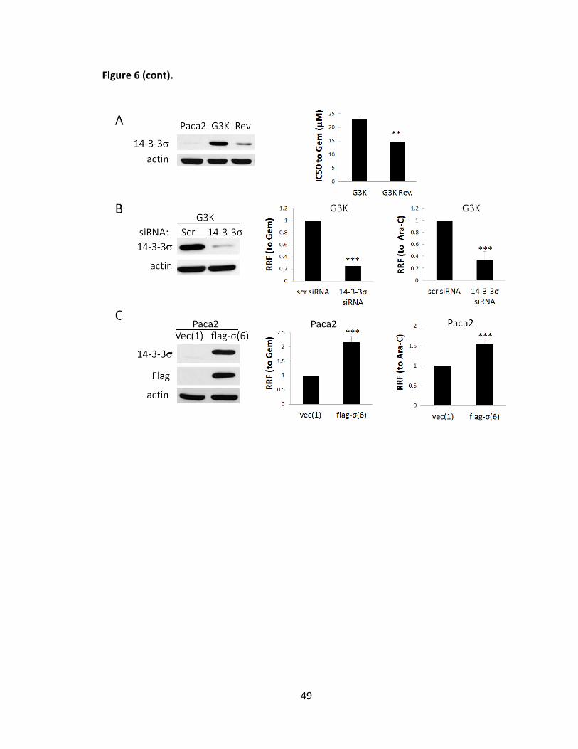
49
Figure 6 (cont).

50
D. Differential methylation of 14-3-3σ gene in MiaPaca-2 and G3K cells.
The 14-3-3σ gene was found frequently hypermethylated and thus its expression
was suppressed in several cancer cells [54, 98, 101-104]. Thus, I hypothesize that the 14-
3-3σ gene in the parental MiaPaca-2 cells may be hypermethylated and 14-3-3σ
expression remains low. Following gemcitabine selection the methylation status of the
14-3-3σ gene may be altered, resulting in increased transcription and expression of 14-
3-3σ. To test this hypothesis, I first treated the parental MiaPaca-2 cells with a well-
known demethylating agent, 5-aza-2'-deoxycytidine (5-Aza-dC, decitabine) that inhibits
DNA methyltransferases, and determined 14-3-3σ expression level using Western blot.
As shown in Figure 7A, 5-Aza-dC treatment increased 14-3-3σ expression in a dose-
dependent manner. Thus, the 14-3-3σ gene is likely silenced in the parental MiaPaca-2
cells by methylation and reactivated in G3K cells.
Although it has been reported that gemcitabine does not possess the
pyrimindine ring modification at position 5, which is responsible for inhibition of DNA
methyltransferases (DNMTs) and, thus, do not inhibit DNA methylation [172], a recent
study showed that gemcitabine reactivated several epigenetically silenced genes
possibly by inhibiting DNMT1 [173]. Thus, it is possible that treatment with gemcitabine
during selection reactivated 14-3-3σ gene expression. To test this possibility, I treated
the parental MiaPaca-2 cells with different concentrations of gemcitabine followed by
Western blot analysis of 14-3-3σ expression. As shown in Figure 7A, unlike 5-Aza-dC,

51
gemcitabine treatment did not increase 14-3-3σ expression. Thus, reactivation of 14-3-
3σ gene in G3K cells is unlikely due to direct gemcitabine-induced demethylation.
Next, I compared the methylation status of the 14-3-3σ gene in G3K and the
parental MiaPaca-2 cells using methylation-specific PCR (MSP). As shown in Figure 7B,
14-3-3σ gene in the parental MiaPaca-2 cells was amplified only by primers for
methylated sequences whereas in G3K cells it was amplified only by primers for
unmethylated sequences. Thus, the 14-3-3σ gene in the parental MiaPaca-2 cells is
heavily methylated whereas it is unmethylated in G3K cells. Analysis of the first exon
with 27 CpG dinucleotides in the 14-3-3σ gene that are known to be methylated in
cancer cells [54] using sodium bisulfite sequencing shows that 25 of these CpG
dinucleotides in the parental MiaPaca-2 cells are fully methylated and the remaining 2
are partially methylated (Figure 7C). However, in G3K cells 23 of the 27 CpG
dinucleotides are unmethylated and the remaining 4 are partially methylated. Clearly,
the methylations of the 14-3-3σ gene in the parental MiaPaca-2 cells have been
removed in G3K cells.

52
Figure 7. Methylation status of 14-3-3σ gene in MiaPaca-2 and G3K cells. (A) Effect of
5-Aza-dC or gemcitabine treatment on 14-3-3σ expression. MiaPaca-2 cells were treated
with increasing concentrations of 5-Aza-dC or gemcitabine for 5 days or 72hrs
respectively, followed by Western blot analysis of 14-3-3σ, cleaved Parp-1 and actin as a
loading control. (B) Methylation-specific PCR (MSP) analysis of MiaPaCa-2 and G3K cells.
Con., control MSP without genomic DNA input. (C) Sodium bisulfite sequencing analysis
of MiaPaCa-2 and G3K cells. The 27 CpG islands in the first exon of 14-3-3σ gene is
shown with solid circles for fully methylated, open circles for unmethylated, and half
filled circles for partially methylated CpG dinucleotides. The sequence profile containing
CpG dinucleotides #10-14 is shown for both MiaPaca-2 and G3K cells. (D) Methylation
status of 14-3-3σ gene in the revertant G3KRev cells. MSP were used to determine the
methylation status of 14-3-3σ gene in G3KRev cells as with MiaPaca-2 and G3K cells as
controls. U, primers for unmethylated; M, primers for methylated.
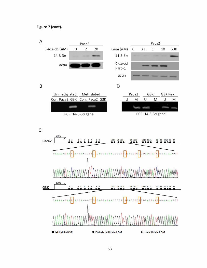
53
Figure 7 (cont).

54
E. Demethylation of the 14-3-3σ gene is reversible.
As shown above, the increased expression of 14-3-3σ in G3K cells is partially
reversed in the revertant G3KRev cells. The partial reversion in 14-3-3σ expression may
be due to partial reversal of the methylation status of the 14-3-3σ gene. To test this
possibility, I performed MSP of isolated genomic DNAs from the partially revertant
G3KRev cells as described above. Figure 7D shows that the 14-3-3σ gene can be
amplified by primers for both methylated and unmethylated sequences, indicating that
the 14-3-3σ gene in G3KRev cells is likely partially reversed and that some cells have
restored the methylation of their 14-3-3σ gene while others retains the demethylated
14-3-3σ gene. Thus, demethylation of the 14-3-3σ gene is likely partially reversible. The
incomplete reversal of 14-3-3σ gene methylation following removal of gemcitabine
suggests that the increased 14-3-3σ expression in G3K cells is unlikely due to direct
gemcitabine-induced demethylation, consistent with the observation shown in Figure
7A.
Previously, up-regulation of 14-3-3σ expression was also observed in an
Adriamycin-selected breast cancer cell line MCF7/AdVp3000 and it was decreased in the
revertant MCF7/AdVp3000/Rev cells [63] (see also Figure 8A). However, the mechanism
of 14-3-3σ regulation in these cell lines is not yet known. To determine if gene
methylation is involved in regulating 14-3-3σ expression in these cells, I performed MSP
of isolated genomic DNAs from the parental MCF7, Adriamycin-selected

55
MCF7/AdVp3000, and the revertant MCF7/AdVpG3K/Rev cells with MiaPaca-2 cells as a
positive control. As shown in Figure 8B, while the 14-3-3σ gene in MiaPaca-2 cells can
be amplified only using primers for methylated sequences, in all breast cancer cells it
can be amplified only using primers for the unmethylated sequences. Thus, up-
regulation of 14-3-3σ expression in MCF7/AdVp3000 cells and its reversal in
MCF7/AdVp3000/Rev cells is unlikely due to changes in methylation status of the 14-3-
3σ gene.

56
Figure 8. 14-3-3σ up-regulation in Adriamycin-selected MCF7/AdVp3000 cells is not
due to gene demethylation. (A) Western blot analysis of 14-3-3σ expression in MCF7,
MCF7/Advp3000 (AdVp), and MCF7/AdVp/Rev (Rev) cells. Actin was used as a loading
control. (B) MSP analysis of 14-3-3σ gene in MCF7, MCF7/Advp3000 (AdVp), and
MCF7/AdVp/Rev (Rev) cells with MiaPaCa-2 cells as a control. U, primers for
unmethylated; M, primers for methylated. Contl., control MSP without genomic DNA
input.

57
F. Uhrf1 and DNMT1 play important roles in regulating 14-3-3σ expression.
To determine what regulates the reversible methylation of 14-3-3σ gene in
MiaPaca-2 and G3K cells, I first compared the expression level of enzymes important for
DNA methylation among the parental MiaPaca-2, gemcitabine resistant G3K, and the
partially revertant G3KRev cells using Western blot or real-time RT-PCR. As shown in
Figure 9A, while DNMT3a and DNMT3b were increased in G3K and remained high in
G3KRev cells, DNMT1 is increased in G3K but reduced back to the basal level in G3KRev
cells. The expression pattern of these methyltransferases is peculiar and inconsistent
with the expression pattern of 14-3-3σ in MiaPaca-2, G3K, and G3KRev cells.
Another protein of importance in gene methylation, Uhrf1, has been shown to
play a major role in recruiting DNMTs [174] and loss of Uhrf1 results in 75% reduction in
genomic methylation [175]. Thus, I tested the expression pattern of Uhrf1. As shown in
Figure 9A, Uhrf1 protein is reduced in G3K cells and increased in the G3KRev cells. The
profile of Uhrf1 expression in these cells is consistent with the partially reversible
change in 14-3-3σ expression and gene methylation in these cells. The consequence of
Uhrf1 binding is recruitment of DNMT1 and histone deacetylase 1, resulting in
methylation of newly synthesized DNA strands [124, 126-128]. Thus, reduced Uhrf1
expression in G3K cells is likely responsible for reduced methylation of 14-3-3σ gene by
reducing recruitment of DNMT1 protein to the methylated CpG islands despite the
presence of high level of DNMT1. In MiaPaca-2 cells, however, the high level of Uhrf1

58
may efficiently recruit enough DNMT1 despite low DNMT1 level for efficient
methylation of 14-3-3σ gene. If so, knocking down either Uhrf1 or DNMT1 in MiaPaca-2
cells would effectively reduce the level of the recruiter (Uhrf1) or the pool of DNMT1 to
be recruited for methylation of 14-3-3σ gene and consequently increase 14-3-3σ
expression. Furthermore, the slight increase of Uhrf1 expression in the G3KRev cells
may be responsible for the increased methylation and reduced expression of 14-3-3σ
gene. In addition, due to the fact that Uhrf1 has been shown to regulate p21 expression
[132], I next tested the expression of p21 in MiaPaca-2, G3K, and G3KRev cells. As
shown in Figure 9A, p21 protein level shows an expression profile in these cells similar
to that of 14-3-3σ, consistent with the possible regulatory role of Uhrf1 in these cells.
To determine if the reduced Uhrf1 expression is possibly responsible for
increased expression of the 14-3-3σ gene in G3K cells, I knocked down Uhrf1 in the
parental MiaPaca-2 cells using siRNA and tested its effect on 14-3-3σ expression. Figure
9B shows that 14-3-3σ protein level is dramatically increased by Uhrf1 knockdown. This
observation is confirmed by real time RT-PCR analysis of 14-3-3σ mRNA, with ~1.4 fold
and ~2.4 fold increase of 14-3-3σ mRNA by Uhrf1 and DNMT1 knockdown, respectively
(Figure 9C).
To determine if DNMTs also contribute to 14-3-3σ expression regulation, I
performed similar experiment by knocking down DNMT1, DNMT3a, and DNMT3b in
MiaPaca-2 cells. Figure 9D shows successful knockdown of DNMT1, DNMT3a, and

59
DNMT3b as determined using Western blot or real-time RT-PCR. However, only DNMT1
knockdown significantly up-regulated the protein level of 14-3-3σ. DNMT3a and
DNMT3b knockdown did not appear to affect 14-3-3σ expression. The effect of DNMT1
knockdown on 14-3-3σ expression was also confirmed by real time RT-PCR analysis
(Figure 9C). Taken together, I conclude that likely both Uhrf1 and DNMT1 play
important roles in regulating 14-3-3σ expression.

60
Figure 9. Role of Uhrf1 and DNMT1 in 14-3-3σ expression. (A) Western blot analysis of
Uhrf1, DNMT1, DNMT3a, and p21 expression as well as real-time RT-PCR analysis of
DNMT3b expression in MiaPaca-2, G3K, and G3KRev cells. (B-D) Effect of Uhrf1, DNMT1,
DNMT3a and DNMT3b knockdown on 14-3-3σ expression. MiaPaca-2 cells were
transiently transfected with scrambled control siRNA (Scr) or siRNAs targeting Uhrf1 (B),
DNMT1, DNMT3a, and DNMT3b (D) followed by Western blot analysis of Uhrf1, DNMT1,
DNMT3a, and 14-3-3σ protein levels (B, D) or real time RT-PCR analysis of DNMT3b and
14-3-3σ mRNA levels (C-D). (N=4-5, *p<0.05, ***p<0.001). Actin and GAPDH were used
as a loading control for Western blot and internal control for PCR analyses, respectively.

61
Figure 9 (cont).

62
G. Uhrf1 binds and helps recruit DNMT1 to methylated region of 14-3-3σ gene.
To further determine the role of Uhrf1 and DNMT1 in regulating 14-3-3σ
expression and gene methylation, I first determined if Uhrf1 binds to the methylated
CpG islands in the first exon of 14-3-3σ gene in MiaPaca-2 cells using ChIP assay. As
shown in Figure 10A, Uhrf1 was bound to the CpG islands but not to the CpG-free
sequences in the promoter region of the 14-3-3σ gene in MiaPaca-2 cells. Knocking
down Uhrf1 also reduced Uhrf1 binding to the CpG islands of 14-3-3σ gene (Figure 10B).
Thus, Uhrf1 likely can bind to the methylated CpG island sequences of the 14-3-3σ gene.
As shown in Figure 9A, DNMT1 expression was found increased in G3K cells,
which is peculiar since methylation of 14-3-3σ gene in G3K cells is reduced. However,
because Uhrf1 binding to CpG island sequences helps recruit DNMT1 to maintain the
methylation status of the DNA, it is possible that the reduced Uhrf1 expression in G3K
cells reduces DNMT1 recruitment despite the higher level of DNMT1 in G3K cells. To test
this possibility, I first compared the binding of DNMT1 to the CpG island sequences of
the 14-3-3σ gene between MiaPaca-2 and G3K cells. As shown in Figure 10C, binding of
DNMT1 to the CpG island sequences of 14-3-3σ gene is indeed much less in G3K than in
MiaPaca-2 cells despite the higher expression level of DNMT1 in G3K cells. Importantly,
Uhrf1 knockdown in MiaPaca-2 cells dramatically reduced DNMT1 binding to the CpG
island sequences of the 14-3-3σ gene (Figure 10D), indicating that DNMT1 could not be
effectively recruited to the CpG island sequences of the 14-3-3σ gene in the absence of

63
Uhrf1, and hence Uhrf1 is the recruiter of DNMT1. Thus, I conclude that Uhrf1 likely
plays a major role in regulating 14-3-3σ expression by binding to its CpG-rich sequences
and helps recruit DNMT1 to the site to reversibly methylate the 14-3-3σ gene during
replication. The reduced Uhrf1 expression by gemcitabine selection in G3K cells likely
decreased DNMT1 recruitment and methylation of 14-3-3σ gene while the slightly
increase in Uhrf1 level in G3KRev cells is responsible for partial reversal of 14-3-3σ gene
methylation.

64
Figure 10. Binding of Uhrf1 and DNMT1 to the CpG islands of the 14-3-3σ gene. (A)
ChIP analysis of Uhrf1 binding to the CpG island sequences or nonCpG island
sequences of 14-3-3σ gene. (B, D) Effect of Uhrf1 knockdown on Uhrf1 and DNMT1
binding to the CpG island sequences of 14-3-3σ gene in MiaPaca-2 cells. MiaPaca-2 cells
were transiently transfected with scrambled control or Uhrf1 siRNAs followed by ChIP
analysis of Uhrf1 and DNMT1 binding. (C) DNMT1 binding to the CpG island sequence of
14-3-3σ gene in G3K cells is less than that in MiaPaca-2 cells. ChIP with histone H3
antibody or without any primary antibody were used as positive and negative controls,
respectively. Normal IgG was also used as negative control, which did not
immunoprecipitate any DNA (data not shown).

65
H. Gemcitabine treatment does not affect Uhrf1 and DNMT1 expression.
Finally, to investigate if the altered expression of Uhrf1 and DNMT1 in G3K cells
is potentially due to gemcitabine treatments during selection, I performed a Western
blot analysis to detect both Uhrf1 and DNMT1 expression in MiaPaca-2 cells following
treatments with gemcitabine at different concentrations for 72 hrs. As shown in Figure
11, gemcitabine treatments had no effect on Uhrf1 and DNMT1 expression. Therefore,
the mechanism of regulation of Uhrf1 and DNMT1 expression in the drug resistant cells
remains to be determined.
Overall, this section of my thesis demonstrated that 14-3-3σ over-expression
contributed to acquired gemcitabine resistance and that the up-regulated 14-3-3σ
expression was caused by lack of gene methylation regulation during gemcitabine
selection. As summarized in Figure 12, Uhrf1 and DNMT1 bind to the methylated region
of 14-3-3σ gene in parental MiaPaca-2 cells to maintain the methylation status, whereas
during gemcitabine selection, the expression of Uhrf1 is dramatically decreased and
thus insufficient to recruit DNMT1 to the site to methylate the DNA, resulting in 14-3-3σ
gene demethylation and re-expression. After gemcitabine removal, however, the slight
increase of Uhrf1 expression likely functions to recruit the methylation machinery to
restore the methylation of 14-3-3σ gene.

66
Figure 11. Effect of gemcitabine treatment on Uhrf1 and DNMT1 expression. MiaPaca-
2 were treated with/without increasing concentrations of gemcitabine, and G3K cells
were used as control without treatment, followed by Western blot analysis of Uhrf1,
DNMT1. Actin was used as a loading control.

67
Figure 12. Schematic model of epigenetic regulation of 14-3-3σ gene following
gemcitabine selection and drug removal. The relative expression levels of Uhrf1 and
DNMT1, as well as their binding status to 14-3-3σ gene during gemcitabine selection or
drug removal are shown in the model. Solid circles represent fully methylated CpG
islands of 14-3-3σ gene, and open circles represent unmethylated CpGs.

68
Section II: Identification of PDGFD as a potential contributor to acquired gemcitabine
resistance by Reduced Representation Bisulfite Sequencing (RRBS)
A. Identification of reversibly-methylated genes using Reduced Representation
Bisulfite Sequencing (RRBS).
The finding of demethylation of the 14-3-3σ gene in gemcitabine resistant cells
led to a hypothesis that other genes may also have changed epigenetically following
gemcitabine selection. To profile global changes in gene methylation in response to
gemcitabine selection, genomic DNAs from MiaPaca-2, G3K, and G3KRev cells were
isolated and subjected to RRBS sequencing, which yield genome-
wide methylation profiles on a single nucleotide level, by BGI (Beijing Genomics
Institute). The RRBS data were analysed in collaboration with Dr. Yunlong-Liu’s lab.
As a result, the RRBS yields about 65~70 Million clean reads for each sample,
with map rate of >92%, unique mapping rate of >70%, and >97% of unique reads with
enzyme cutting site (Figure 13A). Moreover, the RRBS targeted a reasonable proportion
of genome of interest in all samples, with >70% of promoters and >83% of CG islands
were covered respectively (Figure 13B). The individual CpG sites within CG islands and
promoters were also examined and good depth coverage at 4X and 10X were achieved,
and all samples showed consistent coverage (data not shown).

69
Figure 13. RRBS sequencing results comparison and characterization of genomic
coverage. (A) RRBS sequencing results comparison. (B) Genomic coverage
characterization of promoter and CG island (CGI) numbers.

70
According to RRBS outcome, 845 genes are differentially methylated between
MiaPaca-2 and G3K cells, and 282 genes are differentially methylated between G3K and
G3KRev cells. The list of these differentially methylated genes is shown in Appendices.
Because the methylation changes in the promoter region most likely influence gene
transcription and expression, I filtered these genes with differential methylation level in
the promoter region and with a 2-fold change cutoff. As shown in Figure 14, 159 genes
have increased methylation while 459 genes have decreased methylation in G3K cells
compared with MiaPaca-2 cells. By comparing with G3K cells, G3KRev cells have 104
genes with increased and 88 genes with decreased methylation. Together, there are 65
genes that have reversible methylation changes from MiaPaca-2 to G3K and to G3KRev
cells (Table 2).

71
Figure 14. Flowchart of DMR filteration of genes. Genes with DMR p-value<0.01 were
first filtered by DMR at the promoter regions, followed by further filter with 2-fold
methylation level change.

72
Table 2. List of reversibly methylated genes.
Paca2 vs G3K up, G3K vs G3KRev down Paca2 vs G3K down, G3K vs G3KRev up
ADORA2B, CDC42EP3, CPNE7, FBXO27,
JAG2, MIR1915, MRPS6, NPAS2, OLIG1,
PTPRG, PWWP2B, SLC45A1, SNORD56B,
SOX13, TNRC18, WNT9A
AFAP1L2, ANXA2P3, APBA1, CCNO,
CDC42BPB, CDK6, CHRNA7, CRAMP1L,
DDIT4L, DSE, DUSP6, EFNA5, ELOVL2-AS1,
FAM133B, FRAS1, FUOM, GALNT7, GNS,
GRM4, H2AFY2, JMJD8, KIAA1324L,
KIF21B, LOC100506190, LONRF3, MCTP1,
MDFIC, MIR205HG, MMP25, MTA1, NKX1-
2, PDGFD, PRDM1, RAET1G, RASD2,
RNASET2, SEMA5B, SLC25A27, SLC31A2,
SLC35D3, SOWAHD, TMEM181, TRPC3,
WDR1, WNT11, ZNF593, ZNF669, ZNF808,
ZNF83
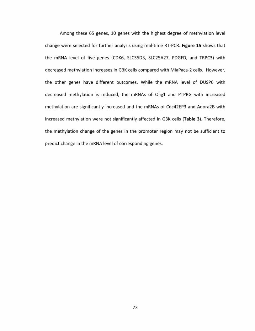
73
Among these 65 genes, 10 genes with the highest degree of methylation level
change were selected for further analysis using real-time RT-PCR. Figure 15 shows that
the mRNA level of five genes (CDK6, SLC35D3, SLC25A27, PDGFD, and TRPC3) with
decreased methylation increases in G3K cells compared with MiaPaca-2 cells. However,
the other genes have different outcomes. While the mRNA level of DUSP6 with
decreased methylation is reduced, the mRNAs of Olig1 and PTPRG with increased
methylation are significantly increased and the mRNAs of Cdc42EP3 and Adora2B with
increased methylation were not significantly affected in G3K cells (Table 3). Therefore,
the methylation change of the genes in the promoter region may not be sufficient to
predict change in the mRNA level of corresponding genes.

74
Figure 15. Identification of transcriptional changes in candidate genes. (A-B) Real time
RT-PCR analysis of mRNA level of candidate genes including DUSP6, Olig1, Cdc42EP3,
Adora2B, PTPRG, CDK6, SLC35D3, SLC25A27, PDGFD, and TRPC3 gene between
MiaPaca-2 and G3K cells. GAPDH was used as internal control. (N=3-5, *p<0.05,
**p<0.01, ***p<0.001). ↑: methylation is increased in G3K cells, ↓: methylation is
decreased in G3K cells.

75
Table 3. Identification and characterization of candidate genes.
DMR (G3K-MiaPaca-2)
mRNA level change (G3K/MiaPaca-2) Significance
Negative correlation of methylation change with mRNA level change CDK6 -0.5 1.684548 p<0.001
SLC35D3 -0.57 2.171952 p<0.05
SLC25A27 -0.39 2.566402 p<0.05
PDGFD -0.41 6.164361 p<0.05
TRPC3 -0.48 15.42079 p<0.001
Positive correlation of methylation change with mRNA level change
PTPRG 0.21 1.551086 p<0.01
Olig1 0.34 4.334516 p<0.001
DUSP6 -0.41 0.596951 p<0.01
No correlation of methylation change with mRNA level change
cdc42EP3 0.28 1.074495 NS
Adora2B 0.26 1.148135 NS

76
B. Reversible up-regulation of PDGFD.
Next, TRPC3 and PDGFD were selected for validation of their expression at
protein level. Firstly, membrane fractions were prepared from both MiaPaca-2 and G3K
cells for Western blot analysis of TRPC3. As shown in Figure 16A, TRPC3 protein level did
not appear to change despite its mRNA level increased ~15 fold in G3K cells. Thus,
TRPC3 mRNA level does not seem to correlate with its protein level. To determine
PDGFD protein level, MiaPaca-2 and G3K cells were cultured in serum-free medium for
24 hrs, followed by collection of conditioned medium (CM) and TCA precipitation. The
precipitated proteins were subjected to Western blot analysis. As shown in Figure 16C,
PDGFD production was dramatically elevated in G3K cells compared with MiaPaca-2
cells.
To determine if increased PDGFD production in G3K cells was reversed in G3KRev
cells, real-time RT-PCR analysis was performed. As shown in Figure 17A, the increased
PDGFD mRNA level in G3K cells (~16 fold increase compared with MiaPaca-2 cells) was
dramatically decreased in the G3KRev cells (only ~2.4 fold increase compared with
MiaPaca-2 cells). Moreover, the Western blot analysis confirmed this finding (Figure
17C). Thus, the expression of PDGFD is reversibly up-regulated in G3K cells.
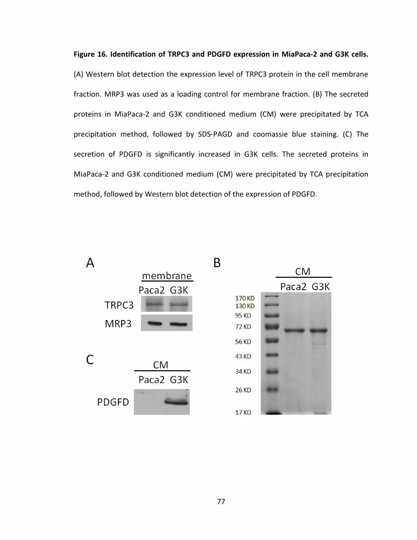
77
Figure 16. Identification of TRPC3 and PDGFD expression in MiaPaca-2 and G3K cells.
(A) Western blot detection the expression level of TRPC3 protein in the cell membrane
fraction. MRP3 was used as a loading control for membrane fraction. (B) The secreted
proteins in MiaPaca-2 and G3K conditioned medium (CM) were precipitated by TCA
precipitation method, followed by SDS-PAGD and coomassie blue staining. (C) The
secretion of PDGFD is significantly increased in G3K cells. The secreted proteins in
MiaPaca-2 and G3K conditioned medium (CM) were precipitated by TCA precipitation
method, followed by Western blot detection of the expression of PDGFD.

78
Figure 17. Reversible transcription and expression of PDGFD. (A) Real time RT-PCR
analysis of PDGFD mRNA level in MiaPaca-2, G3K, and G3KRev cells. GAPDH was used as
internal control. (N=4, **p<0.01). (B) The secreted proteins in MiaPaca-2, G3K, and
G3KRev conditioned medium (CM) were precipitated by TCA precipitation method,
followed by SDS-PAGD and coomassie blue staining. (C) PDGFD expression is reversibly
increased in G3K cells. The secreted proteins in MiaPaca-2, G3K, and G3KRev
conditioned medium (CM) were precipitated by TCA precipitation method, followed by
Western blot detection of PDGFD expression.

79
C. Validation of reversible methylation change of PDGFD gene.
To validate the RRBS data that suggest a reversible methylation change of PDGFD
gene in G3K cells, I performed sodium-bisulfite gene sequencing analysis of the 26 CpG
dinucleotides residing in the promoter region of PDGFD gene in all three cell lines. As
shown in Figure 18, 14-22 out of 26 CpG dinucleotides were methylated in MiaPaca-2
cells. However, these CpG dinucleotides were all unmethylated in G3K cells.
Interestingly, in G3KRev cells, 16-17 out of 26 CpG dinucleotides were again methylated.
Thus, the methylation change of PDGFD promoter is reversible, and consistent with
RRBS data.

80
Figure 18. Reversible methylation change of PDGFD gene. Sodium bisulfite sequencing
analysis of PDGFD gene in MiaPaca-2, G3K, and G3KRev cells was conducted. Briefly,
genomic DNA was treated with sodium bisulfite and the promoter region of PDGFD
gene was amplified by PCR, followed by cloning into pGEM-T vector. 4 clones from
each cell line were picked for sequencing. The 26 CpG islands in promoter region of
PDGFD gene were shown with solid circles for fully methylated, open circles for
unmethylated. The chromatogram represents sequence profile containing CpG
dinucleotides #2-13 for all three cells.
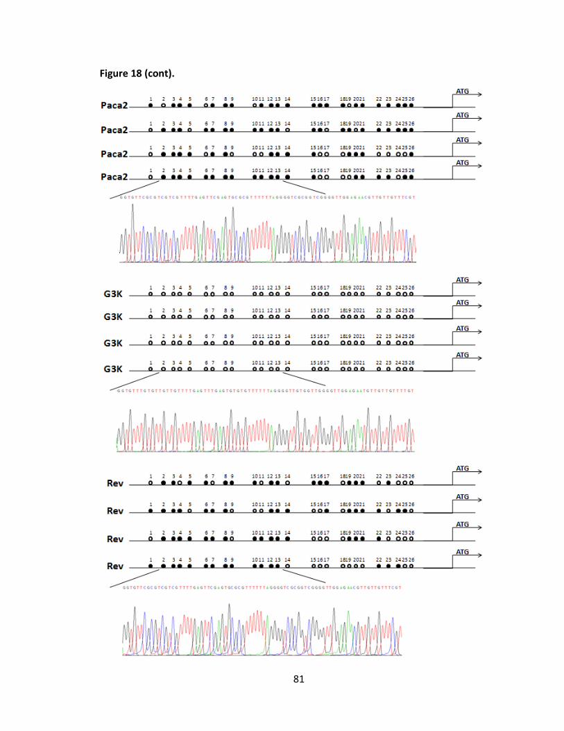
81
Figure 18 (cont).
82
D. PDGFD up-regulation plays an important role in gemcitabine resistance.
It has been reported that PDGFD signaling is frequently deregulated in human
malignancies with up-regulated expression in prostate, lung, renal, ovarian, brain, and
pancreatic cancer [146, 147, 149-152]. However, its relationship with therapeutic drug
resistance is limited to the discovery of its role in regulation of radiosensitivity in
prostate adenocarcinoma, as well as its influence on sensitivity and resistance of
glioblastoma to semustine [154, 158]. To determine if PDGFD potentially contributes to
gemcitabine resistance in pancreatic cancer, I knocked down PDGFD in both G3K and
Aspc-1 cells and performed the MTT assay to test its response to gemcitabine. Figure
19A and Figure 19C show knockdown of PDGFD in G3K and Aspc-1 cells at both protein
and mRNA levels, respectively. As shown in Figure 19B and Figure 19D, the sufficient
knockdown of PDGFD is accompanied with a dramatic reduction of gemcitabine
resistance by 63.7%-76.5% in G3K and 46.4%-84.8% in Aspc-1 cells, respectively. Thus,
PDGFD knockdown likely reduces gemcitabine resistance in both cells.
Next, to further test the influence of PDGFD over-expression on gemcitabine
sensitivity, stable clones with PDGFD over-expression were generated from MiaPaca-2
cells. Figure 20A-B show increased PDGFD production in these clones compared with
vector control clone by Western blot analysis of conditioned medium. Moreover, the
increased production of PDGFD is accompanied with an increased resistance to
gemcitabine by 4.8-4.9 fold (Figure 20C). Furthermore, MiaPaca-2 cells cultured in

83
conditioned medium from G3K cells are also more resistant to gemcitabine (by 2.4-fold
increase averagely), than cells cultured with fresh medium (Figure 20D). Together, these
results suggest that PDGFD plays an important role in gemcitabine resistance in human
pancreatic cancers and that there may be bystander effect of resistant cells on sensitive
cells.
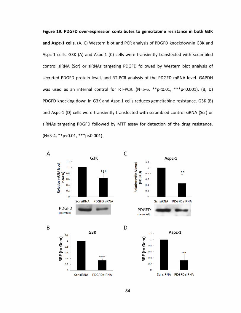
84
Figure 19. PDGFD over-expression contributes to gemcitabine resistance in both G3K
and Aspc-1 cells. (A, C) Western blot and PCR analysis of PDGFD knockdownin G3K and
Aspc-1 cells. G3K (A) and Aspc-1 (C) cells were transiently transfected with scrambled
control siRNA (Scr) or siRNAs targeting PDGFD followed by Western blot analysis of
secreted PDGFD protein level, and RT-PCR analysis of the PDGFD mRNA level. GAPDH
was used as an internal control for RT-PCR. (N=5-6, **p<0.01, ***p<0.001). (B, D)
PDGFD knocking down in G3K and Aspc-1 cells reduces gemcitabine resistance. G3K (B)
and Aspc-1 (D) cells were transiently transfected with scrambled control siRNA (Scr) or
siRNAs targeting PDGFD followed by MTT assay for detection of the drug resistance.
(N=3-4, **p<0.01, ***p<0.001).
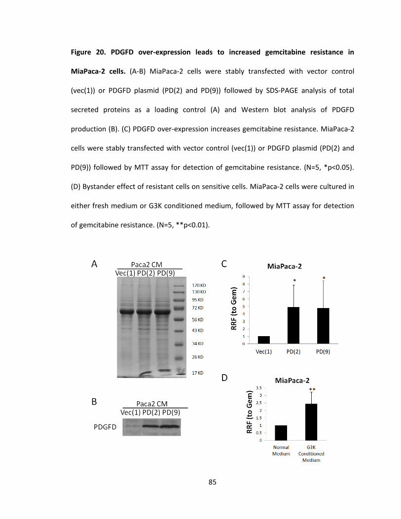
85
Figure 20. PDGFD over-expression leads to increased gemcitabine resistance in
MiaPaca-2 cells. (A-B) MiaPaca-2 cells were stably transfected with vector control
(vec(1)) or PDGFD plasmid (PD(2) and PD(9)) followed by SDS-PAGE analysis of total
secreted proteins as a loading control (A) and Western blot analysis of PDGFD
production (B). (C) PDGFD over-expression increases gemcitabine resistance. MiaPaca-2
cells were stably transfected with vector control (vec(1)) or PDGFD plasmid (PD(2) and
PD(9)) followed by MTT assay for detection of gemcitabine resistance. (N=5, *p<0.05).
(D) Bystander effect of resistant cells on sensitive cells. MiaPaca-2 cells were cultured in
either fresh medium or G3K conditioned medium, followed by MTT assay for detection
of gemcitabine resistance. (N=5, **p<0.01).

86
E. PDGFD over-expression contributes to gemcitabine resistance possibly by regulating
STAT3 signaling pathway.
It has been reported that PDGFD activation triggers a number of downstream
signaling pathways including activation of phosphatidylinositol 3 kinase (PI3K), Akt,
nuclear factor-κB (NF-κB), Notch, and extracellular signal-regulated kinase (ERK) [139,
145-147]. It has also been reported that STAT3 is up-regulated in several drug-selected
cancer cells including acquired resistance to erlotinib, vemurafenib, temozolomide,
cisplatin, and paclitaxel [176-180]. Thus, I hypothesize that PDGFD over-expression may
contribute to gemcitabine resistance by regulating STAT3 signaling pathway.
To test this hypothesis, I first compared the activation of STAT3 signaling
pathway in MiaPaca-2 and G3K cells. As shown in Figure 21A, both the p-stat3 and total
Stat3 as well as Stat3 downstream targets including VEGF and MMP2 were up-regulated
in G3K cells. More importantly, knocking down PDGFD in G3K cells led to significant
reduction of p-stat3/Stat3 and Stat3 downstream targets (Figure 21B). Similarly,
knocking down PDGFD in Aspc-1 cells also reduced the level of p-stat3 and total Stat3 as
well as Stat3 downstream targets (data not shown) while over-expression of PDGFD in
parental MiaPaca-2 cells caused markedly increased level of p-stat3, total Stat3 and
Stat3 downstream target VEGF (Figure 21C). Interestingly, the revertant G3KRev cells
showed dramatically decreased expression of p-stat3 to the basal level compared with
G3K cells although the total Stat3 level seems unchanged (Figure 22C). Taken together,

87
these findings strongly suggest that PDGFD actively regulates STAT3 signaling pathway,
which may mediate PDGFD-induced gemcitabine resistance in pancreatic cancer cells.

88
Figure 21. PDGFD actively regulates STAT3 signaling pathway. (A) Western blot analysis
of secreted PDGFD, p-stat3, stat3, VEGF, secreted VEGF, and secreted MMP2 expression
in both MiaPaca-2 and G3K cells. (B) Effect of PDGFD knockdown on Stat3 signaling in
G3K cells. G3K cells were transiently transfected with scrambled control siRNA (Scr) or
siRNAs targeting PDGFD followed by Western blot analysis of p-stat3, stat3, VEGF, and
MMP2 secretion. Actin was used as a loading control. (C) Effect of PDGFD over-
expression on Stat3 signaling in MiaPaca-2 cells. MiaPaca-2 cells were stably transfected
with vector control (vec(1)) or PDGFD plasmid (PD(9)) followed by Western blot analysis
of p-stat3, stat3, and VEGF expression. Actin was used as a loading control.
89
F. Stat3 potentially contributes to gemcitabine resistance.
Finally, the potential role of Stat3 in gemcitabine resistance was tested by over-
expressing wild type and constitutively activated mutant Stat3 (Stat3c) in parental
MiaPaca-2 cells (Figure 22A). Figure 22B shows that over-expressing either the wild type
or the constitutively activated Stat3 in parental MiaPaca-2 cells significantly increased
gemcitabine resistance, by ~1.7 fold and ~2.3 fold increase respectively. Therefore, it is
possible that PDGFD-induced up-regulation of Stat3 signaling may serve as a mediator
for PDGFD-induced gemcitabine resistance.
Thus, I conclude that gemcitabine selection causes PDGFD gene demethylation
and protein up-regulation, and its over-expression in turn contributes to gemcitabine
resistance by activating STAT3 signaling pathway (Figure 23).

90
Figure 22. Stat3 over-expression promotes gemcitabine resistance in MiaPaca-2 cells.
(A-B) Effect of PDGFD over-expression on gemcitabine resistance in MiaPaca-2 cells.
MiaPaca-2 cells were stably transfected with vector control, stat3, or stat3c plasmid
followed by Western blot analysis for detecting the expression of stat3 (A) or MTT assay
for detecting the gemcitabine resistance. Actin was used as a loading control for
Western blot. (N=3-4, **p<0.01). (C) Western blot detection of the expression of total
Stat3 and p-stat3 in MiaPaca-2, G3K, and G3KRev cells. Actin was used as a loading
control.

91
Figure 23. Schematic model of PDGFD-mediated gemcitabine resistance during drug
selection. Gemcitabine selection leads to PDGFD gene demethylation and protein up-
regulation, which binds to its cognate receptor and activates Stat3 signaling. Activation
of Stat3 signaling pathway causes transcription of its downstream target genes and
contributes to gemcitabine resistance.

92
Section III: Molecular mechanism of 14-3-3σ-mediated gemcitabine resistance in
pancreatic cancer
The previous studies showed that 14-3-3σ over-expression in G3K cells
contribute to gemcitabine resistance. However, the underlying mechanism through
which 14-3-3σ contributes to acquired gemcitabine resistance remains unknown. Here,
based on the escalating interest in recent years on one potential binding partner of 14-
3-3σ, YAP1, I tested the hypothesis that 14-3-3σ contributes to acquired gemcitabine
resistance by binding to YAP1.
A. YAP1 over-expression in G3K cells and its contribution to gemcitabine resistance.
To determine the potential role of YAP1 in 14-3-3σ-mediated gemcitabine
resistance, I first tested the level of total YAP1 and p-YAP1 in both MiaPaca-2 and G3K
cells. As shown in Figure 24, the level of both YAP1 and p-YAP1 as well as the mRNA
level of YAP1 (~4.7 fold increase) were found highly elevated in G3K compared with
MiaPaca-2 cells, suggesting that YAP1 may be transcriptionally up-regulated in G3K cells.
Interestingly, 14-3-3σ knockdown in G3K cells also reduced total YAP1, p-YAP1, and
YAP1 mRNA levels (~50% reduction) (Figure 25A-B). Moreover, over-expression of 14-3-
3σ in MiaPaca-2 cells resulted in an increase of YAP1, p-YAP1, and YAP1 mRNA levels
(~2.5 fold increase) (Figure 25C-D), indicating that YAP1 may be transcriptionally
regulated by 14-3-3σ. However, knocking down of YAP1 in G3K cells did not affect the

93
protein level of 14-3-3σ (Figure 26A). Thus, 14-3-3σ may regulate the transcription and
expression of YAP1, but not vice versa.
To determine whether the increased expression of YAP1 in G3K cells contributes
to gemcitabine resistance, I first knocked down YAP1 in G3K cells by using specific siRNA
followed by examining the difference in gemcitabine resistance using MTT assay. As
shown in Figure 26A-B, knocking down YAP1 dramatically reduced the drug resistance in
G3K cells, by up to 80% reduction. However, over-expression of YAP1 in the parental
MiaPaca-2 cells did not significantly influence the gemcitabine resistance (Figure 26C-D).
The reason for the discrepancy between these two experiments could be that YAP1
over-expression in MiaPaca2 cells may need 14-3-3σ for gemcitabine resistance. To test
this possibility, I over-expressed YAP1 in MiaPaca-2 cells with stable flag-14-3-3σ-over-
expression and tested the effect on gemcitabine resistance. As shown in Figure 26E-F,
over-expression of YAP1 in these cells further increase gemcitabine resistance by ~2.4
fold. Thus, it is likely that YAP1 requires 14-3-3σ to contribute to gemcitabine resistance.

94
Figure 24. YAP1 is over-expressed in resistant G3K cells. (A) Western blot analysis of
YAP1, p-YAP1 and 14-3-3σ expression in both MiaPaca-2 and G3K cells. Actin was used
as a loading control. (B) Real time RT-PCR analysis of YAP1 mRNA level in MiaPaca-2 and
G3K cells (N=3, **p<0.01).

95
Figure 25. Regulation of YAP1 expression by 14-3-3σ protein level. (A) Effect of 14-3-3σ
knockdown on YAP1/p-YAP1 expression. G3K cells were transiently transfected with
scrambled control siRNA (Scr) or siRNAs targeting 14-3-3σ followed by Western blot
analysis of YAP1, p-YAP1 and 14-3-3σ protein level. Actin was used as a loading control.
(B) Real time RT-PCR analysis of YAP1 mRNA level in G3K cells stably transfected with
scrambled control shRNA (scr sh3’) or shRNA targeting 14-3-3σ (14-3-3 sh(11)). GAPDH
was used as internal control. (N=3, **p<0.01). (C-D). Effect of 14-3-3σ over-expression
on YAP1 expression. MiaPaca-2 cells were stably transfected with vector control (vec(1))
or pcDNA3.1(+)-flag-14-3-3σ plasmid (flag-σ(6)) followed by Western blot analysis of
YAP1, p-YAP1, and 14-3-3σ protein levels (C) or real time RT-PCR analysis of YAP1 mRNA
level (D). (N=3, **p<0.01). Actin and GAPDH were used as a loading control for Western
blot and internal control for PCR analysis, respectively.
96
Figure 26. YAP1 over-expression contributes to gemcitabine resistance, but requires
the presence of 14-3-3σ. (A-B) Effect of YAP1 knockdown on gemcitabine resistance in
G3K cells. G3K cells were transiently transfected with scrambled control siRNA (Scr) or
siRNAs targeting YAP1 followed by Western blot analysis of YAP1 and 14-3-3σ protein
level (A) or MTT assay for detection of drug resistance(B). (N=4, **p<0.01). Actin was
used as a loading control for Western blot. (C-D) Effect of YAP1 over-expression on
gemcitabine resistance in MiaPaca-2 cells. MiaPaca-2 cells were transfected with vector
control (GFP-C2) or GFP-YAP plasmid followed by Western blot analysis of YAP1 protein
level (C) or MTT assay for detection of drug resistance (D). (N=4, not significant). Actin
was used as a loading control for Western blot. (E-F) Effect of YAP1 over-expression on
gemcitabine resistance in 14-3-3σ-over-expressing MiaPaca-2 cells. 14-3-3σ-over-
expressing MiaPaca-2 cells (MiaPaca-2 flag-σ (6)) were transfected with vector control
(GFP-C2) or GFP-YAP plasmid followed by Western blot analysis of YAP1 protein level (E)
or MTT assay for detection of drug resistance (F). (N=3, **p<0.01). Actin was used as a
loading control for Western blot.

97
Figure 26 (cont).

98
B. Gemcitabine resistance requires both 14-3-3σ and YAP1.
To better understand the mechanism of 14-3-3σ and YAP1 in contributing to
acquired gemcitabine resistance, either 14-3-3σ or YAP1 was up-regulated but down-
regulated the other, followed by MTT assay to determine gemcitabine response. As
shown in Figure 27A-B, over-expressing 14-3-3σ markedly increased YAP1 protein level
and gemcitabine resistance in MiaPaca-2 cells. However, knocking down YAP1 in 14-3-
3σ-over-expressing MiaPaca-2 cells diminished the 14-3-3σ-induced gemcitabine
resistance. Similarly, up-regulation of YAP1 in 14-3-3σ-over-expressing MiaPaca-2 cells
significantly increased gemcitabine resistance, whereas knocking down 14-3-3σ in these
cells abolished gemcitabine resistance despite that the ectopic YAP1 expression
maintains at high level (Figure 27C-D). Together, these findings suggest that both 14-3-
3σ and YAP1 are required for gemcitabine resistance.
To better address this observation, I performed the double knockdown
experiment. Either 14-3-3σ or YAP1, or both proteins were knocked down in G3K (Figure
28A-B) or Aspc-1 (Figure 28C-D) cells to test the changes in gemcitabine resistance. Not
surprisingly, knocking down either 14-3-3σ or YAP1 alone dramatically reduces
gemcitabine resistance in both cell lines, by ~61% and ~69% reduction in G3K cells
respectively. However, knocking down both 14-3-3σ and YAP1 simultaneously did not
further reduce drug resistance, by ~72% reduction in G3K cells. Thus, it is possible that
14-3-3σ and YAP1 cooperates with each other to contribute to gemcitabine resistance,

99
and hence it is likely that the mechanism of 14-3-3σ-mediated gemcitabine resisance is
the same as YAP1-mediated gemcitabine resistance.

100
Figure 27. Gemcitabine resistance requires both 14-3-3σ and YAP1. (A-B) YAP1
knockdown counteracts increased gemcitabine resistance caused by 14-3-3σ over-
expression. MiaPaca-2 vec(1) and MiaPaca-2 flag-σ(6) cells were transiently transfected
with scrambled control siRNA (Scr) or siRNAs targeting YAP1 followed by Western blot
analysis of YAP1 and 14-3-3σ (A) or MTT assay for detection of drug resistance (B). (N=3,
**p<0.01, ***p<0.001). Actin was used as a loading control for Western blot. (C-D) 14-3-
3σ knockdown counteracts with increased gemcitabine resistance caused by YAP1 over-
expression. MiaPaca-2 flag-σ(6) cells were transiently co-transfected with (1) GFP-C2
vector and scrambled control siRNA (GFP-C2/Scr si), or (2) GFP-YAP over-expression
plamid and scrambled control siRNA ( GFP-YAP/scr si), or (3) or GFP-YAP over-expression
plamid and siRNA targeting 14-3-3σ (GFP-YAP/14-3-3σ si), followed by Western blot
analysis of YAP1 and 14-3-3σ (C) or MTT assay for detection of drug resistance (D). (N=3,
**p<0.01, ***p<0.001). Actin was used as a loading control for Western blot.

101
Figure 27 (cont).
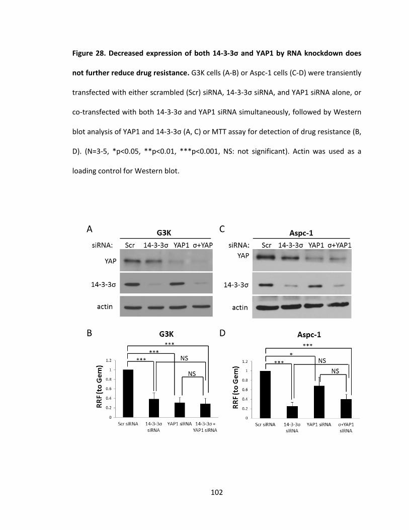
102
Figure 28. Decreased expression of both 14-3-3σ and YAP1 by RNA knockdown does
not further reduce drug resistance. G3K cells (A-B) or Aspc-1 cells (C-D) were transiently
transfected with either scrambled (Scr) siRNA, 14-3-3σ siRNA, and YAP1 siRNA alone, or
co-transfected with both 14-3-3σ and YAP1 siRNA simultaneously, followed by Western
blot analysis of YAP1 and 14-3-3σ (A, C) or MTT assay for detection of drug resistance (B,
D). (N=3-5, *p<0.05, **p<0.01, ***p<0.001, NS: not significant). Actin was used as a
loading control for Western blot.

103
C. 14-3-3σ and YAP1 form a complex.
The finding of indispensable roles of both 14-3-3σ and YAP1 in gemcitabine
resistance suggests that 14-3-3σ may form a complex with YAP1. To test this possibility,
co-immunoprecipitation assay was performed in G3K cells transiently transected with
GFP-YAP1 and flag-14-3-3σ. As shown in Figure 29A, YAP1 and p-YAP1 proteins could be
co-immunoprecipitated by 14-3-3σ antibody and flag-14-3-3σ protein could be co-
immunoprecipitated by GFP antibody. Thus, 14-3-3σ may bind with YAP1/p-YAP1 to
form a complex in G3K cells.
Next, co-localization analysis of these two proteins was performed using
immunofluorescence staining. As shown in Figure 29B, both 14-3-3σ and YAP1 appear to
localize to the cytoplasm of G3K cells and the overlay image indicates their co-
localization, which supports the possibility that these two proteins reside and form a
complex in the cytoplasm.

104
Figure 29. 14-3-3σ interacts and binds with YAP1 in vitro. (A) 14-3-3σ co-
immunoprecipitates with YAP1, and vise versa. G3K cells were transiently transfected
with pEGFP-YAP1 plasmid or co-transfected with both pEGFP-YAP1 and pcDNA3.1-flag-σ
plasmid, followed by immunoprecipitation with either 14-3-3σ or GFP antibody, and
Western blot detection the existence of YAP1, p-YAP1 or 14-3-3σ in the complex. (B)
YAP1 co-localizes with 14-3-3σ in vitro. The localization of both YAP1 and 14-3-3σ in the
G3K cells was visualized by confocal microscopy.

105
D. Both 14-3-3σ and YAP1 protect against gemcitabine-induced caspase-8 activation
and apoptosis.
To further investigate the mechanism of 14-3-3σ and YAP1-induced gemcitabine
resistance, I tested their effect on gemcitabine-induced apoptosis. 14-3-3σ was knocked
down in G3K cells to test its influence on gemcitabine-induced apoptosis. As shown in
Figure 30A, knocking down 14-3-3σ in G3K cells led to dose-dependent increase in
gemcitabine-induced Parp-1 cleavage, suggesting its potential protective role in
apoptosis. To confirm this result, a stable knockdown clone was established by
transfecting the G3K cells with 14-3-3σ shRNA followed by apoptotic assay. Treatment
the G3K cells with scrambled shRNA with 40 μM or 80 μM of gemcitabine for 24 hrs
induced apoptosis by ~1.9 fold and ~4.8 fold increase compared with no treatment
control respectively, whereas in 14-3-3σ knockdown cells it induced more apoptosis, by
~4.8 fold and ~9.5 fold increase (Figure 30B). The apoptotic assay showed that 14-3-3σ
knockdown cells appeared to undergo more apoptosis following gemcitabine treatment,
confirming the protective role of 14-3-3σ against gemcitabine-induced apoptosis.
Moreover, gemcitabine-induced Parp-1 cleavage was found to be markedly increased in
14-3-3σ knockdown cells compared with vector shRNA transfected cells (Figure 31A).
Similarly, knocking down YAP1 in G3K cells also led to dose-dependent increase in
gemcitabine-induced Parp-1 cleavage, suggesting that YAP1 over-expression may also
protect G3K cells from gemcitabine-induced apoptosis (Figure 31B). Together, these

106
experiments strongly suggest that both 14-3-3σ and YAP1 up-regulation contribute to
gemcitabine resistance by inhibiting Parp-1 cleavage and gemcitabine-induced apoptosis.
To further investigate the specific apoptotic pathway that 14-3-3σ and YAP1
participated in, the Western blot analysis was conducted to detect the caspase-8 and
caspase-9 activation under gemcitabine treatment in G3K cells. As shown in Figure 31A,
cleaved and active caspase-8 was found to be dramatically elevated in 14-3-3σ
knockdown G3K cells compared with control cells upon gemcitabine treatment. Similarly,
knocking down YAP1 in G3K cells also led to an increased activation of caspase-8
following gemcitabine treatment (Figure 31B). However, caspase-9 activation was not
affected (data not shown). Thus, likely 14-3-3σ and YAP1 function in inhibiting
gemcitabine-induced apoptosis by attenuating caspase-8 activation.

107
Figure 30. Decreased expression of 14-3-3σ by RNA knockdown promotes parp-1
cleavage and apoptotic cell death. (A) G3K cells were transiently transfected with either
scrambled (Scr) siRNA or siRNA targeting 14-3-3σ, followed by Western blot analysis of
cleaved parp-1 and 14-3-3σ. Actin was used as a loading control. (B) 14-3-3σ knocking
down leads to compromised apoptosis. G3K cells stably transfected with either
scrambled shRNA (scr sh3’) or 14-3-3σ shRNA (14-3-3σ sh(11)) were treated
with/without various dose of gemcitabine, followed by apoptotic assay to measure the
enrichment of nucleosomes released in the cytoplasm. (N=5, *p<0.05, **p<0.001).

108
Figure 31. Decreased expression of either 14-3-3σ or YAP1 by RNA knockdown leads
to increased parp-1 cleavage and caspase-8 activation. (A) G3K cells stably transfected
with either scrambled shRNA (scr sh3’) or 14-3-3σ shRNA (14-3-3σ sh(11)) were treated
with or without various dose of gemcitabine, followed by Western blot analysis of
cleaved parp-1, cleaved caspase-8 and 14-3-3σ. Actin was used as a loading control. (B)
G3K cells were transiently transfected with either scrambled (Scr) siRNA or siRNA
targeting YAP1, followed by Western blot analysis of cleaved parp-1, caspase-8
activation and YAP1. Actin was used as a loading control.

109
Finally, in order to further confirm its protective role in gemcitabine-induced
apoptosis, 14-3-3σ was over-expressed in parental MiaPaca-2 cells followed by Western
blot detection of caspase-8 activation. As shown in Figure 32A, following treatment with
various doses of gemcitabine, 14-3-3σ over-expression led to much less Parp-1 cleavage
and caspase-8 activation, with Parp-1 cleavage and caspase-8 activation saw at 20 nM of
gemcitabine treatment compared to 10 nM in vector control cells, confirming that 14-3-
3σ up-regulation protects MiaPaca-2 cells from gemcitabine-induced caspase-8
activation and apoptosis. Furthermore, 14-3-3σ over-expression in MiaPaca-2 cells also
caused a delay of caspase-8 activation at different time points under 100 nM of
gemcitabine treatment, with caspase-8 activation at 48 hrs compared to 24 hrs in vector
control cells (Figure 32B), suggesting that the protective role of 14-3-3σ in gemcitabine-
induced apoptosis and caspase-8 activation is not only dose-dependent, but also time-
dependent.
Taken together, 14-3-3σ appears to contribute to gemcitabine resistance by up-
regulating YAP1 protein level and then forming a complex with YAP1 and p-YAP1, then
both 14-3-3σ and YAP1 in the complex may protect against gemcitabine-induced
apoptosis via attenuating caspase-8 activation (Figure 33).

110
Figure 32. 14-3-3σ over-expression in MiaPaca-2 cells protects against parp-1 cleavage
and caspase-8 activation. (A) MiaPaca-2 cells stably transfected with vector control or
pcDNA3.1-flag-σ plasmid were treated with/without various dose of gemcitabine,
followed by Western blot detection the expression level of cleaved parp-1, cleaved
caspase-8, and 14-3-3σ. Actin was used as a loading control. (B) MiaPaca-2 cells stably
transfected with vector control or pcDNA3.1-flag-σ plasmid were treated with 100nM of
gemcitabine, followed by Western blot detection the expression level of cleaved
caspase-8 and 14-3-3σ at different time points. Actin was used as a loading control.

111
Figure 33. Schematic model of 14-3-3σ-mediated gemcitabine. 14-3-3σ up-regulation
in resistant G3K cells mediated gemcitabine resistance by up-regulating YAP1
expression, binding with YAP1/p-YAP1 as a complex, and together inhibiting
gemcitabine-induced caspase-8 activation.

112
Discussion
Stepwise selections with anticancer drugs have been used as a standard method
to create model cell lines for laboratory studies and identify novel mechanisms of
acquired resistance. G3K cells selected with gemcitabine in this study are clonal and
cross resistant to Ara-C. Considering the similarity in structure, mechanism of action,
and metabolism between gemcitabine and Ara-C, it was not surprising to find the cross-
resistance of G3K cells to Ara-C. The observation of increased expression of RRM is also
as expected as these proteins have previously been shown to be up-regulated and
contribute to gemcitabine resistance in other gemcitabine-selected cells [181, 182].
However, the findings of the up-regulated expression of 14-3-3σ via reversible
epigenetic regulation during gemcitabine selection and its role in acquired gemcitabine
resistance are unexpected and novel.
14-3-3σ, a homo-dimeric protein that functions as a chaperone, binds to >100
phospho-serine/phospho-threonine proteins and plays an important role in cell survival
[183]. Up-regulated expression of 14-3-3σ has been found in PDAC and appears to
associate with poor prognosis of PDAC by causing resistance to gemcitabine [46, 184].
However, it remains to be determined if 14-3-3σ plays an important role in clinically
acquired gemcitabine resistance in PDAC. The possible role of 14-3-3σ in resistance to
other anticancer drugs such as Adriamycin and cisplatin has also been reported
previously [60, 61, 63, 65]. Similar to the finding of this study that 14-3-3σ can be

113
selected and responsible for acquired gemcitabine resistance, it can also be selected
and responsible for acquired Adriamycin resistance [63].
Although some of the other members of the 14-3-3 protein family such as 14-3-
3ζ and 14-3-3θ/τ have been reported to also contribute to [185, 186] and associate with
[187] drug resistance, respectively, I did not find up-regulation in expression of 14-3-3ζ
and 14-3-3θ/τ in the gemcitabine resistant G3K cells. This observation suggests that 14-
3-3ζ and 14-3-3θ/τ may not participate in the acquired gemcitabine resistance in PDAC.
This study is the first time to find that 14-3-3σ-mediated gemcitabine resistance
may be possibly through binding with YAP1 and protecting against gemcitabine-induced
caspase-8 activation and apoptosis. There are two distinct apoptotic pathways, the
mitochondria pathway with subsequent activation of caspase-9, or the death receptor
pathway via activation of caspase-8. In this study, I found that 14-3-3σ exerts its
protective effect mainly by binding to YAP1 and inhibiting gemcitabine-induced
apoptosis and caspase-8 activation, suggesting that the apoptotic pathway induced by
gemcitabine is likely the cell death receptor pathway although it remains unclear how
14-3-3σ/YAP1 complex protects against caspase-8 activation. This study is consistent
with previous report that gemcitabine induces caspase-8 and caspase-3 activation in
H292 cells and enhances cell sensitivity to Fas-mediated cytotoxic activity [188], which
supported our evidence that gemcitabine induces apoptosis by activating caspase-8.
Moreover, it is also reported that gemcitabine induces apoptosis in non-small cell lung

114
cancer (NSCLC) cells by increasing functionally active Fas (CD95, APO-1) expression, as
well as up-regulation of Fas ligand (FasL) which triggers cell apoptosis via an
autocrine/paracrine loop [188, 189], demonstrating the important role of death
receptor pathway (extrinsic pathway) in contributing to gemcitabine-induced apoptosis.
Therefore, it will be interesting to investigate whether or not 14-3-3σ and YAP1-
mediated inhibition of gemcitabine-induced caspase-8 activation by down-regulating or
inactivating Fas or FasL. Fas stimulation triggers apoptosis via the so-called type I
extrinsic signaling pathway. Central to this pathway is the direct caspase-8-mediated
cleavage and activation of caspase-3 as compared to the type II pathway which first
requires caspase-8-mediated Bid cleavage to trigger mitochondrial cytochrome c release
for caspase-3 activation. However, in this study, caspase-9 activation under gemcitabine
treatment seems not be interfered by 14-3-3σ and YAP1 expression, suggesting type I
extrinsic signaling pathway as the main pathway. In contrast to the well established role
of caspase-3 as an effector caspase, caspase-3 is also delineated to
induce feedback activation of the apical caspases including caspase-2, caspase-8,
caspase-9 and caspase-10A in doxorubicin and TNFα-induced apoptosis [190]. Moreover,
the positive feedback loop between active caspases-3 and caspase-8 is also reported in
lymphocytes, hepatocytes and Hela cells [191]. It has shown that fully processed, active
p17 caspase-3 feeds back on caspase-8 by cleaving its partially processed p43 form into
the fully processed p18 species [191], suggesting a possibility that 14-3-3σ protects
against gemcitabine-induced caspase-8 activation by suppressing caspase-3 activation

115
first. Therefore, it will be intriguing to investigate if 14-3-3σ-mediated inhibition of
gemcitabine-induced caspase-8 activation is dependent on caspase-3 activation or not.
Although it is unknown if other mechanisms of 14-3-3σ-induced gemcitabine
resistance exist, the fact that it binds to various proteins important for different cellular
processes suggest that other protein partners may also mediate 14-3-3σ-induced
gemcitabine resistance. Somatic knockout of 14-3-3σ in colon cancer cells has been
shown to cause drug-induced mitotic catastrophe by reducing cellular ability to arrest in
G2/M phase [55]. In addition, increased 14-3-3σ expression in breast cancer cells was
found to make cancer cells more resistant to drug-induced apoptosis [63], possibly due
to 14-3-3σ binding and arresting cyclin B1 and CDC2 [55, 65] and pro-apoptotic proteins
such as Bax and Bad [192, 193] in cytoplasm. 14-3-3σ over-expression was also found to
contribute to cisplatin resistance in gastric cancer cells through Erk and p38 activation
[194], suggesting a possible role of Erk and p38 in mediating 14-3-3σ-induced
gemcitabine resistance. Nevertheless, it remains to be determined if 14-3-3σ-induced
gemcitabine resistance is also via cytoplasmic retention of cyclin B1 and CDC2,
interference of Erk and p38 signaling pathway, and blockade of mitotic catastrophe. In
addition, based on similar expression profile of RRM1, RRM2, and 14-3-3σ, and the
regulatory role of 14-3-3σ on YAP1 expression, it is intriguing to determine whether or
not 14-3-3σ potentially regulates the expression of RRM1 and RRM2. The possible role
of 14-3-3σ in regulating gemcitabine metabolism pathway is not studied and remains to
be investigated. I am currently working toward this direction.

116
The regulation of 14-3-3σ expression occurs in multiple steps and epigenetic
regulation by methylation has been observed in cell line models and clinical samples
although the mechanism of regulation by methylation was unknown [195, 196].
However, the increased 14-3-3σ expression following drug selection in different cell
lines appears to be regulated by different mechanisms. Epigenetic regulation likely
occurs only in the parental cells where the 14-3-3σ gene is epigenetically silenced in
MiaPaca-2 cells due to hypermethylation of the promoter. Up-regulation of 14-3-3σ
expression in Adriamycin-selected and resistant MCF7 cells is likely mediated by other
mechanisms. In response to DNA damage, 14-3-3σ is induced in a p53-dependent
manner, and in turn it positively regulates p53 and suppresses tumor cell growth,
indicating a positive feedback between 14-3-3σ and p53 [197]. Similar to our finding
that 14-3-3σ over-expression in Adriamycin-selected and resistant MCF7 cells is not
mediated by epigenetic regulation, 14-3-3σ mRNA level change in 5-fluorouracil (5-FU)-
selected and resistant MCF7 cells was due to p53 protein expression but not
methylation level change [198]. Like p53, 14-3-3σ was also found to be transactivated by
p73 and, in turn, stabilize p73 and enhance the p73-mediated transcriptional activity as
well as its pro-apoptotic function [199]. Our preliminary study has found that p73
expression was increased in G3K cells, whereas in parental MiaPaca-2 cells p53 gene is
mutated, thus it is likely that in G3K cells 14-3-3σ is transactivated by p73. Furthermore,
except for being regulated by gene methylation and transcription, 14-3-3σ protein was
also found to be regulated by phosphorylation and proteolytic inactivation [200, 201]. It
was found that TGFβ1 induced phosphorylation of 14-3-3σ at Ser69 and Ser74, which

117
regulates MCF7 tumor progenitor population and acts as a feed-forward mechanism in
TGFβ/Smad3-dependent transcription [201]. In addition, the estrogen-responsive E3
ubiquitin ligase Efp was identified to specifically targets 14-3-3σ for degradation in
breast cancers [200]. Therefore, both epigenetic silencing by gene methylation, p53
inactivation, and proteasome-dependent proteolysis leads to loss of 14-3-3σ.
In this study, it is the first time to discover that Uhrf1 and DNMT1 bind to
methylated region and regulate and maintain the hypermethylation status of 14-3-3σ
gene. The finding that the 14-3-3σ gene could be reversibly methylated by DNMT1 and
Uhrf1 during gemcitabine selection is consistent with the observation of reduced Uhrf1
expression in G3K cells, which recruits DNMT1. The finding that 14-3-3σ expression is
progressively up-regulated during gemcitabine selection suggests that the gene
demethylation may also be progressive. Although the decreased methylation of the 14-
3-3σ gene in the drug resistant G3K cells is inconsistent with the increased expression of
DNMT1, but it is consistent with the reduced level of Uhrf1.
Uhrf1, a multi-domain protein associated with cellular proliferation and
epigenetic regulation, binds to histones and methyl-CpG dinucleotides with a preference
for hemi-methylated CpG sites. The consequence of Uhrf1 binding was recruitment of
DNMT1 and histone deacetylase 1, resulting in methylation of nascent DNA strands [124,
126-128]. Thus, reduced Uhrf1 expression in G3K cells is likely responsible for reduced
methylation of 14-3-3σ gene by reducing recruitment of DNMT1 to the methylated CpG

118
islands despite the presence of high level of DNMT1. In MiaPaca-2 cells, however, the
high level of Uhrf1 may efficiently recruit enough DNMT1 despite low DNMT1 level for
sufficient methylation of 14-3-3σ gene. Therefore, knocking down either Uhrf1 or
DNMT1 in MiaPaca-2 cells would effectively reduce the level of the recruiter (Uhrf1) or
the pool of DNMT1 to be recruited for methylation of 14-3-3σ gene and consequently
increase 14-3-3σ expression. Furthermore, the slight increase of Uhrf1 expression in the
G3KRev cells may be responsible for the increased methylation and reduced expression
of 14-3-3σ gene. These findings also suggest that other genes that are under epigenetic
regulation, specifically DNA methylation, may also change in their expression similar as
14-3-3σ. In addition, I found that p21, a known Uhrf1 downstream target gene, has a
similar expression profile as 14-3-3σ in the parental MiaPaca-2, gemcitabine resistant
G3K and revertant G3KRev cells. What other genes have similar expression profile in
these cells and whether these genes such as p21 also contribute to the acquired
gemcitabine resistance are very intriguing, and have been partly answered by RRBS
analysis of global gene methylations.
There are several methods available for global DNA methylation evaluation.
Comparing with MeDIP, MethylCap, the bisulfite-based method RRBS is slightly more
accurate than the other two enrichment-based methods in identifying Differential
Methylated Regions (DMRs) [202]. Due to single nucleotide resolution, RRBS has the
advantage of offering a direct measure of methylation levels without requiring statistical
correction of CpG bias as in MeDIP and MethylCap. However, the method design of

119
RRBS only focuses on CpG-rich regions (promoters and CG islands) and thus has a much
lower genomic coverage (RRBS reads are clustered in approximately 1% of the genome)
than MeDIP and MethylCap, the latter two methods are theoretically capable of
covering all methylation regions across the whole genome. Despite the great difference
in genomic coverage, the identified DMRs are similar among these three methods due
to depth requirement for reliably detecting DMRs that are statistically significant.
14-3-3σ gene, which was confirmed to be differentially methylated, nevertheless
was not listed among the total 65 candidate genes. The reason is that the RRBS read
coverage for 14-3-3σ gene is only 11.71% and among 27 CpG dinucleotides analyzed
using traditional bisulfite sequencing, only 4 were covered in the RRBS sequencing
regions. It is, however, noteworthy that RRBS revealed significant difference in
differential methylation of 14-3-3σ gene between MiaPaca-2 and G3K cells with a P-
value of 9.23e-15. Moreover, studies on 14-3-3σ gene methylation are focused on its
first exon, and whether or not 14-3-3σ gene is methylated at its promoter or other
regions is unknown. Based on our RRBS study that significantly differential methylation
of 14-3-3σ gene appears only in the region of first exon, CpG islands of 14-3-3σ gene at
promoter and other regions may not be frequently methylated.
It is widely recognized that gene methylation at the promoter may affect gene
transcription and expression. However, among the 10 candidate genes selected by
DMRs from RRBS analysis, two had no change in mRNA levels as determined using real
120
time RT-PCR. Thus gene methylation change in the promoter region may not necessarily
lead to mRNA level change. In general, gene silencing has been thought to be either due
to direct inhibition of transcription factor binding by DNA methylation or mediated by
methyl-binding domain (MBD) proteins that recruit chromatin-modifying complexes to
methylated DNA to bring about further changes in chromatin structure: prototypically
those associated with nucleosomal compaction and transcriptional silencing [203, 204].
The linkage between gene promoter methylation and heritable transcriptional
suppression is well recognized, however, it seems that CG Island (CGI) methylation is not
the initiating event in gene silencing, but acts to lock in the silent state. For example,
during X-chromosome inactivation, X-linked CGIs do not become methylated until after
gene silencing and the acquisition of several silencing chromatin modifications, such as
H3K27me3 [205, 206]. Moreover, the majority of CGIs that gain methylation during
differentiation are already silent in embryonic stem cells, providing further evidence
that gene silencing precedes DNA methylation [207]. Therefore, the causative
relationship between gemcitabine selection and differential gene methylation is unclear.
In addition, using regionally methylated plasmids, it has been reported that parameters
such as position, length, or density of methylated cytosines are crucial for the efficiency
of gene repression [208-210]. Furthermore, other studies also suggested that
transcriptional inhibition relies on methylation at specifically critical CpG sites [211-213].
Thus, it is likely that DMRs of some genes in our RRBS analysis are not the key CpG sites
and hence the gene transcription is not affected. Noticeably, it was also reported that
densely methylated elements (DMEs) of the genome are disproportionately enriched for

121
exons, and that DNA methylation in the region of the first exon is much more tightly
linked to transcriptional silencing than methylation in the upstream promoter region
[214], indicating a more predictive role of first exon rather than promoter region in gene
silencing. Moreover, it was demonstrated that methylation of promoter sequences does
not have a greater repressive effect than the modification of flanking, nonregulatory
DNA sequences, and that the presence of a transcription factor can compete for the
establishment of an inactive promoter conformation, thus reducing the silencing effect
[215]. Additionally, the existence of factors indifferent to DNA methylation status and
the demonstration that this modification is often capable of repressing transcription
only after chromatin has been assembled suggest that other factors such as chromatin
structure may impede DNA methylation-mediated gene silencing [210, 216, 217].
Thereby, gene transcription and repression is a complicated process and the
methylation of CpG islands located in gene promoters is not always equated with
transcriptional inactivity, other factors such as methylation density, critical CpG
methylation sites, competing transcription factors as well as chromatin status and other
player participating in gene transcription and repression complex should also be
considered.
PDGFD, as the latest addition to PDGF family, has generated considerable
interest in recent years because of its up-regulation and involvement in the progression
of many types of human cancers [147-153]. The growing body of literature strongly
suggests that PDGFD may function as a key player in the development and progression

122
of human cancers by regulating the processes of cell proliferation, apoptosis, migration,
invasion, angiogenesis, and metastasis. It has been reported that PDGFD signaling is
frequently deregulated in human malignancies and PDGFD expression was up-regulated
in many human cancers including prostate, lung, renal, ovarian, brain, and pancreatic
cancer [146, 147, 149-152]. Pancreatic cancer, like many other tumors, has been shown
to over-express PDGFD. However, the association of PDGFD with drug resistance,
especially gemcitabine resistance in pancreatic cancers, is unknown. This study not only
identified PDGFD as an important contributor of gemcitabine resistance for the first
time, but also newly investigated its methylation status in the promoter region. Thus it
is very possible that the up-regulated expression of PDGFD found in many other cancers
was also accredited to gene demethylation, and if so, targeting PDGFD gene methylation
and over-expression may be a useful therapeutic approach to overcome different
human tumors.
It is known that PDGFD upregulation triggers a number of downstream signaling
pathways including activation of phosphatidylinositol 3 kinase (PI3K), Akt, nuclear
factor-κB (NF-κB), Notch, and extracellular signal-regulated kinase (ERK) [139, 145-147].
Here, it is the first time to show a direct association of PDGFD expression and activation
of Stat3 signaling pathway. However, other signaling pathways that might regulate
PDGFD-mediated gemcitabine resistance are not excluded. Thus it is interesting to
investigate other downstream signaling pathways of PDGFD such as NF-κB and ERK to
complete the study of molecular mechanisms of PDGFD-induced gemcitabine resistance.

123
It is noteworthy that in addition to PDGFD, the methylations of PDGFB and PDGFC genes
were also found significantly reduced in G3K cells compared with MiaPaca-2 cells, based
on the RRBS analysis. Therefore, it will be of interest to determine if the methylation
change of both PDGFB and PDGFC genes also leads to increased gene transcription and
protein expression, and whether these two genes also play important roles in acquired
gemcitabine resistance.
The overall outcome of this study suggests the importance of epigenetic
alterations, specifically methylation of chromatin DNA, which may play an important
role in gemcitabine resistance. It also revealed a critical role of 14-3-3σ and PDGFD in
acquired gemcitabine resistance. Finally, targeting 14-3-3σ, PDGFD, and perhaps DNA
methylation may be considered for therapeutic development in combinational
treatment to overcome gemcitabine resistance.

124
Summary and Conclusion
The experimental results of this dissertation can be summarized as follows:
1. Acquired gemcitabine resistant cell line G3K is cross-resistant to Ara-C, but not
to other anticancer therapeutic drugs been tested.
2. In addition to increased RRM1/2, 14-3-3σ expression and transcription level
are dramatic elevated in gemcitabine-resistant cells.
3. Down-regulation of 14-3-3σ expression in gemcitabine-resistant cell line G3K
decreases drug resistance level to both gemcitabine and Ara-C.
4. Ectopic over-expression of 14-3-3σ in the parental MiaPaca-2 cells increases
resistance to both gemcitabine and Ara-C.
5. 14-3-3σ gene in the parental MiaPaca-2 cells is hyper-methylated, whereas in
gemcitabine-resistant G3K cells it is hypo-methylated.
6. Demethylation of 14-3-3σ gene during gemcitabine selection is partially
reversible followed by drug retrieval.
7. Although 14-3-3σ expression is up-regulated in Adriamycin-resistant breast
cancer cell line MCF7/Advp3000, the methylation status of 14-3-3σ gene remains
unchanged during Adriamycin selection.
8. The expression level of DNMT1 in gemcitabine-resistant G3K cells is markedly
increased, while down-regulation of DNMT1 in parental MiaPaca-2 cells restores 14-3-
3σ gene expression.

125
9. The expression level of Uhrf1 in gemcitabine-resistant G3K cells is dramatically
decreased, and down-regulation of Uhrf1 in parental MiaPaca-2 cells leads to 14-3-3σ
gene re-expression.
10. Both Uhrf1 and DNMT1 bind to methylated region of 14-3-3σ gene in the
parental MiaPaca-2 cells, together function to repress 14-3-3σ gene expression.
11. DNMT1 protein is recruited by Uhrf1 for binding to 14-3-3σ gene, and down-
regulation of Uhrf1 in MiaPaca-2 cells affect DNMT1 binding.
12. Through RRBS, 845 genes are found differentially methylated comparing
MiaPaca-2 and G3K cells, and 282 genes found differentially methylated comparing G3K
and G3KRev cells.
13. PDGFD gene is reversibly demethylated during gemcitabine selection, and
the reversible demethylation leads to corresponding increase/decrease of gene
transcription and protein expression.
14. Down-regulation of PDGFD in gemcitabine resistant G3K cells and Aspc-1 cells
compromises gemcitabine resistance.
15. Ectopic over-expression of PDGFD in parental MiaPaca-2 cells escalates cell
resistance to gemcitabine.
16. The expression level of STAT3, p-stat3, and STAT3 downstream targets MMP2
and VEGF is increased in gemcitabine resistant G3K cells compared with MiaPaca-2 cells.
17. Down-regulation of PDGFD in gemcitabine resistant G3K cells impairs STAT3
signaling pathway, whereas ectopic over-expression of PDGFD in parental MiaPaca-2
cells activates STAT3 signaling pathway.

126
18. Ectopic over-expression of STAT3 in parental MiaPaca-2 cells increases drug
resistance to gemcitabine.
19. The expression level of YAP1/p-YAP1 is increased in gemcitabine-resistant
G3K cells.
20. Both the protein and mRNA levels of YAP1 are regulated by 14-3-3σ
expression.
21. Down-regulation of YAP1 in gemcitabine-resistant G3K cells leads to
sensitization to gemcitabine treatment.
22. Ectopic over-expression of YAP1 in MiaPaca-2 cells dramatically increases
gemcitabine resistance, whereas it requires existence of 14-3-3σ.
23. Down-regulation of 14-3-3σ or YAP1 reduces gemcitabine resistance caused
by the up-regulation of YAP1 or 14-3-3σ.
24. Down-regulation of either 14-3-3σ or YAP1 in gemcitabine resistant G3K cells
and Aspc-1 cells reduces gemcitabine resistance, while down-regulation of both proteins
do not further reduce drug resistance.
25. 14-3-3σ and YAP1/p-YAP1 binds to each other in gemcitabine resistant G3K
cells, and co-localizes at the cytoplasm.
26. Down-regulation of either 14-3-3σ or YAP1 in gemcitabine resistant G3K cells
leads to increased PARP-1 cleavage and caspase-8 activation under gemcitabine
treatment.
27. Over-expression of 14-3-3σ inhibits and delays parental MiaPaca-2 cells from
gemcitabine-induced PARP-1 cleavage and caspase-8 activation.

127
Future Plans
The key findings of my current study showed: 1) the contribution of 14-3-3σ
over-expression in acquired gemcitabine resistance and its reversibly epigenetic
regulation by Uhrf1 and DNMT1 during gemcitabine selection; 2) identification of
PDGFD as a differentially methylated gene during gemcitabine selection and as a critical
contributor to gemcitabine resistance; 3) 14-3-3σ mediated gemcitabine resistance at
least partially by binding with YAP1 protein and together protecting against caspase-8
activation and apoptosis induced by gemcitabine. Future directions that may extend the
current are:
1. Based on similar expression and methylation profile of 14-3-3σ and PDGFD, it
is possible that PDGFD gene is epigenetically regulated by the same machinery as 14-3-
3σ. Therefore, it is very interesting to investigate whether or not Uhrf1 and DNMT1 also
binds to the methylated region of PDGFD gene and function to repress its expression.
Another gene of interest is p21 the protein expression profile of which is found to be
same as 14-3-3σ, yet other studies has demonstrated that Uhrf1 binds to p21 promoter
to repress its gene expression. Thereby, it will be interesting to investigate the
methylation status of p21 gene during gemcitabine selection and whether or not the
increased expression of p21 contributes to acquired gemcitabine resistance. In addition,
other proteins such as HDAC1 and methyl-CpG binding proteins like MeCP2 should also

128
be tested to better understand dynamic changes of "Epigenetic Code Replication
Machinery" of 14-3-3σ and PDGFD gene during gemcitabine selection.
2. RRBS data shows a global methylation profile change during gemcitabine
selection, yet our top candidate gene PDGFD was only screened among 10 genes
transcriptionally tested. Therefore, it is highly possible that there are other genes with
methylation status change also play important roles in acquired gemcitabine resistance.
Hence continuous discovery of other genes participating in gemcitabine resistance is
critical and helps to better understand the complex process of gemcitabine resistance,
as well as helps improvement of clinically therapeutic treatment for pancreatic cancer
patients in the future.
3. 14-3-3σ and YAP1 over-expression are shown to inhibit gemcitabine-induced
caspase-8 activation, which belongs to death receptor pathway. Therefore, whether or
not 14-3-3σ and YAP1 mediate through extracellular signals and what is the detail
mechanism of inhibiting caspase-8 activation are intriguing and need to be further
investigated. Moreover, solid evidence is lacked to prove the importance of caspase-8
inhibition in 14-3-3σ-mediated gemcitabine resistance, and thus the usage of caspase-8
inhibitor or siRNA would help demonstrate this hypothesis. Furthermore, since caspase-
3 feeds back on caspase-8 for its activation, it is very interesting to dissect the process of
which 14-3-3σ and YAP1 inhibit caspase-8 activation and whether or not inhibition of
caspase-3 activation precedes inhibition of capsase-8 activation. Therefore, utilizing a

129
caspase-3 inhibitor or siRNA would be necessary to help understand the process. If
inhibition of caspase-3 precedes inhibition of caspase-8 activation, then future research
should focus on the molecular mechanism by which 14-3-3σ and YAP1 inhibit
gemcitabine-induced caspase-3 activation.
4. This study showed that 14-3-3σ up-regulation contributes to acquired
gemcitabine resistance by up-regulating YAP1 and together inhibit gemcitabine-induced
apoptosis. However, since 14-3-3σ is a chaperon protein and binds >100 protein ligands,
YAP1 binding followed by inhibition of gemcitabine-induced apoptosis may not the sole
mechanism by which 14-3-3σ contributes to gemcitabine resistance. Thereby, other
possibilities including but not limited to cytoplasmic retention of CDC2-Cyclin B1
complexes, prevention of mitotic catastrophe, and interference of gemcitabine
metabolic pathway should also be investigated. In addition, this study also showed that
PDGFD over-expression contributes to acquired gemcitabine resistance possibly by
activating Stat3 signaling pathway, yet other signaling pathways downstream of PDGFD
activation are not investigated. Therefore, future studies should also focus on other
possible signaling pathways like NF-κB and ERK pathway that might mediate PDGFD-
induced acquired gemcitabine resistance.
5. This study suggests a bystander effect of resistant cells on sensitive cells as
shown in Figure 20D, indicating that co-culturing MiaPaca-2 cells with G3K cells likely
makes MiaPaca-2 cells more resistant to gemcitabine treatment compared to MiaPaca-2

130
cells being surrounded by its own. Hence, it will be very interesting to test this
hypothesis. In order to do so, stable clones of MiaPaca-2 cells with GFP vector plasmid
(MiaPaca-2-GFP cells) should first be established, followed by performing a colony
formation assay comparing colonies with green fluorescence under gemcitabine
treatment in both experimental group of MiaPaca-2-GFP cells co-cultured with G3K cells
and control group of MiaPaca-2-GFP cells co-cultured with MiaPaca-2 cells. The long-
term goal of this direction is to inject MiaPaca-2-GFP cells with either G3K cells or
MiaPaca-2 cells into mice xenograft model to compare the growth of MiaPaca-2-GFP
cells in each group and their responses to gemcitabine treatment.
6. Overall, this research identified 14-3-3σ and PDGFD overexpression by
epigenetic regulation and their contribution to acquired gemcitabine resistance, and
long-term goals should move forward toward this direction. I have previously
established stable clones of 14-3-3σ over-expression and PDGFD over-expression in
MiaPaca-2 cells, and stable knockdown clones of 14-3-3σ in G3K cells, and thus stable
knockdown clones of PDGFD should also be generated. Then these four pairs of stably
over-expression or knockdown cells will be injected into nude mice xenograft, followed
by gemcitabine treatment and comparison of parameters including tumor size, tumor
volume, mouse survival and death, response to gemcitabine treatment, tumor growth
and apoptosis, and tumor angiogenesis and metastasis. These in vivo experiments will
help verify the critical role of 14-3-3σ and PDGFD in gemcitabine resistance. Because
both 14-3-3σ and PDGFD were identified to be up-regulated in tissue specimens of
131
pancreatic cancer patients [46, 147], if these in vivo experiments show any significance
of survival benefit, it will be very intriguing and important to test this effect in
pancreatic cancer patients. First of all, inhibitors of both 14-3-3σ and PDGFD need to be
developed. Then pancreatic cancer patients, especially those with advanced and
resistant pancreatic cancers, will receive combinational therapies of gemcitabine with
14-3-3σ inhibitor or gemcitabine with PDGFD inhibitor within a safe dose range. This
study may finally lead to a discovery and improvement of a successful treatment
regimen for pancreatic cancer patients.

132
Appendices
Appendix 1. List of 230 genes the methylations of which are increased in G3K cells
compared to MiaPaca-2 cells:
ABHD14A, ABHD14A-ACY1, ACSS1, ADAM23, ADCY3, ADM, ADORA2A, ADORA2B,
ADRA2B, AGXT2L1, AKNA, ANGPTL6, ANKRD20A2, AP3B1, ARHGAP22, ARHGDIA,
ARHGEF6, ARHGEF7, ART5, ASAH2B, ATG5, B4GALT6, BCKDK, BCL11B, BDH1, BMP6,
BMPR1B, BTBD11, C16orf59, C17orf96, C17orf97, C1orf229, C22orf34, C4orf48, C7orf25,
CABP1, CACNG2, CAMK2D, CD8A, CDC42EP1, CDC42EP3, CDH15, CDH4, CDK5R1,
CDKN2AIP, CERKL, CHID1, CHRND, CHSY1, COBLL1, CPLX2, CPNE7, CRHR1, CTAGE1,
CYP1B1, DAB2IP, DACH1, DACT2, DFNB59, DUSP4, EBF3, ECHDC2, EFNA3, EPHA4, EPHA8,
ERVK13-1, ESRP2, FAM167A, FAM176C, FAM219A, FASN, FBXO27, FBXO44, FGFRL1,
FLJ45983, FLJ46257, FOS, FOXF2, FYCO1, GADD45B, GALNTL4, GBX2, GJB2, GLIS3, GPC2,
GPR160, GPSM1, HBQ1, HCN2, HMHA1, HRASLS, HTT-AS1, IAH1, IGF2-AS, IQSEC2, ITPKA,
JAG2, JDP2, KBTBD11, KCNQ2, KCNQ4, KCNS2, KCTD15, KDELC1, KIF1A, KLF11, KLF8,
KLHDC7B, KREMEN2, LARGE, LCP1, LEMD3, LINC00159, LINC00293, LINC00461,
LINC00473, LLGL2, LMF1, LOC100128946, LOC100131825, LOC285577, LOC286083,
LOC643923, LOC728613, LOC728716, LRRC32, LRRFIP1, LYNX1, MAFK, MAP2K3,
MAPRE2, MAST3, MDFI, MESDC1, MGMT, MIR1915, MIR3201, MIR3621, MIR4724,
MIR598, MMP17, MORN3, MRPS6, MST1P2, MYH11, MYO1B, NCR3LG1, NFATC1, NFIB,
NGEF, NINL, NOD2, NPAS2, NRIP3, OLIG1, OR1F1, OSMR, OVOL2, PALLD, PARD3B,

133
PCGF5, PCSK6, PDLIM1, PEX6, PODXL2, POU3F1, PPM1E, PPOX, PRAF2, PRELID1, PTGFRN,
PTK6, PTMA, PTPN21, PTPRG, PWWP2B, RASSF2, RHBDD1, RHOB, RIMBP2, RNF144B,
RPS6KL1, RRAGC, RSPO1, SALL1, SAMD4A, SIM2, SLC16A14, SLC3A2, SLC45A1, SLC7A5P2,
SLCO5A1, SMARCA2, SNORD56B, SNORD68, SNTG2, SOWAHC, SOX13, ST6GAL1, STK3,
SUPT7L, SYCE3, TBR1, TFCP2L1, TMEM17, TMEM179, TMEM181, TMEM200B, TNFRSF6B,
TNRC18, TPO, TRABD2B, TSC22D4, USH1G, VEGFC, VPS37D, VWCE, WNT3, WNT9A,
XIRP1, ZBTB47, ZDHHC2, ZNF213, ZNF232, ZNF396, ZNF442, ZNF512, ZNF717, ZNF763,
ZYX

134
Appendix 2. List of 615 genes the methylations of which are decreased in G3K cells
compared to MiaPaca-2 cells:
AATK-AS1, ABCG1, ABHD8, ABLIM2, ABR, ABRACL, ACTB, ADAM19, ADAMTS3,
ADAMTSL4, ADM, ADRA2A, ADRB1, ADRB2, ADSSL1, AFAP1L2, AFF3, AGFG2, AGPAT3,
AKR1B1, ALKBH7, ALS2CR8, AMIGO1, AMOTL1, ANK1, ANKRD18DP, ANKRD6, ANXA2P3,
APBA1, APBB2, AQP7P1, ARAP3, ARC, ARHGAP5, ARNTL, ARNTL2, ASTE1, ATP8B2,
ATRNL1, BAD, BANK1, BARX2, BATF3, BBX, BCL7A, BCOR, BCR, BDH1, BIK, BIN1, BLM,
BMP6, BRSK2, C10orf10, C10orf129, C11orf70, C14orf132, C14orf180, C15orf59,
C18orf25, C1orf204, C22orf34, C2orf82, C5orf55, C6orf141, C9orf106, C9orf96, CABLES2,
CACNA1D, CACNA1I, CACNG4, CAMK2B, CAMK2G, CAV2, CBR3-AS1, CBS, CBX8,
CCDC109B, CCDC149, CCDC174, CCDC71L, CCNO, CDC42BPB, CDK6, CDYL, CEBPA,
CELSR1, CERS4, CHAC1, CHD7, CHRM4, CHRNA7, CHST1, CHST15, CIB2, CIDEA, CKAP4,
CLDN11, CLDN4, CLK3, CLPTM1L, CMTM3, COL11A2, CPE, CPEB2, CPNE5, CRADD,
CRAMP1L, CREG2, CT62, CTAG1A, CTAG1B, CTIF, CTSO, CXCL1, CXXC5, CYP2W1, DACT2,
DBNDD1, DBP, DCAF5, DCTD, DDIT4L, DENND3, DGAT1, DGKZ, DIXDC1, DLL1, DLX4,
DNAJB1, DNAJB2, DNMT3B, DOC2A, DPF1, DPYSL5, DRD3, DSE, DUS3L, DUSP15, DUSP6,
EBF3, ECE1, EFNA5, EGLN3, EHBP1L1, ELANE, ELFN1, ELOVL2-AS1, ELOVL7, ENO3, ENOX1,
EPS8, ERF, ERICH1, ESYT2, ETHE1, ETS2, ETV2, EXD3, EXPH5, F3, FAM102A, FAM105A,
FAM133B, FAM155A, FAM160B1, FAM196A, FAM198B, FAM211A, FAM213A, FAT4,
FBXL16, FBXO6, FGF18, FGF19, FGF8, FHOD1, FLJ30403, FLJ42102, FMNL1, FMNL3,
FOXF1, FRAS1, FTMT, FUCA1, FUOM, GAB1, GADD45B, GADD45G, GALNT7, GALNTL4,

135
GAS1, GGN, GLCCI1, GMNC, GNG7, GNS, GORASP1, GPR123, GPR132, GPR146, GPS1,
GPX4, GRHL1, GRIA2, GRM4, GSN, GSTO2, H2AFY2, H3F3AP4, HDAC5, HERPUD2, HEY1,
HIPK3, HIVEP2, HLA-B, HLA-C, HLA-L, HLX, HMP19, HNRNPD, HOXA3, HOXB2, HOXB9,
HS3ST3B1, HSD11B2, HTR6, HUNK, IDH2, IFITM3, IGF2BP2, IGSF3, IKBKE, IL15RA, IL17RA,
IL20RB, INPP4B, INS, INSIG1, INSIG2, INTU, IRF2BPL, IRF5, IRS1, ITGA9, JARID2, JMJD8,
KANK3, KANK4, KC6, KCNC1, KCNH3, KCNJ12, KCNK18, KCNK9, KIAA1024, KIAA1199,
KIAA1324L, KIF1A, KIF21B, KIF6, KIT, KLF13, KLHL36, KLHL4, KLK6, KNDC1, KRBA1,
KRT19P2, KSR2, L3MBTL2, LACC1, LEF1-AS1, LGI3, LHB, LIMCH1, LINC00112, LINC00290,
LINC00347, LINC00461, LINC00667, LITAF, LMO1, LOC100128511, LOC100271722,
LOC100288974, LOC100506190, LOC158572, LOC255512, LOC283663, LOC283922,
LOC284751, LOC389895, LOC643355, LOC645752, LOC648987, LOC728875, LONRF3,
LPAL2, LPHN2, LPPR3, LRIG1, LRRC43, LRRC6, LRRCC1, LRRK1, LSP1P3, LTBP3, LYPD3,
MAML3, MAMSTR, MAN1A1, MAN1C1, MAPK4, MARK1, MB21D1, MBOAT2, MC4R,
MCOLN3, MCTP1, MDFIC, MED12L, MEGF9, MEIS2, MERTK, METTL10, MFAP3L, MGAT5B,
MGC21881, MGC45800, MICALL1, MIDN, MIR101-1, MIR130B, MIR153-1, MIR205HG,
MIR3182, MIR4456, MIR4479, MIR4634, MIR4787, MIR548I2, MIR573, MIR589, MLL3,
MLL4, MLLT6, MMP25, MMP28, MN1, MNX1, MSI1, MTA1, MUC6, MYO6, NAAA,
NAGPA-AS1, NAT8L, NCKAP5L, NDRG4, NDUFV2, NEIL1, NEK11, NINJ2, NIPAL1, NIPAL4,
NKX1-2, NMU, NOTUM, NQO2, NR4A1, NR4A2, NRARP, NRN1, NSG1, NTNG2, NTSR1,
NUDT16, OGDHL, OR2L1P, OSBPL7, OSTF1, OTOF, PABPC1L, PABPC5, PALM, PANX2,
PAOX, PAQR5, PAQR8, PARD6G, PARP11, PARP8, PARP9, PAX2, PCDHGC3, PDGFB,
PDGFC, PDGFD, PDK3, PDPR, PDZD2, PF4, PGAP2, PGF, PHLDA2, PHLDA3, PIK3IP1,

136
PIK3R1, PITPNM2, PKNOX2, PLEKHA5, PLEKHG1, PLXNA2, PMAIP1, PPCS, PPFIBP2,
PPM1E, PPM1H, PPM1K, PPP1R14C, PPP4R4, PRDM1, PRDM5, PRDM6, PRKCDBP, PRKCZ,
PSD2, PSMD1, PTBP1, PTCH2, PTPRK, PXDN, QPCT, RAB12, RAB26, RAB3B, RAET1G,
RANBP17, RAPGEF4, RASAL2-AS1, RASD2, RASSF9, RCCD1, RHPN1-AS1, RIMBP3B, RIPK4,
RNASET2, RNF126, RNF152, RNF207, RNLS, RNPEPL1, ROBO2, ROCK2, RPA4, RTN1, RXRA,
SAPCD2, SARM1, SBNO2, SCAND2, SCD5, SCOC, SCRN1, SDC3, SDK1, SDK2, SEL1L3,
SEMA4F, SEMA5B, SEPHS2, SEPT11, SEPT4, SEPT5, SEPT8, SEPT9, SERPINE2, SETMAR,
SFRP1, SH3BGRL2, SHH, SHOX2, SKIDA1, SLC16A7, SLC16A9, SLC22A20, SLC25A27,
SLC27A1, SLC2A11, SLC31A2, SLC35D3, SLC37A2, SLC44A3, SLC4A8, SLC6A8, SLITRK5,
SMCR8, SMTNL2, SNORA47, SNORD82, SNX24, SNX33, SOCS1, SOCS3, SOWAHD, SOX4,
SOX6, SOX7, SPAG1, SPATA31D1, SPATA6, SPATA6L, SPECC1, SPHK1, SQSTM1, ST3GAL1,
ST3GAL5, STK11, STK32C, STOX1, STOX2, SULF2, SUV420H1, SYNE4, SYT12, SYT7, TACC3,
TALDO1, TBC1D14, TBC1D9, TBL1XR1, TBX2, TBXAS1, TCF7, TCTE1, TDRD7, TEAD3,
TENM3, TFAP2A, TFAP4, THBS2, THEMIS2, TIGD2, TMEFF1, TMEM150C, TMEM179B,
TMEM181, TMEM38A, TMEM56-RWDD3, TMX4, TNFSF9, TNK2, TNNI3, TNS3, TP53TG1,
TPBG, TPM1, TPM2, TPRA1, TRIL, TRPC3, TSKU, TSPAN14, TSPAN15, TSPAN18, TSPAN5,
TTC23L, TTC9, TXN2, TXNRD1, UBE2E2, UHRF1, ULK1, UPP1, UST, VAX2, VEGFC, VPS13D,
VTI1B, WDR1, WIPF3, WNT11, WNT2B, WNT4, WNT8B, WSCD2, WWC2-AS2, ZC3HAV1L,
ZFAND2A, ZFAND4, ZFHX3, ZFP36, ZFYVE28, ZNF136, ZNF14, ZNF223, ZNF253, ZNF32,
ZNF362, ZNF385C, ZNF419, ZNF44, ZNF440, ZNF469, ZNF516, ZNF517, ZNF555, ZNF593,
ZNF669, ZNF700, ZNF771, ZNF808, ZNF83, ZNF833P

137
Appendix 3. List of 140 genes of which the methylations are increased in G3KRev cells
compared to G3K cells:
AFAP1L2, ANXA2P3, APBA1, APOBEC3A, B3GNT5, C10orf11, C14orf37, C1orf21,
C22orf26, CAMK2D, CCNO, CDC42BPB, CDCA7L, CDK6, CHRNA7, CHURC1-FNTB, CNN3,
CRAMP1L, CTAGE1, CYP11A1, DDIT4L, DDX60, DGCR6, DSE, DUSP6, EFNA5, EGR4,
ELOVL2-AS1, EPHA7, FAM133B, FAR2, FLJ30838, FRAS1, FRMD6, FUOM, GALNT6,
GALNT7, GALNTL1, GDPD5, GFPT2, GLIS3, GNS, GPR160, GREB1L, GRM4, GULP1, H2AFY2,
HIC2, HIVEP2, HLA-A, HLX, HNRNPD, HRASLS, IL15, INSIG1, IRAK1BP1, JMJD8, KCNQ4,
KIAA1324L, KIAA1609, KIF21A, KIF21B, KLF15, KLHL13, LINC00290, LOC100287042,
LOC100506190, LOC285577, LOC648987, LONRF3, LRR1, MCTP1, MDFIC, MIR205HG,
MIR4473, MIR4479, MIR573, MLPH, MMP25, MTA1, NFIA, NKX1-2, NOBOX, PARP4,
PCDH9, PDGFD, PHF10, PLXNA1, PPM1H, PRDM1, PRKAG2, PRR5L, PTGES, RAB39B,
RAD21L1, RAET1G, RASD2, RASSF1, RGS2, RNASET2, ROR2, RSPO4, RUNDC3A, SAMD13,
SAMD5, SEMA5B, SERTAD1, SFXN3, SLC16A7, SLC19A2, SLC25A27, SLC31A2, SLC35D3,
SLC35G2, SLC7A5P1, SOWAHD, SPATA31D1, STK17A, TMA16, TMEM181, TMEM200B,
TRABD2A, TRIL, TRPC3, VDR, VENTXP7, VGLL3, WDR1, WNT11, WNT6, ZNF160, ZNF470,
ZNF593, ZNF611, ZNF630, ZNF669, ZNF704, ZNF789, ZNF808, ZNF83

138
Appendix 4. List of 142 genes of which the methylations are decreased in G3KRev
cells compared to G3K cells:
ABCB6, ACAP3, ACOT11, ADAP1, ADORA2B, AHNAK2, AKT3, AMOTL2, ARAF, ARHGAP29,
ARHGEF6, ARX, ASAP1-IT1, BCL7A, BCOR, C15orf59, C9orf106, CACNG6, CBFA2T3, CBX8,
CDC42EP3, CELSR1, COL18A1, COL18A1-AS1, CPNE7, CSTB, CUX1, CXXC5, DGKE, DOCK3,
DPYSL2, EDN2, EIF4E3, ELFN1, EPS8L1, ERVK13-1, EXD3, FAM131B, FAM174A, FAM84B,
FBXO27, FGFRL1, FOXQ1, GADD45B, GAS1, GBX2, GFOD1, GPM6B, GPSM1, GRIN1, HCN2,
IKBKE, IMPA2, INS, IRX3, JAG2, KCNH3, KLF4, KLHL29, LHX4, LHX9, LINC00523,
LINC00674, LMX1B, LOC284751, LOC339874, LOC643355, LRRK1, MAGED4, MAGIX,
MB21D1, MFI2, MIR124-3, MIR1915, MIR4456, MIR4516, MIR4686, MMEL1, MN1, MPG,
MRPS6, MUC17, MXRA7, NCOR2, NEDD4L, NFIA, NPAS2, NR2F6, NT5DC2, NUDT11,
NUP210, NXN, OAZ1, OLIG1, OR1F1, PAX2, PDGFB, PFKFB2, PIP5K1C, PLEC, PNPLA7,
PPARG, PRDM16, PRR5, PTPRG, PWWP2B, RALGPS2, RBMS1, RIPK4, RNU6ATAC, SATB1,
SCARNA3, SERPINE2, SESTD1, SH3GL1, SHOX2, SIM2, SLC25A17, SLC25A39, SLC45A1,
SLC9A3R2, SMAD3, SNCG, SNORD56B, SNORD68, SOCS1, SOX13, SSTR5, TENC1, TET3,
TJP3, TNRC18, TPRN, TTYH1, TXN2, WNT9A, WSCD2, WWC3, XIRP1, ZNF213, ZNF555,
ZNF761

139
Appendix 5. List of 309 genes of which the methylations are increased in G3KRev cells
comparing with MiaPaca2 cells:
ABHD14A, ABHD14A-ACY1, ACSS1, ADM, ADORA2A, AGXT2L1, AKNA, ANGPTL6, ANKH,
ANKRD20A2, APOBEC3A, ARHGAP22, ARHGDIA, ARHGEF16, ARHGEF7, ARL6IP4, ARRDC4,
ARSD, ART5, ASAH2B, ATF3, ATG5, B3GNT5, B4GALT6, BAHCC1, BBX, BCKDK, BCL11B,
BDH1, BMP6, BMPR1B, BSX, BTBD11, C14orf132, C17orf96, C17orf97, C1orf21, C1orf229,
C20orf112, C22orf34, C4orf48, C7orf25, CA12, CABP1, CALN1, CAMK2D, CCND2, CD8A,
CDC42EP1, CDCA7L, CDH15, CDH4, CDS1, CERKL, CHID1, CHRND, CHSY1, CNNM1,
COBLL1, COL5A1, CPLX2, CPOX, CRHR1, CTAGE1, CTBP2, CYP1B1, DACH1, DDIT4L,
DFNB59, DHPS, DLGAP1-AS1, DLGAP2, DSE, DUSP4, EBF3, ECHDC2, EGR1, EGR4, ELFN1,
EPHA4, EPHA8, FAHD1, FAM176C, FAM219A, FBXO27, FBXO44, FGFRL1, FLJ44511,
FLJ45983, FLJ46257, FOS, FOXA1, FOXF2, FOXP1, FZD1, FZD10, GADD45B, GALNTL4,
GBX2, GFPT2, GJB2, GLIS3, GPC1, GPC2, GPR160, GPSM1, GRIN3B, GULP1, H19, HBQ1,
HDAC9, HLA-A, HLA-E, HMHA1, HRASLS, IAH1, IFT140, IGF2-AS, IQSEC2, IRX2, ITGB8,
ITPKA, JDP2, JHDM1D, KBTBD11, KCNQ2, KCNQ4, KCNS2, KCTD15, KDELC1, KIAA0284,
KIAA0513, KIF1A, KLF11, KLF15, KLF6, KLHDC7B, KREMEN2, LARGE, LCP1, LINC00461,
LINC00473, LINC00628, LLGL2, LMF1, LOC100128946, LOC100131825, LOC100287042,
LOC285577, LOC286083, LOC339807, LOC643923, LOC648987, LOC728613, LOC728716,
LPAR3, LRRC32, LRRFIP1, LUZP2, LYNX1, MAFK, MAPRE2, MAST3, MBLAC1, MESDC1,
MGMT, MIR3201, MIR3621, MIR3914-2, MIR4655, MIR4724, MIR581, MIR598, MKI67,
MLPH, MMP17, MORN3, MST1P2, MYH11, MYO1B, NCR3LG1, NDST3, NFASC, NFIA,

140
NFIB, NGEF, NINL, NKX6-1, NOD2, NRIP3, NRL, NUDT16L1, OR1F1, OSMR, OVOL2, P4HA2,
PALLD, PARD3B, PCDH9, PCGF5, PCOLCE-AS1, PDE4D, PDLIM1, PEX6, PGM2L1, PGPEP1L,
PODXL2, POU3F1, PPM1E, PPM1H, PRAF2, PRELID1, PRR5L, PTGFRN, PTK6, PTMA,
PTPN21, PWWP2B, RAB39B, RAD21L1, RASGEF1A, RASSF1, RASSF2, RBM33, RHBDD1,
RHOB, RIMBP2, RNF130, ROR2, RPP40, RPS6KL1, RPUSD1, RRAGC, RSPO1, RUNDC3A,
S1PR1, SALL1, SAMD13, SAMD4A, SARM1, SATB2-AS1, SCAMP1, SERTAD1, SFXN3, SFXN4,
SLAIN1, SLC10A3, SLC16A14, SLC35G2, SLC3A2, SLC7A5P1, SLC7A5P2, SLCO5A1,
SMARCA2, SNTG2, SOWAHC, ST6GAL1, STK17A, STK3, SUPT7L, SVIL, SYCE3, SYNPO,
SYT17, TBR1, TFCP2L1, TGFA, TGFB1I1, TMEM171, TMEM179, TMEM181, TMEM200B,
TMEM238, TNRC6C, TPBG, TPO, TRABD2A, TRABD2B, TSC22D3, TSC22D4, TUB, TUBA4A,
UBB, USH1G, USP25, VDR, VEGFC, VGLL3, VLDLR, VPS37D, VWCE, WNT2B, WNT3, WNT6,
ZDHHC2, ZNF160, ZNF20, ZNF396, ZNF423, ZNF442, ZNF470, ZNF512, ZNF528, ZNF611,
ZNF669, ZNF717, ZNF789, ZNF808, ZNRF1

141
Appendix 6. List of 645 genes of which the methylations are decreased in G3KRev
cells comparing with MiaPaca2 cells:
AATK-AS1, ABCB6, ABHD8, ABLIM2, ACAP3, ACOT11, ACTB, ADAM19, ADAMTS3,
ADAMTSL4, ADAP1, ADM, ADRB1, ADRB2, ADSSL1, AFF3, AGFG2, AKT3, ALG10, AMOTL1,
AMOTL2, ANK1, ANKRD18DP, ANKRD29, APBB2, AQP7P1, ARAP3, ARC, ARHGAP23,
ARHGAP29, ARHGAP5, ARHGEF6, ARNTL, ARTN, ARX, ASTE1, ATP8B2, ATRNL1,
B4GALNT4, BAHCC1, BARX2, BATF3, BCL2L14, BCL7A, BCOR, BCR, BIK, BIN1, BLM, BMP6,
BRSK2, BTNL9, C10orf10, C10orf129, C11orf70, C12orf57, C14orf132, C15orf59,
C18orf25, C1orf115, C1orf170, C1orf204, C22orf34, C2CD4C, C3orf79, C6orf141,
C9orf106, CABLES2, CACHD1, CACNA1D, CACNA1I, CACNG4, CACNG6, CAMK2B,
CAMK2G, CAV2, CBFA2T3, CBLN2, CBR3-AS1, CBX4, CBX8, CCDC109B, CCDC149,
CCDC174, CCDC71L, CCNO, CD34, CDC42EP1, CDH8, CDK6, CEBPA, CELSR1, CERS4,
CHAC1, CHD7, CHRM4, CHST1, CIB2, CIDEA, CKAP4, CLDN4, CLK3, CMTM3, COL11A2,
COL18A1, COL18A1-AS1, CPE, CPEB2, CPNE5, CPNE9, CRADD, CREG2, CRISPLD2, CSNK1E,
CSTB, CT62, CTAG1A, CTAG1B, CTIF, CXCL1, CXXC5, CYB5R3, CYP1A1, CYP24A1, CYP2W1,
DACT2, DBNDD1, DBP, DCAF5, DCTD, DDHD1, DEFA5, DGKZ, DHRS3, DIXDC1, DLK2, DLL1,
DLX4, DNAJB1, DNAJB2, DNAJC6, DNMT3B, DOC2A, DOCK3, DPYSL2, DPYSL5, DSE,
DUS3L, DUSP1, DUSP15, EBF3, ECE1, EDN2, EFNA5, EGLN3, EHBP1L1, EIF4E3, ELANE,
ELFN1, ELOVL6, ENTHD2, EPHB3, EPS8L1, ERICH1, ESYT2, ETHE1, ETV2, EXD3, FAM105A,
FAM184A, FAM196A, FAM198B, FAM20C, FAM211A, FAM213A, FAM27A, FAM65A,
FAT4, FBXL16, FBXO6, FGF8, FGFRL1, FLJ42102, FMNL1, FMNL3, FOXF1, FOXQ1, FRG2,

142
FTMT, FUCA1, FUOM, GAB1, GAD1, GADD45B, GADD45G, GALNT11, GAS1, GBX1, GBX2,
GFRA2, GGN, GINS2, GLCCI1, GLIS3, GMNC, GNG7, GORASP1, GPM6B, GPR132,
GPR137B, GPR146, GPS1, GRIA2, GRM4, GSTO2, GUK1, H1F0, H3F3AP4, HDAC11, HDAC5,
HERPUD2, HEY1, HHEX, HIPK3, HIVEP2, HLA-L, HLX, HNRNPD, HOXA3, HOXB1, HOXB2,
HOXB9, HRC, HS3ST3B1, HSD11B2, HTR6, HUNK, IFITM3, IGF2BP2, IGSF3, IGSF8, IKBKE,
IL15RA, IL17RA, IL20RB, IMPA2, INF2, INPP4B, INS, INSIG2, INTU, IRF2BPL, IRF5, IRS1,
IRX3, ITGA9, JARID2, JMJD8, JUNB, KANK3, KCNC3, KCND3, KCNG3, KCNH3, KCNK18,
KCNK9, KIAA0895L, KIAA1024, KIAA1324L, KIF1A, KIF6, KLC2, KLF13, KLF16, KLF4, KLF9,
KLHL29, KLHL36, KLK6, KRT19P2, KSR2, L3MBTL2, LEF1-AS1, LGI3, LHB, LHX2, LHX4, LHX9,
LIMCH1, LINC00112, LINC00290, LINC00461, LINC00518, LINC00523, LINC00667,
LINC00674, LITAF, LMO1, LMO2, LMX1B, LOC100128511, LOC100271722,
LOC100288974, LOC100506190, LOC255512, LOC283663, LOC283856, LOC283922,
LOC284751, LOC339874, LOC400958, LOC643355, LOC645752, LOC648987, LPAL2,
LPIN1, LPPR3, LRIG1, LRRC43, LRRCC1, LRRK1, LSP1P3, LTBP3, LYPD3, MAGED4, MAGIX,
MAML3, MAMSTR, MAN1A1, MAN1C1, MAP1B, MAPK4, MARK1, MB21D1, MBOAT2,
MCM5, MCOLN3, MDFIC, MECOM, MED12L, MEIS2, METTL10, MFI2, MFNG, MGAT4A,
MGAT5B, MGC45800, MICALL1, MIDN, MIR101-1, MIR124-3, MIR130B, MIR153-1,
MIR183, MIR3180-4, MIR339, MIR3615, MIR4456, MIR4634, MIR4664, MIR4686,
MIR4787, MIR5189, MIR548I2, MIR573, MIR589, MLL3, MLLT6, MMEL1, MMP25, MN1,
MNX1, MRPS10, MSI1, MUC17, MUC6, MYZAP, N4BP2L1, NAAA, NBL1, NCOA1, NCOR2,
NDRG4, NDUFV2, NEDD4L, NEIL1, NFIA, NINJ2, NIPAL1, NIPAL4, NKX1-2, NOTUM, NPAS2,
NPPC, NQO2, NR2F6, NR4A2, NRARP, NRN1, NSUN5, NTNG2, NTSR1, NUDT11, NUDT16,

143
NUP210, OAZ1, OR1F1, OSBPL7, OTOF, PABPC1L, PAOX, PAQR5, PAQR8, PARD6G, PARP9,
PAX2, PCDHGC3, PDGFA, PDGFB, PDGFC, PDPR, PDZD2, PF4, PFKFB2, PHKA2-AS1,
PIK3IP1, PIP5K1C, PITPNM2, PLEC, PLEKHA5, PLEKHG1, PLXNA2, PLXNA3, PMAIP1,
PNMA2, PNPLA7, PPAPDC3, PPARG, PPCS, PPFIBP2, PPM1H, PPM1K, PPP1R14C, PPP4R4,
PRDM1, PRDM16, PRDM6, PRKCQ, PRKCZ, PRR5, PRRX2, PRTG, PSD2, PSMD1, PTBP1,
PTCH2, PTDSS2, PTGS1, PTPRK, PXDN, RAB26, RABAC1, RALGPS2, RANBP17, RAPGEF3,
RAPGEF4, RASAL2-AS1, RASGEF1A, RASSF9, RBM20, RBMS1, RBPMS, RCAN2, RCCD1,
RHPN1-AS1, RIMBP3B, RIPK4, RNF126, RNF152, RNF207, RNLS, RNPEPL1, RNU6ATAC,
ROBO2, RSPH1, SAMD14, SBNO2, SCAND2, SCARNA3, SCD5, SCN8A, SCRN1, SDK1, SDK2,
SEL1L3, SEMA4F, SEPHS2, SEPT11, SEPT5, SEPT8, SEPT9, SERPINE2, SFRP1, SH3BGRL2,
SH3KBP1, SHOX2, SIM2, SKIDA1, SLC16A9, SLC22A20, SLC25A17, SLC25A27, SLC27A1,
SLC29A4, SLC2A11, SLC37A2, SLC38A1, SLC44A3, SLC45A1, SLC4A8, SLC52A3, SLC6A8,
SLC9A3R2, SMAD3, SMCR8, SMTNL2, SNCG, SNORA47, SNORD56B, SNORD82, SOCS3,
SOX4, SOX6, SOX7, SOX8, SOX9, SPAG1, SPATA6, SPATA6L, SPC24, SPECC1, SPHK1,
SQSTM1, SRCIN1, ST3GAL1, STK11, STOX2, STX17, SULF2, SUV420H1, SYNE4, SYNM,
SYT12, SYT7, TACC1, TACC3, TALDO1, TAOK3, TBC1D14, TBC1D9, TBL1XR1, TBX2, TCF7,
TDRD7, TEAD3, TENC1, TENM3, TFAP2A, THEMIS2, TIGD2, TJP3, TMC6, TMEM107,
TMEM150C, TMEM179B, TMEM38A, TMEM56-RWDD3, TMTC2, TMX4, TNFSF9, TNNI3,
TNRC18, TNS3, TP53TG1, TPBG, TPM1, TPM2, TPRN, TRIM62, TRPC3, TSKU, TSPAN14,
TSPAN18, TSPAN5, TTC23L, TTC9, TTYH1, TXN2, TXNRD1, UBAC1, UBE2E2, UHRF1, ULK1,
UNC5A, USP49, UST, VAMP3, VEGFC, VPS13D, VTI1B, VWA1, WDR81, WDR83, WIPF3,
WNK2, WNT11, WNT2B, WNT4, WNT9A, WSCD2, WWC2-AS2, WWC3, ZBTB7A,

144
ZC3HAV1L, ZFAND2A, ZFAND4, ZFP36, ZNF136, ZNF14, ZNF223, ZNF304, ZNF362,
ZNF385C, ZNF419, ZNF44, ZNF440, ZNF469, ZNF516, ZNF517, ZNF555, ZNF700, ZNF761,
ZNF771, ZNF777, ZNF833P

145
References
1. Siegel R, Naishadham D, Jemal A: Cancer statistics, 2013. CA: a cancer journal for clinicians 2013, 63(1):11-30.
2. Alexakis N, Halloran C, Raraty M, Ghaneh P, Sutton R, Neoptolemos JP: Current standards of surgery for pancreatic cancer. The British journal of surgery 2004, 91(11):1410-1427.
3. Michaud DS: Epidemiology of pancreatic cancer. Minerva chirurgica 2004, 59(2):99-111.
4. Jemal A, Bray F, Center MM, Ferlay J, Ward E, Forman D: Global cancer statistics. CA: a cancer journal for clinicians 2011, 61(2):69-90.
5. Vincent A, Herman J, Schulick R, Hruban RH, Goggins M: Pancreatic cancer. Lancet 2011, 378(9791):607-620.
6. el-Kamar FG, Grossbard ML, Kozuch PS: Metastatic pancreatic cancer: emerging strategies in chemotherapy and palliative care. The oncologist 2003, 8(1):18-34.
7. Rosemurgy AS, Serafini FM: New directions in systemic therapy of pancreatic cancer. Cancer control : journal of the Moffitt Cancer Center 2000, 7(5):437-451.
8. Mini E, Nobili S, Caciagli B, Landini I, Mazzei T: Cellular pharmacology of gemcitabine. Ann Oncol 2006, 17 Suppl 5:v7-12.
9. Moore MJ, Goldstein D, Hamm J, Figer A, Hecht JR, Gallinger S, Au HJ, Murawa P, Walde D, Wolff RA et al: Erlotinib plus gemcitabine compared with gemcitabine alone in patients with advanced pancreatic cancer: a phase III trial of the National Cancer Institute of Canada Clinical Trials Group. J Clin Oncol 2007, 25(15):1960-1966.
10. Stathopoulos GP, Androulakis N, Souglakos J, Stathopoulos J, Georgoulias V: Present treatment and future expectations in advanced pancreatic cancer. Anticancer Res 2008, 28(2B):1303-1308.
11. Garcia-Manteiga J, Molina-Arcas M, Casado FJ, Mazo A, Pastor-Anglada M: Nucleoside transporter profiles in human pancreatic cancer cells: role of hCNT1 in 2',2'-difluorodeoxycytidine- induced cytotoxicity. Clinical cancer research : an official journal of the American Association for Cancer Research 2003, 9(13):5000-5008.
12. Veltkamp SA, Beijnen JH, Schellens JH: Prolonged versus standard gemcitabine infusion: translation of molecular pharmacology to new treatment strategy. The oncologist 2008, 13(3):261-276.
13. Heinemann V, Xu YZ, Chubb S, Sen A, Hertel LW, Grindey GB, Plunkett W: Cellular elimination of 2',2'-difluorodeoxycytidine 5'-triphosphate: a mechanism of self-potentiation. Cancer research 1992, 52(3):533-539.
14. Ruiz van Haperen VW, Veerman G, Boven E, Noordhuis P, Vermorken JB, Peters GJ: Schedule dependence of sensitivity to 2',2'-difluorodeoxycytidine (Gemcitabine) in relation to accumulation and retention of its triphosphate in

146
solid tumour cell lines and solid tumours. Biochem Pharmacol 1994, 48(7):1327-1339.
15. Hertel LW, Boder GB, Kroin JS, Rinzel SM, Poore GA, Todd GC, Grindey GB: Evaluation of the antitumor activity of gemcitabine (2',2'-difluoro-2'-deoxycytidine). Cancer research 1990, 50(14):4417-4422.
16. Huang P, Chubb S, Hertel LW, Grindey GB, Plunkett W: Action of 2',2'-difluorodeoxycytidine on DNA synthesis. Cancer research 1991, 51(22):6110-6117.
17. Galmarini CM, Mackey JR, Dumontet C: Nucleoside analogues: mechanisms of drug resistance and reversal strategies. Leukemia 2001, 15(6):875-890.
18. Fukunaga AK, Marsh S, Murry DJ, Hurley TD, McLeod HL: Identification and analysis of single-nucleotide polymorphisms in the gemcitabine pharmacologic pathway. Pharmacogenomics J 2004, 4(5):307-314.
19. Maring JG, Groen HJ, Wachters FM, Uges DR, de Vries EG: Genetic factors influencing pyrimidine-antagonist chemotherapy. Pharmacogenomics J 2005, 5(4):226-243.
20. Joerger M, Bosch TM, Doodeman VD, Beijnen JH, Smits PH, Schellens JH: Novel deoxycytidine kinase gene polymorphisms: a population screening study in Caucasian healthy volunteers. Eur J Clin Pharmacol 2006, 62(8):681-684.
21. Ohuchida K, Mizumoto K, Kayashima T, Fujita H, Moriyama T, Ohtsuka T, Ueda J, Nagai E, Hashizume M, Tanaka M: MicroRNA expression as a predictive marker for gemcitabine response after surgical resection of pancreatic cancer. Ann Surg Oncol 2011, 18(8):2381-2387.
22. Ali S, Ahmad A, Banerjee S, Padhye S, Dominiak K, Schaffert JM, Wang Z, Philip PA, Sarkar FH: Gemcitabine sensitivity can be induced in pancreatic cancer cells through modulation of miR-200 and miR-21 expression by curcumin or its analogue CDF. Cancer research 2010, 70(9):3606-3617.
23. Mahon PC, Baril P, Bhakta V, Chelala C, Caulee K, Harada T, Lemoine NR: S100A4 contributes to the suppression of BNIP3 expression, chemoresistance, and inhibition of apoptosis in pancreatic cancer. Cancer research 2007, 67(14):6786-6795.
24. Ueno H, Kiyosawa K, Kaniwa N: Pharmacogenomics of gemcitabine: can genetic studies lead to tailor-made therapy? British journal of cancer 2007, 97(2):145-151.
25. Ohhashi S, Ohuchida K, Mizumoto K, Fujita H, Egami T, Yu J, Toma H, Sadatomi S, Nagai E, Tanaka M: Down-regulation of deoxycytidine kinase enhances acquired resistance to gemcitabine in pancreatic cancer. Anticancer Res 2008, 28(4B):2205-2212.
26. Spratlin J, Sangha R, Glubrecht D, Dabbagh L, Young JD, Dumontet C, Cass C, Lai R, Mackey JR: The absence of human equilibrative nucleoside transporter 1 is associated with reduced survival in patients with gemcitabine-treated pancreas adenocarcinoma. Clinical cancer research : an official journal of the American Association for Cancer Research 2004, 10(20):6956-6961.

147
27. Wang H, Word BR, Lyn-Cook BD: Enhanced efficacy of gemcitabine by indole-3-carbinol in pancreatic cell lines: the role of human equilibrative nucleoside transporter 1. Anticancer Res 2011, 31(10):3171-3180.
28. Santini D, Vincenzi B, Fratto ME, Perrone G, Lai R, Catalano V, Cass C, Ruffini PA, Spoto C, Muretto P et al: Prognostic role of human equilibrative transporter 1 (hENT1) in patients with resected gastric cancer. J Cell Physiol 2010, 223(2):384-388.
29. Mori R, Ishikawa T, Ichikawa Y, Taniguchi K, Matsuyama R, Ueda M, Fujii Y, Endo I, Togo S, Danenberg PV et al: Human equilibrative nucleoside transporter 1 is associated with the chemosensitivity of gemcitabine in human pancreatic adenocarcinoma and biliary tract carcinoma cells. Oncol Rep 2007, 17(5):1201-1205.
30. Nakano Y, Tanno S, Koizumi K, Nishikawa T, Nakamura K, Minoguchi M, Izawa T, Mizukami Y, Okumura T, Kohgo Y: Gemcitabine chemoresistance and molecular markers associated with gemcitabine transport and metabolism in human pancreatic cancer cells. British journal of cancer 2007, 96(3):457-463.
31. Ohmine K, Kawaguchi K, Ohtsuki S, Motoi F, Egawa S, Unno M, Terasaki T: Attenuation of phosphorylation by deoxycytidine kinase is key to acquired gemcitabine resistance in a pancreatic cancer cell line: targeted proteomic and metabolomic analyses in PK9 cells. Pharm Res 2012, 29(7):2006-2016.
32. Kagawa S, Takano S, Yoshitomi H, Kimura F, Satoh M, Shimizu H, Yoshidome H, Ohtsuka M, Kato A, Furukawa K et al: Akt/mTOR signaling pathway is crucial for gemcitabine resistance induced by Annexin II in pancreatic cancer cells. J Surg Res 2012, 178(2):758-767.
33. Wang YW, Wang SJ, Zhou YN, Pan SH, Sun B: Escin augments the efficacy of gemcitabine through down-regulation of nuclear factor-kappaB and nuclear factor-kappaB-regulated gene products in pancreatic cancer both in vitro and in vivo. J Cancer Res Clin Oncol 2012, 138(5):785-797.
34. Wang SJ, Gao Y, Chen H, Kong R, Jiang HC, Pan SH, Xue DB, Bai XW, Sun B: Dihydroartemisinin inactivates NF-kappaB and potentiates the anti-tumor effect of gemcitabine on pancreatic cancer both in vitro and in vivo. Cancer letters 2010, 293(1):99-108.
35. Wang R, Cheng L, Xia J, Wang Z, Wu Q, Wang Z: Gemcitabine Resistance is Associated with Epithelial-Mesenchymal Transition and Induction of HIF-1 in Pancreatic Cancer Cells. Curr Cancer Drug Targets 2014.
36. Wang Z, Li Y, Kong D, Banerjee S, Ahmad A, Azmi AS, Ali S, Abbruzzese JL, Gallick GE, Sarkar FH: Acquisition of epithelial-mesenchymal transition phenotype of gemcitabine-resistant pancreatic cancer cells is linked with activation of the notch signaling pathway. Cancer research 2009, 69(6):2400-2407.
37. Arumugam T, Ramachandran V, Fournier KF, Wang H, Marquis L, Abbruzzese JL, Gallick GE, Logsdon CD, McConkey DJ, Choi W: Epithelial to mesenchymal transition contributes to drug resistance in pancreatic cancer. Cancer research 2009, 69(14):5820-5828.
148
38. Shah AN, Summy JM, Zhang J, Park SI, Parikh NU, Gallick GE: Development and characterization of gemcitabine-resistant pancreatic tumor cells. Ann Surg Oncol 2007, 14(12):3629-3637.
39. Li Y, VandenBoom TG, 2nd, Kong D, Wang Z, Ali S, Philip PA, Sarkar FH: Up-regulation of miR-200 and let-7 by natural agents leads to the reversal of epithelial-to-mesenchymal transition in gemcitabine-resistant pancreatic cancer cells. Cancer research 2009, 69(16):6704-6712.
40. Du Z, Qin R, Wei C, Wang M, Shi C, Tian R, Peng C: Pancreatic cancer cells resistant to chemoradiotherapy rich in "stem-cell-like" tumor cells. Dig Dis Sci 2011, 56(3):741-750.
41. Croker AK, Allan AL: Cancer stem cells: implications for the progression and treatment of metastatic disease. J Cell Mol Med 2008, 12(2):374-390.
42. Ponti D, Zaffaroni N, Capelli C, Daidone MG: Breast cancer stem cells: an overview. Eur J Cancer 2006, 42(9):1219-1224.
43. Soltysova A, Altanerova V, Altaner C: Cancer stem cells. Neoplasma 2005, 52(6):435-440.
44. Hugo H, Ackland ML, Blick T, Lawrence MG, Clements JA, Williams ED, Thompson EW: Epithelial--mesenchymal and mesenchymal--epithelial transitions in carcinoma progression. J Cell Physiol 2007, 213(2):374-383.
45. Min C, Eddy SF, Sherr DH, Sonenshein GE: NF-kappaB and epithelial to mesenchymal transition of cancer. J Cell Biochem 2008, 104(3):733-744.
46. Li Z, Dong Z, Myer D, Yip-Schneider M, Liu J, Cui P, Schmidt CM, Zhang JT: Role of 14-3-3sigma in poor prognosis and in radiation and drug resistance of human pancreatic cancers. BMC Cancer 2010, 10:598.
47. Li Z, Liu JY, Zhang JT: 14-3-3sigma, the double-edged sword of human cancers. American journal of translational research 2009, 1(4):326-340.
48. Hermeking H: The 14-3-3 cancer connection. Nature reviews Cancer 2003, 3(12):931-943.
49. Muslin AJ, Tanner JW, Allen PM, Shaw AS: Interaction of 14-3-3 with signaling proteins is mediated by the recognition of phosphoserine. Cell 1996, 84(6):889-897.
50. Yaffe MB, Rittinger K, Volinia S, Caron PR, Aitken A, Leffers H, Gamblin SJ, Smerdon SJ, Cantley LC: The structural basis for 14-3-3:phosphopeptide binding specificity. Cell 1997, 91(7):961-971.
51. van Hemert MJ, Steensma HY, van Heusden GP: 14-3-3 proteins: key regulators of cell division, signalling and apoptosis. BioEssays : news and reviews in molecular, cellular and developmental biology 2001, 23(10):936-946.
52. Lee MH, Lozano G: Regulation of the p53-MDM2 pathway by 14-3-3 sigma and other proteins. Seminars in cancer biology 2006, 16(3):225-234.
53. Hermeking H, Lengauer C, Polyak K, He TC, Zhang L, Thiagalingam S, Kinzler KW, Vogelstein B: 14-3-3 sigma is a p53-regulated inhibitor of G2/M progression. Molecular cell 1997, 1(1):3-11.
54. Ferguson AT, Evron E, Umbricht CB, Pandita TK, Chan TA, Hermeking H, Marks JR, Lambers AR, Futreal PA, Stampfer MR et al: High frequency of hypermethylation

149
at the 14-3-3 sigma locus leads to gene silencing in breast cancer. Proceedings of the National Academy of Sciences of the United States of America 2000, 97(11):6049-6054.
55. Chan TA, Hermeking H, Lengauer C, Kinzler KW, Vogelstein B: 14-3-3Sigma is required to prevent mitotic catastrophe after DNA damage. Nature 1999, 401(6753):616-620.
56. Chu K, Teele N, Dewey MW, Albright N, Dewey WC: Computerized video time lapse study of cell cycle delay and arrest, mitotic catastrophe, apoptosis and clonogenic survival in irradiated 14-3-3sigma and CDKN1A (p21) knockout cell lines. Radiat Res 2004, 162(3):270-286.
57. Sanchez Y, Wong C, Thoma RS, Richman R, Wu Z, Piwnica-Worms H, Elledge SJ: Conservation of the Chk1 checkpoint pathway in mammals: linkage of DNA damage to Cdk regulation through Cdc25. Science 1997, 277(5331):1497-1501.
58. Peng CY, Graves PR, Thoma RS, Wu Z, Shaw AS, Piwnica-Worms H: Mitotic and G2 checkpoint control: regulation of 14-3-3 protein binding by phosphorylation of Cdc25C on serine-216. Science 1997, 277(5331):1501-1505.
59. Lopez-Girona A, Furnari B, Mondesert O, Russell P: Nuclear localization of Cdc25 is regulated by DNA damage and a 14-3-3 protein. Nature 1999, 397(6715):172-175.
60. Han Z, Dimas K, Tian X, Wang Y, Hemmi H, Yamada K, Kato N, Pantazis P, Ramanujam RJ, Anant S et al: 14-3-3sigma-dependent resistance to cisplatin. Anticancer Res 2009, 29(6):2009-2014.
61. Neupane D, Korc M: 14-3-3sigma Modulates pancreatic cancer cell survival and invasiveness. Clinical cancer research : an official journal of the American Association for Cancer Research 2008, 14(23):7614-7623.
62. Cetintas VB, Tetik A, Cok G, Kucukaslan AS, Kosova B, Gunduz C, Veral A, Eroglu Z: Role of 14-3-3sigma in resistance to cisplatin in non-small cell lung cancer cells. Cell Biol Int 2013, 37(1):78-86.
63. Liu Y, Liu H, Han B, Zhang JT: Identification of 14-3-3sigma as a contributor to drug resistance in human breast cancer cells using functional proteomic analysis. Cancer research 2006, 66(6):3248-3255.
64. Sinha P, Hutter G, Kottgen E, Dietel M, Schadendorf D, Lage H: Increased expression of epidermal fatty acid binding protein, cofilin, and 14-3-3-sigma (stratifin) detected by two-dimensional gel electrophoresis, mass spectrometry and microsequencing of drug-resistant human adenocarcinoma of the pancreas. Electrophoresis 1999, 20(14):2952-2960.
65. Han B, Xie H, Chen Q, Zhang JT: Sensitizing hormone-refractory prostate cancer cells to drug treatment by targeting 14-3-3sigma. Molecular cancer therapeutics 2006, 5(4):903-912.
66. Sudol M: Yes-associated protein (YAP65) is a proline-rich phosphoprotein that binds to the SH3 domain of the Yes proto-oncogene product. Oncogene 1994, 9(8):2145-2152.

150
67. Yagi R, Chen LF, Shigesada K, Murakami Y, Ito Y: A WW domain-containing yes-associated protein (YAP) is a novel transcriptional co-activator. EMBO J 1999, 18(9):2551-2562.
68. Vassilev A, Kaneko KJ, Shu H, Zhao Y, DePamphilis ML: TEAD/TEF transcription factors utilize the activation domain of YAP65, a Src/Yes-associated protein localized in the cytoplasm. Genes Dev 2001, 15(10):1229-1241.
69. Sudol M, Bork P, Einbond A, Kastury K, Druck T, Negrini M, Huebner K, Lehman D: Characterization of the mammalian YAP (Yes-associated protein) gene and its role in defining a novel protein module, the WW domain. The Journal of biological chemistry 1995, 270(24):14733-14741.
70. Linn H, Ermekova KS, Rentschler S, Sparks AB, Kay BK, Sudol M: Using molecular repertoires to identify high-affinity peptide ligands of the WW domain of human and mouse YAP. Biol Chem 1997, 378(6):531-537.
71. Chen HI, Sudol M: The WW domain of Yes-associated protein binds a proline-rich ligand that differs from the consensus established for Src homology 3-binding modules. Proceedings of the National Academy of Sciences of the United States of America 1995, 92(17):7819-7823.
72. Huang J, Wu S, Barrera J, Matthews K, Pan D: The Hippo signaling pathway coordinately regulates cell proliferation and apoptosis by inactivating Yorkie, the Drosophila Homolog of YAP. Cell 2005, 122(3):421-434.
73. Mohler PJ, Kreda SM, Boucher RC, Sudol M, Stutts MJ, Milgram SL: Yes-associated protein 65 localizes p62(c-Yes) to the apical compartment of airway epithelia by association with EBP50. The Journal of cell biology 1999, 147(4):879-890.
74. Zhao B, Wei X, Li W, Udan RS, Yang Q, Kim J, Xie J, Ikenoue T, Yu J, Li L et al: Inactivation of YAP oncoprotein by the Hippo pathway is involved in cell contact inhibition and tissue growth control. Genes Dev 2007, 21(21):2747-2761.
75. Hao Y, Chun A, Cheung K, Rashidi B, Yang X: Tumor suppressor LATS1 is a negative regulator of oncogene YAP. The Journal of biological chemistry 2008, 283(9):5496-5509.
76. Oka T, Mazack V, Sudol M: Mst2 and Lats kinases regulate apoptotic function of Yes kinase-associated protein (YAP). The Journal of biological chemistry 2008, 283(41):27534-27546.
77. Zhang L, Ren F, Zhang Q, Chen Y, Wang B, Jiang J: The TEAD/TEF family of transcription factor Scalloped mediates Hippo signaling in organ size control. Dev Cell 2008, 14(3):377-387.
78. Dong J, Feldmann G, Huang J, Wu S, Zhang N, Comerford SA, Gayyed MF, Anders RA, Maitra A, Pan D: Elucidation of a universal size-control mechanism in Drosophila and mammals. Cell 2007, 130(6):1120-1133.
79. Espanel X, Sudol M: Yes-associated protein and p53-binding protein-2 interact through their WW and SH3 domains. The Journal of biological chemistry 2001, 276(17):14514-14523.

151
80. Samuels-Lev Y, O'Connor DJ, Bergamaschi D, Trigiante G, Hsieh JK, Zhong S, Campargue I, Naumovski L, Crook T, Lu X: ASPP proteins specifically stimulate the apoptotic function of p53. Molecular cell 2001, 8(4):781-794.
81. Strano S, Munarriz E, Rossi M, Castagnoli L, Shaul Y, Sacchi A, Oren M, Sudol M, Cesareni G, Blandino G: Physical interaction with Yes-associated protein enhances p73 transcriptional activity. The Journal of biological chemistry 2001, 276(18):15164-15173.
82. Kanai F, Marignani PA, Sarbassova D, Yagi R, Hall RA, Donowitz M, Hisaminato A, Fujiwara T, Ito Y, Cantley LC et al: TAZ: a novel transcriptional co-activator regulated by interactions with 14-3-3 and PDZ domain proteins. EMBO J 2000, 19(24):6778-6791.
83. Basu S, Totty NF, Irwin MS, Sudol M, Downward J: Akt phosphorylates the Yes-associated protein, YAP, to induce interaction with 14-3-3 and attenuation of p73-mediated apoptosis. Molecular cell 2003, 11(1):11-23.
84. Schumacher B, Skwarczynska M, Rose R, Ottmann C: Structure of a 14-3-3sigma-YAP phosphopeptide complex at 1.15 A resolution. Acta Crystallogr Sect F Struct Biol Cryst Commun 2010, 66(Pt 9):978-984.
85. Feinberg AP, Ohlsson R, Henikoff S: The epigenetic progenitor origin of human cancer. Nature reviews Genetics 2006, 7(1):21-33.
86. Jones PA, Baylin SB: The fundamental role of epigenetic events in cancer. Nature reviews Genetics 2002, 3(6):415-428.
87. Sarkar S, Goldgar S, Byler S, Rosenthal S, Heerboth S: Demethylation and re-expression of epigenetically silenced tumor suppressor genes: sensitization of cancer cells by combination therapy. Epigenomics 2013, 5(1):87-94.
88. Fukushige S, Horii A: DNA methylation in cancer: a gene silencing mechanism and the clinical potential of its biomarkers. The Tohoku journal of experimental medicine 2013, 229(3):173-185.
89. Jacinto FV, Esteller M: Mutator pathways unleashed by epigenetic silencing in human cancer. Mutagenesis 2007, 22(4):247-253.
90. Esteller M: Epigenetic gene silencing in cancer: the DNA hypermethylome. Human molecular genetics 2007, 16 Spec No 1:R50-59.
91. Dammann R, Schagdarsurengin U, Seidel C, Strunnikova M, Rastetter M, Baier K, Pfeifer GP: The tumor suppressor RASSF1A in human carcinogenesis: an update. Histology and histopathology 2005, 20(2):645-663.
92. Fukushima N, Sato N, Ueki T, Rosty C, Walter KM, Wilentz RE, Yeo CJ, Hruban RH, Goggins M: Aberrant methylation of preproenkephalin and p16 genes in pancreatic intraepithelial neoplasia and pancreatic ductal adenocarcinoma. The American journal of pathology 2002, 160(5):1573-1581.
93. Fukushima N, Sato N, Sahin F, Su GH, Hruban RH, Goggins M: Aberrant methylation of suppressor of cytokine signalling-1 (SOCS-1) gene in pancreatic ductal neoplasms. British journal of cancer 2003, 89(2):338-343.
94. Miyamoto K, Asada K, Fukutomi T, Okochi E, Yagi Y, Hasegawa T, Asahara T, Sugimura T, Ushijima T: Methylation-associated silencing of heparan sulfate D-
152
glucosaminyl 3-O-sulfotransferase-2 (3-OST-2) in human breast, colon, lung and pancreatic cancers. Oncogene 2003, 22(2):274-280.
95. Waraya M, Yamashita K, Katoh H, Ooki A, Kawamata H, Nishimiya H, Nakamura K, Ema A, Watanabe M: Cancer specific promoter CpG Islands hypermethylation of HOP homeobox (HOPX) gene and its potential tumor suppressive role in pancreatic carcinogenesis. BMC cancer 2012, 12:397.
96. Zhao G, Qin Q, Zhang J, Liu Y, Deng S, Liu L, Wang B, Tian K, Wang C: Hypermethylation of HIC1 Promoter and Aberrant Expression of HIC1/SIRT1 Might Contribute to the Carcinogenesis of Pancreatic Cancer. Annals of surgical oncology 2012.
97. Zhao L, Cui Q, Lu Z, Chen J: Aberrant methylation of RASSF2A in human pancreatic ductal adenocarcinoma and its relation to clinicopathologic features. Pancreas 2012, 41(2):206-211.
98. Zurita M, Lara PC, del Moral R, Torres B, Linares-Fernandez JL, Arrabal SR, Martinez-Galan J, Oliver FJ, Ruiz de Almodovar JM: Hypermethylated 14-3-3-sigma and ESR1 gene promoters in serum as candidate biomarkers for the diagnosis and treatment efficacy of breast cancer metastasis. BMC Cancer 2010, 10:217.
99. Okumura H, Kita Y, Yokomakura N, Uchikado Y, Setoyama T, Sakurai H, Omoto I, Matsumoto M, Owaki T, Ishigami S et al: Nuclear expression of 14-3-3 sigma is related to prognosis in patients with esophageal squamous cell carcinoma. Anticancer Res 2010, 30(12):5175-5179.
100. Hayashi E, Kuramitsu Y, Fujimoto M, Zhang X, Tanaka T, Uchida K, Fukuda T, Furumoto H, Ueyama Y, Nakamura K: Proteomic profiling of differential display analysis for human oral squamous cell carcinoma: 14-3-3 sigma Protein is upregulated in human oral squamous cell carcinoma and dependent on the differentiation level. Proteomics Clin Appl 2009, 3(11):1338-1347.
101. Uchida D, Begum NM, Almofti A, Kawamata H, Yoshida H, Sato M: Frequent downregulation of 14-3-3 sigma protein and hypermethylation of 14-3-3 sigma gene in salivary gland adenoid cystic carcinoma. British journal of cancer 2004, 91(6):1131-1138.
102. Suzuki H, Itoh F, Toyota M, Kikuchi T, Kakiuchi H, Imai K: Inactivation of the 14-3-3 sigma gene is associated with 5' CpG island hypermethylation in human cancers. Cancer research 2000, 60(16):4353-4357.
103. Kunze E, Wendt M, Schlott T: Promoter hypermethylation of the 14-3-3 sigma, SYK and CAGE-1 genes is related to the various phenotypes of urinary bladder carcinomas and associated with progression of transitional cell carcinomas. Int J Mol Med 2006, 18(4):547-557.
104. Henrique R, Jeronimo C, Hoque MO, Carvalho AL, Oliveira J, Teixeira MR, Lopes C, Sidransky D: Frequent 14-3-3 sigma promoter methylation in benign and malignant prostate lesions. DNA Cell Biol 2005, 24(4):264-269.
105. Lodygin D, Yazdi AS, Sander CA, Herzinger T, Hermeking H: Analysis of 14-3-3sigma expression in hyperproliferative skin diseases reveals selective loss

153
associated with CpG-methylation in basal cell carcinoma. Oncogene 2003, 22(35):5519-5524.
106. Umbricht CB, Evron E, Gabrielson E, Ferguson A, Marks J, Sukumar S: Hypermethylation of 14-3-3 sigma (stratifin) is an early event in breast cancer. Oncogene 2001, 20(26):3348-3353.
107. Iwata N, Yamamoto H, Sasaki S, Itoh F, Suzuki H, Kikuchi T, Kaneto H, Iku S, Ozeki I, Karino Y et al: Frequent hypermethylation of CpG islands and loss of expression of the 14-3-3 sigma gene in human hepatocellular carcinoma. Oncogene 2000, 19(46):5298-5302.
108. Osada H, Tatematsu Y, Yatabe Y, Nakagawa T, Konishi H, Harano T, Tezel E, Takada M, Takahashi T: Frequent and histological type-specific inactivation of 14-3-3sigma in human lung cancers. Oncogene 2002, 21(15):2418-2424.
109. Gasco M, Bell AK, Heath V, Sullivan A, Smith P, Hiller L, Yulug I, Numico G, Merlano M, Farrell PJ et al: Epigenetic inactivation of 14-3-3 sigma in oral carcinoma: association with p16(INK4a) silencing and human papillomavirus negativity. Cancer research 2002, 62(7):2072-2076.
110. Gasco M, Sullivan A, Repellin C, Brooks L, Farrell PJ, Tidy JA, Dunne B, Gusterson B, Evans DJ, Crook T: Coincident inactivation of 14-3-3sigma and p16INK4a is an early event in vulval squamous neoplasia. Oncogene 2002, 21(12):1876-1881.
111. Goll MG, Bestor TH: Eukaryotic cytosine methyltransferases. Annual review of biochemistry 2005, 74:481-514.
112. Cheng X, Blumenthal RM: Mammalian DNA methyltransferases: a structural perspective. Structure 2008, 16(3):341-350.
113. Hermann A, Goyal R, Jeltsch A: The Dnmt1 DNA-(cytosine-C5)-methyltransferase methylates DNA processively with high preference for hemimethylated target sites. The Journal of biological chemistry 2004, 279(46):48350-48359.
114. Leonhardt H, Page AW, Weier HU, Bestor TH: A targeting sequence directs DNA methyltransferase to sites of DNA replication in mammalian nuclei. Cell 1992, 71(5):865-873.
115. Liu Y, Oakeley EJ, Sun L, Jost JP: Multiple domains are involved in the targeting of the mouse DNA methyltransferase to the DNA replication foci. Nucleic acids research 1998, 26(4):1038-1045.
116. Bostick M, Kim JK, Esteve PO, Clark A, Pradhan S, Jacobsen SE: UHRF1 plays a role in maintaining DNA methylation in mammalian cells. Science 2007, 317(5845):1760-1764.
117. Sharif J, Muto M, Takebayashi S, Suetake I, Iwamatsu A, Endo TA, Shinga J, Mizutani-Koseki Y, Toyoda T, Okamura K et al: The SRA protein Np95 mediates epigenetic inheritance by recruiting Dnmt1 to methylated DNA. Nature 2007, 450(7171):908-912.
118. Bronner C, Achour M, Arima Y, Chataigneau T, Saya H, Schini-Kerth VB: The UHRF family: oncogenes that are drugable targets for cancer therapy in the near future? Pharmacol Ther 2007, 115(3):419-434.

154
119. Unoki M, Brunet J, Mousli M: Drug discovery targeting epigenetic codes: the great potential of UHRF1, which links DNA methylation and histone modifications, as a drug target in cancers and toxoplasmosis. Biochem Pharmacol 2009, 78(10):1279-1288.
120. Crnogorac-Jurcevic T, Gangeswaran R, Bhakta V, Capurso G, Lattimore S, Akada M, Sunamura M, Prime W, Campbell F, Brentnall TA et al: Proteomic analysis of chronic pancreatitis and pancreatic adenocarcinoma. Gastroenterology 2005, 129(5):1454-1463.
121. Unoki M, Kelly JD, Neal DE, Ponder BA, Nakamura Y, Hamamoto R: UHRF1 is a novel molecular marker for diagnosis and the prognosis of bladder cancer. British journal of cancer 2009, 101(1):98-105.
122. Berkyurek AC, Suetake I, Arita K, Takeshita K, Nakagawa A, Shirakawa M, Tajima S: The DNA methyltransferase Dnmt1 directly interacts with the SET and RING finger-associated (SRA) domain of the multifunctional protein Uhrf1 to facilitate accession of the catalytic center to hemi-methylated DNA. The Journal of biological chemistry 2014, 289(1):379-386.
123. Liu X, Gao Q, Li P, Zhao Q, Zhang J, Li J, Koseki H, Wong J: UHRF1 targets DNMT1 for DNA methylation through cooperative binding of hemi-methylated DNA and methylated H3K9. Nat Commun 2013, 4:1563.
124. Hervouet E, Lalier L, Debien E, Cheray M, Geairon A, Rogniaux H, Loussouarn D, Martin SA, Vallette FM, Cartron PF: Disruption of Dnmt1/PCNA/UHRF1 interactions promotes tumorigenesis from human and mice glial cells. PLoS One 2010, 5(6):e11333.
125. Cartron PF, Blanquart C, Hervouet E, Gregoire M, Vallette FM: HDAC1-mSin3a-NCOR1, Dnmt3b-HDAC1-Egr1 and Dnmt1-PCNA-UHRF1-G9a regulate the NY-ESO1 gene expression. Molecular oncology 2012.
126. Felle M, Joppien S, Nemeth A, Diermeier S, Thalhammer V, Dobner T, Kremmer E, Kappler R, Langst G: The USP7/Dnmt1 complex stimulates the DNA methylation activity of Dnmt1 and regulates the stability of UHRF1. Nucleic Acids Res 2011, 39(19):8355-8365.
127. Achour M, Jacq X, Ronde P, Alhosin M, Charlot C, Chataigneau T, Jeanblanc M, Macaluso M, Giordano A, Hughes AD et al: The interaction of the SRA domain of ICBP90 with a novel domain of DNMT1 is involved in the regulation of VEGF gene expression. Oncogene 2008, 27(15):2187-2197.
128. Cartron PF, Blanquart C, Hervouet E, Gregoire M, Vallette FM: HDAC1-mSin3a-NCOR1, Dnmt3b-HDAC1-Egr1 and Dnmt1-PCNA-UHRF1-G9a regulate the NY-ESO1 gene expression. Molecular oncology 2013, 7(3):452-463.
129. Alhosin M, Sharif T, Mousli M, Etienne-Selloum N, Fuhrmann G, Schini-Kerth VB, Bronner C: Down-regulation of UHRF1, associated with re-expression of tumor suppressor genes, is a common feature of natural compounds exhibiting anti-cancer properties. J Exp Clin Cancer Res 2011, 30:41.
130. Bronner C, Chataigneau T, Schini-Kerth VB, Landry Y: The "Epigenetic Code Replication Machinery", ECREM: a promising drugable target of the epigenetic cell memory. Curr Med Chem 2007, 14(25):2629-2641.

155
131. Unoki M, Nishidate T, Nakamura Y: ICBP90, an E2F-1 target, recruits HDAC1 and binds to methyl-CpG through its SRA domain. Oncogene 2004, 23(46):7601-7610.
132. Kim JK, Esteve PO, Jacobsen SE, Pradhan S: UHRF1 binds G9a and participates in p21 transcriptional regulation in mammalian cells. Nucleic Acids Res 2009, 37(2):493-505.
133. Wang F, Yang YZ, Shi CZ, Zhang P, Moyer MP, Zhang HZ, Zou Y, Qin HL: UHRF1 promotes cell growth and metastasis through repression of p16(ink(4)a) in colorectal cancer. Ann Surg Oncol 2012, 19(8):2753-2762.
134. Jin W, Chen L, Chen Y, Xu SG, Di GH, Yin WJ, Wu J, Shao ZM: UHRF1 is associated with epigenetic silencing of BRCA1 in sporadic breast cancer. Breast Cancer Res Treat 2010, 123(2):359-373.
135. Andrae J, Gallini R, Betsholtz C: Role of platelet-derived growth factors in physiology and medicine. Genes Dev 2008, 22(10):1276-1312.
136. LaRochelle WJ, Jeffers M, McDonald WF, Chillakuru RA, Giese NA, Lokker NA, Sullivan C, Boldog FL, Yang M, Vernet C et al: PDGF-D, a new protease-activated growth factor. Nature cell biology 2001, 3(5):517-521.
137. Bergsten E, Uutela M, Li X, Pietras K, Ostman A, Heldin CH, Alitalo K, Eriksson U: PDGF-D is a specific, protease-activated ligand for the PDGF beta-receptor. Nature cell biology 2001, 3(5):512-516.
138. Li X, Eriksson U: Novel PDGF family members: PDGF-C and PDGF-D. Cytokine & growth factor reviews 2003, 14(2):91-98.
139. Wang Z, Kong D, Li Y, Sarkar FH: PDGF-D signaling: a novel target in cancer therapy. Curr Drug Targets 2009, 10(1):38-41.
140. Ostman A, Thyberg J, Westermark B, Heldin CH: PDGF-AA and PDGF-BB biosynthesis: proprotein processing in the Golgi complex and lysosomal degradation of PDGF-BB retained intracellularly. The Journal of cell biology 1992, 118(3):509-519.
141. Siegfried G, Khatib AM, Benjannet S, Chretien M, Seidah NG: The proteolytic processing of pro-platelet-derived growth factor-A at RRKR(86) by members of the proprotein convertase family is functionally correlated to platelet-derived growth factor-A-induced functions and tumorigenicity. Cancer research 2003, 63(7):1458-1463.
142. Fredriksson L, Li H, Eriksson U: The PDGF family: four gene products form five dimeric isoforms. Cytokine & growth factor reviews 2004, 15(4):197-204.
143. Li X, Ponten A, Aase K, Karlsson L, Abramsson A, Uutela M, Backstrom G, Hellstrom M, Bostrom H, Li H et al: PDGF-C is a new protease-activated ligand for the PDGF alpha-receptor. Nature cell biology 2000, 2(5):302-309.
144. Gilbertson DG, Duff ME, West JW, Kelly JD, Sheppard PO, Hofstrand PD, Gao Z, Shoemaker K, Bukowski TR, Moore M et al: Platelet-derived growth factor C (PDGF-C), a novel growth factor that binds to PDGF alpha and beta receptor. The Journal of biological chemistry 2001, 276(29):27406-27414.
145. Kong D, Wang Z, Sarkar SH, Li Y, Banerjee S, Saliganan A, Kim HR, Cher ML, Sarkar FH: Platelet-derived growth factor-D overexpression contributes to epithelial-

156
mesenchymal transition of PC3 prostate cancer cells. Stem Cells 2008, 26(6):1425-1435.
146. Kong D, Banerjee S, Huang W, Li Y, Wang Z, Kim HR, Sarkar FH: Mammalian target of rapamycin repression by 3,3'-diindolylmethane inhibits invasion and angiogenesis in platelet-derived growth factor-D-overexpressing PC3 cells. Cancer research 2008, 68(6):1927-1934.
147. Wang Z, Kong D, Banerjee S, Li Y, Adsay NV, Abbruzzese J, Sarkar FH: Down-regulation of platelet-derived growth factor-D inhibits cell growth and angiogenesis through inactivation of Notch-1 and nuclear factor-kappaB signaling. Cancer research 2007, 67(23):11377-11385.
148. LaRochelle WJ, Jeffers M, Corvalan JR, Jia XC, Feng X, Vanegas S, Vickroy JD, Yang XD, Chen F, Gazit G et al: Platelet-derived growth factor D: tumorigenicity in mice and dysregulated expression in human cancer. Cancer research 2002, 62(9):2468-2473.
149. Lokker NA, Sullivan CM, Hollenbach SJ, Israel MA, Giese NA: Platelet-derived growth factor (PDGF) autocrine signaling regulates survival and mitogenic pathways in glioblastoma cells: evidence that the novel PDGF-C and PDGF-D ligands may play a role in the development of brain tumors. Cancer research 2002, 62(13):3729-3735.
150. Ustach CV, Taube ME, Hurst NJ, Jr., Bhagat S, Bonfil RD, Cher ML, Schuger L, Kim HR: A potential oncogenic activity of platelet-derived growth factor d in prostate cancer progression. Cancer research 2004, 64(5):1722-1729.
151. Xu L, Tong R, Cochran DM, Jain RK: Blocking platelet-derived growth factor-D/platelet-derived growth factor receptor beta signaling inhibits human renal cell carcinoma progression in an orthotopic mouse model. Cancer research 2005, 65(13):5711-5719.
152. Ustach CV, Kim HR: Platelet-derived growth factor D is activated by urokinase plasminogen activator in prostate carcinoma cells. Molecular and cellular biology 2005, 25(14):6279-6288.
153. Zhao L, Zhang C, Liao G, Long J: RNAi-mediated inhibition of PDGF-D leads to decreased cell growth, invasion and angiogenesis in the SGC-7901 gastric cancer xenograft model. Cancer Biol Ther 2010, 9(1):42-48.
154. Christensen M, Najy AJ, Snyder M, Movilla LS, Kim HR: A critical role of the PTEN/PDGF signaling network for the regulation of radiosensitivity in adenocarcinoma of the prostate. Int J Radiat Oncol Biol Phys 2014, 88(1):151-158.
155. Wang Z, Ahmad A, Li Y, Kong D, Azmi AS, Banerjee S, Sarkar FH: Emerging roles of PDGF-D signaling pathway in tumor development and progression. Biochim Biophys Acta 2010, 1806(1):122-130.
156. Li H, Fredriksson L, Li X, Eriksson U: PDGF-D is a potent transforming and angiogenic growth factor. Oncogene 2003, 22(10):1501-1510.
157. Liu J, Liao S, Huang Y, Samuel R, Shi T, Naxerova K, Huang P, Kamoun W, Jain RK, Fukumura D et al: PDGF-D improves drug delivery and efficacy via vascular normalization, but promotes lymphatic metastasis by activating CXCR4 in

157
breast cancer. Clinical cancer research : an official journal of the American Association for Cancer Research 2011, 17(11):3638-3648.
158. Zhao Z, Liu Y, He H, Chen X, Chen J, Lu YC: Candidate genes influencing sensitivity and resistance of human glioblastoma to Semustine. Brain Res Bull 2011, 86(3-4):189-194.
159. Dong Z, Liu Y, Zhang JT: Regulation of ribonucleotide reductase M2 expression by the upstream AUGs. Nucleic Acids Res 2005, 33(8):2715-2725.
160. Liu H, Liu Y, Zhang JT: A new mechanism of drug resistance in breast cancer cells: fatty acid synthase overexpression-mediated palmitate overproduction. Mol Cancer Ther 2008, 7(2):263-270.
161. Yang Y, Chen Q, Zhang JT: Structural and functional consequences of mutating cysteine residues in the amino terminus of human multidrug resistance-associated protein 1. The Journal of biological chemistry 2002, 277(46):44268-44277.
162. Yang Y, Liu Y, Dong Z, Xu J, Peng H, Liu Z, Zhang JT: Regulation of function by dimerization through the amino-terminal membrane-spanning domain of human ABCC1/MRP1. J Biol Chem 2007, 282(12):8821-8830.
163. Huang W, Dong Z, Wang F, Peng H, Liu JY, Zhang JT: A small molecule compound targeting STAT3 DNA-binding domain inhibits cancer cell proliferation, migration, and invasion. ACS Chem Biol 2014, 9(5):1188-1196.
164. Liu Z, Dong Z, Yang Z, Chen Q, Pan Y, Yang Y, Cui P, Zhang X, Zhang JT: Role of eIF3a (eIF3 p170) in intestinal cell differentiation and its association with early development. Differentiation 2007, 75(7):652-661.
165. Dong Z, Liu Z, Cui P, Pincheira R, Yang Y, Liu J, Zhang JT: Role of eIF3a in regulating cell cycle progression. Exp Cell Res 2009, 315(11):1889-1894.
166. Yang Y, Li Z, Mo W, Ambadipudi R, Arnold RJ, Hrncirova P, Novotny MV, Georges E, Zhang JT: Human ABCC1 interacts and colocalizes with ATP synthase alpha, revealed by interactive proteomics analysis. J Proteome Res 2012, 11(2):1364-1372.
167. Herman JG, Graff JR, Myohanen S, Nelkin BD, Baylin SB: Methylation-specific PCR: a novel PCR assay for methylation status of CpG islands. Proceedings of the National Academy of Sciences of the United States of America 1996, 93(18):9821-9826.
168. Wang L, Sun J, Wu H, Liu S, Wang J, Wu B, Huang S, Li N, Wang J, Zhang X: Systematic assessment of reduced representation bisulfite sequencing to human blood samples: A promising method for large-sample-scale epigenomic studies. Journal of biotechnology 2012, 157(1):1-6.
169. Li R, Yu C, Li Y, Lam TW, Yiu SM, Kristiansen K, Wang J: SOAP2: an improved ultrafast tool for short read alignment. Bioinformatics 2009, 25(15):1966-1967.
170. Li Y, Zhu J, Tian G, Li N, Li Q, Ye M, Zheng H, Yu J, Wu H, Sun J et al: The DNA methylome of human peripheral blood mononuclear cells. PLoS biology 2010, 8(11):e1000533.
171. Nakahira S, Nakamori S, Tsujie M, Takahashi Y, Okami J, Yoshioka S, Yamasaki M, Marubashi S, Takemasa I, Miyamoto A et al: Involvement of ribonucleotide

158
reductase M1 subunit overexpression in gemcitabine resistance of human pancreatic cancer. International journal of cancer Journal international du cancer 2007, 120(6):1355-1363.
172. Goffin J, Eisenhauer E: DNA methyltransferase inhibitors-state of the art. Ann Oncol 2002, 13(11):1699-1716.
173. Gray SG, Baird AM, O'Kelly F, Nikolaidis G, Almgren M, Meunier A, Dockry E, Hollywood D, Ekstrom TJ, Perry AS et al: Gemcitabine reactivates epigenetically silenced genes and functions as a DNA methyltransferase inhibitor. Int J Mol Med 2012, 30(6):1505-1511.
174. Bronner C, Fuhrmann G, Chedin FL, Macaluso M, Dhe-Paganon S: UHRF1 Links the Histone code and DNA Methylation to ensure Faithful Epigenetic Memory Inheritance. Genetics & epigenetics 2010, 2009(2):29-36.
175. Cokus SJ, Feng S, Zhang X, Chen Z, Merriman B, Haudenschild CD, Pradhan S, Nelson SF, Pellegrini M, Jacobsen SE: Shotgun bisulphite sequencing of the Arabidopsis genome reveals DNA methylation patterning. Nature 2008, 452(7184):215-219.
176. Li R, Hu Z, Sun SY, Chen ZG, Owonikoko TK, Sica GL, Ramalingam SS, Curran WJ, Khuri FR, Deng X: Niclosamide overcomes acquired resistance to erlotinib through suppression of STAT3 in non-small cell lung cancer. Molecular cancer therapeutics 2013, 12(10):2200-2212.
177. Liu F, Cao J, Wu J, Sullivan K, Shen J, Ryu B, Xu Z, Wei W, Cui R: Stat3-targeted therapies overcome the acquired resistance to vemurafenib in melanomas. The Journal of investigative dermatology 2013, 133(8):2041-2049.
178. Huang S, Chen M, Shen Y, Shen W, Guo H, Gao Q, Zou X: Inhibition of activated Stat3 reverses drug resistance to chemotherapeutic agents in gastric cancer cells. Cancer letters 2012, 315(2):198-205.
179. Duan Z, Foster R, Bell DA, Mahoney J, Wolak K, Vaidya A, Hampel C, Lee H, Seiden MV: Signal transducers and activators of transcription 3 pathway activation in drug-resistant ovarian cancer. Clinical cancer research : an official journal of the American Association for Cancer Research 2006, 12(17):5055-5063.
180. Lee ES, Ko KK, Joe YA, Kang SG, Hong YK: Inhibition of STAT3 reverses drug resistance acquired in temozolomide-resistant human glioma cells. Oncology letters 2011, 2(1):115-121.
181. Goan YG, Zhou B, Hu E, Mi S, Yen Y: Overexpression of ribonucleotide reductase as a mechanism of resistance to 2,2-difluorodeoxycytidine in the human KB cancer cell line. Cancer Res 1999, 59(17):4204-4207.
182. Davidson JD, Ma L, Flagella M, Geeganage S, Gelbert LM, Slapak CA: An increase in the expression of ribonucleotide reductase large subunit 1 is associated with gemcitabine resistance in non-small cell lung cancer cell lines. Cancer Res 2004, 64(11):3761-3766.
183. Li Z, Liu J-Y, Zhang J-T: 14-3-3σ, the double-edged sword of human cancers. American Journal of Translational Research 2009, 1(4):326-340.
184. Hustinx SR, Fukushima N, Zahurak ML, Riall TS, Maitra A, Brosens L, Cameron JL, Yeo CJ, Offerhaus GJ, Hruban RH et al: Expression and Prognostic Significance of

159
14-3-3sigma and ERM Family Protein Expression in Periampullary Neoplasms. Cancer Biol Ther 2005, 4(5):596-601.
185. Maxwell SA, Li Z, Jaye D, Ballard S, Ferrell J, Fu H: 14-3-3zeta mediates resistance of diffuse large B cell lymphoma to an anthracycline-based chemotherapeutic regimen. J Biol Chem 2009, 284(33):22379-22389.
186. Neal CL, Yao J, Yang W, Zhou X, Nguyen NT, Lu J, Danes CG, Guo H, Lan KH, Ensor J et al: 14-3-3zeta overexpression defines high risk for breast cancer recurrence and promotes cancer cell survival. Cancer Res 2009, 69(8):3425-3432.
187. Hodgkinson VC, D EL, Agarwal V, Garimella V, Russell C, Long ED, Fox JN, McManus PL, Mahapatra TK, Kneeshaw PJ et al: Proteomic identification of predictive biomarkers of resistance to neoadjuvant chemotherapy in luminal breast cancer: a possible role for 14-3-3 theta/tau and tBID? Journal of proteomics 2012, 75(4):1276-1283.
188. Siena L, Pace E, Ferraro M, Di Sano C, Melis M, Profita M, Spatafora M, Gjomarkaj M: Gemcitabine sensitizes lung cancer cells to Fas/FasL system-mediated killing. Immunology 2014, 141(2):242-255.
189. Pace E, Melis M, Siena L, Bucchieri F, Vignola AM, Profita M, Gjomarkaj M, Bonsignore G: Effects of gemcitabine on cell proliferation and apoptosis in non-small-cell lung cancer (NSCLC) cell lines. Cancer chemotherapy and pharmacology 2000, 46(6):467-476.
190. Yang S, Thor AD, Edgerton S, Yang X: Caspase-3 mediated feedback activation of apical caspases in doxorubicin and TNF-alpha induced apoptosis. Apoptosis : an international journal on programmed cell death 2006, 11(11):1987-1997.
191. Ferreira KS, Kreutz C, Macnelly S, Neubert K, Haber A, Bogyo M, Timmer J, Borner C: Caspase-3 feeds back on caspase-8, Bid and XIAP in type I Fas signaling in primary mouse hepatocytes. Apoptosis : an international journal on programmed cell death 2012, 17(5):503-515.
192. Samuel T, Weber HO, Rauch P, Verdoodt B, Eppel JT, McShea A, Hermeking H, Funk JO: The G2/M regulator 14-3-3sigma prevents apoptosis through sequestration of Bax. J Biol Chem 2001, 276(48):45201-45206.
193. Subramanian RR, Masters SC, Zhang H, Fu H: Functional conservation of 14-3-3 isoforms in inhibiting bad-induced apoptosis. Exp Cell Res 2001, 271(1):142-151.
194. Kim IK, Park SM, Cho HJ, Baek KE, Nam IK, Park SH, Ryu KJ, Ryu J, Choi J, Hong SC et al: 14-3-3sigma attenuates RhoGDI2-induced cisplatin resistance through activation of Erk and p38 in gastric cancer cells. Oncotarget 2013, 4(11):2045-2056.
195. Ferguson AT, Evron E, Umbricht CB, Pandita TK, Chan TA, Hermeking H, Marks JR, Lambers AR, Futreal PA, Stampfer MR et al: High frequency of hypermethylation at the 14-3-3 sigma locus leads to gene silencing in breast cancer. PNAS 2000, 97(11):6049-6054.
196. Suzuki H, Itoh F, Toyota M, Kikuchi T, Kakiuchi H, Imai K: Inactivation of the 14-3-3 {{sigma}} Gene Is Associated with 5' CpG Island Hypermethylation in Human Cancers. Cancer Res 2000, 60(16):4353-4357.

160
197. Yang HY, Wen YY, Chen CH, Lozano G, Lee MH: 14-3-3 sigma positively regulates p53 and suppresses tumor growth. Molecular and cellular biology 2003, 23(20):7096-7107.
198. Zheng G, Xiong Y, Yi S, Zhang W, Peng B, Zhang Q, He Z: 14-3-3sigma regulation by p53 mediates a chemotherapy response to 5-fluorouracil in MCF-7 breast cancer cells via Akt inactivation. FEBS letters 2012, 586(2):163-168.
199. Sang M, Li Y, Ozaki T, Ono S, Ando K, Yamamoto H, Koda T, Geng C, Nakagawara A: p73-dependent induction of 14-3-3sigma increases the chemo-sensitivity of drug-resistant human breast cancers. Biochemical and biophysical research communications 2006, 347(1):327-333.
200. Horie-Inoue K, Inoue S: Epigenetic and proteolytic inactivation of 14-3-3sigma in breast and prostate cancers. Seminars in cancer biology 2006, 16(3):235-239.
201. Zakharchenko O, Cojoc M, Dubrovska A, Souchelnytskyi S: A role of TGFss1 dependent 14-3-3sigma phosphorylation at Ser69 and Ser74 in the regulation of gene transcription, stemness and radioresistance. PloS one 2013, 8(5):e65163.
202. Bock C, Tomazou EM, Brinkman AB, Muller F, Simmer F, Gu H, Jager N, Gnirke A, Stunnenberg HG, Meissner A: Quantitative comparison of genome-wide DNA methylation mapping technologies. Nature biotechnology 2010, 28(10):1106-1114.
203. Bogdanovic O, Veenstra GJ: DNA methylation and methyl-CpG binding proteins: developmental requirements and function. Chromosoma 2009, 118(5):549-565.
204. Klose RJ, Bird AP: Genomic DNA methylation: the mark and its mediators. Trends in biochemical sciences 2006, 31(2):89-97.
205. Payer B, Lee JT: X chromosome dosage compensation: how mammals keep the balance. Annual review of genetics 2008, 42:733-772.
206. Okamoto I, Heard E: Lessons from comparative analysis of X-chromosome inactivation in mammals. Chromosome research : an international journal on the molecular, supramolecular and evolutionary aspects of chromosome biology 2009, 17(5):659-669.
207. Mohn F, Weber M, Rebhan M, Roloff TC, Richter J, Stadler MB, Bibel M, Schubeler D: Lineage-specific polycomb targets and de novo DNA methylation define restriction and potential of neuronal progenitors. Molecular cell 2008, 30(6):755-766.
208. Hsieh CL: Dependence of transcriptional repression on CpG methylation density. Molecular and cellular biology 1994, 14(8):5487-5494.
209. Hsieh CL: Stability of patch methylation and its impact in regions of transcriptional initiation and elongation. Molecular and cellular biology 1997, 17(10):5897-5904.
210. Kass SU, Goddard JP, Adams RL: Inactive chromatin spreads from a focus of methylation. Molecular and cellular biology 1993, 13(12):7372-7379.
211. Chen C, Yang MC, Yang TP: Evidence that silencing of the HPRT promoter by DNA methylation is mediated by critical CpG sites. The Journal of biological chemistry 2001, 276(1):320-328.

161
212. Graessmann A, Sandberg G, Guhl E, Graessmann M: Methylation of single sites within the herpes simplex virus tk coding region and the simian virus 40 T-antigen intron causes gene inactivation. Molecular and cellular biology 1994, 14(3):2004-2010.
213. Yisraeli J, Frank D, Razin A, Cedar H: Effect of in vitro DNA methylation on beta-globin gene expression. Proceedings of the National Academy of Sciences of the United States of America 1988, 85(13):4638-4642.
214. Brenet F, Moh M, Funk P, Feierstein E, Viale AJ, Socci ND, Scandura JM: DNA methylation of the first exon is tightly linked to transcriptional silencing. PloS one 2011, 6(1):e14524.
215. Curradi M, Izzo A, Badaracco G, Landsberger N: Molecular mechanisms of gene silencing mediated by DNA methylation. Molecular and cellular biology 2002, 22(9):3157-3173.
216. Buschhausen G, Wittig B, Graessmann M, Graessmann A: Chromatin structure is required to block transcription of the methylated herpes simplex virus thymidine kinase gene. Proceedings of the National Academy of Sciences of the United States of America 1987, 84(5):1177-1181.
217. Kass SU, Landsberger N, Wolffe AP: DNA methylation directs a time-dependent repression of transcription initiation. Current biology : CB 1997, 7(3):157-165.

Curriculum Vitae
Li Qin
EDUCATION
2009-2014 Indiana University, Indianapolis, United States Ph.D. Pharmacology
2005-2009 Nankai University, Tianjin, China B.S. Biological Science
ACADEMIC EXPERIENCES
2009-2014 Graduate student
Indiana University, Indianapolis, United States with Dr. Jian-Ting Zhang
Thesis title: Molecular mechanisms of acquired gemcitabine resistance in pancreatic
cancer
2005-2009 Undergraduate Research
Nankai University, Tianjin, China with Dr. Zhi-Nan Yin
ACADEMIC ACTIVITIES
5/2014
3/2014
The 9th Annual Meeting of the Great Lakes Drug Metabolism &
Disposition Group, Indianapolis, IN, United States
The 6th Yao Yuan Biotech-Pharma Symposium, Chicago, IL, United
States
7/2013 Molecular Therapeutics of Cancer Meeting, Boulder, CO, United States
3/2012 2012 American Association for Cancer Research Annual Meeting,
Chicago, IL, United States
AWARDS
1/2014 Paradise Travel Award, Indiana University
2009-2014 Indiana University Scholarship
7/2007 Top-grade National Encouragement Scholarship, Nankai University
7/2006 The Scholarship for Excellent Undergraduate of Nankai University
PUBLICATIONS
Qin L, Dong ZZ, Zhang JT. Reversible epigenetic regulation of 14-3-3σ expression in acquired gemcitabine resistance by Uhrf1 and DNA methyltransferase 1. Molecular Pharmacology. 2014, 86(5):561-9.
Qin L, Dong ZZ, Zhang JT. Emerging role of 14-3-3σ/YAP1 complex in acquired gemcitabine resistance in pancreatic cancer. (in preparation)
Qin L, Hao YY, Dong ZZ, Liu YL, Zhang JT. Identification of PDGFD as a contributor of gemcitabine resistance by Reduced Representation Bisulfite Sequencing. (in preparation)
Wu X, Qin L, Fako V, Zhang JT. Molecular mechanisms of fatty acid synthase (FASN)-mediated resistance to anti-cancer treatments. Advances in Biological Regulation. 2014, 54:214-21
Li Z, Peng H, Qin L, Qi J, Zuo X, Liu JY, Zhang JT. Determinants of 14-3-3σ protein dimerization and function in drug and radiation resistance. The Journal of Biological Chemistry. 2013, 288(44):31447-57.