aerosol gemcitabine after amputation inhibits...
TRANSCRIPT

Research ArticleAerosol Gemcitabine after Amputation Inhibits OsteosarcomaLung Metastases but Not Wound Healing
Eugenie S. Kleinerman ,1 Ling Yu,2 Jasmine Dao,3 Andrea A. Hayes-Jordan,4
Brock Lindsey,5 Jitesh D. Kawedia,6 John Stewart,7 and Nancy Gordon1
1 e University of Texas MD Anderson Cancer Center, Division of Pediatrics, 1515 Holcombe Blvd., Unit 0853, Houston,TX 77030, USA2 e University of Texas MD Anderson Cancer Center, Stem Cell Transplantation Research, 1515 Holcombe Blvd., Houston,TX 77030, USA3Nightlight Urgent Care, 15551 Southwest Freeway, Sugar Land, TX 77478, USA4Department of Surgical Oncology, e University of Texas MD Anderson Cancer Center, Children’s Cancer Hospital,1515 Holcombe Blvd., Unit 1484, Houston, TX 77030, USA5Department of Orthopedic Surgery, University of West Virginia, P.O. Box 9196, Morgantown, WV 26506-9196, USA6 e University of Texas MD Anderson Cancer Center, Pharmacy Pharmacology Research, 1515 Holcombe Blvd., Unit 0090,Houston, TX 77030, USA7Department of Pathology, e University of Texas MD Anderson Cancer Center, 1515 Holcombe Blvd., Houston, TX 77030, USA
Correspondence should be addressed to Eugenie S. Kleinerman; [email protected]
Received 17 August 2017; Accepted 25 October 2017; Published 21 January 2018
Academic Editor: Fritz C. Eilber
Copyright © 2018 Eugenie S. Kleinerman et al.)is is an open access article distributed under the Creative Commons AttributionLicense, which permits unrestricted use, distribution, and reproduction in any medium, provided the original work isproperly cited.
Background. In newly diagnosed osteosarcoma (OS) patients, the time between surgery and resumption of chemotherapy is2–7 weeks. Delays > 16 days are associated with increased risk of relapse and decreased overall survival. Identifying aneffective therapy that can be used postoperatively may prevent relapse. We investigated whether aerosol gemcitabine (GCB)initiated after tumor resection inhibited the growth of OS lung metastases without affecting the wound-healing process.Methods. Mice were injected intratibially with OS cells. Amputation was performed when the tumor reached 1.5 cm. Full-thickness excisional wounds were also made on the dorsal skin and tail. Aerosol GCB or PBS was initiated 48 hours afteramputation (3 times/week for 3 weeks). Wound sections were evaluated by immunohistochemistry for Ki-67 (proliferation),CD31 (vessels), VEGF, IL-10, bFGF, mast cells, macrophages, and M1/M2 macrophage ratios. )e lungs were analyzed formacro- and micrometastases. Results. Aerosol GCB inhibited the growth of the lung metastases but had no effect on the 3phases of wound healing in the dorsal skin, tail, or bone. Production of cytokines at the wound sites was the same. Conclusion.)ese data indicate that initiating aerosol GCB postoperatively may kill residual lung metastases thereby preventing relapseand improve survival.
1. Introduction
Osteosarcoma is the most common malignant bone tumorin both adults and children. While the introduction ofcombination chemotherapy given in addition to surgeryimproved the overall survival from 20 to 65%, survival rateshave remained stagnant for >25 years [1–3].)e lung is most
common site of metastatic spread, and the majority of newlydiagnosed patients have undetectable microscopic lungmetastases at the time of diagnosis providing the rational forgiving preoperative and postoperative chemotherapy. De-spite the use of aggressive combination chemotherapy, thedevelopment of visible lung metastases continues to be themajor challenge in curing this disease. For patients who
HindawiSarcomaVolume 2018, Article ID 3143096, 12 pageshttps://doi.org/10.1155/2018/3143096

develop relapsed disease in the lung, the 5-year survival rateis only 20% [4, 5], and second-line chemotherapy has notmade a significant impact on improving this outcome [6]. Itis therefore critical to eradicate these micrometastasesduring the initial treatment period.
Prior to surgery, systemic chemotherapy serves tocontrol the growth and size of the primary tumor to decreasethe morbidity of surgery in addition to targeting the mi-croscopic disease in the lung. Chemotherapy is stopped a fewweeks before and for several weeks after surgery due to theinterference of systemic chemotherapy on wound healing.Once patients recover from surgery, chemotherapy is re-sumed to prevent macroscopic metastases from developing.)is results in a break or delay in chemotherapy adminis-tration, leaving patients untreated for a significant period oftime when tumor rebound is possible. It is therefore im-portant that this delay be as short as possible. )e standardtime between the surgery and the resumption of chemo-therapy treatment is 2–4 weeks, but delays as much as 6–8weeks have been reported in patients with postoperativecomplications such as infection or other comorbidities thatlead to delayed healing [7–9]. Delays> 16 days have beenassociated with an increased risk of relapse and a significantdecrease in overall survival [7, 8]. With 21–40% of patientsexperiencing delays [7, 8], identifying an effective “bridgetherapy” against osteosarcoma lung metastases which doesnot interfere with postoperative healing and can be used inthe immediate postoperative period has the potential toprevent relapse and increase both the event-free and overallsurvival.
Wound healing is complex and includes 3 distinct phases[10, 11]. First is the inflammatory phase which consists of therecruitment of inflammatory cells, including neutrophils,mast cells, and macrophages, both M1 and M2. M2 mac-rophages promote cell proliferation and tissue repair, whileM1macrophages inhibit cell proliferation.)e second phaseinvolves the migration and proliferation of fibroblasts andendothelial cells [12, 13]. Tissue remodeling is the thirdphase. Several specific cytokines and growth factors arecritical to the healing process including IL-10 and bFGF[14–17]. A potential bridge therapy candidate should notinterfere with any of the three wound-healing phases.
Gemcitabine (GCB) is a deoxycytidine analogue thatcauses DNA damage, initiating cell death [18]. GCB givensystemically together with Taxol has shown modest activityagainst relapsed large lung metastases [19]. )e activity ofaerosol delivery to target OS lung metastases has been in-vestigated. Aerosol therapy has the advantage of deliveringagents directly to the lung, the organ where micrometastasesexist, resulting in increased drug concentration in the lung,decreased drug levels in the circulation, and decreasedsystemic toxicity [20]. Using 1/10th the systemic dose, theeffect of aerosol GCB against human and mouse OS lungmetastases in vivo was demonstrated [21, 22]. Maximumtherapeutic activity was seen against microscopic disease.Peak serum levels following 1.0mg/kg aerosol GCB were200 ng/ml, significantly lower than the peak serum levels(700 ng/kg) following 1.0mg/kg GCB given systemically. Noliver, kidney, lung, or hematologic toxicity was observed in
the mice following 5 weeks of aerosol GCB given 3 times/wk.In addition, aerosol GCB demonstrated significant thera-peutic benefit in dogs with visible osteosarcoma lung me-tastases [23]. Finally, Phase I/II trials using aerosol GCBdemonstrated that this therapeutic approach is safe with nosignificant toxicity in regard to organ function or hema-tologic effects [24]. Taken together, these studies indicatethat aerosol GCB may be a candidate for bridge therapybetween the surgery and the restarting of systemic che-motherapy. However, there are no data in mice or patientson whether aerosol GCB affects wound healing.
We therefore wished to determine whether aerosol de-livery of GCB is effective against OS lung metastases withoutinterfering with wound healing in the skin and the bonefollowing limb amputation.We evaluated the in vivo effect ofaerosol GCB initiated 48 hours after amputation of a pri-mary OS tumor in the tibia on established OS lung me-tastases and the healing of bone, dorsal skin, and tailwounds. While aerosol GCB successfully eradicated themicroscopic lungmetastases, aerosol GCB had no significanteffect on the in vivo wound-healing process.
2. Materials and Methods
2.1. Cell Lines. K7M3 murine osteosarcoma metastatic cells[21] were cultured in Dulbecco’s modified Eagle’s medium(DMEM) and supplemented with 10% fetal calf serum(HyClone, USA) and 1% penicillin/streptomycin (Lonza,USA). )e cells were maintained at 37°C in a humidifiedatmosphere of 5% carbon dioxide in air and harvested at80–90% confluency.
2.2. Animal Model. BALB/c mice (Charles River, USA)were anesthetized with isoflurane. K7M3 cells suspendedin 10 µl of sterile phosphate-buffered saline (PBS) ata density of 1 ×10⁶ cells were injected into the tibia aspreviously described [22, 25]. Local tumors were detectedat 3-4 weeks after injection, and the metastases werevisible by 5-6 weeks [25].
2.3. Amputation. When tumor size reached 1.5 cm in di-ameter, the mouse was anesthetized with isoflurane and theleg was amputated above the knee joint with a sterile bladefollowed by the application of surgical staples. To minimizethe postoperative pain, buprenorphine (0.1mg/kg) wasgiven as s.c. injection every 8–12 hours. )e amputationwound was monitored thereafter during the aerosol PBS andGCB treatment.
2.4. Preparation ofWoundTissue. Mice were also used in thedorsal cutaneous and tail wound experiments one day afteramputation.)emice were anesthetized with isoflurane, andthe dorsal cutaneous skin was shaved and wiped with 70%ethanol. Full-thickness excisional wounds were made bypicking up a fold of skin using a sterile disposable 6mmbiopsy punch, resulting in the generation of one woundon each side of the midline [26, 27]. A separate tail wound
2 Sarcoma

10mm× 3mm was made on the tail of each mouse down tothe fascia on the same day as the dorsal wound. )e twowounds were photographed every 3 days, and dimensionswere measured every 3 days, particularly noting reepithe-lialization of the skin [28].
2.5. Aerosol GCB Treatment. Ten mice were treated withaerosol PBS (control) or aerosol GCB at 1.0mg/kg in 10mlsaline. For comparison of serum GCB concentration, 10 micewere treated with intraperitoneal injection of 1.0mg/kg GCB in0.2ml of normal saline 3 times weekly for 3 weeks as previouslydescribed [22]. Blood was collected in tubes containing 0.75mgof tetrahydrouridine, a cytidine deaminase inhibitor, toprevent ex vivo metabolism of gemcitabine. Blood sam-ples were processed to separate plasma and kept at −80°Cuntil analysis. Serum was collected at 10min, 30min, and24 hours after GCB treatment. Samples were sent to PharmacyPharmacology Research Department for analysis of the serumGCB level.
2.6. Histology and Immunohistochemistry. Tissues from theamputation wound and dorsal cutaneous and tail woundswere collected on days 7, 14, and two months after thesurgery and aerosol GCB treatment. Tissue sections wereformalin fixed and paraffin embedded. Decalcification wasperformed for the amputation wound.
Mast cell numbers were quantified using toluidine bluestaining and counting the number of toluidine blue pos-itive cells in 10 high-power field (h.p.f) under a 20x ob-jective lens.
)e total number of macrophages was quantified usinganti-F4/80 antibody staining (ab6640, Abcam USA). M1macrophages were identified and quantified using iNOSstaining (PA5-16855, )ermo Fisher Scientific). Anti-mannosereceptor antibody (ab 64,693, AbcamUSA)was used to identifyand quantify M2 macrophages. CD31 antibody (ab 28,364,Abcam USA) was used for endothelial cell staining; anti-VEGFR-2 (ab39256, AbcamUSA)was used for VEGF staining;IL-10 and FGF-2weremeasured using ab189392 (AbcamUSA)and sc-1360 (Santa Cruz Biotechnology), respectively. Ki67antibody (ab15580, AbcamUSA)was used for cell proliferationand fibroblast activation; protein alpha antibody (ab53066,Abcam USA) was used for fibroblast staining. All thesections were incubated with the primary antibody over-night at 4°C in accordance with the protocols provided bycompanies.
2.7. Statistics. To determine the significance between twogroups, we used GraphPad Prism. )e statistical analysisbetween two groups was performed using the unpairedStudent’s t-test. P> 0.05 was considered to have no signif-icant difference.
3. Results
3.1. SerumGCBLevels followingAerosol versus IntraperitonealAdministration. We first compared the serum levels of GCB
following aerosol or intraperitoneal administration. SerumGCB levels in the mice treated with GCB i.p. were 3.5-foldhigher than those treated with aerosol GCB at 10min and30min after administered (Figure 1).
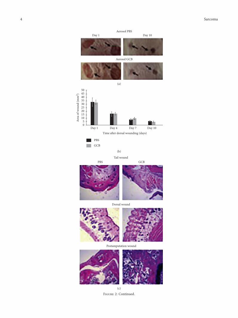
3.2. Aerosol GCB Had No Effect on Wound Healing butInhibited Osteosarcoma LungMetastases. In order to mimicthe clinical course of osteosarcoma, K7M3 cells wereinjected into the tibia. Micrometastases in the lung wereconfirmed by sacrificing 2 mice 3 weeks later. When thetumors measured 1.5 cm, tail and dorsal skin wounding andamputation of the affected limb were performed. )e micewere divided into 2 groups and treated with either aerosolPBS or aerosol GCB 24 hours after amputation. Aerosoltherapy continued twice a week for three weeks. Healing ofthe tail, dorsal skin, and amputation site was monitored.Skin wounds were measured 4, 7, and 10 days after theinitiation of aerosol therapy. )ere was no difference inwound healing of the tail or dorsal skin between the aerosolPBS- and aerosol GCB-treated mice (Figures 2(a) and 2(b)).Similar to dorsal and tail wounds, aerosol GCB also did notaffect the healing of the amputation site as shown by H&Estaining in 3 different regions (Figure 2(c)).
To determine whether aerosol GCB inhibited lung me-tastasis, five mice from each treatment group were sacrificedafter 3 weeks of aerosol therapy. Aerosol GCB significantlydecreased the number of visible lung metastases compared tothe aerosol PBS group (Figure 2(d)). Lung weights and thenumber of micrometastases were also significantly reduced inthe aerosol GCB group (Figures 2(e) and 2(f)).
3.3. Effect of Aerosol GCB on Cell Proliferation and the Numberof Fibroblasts in the Wound Areas. Ki67 is a marker of cell
P < 0.05700.0
600.0
500.0
400.0
300.0
200.0
100.0
0
Gem
cita
bine
seru
m le
vel (
ng/m
l)
10 min 30 min 24 hrs
Aerosol GCBGCB i.p.
Figure 1: Serum GCB levels following intraperitoneal (i.p.) andaerosol GCB. Mice were treated with 1mg/kg GCB given i.p. or byaerosol administration. Blood was collected at various times fol-lowing administration, and serum GCB levels were quantified.Mice treated with aerosol GCB had significantly lower serum levels(P< 0.05).
Sarcoma 3
Aerosol PBSDay 1 Day 10
Aerosol GCB
(a)
PBS
GCB
Time a�er dorsal wounding (days)Day 1 Day 4 Day 7 Day 10
Are
a of w
ound
(mm
2 )
504540353025201510
50
(b)
PBS GCBTail wound
Dorsal wound
Postamputation wound
(c)
Figure 2: Continued.
4 Sarcoma

Aerosol PBS
Aerosol GCB
(d)
Lung
wei
ght (
g)P < 0.01
PBS treated GCB aerosol
30
20
10
0
PBS
GCB
(e)
Lung
met
asta
sis n
umbe
r
P < 0.01
PBS Aerosol GCB
25
20
15
10
5
0
PBSGCB
(f)
Figure 2: Aerosol GCB inhibited lung metastases but had no effect on wound healing in the tail, dorsal skin, or bone. (a) Representativeappearance of the dorsal wound on days 1 and 10 in the mice treated with aerosol PBS (top row) or aerosol GCB (bottom row). (b))e areasof each wound in the aerosol PBS and aerosol GCB groups were measured 1, 4, 7, and 10 days after treatment (P> 0.05 for each group).(c) Representative H&E sections from the wounded tail, dorsal skin, and postamputation area 7 days following wounding. (d–f ) AerosolGCB inhibited the growth of osteosarcoma lung metastases as assessed visually (d), by the lung weight (e), and by the mean number of lungmetastases (f ) (P< 0.01).
Sarcoma 5

proliferation. We therefore quantified the number of Ki67+
cells in the tail and dorsal skin wounds 7 days after treatmentwith aerosol therapy. )ere was no difference in the positiveareas for Ki67 staining between aerosol PBS and aerosol GCB(Figure 3(a), P> 0.05 for both dorsal and tail wounds). Inaddition, quantification of the positive areas for fibroblastsusing immunohistochemistry also showed no difference be-tween aerosol PBS and aerosol GCB (Figure 3(b), P> 0.05 forboth dorsal and tail wounds). )ese results demonstrate thataerosol GCB had no effect on cell proliferation or the numberof fibroblast cells during wound healing.
3.4.AerosolGCBDidNotAffect the 3Critical Phases ofWoundHealing. Wound repair typically consists of three phases:initial inflammation, followed by proliferation and finallyremodeling. To determine whether aerosol GCB affectswound-induced inflammation, we quantified the number of
mast cells in the wound tissues using toluidine staining, thenumber of macrophages using F4/80 antibody staining, andthe ratio of M1 to M2 macrophages. M1 macrophages, whichinhibit cell proliferation and cause tissue damage, wereidentified and quantified using iNOS staining. M2 macro-phages, which promote cell proliferation and tissue repair,were identified and quantified using the anti-mannose re-ceptor antibody. Both types of macrophages are important fornormal wound healing as is the M1/M2 ratio. Aerosol GCBhad no effect on the number of mast cells (Figure 4(a),P> 0.05) or the number of M1 and M2 macrophages (Figure4(b), P> 0.05) in both tail and dorsal wounds. In addition, weshowed that the M1/M2 ratio in the tail and dorsal woundsdid not differ between aerosol PBS- and aerosol GCB-treatedmice (M1 Figure 4(c), P> 0.05; M2 Figure 4(d), P> 0.05).)ese results demonstrated that aerosol GCB does not affectthe first phase of wound healing, that is, wound-inducedinflammation in the mice.
PBSGCB
Tail wound
Posit
ive a
rea
100000
80000
60000
40000
20000
0GCBPBS
Dorsal wound
PBSGCB
60000
40000
20000
0
Posit
ive a
rea
GCBPBS
(a)
Dorsal wound
PBSGCB
15000
10000
5000
0
Posit
ive a
rea
GCBPBS
PBSGCB
20000
15000
10000
5000
0
Posit
ive a
rea
Tail woundGCBPBS
(b)
Figure 3: Effect of aerosol GCB on cell proliferation and fibroblast numbers associated with wound healing.)e dorsal and tail skin woundsfrom themice treated with aerosol PBS or aerosol GCBwere examined on day 7. (a) Cell proliferation was assessed using Ki67. (b) Fibroblastnumbers were assessed using anti-fibroblast antibody.)e positive areas were quantified by SimplePCI groups obtained from five h.p.f areasand compared using Student’s t-test. P> 0.05 for all graphs.
6 Sarcoma

We next determined whether aerosol GCB affectedendothelial cell proliferation and angiogenesis, both criticalfunctions in the second phase of wound healing.
Endothelial cells play an important role in angiogenesisin wound-healing process.
To explore whether GCB affects endothelial cell growthand recruitment in wound healing, we quantified thenumber of CD31+ cells (a marker expressed by endothelialcells) in dorsal and tail wound tissues 7 days after aerosolPBS or aerosol GCB. )ere was no difference in CD31staining in the tail and dorsal skin wounds between aerosolPBS- and aerosol GCB-treated mice (Figure 5(a), P> 0.05).VEGFR-2 has been shown to mediate almost all of theknown cellular responses to VEGF [29]. We thereforemeasured the density of VEGFR-2+ cells in the wound areasand once again found no difference between the aerosol PBS-and aerosol GCB-treated groups (Figure 5(b), P> 0.05). )eseresults indicate that aerosol GCB did not inhibit angiogenesis(an important process in wound dealing) or interfere with thegrowth of endothelial cells.
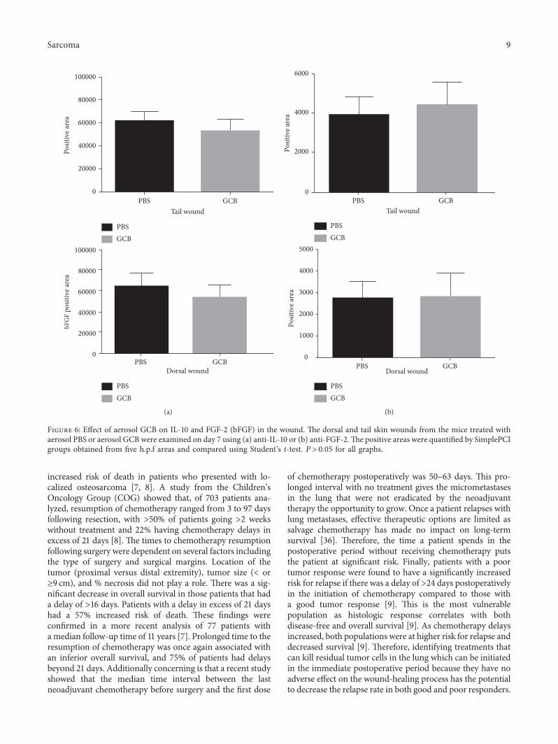
To determine whether aerosol GCB affected tissueremodeling (the third phase of wound healing), we quan-tified IL-10 and FGF-2 (bFGF), both of which are criticalcytokines for the remodeling phase of wound healing.
IL-10 plays an important role in wound healing throughits function to inhibit the infiltration of neutrophils andmacrophages toward the site of the wound area during thisphase [28]. We measured the positive areas of IL-10 indorsal wound and tail wound sections 7 days after GCBtreatment. Again, we showed no difference in tail or dorsal
wounds (Figure 6(a), P> 0.05) between aerosol GCB- andaerosol PBS-treated mice. FGF-2 or bFGF mediates theformation of new blood vessels that are critical in thewound-healing process following surgery. Different studiesdemonstrated a correlation between reduced FGF-2 ex-pression and wound-healing disorders [16, 30, 31, 33].mRNA levels of FGF-2 were reduced during wound healingin healing-impaired genetically diabetic mice comparedwith control mice [38]. Expression of FGF-2 was found tobe upregulated after injury in normal but not in diabeticrats. Impaired would healing was seen in aged mice, andthis impairment was associated with reduced levels ofFGF-2 [33]. Finally, when FGF-2 null mice were used forwound-healing studies, they showed delayed healing, whilethere was no delay seen in FGF-1-knockout mice [30, 31]Similar to our findings with IL-10, there was no differencein the expression of FGF-2 (bFGF) in the dorsal and tailwounds between the mice treated with aerosol PBS andaerosol GCB (Figure 6(b), P> 0.05).
3.5. Effect of Aerosol GCB on Wound Healing followingAmputation. Healing of the bone and tissue followingamputation and tumor removal is a critical part of thepatient recovery process. We therefore monitored healing ofthe amputated area every other day.)e amputation wound-healing process in the mice following aerosol GCB ad-ministration was not delayed compared with the micetreated with aerosol PBS. Similar to what we observed in thetail and dorsal skin wounds, there was no difference in
Tail wound
PBSGCB
PBSGCB
PBS GCB0
2000
4000
6000Po
sitiv
e are
aPo
sitiv
e are
a
Dorsal woundPBS GCB
0
2000
4000
6000
8000
10000
(a)
Dorsal woundPBS GCB
0
20000
40000
60000
80000
Tail woundPBS GCB
PBSGCB
PBSGCB
0
20000
40000
60000
80000
Posit
ive a
rea
Posit
ive a
rea
(b)
Dorsal woundPBS GCB
0
2000
4000
6000
8000
10000
Tail woundPBS GCB
PBSGCB
PBSGCB
0
20000
40000
60000
80000
100000
Posit
ive a
rea
Posit
ive a
rea
(c)
Tail wound
Dorsal woundPBS GCB
PBS GCB
PBSGCB
PBSGCB
0
20000
40000
60000
80000
100000
Posit
ive a
rea
0
2000
4000
6000
8000
Posit
ive a
rea
(d)
Figure 4: Effect of aerosol GCB on mast cell and macrophage infiltration andM1/M2 wound content.)e dorsal and tail skin wounds fromthemice treated with aerosol PBS or aerosol GCBwere examined on day 7 for (a) mast cells using toluidine blue and (b) macrophage contentusing F4/80 antibody. (c) M1macrophages were identified by anti-iNOS; (d) M2macrophages were identified using anti-mannose receptor.)e positive areas were quantified by SimplePCI groups obtained from five h.p.f areas and compared using Student’s t-test. P> 0.05 for allgraphs.
Sarcoma 7

fibroblast proliferation, CD31 expression, VEGFR-2 expression,or the wound-associated cytokines FGF-2 (bFGF) and IL-10between aerosol PBS and GCB groups (Figure 7).
4. Discussion
Combination chemotherapy given both pre- and post-operatively has raised the overall survival of patients withprimary nonmetastatic osteosarcoma from 20 to 65% [1–3].However, despite the use of aggressive multiagent chemo-therapy, 30–35% of patients who have no detectable metas-tasis at the time of diagnosis develop pulmonary metastasesfollowing surgical resection and adjuvant chemotherapy. )isstatistic has not improved in >30 years, and patients whodevelop lung metastases have a significantly reduced long-term survival [4, 5]. Equally disturbing is that there has beenrelatively little success in treating relapsed patients withsurgical resection of metastases in the lung, which is the mosteffective approach [32]. A recent analysis showed that the
median time to progression in multiple Phase II trials in theChildren’s Oncology Group for children and adolescents withrelapsed osteosarcoma was ∼4 months [34]. Furthermore, noresponses were seen in multiple Phase I trials from a singleinstitution [35]. Due to the absence of effective secondaryagents and the poor response rates to date for relapsed pa-tients, it is critical to identify conditions that put the patientsat a higher risk for relapse.
Postoperative chemotherapy is usually not initiated for2–8 weeks after tumor resection, depending upon the post-operative course, as chemotherapy can interfere with thewound-healing process and the production of cytokines at thewound site that have been shown to be critical to the healingprocess. )is leaves the patient in a potential vulnerablesetting as the tumor cells are free to divide and grow un-checked. Delays> 2 weeks postoperatively correlate witha poorer overall survival [7, 8]. Two independent retrospectivestudies showed that increased time from the definitive surgeryto the resumption of chemotherapy was associated with an
Tail wound
Dorsal wound
PBS
GCB
PBS
GCB
PBS GCB
PBS GCB
0
20000
40000
60000
80000
0
20000
40000
60000
80000
Posit
ive a
rea
Posit
ive a
rea
(a)
Tail wound
Dorsal woundPBS GCB
PBS GCB
PBS
GCB
PBS
GCB
0
10000
20000
30000
40000
0
20000
40000
60000
80000
100000
Posit
ive a
rea
Posit
ive a
rea
(b)
Figure 5: Effect of aerosol GCB on CD31 and VEGFR in the wound area.)e dorsal and tail skin wounds from the mice treated with aerosolPBS or aerosol GCB were examined on day 7 using (a) anti-CD31 or (b) anti-VEGFR )e positive areas were quantified by SimplePCIgroups obtained from five h.p.f areas and compared using Student’s t-test. P> 0.05 for all graphs.
8 Sarcoma
increased risk of death in patients who presented with lo-calized osteosarcoma [7, 8]. A study from the Children’sOncology Group (COG) showed that, of 703 patients ana-lyzed, resumption of chemotherapy ranged from 3 to 97 daysfollowing resection, with >50% of patients going >2 weekswithout treatment and 22% having chemotherapy delays inexcess of 21 days [8]. )e times to chemotherapy resumptionfollowing surgery were dependent on several factors includingthe type of surgery and surgical margins. Location of thetumor (proximal versus distal extremity), tumor size (< or≥9 cm), and % necrosis did not play a role. )ere was a sig-nificant decrease in overall survival in those patients that hada delay of >16 days. Patients with a delay in excess of 21 dayshad a 57% increased risk of death. )ese findings wereconfirmed in a more recent analysis of 77 patients witha median follow-up time of 11 years [7]. Prolonged time to theresumption of chemotherapy was once again associated withan inferior overall survival, and 75% of patients had delaysbeyond 21 days. Additionally concerning is that a recent studyshowed that the median time interval between the lastneoadjuvant chemotherapy before surgery and the first dose
of chemotherapy postoperatively was 50–63 days. )is pro-longed interval with no treatment gives the micrometastasesin the lung that were not eradicated by the neoadjuvanttherapy the opportunity to grow. Once a patient relapses withlung metastases, effective therapeutic options are limited assalvage chemotherapy has made no impact on long-termsurvival [36]. )erefore, the time a patient spends in thepostoperative period without receiving chemotherapy putsthe patient at significant risk. Finally, patients with a poortumor response were found to have a significantly increasedrisk for relapse if there was a delay of >24 days postoperativelyin the initiation of chemotherapy compared to those witha good tumor response [9]. )is is the most vulnerablepopulation as histologic response correlates with bothdisease-free and overall survival [9]. As chemotherapy delaysincreased, both populations were at higher risk for relapse anddecreased survival [9]. )erefore, identifying treatments thatcan kill residual tumor cells in the lung which can be initiatedin the immediate postoperative period because they have noadverse effect on the wound-healing process has the potentialto decrease the relapse rate in both good and poor responders.
Tail wound
Dorsal wound
PBS GCB
PBS GCB
0
20000
40000
60000
80000
100000
0
20000
40000
60000
80000
100000
Posit
ive a
rea
bFG
F po
sitiv
e are
a
PBSGCB
PBSGCB
(a)
Tail wound
Dorsal woundPBS GCB
PBS GCB0
2000
4000
6000
0
1000
2000
3000
4000
5000
Posit
ive a
rea
Posit
ive a
rea
PBSGCB
PBSGCB
(b)
Figure 6: Effect of aerosol GCB on IL-10 and FGF-2 (bFGF) in the wound. )e dorsal and tail skin wounds from the mice treated withaerosol PBS or aerosol GCB were examined on day 7 using (a) anti-IL-10 or (b) anti-FGF-2.)e positive areas were quantified by SimplePCIgroups obtained from five h.p.f areas and compared using Student’s t-test. P> 0.05 for all graphs.
Sarcoma 9

Such a strategy can have a significant impact on the long-termsurvival of patients.
Our current investigations show that aerosol GCB resultsin significantly lower serum levels compared with its systemicadministration. )e dose used in our studies was 1/10th thesystemic dose normally given to evaluate efficacy [22]. Wehave previously evaluated the efficacy of aerosol versus i.p.GCB against both primary OS and OS lung metastases [22].Treatment in these studies was similar to that in the presentinvestigations. When the primary tumor reached 130mm3
and micrometastases were present in the lung, the micewere treated 3 times weekly for 3 weeks. While aerosol GCB
was effective against both the primary tumor and the lungmetastases, i.p. GCB at the equivalent dose was only effectiveagainst the primary tumor. GCB given i.p. was not effectiveagainst the OS lung metastases [22]. Since i.p. GCB was noteffective against lungmetastases, we did not evaluate the effectof systemic GCB on wound healing. Our present studiesfocused on evaluating the simultaneous efficacy of aerosolGCB against pulmonary metastases and its effect on woundhealing. )e therapeutically appropriate dose of i.p. GCBwould yield even higher serum GCB levels which are morelikely to interfere with the healing process. )e goal of ourstudy was to evaluate the simultaneous activity of aerosoldelivery of the drug on established lung metastases in thesetting of a surgical resection. Supportive of the safety andtolerability of aerosol GCB are investigations showing thatthere was no organ or hematologic toxicity following 5 weeksof aerosol GCB given 3 times per week [21, 22]. More im-portantly, our present investigations show that aerosol GCBinitiated 48 hours following amputation of the limb with theprimary osteosarcoma tumor inhibited the growth anderadicated established micrometastases in the lung withoutinterfering with wound healing in the skin or bone. AerosolGCB had no effect on the 3 phases of wound healing in thedorsal skin, tail, or bone. Cell proliferation, the number offibroblasts and mast cells, macrophage recruitment, and theratio of Type I to Type II macrophages in the dorsal skin andtail wounds of the mice treated with aerosol GCB were similarto those of the mice treated with aerosol PBS.
Anti-VEGF therapy has been shown to result in latewound dehiscence [37]. Aerosol GCB did not inhibit en-dothelial cell growth or VEGF levels in the wounds and hadno effect on the production of either IL-10 or FGF-2 (bFGF).Similarly, when the wound at the amputation site was ex-amined, treatment with aerosol GCB had no effect on CD31,VEGF, bFGF, IL-10, or the number of fibroblasts. )is is thefirst demonstration that aerosol GCB can have a therapeuticeffect on lung metastases without interfering with woundhealing following surgical resection of the primary tumor,a critical process in the treatment regimen for osteosarcoma.Taken together, our data suggest that the initiation of aerosolGCB in the immediate postoperative period has the potentialto eradicate lung micrometastases without fear of interferingwith the postoperative healing process in the skin or thebone which can delay the resumption of the systemicchemotherapy. Eradication of lung metastases during thisperiod when systemic chemotherapy has been suspendedmay prevent relapse and result in an increase in the event-free and overall survival. Patients who develop lung me-tastases have a 5-year survival rate of only 20% [4, 5].
5. Conclusion
Salvage chemotherapy has made little impact on this sur-vival rate. Aggressive surgical removal of the metastases incombination with salvage chemotherapy only rescues about40% of patients [6]. As the safety of aerosol GCB has alreadybeen demonstrated in adult patients [24], our data support theconcept of using aerosol GCB as a bridge after tumor re-section and the initiation of clinical trials using aerosol GCB
Fibroblast staining
CD31 staining
VEGF receptor staining
bFGF staining
IL-10 staining
PBS GCB
Figure 7: Aerosol GCB did not affect bone healing followingamputation. Representative sections of immunohistochemistry ofthe mice 14 days after amputation for fibroblasts, CD31, VEGFR-2,FGF-2 (bFGF), and IL-10.
10 Sarcoma

in children and adolescents with osteosarcoma in the im-mediate postoperative period.
Conflicts of Interest
)e authors declare that there are no conflicts of interestregarding the publication of this article.
Acknowledgments
)e authors thank Ms. Barbara Liddle for her excellentassistance with manuscript preparation.
References
[1] F. Eilber, A. Giuliano, J. Eckardt, K. Patterson, S. Moseley, andJ. Goodnight, “Adjuvant chemotherapy for osteosarcoma:a randomized prospective trial,” Journal of Clinical Oncology,vol. 5, no. 1, pp. 21–26, 1987.
[2] N. Jaffe, “Historical perspective on the introduction and use ofchemotherapy for the treatment of osteosarcoma,” Advancesin Experimental Medicine and Biology, vol. 804, pp. 1–30, 2014.
[3] L. Mirabello, R. J. Troisi, and S. A. Savage, “Osteosarcomaincidence and survival rates from 1973 to 2004: data from thesurveillance, epidemiology, and end results program,” Cancer,vol. 115, no. 7, pp. 1531–1543, 2009.
[4] J. F. Huth and F. R. Eilber, “Patterns of recurrence afterresection of osteosarcoma of the extremity. Strategies fortreatment of metastases,” Archives of Surgery, vol. 124, no. 1,pp. 122–126, 1989.
[5] M. D. Tabone, “Osteosarcoma recurrences in pediatricpatients previously treated with intensive chemotherapy,”Journal of Clinical Oncology, vol. 12, no. 12, pp. 2614–2620,1994.
[6] M. A. Smith, N. L. Seibel, S. F. Altekruse et al., “Outcomes forchildren and adolescents with cancer: challenges for thetwenty-first century,” Journal of Clinical Oncology, vol. 28,no. 15, pp. 2625–2634, 2010.
[7] P. Berlanga, A. Canete, R. Diaz et al., “Presentation and long-term outcome of high-grade osteosarcoma: a single-institutionexperience,” Journal of Pediatric Hematology/Oncology, vol. 37,no. 5, pp. e272–e277, 2015.
[8] H. Imran, F. Enders, M. Krailo et al., “Effect of time to re-sumption of chemotherapy after definitive surgery on prog-nosis for non-metastatic osteosarcoma,” Journal of Bone andJoint Surgery-American Volume, vol. 91, no. 3, pp. 604–612,2009.
[9] P. A. Meyers, G. Heller, J. Healey et al., “Chemotherapy fornonmetastatic osteogenic sarcoma: the Memorial Sloan-Kettering experience,” Journal of Clinical Oncology, vol. 10,no. 1, pp. 5–15, 1992.
[10] C. N. Serhan and N. Chiang, “Novel endogenous smallmolecules as the checkpoint controllers in inflammation andresolution: entree for resoleomics,” Rheumatic Disease Clinicsof North America, vol. 30, no. 1, pp. 69–95, 2004.
[11] J. A. Schilling, “Wound healing,” Surgical Clinics of NorthAmerica, vol. 56, no. 4, pp. 859–874, 1976.
[12] P. Bainbridge, “Wound healing and the role of fibroblasts,”Journal of Wound Care, vol. 22, no. 8, pp. 407-408, 2013.
[13] G. Broughton II, J. E. Janis, and C. E. Attinger, “Woundhealing: an overview,” Plastic and Reconstructive Surgery,vol. 117, no. 7, pp. 1e-S–32e-S, 2006.
[14] J. Hart, “Inflammation. 2: its role in the healing of chronicwounds,” Journal ofWound Care, vol. 11, no. 7, pp. 245–249, 2002.
[15] J. E. Park and A. Barbul, “Understanding the role of immuneregulation in wound healing,” American Journal of Surgery,vol. 187, no. 5, pp. 11S–16S, 2004.
[16] S. Werner and R. Grose, “Regulation of wound healing bygrowth factors and cytokines,” Physiological Reviews, vol. 83,no. 3, pp. 835–870, 2003.
[17] A. J. Singer and R. A. Clark, “Cutaneous wound healing,”NewEngland Journal of Medicine, vol. 341, no. 10, pp. 738–746,1999.
[18] W. Plunkett, P. Huang, Y. Z. Xu, V. Heinemann, R. Grunewald,and V. Gandhi, “Gemcitabine: metabolism, mechanisms ofaction, and self-potentiation,” Seminars in Oncology, vol. 22,no. 4, pp. 3–10, 1995.
[19] S. Okuno, J. Edmonson, M. Mahoney, J. C. Buckner, S. Frytak,and E. Galanis, “Phase II trial of gemcitabine in advancedsarcomas,” Cancer, vol. 94, no. 12, pp. 3225–3229, 2002.
[20] N. V. Koshkina, B. E. Gilbert, J. C. Waldrep, A. Seryshev, andV. Knight, “Distribution of camptothecin after delivery asa liposome aerosol or following intramuscular injection inmice,”Cancer Chemotherapy and Pharmacology, vol. 44, no. 3,pp. 187–192, 1999.
[21] N. Gordon and E. S. Kleinerman, “Aerosol therapy for thetreatment of osteosarcoma lung metastases: targeting theFas/FasL pathway and rationale for the use of gemcitabine,”Journal of Aerosol Medicine and Pulmonary Drug Delivery,vol. 23, no. 4, pp. 189–196, 2010.
[22] N. V. Koshkina and E. S. Kleinerman, “Aerosol gemcitabineinhibits the growth of primary osteosarcoma and osteosar-coma lung metastases,” International Journal of Cancer,vol. 116, no. 3, pp. 458–463, 2005.
[23] C. O. Rodriguez Jr., R. A. Crabbs, D. W. Wilson et al.,“Aerosol gemcitabine: preclinical safety and in vivo antitumoractivity in osteosarcoma-bearing dogs,” Journal of AerosolMedicine and Pulmonary Drug Delivery, vol. 23, no. 4,pp. 197–206, 2010.
[24] E. Lemarie, L. Vecellio, J. Hureaux et al., “Aerosolizedgemcitibine in patients with carcinoma of the lung: feasibilityand safety study,” Journal of Aerosol Medicine and PulmonaryDrug Delivery, vol. 24, no. 6, pp. 261–270, 2011.
[25] N. Gordon, N. V. Koshkina, S.-F. Jia et al., “Corruption of thefas pathway delays the pulmonary clearance of murine os-teosarcoma cells, enhances their metastatic potential, andreduces the effect of aerosol gemcitabine,” Clinical CancerResearch, vol. 13, no. 15, pp. 4503–4510, 2007.
[26] Y. Ishida, J. L. Gao, and P. M. Murphy, “Chemokine receptorCX3CR1 mediates skin wound healing by promoting mac-rophage and fibroblast accumulation and function,” Journal ofImmunology, vol. 180, no. 1, pp. 569–579, 2008.
[27] H. Tomita, Y. Iwata, F. Ogawa et al., “P-selectin glycoproteinligand-1 contributes to wound healing predominantly asa p-selectin ligand and partly as an e-selectin ligand,” Journalof Investigative Dermatology, vol. 129, no. 8, pp. 2059–2067,2009.
[28] T. Kimura, M. Sugaya, A. Blauvelt, H. Okochi, and S. Sato,“Delayed wound healing due to increased interleukin-10expression in mice with lymphatic dysfunction,” Journal ofLeukocyte Biology, vol. 94, no. 1, pp. 137–145, 2013.
[29] K. Holmes, O. L. Roberts, A. M. )omas, and M. J. Corss,“Vascular endothelial growth factor receptor-2: structure,function, intracellular signalling and therapeutic inhibition,”Cellular Signalling, vol. 19, no. 10, pp. 2003–2012, 2007.
Sarcoma 11
[30] D. L. Miller, S. Ortega, O. Bashayan, R. Basch, and C. Basilico,“Compensation by fibroblast growth factor 1 (FGF1) does notaccount for the mild phenotypic defects observed in FGF2null mice,” Molecular and Cellular Biology, vol. 20, no. 6,pp. 2260–2268, 2000.
[31] S. Ortega, M. Ittmann, S. H. Tsang,M. Ehrlich, and C. Basilico,“Neuronal defects and delayed wound healing in mice lackingfibroblast growth factor 2,” Proceedings of the NationalAcademy of Sciences, vol. 95, no. 10, pp. 5672–5677, 1998.
[32] K. A. Skinner, F. R. Eilber, E. C. Holmes, J. Eckardt, andG. Rosen, “Surgical treatment and chemotherapy for pul-monary metastases from osteosarcoma,” Archives of Surgery,vol. 127, no. 9, pp. 1065–1070, 1992.
[33] M. E. Swift, E. S. Kleinman, and L. A. DiPietro, “Impairedwound repair and delayed angiogenesis in aged mice,” Lab-oratory Investigation, vol. 79, no. 12, pp. 1479–1487, 1999.
[34] J. P. Lagmay, M. D. Krailo, H. Dang et al., “Outcome ofpatients with recurrent osteosarcoma enrolled in seven phaseII trials through children’s cancer group, pediatric oncologygroup, and children’s oncology group: learning from the pastto move forward,” Journal of Clinical Oncology, vol. 34, no. 25,pp. 3031–3038, 2016.
[35] J. A. Livingston, K. R. Hess, A. Naing et al., “Validation ofprognostic scoring and assessment of clinical benefit forpatients with bone sarcomas enrolled in phase I clinical trials,”Oncotarget, vol. 7, no. 39, pp. 64421–64430, 2016.
[36] B. Kempf-Bielack, S. S. Bielack, H. Jurgens et al., “Osteo-sarcoma relapse after combined modality therapy: an analysisof unselected patients in the Cooperative Osteosarcoma StudyGroup (COSS),” Journal of Clinical Oncology, vol. 23, no. 3,pp. 559–568, 2005.
[37] H. Zhang, Z. Huang, X. Zou, and T. Liu, “Bevacizumab andwound-healing complications: a systematic review and meta-analysis of randomized controlled trials,” Oncotarget, vol. 7,no. 50, pp. 82473–82481, 2016.
[38] S. Werner, M. Breeden, G. Hubner, D. G. Greenhalgh, andM. T. Longaker, “Induction of keratinocyte growth factorexpression is reduced and delayed during wound healing inthe genetically diabetic mouse,” Journal of InvestigativeDermatology, vol. 103, no. 4, pp. 469–473, 1994.
12 Sarcoma

Stem Cells International
Hindawiwww.hindawi.com Volume 2018
Hindawiwww.hindawi.com Volume 2018
MEDIATORSINFLAMMATION
of
EndocrinologyInternational Journal of
Hindawiwww.hindawi.com Volume 2018
Hindawiwww.hindawi.com Volume 2018
Disease Markers
Hindawiwww.hindawi.com Volume 2018
BioMed Research International
OncologyJournal of
Hindawiwww.hindawi.com Volume 2013
Hindawiwww.hindawi.com Volume 2018
Oxidative Medicine and Cellular Longevity
Hindawiwww.hindawi.com Volume 2018
PPAR Research
Hindawi Publishing Corporation http://www.hindawi.com Volume 2013Hindawiwww.hindawi.com
The Scientific World Journal
Volume 2018
Immunology ResearchHindawiwww.hindawi.com Volume 2018
Journal of
ObesityJournal of
Hindawiwww.hindawi.com Volume 2018
Hindawiwww.hindawi.com Volume 2018
Computational and Mathematical Methods in Medicine
Hindawiwww.hindawi.com Volume 2018
Behavioural Neurology
OphthalmologyJournal of
Hindawiwww.hindawi.com Volume 2018
Diabetes ResearchJournal of
Hindawiwww.hindawi.com Volume 2018
Hindawiwww.hindawi.com Volume 2018
Research and TreatmentAIDS
Hindawiwww.hindawi.com Volume 2018
Gastroenterology Research and Practice
Hindawiwww.hindawi.com Volume 2018
Parkinson’s Disease
Evidence-Based Complementary andAlternative Medicine
Volume 2018Hindawiwww.hindawi.com
Submit your manuscripts atwww.hindawi.com