molecular mechanisms of cholangiocarcinoma progression
TRANSCRIPT

DOCTORAL SCHOOL
UNIVERSITY OF MILANO-BICOCCA
School of Medicine and Surgery
PhD Program in Translational and Molecular Medicine (DIMET)
Cycle XXX
Molecular mechanisms of
cholangiocarcinoma progression:
emphasizing the role of tumor-stroma
interactions
Dr. Simone Brivio
Registration number: 708864
Tutor: Prof. Mario Strazzabosco
Co-tutor: Prof. Luca Fabris
Coordinator: Prof. Andrea Biondi
ACADEMIC YEAR 2016/2017

2

3

4

5
Table of contents
CHAPTER 1 General introduction
8
1. Cholangiocarcinoma
9
1.1 Classification and pathological features
10
1.2 Epidemiology
15
1.3 Etiology
16
1.4 Molecular pathogenesis
16
1.4.1 Deregulated signaling pathways
18
1.4.2 Genetic and epigenetic abnormalities
21
1.5 Management
24
2. The tumor reactive stroma
26
2.1 The pathological relevance of the tumor reactive stroma in cholangiocarcinoma
32

6
2.2 Cancer-associated fibroblasts
34
2.3 Tumor-associated macrophages
37
2.4 Lymphatic endothelial cells
40
2.5 Non-cellular components of the tumor reactive stroma
43
2.5.1 The extracellular matrix
43
2.5.2 Intratumoral hypoxia
45
3. Molecular mechanisms underpinning cholangiocarcinoma progression: an overview
48
3.1 Molecular mechanisms of chemoresistance
48
3.2 Molecular mechanisms of cancer cell invasiveness
52
Scope of the thesis
56
References
59

7
CHAPTER 2 Leukemia inhibitory factor protects cholangiocarcinoma cells from drug-induced apoptosis via a PI3K/AKT-dependent Mcl-1 activation
85
CHAPTER 3 Low-dose paclitaxel reduces S100A4 nuclear import to inhibit invasion and hematogenous metastasis of cholangiocarcinoma
129
CHAPTER 4 Platelet-derived growth factor D enables cancer-associated fibroblasts to promote tumor lymphangiogenesis in cholangiocarcinoma
174
CHAPTER 5 Summary, conclusions and future perspectives
222
References
230
Contribution to international publications
234

8
CHAPTER 1
General introduction

9
1. Cholangiocarcinoma
Cholangiocarcinoma (CCA) is a highly aggressive epithelial
malignancy stemming from varying locations within the biliary tree.
Nowadays, CCA carries a very poor prognosis, mainly due to an early
metastatic propensity, and an impressive resistance to conventional
chemotherapy [1-3]. The median survival is below 2 years, and the
survival rate is less than 10% [4]. Liver failure, biliary tract sepsis, and
cachexia are the most common causes of CCA-related mortality [4,5].
According to its anatomical location, CCA is classified as either
intrahepatic (iCCA) (also called peripheral) or extrahepatic (eCCA),
with the latter being further divided into perihilar (pCCA) (also called
Klatskin tumor) and distal (dCCA). iCCA occurs within the liver
parenchyma, involving the intrahepatic biliary system in its entirety,
from segmental bile ducts to small bile ductules. pCCA arises from the
right and left hepatic ducts at or near their confluence, whereas dCCA
grows along the common bile duct (Figure 1) [6]. pCCA represents
about 50% of CCA cases, whereas dCCA and iCCA roughly accounts for
40% and 10% of tumors, respectively [3]. Classically, CCA is thought
to originate from the neoplastic transformation of the epithelial cells
lining the bile ducts, named cholangiocytes. However, recent evidence
suggests that iCCA pathogenesis may also involve mature hepatocytes
or hepatic progenitor cells (HPCs) [7,8]. The latter are immature,
bipotential epithelial cells residing in the canals of Hering (i.e., the
physiologic link between the hepatocyte canalicular system and the

10
biliary tree), capable of differentiating towards the cholangiocytic or
hepatocytic lineage [9]. Similar to iCCA, eCCA has been proposed to
arise not only from the lining epithelium of the extrahepatic biliary
tree, but also from peribiliary glands, i.e., mucin-producing glandular
elements scattered throughout the wall of perihilar and extrahepatic
bile ducts, representing a reservoir of multipotent stem cells of
endodermal origin [7,10].
Figure 1 Anatomical classification of cholangiocarcinoma. According to its
location within the biliary tree, cholangiocarcinoma (CCA) can be classified as either
intrahepatic (iCCA) or extrahepatic (eCCA), with the merging point of the second
order bile ducts representing the separation point. eCCA can be further divided into
perihilar (pCCA) or distal (dCCA), with the insertion of the cystic duct acting as the
anatomic boundary. Adapted from [7].
1.1 Classification and pathological features
Going beyond the anatomical location, CCA can be also
classified on the basis of its macroscopic growth pattern, which can be

11
identified as mass-forming, periductal-infiltrating, or intraductal-
growing [11,12]. The mass-forming type produces a well-delimited,
polylobulated mass that early occludes the bile duct lumen, and then
expands beyond the bile duct wall, within the periductal tissue.
Periductal-infiltrating tumors extend lengthwise along the bile duct,
thereby causing wall thickening and eventually, luminal obstruction.
Intraductal-growing tumors preferentially grow towards the lumen of
the bile duct, which undergoes a marked dilatation, and infiltrate the
bile duct wall only at very late stages (Figure 2). Of note, intraductal-
growing CCAs typically spread superficially along the mucosal layer,
thereby generating multiple masses on the inner surface of the bile
ducts [6,11,13]. The most frequent growth pattern of iCCA is the mass-
forming type, with the periductal-infiltrating and intraductal-growing
types exclusively arising at the level of large intrahepatic bile ducts.
pCCA typically originates as a periductal-infiltrating tumor, but it
tends to acquire additional mass-forming features in the course of its
progression. Finally, dCCA are almost exclusively periductal-
infiltrating or intraductal-growing [2,6]. Importantly, mass-forming
and periductal-infiltrating CCAs are generally associated with a poorer
prognosis compared to intraductal-growing tumors [11,14].

12
Figure 2 Morphologic classification of cholangiocarcinoma. Based on its
macroscopic growth characteristics, cholangiocarcinoma can be classified as mass-
forming (A), periductal-infiltrating (B), and intraductal-growing (C). See text for
further details. Adapted from [11].
At the microscopic, histopathological level, 90-95% of CCAs are
well to moderately differentiated adenocarcinomas, with an abundant
fibrous stroma surrounding the neoplastic glands [11,15]. However,
iCCAs are histologically heterogeneous tumors, which likely reflects
the different anatomical sites of origin and therefore, the
topographical heterogeneity of cholangiocytes all along the biliary tree
[14,16]. Indeed, large intrahepatic ducts (i.e., segmental, area and
septal ducts), similarly to extrahepatic bile ducts, are lined by mucin-
producing, columnar cholangiocytes, whereas small bile ductules (or
terminal cholangioles) and interlobular ducts are delimited by mucin-
negative, cuboidal cholangiocytes [16,17]. Although different
classification systems have been proposed over time, iCCA can be
substantially subcategorized in two main histological phenotypes,
which we refer to as bile duct (mucinous) type and bile ductular
(mixed) type. The bile duct type is composed of cylindrical tumor cells
organized in large glandular patterns, with extensive mucin
13
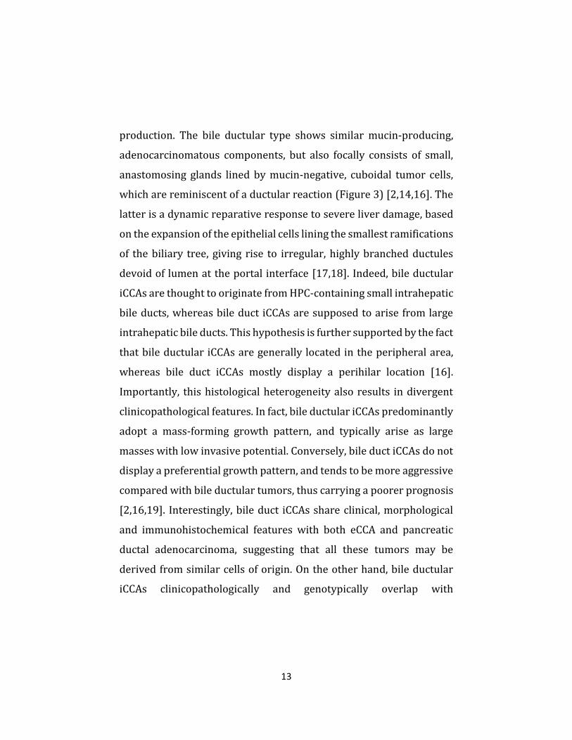
production. The bile ductular type shows similar mucin-producing,
adenocarcinomatous components, but also focally consists of small,
anastomosing glands lined by mucin-negative, cuboidal tumor cells,
which are reminiscent of a ductular reaction (Figure 3) [2,14,16]. The
latter is a dynamic reparative response to severe liver damage, based
on the expansion of the epithelial cells lining the smallest ramifications
of the biliary tree, giving rise to irregular, highly branched ductules
devoid of lumen at the portal interface [17,18]. Indeed, bile ductular
iCCAs are thought to originate from HPC-containing small intrahepatic
bile ducts, whereas bile duct iCCAs are supposed to arise from large
intrahepatic bile ducts. This hypothesis is further supported by the fact
that bile ductular iCCAs are generally located in the peripheral area,
whereas bile duct iCCAs mostly display a perihilar location [16].
Importantly, this histological heterogeneity also results in divergent
clinicopathological features. In fact, bile ductular iCCAs predominantly
adopt a mass-forming growth pattern, and typically arise as large
masses with low invasive potential. Conversely, bile duct iCCAs do not
display a preferential growth pattern, and tends to be more aggressive
compared with bile ductular tumors, thus carrying a poorer prognosis
[2,16,19]. Interestingly, bile duct iCCAs share clinical, morphological
and immunohistochemical features with both eCCA and pancreatic
ductal adenocarcinoma, suggesting that all these tumors may be
derived from similar cells of origin. On the other hand, bile ductular
iCCAs clinicopathologically and genotypically overlap with

14
cholangiolocellular carcinoma (CLC), an HPC-derived tumor that is
almost exclusively arranged in small, monotonous glands, with no
mucin production, showing both hepatocellular and cholangiocellular
differentiation characteristics [2,16]. In this regard, it was suggested
that bile ductular iCCAs, CLC, and combined hepatocellular-
cholangiocarcinoma actually all originate from HPCs, though the
neoplastic transformation apparently occurs at distinct stages of HPC
differentiation towards biliary or hepatocyte lineage [2]. In conclusion,
as the anatomical-based classification of CCA is challenged by the
highly branched, three-dimensional structure of the biliary system, the
histological subtyping has drawn increasing attention over time, also
because it carries more detailed information about tumor biology and
clinical course [2,16].
Figure 3 Hematoxylin and eosin staining of distinct histopathological subtypes
of intrahepatic cholangiocarcinoma. The bile duct type intrahepatic
cholangiocarcinoma (iCCA) is solely composed of large tubular, acinar, or papillary
structures arising from the abnormal proliferation of columnar cholangiocytes lining
the large intrahepatic ducts (A). The bile ductular type iCCA also focally consists of
small glandular patterns with irregular lumen (arrowheads) arising from the

15
abnormal proliferation of cuboidal cholangiocytes lining the small bile ductules and
the interlobular ducts (B). Modified from [2].
1.2 Epidemiology
CCA is the second most common primary neoplasm of the liver
after hepatocellular carcinoma (HCC), and overall accounts for 10-
20% of the deaths for hepatobiliary malignancies. Whereas liver
tumors are the second cause of cancer-related mortality worldwide, it
is clear that CCA represents a global health problem [5,20]. Of note,
CCA incidence rates are geographically heterogeneous, likely due to a
differential exposure to environmental risk factors, as well as to
genetic diversity among various populations [21]. The highest
incidence rates are found in China, South Korea and North Thailand,
which indeed are all areas where infestation with liver flukes is
endemic (see below). Conversely, CCA is a rare cancer (i.e., less than 6
cases per 100,000 people) in Europe, United States and Australia. For
instance, the incidence rate of CCA in Italy is 3.36/100,000 [2].
However, the occurrence of CCA, more specifically of iCCA, markedly
increased in Western Countries during the last three decades of the
twentieth century, thus strongly renewing the interest of the scientific
community towards this malignancy, though the underlying reasons
have not yet been fully understood [2]. It is also worth noting that,
owing to the poor outcome, mortality and incidence rates are nearly
comparable [5].

16
1.3 Etiology
Unlike HCC, CCA typically occurs in the absence of an evident
pre-existing chronic liver disease. Nevertheless, several risk factors
have been identified over time, and recent observations also point out
that iCCA is increasingly detected in the context of cirrhotic liver [1].
Hepatobiliary fluke infestation (e.g., Opisthorchis viverrini, Clonorchis
sinensis), hepatitis B viral infection, and hepatolithiasis are common
risk factors for CCA in Southeast Asia, where the prevalence of these
conditions is high. Conversely, the association between CCA and
primary sclerosing cholangitis (PSC) or chronic hepatitis C infection is
statistically relevant in Western Countries [2-4]. More specifically, 8-
40% of PSC patients eventually develop CCA in their lifetime, and this
usually occurs at a younger age (30-50 years) compared to the general
population (60-70 years) [15]. Interestingly, the burden of viral
hepatitis C has been even claimed to partly account for the increasing
incidence of iCCA in Europe and United States [2]. Other major risk
factors for CCA are metabolic syndrome and congenital malformations
of the bile ducts (e.g., Caroli’s disease, choledochal cysts) [2-4].
1.4 Molecular pathogenesis
The molecular pathogenesis of CCA is a complex, multistep
process relying on genomic (point mutations, copy number variations,
chromosome fusions) and epigenetic (promoter hypermethylation,
histone deacetylation) alterations, as well as on non-genetically

17
determined signaling pathway deregulations [3,8]. In this regard,
chronic inflammation of the biliary tract is unanimously recognized as
a paramount force behind the neoplastic transformation, regardless of
etiology. Indeed, a persistent inflammatory state is capable of
generating a pro-carcinogenic microenvironment abnormally
enriched with cytokines and growth factors, which are broadly
released by both inflammatory and epithelial cells. On the one hand,
pro-inflammatory cytokines elicit DNA damage by fueling the
generation of nitric oxide, ultimately leading to an increased mutation
rate. On the other hand, the wide web of local inflammatory cues
directly prompts cholangiocytes to undertake a sustained proliferative
program, generally coupled with apoptosis evasion. Of note, the
acceleration of the cell cycle, along with the impossibility of
eliminating dysfunctional cells by programmed cell death, further
supports the accumulation of somatic mutations potentially conducive
to neoplastic growth [5,22]. In addition, chronic inflammation is
typically comorbid with cholestasis, which for its part, dangerously
promotes cholangiocyte turnover in this hectic microenvironment,
again increasing the risk of malignant transformation (Figure 4)
[3,22].

18
Figure 4 Cellular mechanisms driving cholangiocarcinogenesis. Chronic
inflammation of the biliary tract, coupled with sever obstruction of bile flow, may
trigger the pathogenesis of cholangiocarcinoma, both by directly fostering the
unrestrained growth of the biliary epithelium, and by leading to nitric oxide-
mediated DNA damage. Modified from [22].
1.4.1 Deregulated signaling pathways
Among cytokines promoting cholangiocarcinogenesis,
interleukin (IL)-6, a well-known biliary mitogen, is recognized to play
a key role. IL-6 is constitutively expressed by both normal and
neoplastic cholangiocytes, and its secretion can be further enhanced
by inflammatory mediators such as IL-1β and tumor necrosis factor
(TNF)-α [23]. At the mechanistic level, IL-6 markedly promotes CCA
cell survival by up-regulating the anti-apoptotic protein myeloid cell
leukemia (Mcl)-1, via activation of signal transducer and activator of
transcription (STAT)3, phosphatidylinositol-3 kinase (PI3K), and p38
mitogen-activated protein kinase (MAPK) pathways [24-26].

19
Mitogenic effects of IL-6 on CCA cells are also reported, and mainly rely
on p44/p42 (also known as extracellular signal-regulated kinase
(ERK)1/2) and p38 MAPK activation [23]. Importantly, in mice with
genetically primed biliary tract, the pro-inflammatory cytokine IL-33
was shown to greatly facilitate CCA development by promoting IL-6
expression, further validating the intimate relationship between
chronic inflammation and biliary carcinogenesis [27].
Deregulation of growth factor signaling also represents a major
pro-tumorigenic mechanism in CCA. In particular, epidermal growth
factor receptor (EGFR) (also called ErbB1 or HER1), ErbB2/HER2, and
MET pathways, which generally involve the activation of MAPKs, are
deeply connected to the acquisition of malignant traits by biliary
epithelial cells, as a result of sustained ligand stimulation, receptor
overexpression or inactivation of negative feedback mechanisms.
Classically, constitutive activation of these signaling cascades exerts
potent mitogenic effects on the biliary epithelium, ultimately
encouraging tumor outgrowth in an autocrine fashion [7,12]. For
instance, EGFR and ErbB2 are potent inducers of cyclooxygenase
(COX)-2 expression, which fosters CCA growth by favoring
uncontrolled proliferation and evasion of apoptosis [4]. Of note, COX-
2 up-regulation may also be induced by inflammatory cytokine (e.g.,
TNF-α), bile acids, nitrosative stress, and oxysterols (i.e., cholesterol
oxidation products) [5].

20
Notch and Hedgehog (Hh) signaling are evolutionarily
conserved morphogen pathways fulfilling essential roles in both liver
morphogenesis and liver regeneration following chronic injury.
Therefore, it is not surprising that the aberrant activation of both
pathways has been widely related to cholangiocarcinogenesis [2]. In
particular, studies in mice have shown that a persistent hepatic Notch1
activation contributed to the development of CCA, based on an
abnormal transdifferentiation of normal adult hepatocytes into
neoplastic cholangiocytes [28,29]. Of note, inducible NO synthase
(iNOS) was reported to substantially promote Notch1 expression in
murine cholangiocytes via production of NO [30]. Similarly, the
pharmacological inhibition of autocrine Hh signaling dramatically
impaired CCA cell viability, both in vitro and in xenograft mouse
models, arguing for a leading role of this pathway in the emergence of
malignant growth features [31]. The molecular mechanisms
underlying Hh overactivation have yet to be elucidated, though the
accumulation of oxysterols in bile has been suggested to play a role
[32].
Cholangiocytes, especially those lining the intrahepatic portion
of the biliary tree, are potentially able to secrete and respond to a wide
range of neuropeptides and hormones, since they classically adopt a
neuroendocrine-like phenotype in the course of chronic liver injury
[5,33]. In this regard, several endocrine factors have been claimed to
boost the neoplastic transformation of biliary epithelial cells, either by

21
chronically fueling proliferation or by preventing apoptosis. For
instance, malignant cholangiocytes up-regulate estrogen receptor
(ER)α, whose stimulation actually results in enhanced cell growth, as
well as in increased expression of IL-6 and vascular endothelial growth
factor (VEGF), both critical mediators of CCA biology. Moreover, a
relevant role in promoting CCA cell proliferation has also been
attributed to dopamine, serotonin, histamine, and leptin [2].
1.4.2 Genetic and epigenetic abnormalities
Somatic mutations in well-known proto-oncogenes (e.g., KRAS,
FGFR2, EGFR, ERBB2, MET, IDH1/2) or tumor suppressor genes (e.g.,
TP53, SMAD4, ARID1A, PBRM1, BAP1) have been widely documented
in CCA [34,35]. Importantly, the pattern of oncogenic mutations in CCA
is heavily influenced by both etiology and tumor anatomical location
[2,4,35]. For instance, TP53 mutations are more frequent in liver fluke-
related CCAs compared with non-infection-related tumors, whereas
IDH1/2 and BAP1 mutations generally show an opposite trend [36].
Regarding the anatomic subtype, FGFR2, MET, IDH1/2, ARID1A,
PBRM1, and BAP1 mutations are characteristic of iCCA, whereas KRAS
and ERBB2 mutations are more common in eCCA [34,35].
Activating mutations in KRAS, as well as loss-of-function
mutations in TP53 are frequent events in cancer, and CCA is no
exception, with mutation frequencies of 9-40% and 3-45%,
respectively [34]. Specifically, KRAS belongs to the small GTPase

22
superfamily, and actively regulates several cellular processes such as
proliferation, survival, and motility, through the activation of its
downstream effectors, including p44/42 MAPK and
PI3K/Akt/mammalian target of rapamycin (mTOR) pathways [35].
p53 is deeply involved in cellular stress responses, by acting as a
transcription factor to regulate cell cycle arrest, DNA repair and
apoptosis. Loss of p53 activity paves the way for an uncontrolled
proliferation of damaged cells, ultimately favoring tumorigenesis. In
addition, alterations in p53 expression may lead to an aberrant
accumulation of β-catenin, a signaling molecule that modulates the
expression of several oncogenic genes [3,37,38]. Unlike KRAS and
TP53 point mutations, which are widespread genetic abnormalities,
chromosomal translocations involving the FGFR2 gene are almost
exclusively found in CCA, thus representing a potential diagnostic
marker [2]. Fibroblast growth factor receptor (FGFR)2 is tyrosine
kinase protein acting as a receptor for various FGFs, which are well-
known regulators of mitogenesis and differentiation [39]. Typically,
FGFR2 fusion proteins undergo an enforced, ligand-independent
dimerization, which is eventually responsible for an unrestrained
activation of the FGFR2 kinase domain [8]. Still on the subject of
receptor kinase signaling, a decreased expression of SMAD4, a
common signal transducer of transforming growth factor (TGF)-β
pathway, is frequently found in CCA, and undermines the tumor

23
suppressor functions typically exerted by TGF-β during early stages of
carcinogenesis [4,40].
Interestingly, several recurrently mutated genes in CCA (e.g.,
IDH1/2, ARID1A, PBRM1, BAP1) directly or indirectly impinge on
epigenetic dynamics and chromatin architecture, thereby deeply
influencing the transcriptomic profile of cancer cells [3]. For instance,
mutant forms of isocitrate dehydrogenases (IDH)1 and 2 drive the
production of 2-hydroxyglutarate, an oncometabolite that elicits
aberrant epigenetic changes by impairing the function of multiple
enzymes involved in histone and DNA methylation [3,8,35].
Specifically, it was shown that mutant IDH1/2 could contribute to the
pathogenesis of HPC-derived iCCA by epigenetically blunting the
expression of hepatocyte nuclear factor 4α, a master regulator of
hepatocytic differentiation [41]. Regardless of the underlying cause,
epigenetic silencing via promoter hypermethylation has been
frequently described in CCA, involving well-known tumor suppressor
genes such as SOCS3, CDKN2A, APC, and RASSF1 [7,8]. In particular,
reduced expression of suppressor of cytokine signaling (SOCS)3
partially accounts for the aberrant activation of IL-6 signaling typically
observed in CCA, as it acts as a negative-feedback regulator of the Janus
kinases/STAT3 axis [42]. In contrast to IDH1/2 gene, ARID1A, PBRM1,
and BAP1 genes are classically endowed with tumor suppressive
functions, and frequently harbor inactivating mutations in CCA
patients [43]. In particular, AT-rich interaction domain (ARID)1A and

24
polybromo (PBRM)1 are subunits of the switch/sucrose non-
fermentable chromatin-remodeling complex, which finely regulates
gene transcription by mobilizing nucleosomes [44], while BRCA1
associated protein (BAP)1 is a nuclear deubiquitylating enzyme,
capable of altering chromatin architecture by modulating the
ubiquitylated status of histone 2A [45].
1.5 Management
As previously mentioned, the prognosis of CCA is still very grim,
basically due to the late diagnosis and the lack of effective therapeutic
strategies [22]. Indeed, CCA is difficult to detect and diagnose early,
since it remains clinically silent until advanced stages, when it finally
presents with non-specific symptoms such as painless jaundice
(eCCA), weight loss or abdominal pain (iCCA) [3,4]. Therefore, most of
patients are diagnosed when metastatic dissemination has already
occurred [2]. In fact, CCA is an highly invasive cancer, which tends to
spread along the duct walls, and pervasively infiltrate the adjacent
structures (branches of the portal vein, lymphatic vessels, nerve
fibers), thereby spawning metastases within the liver, at regional
lymph nodes, or at distant sites (notably, lungs or peritoneum) (Figure
5) [5,46]. Diagnosis of CCA frequently occurs as an incidental finding,
and is classically based on a combination of clinical data, non-specific
(biochemical and/or histological) biomarkers, and imaging modalities
[2]. Surgical resection by partial hepatectomy and orthotopic liver

25
transplantation are the only treatments with curative intent, but their
applicability is limited to early-stage disease [2]. Accordingly, only
30% of CCA patients are actually eligible for radical surgery [3].
Moreover, chances of recurrence after resection are still very high (49-
64%), ultimately resulting in discouraging outcomes, with a 5-year
survival after resection lower than 45% [2,12]. Although liver
transplantation was initially hailed as a promising therapy for
unresectable tumors without evidence of metastasis, its actual
reliability remains controversial. Indeed, encouraging results were
obtained only in highly specialized centers implementing stringent
patient selection criteria, whereas historically, liver transplant is in
turn associated with high recurrence rates and poor long-term
survival [2,12]. CCA patients who are not candidate for surgical
therapy have no other chance but palliative procedures, which, by
definition, are merely aimed at turning the tumor into a clinically
manageable chronic disease and improving the quality of life [4]. The
median survival of patients diagnosed at inoperative stage is 6-12
months, with a 5-year overall survival of 5% [4,5,8]. Systemic
chemotherapy with gemcitabine plus cisplatin currently represents
the standard of practice in the palliative setting, though pledging a
median overall survival of only 11.7 months [2,47]. In fact, CCA is
characterized by a remarkable resistance to conventional
chemotherapeutic agents, directly resulting from the intrinsic ability
of CCA cells to efficiently escape from drug-induced cytotoxicity [5,48].

26
Unfortunately, there are still no targeted molecular therapies
approved for the treatment of CCA, likely owing to the poor knowledge
on the molecular mechanisms driving its development and
progression [35].
Figure 5 Metastatic patterns of cholangiocarcinoma. Cholangiocarcinoma is
characterized by a highly invasive behavior. Lymphatic vessels, nerve fibers, and
branches of the portal vein offer CCA cells multiple routes to escape from the primary
site of growth. Moreover, the periductal wall may be directly invaded by tumoral
infiltrates. Early metastases are more common within the liver and at regional lymph
nodes rather than at distant sites (notably, lung or peritoneum). Adapted from [46].
2. The tumor reactive stroma
Normal tissues encompass two distinct but interdependent
compartments, namely the parenchyma, which accounts for the
specific tissue function, and the stroma, which provides a multifaceted
support for the parenchyma, and predominantly consists of

27
extracellular matrix (ECM) proteins (notably, type I collagen and
fibronectin), resting fibroblasts, a few resident leukocytes, and
vascular elements. The structure of solid tumors is roughly similar,
with the parenchymal component being represented by the malignant
cells themselves [49,50]. However, in sharp contrast to a normal
stroma, which typically contains a small amount of quiescent
fibroblasts embedded within a physiological ECM, the tumor stroma is
characterized by a large number of activated fibroblasts, an
abnormally rich inflammatory infiltrate, and an enhanced capillary
density. By undergoing activation, fibroblasts not only increase their
proliferative activity, but also secrete starkly higher amounts of ECM
components (e.g., fibrillar collagens, fibronectin, tenascin C, and
proteoglycans), thereby generating a highly desmoplastic
microenvironment that is reminiscent of organ fibrosis [51]. Of note,
in human cancers, the stromal compartment can represent up to 90%
of the total mass [52]. However, its relative amount is highly variable
among tumors, and does not necessarily correlate with the degree of
malignancy [49].
In 1986, Harold F. Dvorak first called attention to the
similarities existing between the generation of tumor stroma and
wound healing [52]. Indeed, both processes are initially triggered by
an increased microvascular permeability, which enables a massive
extravasation of plasma proteins such as fibrinogen, plasminogen, and
fibronectin, ultimately resulting in the deposition of an extravascular

28
clot of cross-linked fibrin enriched with fibronectin. This bulk of fibrin
acts as a promiscuous substrate supporting the migration of
inflammatory cells (mainly macrophages), endothelial cells, and
fibroblasts, which in the tumoral context, are powerfully recruited and
activated by a plethora of cancer cell-derived soluble factors. A broad
synthesis of matrix components, coupled with extensive angiogenesis,
then leads to a gradual transformation of the original fibrin-
fibronectin gel matrix into a collagenous and vascularized stroma,
which in wound healing, is usually referred to as granulation tissue.
Upon further deposition of interstitial collagens, the fibrin clot is little
by little degraded, new vessels are partially resorbed, and the number
of activated fibroblasts markedly decreases, thus giving rise to a poorly
vascularized, densely collagenous, and nearly acellular connective
tissue. However, unlike physiological wound healing, which is a self-
limiting process, the remodeling of tumor stroma is continuously
evolving, since in tumors, the molecular cues that classically evoke the
wound healing cascade are released in an unrestrained manner, which
led Dvorak to describe neoplastic lesions as “wounds that do not heal”.
As a result, the tumor core is typically associated with a scar tissue-like
stroma, whereas cancer cells located at the invasive front are generally
encased within a highly cellular stroma mimicking the active stages of
wound healing. Among the mediators of tumor stroma generation,
VEGF is suggested to play a paramount role, since it is constitutively
expressed at high levels by the vast majority of human neoplasms, and

29
may potentially account for both chronic vascular hyperpermeability
and tumor angiogenesis. Of note, besides being secreted by cancer
cells, VEGF can also be plentifully produced by macrophages and
fibroblast populating the tumor microenvironment [50-52].
In summary, by behaving like self-perpetuating wounds,
tumors can effectively hijack a physiological host process, in an effort
to build up a personalized microenvironment that may adequately
satisfy their high metabolic needs, and actively support their
malignant growth [50,53,54]. Indeed, a dense, mutual paracrine
communication typically takes place between cancer and stromal cells,
giving rise to a hectic microenvironment that is increasingly
recognized as a key determinant of tumor progression. As mentioned
above, neoplastic cells act as leading actors in the remodeling of the
neighboring stroma, by chronically recruiting, activating, and co-
opting several inflammatory and mesenchymal cell types. Stromal
cells, for their side, secrete a plethora of cyto/chemokines, growth
factors, and proteinases that directly foster the emergence of
malignant phenotypes, and/or contribute to the recruitment and
aberrant activation of additional stromal components [55,56].
Furthermore, intratumoral hypoxia and aberrant changes in the ECM
may further impinge on cancer cell behavior [57]. Interestingly, a
growing body of evidence suggests that oncogenic signals emanating
from the TRS may even modify the epigenome of cancer cells [58-60].
In the light of the above, it is clear that the naïve stroma represents a

30
highly plastic and dynamic compartment that is prone to pandering to
the neoplastic evolution of the adjacent parenchyma, thereby
undergoing a change from gatekeeper of tissue homeostasis to
pathological niche fueling cancer aggressiveness [61,62]. Therefore,
the complex milieu harboring tumor growth is currently termed as
“tumor reactive stroma” (TRS). It is worth noting that normal epithelia,
as well as early-stage carcinomas, are surrounded by a well-delineated
basement membrane that effectively separates epithelial cells from
the adjoining connective tissue. Although there is evidence that a
primal tumor-stroma crosstalk still occurs despite the presence of an
intact (but altered) basal lamina, reactive stromal cells are empowered
to fully exert their pro-neoplastic effects only upon basement
membrane degradation, which represents one of the first steps of the
invasion-metastasis cascade (Figure 6) [51,63].

31
Figure 6 Cellular and molecular mechanisms underlying the generation of the
tumor reactive stroma in cholangiocarcinoma. Upon neoplastic transformation,
cholangiocytes acquire the ability to broadly secrete a rich repertoire of
cyto/chemokines and growth factors that enable them to establish an intense
crosstalk with various cell types (e.g., portal fibroblasts, hepatic stellate cells,
inflammatory cells, endothelial cells). Moreover, the basement membrane is
progressively dismantled through the massive release of matrix metalloproteinases
(MMPs), further supporting the interplay between the neoplastic epithelium and its
stromal microenvironment. It is against this background that multiple mesenchymal
and inflammatory cells are plentifully recruited to the tumor mass through paracrine
signals chronically released by malignant cholangiocytes. For instance, platelet-
derived growth factor (PDGF)-DD triggers the chemotaxis of resident fibroblasts, C-
C motif chemokine ligand (CCL)2 and colony stimulating factor (CSF)-1 dictate the
monocyte homing from blood circulation, and vascular endothelial growth factor
(VEGF)-C orchestrates lymphangiogenesis by stimulating the proliferation and
migration of lymphatic endothelial cells. Under the influence of the tumor
microenvironment, fibroblasts and monocytes transdifferentiate into cancer-
associated fibroblasts (CAFs) and tumor-associated macrophages (TAMs),
respectively, and then foster tumor aggressiveness in a paracrine fashion (see text
for further details). Importantly, cholangiocarcinoma cells, CAFs, and TAMs all

32
contribute to the aberrant remodeling of the extracellular matrix (ECM), an
additional mechanism supporting cancer progression. Modified from [53].
2.1 The pathological relevance of the tumor reactive stroma in
cholangiocarcinoma
In CCA, as well as in other epithelial tumors with strong
invasiveness, such as breast and pancreatic carcinomas, the stromal
compartment is particularly prominent, thus representing a
histological hallmark (Figure 7). More importantly, it is now a fact that
the TRS hugely promotes CCA growth and dissemination, which is why
it is drawing a growing interest as a potential therapeutic target
[55,56]. Interestingly, it was shown that the genomic profile of CCA-
associated stroma markedly differed from that of fibrous tissue from
peritumoral areas. Furthermore, specific genetic changes in the tumor
stroma were found to correlate with clinicopathological variables
predictive of bad prognosis. Of note, most of the differentially
expressed genes were involved in cell metabolism, cell cycle
progression, ECM composition, and intracellular signaling [64]. In line
with these findings, a genome-wide transcriptome profiling of tumor
epithelium and stroma from resected CCAs revealed a stromal gene
signature significantly associated with poor prognosis. In particular,
the stromal compartment was markedly enriched with inflammatory
cyto/chemokines, including IL-6, IL-33, TGF-β3, and C-C motif
chemokine ligand (CCL)2 (also called monocyte chemoattractant
protein (MCP)-1) [65]. Overall, these studies highlight not only the

33
uniqueness of the TRS as a biological entity, but also the prognostic
relevance of the molecular aberrations underlying its pathological
remodeling. In the next paragraphs, we will discuss in detail the
characteristics of the main (cellular and non-cellular) components of
the TRS, particularly focusing on how they are supposed to elicit the
emergence of malignant phenotypes in CCA cells.

34
Figure 7 Phenotyping the tumor reactive stroma in cholangiocarcinoma.
Immunohistochemistry of multiple markers to characterize the main cellular and
structural components of the tumor reactive stroma in intrahepatic
cholangiocarcinoma. Cancer-associated fibroblasts (α-SMA) (A); extracellular matrix
(fibronectin) (B); inflammatory cells (CD45) (C); tumor-associated macrophages
(arrows) (CD206) (D); lymphatic endothelial cells (podoplanin) (E); vascular
endothelial cells (CD34) (F). Original magnification: 200x. Modified from [53].
2.2 Cancer-associated fibroblasts
Cancer-associated fibroblasts (CAFs) are perpetually activated
fibroblasts, phenotypically characterized by the expression of
vimentin, S100A4 (otherwise called fibroblast-specific protein 1),
fibroblast activation protein (FAP), and especially, α-smooth muscle
actin (α-SMA), a widely recognized hallmark of fibroblast activation
[66,67]. Gene expression profiling of CAFs from CCA specimens
unveiled thousands of differentially expressed genes compared to
fibroblasts from matched peritumoral tissue, with the majority of
them being involved in cellular metabolism [68]. Indeed, CAFs feature
a high proliferation rate, and are deeply engaged in the remodeling of
tumor-associated ECM, due to the enhanced production of both matrix
components (e.g., collagen type I, fibronectin, tenascin C) and matrix-
degrading enzymes. Furthermore, CAFs are capable of secreting a rich
repertoire of cyto/chemokines and growth factors, which mediate the
crosstalk with cancer, endothelial and inflammatory cells [49,51]. For
instance, CAF-derived CCL2 effectively triggers macrophage
recruitment to the tumor neighborhood [53,69], whereas the broad

35
secretion of VEGF accounts for the ability of CAFs to sustain tumor-
associated angiogenesis and lymphangiogenesis [70]. Hepatic stellate
cells (HSCs) and portal fibroblasts are the main precursors of CAFs,
with a minor contribution by bone marrow-derived mesenchymal
stem cells (MSCs) [71]. Interestingly, conditioned medium from CCA
cells was found to foster the activation of HSCs [72] and primary liver
myofibroblasts [73], as well as to induce the differentiation of MSCs
into myofibroblast-like cells [74], further confirming that cancer cells
are largely responsible for the generation of CAFs. In particular, among
the pro-fibrotic factors driving the recruitment and/or activation of
fibroblasts within the tumor microenvironment, TGF-β, FGF, and
platelet-derived growth factor (PDGF), which may be released by both
cancer and inflammatory cells, have been widely reported to play a
prominent role [51,57,75]. In this regard, our group has recently
unveiled that PDGF-DD, which is aplenty released by CCA cells under
severe hypoxia, can massively promote the migration of fibroblasts
towards the tumor mass, by activating the small Rho GTPases Rac1 and
Cdc42, as well as the c-Jun N-terminal kinases (JNK) pathway, through
PDGFRβ binding [75].
Co-culture and conditioned medium experiments clearly
demonstrated that both CAFs and their precursors (i.e., HSCs) are able
to provide CCA cells with enhanced proliferative, survival, and
migratory capabilities, in vitro [67,72,76-82]. Furthermore, co-
transplantation of CCA cells with hepatic myofibroblasts [73] or HSCs

36
[72] into the flank of nude mice markedly boosted the growth of CCA
xenografts. Similarly, the selective depletion of CAFs by navitoclax in a
syngeneic, orthotopic rat model of CCA dramatically impaired tumor
burden and metastasis, while improving host survival [83]. Overall,
these studies argue for a tumor-promoting role of CAFs in CCA, which
is further validated by the finding that α-SMA expression proved to be
an independent negative prognostic factor for survival in iCCA patients
[77]. In particular, various soluble factors have been shown to mediate
the pro-neoplastic effects of CAFs, including C-X-C motif chemokine
ligand (CXCL)12 (also named stromal cell-derived factor (SDF)-1),
heparin-binding epidermal growth factor (HB-EGF), and PDGF-BB. For
instance, both CXCL12 [78] and PDGF-BB [81,82] strongly encourage
apoptosis resistance in CCA cells, by up-regulating the anti-apoptotic
protein B-cell lymphoma (Bcl)-2, and activating the Hh signaling,
respectively. CXCL12 also promotes CCA cell invasiveness, through
activation of ERK1/2 and PI3K pathways [78,79]. The migratory and
invasive properties of neoplastic cholangiocytes are also enhanced by
HB-EGF, which binds to EGFR on the cancer cell surface, and thus starts
a transcriptional program that involves ERK1/2, STAT3 and β-catenin
[73]. It is worth noting that CCA cells and CAFs are engaged in an
intricate network of reciprocal, self-perpetuating paracrine loops,
which well exemplify the ability of cancer cells to tirelessly shape the
behavior of their stromal neighbors. For instance, the expression of
HB-EGF by CAFs can be induced by CCA cell-derived TGF-β1, whose
37
production is in turn sustained by HB-EGF in a paracrine fashion [73].
Similarly, studies in rats showed that CCA cells lead CAFs to massively
secrete hepatocyte growth factor (HGF), which in turn can up-regulate
C-X-C motif chemokine receptor (CXCR)4 (the cognate receptor for
CXCL12) on the surface of CCA cells, thus making them hyper-
responsive to CAF-derived CXCL12 [84].
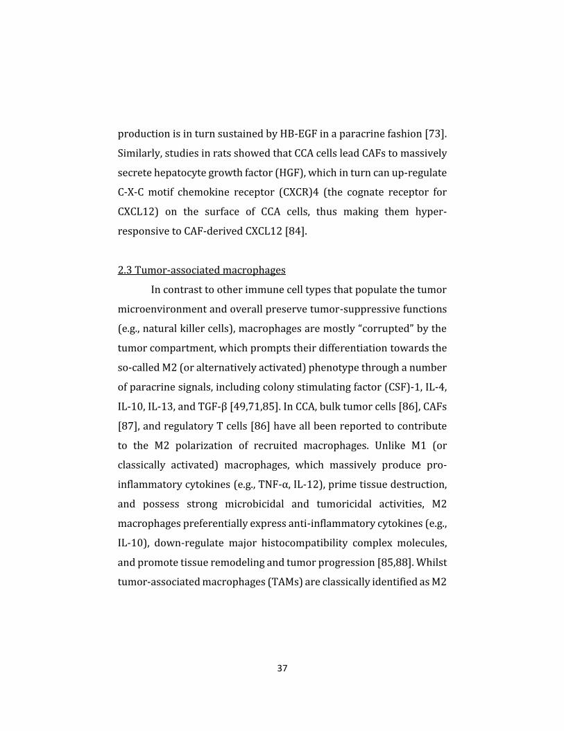
2.3 Tumor-associated macrophages
In contrast to other immune cell types that populate the tumor
microenvironment and overall preserve tumor-suppressive functions
(e.g., natural killer cells), macrophages are mostly “corrupted” by the
tumor compartment, which prompts their differentiation towards the
so-called M2 (or alternatively activated) phenotype through a number
of paracrine signals, including colony stimulating factor (CSF)-1, IL-4,
IL-10, IL-13, and TGF-β [49,71,85]. In CCA, bulk tumor cells [86], CAFs
[87], and regulatory T cells [86] have all been reported to contribute
to the M2 polarization of recruited macrophages. Unlike M1 (or
classically activated) macrophages, which massively produce pro-
inflammatory cytokines (e.g., TNF-α, IL-12), prime tissue destruction,
and possess strong microbicidal and tumoricidal activities, M2
macrophages preferentially express anti-inflammatory cytokines (e.g.,
IL-10), down-regulate major histocompatibility complex molecules,
and promote tissue remodeling and tumor progression [85,88]. Whilst
tumor-associated macrophages (TAMs) are classically identified as M2

38
macrophages, it is worth considering that the boundary between the
M1 and M2 activation states is quite blurred. In fact, macrophages
feature a considerable degree of plasticity, and actually go through a
wide spectrum of highly changing phenotypes [88]. Therefore, it is not
surprising that the tumor-promoting functions of TAMs frequently
rely on ‘‘M1 cytokines”, such as IL-6 and TNF-α [85]. Furthermore, in
CCA, Raggi et al. identified a peculiar subset of TAMs that is directly
molded by cancer stem cells (CSCs), and is characterized by a mixed
expression of M1 and M2 markers. Interestingly, CSC-associated TAMs
exhibit pronounced adhesive and invasive properties, as well as a
heightened expression of matrix-remodeling-related genes (e.g.,
ADAM17, MMP2), suggesting a critical role in ECM reorganization [89].
Importantly, TAM accumulation within the TRS mainly results from
the recruitment of blood monocytes (notably, CD14+/CD16+
monocytes) by tumor- or CAF-derived chemoattractants (e.g., CCL2,
CSF-1/2/3, VEGF), rather than from the in situ proliferation of resident
(CD68+) macrophages [49,90-92].
Similar to CAFs, TAMs are believed to play a critical part in CCA
progression. Indeed, high tumor infiltration by M2 (CD163+)
macrophages was found to correlate with poor disease-free survival of
CCA patients [86]. Furthermore, in CCA tissue, the density of M2 TAMs
is greatest at the tumor front, and positively correlated with tumor
pathological grade [89], and the development of extrahepatic
metastases [93]. In line with the immunohistochemical data, the

39
selective depletion of macrophages by liposomal clodronate in rodent
models of CCA strikingly decreased tumor burden [94]. In particular,
TAMs have been suggested to foster CCA growth by sustaining the
chronic activation of the Wnt/β-catenin signaling in cancer cells,
through the release of Wnt ligands [94,95]. In fact, the canonical Wnt
pathway is able to promote the unrestrained proliferation of
neoplastic cholangiocytes by up-regulating a number of cell cycle-
related genes [94,96]. In addition, macrophages may paracrinally
contribute to the aberrant activation of the IL-6/STAT3 axis frequently
observed in CCA cells [97,98], a fundamental mechanism underlying
their heightened survival abilities [24]. TAMs are also likely to mediate
cancer invasion, by both directly impinging on cancer cell behavior,
and shaping the tumor microenvironment. For instance, TAMs are the
primary source of TNF-α [99], which has been shown to provide CCA
cells with increased migratory functions by activating transcription
factors Snail and ZEB2 [97,100,101]. Consistently, conditioned
medium from M2 macrophages was found to foster CCA cell motility,
in vitro [93]. On the other hand, in several epithelial cancers, including
CCA, TAMs aplenty produce various MMPs, most notably MMP-9 [102],
thereby aiding tumor cells in degrading the surrounding ECM, a
process of utmost importance for local invasion [103]. Finally, a
variety of pro-angiogenic and/or pro-lymphangiogenic factors, such as
VEGF, FGF-2, and angiopoietin 1, are typically released by TAMs, which
thus promote the generation of a microenvironment conducive to

40
metastatic dissemination [49,88]. Indeed, in CCA samples, the number
of M2 macrophages was directly associated with microvascular
density [86].
2.4 Lymphatic endothelial cells
In CCA, tumor-associated angiogenesis and lymphangiogenesis
are both markedly induced, and were found to confer a significant
survival disadvantage on patients [104-106]. However, the formation
of new blood vessels within the tumor mass is generally not as striking
as the expansion of the lymphatic vasculature, which actually
represents the preferential route for CCA cells to escape from the
primary site of growth [53]. In this regard, immunohistochemical
staining of resected iCCAs and eCCAs for the lymphatic vessel marker
D2-40 (otherwise named podoplanin) revealed that a high lymphatic
microvessel density correlated with lymph node involvement
[104,106]. Importantly, lymph node metastasis is a well-known
negative prognostic factor for the survival of patients with CCA [107-
110]. The lymphatic capillaries, also called initial lymphatics, are
physiologically responsible for the continuous uptake of the
interstitial fluids, and their structure is particularly suitable for the
intravasation of metastasizing cancer cells. Indeed, they are only
composed of a monolayer of lymphatic endothelial cells (LECs), joined
together by weak cell-cell junctions, and surrounded by a
discontinuous basement membrane. However, the caliber of the initial

41
lymphatics gradually increases to form the pre-collecting lymphatics
first, and then the collecting lymphatics, which are lined by an intact
basal lamina, and contain both smooth muscle cells in their walls, and
intraluminal valves regulating the fluid flow. During the transit from
the collecting lymphatics to the thoracic duct, the fluid goes through
the regional lymph nodes, where metastasizing cancer cells can reside
before disseminating to distant organs [92,111].
Tumor-associated lymphangiogenesis essentially consists in
the formation of new lymphatic vessels arising from pre-existing
lymphatic capillaries, through the proliferation and migration of LECs.
However, both the recruitment of bone marrow-derived endothelial
progenitor cells, and the transdifferentiation of mature blood
endothelial cells have been suggested to play a role in this context,
though providing a minor contribution. Overexpression of the
archetypical lymphangiogenic factor VEGF-C by cancer and reactive
stromal cells is widely recognized as the main driver of neo-
lymphangiogenesis. Indeed, VEGFR-3, the cognate receptor for VEGF-
C, is selectively and highly expressed by LECs, and the maintenance of
the VEGF-C/VEGFR-3 signaling is of crucial importance for the
expansion of the lymphatic vasculature network in the embryo
[92,112]. In CCA, a high expression of VEGF-C was shown to correlate
with lymph node metastasis, and was also reported as an independent
prognostic factor for the survival of patients [113-115]. Additional
VEGF family members, namely VEGF-A and VEGF-D, are also claimed

42
to participate in tumor-associated lymphangiogenesis, even though
they are just ancillary mediators of embryonic lymphatic vessel
development [92,112]. Of note, VEGF-C and VEGF-D selectively bind to
VEGFR-2 and VEGFR-3, whereas VEGF-A selectively binds to VEGFR-1
and VEGFR-2. Unlike VEGFR-3, VEGFR-2 is primarily expressed by
vascular endothelial cells, where it acts as a positive modulator of
angiogenesis [116]. Nevertheless, critical functions for VEGFR-2 in the
lymphatic biology are emerging, and VEGFR-2 overexpression on
tumor-associated LECs has been described [117]. In addition to VEGF
ligands, other well-recognized tumor lymphangiogenic growth factors
include angiopoietins 1 and 2 (i.e., growth factors classically involved
in lymphatic remodeling and maturation), PDGF-BB, and FGF-2
[92,112].
Besides being involved in lymphangiogenesis, the initial
lymphatics may also be driven by cancer cell-derived factors (notably,
VEGF-C) to undergo a substantial enlargement, which is mediated by
the proliferation of LECs, and ultimately increases the interface for
lymphatic invasion by cancer cells. Enlargement of the collecting
lymphatics has also been reported, and likely results in an increased
lymph flow that further supports the metastatic dissemination. Of
further interest, VEGF-C is capable of altering the structure of pre-
existing and/or newly formed vessels by inducing the formation of
intercellular gaps, which can be deleteriously exploited by
metastasizing cancer cells. It is finally worth noting that LECs are not

43
only passively “educated” by cancer cells to build up a pro-metastatic
lymphatic network, but also actively promote the intravasation of
tumor cells by generating a chemoattracting gradient. Indeed, they
broadly release endogenous chemokines, such as CCL21 and CXCL12,
whose cognate receptors are generally located on the surface of cancer
cells [92,112].
2.5 Non-cellular components of the tumor reactive stroma
2.5.1 The extracellular matrix
The so-called ECM encompasses the interstitial connective
tissue matrix, and the basement membrane (or basal lamina). The
interstitial matrix is a three-dimensional network of collagens
(especially, type I collagen), proteoglycans, and glycoproteins (e.g.,
fibronectin, tenascin C, elastin), which fills the intercellular spaces, and
constitutes a structural scaffold for the tissue. Specifically, collagen
bundles confer tensile strength upon the whole tissue, while
proteoglycans mostly perform hydration functions. On the other hand,
the basement membrane is a specialized, sheet-like type of ECM,
interposed between the epithelium and the stroma, and mainly
composed of type IV collagen and laminin. Importantly, the ECM not
only provides a physical support to cells, but also actively impinges on
their behavior by communicating with them. Indeed, several ECM
constituents, especially glycoproteins, may act as ligands for cell

44
surface receptors such as integrins. Furthermore, both proteoglycans
and glycoproteins physically interact with a number of growth factors
and cyto/chemokines, which are thus efficiently sequestered within
the ECM, and may be subsequently locally unleashed through
proteolytic cleavage. Therefore, it is not surprising that the extensive,
dysregulated ECM remodeling that typically accompanies the
emergence of neoplastic lesions has been widely reported to support
cancer progression at multiple levels, including the induction of
angiogenesis, the activation of stromal cells, and the direct promotion
of cancer cell aggressiveness [49,51,118]. Throughout carcinogenesis,
the ECM progressively becomes stiffer, due to type I collagen
deposition and cross-linking, and concomitantly undergoes
compositional changes, resulting from both the proteolytic activity of
overexpressed MMPs, and the accumulation of newly synthesized
glycoproteins [55]. Importantly, the unrestrained ECM breakdown by
proteases leads to the generation of reactive ECM fragments endowed
with unique signaling functions, which in turn may elicit specific
biological responses [49,118]. In addition, ECM stiffening also
profoundly affects cell behavior, by impinging on the activity of
intracellular mechanosensors [55,118].
Similar to reactive stromal cells, the TRS-associated ECM has
been shown to foster CCA aggressiveness. Indeed, CCA cells cultured
on a mixture of collagen type IV and laminin demonstrated greater
invasive abilities than cells grown on uncoated plates, an effect likely

45
mediated by the up-regulation of the actin-binding protein L-plastin
[119]. Furthermore, in CCA tissue, increased stromal expression of
certain non-structural ECM proteins, namely tenascin [120], periostin
[68], and osteopontin [64], correlated with poor outcome and
tendency to invade. In particular, the tumor-promoting functions of
tenascin are suggested to rely on its binding to EGFR [120], while
periostin was found to promote CCA cell invasiveness in a α5β1
integrin-dependent manner [68,121]. Of note, such ECM constituents
are primarily produced by CAFs, though CCA cells themselves may
represent an ancillary source [68,120,121].
2.5.2 Intratumoral hypoxia
In most of solid tumors, the unrestrained proliferation of
neoplastic cells, and the concomitant formation of an inadequate and
highly disorganized vascular bed, necessarily lead to the generation of
a markedly hypoxic microenvironment. Nowadays, hypoxia is widely
recognized as a critical tumor-promoting player, deeply involved in
the emergence of the main hallmarks of tumor progression. In fact,
malignant cells are able to deleteriously hijack the adaptive cellular
responses to oxygen deprivation, which are physiologically aimed at
enabling injured cells to survive, migrate towards less hypoxic regions,
and orchestrate compensatory angiogenesis. Moreover, hypoxia
represents a potent microenvironmental stressors, and thus exerts a
selective pressure on cancer cells, ultimately resulting in the clonal

46
expansion of the most aggressive tumor phenotypes. Cellular
responses to hypoxia are primarily mediated by the activation of the
hypoxia-inducible factors (HIFs), i.e., helix-loop-helix transcription
factors that bind to specific DNA sequences known as hypoxia-
response elements. HIFs consist of an α (HIF-1α, HIF-2α, or HIF-3α)
and a β (HIF-1β) subunit. Among HIF-α isoforms, HIF-1α is the best
characterized, due to its ubiquitous expression. Under normoxic
conditions, HIF-1α is continuously primed for proteasome-mediated
degradation by prolyl hydroxylase domain-containing (PHD) enzymes,
whereas in the absence of adequate oxygen levels, PHD enzymes are
unable to properly hydroxylate HIF-1α, which can thus accumulate
and translocate to the nucleus. Here, HIF-1α dimerizes with HIF-1β,
which is constitutively expressed regardless of the oxygen
concentration. The resulting heterodimer interacts with
transcriptional co-activators, and regulates the expression of
hundreds of genes involved in cell survival and motility, glucose
metabolism, and angiogenesis [122-124].
In CCA tissue, HIF-1α is typically overexpressed compared with
bile ducts of peritumoral areas [125,126], and its high expression was
reported as an independent prognostic factor for overall and disease-
free survival [127,128]. Consistently, in vitro studies clearly
demonstrated that hypoxia-induced signaling leads CCA cells to gain a
more malignant phenotype. For instance, low-oxygen culture
conditions, as well as treatment with cobalt chloride (i.e., a chemical

47
inducer of HIF-1α), promoted a substantial increase in CCA cell
motility and invasiveness [126,128,129]. Specifically, the pro-invasive
effects of hypoxia were suggested to rely on the aberrant activation of
the pro-oncogenic MET/ERK axis [126], as well as on the induction of
autophagy [128], which is actually emerging as a key regulator of cell
invasion in several carcinomas, including CCA [130]. Of further
interest, hypoxia was found to boost the resistance of CCA cells to both
radiation- and drug-induced cytotoxicity [131], an effect likely
dependent on the up-regulation of the anti-apoptotic protein X-linked
inhibitor of apoptosis (XIAP) at the translational level [132]. It is worth
noting that, in sharp contrast to CCA cells, normal cholangiocytes
proved to be highly sensitive to oxygen deprivation [131,132], further
highlighting the ability of cancer cells to adapt to otherwise
detrimental conditions. Despite this growing amount of evidence, the
overall impact of hypoxia on CCA progression has yet to be fully
understood. In this regard, it was shown that a reduced oxygen
availability extensively impinged on the transcriptomic profile of
neoplastic cholangiocytes, altering the expression of several genes
involved in cancer cell invasion and apoptosis resistance (e.g., TFF1,
ADAM12, ITGA5, BIRC5, UMPS, S100P) [131]. A hypoxic
microenvironment could also endow CCA cells with heightened pro-
angiogenic and/or pro-lymphangiogenic functions, since HIF-1α
stabilization by cobalt chloride was shown to promote a massive up-
regulation of VEGF [129]. Finally, HIF-1α accumulation could drive

48
genomic instability by directly triggering the expression of iNOS,
which may indeed be responsible for NO-mediated DNA damage [125].
3. Molecular mechanisms underpinning cholangiocarcinoma
progression: an overview
3.1 Molecular mechanisms of chemoresistance
As previously discussed, CCA cells are characterized by a
substantial lack of sensitivity to drug-induced cytotoxicity, which
accounts for the disappointing results of conventional chemotherapy
[5]. The mechanisms of chemoresistance typically employed by CCA
cells are redundant and multifaceted, and most of them are borrowed
by normal cholangiocytes, which are required to chronically deal with
toxic compounds from bile and blood, and are thus equipped with
appropriate defense enzymatic activities [2,48]. A first strategy to
circumvent drug toxicity consists in keeping at bay the intracellular
drug concentration, either by restricting its uptake, through the down-
regulation of solute carriers transporters (e.g., organic anion-
transporting polypeptides, organic cation transporters, nucleoside
transporters), or by encouraging its efflux, through the up-regulation
of ATP-binding cassette (ABC) transporters (e.g., multidrug resistance
protein 1 or P-glycoprotein, multidrug resistance-associated proteins
(MRP)). Alternatively, CCA cells may selectively decrease the
intracellular amount of the active drug metabolite, thanks to an

49
impaired expression (or activity) of the enzymes responsible for pro-
drug activation, or to an enhanced expression (or activity) of the
enzymes involved in the detoxification of the active compound [2]. For
instance, glutathione S-transferase P1-1 is highly expressed in CCA
cells, and promotes cellular resistance to various xenobiotics (e.g.,
adriamycin, cisplatin, melphalan, cyclophosphamide) by catalyzing
their conjugation with reduced glutathione [133]. A large fraction of
anti-cancer drugs are DNA-damaging agents; therefore, the
optimization of DNA repair strategies also represents an efficient
mechanisms of chemoresistance [2]. In this regard, in CCA cells, the up-
regulation of uracil-DNA glycosylases, which initiates the base excision
repair pathway, is likely to underlie the acquired resistance to 5-
fluorouracil [134], whereas high expression of the p53-inducible
ribonucleotide reductase subunit M2B, which supplies
deoxyribonucleoside diphosphates throughout DNA repair, is
associated with decreased sensitivity to gemcitabine [135]. Regardless
of the nature of the primary cytotoxic insult, drug-induced cell death
typically results from the activation of the apoptotic machinery. It is no
accident that in CCA cells, the balance between anti-apoptotic and pro-
apoptotic proteins is dramatically altered in favor of the former [2].
Anti-apoptotic factors that are frequently overexpressed or aberrantly
activated in CCA include Mcl-1 [136], XIAP [137], and survivin [138].
Mcl-1 is a member of the Bcl-2 protein family, upon which the intrinsic
apoptosis pathway is based. Specifically, Mcl-1 binds to the pro-

50
apoptotic member Bak, thereby preventing its oligomerization and
activation. However, different intracellular stresses (e.g., trophic
factor withdrawal, DNA damage) can lead to the activation of BH3-only
proteins (e.g., Noxa, Bim, Puma), which effectively displace Bak by
competitively interacting with Mcl-1. Once unleashed, Bak
permeabilizes the outer mitochondrial membrane, thus allowing the
release of cytochrome c, which eventually triggers the activation of the
caspase cascade [139-141]. In this context, XIAP and survivin (also
called BIRC5) counteract cell death by inhibiting caspase activity
(notably, caspase-3, -7, and -9) [142]. Importantly, a defective
activation of the extrinsic apoptosis pathway, which is triggered by the
stimulation of cell surface death receptors (e.g., Fas receptor or CD95,
death receptor (DR)4) by cognate ligands (e.g., Fas ligand, TNF-related
apoptosis inducing ligand (TRAIL)), and in turn converges on caspase
activation, is also believed to underpin CCA chemoresistance [48]. It is
finally worth noting that, even where the drug cytotoxic mechanisms
cannot be intrinsically neutralized, chemoresistance may still be
acquired through an altered expression of molecular therapeutic
targets. For instance, in CCA cells, a decreased expression of ERs, and
an increased expression of thymidylate synthase can markedly
hampered the therapeutic effectiveness of tamoxifen and 5-
fluorouracil, respectively [2].
All these mechanisms of chemoresistance are fueled by
microenvironmental signals originating from either cancer or reactive

51
stromal cells, which thus enable CCA cells to chronically avoid cell
death [48]. In addition to the already mentioned anti-apoptotic effects
of IL-6 and PDGF-BB, the aberrant activation of morphogenetic
signaling pathways is also crucial for the emergence of chemoresistant
phenotypes. For instance, the scarce sensitivity of CCA cells to different
chemotherapeutics (i.e., 5-fluorouracil, cisplatin, vincristine) was
found to largely rely on the overactivation of the Wnt/β-catenin
pathway, which is indeed responsible for the up-regulation of the
efflux pump P-glycoprotein [143]. Interestingly, both TAMs [95] and
MSCs [144] were identified as important contributors to the activation
of the Wnt/β-catenin axis in CCA cells and consistently, conditioned
medium from MSCs was found to endow CCA cells with enhanced
apoptosis resistance [144]. The overexpression of ABC transporters,
namely P-glycoprotein and MRP1, is also sustained by Notch1
signaling, whose down-modulation dramatically increased the
apoptotic rate of CCA cells upon 5-fluorouracil exposure [145]. Unlike
Wnt and Notch signaling, the oncogenic Hh pathway decreased the
sensitivity of CCA cells to TRAIL-mediated apoptosis by down-
regulating the expression of DR4 at the transcriptional and post-
transcriptional level [146,147]. Indeed, the Hh-related transcription
factor glioma-associated oncogene (GLI)3 can negatively modulate the
DR4 promoter activity [146], while DR4 mRNA is a direct target of
microRNA (miR)-25, whose expression is strongly promoted by Hh
signaling [147]. Of further interest, the Hh-related transcription

52
factors GLI1 and GLI2 directly stimulate the expression of polo-like
kinase 2 (PLK2), a serine/threonine kinase classically involved in the
regulation of cell division. In CCA cells, PLK2 may also inhibit the
proteasomal degradation of Mcl-1, thereby further providing cells
with potent pro-survival signals [148]. Interestingly, the expression of
TRAIL in CCA cells can be transcriptionally down-modulated by the
Hippo pathway through its downstream effector yes-associated
protein (YAP), whose forced overexpression actually dampened the
induction of cancer cell apoptosis by Nutlin-3, a small molecule
supporting p53 activity [149].
3.2 Molecular mechanisms of cancer cell invasiveness
The acquisition of an invasive phenotype by cancer cells is a fist
and crucial step towards the development of metastases, which in CCA
represents a major hindrance to therapeutic success [46,150]. Cancer
cells need to be equipped with pronounced invasive properties in
order to degrade the surrounding basement membrane, detach from
the primary tumor mass, and easily move across the stroma towards
the blood and/or lymphatic vessels [150,151]. Basically, the
emergence of pro-invasive changes results from an aberrant and
incomplete rehash of an embryonic program known as epithelial-to-
mesenchymal transition (EMT), through which epithelial cells can
transdifferentiate into mesenchymal cells [63]. Regardless of the
background (i.e., morphogenesis, wound healing, cancer), the EMT

53
program is ultimately driven by a set of master transcription factors,
notably Snail (or Snail1), Slug (or Snail2), Twist1/2 and ZEB1/2, which
directly repress the expression of cardinal epithelial genes, while
simultaneously inducing the de novo expression of mesenchymal
genes. For instance, a major outcome of the EMT-associated gene
expression reprogramming is the so-called “cadherin switch”, that is
the exchange of E-cadherin, the main constituent of adherens
junctions, for N-cadherin, which allows dynamic interactions among
invading cells. Importantly, upon E-cadherin down-regulation, β-
catenin can be no longer retained at the membrane, and either is
degraded by the proteasome (in the absence of Wnt ligands) or enter
the nucleus to regulate gene expression (upon Wnt pathway
activation). The destabilization of adherens junctions occurs alongside
the repression of tight junctions proteins (e.g., occludin, claudins),
desmosomal proteins (e.g., desmoplakin), and polarity complex
proteins (e.g., Crumbs complex), overall leading to a definitive loss of
cell-cell adhesion and apical-basal polarity. In addition, the expression
pattern of integrins is deeply modified, and the cytoskeleton is
throughout remodeled, due to the down-regulation of cytokeratins
(CK) and the up-regulation of vimentin. The production of MMPs is
also greatly enhanced, thus enabling cells to massively degrade the
ECM [152,153]. The expression of prototypical EMT features by
neoplastic bile ducts is a well-established fact, generally predicting
metastatic behavior and poor survival [63,154]. CCA cells, especially

54
those located at the invasive front, typically exhibit an elongated,
spindle-shaped morphology, and express high levels of N-cadherin,
vimentin, S100A4, and nuclear β-catenin, and low levels of E-cadherin
and CK19. Overexpression of EMT master transcription factors is also
frequently observed [75,154,155].
Although there is still a long way to go, the molecular
mechanisms orchestrating this pro-invasive phenotypic switch are
gradually emerging. They rely on secreted factors permeating the
tumor microenvironment, as well as on aberrantly expressed
intracellular players that directly or indirectly interact with the EMT
machinery. For instance, TGF-β1 [156-160] and EGF-like family
members (notably, EGF and HB-EGF) [73,161-163] are prototypical
EMT inducers that are generally overexpressed by either CCA cells or
reactive stromal cells, and are well-known to provide malignant
cholangiocytes with strong migratory and invasive capabilities.
Similarly, TAM-derived TNF-α up-regulates the expression of Snail and
ZEB2 [100,101], and dramatically enhances the production of MMP-9
by activating focal adhesion kinase [164], and promoting the
expression of COX-2 [165]. Other pro-invasive microenvironmental
cues encompass IL-6 [166,167], SDF-1 [78,79,168-170], HGF
[171,172] and FGF-19 [173]. On the other hand, stochastic mutations
and oncogenic pathways may deleteriously impinge on the expression
or activity of several intracellular proteins and miRNAs that in turn can
potentially modulate the invasive potential of CCA cells. In this context,

55
pro-invasive transcription factors such as Notch1 [145,174-177], YAP
[178], and sal-like protein (SALL)4 [179,180], which are typically up-
regulated in CCA, are suggested to play a prominent role. For example,
SALL4 may unleash the pro-metastatic PI3K/Akt and Wnt/β-catenin
pathways by down-regulating phosphatase and tensin homolog, a
negative regulator of Akt, and up-regulating BMI-1, an epigenetic
repressor of Wnt inhibitors [180]. Conversely, the E3 ubiquitin ligase
F-box and WD repeat domain-containing (FBXW)7 [181], and the
MAPK kinase kinase (MAP3K)4 [155] counteract the invasive
functions of CCA cells and consistently, are frequently down-regulated
in neoplastic bile ducts. FBXW7 keeps at bay the protein levels of its
direct target mTOR, which can otherwise induce EMT through ZEB1
up-regulation [181]; MAP3K4 triggers the activation of p38 MAPK,
which then prevents nuclear factor κB from entering the nucleus and
promoting Snail expression [155].

56
Scope of the thesis
During my PhD studies, I sought to dissect the nature and the
biological relevance of the dense, multifaceted paracrine
communications between stromal and cancer cells in CCA, in an effort
to unveil the intimate molecular mechanisms driving tumor
progression.
Chapter 2: Leukemia inhibitory factor protects
cholangiocarcinoma cells from drug-induced apoptosis via a
PI3K/AKT-dependent Mcl-1 activation.
Leukemia inhibitory factor (LIF) is a pleiotropic cytokine
belonging to the IL-6 family, which can be secreted by both epithelial
and stromal cell types (e.g., fibroblasts, monocytes, macrophages), and
whose pro-oncogenic activity has been described in several human
cancers. Our aim was to investigate the role of LIF in the progression
of CCA, by evaluating the expression of LIF and its receptor in tumor
tissue and cell lines, and testing the effects of LIF on the hallmarks of
cancer, in vitro. In addition, we also intended to dissect the signaling
pathways triggered by LIF in CCA cells.
Chapter 3: Low-dose paclitaxel reduces S100A4 nuclear
import to inhibit invasion and hematogenous metastasis of
cholangiocarcinoma.

57
EMT of cancer cells is a paramount process underlying
carcinoma invasion and metastasis, and is widely recognized as a
classic readout of tumor-stroma interactions. In a previous study, our
group has revealed that in CCA, nuclear expression of the EMT-related
protein S100A4 is both a powerful predictor of poor outcome, and a
mechanistic determinant of cancer cell invasiveness. In this work, we
aimed at clarifying the mechanisms underlying the pro-metastatic
activity of nuclear S100A4 in CCA cells, as well as the mechanisms
responsible for its nuclear import. Since studies from the early ‘90s
have described the ability of paclitaxel (PTX) to downregulate the
expression of S100A4 in mouse melanoma cells, we preliminary
assessed whether PTX could be useful in modulating the intracellular
levels of S100A4 in our cell lines.
Chapter 4: Platelet-derived growth factor D enables
cancer-associated fibroblasts to promote tumor
lymphangiogenesis in cholangiocarcinoma.
CAFs are recognized to play a major role in the induction of
lymphangiogenesis in several human cancers, mainly due to a broad
secretion of VEGFs. In a previous study, our group has identified PDGF-
DD as a paramount mediator of fibroblasts recruitment by CCA cells.
Interestingly, PDGF ligands, especially PDGF-BB, have been reported
to stimulate the production of VEGFs in a variety of cell systems,
including hepatic stellate cells and pulmonary fibroblasts. In this
58
study, we therefore tested the hypothesis that PDGF-DD may also
endow CAFs with pro-lymphangiogenic functions. To this end, we
measured the secretion of various lymphangiogenic growth factors by
human fibroblasts upon PDGF-DD treatment (with a focus on the
intracellular pathways activated downstream of PDGFRβ), and
evaluated the effects of conditioned medium from PDGF-DD-treated
fibroblasts on the behavior of LECs, in terms of motility and
tubulogenesis.

59
References
1. Razumilava N, Gores GJ. Cholangiocarcinoma. Lancet.
2014;383:2168-79.
2. Banales JM, Cardinale V, Carpino G, Marzioni M, Andersen JB,
Invernizzi P, Lind GE, Folseraas T, Forbes SJ, Fouassier L, Geier A,
Calvisi DF, Mertens JC, Trauner M, Benedetti A, Maroni L, Vaquero J,
Macias RI, Raggi C, Perugorria MJ, Gaudio E, Boberg KM, Marin JJ,
Alvaro D. Expert consensus document: Cholangiocarcinoma: current
knowledge and future perspectives consensus statement from the
European Network for the Study of Cholangiocarcinoma (ENS-CCA).
Nat Rev Gastroenterol Hepatol. 2016;13:261-80.
3. Høgdall D, O'Rourke CJ, Taranta A, Oliveira DV, Andersen JB.
Molecular Pathogenesis and Current Therapy in Intrahepatic
Cholangiocarcinoma. Dig Dis. 2016;34:440-51.
4. Brito AF, Abrantes AM, Encarnação JC, Tralhão JG, Botelho MF.
Cholangiocarcinoma: from molecular biology to treatment. Med
Oncol. 2015;32:245.
5. Gatto M, Bragazzi MC, Semeraro R, Napoli C, Gentile R, Torrice A,
Gaudio E, Alvaro D. Cholangiocarcinoma: update and future
perspectives. Dig Liver Dis. 2010;42:253-60.
6. Blechacz B, Komuta M, Roskams T, Gores GJ. Clinical diagnosis and
staging of cholangiocarcinoma. Nat Rev Gastroenterol Hepatol.
2011;8:512-22.
7. Rizvi S, Gores GJ. Pathogenesis, diagnosis, and management of
cholangiocarcinoma. Gastroenterology. 2013;145:1215-29.

60
8. Moeini A, Sia D, Bardeesy N, Mazzaferro V, Llovet JM. Molecular
Pathogenesis and Targeted Therapies for Intrahepatic
Cholangiocarcinoma. Clin Cancer Res. 2016;22:291-300.
9. Roskams TA, Libbrecht L, Desmet VJ. Progenitor cells in diseased
human liver. Semin Liver Dis. 2003;23:385-96.
10. Cardinale V, Carpino G, Reid L, Gaudio E, Alvaro D. Multiple cells of
origin in cholangiocarcinoma underlie biological, epidemiological
and clinical heterogeneity. World J Gastrointest Oncol. 2012;4:94-
102.
11. Lim JH. Cholangiocarcinoma: morphologic classification according to
growth pattern and imaging findings. AJR Am J Roentgenol.
2003;181:819-27.
12. Blechacz B. Cholangiocarcinoma: Current Knowledge and New
Developments. Gut Liver. 2017;11:13-26.
13. Vijgen S, Terris B, Rubbia-Brandt L. Pathology of intrahepatic
cholangiocarcinoma. Hepatobiliary Surg Nutr. 2017;6:22-34.
14. Aishima S, Oda Y. Pathogenesis and classification of intrahepatic
cholangiocarcinoma: different characters of perihilar large duct type
versus peripheral small duct type. J Hepatobiliary Pancreat Sci.
2015;22:94-100.
15. Aljiffry M, Walsh MJ, Molinari M. Advances in diagnosis, treatment
and palliation of cholangiocarcinoma: 1990-2009. World J
Gastroenterol. 2009;15:4240-62.
16. Komuta M, Govaere O, Vandecaveye V, Akiba J, Van Steenbergen W,
Verslype C, Laleman W, Pirenne J, Aerts R, Yano H, Nevens F, Topal B,
Roskams T. Histological diversity in cholangiocellular carcinoma
61
reflects the different cholangiocyte phenotypes. Hepatology.
2012;55:1876-88.
17. Strazzabosco M, Fabris L. Functional anatomy of normal bile ducts.
Anat Rec (Hoboken). 2008;291:653-60.
18. Fabris L, Brivio S, Cadamuro M, Strazzabosco M. Revisiting Epithelial-
to-Mesenchymal Transition in Liver Fibrosis: Clues for a Better
Understanding of the "Reactive" Biliary Epithelial Phenotype. Stem
Cells Int. 2016;2016:2953727.
19. Liau JY, Tsai JH, Yuan RH, Chang CN, Lee HJ, Jeng YM. Morphological
subclassification of intrahepatic cholangiocarcinoma: etiological,
clinicopathological, and molecular features. Mod Pathol.
2014;27:1163-73.
20. Ferlay J, Soerjomataram I, Ervik M, Dikshit R, Eser S, Mathers C,
Rebelo M, Parkin DM, Forman D, Bray F. GLOBOCAN 2012 v1.0,
Cancer Incidence and Mortality Worldwide: IARC CancerBase No. 11
[Internet]. Lyon, France: International Agency for Research on
Cancer; 2013. Available from: http://globocan.iarc.fr, accessed on
25/08/2017.
21. Ebata T, Ercolani G, Alvaro D, Ribero D, Di Tommaso L, Valle JW.
Current Status on Cholangiocarcinoma and Gallbladder Cancer. Liver
Cancer. 2016;6:59-65.
22. Wise C, Pilanthananond M, Perry BF, Alpini G, McNeal M, Glaser SS.
Mechanisms of biliary carcinogenesis and growth. World J
Gastroenterol. 2008;14:2986-9.
23. Park J, Tadlock L, Gores GJ, Patel T. Inhibition of interleukin 6-
mediated mitogen-activated protein kinase activation attenuates

62
growth of a cholangiocarcinoma cell line. Hepatology. 1999;30:1128-
33.
24. Isomoto H, Kobayashi S, Werneburg NW, Bronk SF, Guicciardi ME,
Frank DA, Gores GJ. Interleukin 6 upregulates myeloid cell leukemia-
1 expression through a STAT3 pathway in cholangiocarcinoma cells.
Hepatology. 2005;42:1329-38.
25. Kobayashi S, Werneburg NW, Bronk SF, Kaufmann SH, Gores GJ.
Interleukin-6 contributes to Mcl-1 up-regulation and TRAIL
resistance via an Akt-signaling pathway in cholangiocarcinoma cells.
Gastroenterology. 2005;128:2054-65.
26. Meng F, Yamagiwa Y, Ueno Y, Patel T. Over-expression of interleukin-
6 enhances cell survival and transformed cell growth in human
malignant cholangiocytes. J Hepatol. 2006;44:1055-65.
27. Yamada D, Rizvi S, Razumilava N, Bronk SF, Davila JI, Champion MD,
Borad MJ, Bezerra JA, Chen X, Gores GJ. IL-33 facilitates oncogene-
induced cholangiocarcinoma in mice by an interleukin-6-sensitive
mechanism. Hepatology. 2015;61:1627-42.
28. Fan B, Malato Y, Calvisi DF, Naqvi S, Razumilava N, Ribback S, Gores
GJ, Dombrowski F, Evert M, Chen X, Willenbring H.
Cholangiocarcinomas can originate from hepatocytes in mice. J Clin
Invest. 2012;122:2911–15.
29. Sekiya S, Suzuki A. Intrahepatic cholangiocarcinoma can arise from
Notch-mediated conversion of hepatocytes. J Clin Invest.
2012;122:3914–18.
30. Ishimura N, Bronk SF, Gores GJ. Inducible nitric oxide synthase up-
regulates Notch-1 in mouse cholangiocytes: implications for
carcinogenesis. Gastroenterology 2005;128:1354-68.

63
31. El Khatib M, Kalnytska A, Palagani V, Kossatz U, Manns MP, Malek NP,
Wilkens L, Plentz RR. Inhibition of hedgehog signaling attenuates
carcinogenesis in vitro and increases necrosis of cholangiocellular
carcinoma. Hepatology 2013;57:1035-45.
32. Nachtergaele S, Mydock LK, Krishnan K, Rammohan J, Schlesinger PH,
Covey DF, Rohatgi R. Oxysterols are allosteric activators of the
oncoprotein Smoothened. Nat Chem Biol. 2012;8:211-20.
33. Alvaro D, Mancino MG, Glaser S, et al. Proliferating cholangiocytes: a
neuroendocrine compartment in the diseased liver.
Gastroenterology. 2007;132:415–31.
34. Chong DQ, Zhu AX. The landscape of targeted therapies for
cholangiocarcinoma: current status and emerging targets.
Oncotarget. 2016;7:46750-67.
35. Rizvi S, Gores GJ. Emerging molecular therapeutic targets for
cholangiocarcinoma. J Hepatol. 2017;67:632-44.
36. Chan-On W, Nairismägi ML, Ong CK, Lim WK, Dima S, Pairojkul C, Lim
KH, McPherson JR, Cutcutache I, Heng HL, Ooi L, Chung A, Chow P,
Cheow PC, Lee SY, Choo SP, Tan IB, Duda D, Nastase A, Myint SS, Wong
BH, Gan A, Rajasegaran V, Ng CC, Nagarajan S, Jusakul A, Zhang S,
Vohra P, Yu W, Huang D, Sithithaworn P, Yongvanit P, Wongkham S,
Khuntikeo N, Bhudhisawasdi V, Popescu I, Rozen SG, Tan P, Teh BT.
Exome sequencing identifies distinct mutational patterns in liver
fluke-related and non-infection-related bile duct cancers. Nat Genet.
2013;45:1474-8.
37. Sugimachi K, Taguchi K, Aishima S, Tanaka S, Shimada M, Kajiyama K,
Sugimachi K, Tsuneyoshi M. Altered expression of beta-catenin

64
without genetic mutation in intrahepatic cholangiocarcinoma. Mod
Pathol. 2001;14:900-5.
38. Sadot E, Geiger B, Oren M, Ben-Ze'ev A. Down-regulation of beta-
catenin by activated p53. Mol Cell Biol. 2001;21:6768-81.
39. Ornitz DM, Itoh N. The Fibroblast Growth Factor signaling pathway.
Wiley Interdiscip Rev Dev Biol. 2015;4:215-66.
40. Principe DR, Doll JA, Bauer J, Jung B, Munshi HG, Bartholin L, Pasche
B, Lee C, Grippo PJ. TGF-β: duality of function between tumor
prevention and carcinogenesis. J Natl Cancer Inst. 2014;106:djt369.
41. Saha SK, Parachoniak CA, Ghanta KS, Fitamant J, Ross KN, Najem MS,
Gurumurthy S, Akbay EA, Sia D, Cornella H, Miltiadous O, Walesky C,
Deshpande V, Zhu AX, Hezel AF, Yen KE, Straley KS, Travins J,
Popovici-Muller J, Gliser C, Ferrone CR, Apte U, Llovet JM, Wong KK,
Ramaswamy S, Bardeesy N. Mutant IDH inhibits HNF-4α to block
hepatocyte differentiation and promote biliary cancer. Nature.
2014;513:110-4.
42. Isomoto H. Epigenetic alterations in cholangiocarcinoma-sustained
IL-6/STAT3 signaling in cholangiocarcinoma due to SOCS3
epigenetic silencing. Digestion. 2009;79:2-8.
43. Jiao Y, Pawlik TM, Anders RA, Selaru FM, Streppel MM, Lucas DJ,
Niknafs N, Guthrie VB, Maitra A, Argani P, Offerhaus GJA, Roa JC,
Roberts LR, Gores GJ, Popescu I, Alexandrescu ST, Dima S, Fassan M,
Simbolo M, Mafficini A, Capelli P, Lawlor RT, Ruzzenente A, Guglielmi
A, Tortora G, de Braud F, Scarpa A, Jarnagin W, Klimstra D, Karchin R,
Velculescu VE, Hruban RH, Vogelstein B, Kinzler KW, Papadopoulos
N, Wood LD. Exome sequencing identifies frequent inactivating

65
mutations in BAP1, ARID1A and PBRM1 in intrahepatic
cholangiocarcinomas. Nat Genet. 2013;45:1470-73.
44. Kadoch C, Crabtree GR. Mammalian SWI/SNF chromatin remodeling
complexes and cancer: Mechanistic insights gained from human
genomics. Sci Adv. 2015;1:e1500447.
45. Carbone M, Yang H, Pass HI, Krausz T, Testa JR, Gaudino G. BAP1 and
cancer. Nat Rev Cancer. 2013;13:153-9.
46. Fabris L, Alvaro D. The prognosis of perihilar cholangiocarcinoma
after radical treatments. Hepatology 2012;56:800-2.
47. Valle J, Wasan H, Palmer DH, Cunningham D, Anthoney A, Maraveyas
A, Madhusudan S, Iveson T, Hughes S, Pereira SP, Roughton M,
Bridgewater J; ABC-02 Trial Investigators. Cisplatin plus gemcitabine
versus gemcitabine for biliary tract cancer. N Engl J Med.
2010;362:1273-81.
48. Cadamuro M, Brivio S, Spirli C, Joplin RE, Strazzabosco M, Fabris L.
Autocrine and Paracrine Mechanisms Promoting Chemoresistance in
Cholangiocarcinoma. Int J Mol Sci. 2017;18.
49. Mueller MM, Fusenig NE. Friends or foes - bipolar effects of the
tumour stroma in cancer. Nat Rev Cancer. 2004;4:839-49.
50. Dvorak HF. Tumors: wounds that do not heal-redux. Cancer Immunol
Res. 2015;3:1-11.
51. Kalluri R, Zeisberg M. Fibroblasts in cancer. Nat Rev Cancer.
2006;6:392-401.
52. Dvorak HF. Tumors: wounds that do not heal. Similarities between
tumor stroma generation and wound healing. N Engl J Med.
1986;315:1650-9.

66
53. Cadamuro M, Morton SD, Strazzabosco M, Fabris L. Unveiling the role
of tumor reactive stroma in cholangiocarcinoma: An opportunity for
new therapeutic strategies. Transl Gastrointest Cancer. 2013;2:130-
44.
54. Bhome R, Bullock MD, Al Saihati HA, Goh RW, Primrose JN, Sayan AE,
Mirnezami AH. A top-down view of the tumor microenvironment:
structure, cells and signaling. Front Cell Dev Biol. 2015;3:33.
55. Brivio S, Cadamuro M, Strazzabosco M, Fabris L. Tumor reactive
stroma in cholangiocarcinoma: The fuel behind cancer
aggressiveness. World J Hepatol. 2017;9:455-68.
56. Cadamuro M, Stecca T, Brivio S, Mariotti V, Fiorotto R, Spirli C,
Strazzabosco M, Fabris L. The deleterious interplay between tumor
epithelia and stroma in cholangiocarcinoma. Biochim Biophys Acta.
2017.
57. Quail DF, Joyce JA. Microenvironmental regulation of tumor
progression and metastasis. Nat Med. 2013;19:1423-37.
58. Lin HJ, Zuo T, Lin CH, Kuo CT, Liyanarachchi S, Sun S, Shen R,
Deatherage DE, Potter D, Asamoto L, Lin S, Yan PS, Cheng AL,
Ostrowski MC, Huang TH. Breast cancer-associated fibroblasts confer
AKT1-mediated epigenetic silencing of Cystatin M in epithelial cells.
Cancer Res. 2008;68:10257-66.
59. Xu L, Deng Q, Pan Y, Peng M, Wang X, Song L, Xiao M, Wang Z. Cancer-
associated fibroblasts enhance the migration ability of ovarian cancer
cells by increasing EZH2 expression. Int J Mol Med. 2014;33:91-6.
60. Kurashige J, Mima K, Sawada G, Takahashi Y, Eguchi H, Sugimachi K,
Mori M, Yanagihara K, Yashiro M, Hirakawa K, Baba H, Mimori K.
Epigenetic modulation and repression of miR-200b by cancer-

67
associated fibroblasts contribute to cancer invasion and peritoneal
dissemination in gastric cancer. Carcinogenesis. 2015;36:133-41.
61. Coulouarn C, Clément B. Stellate cells and the development of liver
cancer: therapeutic potential of targeting the stroma. J Hepatol.
2014;60:1306-09.
62. Chen F, Zhuang X, Lin L, Yu P, Wang Y, Shi Y, Hu G, Sun Y. New horizons
in tumor microenvironment biology: challenges and opportunities.
BMC Med. 2015;13:45.
63. Brivio S, Cadamuro M, Fabris L, Strazzabosco M. Epithelial-to-
Mesenchymal Transition and Cancer Invasiveness: What Can We
Learn from Cholangiocarcinoma? J Clin Med. 2015;4:2028-41.
64. Sulpice L, Rayar M, Desille M, Turlin B, Fautrel A, Boucher E, Llamas-
Gutierrez F, Meunier B, Boudjema K, Clément B, Coulouarn C.
Molecular profiling of stroma identifies osteopontin as an
independent predictor of poor prognosis in intrahepatic
cholangiocarcinoma. Hepatology. 2013;58:1992-2000.
65. Andersen JB, Spee B, Blechacz BR, Avital I, Komuta M, Barbour A,
Conner EA, Gillen MC, Roskams T, Roberts LR, Factor VM,
Thorgeirsson SS. Genomic and genetic characterization of
cholangiocarcinoma identifies therapeutic targets for tyrosine kinase
inhibitors. Gastroenterology. 2012;142:1021-31.e15.
66. Augsten M. Cancer-associated fibroblasts as another polarized cell
type of the tumor microenvironment. Front Oncol. 2014;4:62.
67. Chuaysri C, Thuwajit P, Paupairoj A, Chau-In S, Suthiphongchai T,
Thuwajit C. Alpha-smooth muscle actin-positive fibroblasts promote
biliary cell proliferation and correlate with poor survival in
cholangiocarcinoma. Oncol Rep. 2009;21:957-69.

68
68. Utispan K, Thuwajit P, Abiko Y, Charngkaew K, Paupairoj A, Chauin S,
Thuwajit C. Gene expression profiling of cholangiocarcinoma-derived
fibroblast reveals alterations related to tumor progression and
indicates periostin as a poor prognostic marker. Mol Cancer.
2010;9:13.
69. Yang X, Lin Y, Shi Y, Li B, Liu W, Yin W, Dang Y, Chu Y, Fan J, He R. FAP
Promotes Immunosuppression by Cancer-Associated Fibroblasts in
the Tumor Microenvironment via STAT3-CCL2 Signaling. Cancer Res.
2016;76:4124-35.
70. Shiga K, Hara M, Nagasaki T, Sato T, Takahashi H, Takeyama H.
Cancer-Associated Fibroblasts: Their Characteristics and Their Roles
in Tumor Growth. Cancers (Basel). 2015;7:2443-58.
71. Leyva-Illades D, McMillin M, Quinn M, Demorrow S.
Cholangiocarcinoma pathogenesis: Role of the tumor
microenvironment. Transl Gastrointest Cancer. 2012;1:71-80.
72. Okabe H, Beppu T, Hayashi H, Ishiko T, Masuda T, Otao R, Horlad H,
Jono H, Ueda M, Phd SS, Ando Y, Baba H. Hepatic stellate cells
accelerate the malignant behavior of cholangiocarcinoma cells. Ann
Surg Oncol. 2011;18:1175-84.
73. Clapéron A, Mergey M, Aoudjehane L, Ho-Bouldoires TH, Wendum D,
Prignon A, Merabtene F, Firrincieli D, Desbois-Mouthon C, Scatton O,
Conti F, Housset C, Fouassier L. Hepatic myofibroblasts promote the
progression of human cholangiocarcinoma through activation of
epidermal growth factor receptor. Hepatology. 2013;58:2001-11.
74. Haga H, Yan IK, Takahashi K, Wood J, Zubair A, Patel T. Tumour cell-
derived extracellular vesicles interact with mesenchymal stem cells

69
to modulate the microenvironment and enhance cholangiocarcinoma
growth. J Extracell Vesicles. 2015;4:24900.
75. Cadamuro M, Nardo G, Indraccolo S, Dall’olmo L, Sambado L, Moserle
L, Franceschet I, Colledan M, Massani M, Stecca T, Bassi N, Morton S,
Spirli C, Fiorotto R, Fabris L, Strazzabosco M. Platelet-derived growth
factor-D and Rho GTPases regulate recruitment of cancer-associated
fibroblasts in cholangiocarcinoma. Hepatology. 2013;58:1042-53.
76. Heits N, Heinze T, Bernsmeier A, Kerber J, Hauser C, Becker T, Kalthoff
H, Egberts JH, Braun F. Influence of mTOR-inhibitors and
mycophenolic acid on human cholangiocellular carcinoma and
cancer associated fibroblasts. BMC Cancer. 2016;16:322.
77. Okabe H, Beppu T, Hayashi H, Horino K, Masuda T, Komori H,
Ishikawa S, Watanabe M, Takamori H, Iyama K, Baba H. Hepatic
stellate cells may relate to progression of intrahepatic
cholangiocarcinoma. Ann Surg Oncol. 2009;16:2555-64.
78. Gentilini A, Rombouts K, Galastri S, Caligiuri A, Mingarelli E, Mello T,
Marra F, Mantero S, Roncalli M, Invernizzi P, Pinzani M. Role of the
stromal-derived factor-1 (SDF-1)-CXCR4 axis in the interaction
between hepatic stellate cells and cholangiocarcinoma. J Hepatol.
2012;57:813-20.
79. Okamoto K, Tajima H, Nakanuma S, Sakai S, Makino I, Kinoshita J,
Hayashi H, Nakamura K, Oyama K, Nakagawara H, Fujita H, Takamura
H, Ninomiya I, Kitagawa H, Fushida S, Fujimura T, Harada S,
Wakayama T, Iseki S, Ohta T. Angiotensin II enhances epithelial-to-
mesenchymal transition through the interaction between activated
hepatic stellate cells and the stromal cell-derived factor-1/CXCR4
axis in intrahepatic cholangiocarcinoma. Int J Oncol. 2012;41:573-82.

70
80. Kim Y, Kim MO, Shin JS, Park SH, Kim SB, Kim J, Park SC, Han CJ, Ryu
JK, Yoon YB, Kim YT. Hedgehog signaling between cancer cells and
hepatic stellate cells in promoting cholangiocarcinoma. Ann Surg
Oncol. 2014;21:2684-98.
81. Fingas CD, Bronk SF, Werneburg NW, Mott JL, Guicciardi ME,
Cazanave SC, Mertens JC, Sirica AE, Gores GJ. Myofibroblast-derived
PDGF-BB promotes Hedgehog survival signaling in
cholangiocarcinoma cells. Hepatology. 2011; 54: 2076-2088.
82. Fingas CD, Mertens JC, Razumilava N, Bronk SF, Sirica AE, Gores GJ.
Targeting PDGFR-β in Cholangiocarcinoma. Liver Int. 2012;32:400-9.
83. Mertens JC, Fingas CD, Christensen JD, Smoot RL, Bronk SF,
Werneburg NW, Gustafson MP, Dietz AB, Roberts LR, Sirica AE, Gores
GJ. Therapeutic effects of deleting cancer-associated fibroblasts in
cholangiocarcinoma. Cancer Res. 2013;73:897-907.
84. Campbell DJ, Dumur CI, Lamour NF, Dewitt JL, Sirica AE. Novel
organotypic culture model of cholangiocarcinoma progression.
Hepatol Res. 2012;42:1119-30.
85. Grivennikov SI, Greten FR, Karin M. Immunity, inflammation, and
cancer. Cell. 2010;140:883-99.
86. Hasita H, Komohara Y, Okabe H, Masuda T, Ohnishi K, Lei XF, Beppu
T, Baba H, Takeya M. Significance of alternatively activated
macrophages in patients with intrahepatic cholangiocarcinoma.
Cancer Sci. 2010;101:1913-19.
87. Chang J, Hisamatsu T, Shimamura K, Yoneno K, Adachi M, Naruse H,
Igarashi T, Higuchi H, Matsuoka K, Kitazume MT, Ando S, Kamada N,
Kanai T, Hibi T. Activated hepatic stellate cells mediate the
differentiation of macrophages. Hepatol Res. 2013;43:658-69.

71
88. Sica A, Invernizzi P, Mantovani A. Macrophage plasticity and
polarization in liver homeostasis and pathology. Hepatology. 2014;
59:2034-42.
89. Raggi C, Correnti M, Sica A, Andersen JB, Cardinale V, Alvaro D,
Chiorino G, Forti E, Glaser S, Alpini G, Destro A, Sozio F, Di Tommaso
L, Roncalli M, Banales JM, Coulouarn C, Bujanda L, Torzilli G,
Invernizzi P. Cholangiocarcinoma stem-like subset shapes tumor-
initiating niche by educating associated macrophages. J Hepatol.
2017;66:102-15.
90. Mantovani A, Allavena P, Sica A, Balkwill F. Cancer-related
inflammation. Nature. 2008;454:436-44.
91. Subimerb C, Pinlaor S, Lulitanond V, Khuntikeo N, Okada S, McGrath
MS, Wongkham S. Circulating CD14(+) CD16(+) monocyte levels
predict tissue invasive character of cholangiocarcinoma. Clin Exp
Immunol. 2010;161:471-9.
92. Duong T, Koopman P, Francois M. Tumor lymphangiogenesis as a
potential therapeutic target. J Oncol. 2012;2012:204946.
93. Thanee M, Loilome W, Techasen A, Namwat N, Boonmars T, Pairojkul
C, Yongvanit P. Quantitative changes in tumor-associated M2
macrophages characterize cholangiocarcinoma and their association
with metastasis. Asian Pac J Cancer Prev. 2015;16:3043-50.
94. Boulter L, Guest RV, Kendall TJ, Wilson DH, Wojtacha D, Robson AJ,
Ridgway RA, Samuel K, Van Rooijen N, Barry ST, Wigmore SJ, Sansom
OJ, Forbes SJ. WNT signaling drives cholangiocarcinoma growth and
can be pharmacologically inhibited. J Clin Invest. 2015; 125:1269-85.
95. Loilome W, Bungkanjana P, Techasen A, Namwat N, Yongvanit P,
Puapairoj A, Khuntikeo N, Riggins GJ. Activated macrophages

72
promote Wnt/β-catenin signaling in cholangiocarcinoma cells.
Tumour Biol. 2014;35:5357-67.
96. MacDonald BT, Tamai K, He X. Wnt/beta-catenin signaling:
components, mechanisms, and diseases. Dev Cell. 2009;17:9-26.
97. Techasen A, Loilome W, Namwat N, Dokduang H, Jongthawin J,
Yongvanit P. Cytokines released from activated human macrophages
induce epithelial mesenchymal transition markers of
cholangiocarcinoma cells. Asian Pac J Cancer Prev. 2012;13
Suppl:115-8.
98. Dokduang H, Techasen A, Namwat N, Khuntikeo N, Pairojkul C,
Murakami Y, Loilome W, Yongvanit P. STATs profiling reveals
predominantly-activated STAT3 in cholangiocarcinoma genesis and
progression. J Hepatobiliary Pancreat Sci. 2014;21:767-6.
99. Ohira S, Sasaki M, Harada K, Sato Y, Zen Y, Isse K, Kozaka K, Ishikawa
A, Oda K, Nimura Y, Nakanuma Y. Possible regulation of migration of
intrahepatic cholangiocarcinoma cells by interaction of CXCR4
expressed in carcinoma cells with tumor necrosis factor-alpha and
stromal-derived factor-1 released in stroma. Am J Pathol.
2006;168:1155-68.
100. Techasen A, Namwat N, Loilome W, Bungkanjana P, Khuntikeo N,
Puapairoj A, Jearanaikoon P, Saya H, Yongvanit P. Tumor necrosis
factor-α (TNF-α) stimulates the epithelial-mesenchymal transition
regulator Snail in cholangiocarcinoma. Med Oncol 2012; 29:3083-91.
101. Techasen A, Namwat N, Loilome W, Duangkumpha K, Puapairoj A,
Saya H, Yongvanit P. Tumor necrosis factor-α modulates epithelial
mesenchymal transition mediators ZEB2 and S100A4 to promote

73
cholangiocarcinoma progression. J Hepatobiliary Pancreat Sci.
2014;21:703-11.
102. Subimerb C, Pinlaor S, Khuntikeo N, Leelayuwat C, Morris A, McGrath
MS, Wongkham S. Tissue invasive macrophage density is correlated
with prognosis in cholangiocarcinoma. Mol Med Rep. 2010;3:597-
605.
103. Brown GT, Murray GI. Current mechanistic insights into the roles of
matrix metalloproteinases in tumour invasion and metastasis. J
Pathol. 2015;237:273-81.
104. Thelen A, Scholz A, Benckert C, Weichert W, Dietz E, Wiedenmann B,
Neuhaus P, Jonas S. Tumor-associated lymphangiogenesis correlates
with lymph node metastases and prognosis in hilar
cholangiocarcinoma. Ann Surg Oncol. 2008;15:791-9.
105. Thelen A, Scholz A, Benckert C, Schröder M, Weichert W,
Wiedenmann B, Neuhaus P, Jonas S. Microvessel density correlates
with lymph node metastases and prognosis in hilar
cholangiocarcinoma. J Gastroenterol. 2008;43:959-66.
106. Thelen A, Scholz A, Weichert W, Wiedenmann B, Neuhaus P, Gessner
R, Benckert C, Jonas S. Tumor-associated angiogenesis and
lymphangiogenesis correlate with progression of intrahepatic
cholangiocarcinoma. Am J Gastroenterol. 2010;105:1123-32.
107. de Jong MC, Nathan H, Sotiropoulos GC, Paul A, Alexandrescu S,
Marques H, Pulitano C, Barroso E, Clary BM, Aldrighetti L, Ferrone CR,
Zhu AX, Bauer TW, Walters DM, Gamblin TC, Nguyen KT, Turley R,
Popescu I, Hubert C, Meyer S, Schulick RD, Choti MA, Gigot JF, Mentha
G, Pawlik TM. Intrahepatic cholangiocarcinoma: an international

74
multi-institutional analysis of prognostic factors and lymph node
assessment. J Clin Oncol. 2011;29:3140-5.
108. Oshiro Y, Sasaki R, Kobayashi A, Murata S, Fukunaga K, Kondo T, Oda
T, Ohkohchi N. Prognostic relevance of the lymph node ratio in
surgical patients with extrahepatic cholangiocarcinoma. Eur J Surg
Oncol. 2011;37:60-4.
109. Kiriyama M, Ebata T, Aoba T, Kaneoka Y, Arai T, Shimizu Y, Nagino M;
Nagoya Surgical Oncology Group. Prognostic impact of lymph node
metastasis in distal cholangiocarcinoma. Br J Surg. 2015;102:399-
406.
110. Zhang JW, Chu YM, Lan ZM, Tang XL, Chen YT, Wang CF, Che X.
Correlation between metastatic lymph node ratio and prognosis in
patients with extrahepatic cholangiocarcinoma. World J
Gastroenterol. 2015;21:4255-60.
111. Margaris KN, Black RA. Modelling the lymphatic system: challenges
and opportunities. J R Soc Interface. 2012;9:601-12.
112. Stacker SA, Williams SP, Karnezis T, Shayan R, Fox SB, Achen MG.
Lymphangiogenesis and lymphatic vessel remodelling in cancer. Nat
Rev Cancer. 2014;14:159-72.
113. Park BK, Paik YH, Park JY, Park KH, Bang S, Park SW, Chung JB, Park
YN, Song SY. The clinicopathologic significance of the expression of
vascular endothelial growth factor-C in intrahepatic
cholangiocarcinoma. Am J Clin Oncol. 2006;29:138-42.
114. Aishima S, Nishihara Y, Iguchi T, Taguchi K, Taketomi A, Maehara Y,
Tsuneyoshi M. Lymphatic spread is related to VEGF-C expression and
D2-40-positive myofibroblasts in intrahepatic cholangiocarcinoma.
Mod Pathol. 2008;21:256-64.

75
115. Zhao R, Chang Y, Liu Z, Liu Y, Guo S, Yu J, Wang J. Effect of vascular
endothelial growth factor-C expression on lymph node metastasis in
human cholangiocarcinoma. Oncol Lett. 2015;10:1011-5.
116. Ellis LM, Hicklin DJ. VEGF-targeted therapy: mechanisms of anti-
tumour activity. Nat Rev Cancer. 2008;8:579-91.
117. Hirakawa S, Kodama S, Kunstfeld R, Kajiya K, Brown LF, Detmar M.
VEGF-A induces tumor and sentinel lymph node lymphangiogenesis
and promotes lymphatic metastasis. J Exp Med. 2005;201:1089-99.
118. Bonnans C, Chou J, Werb Z. Remodelling the extracellular matrix in
development and disease. Nat Rev Mol Cell Biol. 2014;15:786-801.
119. Chaijan S, Roytrakul S, Mutirangura A, Leelawat K. Matrigel induces
L-plastin expression and promotes L-plastin-dependent invasion in
human cholangiocarcinoma cells. Oncol Lett. 2014;8:993-1000.
120. Aishima S, Taguchi K, Terashi T, Matsuura S, Shimada M, Tsuneyoshi
M. Tenascin expression at the invasive front is associated with poor
prognosis in intrahepatic cholangiocarcinoma. Mod Pathol.
2003;16:1019-27.
121. Utispan K, Sonongbua J, Thuwajit P, Chau-In S, Pairojkul C,
Wongkham S, Thuwajit C. Periostin activates integrin α5β1 through
a PI3K/AKT‑dependent pathway in invasion of cholangiocarcinoma.
Int J Oncol. 2012;41:1110-8.
122. Keith B, Simon MC. Hypoxia-inducible factors, stem cells, and cancer.
Cell. 2007;129:465-72.
123. Ruan K, Song G, Ouyang G. Role of hypoxia in the hallmarks of human
cancer. J Cell Biochem. 2009;107:1053-62.
124. Lu X, Kang Y. Hypoxia and hypoxia-inducible factors: master
regulators of metastasis. Clin Cancer Res. 2010;16:5928-35.

76
125. Pinlaor S, Sripa B, Ma N, Hiraku Y, Yongvanit P, Wongkham S,
Pairojkul C, Bhudhisawasdi V, Oikawa S, Murata M, Semba R,
Kawanishi S. Nitrative and oxidative DNA damage in intrahepatic
cholangiocarcinoma patients in relation to tumor invasion. World J
Gastroenterol. 2005;11:4644-9.
126. Vanichapol T, Leelawat K, Hongeng S. Hypoxia enhances
cholangiocarcinoma invasion through activation of hepatocyte
growth factor receptor and the extracellular signal-regulated kinase
signaling pathway. Mol Med Rep. 2015;12:3265-72.
127. Morine Y, Shimada M, Utsunomiya T, Imura S, Ikemoto T, Mori H,
Hanaoka J, Kanamoto M, Iwahashi S, Miyake H. Hypoxia inducible
factor expression in intrahepatic cholangiocarcinoma.
Hepatogastroenterology. 2011;58:1439-44.
128. Thongchot S, Yongvanit P, Loilome W, Seubwai W, Phunicom K,
Tassaneeyakul W, Pairojkul C, Promkotra W, Techasen A, Namwat N.
High expression of HIF-1α, BNIP3 and PI3KC3: hypoxia-induced
autophagy predicts cholangiocarcinoma survival and metastasis.
Asian Pac J Cancer Prev. 2014;15:5873-8.
129. Thongchot S, Loilome W, Yongvanit P, Dokduang H, Thanan R,
Techasen A, Namwat N. Chloroquine exerts anti-metastatic activities
under hypoxic conditions in cholangiocarcinoma cells. Asian Pac J
Cancer Prev. 2015;16:2031-5.
130. Nitta T, Sato Y, Ren XS, Harada K, Sasaki M, Hirano S, Nakanuma Y.
Autophagy may promote carcinoma cell invasion and correlate with
poor prognosis in cholangiocarcinoma. Int J Clin Exp Pathol.
2014;7:4913–21.

77
131. Seubwai W, Kraiklang R, Wongkham C, Wongkham S. Hypoxia
enhances aggressiveness of cholangiocarcinoma cells. Asian Pac J
Cancer Prev. 2012;13 Suppl:53-8.
132. Marienfeld C, Yamagiwa Y, Ueno Y, Chiasson V, Brooks L, Meng F,
Patel T. Translational regulation of XIAP expression and cell survival
during hypoxia in human cholangiocarcinoma. Gastroenterology.
2004;127:1787-97.
133. Nakajima T, Takayama T, Miyanishi K, Nobuoka A, Hayashi T, Abe T,
Kato J, Sakon K, Naniwa Y, Tanabe H, Niitsu Y. Reversal of multiple
drug resistance in cholangiocarcinoma by the glutathione S-
transferase-pi-specific inhibitor O1-hexadecyl-gamma-glutamyl-S-
benzylcysteinyl-D-phenylglycine ethylester. J Pharmacol Exp Ther.
2003;306:861-9.
134. Namwat N, Amimanan P, Loilome W, Jearanaikoon P, Sripa B,
Bhudhisawasdi V, Tassaneeyakul W. Characterization of 5-
fluorouracil-resistant cholangiocarcinoma cell lines. Chemotherapy.
2008;54:343-51.
135. Sato J, Kimura T, Saito T, Anazawa T, Kenjo A, Sato Y, Tsuchiya T,
Gotoh M. Gene expression analysis for predicting gemcitabine
resistance in human cholangiocarcinoma. J Hepatobiliary Pancreat
Sci. 2011;18:700-11.
136. Taniai M, Grambihler A, Higuchi H, Werneburg N, Bronk SF, Farrugia
DJ, Kaufmann SH, Gores GJ. Mcl-1 mediates tumor necrosis factor-
related apoptosis-inducing ligand resistance in human
cholangiocarcinoma cells. Cancer Res. 2004;64:3517-24.

78
137. Zhou F, Xu J, Ding G, Cao L. Overexpressions of CK2β and XIAP are
associated with poor prognosis of patients with cholangiocarcinoma.
Pathol Oncol Res. 2014;20:73-9.
138. Obama K, Ura K, Li M, Katagiri T, Tsunoda T, Nomura A, Satoh S,
Nakamura Y, Furukawa Y. Genome-wide analysis of gene expression
in human intrahepatic cholangiocarcinoma. Hepatology.
2005;41:1339-48.
139. Adams JM, Cory S. The Bcl-2 apoptotic switch in cancer development
and therapy. Oncogene. 2007;26:1324-37.
140. Akgul C. Mcl-1 is a potential therapeutic target in multiple types of
cancer. Cell Mol Life Sci. 2009;66:1326-36.
141. Thomas LW, Lam C, Edwards SW. Mcl-1; the molecular regulation of
protein function. FEBS Lett. 2010;584:2981-9.
142. Dubrez L, Berthelet J, Glorian V. IAP proteins as targets for drug
development in oncology. Onco Targets Ther. 2013;9:1285-304.
143. Shen DY, Zhang W, Zeng X, Liu CQ. Inhibition of Wnt/β-catenin
signaling downregulates P-glycoprotein and reverses multi-drug
resistance of cholangiocarcinoma. Cancer Sci. 2013;104:1303-8.
144. Wang W, Zhong W, Yuan J, Yan C, Hu S, Tong Y, Mao Y, Hu T, Zhang B,
Song G. Involvement of Wnt/β-catenin signaling in the mesenchymal
stem cells promote metastatic growth and chemoresistance of
cholangiocarcinoma. Oncotarget. 2015;6:42276-89.
145. Wu WR, Zhang R, Shi XD, Zhu MS, Xu LB, Zeng H, Liu C. Notch1 is
overexpressed in human intrahepatic cholangiocarcinoma and is
associated with its proliferation, invasiveness and sensitivity to 5-
fluorouracil in vitro. Oncol Rep. 2014;31:2515-24.

79
146. Kurita S, Mott JL, Almada LL, Bronk SF, Werneburg NW, Sun SY,
Roberts LR, Fernandez-Zapico ME, Gores GJ. GLI3-dependent
repression of DR4 mediates hedgehog antagonism of TRAIL-induced
apoptosis. Oncogene. 2010;29:4848-58.
147. Razumilava N, Bronk SF, Smoot RL, Fingas CD, Werneburg NW,
Roberts LR, Mott JL. miR-25 targets TNF-related apoptosis inducing
ligand (TRAIL) death receptor-4 and promotes apoptosis resistance
in cholangiocarcinoma. Hepatology. 2012;55:465-75.
148. Fingas CD, Mertens JC, Razumilava N, Sydor S, Bronk SF, Christensen
JD, Rizvi SH, Canbay A, Treckmann JW, Paul A, Sirica AE, Gores GJ.
Polo-like kinase 2 is a mediator of hedgehog survival signaling in
cholangiocarcinoma. Hepatology. 2013;58:1362-74.
149. Marti P, Stein C, Blumer T, Abraham Y, Dill MT, Pikiolek M, Orsini V,
Jurisic G, Megel P, Makowska Z, Agarinis C, Tornillo L, Bouwmeester
T, Ruffner H, Bauer A, Parker CN, Schmelzle T, Terracciano LM, Heim
MH, Tchorz JS. YAP promotes proliferation, chemoresistance, and
angiogenesis in human cholangiocarcinoma through TEAD
transcription factors. Hepatology. 2015;62:1497-510.
150. Lambert AW, Pattabiraman DR, Weinberg RA. Emerging Biological
Principles of Metastasis. Cell. 2017;168:670-91.
151. Clark AG, Vignjevic DM. Modes of cancer cell invasion and the role of
the microenvironment. Curr Opin Cell Biol. 2015;36:13-22.
152. Lamouille S, Xu J, Derynck R. Molecular mechanisms of epithelial-
mesenchymal transition. Nat Rev Mol Cell Biol. 2014;15:178-96.
153. Gandalovičová A, Vomastek T, Rosel D, Brábek J. Cell polarity
signaling in the plasticity of cancer cell invasiveness. Oncotarget.
2016;7:25022-49.

80
154. Vaquero J, Guedj N, Clapéron A, Nguyen Ho-Bouldoires TH, Paradis V,
Fouassier L. Epithelial-mesenchymal transition in
cholangiocarcinoma: From clinical evidence to regulatory networks.
J Hepatol. 2017;66:424-41.
155. Yang LX, Gao Q, Shi JY, Wang ZC, Zhang Y, Gao PT, Wang XY, Shi YH,
Ke AW, Shi GM, Cai JB, Liu WR, Duan M, Zhao YJ, Ji Y, Gao DM, Zhu K,
Zhou J, Qiu SJ, Cao Y, Tang QQ, Fan J. Mitogen-activated protein kinase
kinase kinase 4 deficiency in intrahepatic cholangiocarcinoma leads
to invasive growth and epithelial-mesenchymal transition.
Hepatology. 2015;62:1804-16.
156. Sato Y, Harada K, Itatsu K, Ikeda H, Kakuda Y, Shimomura S, Shan Ren
X, Yoneda N, Sasaki M, Nakanuma Y. Epithelial-mesenchymal
transition induced by transforming growth factor-β1/Snail
activation aggravates invasive growth of cholangiocarcinoma. Am J
Pathol. 2010;177:141-52.
157. Araki K, Shimura T, Suzuki H, Tsutsumi S, Wada W, Yajima T,
Kobayahi T, Kubo N, Kuwano H. E/N-cadherin switch mediates
cancer progression via TGF-β-induced epithelial-to-mesenchymal
transition in extrahepatic cholangiocarcinoma. Br J Cancer.
2011;105:1885-93.
158. Duangkumpha K, Techasen A, Loilome W, Namwat N, Thanan R,
Khuntikeo N, Yongvanit P. BMP-7 blocks the effects of TGF-β-induced
EMT in cholangiocarcinoma. Tumour Biol. 2014;35:9667-76.
159. Chen Y, Ma L, He Q, Zhang S, Zhang C, Jia W. TGF-β1 expression is
associated with invasion and metastasis of intrahepatic
cholangiocarcinoma. Biol Res. 2015;48:26.

81
160. Huang CK, Aihara A, Iwagami Y, Yu T, Carlson R, Koga H, Kim M, Zou
J, Casulli S, Wands JR. Expression of transforming growth factor β1
promotes cholangiocarcinoma development and progression. Cancer
Lett. 2016;380:153-62.
161. Lee MJ, Yu GR, Yoo HJ, Kim JH, Yoon BI, Choi YK, Kim DG. ANXA8
down-regulation by EGF-FOXO4 signaling is involved in cell
scattering and tumor metastasis of cholangiocarcinoma.
Gastroenterology. 2009;137:1138-50.
162. Clapéron A, Guedj N, Mergey M, Vignjevic D, Desbois-Mouthon C,
Boissan M, Saubaméa B, Paradis V, Housset C, Fouassier L. Loss of
EBP50 stimulates EGFR activity to induce EMT phenotypic features
in biliary cancer cells. Oncogene. 2012;31:1376-88.
163. Clapéron A, Mergey M, Nguyen Ho-Bouldoires TH, Vignjevic D,
Wendum D, Chrétien Y, Merabtene F, Frazao A, Paradis V, Housset C,
Guedj N, Fouassier L. EGF/EGFR axis contributes to the progression
of cholangiocarcinoma through the induction of an epithelial-
mesenchymal transition. J Hepatol. 2014;61:325-32.
164. Mon NN, Hasegawa H, Thant AA, Huang P, Tanimura Y, Senga T,
Hamaguchi M. A role for focal adhesion kinase signaling in tumor
necrosis factor-alpha-dependent matrix metalloproteinase-9
production in a cholangiocarcinoma cell line, CCKS1. Cancer Res.
2006;66:6778-84.
165. Itatsu K, Sasaki M, Yamaguchi J, Ohira S, Ishikawa A, Ikeda H, Sato Y,
Harada K, Zen Y, Sato H, Ohta T, Nagino M, Nimura Y, Nakanuma Y.
Cyclooxygenase-2 is involved in the up-regulation of matrix
metalloproteinase-9 in cholangiocarcinoma induced by tumor
necrosis factor-alpha. Am J Pathol. 2009;174:829-41.

82
166. Yamada D, Kobayashi S, Wada H, Kawamoto K, Marubashi S, Eguchi
H, Ishii H, Nagano H, Doki Y, Mori M. Role of crosstalk between
interleukin-6 and transforming growth factor-beta 1 in epithelial-
mesenchymal transition and chemoresistance in biliary tract cancer.
Eur J Cancer. 2013;49:1725-40.
167. Zhou QX, Jiang XM, Wang ZD, Li CL, Cui YF. Enhanced expression of
suppresser of cytokine signaling 3 inhibits the IL-6-induced
epithelial-to-mesenchymal transition and cholangiocarcinoma cell
metastasis. Med Oncol. 2015;32:105.
168. Leelawat K, Leelawat S, Narong S, Hongeng S. Roles of the MEK1/2
and AKT pathways in CXCL12/CXCR4 induced cholangiocarcinoma
cell invasion. World J Gastroenterol. 2007;13:1561-8.
169. Tan XY, Chang S, Liu W, Tang HH. Silencing of CXCR4 inhibits tumor
cell proliferation and neural invasion in human hilar
cholangiocarcinoma. Gut Liver. 2014;8:196-204.
170. Zhao S, Wang J, Qin C. Blockade of CXCL12/CXCR4 signaling inhibits
intrahepatic cholangiocarcinoma progression and metastasis via
inactivation of canonical Wnt pathway. J Exp Clin Cancer Res.
2014;33:103.
171. Leelawat K, Leelawat S, Tepaksorn P, Rattanasinganchan P,
Leungchaweng A, Tohtong R, Sobhon P. Involvement of c-
Met/hepatocyte growth factor pathway in cholangiocarcinoma cell
invasion and its therapeutic inhibition with small interfering RNA
specific for c-Met. J Surg Res. 2006;136:78-84.
172. Menakongka A, Suthiphongchai T. Involvement of PI3K and ERK1/2
pathways in hepatocyte growth factor-induced cholangiocarcinoma
cell invasion. World J Gastroenterol. 2010;16:713-22.

83
173. Xu YF, Yang XQ, Lu XF, Guo S, Liu Y, Iqbal M, Ning SL, Yang H, Suo N,
Chen YX. Fibroblast growth factor receptor 4 promotes progression
and correlates to poor prognosis in cholangiocarcinoma. Biochem
Biophys Res Commun. 2014 Mar 28;446(1):54-60.
174. El Khatib M, Bozko P, Palagani V, Malek NP, Wilkens L, Plentz RR.
Activation of Notch signaling is required for cholangiocarcinoma
progression and is enhanced by inactivation of p53 in vivo. PLoS One.
2013;8:e77433.
175. Zhou Q, Wang Y, Peng B, Liang L, Li J. The roles of Notch1 expression
in the migration of intrahepatic cholangiocarcinoma. BMC Cancer.
2013;13:244.
176. Matsushima H, Kuroki T, Kitasato A, Adachi T, Tanaka T, Hirabaru M,
Hirayama T, Kuroshima N, Hidaka M, Soyama A, Takatsuki M,
Kinoshita N, Sano K, Nishida N, Eguchi S. Sox9 expression in
carcinogenesis and its clinical significance in intrahepatic
cholangiocarcinoma. Dig Liver Dis. 2015;47:1067-75.
177. Huang CK, Iwagami Y, Aihara A, Chung W, de la Monte S, Thomas JM,
Olsen M, Carlson R, Yu T, Dong X, Wands J. Anti-Tumor Effects of
Second Generation β-Hydroxylase Inhibitors on Cholangiocarcinoma
Development and Progression. PLoS One. 2016;11:e0150336.
178. Pei T, Li Y, Wang J, Wang H, Liang Y, Shi H, Sun B, Yin D, Sun J, Song R,
Pan S, Sun Y, Jiang H, Zheng T, Liu L. YAP is a critical oncogene in
human cholangiocarcinoma. Oncotarget. 2015;6:17206-20.
179. Deng G, Zhu L, Huang F, Nie W, Huang W, Xu H, Zheng S, Yi Z, Wan T.
SALL4 is a novel therapeutic target in intrahepatic
cholangiocarcinoma. Oncotarget. 2015;6:27416-26.

84
180. Zhu L, Huang F, Deng G, Nie W, Huang W, Xu H, Zheng S, Yi Z, Wan T.
Knockdown of Sall4 inhibits intrahepatic cholangiocarcinoma cell
migration and invasion in ICC-9810 cells. Onco Targets Ther.
2016;9:5297-305.
181. Yang H, Lu X, Liu Z, Chen L, Xu Y, Wang Y, Wei G, Chen Y. FBXW7
suppresses epithelial-mesenchymal transition, stemness and
metastatic potential of cholangiocarcinoma cells. Oncotarget.
2015;6:6310-25.

85
CHAPTER 2
Leukemia inhibitory factor protects
cholangiocarcinoma cells from drug-induced
apoptosis via a PI3K/AKT-dependent Mcl-1
activation
Stuart Duncan Morton#1, Massimiliano Cadamuro#1,2, Simone Brivio2,
Marta Vismara2, Tommaso Stecca3, Marco Massani3, Nicolò Bassi3,4,
Alberto Furlanetto5, Ruth Elizabeth Joplin6, Annarosa Floreani4, Luca
Fabris1,7, and Mario Strazzabosco2,7
1Department of Molecular Medicine, University of Padua, Padua, Italy 2Department of Surgery and Translational Medicine, University of Milan-Bicocca, Milan, Italy 3Fourth Surgery Division, Treviso Regional Hospital, Treviso, Italy 4Department of Surgical, Oncological and Gastroenterological Sciences, University of Padua, Padua, Italy 5Pathology Unit, Treviso Regional Hospital, Treviso, Italy 6School of Immunity and Infection, University of Birmingham, Birmingham, UK 7Section of Digestive Diseases, Yale University School of Medicine, New Haven, CT, USA #these authors contributed equally to this article
Published on: Oncotarget, 2015 Sep 22;6(28):26052-64. doi:
10.18632/oncotarget.4482.

86
ABSTRACT
Cholangiocarcinoma (CCA) is an aggressive, strongly chemoresistant
liver malignancy. Leukemia inhibitory factor (LIF), an interleukin 6
family cytokine, promotes the progression of various carcinomas. To
investigate the role of LIF in CCA, we first evaluated the expression of
LIF and its receptor (LIFR) in human samples. LIF secretion and LIFR
expression were then assessed in established and primary human CCA
cell lines. In CCA cells, we tested the effects of LIF on proliferation,
invasion, induction of a stem cell-like phenotype, and chemotherapy-
induced (gemcitabine+cisplatin) apoptosis. To dissect the intracellular
mechanisms activated in CCA cells in response to LIF, we evaluated the
expression levels of pSTAT3, pERK1/2, and pAKT, as well as of pro-
apoptotic (Bax) and anti-apoptotic (Mcl-1) proteins. LIF effect on
chemotherapy-induced apoptosis was further evaluated after LIFR
silencing and Mcl-1 inactivation. Results showed that LIF and LIFR
expression were higher in neoplastic than in control cholangiocytes. Of
note, LIF was also expressed by tumor stromal cells. LIF had no effects
on CCA cell proliferation, invasion, and stemness signatures, but it
effectively counteracted drug-induced apoptosis. Upon LIF
stimulation, decreased apoptosis was associated with both AKT
activation and Mcl-1 up-regulation, an effect abrogated by PI3K
inhibition. LIFR silencing and Mcl-1 blockade restored drug-induced
apoptosis. In conclusion, autocrine and paracrine LIF signaling
promotes chemoresistance in CCA by up-regulating Mcl-1 via a novel

87
STAT3- and MAPK-independent, PI3K/AKT-dependent pathway.
Targeting LIF signaling may increase CCA responsiveness to
chemotherapy.
INTRODUCTION
Cholangiocarcinoma (CCA) is a highly aggressive cancer arising
from epithelial cells lining intrahepatic (iCCA) or extrahepatic (eCCA)
bile ducts. Although considered a rare tumor, incidence of CCA
(particularly of iCCA) has steadily increased over the last few decades
[1]. Despite this trend, treatment options remain limited to surgical
resection and liver transplantation, and the overall survival beyond a
year from diagnosis still remains less than 5% [1,2]. In fact, resection
can only be offered to a minority of patients (20–40%) because of a
propensity for early intrahepatic or lymph node metastatic
dissemination, whereas liver transplant is available only for carefully
selected cases in a few, highly-specialized liver centers [2,3]. Both
procedures are further complicated by high rates of recurrence [2,3].
For patients ineligible for surgery, palliation including radiotherapy,
photodynamic therapy or stenting to relieve biliary obstruction, may
provide some benefit [2,4]. Notably, chemotherapy is recognized as
largely ineffective, due to the high resistance of CCA cells to drug
cytotoxicity [5]. A recent study shows that combined administration of
gemcitabine (GEM) and cisplatin (CDDP) in the treatment of advanced

88
CCA increases patient overall survival of about four months compared
with patients treated with GEM alone [6].
Although the mechanisms of chemoresistance in CCA are
poorly understood, the extensive desmoplastic reaction typical of CCA
has been suggested to play a role. In fact, the close interplay between
the tumor epithelium and surrounding stromal cells, especially cancer-
associated fibroblasts (CAF) and tumor-associated macrophages
(TAM), may endow CCA cells with several malignant properties,
including enhanced proliferation, motility, invasiveness, and
resistance to apoptosis [7,8]. Among the cytokines released within the
tumor microenvironment, interleukin (IL)-6 plays a pivotal role in CCA
pathogenesis, as a potent stimulator of cancer growth [9,10].
Leukemia inhibitory factor (LIF) is a pleiotropic pro-
inflammatory cytokine belonging to the IL-6 superfamily that is
secreted by a variety of epithelial and stromal cells (e.g., fibroblasts,
monocytes, macrophages, T-cells), albeit generally at very low levels
[11]. However, LIF secretion may be potently stimulated by various
pro-inflammatory cytokines, such as IL-6, IL-1 and tumor necrosis
factor (TNF)-α, leading to elevated serum LIF levels in cancer patients
[12,13]. LIF effects on cell functions are multifaceted, and still not
extensively detailed. These effects closely depend on cell maturity and
cell type, and may influence the balance between maintenance of
pluripotency and differentiation, as well as the balance between
proliferation and apoptosis [11,13,14]. Following LIF binding, the low-

89
affinity LIF receptor (LIFR) dimerizes with glycoprotein (gp)130 to
form a high-affinity complex that transduces LIF signal through
different intracellular pathways, in particular Janus kinase (JAK)-
signal transducer and activator of transcription (STAT), mitogen-
activated protein kinase (MAPK), and phosphatidylinositol-3 kinase
(PI3K)/AKT pathways [15]. An increasing number of studies describe
LIF as an important player in the pathogenesis and metastatic spread
of various epithelial cancers [16-19]. Moreover, LIF and LIFR/gp130
were found to be expressed in the majority of 30 human carcinoma cell
lines of different origin. However, LIF effects on cancer cell behavior
were extremely variable, and often conflicting. For instance, LIF
promoted proliferation in breast and pancreatic carcinomas, but
triggered apoptosis in colon and gastric carcinomas. Of note, these
effects were strictly influenced by the signaling pathways activated
downstream of LIFR [20].
To date, very little is known about the role of LIF in CCA.
Therefore, in this study, we evaluated: 1) the distribution of LIF, LIFR,
and gp130 in human CCA liver tissue derived from surgical resection;
2) the secretion of LIF and the expression of LIFR in primary and
established human CCA cell lines; 3) the functional effects of LIF on
CCA cells with respect to: a) cell proliferation and invasion, b) cell
viability and apoptosis in response to the chemotherapeutic agents
currently used in CCA (GEM, CDDP), c) the induction of a stem cell-like

90
phenotype; d) the expression of pro- and anti-apoptotic proteins; and
e) the downstream effectors of the signal transduction.
RESULTS
LIF, LIFR and gp130 were more extensively expressed by
neoplastic bile ducts compared to peritumoral tissue. Analysis of
histological sections from resected human CCA liver revealed a
significantly more extensive immunoreactivity of LIF (p<0.001) and
LIFR (p<0.001) (Table 1) on bile ducts in tumoral areas (Figure 1A, 1C)
compared with matched, peritumoral tissue (Figure 1B, 1D). Bile ducts
of peritumoral areas were LIF-negative in all 12 samples, whereas
17/19 (89%) of neoplastic tissue contained LIF-positive bile ducts of
different degree (Table 1). Similarly, the tumor reactive stroma
surrounding the neoplastic bile ducts showed more extensive LIF
immunoreactivity than the peribiliary stroma in peritumoral tissue
(p<0.001) (Table 1). Immunofluorescence studies revealed that in the
tumor reactive stroma, LIF was expressed by inflammatory cells
(CD45-positive), likely including macrophages, lymphocytes and
neutrophils as evaluated by immunoperoxidase, as well as by CAF
(alpha smooth muscle actin (α-SMA)-positive) (Figure 1G, 1H). Only
4/12 peritumoral samples (33%) had extensive (>30%) LIFR staining
in bile ducts, whereas extensive LIFR positivity in neoplastic bile ducts
was present in 17/19 (89%) CCA samples (Table 1). Gp130 expression
on bile ducts in CCA and peritumoral tissue paralleled that of LIFR

91
(Figure 1E, 1F). By categorizing the CCA areas, a significantly higher
extent of LIF staining in ductular-like than in mucin-producing
tumoral bile ducts was determined (Supplementary Figure 1A, 1C); in
contrast, no significant difference in the extent of LIFR staining was
found between the two CCA subtypes (Supplementary Figure 1B, 1C).
Table 1. Extent of LIF-positive and LIFR-positive bile ducts/stromal cells in CCA
and peritumoral areas of resected liver tissue sections. (0 = <5%; 1 = 5–30%; 2
= 30–70%; 3 = >70% area of positive ducts).

92
Figure 1. LIF, LIFR and gp130 immunohistochemical expression in CCA and
peritumoral areas of human liver samples. In CCA bile ducts, the extensiveness of
LIF (A) expression was heterogeneously distributed among samples, whereas the

93
staining of LIFR (C) and gp130 (E) was more homogeneous. In contrast, LIF (B), LIFR
(D), and gp130 (F) immunoreactivity was significantly less in bile ducts of matched
peritumoral tissues. By immunohistochemistry (A, black arrows and inset) and dual
immunofluorescence (G and H), we demonstrated that LIF (green) was also
extensively expressed by CD45-positive cells (red, G) and α-SMA-positive cells (red,
H) that juxtaposed neoplastic biliary structures. Original magnification: A-H, 200×;
insets, 400×.
LIFR protein expression was greater in CCA cells than in controls.
Relative amounts of LIFR protein obtained from primary and
established CCA cell lines and control cholangiocytes were evaluated
by Western blotting (WB). Although LIFR protein expression levels
were heterogeneous among CCA cells, the average level was 7 times
greater than that of controls (1.05 ± 0.56 vs. 0.14 ± 0.03) (Figure 2A).
LIF secretion by cholangiocytes was variable. Using ELISA, no
significant difference was found between the amount of LIF secreted
by primary neoplastic or control cholangiocytes (29.9 ± 28.7 vs. 20.7 ±
0.3 pg/mL). However, the amount of LIF secreted by primary CCA cells
was extremely variable, ranging from 0 to 95.7 pg/mL (Figure 2B).
Among the established CCA cell lines, HuCCT-1 (iCCA) and TFK-1
(eCCA) cells expressed LIFR and secreted LIF (Figure 2A, 2B), as
confirmed by immunofluorescence in cultured cells (Figure 2C, 2D).
Therefore, these cell lines were selected for subsequent in vitro
experiments. Data on LIFR expression and LIF secretion (obtained by
WB analysis and ELISA, respectively) were further confirmed by real-
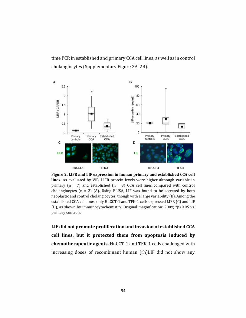
94
time PCR in established and primary CCA cell lines, as well as in control
cholangiocytes (Supplementary Figure 2A, 2B).
Figure 2. LIFR and LIF expression in human primary and established CCA cell
lines. As evaluated by WB, LIFR protein levels were higher although variable in
primary (n = 7) and established (n = 3) CCA cell lines compared with control
cholangiocytes (n = 2) (A). Using ELISA, LIF was found to be secreted by both
neoplastic and control cholangiocytes, though with a large variability (B). Among the
established CCA cell lines, only HuCCT-1 and TFK-1 cells expressed LIFR (C) and LIF
(D), as shown by immunocytochemistry. Original magnification: 200x; *p<0.05 vs.
primary controls.
LIF did not promote proliferation and invasion of established CCA
cell lines, but it protected them from apoptosis induced by
chemotherapeutic agents. HuCCT-1 and TFK-1 cells challenged with
increasing doses of recombinant human (rh)LIF did not show any
95
significant increase in the proliferative rate, except for a minimal
change with the lowest dose in TFK-1 cells (Supplementary Figure 4A,
4B). Additionally, no change in invasive functions was observed with
both CCA cell lines in response to rhLIF (Supplementary Figure 4E, 4F).
To understand whether LIF may stimulate CCA cell proliferation in an
autocrine fashion, thereby precluding further stimulation by
exogenous LIF, we performed an MTS assay in CCA cells with genetic
inactivation of LIFR. Upon transfection of 3 different siRNAs, the extent
of the reduction in LIFR expression in HuCCT-1 and TFK-1 cells was
evaluated by both real-time PCR and WB (Supplementary Figure 3).
Using the 2 most effective siRNAs (siRNA1 and siRNA2), the effect of
autocrine LIF on cell proliferation was evaluated by comparing
silenced cells with scrambled controls in the absence of rhLIF
treatment. No decrease in the proliferative rate was found in LIFR
silenced cells compared with scrambled controls (Supplementary
Figure 4C, 4D). We next turned at evaluating whether LIF can protect
CCA cells from the cytotoxic effects of chemotherapeutic drugs
currently used in the treatment of CCA. To this end, HuCCT-1 and TFK-
1 cells were treated with CDDP, GEM, and GEM+CDDP after a pre-
incubation with/without rhLIF, and their viability was then assessed
by MTS. A drug-induced decrease in cell viability by 34–89% and 23–
64% was observed in HuCCT-1 and TFK-1 cells, respectively (Table 2).
This cytotoxic effect was significantly counteracted by rhLIF, which
augmented cell viability by up to 69.0 ± 36.7% in HuCCT-1 (Figure 3A)

96
and 73.1 ± 17.7% in TFK-1 (Figure 3B) cells. To understand whether
LIF-mediated cytoprotection relies on the ability of LIF to hinder
apoptosis, we assessed the levels of active caspases 3/7, whose up-
regulation is a hallmark of an ongoing apoptotic process. As expected,
a marked increase in the activation of caspase 3/7 was observed in
CCA cells exposed to GEM+CDDP. However, pre-treatment with rhLIF
significantly reduced caspases 3/7 activity by 24% in HuCCT-1 and
22% in TFK-1 cells compared with cells challenged with GEM+CDDP
in the absence of rhLIF pre-treatment (Figure 3C, 3D). The ability of
LIF to exert anti-apoptotic effects in CCA was further confirmed in cells
silenced for LIFR. Genetic inactivation of LIFR in cells exposed to rhLIF
resulted in an increased drug-induced activation of caspases 3/7,
comparable to that observed in cells without rhLIF pre-treatment
(Figure 3C, 3D).
Table 2. Percentage of viable CCA cells following a 24-hour treatment with
chemotherapeutic drugs normalized to untreated cells (MTS assay).

97
Figure 3. Effects of rhLIF on cell viability and apoptosis of CCA cells challenged
with chemotherapeutic agents (GEM, CDDP, or GEM+CDDP). As evaluated by
MTS assay, the pre-treatment of HuCCT-1 (A) and TFK-1 (B) cells with rhLIF was able
to significantly counteract the cytotoxic effect of the chemotherapeutic agents, GEM,
CDDP and GEM+CDDP (Mix). As assessed by caspase GLO 3/7 assay, the activation of
caspases 3/7 in response to Mix in both CCA cell lines (C and D, pale gray columns)
was significantly reduced by the pre-treatment with rhLIF (100 ng/mL) (C and D,
black columns). In addition, the reduction of LIFR expression by two specific siRNAs
was able to abolish the anti-apoptotic effects of rhLIF (C and D, dark gray columns).
*p<0.05 vs. untreated; ^p<0.01 vs. untreated; °p<0.05 vs Mix treatment; §p<0.05 vs.
Scr treated with Mix; n = minimum of 3 in duplicate.
LIF did not induce a stem cell-like phenotype in established CCA
cell lines. To evaluate whether the anti-apoptotic functions of LIF are
associated with the induction of a stem cell-like phenotype, as
described for LIF in malignant melanoma [17], we evaluated the gene
expression of two well-recognized stem cell markers, i.e. Nanog and

98
Oct4, following rhLIF stimulation. However, no significant up-
regulation of either stem cell marker was detected in rhLIF-treated
cells compared to untreated cells (Supplementary Figure 5A, 5B).
In CCA cells, LIF induced Mcl-1 up-regulation via PI3K/AKT,
without activating STAT3 or ERK1/2. To dissect the intracellular
signaling mediating the protective effect of LIF against drug-induced
apoptosis in CCA cells, the relative expression levels of pBax (pro-
apoptotic protein), Bcl-2, and Mcl-1 (anti-apoptotic proteins) were
analyzed by WB in rhLIF-treated HuCCT-1 and TFK-1 cells. Compared
with untreated cells, pBax levels remained unchanged (Supplementary
Figure 6A, 6B), whereas Mcl-1 levels significantly increased in both
CCA cell lines (Figure 4A, 4B). In contrast with pBax and Mcl-1, Bcl-2
was not expressed in either HuCCT-1 or TFK-1 cells (data not shown).
rhLIF did not induce any significant changes in the phosphorylation
levels of STAT3 (Figure 5A, 5B) or extracellular signal-regulated
kinase (ERK)1/2 (or p44/42 MAPK) (Figure 5C, 5D), two well-known
intracellular effectors of LIF [21], in either CCA cell line. In contrast,
rhLIF potently stimulated the phosphorylation of AKT (Figure 5E, 5F).
The key role of the PI3K/AKT axis in mediating LIF-induced Mcl-1 up-
regulation was revealed by treating CCA cells with LY294002, a
specific PI3K inhibitor, which significantly reduced Mcl-1 expression
in rhLIF-treated HuCCT-1 and TFK-1 cells (Figure 4C, 4D).

99
In CCA cells, Mcl-1 inactivation abrogated the cytoprotective
effects of LIF against chemotherapy-induced apoptosis. To
understand whether Mcl-1 plays a central role in the LIF-dependent
protection of CCA cells from the cytotoxic effects of chemotherapeutic
drugs, we evaluated drug-induced apoptosis in HuCCT-1 and TFK-1
cells treated with the selective Mcl-1 inhibitor UMI-77. At our given
dosage (10 μM) [22], UMI-77 did not induce any change in cell viability
(data not shown), nor affect Mcl-1 expression in both cell lines (data
not shown). By caspase 3/7 activation assay, we showed that the
extent of apoptosis in GEM+CDDP-treated cells exposed to UMI-77 and
rhLIF was comparable to that observed in GEM+CDDP-treated cells
without rhLIF exposure (Figure 4C, 4D). Overall, these data point to a
Mcl-1-mediated anti-apoptotic effect of LIF against chemotherapy
toxicity in CCA, which occurs in a PI3K/AKT-dependent, STAT3- and
MAPK-independent manner.

100
Figure 4. Mcl-1 up-regulation mediates the anti-apoptotic effects of rhLIF in
CCA cells. By WB, we found that rhLIF induced a significant up-regulation of Mcl-1
(anti-apoptotic protein) (A and B, black columns) in both HuCCT-1 (A) and TFK-1 (B)
cells compared with untreated cells. This effect was abrogated by the specific PI3K
inhibitor LY294002 (A and B, gray columns). Representative blots are shown below
each respective graph. Interestingly, the protective effect of rhLIF pre-treatment
against GEM+CDDP (Mix) cytotoxicity (C and D, black columns) was abolished by
UMI-77, a selective small molecule inhibitor of Mcl-1 (C and D, dark gray columns).
*p<0.05 vs. untreated; ^p<0.01 vs. untreated; •p<0.01 vs. LIF 10 without LY294002;
°p<0.05 vs. LIF 100; §p<0.05 vs Mix treatment; §§p < 0.01 vs Mix treatment; n =
minimum of 3.

101
Figure 5. rhLIF acts through a STAT3- and ERK1/2-independent, AKT-
dependent pathway. By WB, we showed that rhLIF did not modify the
phosphorylation levels of STAT3 (A and B) or ERK1/2 (C and D) in either HuCCT-1
(A and C) or TFK-1 (B and D) cell lines, but it significantly promoted the
phosphorylation of AKT (E and F) compared with untreated cells. Representative
blots are shown below each respective graph. *p<0.05 vs. untreated; n = minimum
of 3.
DISCUSSION
In this study, we investigated the role of LIF in CCA, a
malignancy with extremely poor prognosis, with a view to unveiling

102
molecular mechanisms responsible for its peculiar aggressiveness,
possibly amenable to therapeutic intervention. We demonstrated that
in CCA, 1) LIF is de novo expressed both in the bile ducts (especially in
the ductular-like rather than in the mucin-producing subtype) and in
the stromal compartment; 2) its cognate receptor LIFR is selectively
up-regulated in neoplastic cholangiocytes; 3) LIF aids neoplastic
cholangiocytes to resist apoptosis induced by the chemotherapeutic
agents GEM and CDDP, without affecting cell proliferation, invasion or
the gain of stemness signatures; 4) anti-apoptotic effects of LIF are
largely mediated by Mcl-1, through activation of the PI3K/AKT
pathway, without the involvement of the conventional LIF
downstream effectors STAT3 and ERK1/2.
In CCA tissue, both cholangiocytes and stromal cells (CAF and
inflammatory cells) proved to be a source of LIF. Conversely, LIF was
not expressed by bile ducts in the peritumoral regions. In tumor
epithelia, LIF expression was heterogeneous, with a greater
extensiveness in areas exhibiting a ductular phenotype. These findings
are consistent with a recent study showing that LIF is overexpressed
in CCA in conjunction with oncostatin M, another IL-6 family member
that is closely related to LIF, with pleiotropic functions in cell
differentiation, proliferation and invasion [23]. In culture conditions,
we could confirm that LIF was variably secreted by CCA cells, in
keeping with the heterogeneous distribution observed in histological
specimens. Interestingly, LIF secretion may be induced by pro-

103
inflammatory cytokines typically released by macrophages and
activated T-cells populating the local inflammatory
microenvironment, such as TNF-α, IL-6, IL-1β and transforming
growth factor (TGF)-β, [12,13,25], as well as by hypoxia, which is a
typical feature of CCA [17]. Once secreted, LIF itself may induce the
expression of LIFR by malignant cells, thus stimulating a positive loop
[12,17]. Indeed, our immunohistochemical, WB, and real-time PCR
data showed that LIFR was selectively up-regulated in CCA bile ducts.
Of note, LIF receptor complex also consists of gp130, which, unlike
LIFR, can be expressed by every cell type within the human body [26].
Nevertheless, we verified the expression of gp130, and found that it
displayed a profile similar to LIFR. Overall, the de novo expression of
LIF and the up-regulation of its receptor in neoplastic bile ducts, along
with LIF overexpression in the tumor reactive stroma, indicate the
presence of autocrine and paracrine LIF-mediated mechanisms in CCA.
Znoyko et al. suggested that an autocrine LIF/LIFR axis is also active
in reactive ductules of cirrhotic livers, likely acting as an important
signal for ductular reaction [27]. It is interesting to note that in our CCA
series, LIF expression was more prevalent in the tumoral areas
characterized by a ductular-like appearance, rather than by a mucin-
producing phenotype. These two specific iCCA phenotypes have been
recently proposed to originate from topographically distinct
cholangiocyte subpopulations, i.e. the major hilar ducts for the mucin-
producing form, and the smaller ducts associated with hepatic

104
progenitor cells for the ductular-like variant [28]. Further studies are
warranted to understand whether LIF expression may indeed
represent a signature of an iCCA subtype arising from hepatic
progenitor cells.
Despite the observation that LIF was prevalently expressed in
the ductular-like areas of CCA, we found that LIF did not exert any
proliferative or pro-invasive effects in CCA cells. The effects of LIF on
neoplastic cells are highly variable, and its failure to stimulate
proliferation or invasiveness has been already reported in other
epithelial cancers [20]. The nature of the functional effects of LIF in
different cancer cell types is actually dependent upon the signal
transduction pathways that may be activated downstream of its
receptor [11]. Our data illustrate that LIF enabled CCA cells to
overcome apoptosis induced by GEM and CDDP, which have been
recently proposed in the treatment of advanced CCA [6]. In CCA cells
that were exposed to GEM+CDDP, LIF could enhance their viability by
up to 73% compared with LIF-untreated cells. In accordance with
these findings, LIF was able to hamper the increase of active caspases
3/7 induced by GEM+CDDP by 22–24% in both CCA cells. Of note,
activation of caspase 3/7 is a fundamental step initiating the cascade
of events ultimately leading to apoptotic cell death. The relevance of
LIF signaling in conferring anti-apoptotic properties upon CCA cells
was confirmed by the restoration of GEM+CDDP cytotoxicity when
LIFR was silenced.

105
To study the mechanisms underlying the resistance to drug-
induced apoptosis mediated by LIF, we first evaluated the possible
involvement of LIF in inducing a stem cell-like phenotype in CCA cells.
Cancer stem cells have an unlimited capacity for self-renewal, and an
impressive capacity for drug resistance. Therefore, their activation in
CCA may largely account for the failure of current chemotherapies. LIF
has been recently reported to regulate the expression of stemness-
related transcription factors, including Nanog and Oct4, in malignant
melanoma [17]. Importantly, Nanog and Oct4 are recognized as
inducers of a stem cell-like phenotype in multiple types of human
cancer, and their expression was found to correlate with resistance to
gemcitabine or cisplatin treatment [29,30]. However, in our
experimental conditions, LIF failed to modulate their gene expression
levels, implying that its anti-apoptotic functions are unlikely to be
related to a dedifferentiation of CCA cells to a cancer stem cell
phenotype. Therefore, we turned to study the balance between pro-
apoptotic and anti-apoptotic proteins of the Bcl-2 family. The anti-
apoptotic Bcl-2 family member Mcl-1 is known to act as a critical
survival factor in both hematogenous and solid tumors, and is
currently regarded as a major oncogene [31]. Importantly, its
expression has been widely documented in both normal and malignant
cholangiocytes [32]. In this study, we demonstrated that Mcl-1
expression by CCA cells can be further augmented by LIF treatment,
whereas the expression of the pro-apoptotic protein pBax remained

106
unchanged, suggesting that this dysregulation may be responsible for
apoptosis evasion induced by LIF. JAK/STAT3, MAPK and PI3K are
common downstream effectors of LIFR [15,16,33], and they have all
been reported to mediate the IL-6-induced Mcl-1 up-regulation in CCA
cells [9,10,33]. In our model, LIF was unable to significantly alter the
levels of either pSTAT3 or pERK1/2. Conversely, LIF stimulation
increased the expression of pAKT in both CCA cell lines, and treatment
of CCA cells with LY294002, a specific PI3K inhibitor, effectively
prevented LIF from up-regulating Mcl-1. This demonstrates that the
positive modulation of Mcl-1 expression in CCA cells is dependent
upon PI3K/AKT activation, as reported in breast cancer [16],
nasopharyngeal carcinoma [18], and rhabdomyosarcoma cells [34].
Pro-survival effects of LIF have also been reported in colorectal cancer
cells, where LIF can negatively regulate the tumor suppressor p53
through a STAT3-dependent pathway [35]. In our experimental
conditions, Mcl-1 inactivation by UMI-77 restored the sensitivity of
CCA cells challenged with LIF to chemotherapeutic agents. This finding
is in accordance with recent data indicating that maritoclax, a similar
selective inhibitor of Mcl-1 triggering its proteosomal degradation, is
able to enhance apoptosis induction by the small-molecule Bcl-2
inhibitor ABT-737 in melanoma cells [36].
Overall, our results indicate that in CCA, LIF signaling may be a
critical mechanism promoting cancer growth and progression. In fact,
LIF is able to protect CCA cells from chemotherapy-induced apoptosis,

107
via a STAT3- and MAPK-independent, PI3K/AKT-dependent Mcl-1 up-
regulation. The pro-oncogenic effects of LIF rely on its broad secretion
by both cancer and reactive stromal cells, as well as on the aberrant
expression of its receptor by neoplastic bile ducts (Figure 6). In
particular, these pro-oncogenic effects are prominent in the ductular-
like areas of iCCA. Importantly, LIF-mediated paracrine effects
highlight the ability of the tumor reactive stroma to promote
therapeutic resistance in epithelial cancers with abundant
desmoplasia. Since our histological samples were obtained from
patients undergoing surgical resection, and in our center
chemotherapy is only reserved for those with advanced CCA (which
generally do not perform histological evaluation), correlating
LIF/LIFR expression with clinical data was not possible in the present
study. However, we demonstrated that the downstream effectors of
LIF signaling could represent innovative molecular targets amenable
to therapeutic modulation, with a view to increasing CCA
responsiveness to conventional chemotherapy.

108
Figure 6. The working model illustrating the molecular mechanisms
underlying the protective effects of LIF against chemotherapy in neoplastic
cholangiocytes. In CCA cells, LIFR is up-regulated under the influence of LIF
released by both neoplastic cholangiocytes themselves (autocrine loop) and reactive
stromal cells, such as CAF and TAM (paracrine loop). When LIFR dimerizes with
gp130, LIF signaling is transduced through the PI3K/AKT pathway to increase the
expression levels of the anti-apoptotic protein Mcl-1, which confers resistance
against chemotherapeutic agents by hampering the activation of caspases 3/7.
Conflicts of interest: All authors have no conflict of interest regarding this study to declare. Financial support: Progetto di Ricerca Ateneo 2011 (grant #CPD113799/11) to LF, MC and SDM. Associazione Scientifica Gastroenterologica di Treviso (ASGET, “associazione di promozione sociale senza scopo di lucro”) to LF and SDM. Associazione Chirurgica Tarvisium to NB, MM and TS. NIH Grant DK079005, Silvio O. Conte Digestive Diseases Research Core Centers – 5P30DK034989”, grant from “PSC

109
partners for a care”, projects CARIPLO 2011–0470 and PRIN 2009ARYX4T_005 to MS.
MATERIALS AND METHODS
Tissue samples. Formalin-fixed, paraffin-embedded sections of
surgically resected CCA liver from 19 patients were included in the
immunohistochemical study and compared with the corresponding
peritumoral areas where available (n = 12). The patients were
predominantly male (12/19), with a median age of 64 years (min 35;
max 81), and 63% (12/19) were iCCA. CCA areas were then
categorized as ductular-like or mucin-producing according to Komuta
[28].
Cell lines. Three established human CCA cell lines were used: EGI-1,
TFK-1 (both eCCA, purchased from Deutsche Sammlung von
Mikroorganismen und Zellkulturen, DSMZ, Germany), and HuCCT-1
(iCCA, from Health Science Research Resource Bank, HSRRB, Japan),
along with primary biliary cell preparations obtained from surgically
resected human iCCA liver samples (n = 7), as previously described
[37]. Human cholangiocytes isolated from liver explants of alcoholic
liver cirrhosis (n = 2) served as controls. All specimens were reviewed
by the same dedicated pathologist (AF) to confirm diagnosis. Local
regional ethical committee approval was obtained for tissue collection
and cell preparations.

110
LIF, LIFR and gp130 expression in tissues. By
immunohistochemistry, we evaluated the expression of LIF, LIFR and
gp130 in bile ducts and the stroma, in both neoplastic and matched
peritumoral areas. Further details are provided in the supplementary
section. The extent of immunoreactivity was scored by two
independent observers (SDM, MC) as: 0 = < 5%; 1 = 5–30%; 2 = 30–
70%; 3 = > 70% area of positive cells, as previously reported [38]. In
selected tissue specimens, dual immunofluorescence for LIF and α-
SMA (myofibroblast marker) or CD45 (inflammatory cell marker) was
performed to assess the specific contribution of different stromal cell
types to LIF production.
LIF and LIFR expression in cells. To evaluate whether
immunohistochemical findings were consistent with in vitro data, LIF
and LIFR expression was then assessed in cultured cholangiocytes by
immunocytochemistry. LIFR protein expression was also assessed by
WB in both established and primary CCA cell lines. Furthermore, gene
expression of LIF and LIFR in CCA cell lines was evaluated by real-time
PCR. Further details are provided in the supplementary section.
LIF secretion by cultured cholangiocytes. The supernatants of CCA
and control cholangiocytes cultured for 24 h at a density of 5×10⁴ were
analyzed for the presence of secreted LIF using an ELISA kit, according

111
to the supplier's instructions (Raybiotech, Milan). For each
experiment, a LIF standard curve was generated.
Cell proliferation. HuCCT-1 and TFK-1 cells were cultured at a
density of 1×10⁴ for 48 h with/without exposure to increasing doses
(0.1, 1, 10, 100 ng/mL) of recombinant human LIF (rhLIF,
R&DSystems). Proliferation activity was assessed by MTS assay
according to the supplier's instructions (CellTiter 96 AQueous One
Solution Cell Proliferation Assay, Promega).
Cell viability. MTS assay was also used to assess whether LIF (24 h
pre-treatment at 0.1, 1, 10, 100 ng/mL) affected the viability of HuCCT-
1 and TFK-1 cells in response to a 24 h treatment with cisplatin, 17 μM
(CDDP; Sigma-Aldrich) [39] and gemcitabine, 30 μM (GEM; Sigma-
Aldrich) [40], either alone or in combination (GEM+CDDP).
Stem cell-like phenotyping. Real-time PCR was used to assess LIF
effect (100 ng/mL) on Nanog and Oct4 gene expression. RNA was
isolated from cultured cells, as previously described [41]. Further
details are available in the supplementary section.
Cell invasiveness. The invasiveness of HuCCT-1 and TFK-1 cells was
assessed by Boyden chamber assay, as previously described [42].
Methodology is detailed in the supplementary section.

112
Downstream effectors of LIF signaling in CCA cells. After exposure
of HuCCT-1 and TFK-1 cells to rhLIF (10, 100 ng/mL) for 15 min
(STAT3, pSTAT3, ERK1/2, pERK1/2, AKT, pAKT) or 24 h (Bax, pBax,
Bcl-2 and Mcl-1), the expression level of different proteins of interest
was evaluated by WB (see the supplementary section). To unravel the
pathway regulating Mcl-1 expression, its protein expression was also
measured in CCA cells pre-treated with the PI3K chemical inhibitor
LY294002 (10 μM, Sigma) [43] for 10 mins, and then treated with
rhLIF plus LY294002 for 24 h.
Activation of caspases 3/7. HuCCT-1 and TFK-1 cells were seeded
into a 96-well plate at a density of 1×10⁴ per well with/without rhLIF
(100 ng/mL) for 24 h, prior to treatment with GEM+CDDP for 12 h. The
luminescence-based solution Caspase-Glo 3/7 (Promega) was then
used to assess activation of caspases 3/7. Luciferase reaction was
evaluated using a microplate reader (BMG Labtech).
Mcl-1 inactivation. Apoptotic response following GEM+CDDP
treatment, assessed as described above (i.e., activation of caspases
3/7), was also evaluated upon Mcl-1 inhibition in HuCCT-1 and TFK-1
cells. We used a novel, selective, small molecule inhibitor of Mcl-1,
UMI-77 (10 μM), for 24 h [22]. Antagonism of Mcl-1 by UMI-77 does
not rely on the down-regulation of its expression, but on the ability of
UMI-77 to block the heterodimerization of Mcl-1 with pro-apoptotic

113
members of the Bcl-2 family, including Bax and Bak [22, 44].
Specifically, the inhibitory effect of UMI-77 is related to its binding to
the BH3-binding groove of Mcl-1.
Silencing of LIFR. Gene silencing was performed using commercially
available siRNAs against LIFR, and scramble RNA was used as a control
(Life Technologies). HuCCT-1 and TFK-1 cell lines were transfected
using Lipofectamine 2000 transfection reagent (Life Technologies).
Further details are provided in the supplementary section.
Statistical analyses. Results are shown as the mean ± standard
deviation. Statistical comparisons were made using Student's t-test.
Statistical analyses were performed using SPSS 20.0 software (IBM
Corp.). A p value < 0.05 was considered significant.
REFERENCES
1. Bragazzi M, Cardinale V, Carpino G, Venere R, Semeraro R, Gentile R,
Gaudio E, Alvaro D. Cholangiocarcinoma: Epidemiology and risk factors.
Transl Gastrointest Cancer. 2011;1:21–32.
2. Razumilava N, Gores GJ. Classification, Diagnosis, and Management of
Cholangiocarcinoma. Clin Gastroenterol Hepatol. 2013;11:13–21.
3. Bridgewater J, Galle PR, Khan SA, Llovet JM, Park JW, Patel T, Pawlik TM,
Gores GJ. Guidelines for the diagnosis and management of intrahepatic
cholangiocarcinoma. J Hepatol. 2014;60:1268–1289.

114
4. Quyn AJ, Ziyaie D, Polignano FM, Tait IS. Photodynamic therapy is
associated with an improvement in survival in patients with irresectable
hilar cholangiocarcinoma. HPB (Oxford) 2009;11:570–577.
5. Gatto M, Bragazzi MC, Semeraro R, Napoli C, Gentile R, Torrice A, Gaudio E,
Alvaro D. Cholangiocarcinoma: update and future perspectives. Dig Liver Dis.
2010;42:253–260.
6. Valle J, Wasan H, Palmer DH, Cunningham D, Anthoney A, Maraveyas A,
Madhusudan S, Iveson T, Hughes S, Pereira SP, Roughton M. Cisplatin plus
gemcitabine versus gemcitabine for biliary tract cancer. N Engl J Med.
2010;362:1273–1281.
7. Rizvi S, Gores GJ. Pathogenesis, diagnosis, and management of
cholangiocarcinoma. Gastroenterology. 2013;145:1215–1229.
8. Zabron A, Edwards RJ, Khan SA. The challenge of cholangiocarcinoma:
dissecting the molecular mechanisms of an insidious cancer. Dis Model Mech.
2013;6:281–292.
9. Meng F, Yamagiwa Y, Ueno Y, Patel T. Over-expression of interleukin-6
enhances cell survival and transformed cell growth in human malignant
cholangiocytes. J Hepatol. 2006;44:1055–1065.
10. Kobayashi S, Werneburg NW, Bronk SF, Kaufmann SH, Gores GJ.
Interleukin-6 contributes to Mcl-1 up-regulation and TRAIL resistance via an
Akt-signaling pathway in cholangiocarcinoma cells. Gastroenterology.
2005;128:2054–2065.
11. Mathieu ME, Saucourt C, Mournetas V, Gauthereau X, Theze N, Praloran
V, Thiébaud P, Bœuf H. LIF-dependent signaling: new pieces in the Lego. Stem
cell reviews. 2012;8:1–15.
12. Kamohara H, Ogawa M, Ishiko T, Sakamoto K, Baba H. Leukemia
inhibitory factor functions as a growth factor in pancreas carcinoma cells:

115
Involvement of regulation of LIF and its receptor expression. Int J Oncol.
2007;30:977–983.
13. McKenzie RC, Szepietowski J. Cutaneous leukemia inhibitory factor and
its potential role in the development of skin tumors. Dermatol Surg.
2004;30:279–290.
14. Gadient RA, Patterson PH. Leukemia inhibitory factor, Interleukin 6, and
other cytokines using the GP130 transducing receptor: roles in inflammation
and injury. Stem cells. 1999;17:127–137.
15. Zouein FA, Kurdi M, Booz GW. LIF and the heart: just another brick in the
wall? Eur Cytokine Netw. 2013;24:11–19.
16. Li X, Yang Q, Yu H, Wu L, Zhao Y, Zhang C, Yue X, Liu Z, Wu H, Haffty BG,
Feng Z, Hu W. LIF promotes tumorigenesis and metastasis of breast cancer
through the AKT-mTOR pathway. Oncotarget. 2014;5:788–801.
17. Kuphal S, Wallner S, Bosserhoff AK. Impact of LIF expression in malignant
melanoma. Exp Mol Pathol. 2013;95:156–165.
18. Liu SC, Tsang NM, Chiang WC, Chang KP, Hsueh C, Liang Y, Juang JL, Chow
KP, Chang YS. Leukemia inhibitory factor promotes nasopharyngeal
carcinoma progression and radioresistance. J Clin Invest. 2013;123:5269–
5283.
19. Kellokumpu-Lehtinen P, Talpaz M, Harris D, Van Q, Kurzrock R, Estrov Z.
Leukemia-inhibitory factor stimulates breast, kidney and prostate cancer cell
proliferation by paracrine and autocrine pathways. Int J Cancer.
1996;66:515–519.
20. Kamohara H, Sakamoto K, Ishiko T, Masuda Y, Abe T, Ogawa M. Leukemia
inhibitory factor induces apoptosis and proliferation of human carcinoma
cells through different oncogene pathways. Int J Cancer. 1997;72:687–695.

116
21. Heinrich PC, Behrmann I, Haan S, Hermanns HM, Muller-Newen G,
Schaper F. Principles of interleukin (IL)-6-type cytokine signaling and its
regulation. The Biochem J. 2003;374:1–20.
22. Abulwerdi F, Liao C, Liu M, Azmi AS, Aboukameel A, Mady AS, Gulappa T,
Cierpicki T, Owens S, Zhang T, Sun D, Stuckey JA, Mohammad RM, et al. A
novel small-molecule inhibitor of mcl-1 blocks pancreatic cancer growth in
vitro and in vivo. Mol Cancer Ther. 2014;13:565–575.
23. Kang MJ, Kim J, Jang JY, Park T, Lee KB, Kim SW. 22q11-q13 as a hot spot
for prediction of disease-free survival in bile duct cancer: integrative analysis
of copy number variations. Cancer Genet. 2014;207:57–69.
24. Clark EA, Golub TR, Lander ES, Hynes RO. Genomic analysis of metastasis
reveals an essential role for RhoC. Nature. 2000;406:532–535.
25. Albrengues J, Bourget I, Pons C, Butet V, Hofman P, Tartare-Deckert S,
Feral CC, Meneguzzi G, Gaggioli C. LIF mediates proinvasive activation of
stromal fibroblasts in cancer. Cell Rep. 2014;7:1664–1678.
26. Wolf J, Rose-John S, Garbers C. Interleukin-6 and its receptors: A highly
regulated and dynamic system. Cytokine. 2014;70:11–20.
27. Znoyko I, Sohara N, Spicer SS, Trojanowska M, Reuben A. Expression of
oncostatin M and its receptors in normal and cirrhotic human liver. J Hepatol.
2005;43:893–900.
28. Komuta M, Govaere O, Vandecaveye V, Akiba J, Van Steenbergen W,
Verslype C, Laleman W, Pirenne J, Aerts R, Yano H, Nevens F, Topal B,
Roskams T. Histological diversity in cholangiocellular carcinoma reflects the
different cholangiocyte phenotypes. Hepatology. 2012;55:1876–1888.
29. Lu Y, Zhu H, Shan H, Lu J, Chang X, Li X, Lu J, Fan X, Zhu S, Wang Y, Guo Q,
Wang L, Huang Y, et al. Knockdown of Oct4 and Nanog expression inhibits the
stemness of pancreatic cancer cells. Cancer Lett. 2013;340:113–123.

117
30. Noh KH, Kim BW, Song KH, Cho H, Lee YH, Kim JH, Chung JY, Kim JH,
Hewitt SM, Seong SY, Mao CP, Wu TC, Kim TW. Nanog signaling in cancer
promotes stem-like phenotype and immune evasion. J Clin Invest.
2012;122:4077–4093.
31. Beroukhim R, Mermel CH, Porter D, Wei G, Raychaudhuri S, Donovan J, et
al. The landscape of somatic copy-number alteration across human cancers.
Nature. 2010;463:899–905.
32. Okaro AC, Deery AR, Hutchins RR, Davidson BR. The expression of
antiapoptotic proteins Bcl-2, Bcl-X(L), and Mcl-1 in benign, dysplastic, and
malignant biliary epithelium. J Clin Pathol. 2001;54:927–932.
33. Isomoto H, Kobayashi S, Werneburg NW, Bronk SF, Guicciardi ME, Frank
DA, Gores GJ. Interleukin 6 upregulates myeloid cell leukemia-1 expression
through a STAT3 pathway in cholangiocarcinoma cells. Hepatology.
2005;42:1329–1338.
34. Wysoczynski M, Miekus K, Jankowski K, Wanzeck J, Bertolone S,
Janowska-Wieczorek A, Ratajczak J, Ratajczak MZ. Leukemia inhibitory
factor: a newly identified metastatic factor in rhabdomyosarcomas. Cancer
Res. 2007;67:2131–2140.
35. Yu H, Yue X, Zhao Y, Li X, Wu L, Zhang C, Liu Z, Lin K, Xu-Monette ZY, Young
KH, Liu J, Shen Z, Feng Z, et al. LIF negatively regulates tumor-suppressor p53
through Stat3/ID1/MDM2 in colorectal cancers. Nat Commun. 2014;5:5218.
36. Pandey MK, Gowda K, Doi K, Sharma AK, Wang H-G, Amin S. Proteosomal
degradation of Mcl-1 by maritoclax induces apoptosis and enhance the
efficacy of ABT-737 in melanoma cells. PloS One. 2013;8:e78570.
37. Massani M, Stecca T, Fabris L, Caratozzolo E, Ruffolo C, Furlanetto A,
Morton S, Cadamuro M, Strazzabosco M, Bassi N. Isolation and
characterization of biliary epithelial and stromal cells from resected human

118
cholangiocarcinoma: a novel in vitro model to study tumor-stroma
interactions. Oncol Rep. 2013;30:1143–1148.
38. Fabris L, Cadamuro M, Moserle L, Dziura J, Cong X, Sambado L, Nardo G,
Sonzogni A, Colledan M, Furlanetto A, Bassi N, Massani M, Cillo U, et al.
Nuclear expression of S100A4 calcium binding protein increases
cholangiocarcinoma invasiveness and metastasization. Hepatology.
2011;54:890–899.
39. Chattopadhyay S, Machado-Pinilla R, Manguan-García C, Belda-Iniesta C,
Moratilla C, Cejas P, Fresno-Vara JA, de Castro-Carpeño J, Casado E, Nistal M,
Gonzalez-Barón M, Perona R. MKP1/CL100 controls tumor growth and
sensitivity to cisplatin in non-small-cell lung cancer. Oncogene.
2006;25:3335–3345.
40. Shord SS, Camp JR, Young LA. Paclitaxel decreases the accumulation of
gemcitabine and its metabolites in human leukemia cells and primary cell
cultures. Anticancer Res. 2005;25:4165–4171.
41. Spirli C, Locatelli L, Morell CM, Fiorotto R, Morton SD, Cadamuro M, Fabris
L, Strazzabosco M. PKA dependent p-Ser- beta-catenin, a novel signaling
defect in a mouse model of Congenital Hepatic Fibrosis. Hepatology.
2013;58:1713–1723.
42. Cadamuro M, Nardo G, Indraccolo S, Dall'olmo L, Sambado L, Moserle L,
Franceschet I, Colledan M, Massani M, Stecca T, Bassi N, Morton S, Spirli C, et
al. Platelet-derived growth factor-D and Rho GTPases regulate recruitment
of cancer-associated fibroblasts in cholangiocarcinoma. Hepatology.
2013;58:1042–1053.
43. Spirli C, Okolicsanyi S, Fiorotto R, Fabris L, Cadamuro M, Lecchi S, Tian X,
Somlo S, Strazzabosco M. Mammalian target of rapamycin regulates vascular

119
endothelial growth factor-dependent liver cyst growth in polycystin-2-
defective mice. Hepatology. 2010;51:1778–1788.
44. Mohammad RM, Muqbil I, Lowe L, Yedjou C, Hsu HY, Lin LT, Siegelin MD,
Fimognari C, Kumar NB, Dou QP, Yang H, Samadi AK, Russo GL, et al. Broad
targeting of resistance to apoptosis in cancer. Semin Cancer Biol. 2015 in
press.
SUPPLEMENTARY MATERIALS AND METHODS
Immunohistochemistry and immunofluorescence. For
immunohistochemical staining, after deparaffinization, sections were
rehydrated in alcohol and endogenous peroxidase activity was
quenched with methanol containing 3% H2O2 for 30 mins. Antigen
retrieval was performed by steam-heating the slides for 20 mins in
either 10 mmol/L citrate buffer (pH 6) for LIF or 1.27 mM
ethylenediaminetetraacetic acid buffer (pH 8) for LIFR and gp130.
Blocking was achieved using UltraVision protein block
(ThermoScientific) for 8 mins. The sections were incubated with
primary antibodies against LIF (1:50, Santa Cruz), LIFR (1:80, Santa
Cruz) and gp130 (1:25, Santa Cruz) overnight at 4°C. After rinsing with
phosphate-buffered saline 1M (PBS) supplemented with 0.05%
Tween20 (PBS-T; both Sigma), slides were incubated for 30 mins at
room temperature with EnVision horseradish peroxidase (HRP)-
conjugated secondary antibody (DAKO). All antibodies were diluted in
PBS. Specimens were developed using 3,3-diaminobenzedine
tetrahydrochloride 0.04 mg/mL (Sigma) with H2O2 0.01% diluted in

120
PBS and counterstained with Gill’s Hematoxylin N°2 (Sigma).
Specimens were analyzed with an Eclipse E800 microscope (Nikon)
and LuciaG 5.0 software (Nikon), and images were collected with a
digital camera (Nikon, DS-U1). For immunofluorescence, sections
were deparaffinized and rehydrated as described above. Antigen
retrieval was performed as above using 10 mmol/L citrate buffer (pH
6). Blocking was achieved as above. The sections were incubated with
primary antibodies against LIF (1:50, Santa Cruz), and CD45 (1:300,
DAKO) or α-SMA (1:100, DAKO) overnight at 4°C. Slides were then
incubated for 30 mins at room temperature with the respective
secondary Alexa Fluor 488- or 594-conjugated antibody (1:500, Life
Technologies) and mounted with Vectastain + DAPI (Vector
Laboratories).
Immunocytochemistry. After fixation with 4% paraformaldehyde
(Carlo Erba), cells were incubated overnight at 4°C with primary
antibodies against LIF (1:100), and LIFR (1:100). After washing with
PBS-T, the cells were incubated for 30 mins at room temperature with
the appropriate Alexa Fluor 488 secondary antibody (1:500, Life
Technologies) and then mounted with Vectastain + DAPI.
ELISA for LIF quantification. Cells were seeded into a 24-well plate
at 5 × 10⁴ per well. After 24 h, supernatants were harvested, stored at

121
-80°C and then the ELISA was developed according to the supplier
(RayBiotech). A calibration curve was generated for each experiment.
Western blotting (WB). Equal concentrations of total lysate obtained
from cultured cells were electrophoresed on a 4-12% NuPAGE®
Novex Bis-Tris gel (Life Technologies) and proteins were transferred
to a nitrocellulose membrane (Life Technologies). The membrane was
then blocked with 5% non-fat dry milk (Euroclone) in Tris-buffered
saline (TBS) supplemented with 0.1% Tween-20 (TBS-T) for 1 hour
and then incubated overnight at 4°C with rabbit anti-LIFR (1:1000,
Santa Cruz), rabbit anti-Bax (1:500, Santa Cruz), rabbit anti-
phosphorylated Bax (pBax) (1:1000; Bioss), rabbit anti-B-cell
lymphoma (Bcl)-2 (1:500, Santa Cruz), rabbit anti-myeloid cell
leukemia (Mcl)-1 (1:1000, Cell Signaling), rabbit anti-STAT3, rabbit
anti-phosphorylated STAT3 (pSTAT3) (both 1:1000, Cell Signaling),
rabbit anti-AKT (1:1000, Cell Signaling), rabbit anti-phosphorylated
AKT 1/2/3 (pAKT) (1:1000, Santa Cruz), rabbit anti-ERK1/2 (1:1000,
Cell Signaling), and rabbit anti-phosphorylated ERK1/2 (pERK1/2)
(1:1000, Cell Signaling). As a reference protein, mouse anti-GAPDH
(1:10000, Santa Cruz) was used. The membrane was washed 3 times
with TBS-T before incubation with goat anti-mouse (1:2000, Sigma) or
goat anti-rabbit (1:2000, Bio-Rad) HRP-conjugated secondary
antibodies for 1 hour. Proteins were visualized using enhanced
chemiluminescence (SuperSignal West Pico, Thermo Scientific).

122
Stem cell-like phenotyping. Briefly, untreated and rhLIF-treated
(100 ng/mL for 24 hours) HuCCT-1 and TFK-1 cells were homogenized
in 1 mL TRIzol® Reagent (Life Technologies). Template
complementary DNA was obtained by reverse transcription using 0.5
μg of total RNA, Superscript II reverse transcriptase (Life
Technologies), random hexamers (50 pmol), and oligo-dT primers
(100 pmol) (Promega). Relative transcript levels were quantified
using Taqman gene expression probes for human Nanog and Oct4 (Life
Technologies) and the real-time PCR was performed on an ABI 7500
thermocycler (Applied Biosystems). The relative expression of each
gene was normalized against that of GAPDH.
Gene expression of LIF and LIFR. Using the same approach described
for stem-cell phenotyping, we quantified the relative expression levels
of LIF and LIFR in established and primary CCA cell lines using specific
Taqman gene expression commercially available probes (Life
Technologies).
Cell invasiveness. Briefly, 5 × 10⁴ cells were re-suspended in serum-
free medium and seeded over a polyvinylpyrrolidone-free
polycarbonate, 8 μm-pore membrane (Transwell, Costar) coated with
50 μg/mL Matrigel, within a Boyden microchamber. The lower
chambers contained serum-free medium with/without rhLIF (10, 100
ng/mL). After 48 h, cells on the upper surface of the membrane were

123
removed with a cotton swab whilst cells that adhered to the lower
surface were fixed and stained using a Diff-Quick Staining Set (Medion
Diagnostics); ten random fields of each membrane were photographed
to count the number of clearly discernible nuclei.
Silencing of LIFR. Gene silencing of LIFR was performed using
commercially available siRNAs against LIFR; scramble RNA served as
a control (both Life Technologies). HuCCT-1 and TFK-1 cell lines were
transfected using 20 pM of siRNA and Lipofectamine 2000 transfection
reagent (Life Technologies) 24 h after plating. Transfection efficiency
was assessed by WB and real-time PCR for LIFR.

124
SUPPLEMENTARY FIGURES
Supplementary Figure 1. Extensiveness of LIF and LIFR expression in ductular-
like and mucin-producing areas of CCA. LIF expression was more extensive in
ductular-like than in mucin-producing CCA bile ducts (A), whereas no significant
difference in LIFR staining was observed between the two CCA phenotypes (B).
Representative micrographs illustrating LIF and LIFR staining in sequential sections
of mucin-producing (upper panels) and ductular-like (lower panels) CCA areas are
shown (C). Original magnification: 200x; *p<0.05.

125
Supplementary Figure 2. LIFR and LIF gene expression quantification in
human primary control and neoplastic cholangiocytes, and established CCA
cell lines. To confirm data obtained by WB and ELISA, the gene expression of LIFR
(A) and LIF (B) was assessed in primary (n = 7) and established (n = 3) CCA cell lines,
and in control cholangiocytes (n = 2) by real-time PCR. The results show increased
mRNA levels for both receptor and ligand in CCA cells compared with controls.

126
Supplementary Figure 3. Effects of siRNA on LIFR expression in CCA cells. The
ability of three different siRNAs (siRNA1, siRNA2, and siRNA3) to suppress LIFR
expression was evaluated by both WB and real-time PCR in HuCCT-1 (A and C) and
TFK-1 (B and D) cells. All siRNAs induced a pronounced reduction in protein (A and
B) and mRNA levels (C and D) of LIFR. Therefore, siRNA1 and siRNA2 were selected
for the experiments on cell proliferation (shown in Supplementary Figure 4C, 4D)
and drug-induced cytotoxicity (shown in Figure 3C, 3D).

127
Supplementary Figure 4. LIF did not affect cell proliferation and invasion of
HuCCT-1 and TFK-1 cells. By MTS assay, we found that HuCCT-1 (A) and TFK-1 (B)
cells challenged with rhLIF at increasing doses did not show any relevant change in
cell proliferation, except for a minimal increase with the lowest dose in TFK-1
(^p<0.01 vs. LIF 0; n = 3 in duplicate). Interestingly, in the absence of rhLIF
stimulation, LIFR silencing did not reduce cell proliferation of HuCCT-1 (C) and TFK-
1 (D) cells compared with scrambled cells, thus ruling out a possible constitutive
activation of cell proliferation triggered by autocrine LIF. Additionally, no effects
were observed on cell invasion with either HuCCT-1 (E) or TFK-1 (F) cells upon LIF
stimulation, as assessed in Boyden chamber assays (n = minimum of 3). Micrographs
of representative fields of invaded cells in response to medium alone or LIF
supplementation are illustrated. Original magnification: 100×.
Supplementary Figure 5. rhLIF did not increase gene expression of Nanog and
Oct4 in HuCCT-1 and TFK-1 cells. By real-time PCR, we found that rhLIF
stimulation (100 ng/mL) was unable to change Nanog and Oct4 mRNA levels in
HuCCT-1 (A) and TFK-1 (B) cells (n = 3).

128
Supplementary Figure 6. Effects of rhLIF on Bax phosphorylation in CCA cells.
By WB, we showed that rhLIF did not modify pBax:Bax ratio (pro-apoptotic) in either
HuCCT-1 (A) or TFK-1 (B) cells compared with untreated cells. Representative blots
are shown below each respective graph (n = 3).

129
CHAPTER 3
Low-Dose Paclitaxel Reduces S100A4 Nuclear
Import to Inhibit Invasion and Hematogenous
Metastasis of Cholangiocarcinoma
Massimiliano Cadamuro1,2, Gaia Spagnuolo1, Luisa Sambado3, Stefano
Indraccolo4, Giorgia Nardo4, Antonio Rosato4,5, Simone Brivio1, Chiara
Caslini1, Tommaso Stecca6, Marco Massani6, Nicolò Bassi5,6, Eugenio
Novelli7, Carlo Spirli2,8, Luca Fabris2,8,9,*, and Mario Strazzabosco1,2,8,*
1School of Medicine and Surgery, University of Milan-Bicocca, Italy 2International Center for Digestive Health (ICDH), University of Milan-Bicocca 3Metabolism, Disease and Clinical Nutrition Unit, Treviso Regional Hospital, Italy 4Veneto Institute of Oncology IOV – IRCCS, Padua, Italy 5Dep. of Surgery, Oncology and Gastroenterology, University of Padua School of Medicine, Italy 64th Surgery Division, Treviso Regional Hospital 7Biostatistics Unit, Clinica San Gaudenzio, Novara, Italy 8Section of Digestive Diseases, Yale University School of Medicine, New Haven (CT), USA 9Dep. of Molecular Medicine (DMM), University of Padua School of Medicine *these authors contributed equally to this article
Published on: Cancer Research, 2016 Aug 15;76(16):4775-4784. doi:
10.1158/0008-5472.CAN-16-0188.

130
ABSTRACT
Nuclear expression of the calcium-binding protein S100A4 is a
biomarker of increased invasiveness in cholangiocarcinoma (CCA), a
primary liver cancer with scarce treatment opportunities and dismal
prognosis. In this study, we provide evidence that targeting S100A4
nuclear import by low dose paclitaxel (PTX), a microtubule stabilizing
agent, inhibits CCA invasiveness and metastatic spread.
Administration of low dose PTX to established (EGI-1) and primary
(CCA-TV3) CCA cell lines expressing nuclear S100A4 triggered a
marked reduction in nuclear expression of S100A4 without modifying
its cytoplasmic levels, an effect associated with a significant decrease
in cell migration and invasiveness. While low dose PTX did not affect
cellular proliferation, apoptosis or cytoskeletal integrity, it
significantly reduced SUMOylation of S100A4, a critical
posttranslational modification that directs its trafficking to the
nucleus. This effect of lose dose PTX was reproduced by ginkolic acid,
a specific SUMOylation inhibitor. Downregulation of nuclear S100A4
by low dose PTX was associated with a strong reduction in RhoA and
Cdc42 GTPase activity, MT1-MMP expression and MMP-9 secretion. In
a SCID mouse xenograft model, low dose metronomic PTX treatment
decreased lung dissemination of EGI-1 cells without significantly
affecting their local tumor growth. In the tumor mass, nuclear S100A4
expression by CCA cells was significantly reduced, whereas rates of
proliferation and apoptosis were unchanged. Overall, our findings
131
highlight nuclear S100A4 as a candidate therapeutic target in CCA and
establish a mechanistic rationale for the use of low dose PTX in
blocking metastatic progression of cholangiocarcinoma.
INTRODUCTION
Cholangiocarcinoma (CCA), a malignancy arising from either
the intrahepatic or the extrahepatic bile ducts, still carries a severe
prognosis. CCA is responsible for the 10–20% of deaths related to
primary liver tumors [1]. In the Western countries, its incidence is
steadily increasing in the last decades [1,2], but, unfortunately, the
prognosis of CCA has not changed, with less than 5% of patients
surviving up to 5 years from diagnosis [1]. At the time of diagnosis, less
than 30% of patients are eligible for surgical resection or liver
transplantation, the only potentially curative strategies. Thus, in 70%
of patients the stage is advanced, because of the tumor invasiveness
and early extrahepatic dissemination. Furthermore, success of
curative treatments is hindered by the high rate of recurrence, with a
5-year survival after resection around 20–40% [3]. Combined cisplatin
and gemcitabine therapy, the current standard of care for advanced
CCA, increases patient’s overall survival by less than four months with
respect to gemcitabine alone [4]. The lack of effective treatments
reflects the deep gap in knowledge on the molecular mechanisms
underlying CCA invasiveness. Better understanding of these

132
mechanisms is needed to predict the invasiveness of the individual
tumor and to devise molecular targeted therapy [5].
Among the biomarkers of increased tumour invasiveness,
S100A4 has drawn particular attention in the last few years. S100A4,
a low molecular weight, cytoskeleton-associated calcium-binding
protein, is normally expressed by mesenchymal (mostly fibroblasts
and macrophages), but not by epithelial cells. S100A4 may handle
different functions depending upon its cellular localization. When
localized in the cytoplasm, it may interact with cytoskeleton and
plasma membrane proteins (including actin, non-muscle myosin-IIA
and -IIB, p53, liprin-β1, methionine aminopeptidase-2) [6], thereby
contributing to the regulation of cell proliferation, survival,
differentiation, as well as cell reshaping and cytoskeletal
rearrangement. When translocated to the nucleus, S100A4 may act as
transcription factor for several genes, including those encoding
adherence junction proteins, thus controlling cell motility [7]. A
number of studies have shown that S100A4 is a marker of poor
prognosis in breast and colon cancers [7,8]. We have shown that in
CCA patients undergoing surgical resection, nuclear expression of
S100A4 in tumor cells is a strong, independent prognostic marker of
poor outcome in terms of both metastasization and tumor-related
death [9]. Furthermore, S100A4 lentiviral silencing significantly
reduced motility and invasive capabilities of CCA cells [9], suggesting
that nuclear expression of S100A4 is not merely a marker of cancer
133
invasiveness, but is a key determinant of the metastatic phenotype of
CCA.
Aim of this study was to understand the mechanisms by which
nuclear S100A4 induces an invasive phenotype in CCA and the
mechanism regulating nuclear translocation of S100A4. Unfortunately,
mechanisms governing S100A4 expression in the nucleus remain
elusive, and there are no strategies to selectively target S100A4
nuclear import. In planning our experiments, we came across studies
from the early ‘90s showing that paclitaxel (PTX) was able to reduce
the expression of S100A4 in the B16 murine melanoma cells [10,11].
PTX is a semisynthetic derivative of taxol, a natural diterpene alkaloid,
isolated from the bark of Taxus brevifolia. Because of its anti-
proliferative and pro-apoptotic effects, PTX is currently used in
chemotherapy protocols for the treatment of ovary, lung, thyroid and
breast carcinomas [12]. Our study shows that low dose PTX inhibits
tumor invasiveness and hematogenous metastases by blocking
SUMOylation-dependent S100A4 nuclear import in CCA.
MATERIALS AND METHODS
Human established and primary CCA cell lines. The S100A4-
expressing established CCA cell lines, EGI-1 (both in the nucleus and in
the cytoplasm) and TFK-1 (only in the cytoplasm), both obtained from
extrahepatic CCA [9], were purchased from Deutsche Sammlung von
Mikroorganismen und Zellkulturen (DSMZ, Germany). The primary

134
CCA cell line CCA-TV3 was isolated from a human sample derived from
surgical resection of an intrahepatic mass-forming CCA, histologically
categorized as cholangiocellular carcinoma (grading G3), performed in
Treviso Regional Hospital (MM, TS) as described [13]. Local regional
ethical committee approval was obtained for tissue collection and cell
preparation. Cultured cells were grown in RPMI1680 supplemented
with 10% FBS and 1% penicillin at 37°C in a 5% CO2 atmosphere, and
then frozen at low passages (<5). After any resuscitation, cell
authentication was performed by checking morphology and by
evaluating their immunophenotype as characterized by our previous
studies [9,13], including cytokeratin (K)-7, K19, EpCAM (clone
HEA125), E-cadherin, β-catenin and S100A4. Following experiments
were run in cultured cells with <20 passages. Mycoplasma
contamination was excluded using a specific biochemical test (Lonza).
Treatment with PTX. In all experiments, cultured CCA cells were
seeded and grown for 24h (otherwise differently indicated) before
exposure for 24h to PTX at low doses (1.5 and 15nM, diluted in DMSO,
Sigma), except for assessment of cell proliferation, viability, apoptosis
and cytoskeletal integrity, where high doses (150 and 1500nM) were
further tested. Untreated CCA cells served as controls.
Expression of S100A4. Differential expression of S100A4 was
evaluated in cytoplasmic and nuclear cell fractions by Western blotting

135
(WB) using the same primary antibody (DAKO, 1:2000) and the NePer
Kit (Pierce) as detection system, as already performed by us [9,13].
Membrane-type 1 (MT1) matrix metalloproteinase (MMP)
expression. MT1-MMP expression was evaluated by WB in total lysate
of EGI-1 before and after PTX treatment using an anti-MT1-MMP
monoclonal antibody (Millipore, 1:500). The membrane expression
levels of MT1-MMP were then evaluated by assessing the fluorescence
intensity profile on cultured cells in five random fields for each
experiment [14]. See supplemental section for details.
Cell proliferation and cell viability. They were evaluated by BRDU
(GE healthcare) and MTS (Promega) respectively, while cell apoptosis
was assessed by immunofluorescence for cleaved caspase-3 (Cell
Signaling). See supplemental section for details.
Cytoskeletal integrity assessment. In PTX-treated CCA cells, actin
filaments were stained by Alexa Fluor 488-conjugated phalloidin
(Invitrogen), and then the percentage of cells showing a damaged
cytoskeleton on the total cultured cells, was evaluated [15]. In
additional experiments, we assessed the expression of β-tubulin by
WB in EGI-1 cells before and after exposure to PTX at different doses,
in microtubule fractions purified by ultracentrifugation (Cytoskeleton
Inc).

136
Cell migration (wound healing) assay. See supplemental materials
for details.
Cell invasion (Boyden chamber) assay. It was performed as
previously described [9,13].
Rho-A, Rac-1 and Cdc-42 GTP levels. See supplemental section for
details.
MMP-9 secretion. Since ELISA assessed both pro and active MMP-9
forms (RayBiotech), we performed gelatin zymography in EGI-1 with
and without PTX treatment to see whether MMP-9 was actually active.
See supplement for details.
SUMOylation assay. CCA cells were seeded in a 6-well plate and let to
grow until confluence, before exposure to PTX. Cell lysates were
prepared using Cellytic (Sigma) and 200µl (1mg/ml) were loaded into
column coated with VIVAbind SUMO matrix (VivaBioscience), to
capture SUMOylated proteins. SUMOylated protein fraction was then
eluted and analyzed by WB (1:1000). Amount of SUMOylated S100A4
was then related to that of total S100A4 (flow through) and compared
with controls.

137
SUMO E1, E2, and E3 enzyme expression. In CCA cells cultured and
exposed to PTX as before, expression levels of the three enzymatic
subunits, E1 (activating enzyme), E2 (conjugating enzyme) and E3
(ligase), involved in SUMO modification, were measured by Real-Time
PCR using specific probes (LifeTechnologies) and compared to
untreated cells.
Effects of ginkolic acid (GA) on S100A4 nuclearization, and on CCA
cell viability and migration. Selected effects of GA, a well-established
SUMOylation inhibitor, on nuclearization of S100A4 (by WB), cell
viability (by MTS assay) and migration (by wound healing assay) of
CCA cells were studied and compared with PTX. Given the toxicity of
GA, a preliminary dose-response experiment of cell viability was run
treating EGI-1 with increasing concentrations of GA (1, 10, 100µM) for
72h [16].
Xenotransplantation experiments in severe combined immune-
deficient (SCID) mice. EGI-1 (500.000 cells suspended in PBS 100µl)
were injected into the spleen of SCID mice (6–8 weeks old; Charles
River Laboratories), after transduction with a lentiviral vector
encoding the firefly luciferase gene to enable detection of tumor
engraftment by in vivo bioluminescence imaging. Tumor engraftment
was checked at weekly intervals using the Living Image® software
(Xenogen), and considered positive when reaching an average of at

138
least 103 p/sec/cm2/sr, according to our previous studies [9,13]. CCA
cells injected intrasplenically are delivered straight into the liver
through the portal venous axis, giving rise to orthotopic CCAs.
Procedures involving animals and their care were conform to the
institutional guidelines that comply with national and international
laws and policies (EEC Council Directive 86/609, OJ L 358, December
12, 1987), and approved by the Ethical Committee of the University of
Padua.
Low dose metronomic (LDM) PTX treatment of SCID mice
xenotransplanted with human EGI-1 cells. Once tumor engraftment
was confirmed by bioluminescence imaging (time 0), we started
metronomic infusion of PTX (diluted in a 50%/50% solution of
Cremophor EL (BASF) and ethanol (Carlo Erba) (vehicle)) for 2 weeks
at the dosage of 2.6mg/kg/die by i.p. injection using micro-osmotic
pumps (Alzet 1004, Durec). Mice were randomly divided into 2
experimental groups: a) controls (vehicle only, n=14); b) LDM PTX
(n=10). After the first week of treatment, mice were checked by
bioluminescence imaging (time 1) to detect metastatic spread, as
previously performed [9,13]. At the end of treatment (time 2), after a
further bioluminescence analysis, mice were anesthetized and
sacrificed for necroscopic examination and sample harvesting from
spleen (to evaluate the tumor mass at the site of injection) and lungs
(to evaluate hematogenous metastases). Tissue samples were fixed in

139
buffered formalin and embedded in paraffin for immunohistochemical
analysis. The xenograft model is illustrated in Supplementary Fig. 1A.
Assessment of the tumor growth in the site of engraftment. The
tumor growth in the site of injection was evaluated in paraffin-
embedded sections obtained from the tumor-bearing spleen, collected
at the time of sacrifice. The tumor mass area was measured using an
electronic caliper and expressed in mm2.
Metastasis analysis. Serial sections from 10 different cutting plans at
a 200µm-interval were taken from lungs of sacrificed mice and stained
by H&E (Supplementary Fig. 1B). In the same section,
immunohistochemistry for human mitochondria (1:100, Millipore)
was performed to detect two different types of metastatic invasion,
isolated tumor cells (ITC), represented by single cells or small clumps
up to 5 cells, and micro metastases (MM), larger clusters containing
more than 5 cells [17-19]. Immunohistochemistry for human
mitochondria provides a useful tool to improve human cancer cell
detection in xenograft models. The metastases were expressed as total
number of human mitochondria-expressing cells in the 10 cutting
plans.
Immunohistochemistry for S100A4, p-Hist3 and CC3 in SCID mice
spleen specimens. See supplemental section for details.

140
Statistical analysis. In vitro experiments. Results were shown as the
mean ± standard deviation (SD). Statistical comparisons were made
using Student’s t-test. Statistical analyses were performed using SPSS
20.0 software (IBM Corp.). A 2-tailed p value <0.05 was considered
significant. In vivo experiments. Continuous data were shown as
mean±SD, and categorical data as counts and percentages.
Distributions of tumor size, number of MM and ITC in lung samples
were graphically displayed by box plots and scatter dot plots
comparing the two groups (PTX vs vehicle). One-sample Kolmogorov-
Smirnov test was used to check distributions for normality, while
Levene's statistic for homogeneity of variances. Mean differences
between the two groups were analyzed with the two independent
samples t-test or Welch's test, according to distributions
characteristics. Welch's t-test is an adaptation of Student's t-test and is
more reliable when the two samples have unequal variances and
unequal sample sizes. Data were collected and reviewed in Microsoft
Excel, and statistical analysis was performed using SPSS 20.0 software.
All 2-tailed p<0.05 were considered statistically significant.
RESULTS
Low dose PTX decreased expression of S100A4 in the nucleus but
not in the cytoplasm of EGI-1 and primary CCA cell lines. As
previously shown [9], EGI-1 cells constitutively expressed S100A4 in
the nucleus, and therefore represent a good model to study the effects

141
of its nuclear down-modulation. Treatment with low dose PTX (1.5 and
15nM) induced a significant and marked reduction in S100A4 nuclear
expression (of about 60 and 80%, with respect to controls,
respectively) (Fig. 1A), without modifying its cytoplasmic expression
(Fig. 1B). This finding was further confirmed in primary CCA cell lines
(CCA-TV3) obtained from a surgical sample similarly expressing
S100A4 in the nucleus; PTX induced a dose-dependent reduction in the
nuclear levels of S100A4 of 38% (1.5nM) and 62% (15nM) as
compared with controls, again without affecting the cytoplasmic
fraction (Fig. 1C,D).

142
Figure 1. Low dose PTX reduced nuclear but not cytoplasmic S100A4
expression in established (EGI-1) and primary (CCA-TV3) CCA cell lines.
Treatment with PTX at low doses (1.5, 15nM) induced a significant reduction in the
nuclear (A and C) but not in the cytoplasmic S100A4 content (B and D), with respect
to controls, in both cell lines. Below each column plot, representative blots of S100A4
together with histone H3 and GAPDH (markers of nuclear and cytoplasmic fractions,
respectively), are shown (n=5 for EGI-1; n=3 for CCA-TV3). *p<0.05 vs Ctrl, **p<0.01
vs Ctrl.
Low dose PTX reduced cell motility and invasiveness of EGI-1 and
primary CCA cell lines, without affecting cell proliferation, cell
viability and apoptosis. Following exposure to PTX at 1.5 and 15nM,
both EGI-1 (Fig. 2A) and CCA-TV3 cells (Supplementary Fig. 2A)
showed a significant dose-dependent reduction in cell motility,
compared with controls. Cell motility and cell invasiveness of EGI-1
and CCA-TV3 were also significantly reduced in a dose-dependent
manner by low dose PTX (Fig. 2B and Supplementary Fig. 2B,
respectively). Interestingly, by comparing PTX 1.5 and 15nM effects on
CCA cells, we found that the degree of cell motility inhibition paralleled
the extent of S100A4 nuclear reduction (Fig. 2A–B). In contrast, low
dose PTX did not induce significant change in cell proliferation (Fig. 3A
for EGI-1, Supplementary Fig. 3A for CCA-TV3), cell viability (Fig. 3B
for EGI-1, Supplementary Fig. 3B for CCA-TV3) and apoptosis (Fig. 3C
for EGI-1, Supplementary Fig. 3C for CCA-TV3). Cell proliferation,
viability and apoptosis were instead strongly affected by high dose
PTX (Fig. 3A–C, Supplementary Fig. 3A–C) in both CCA cell lines. These
data indicate that in CCA cells, nuclear expression of S100A4 exerts
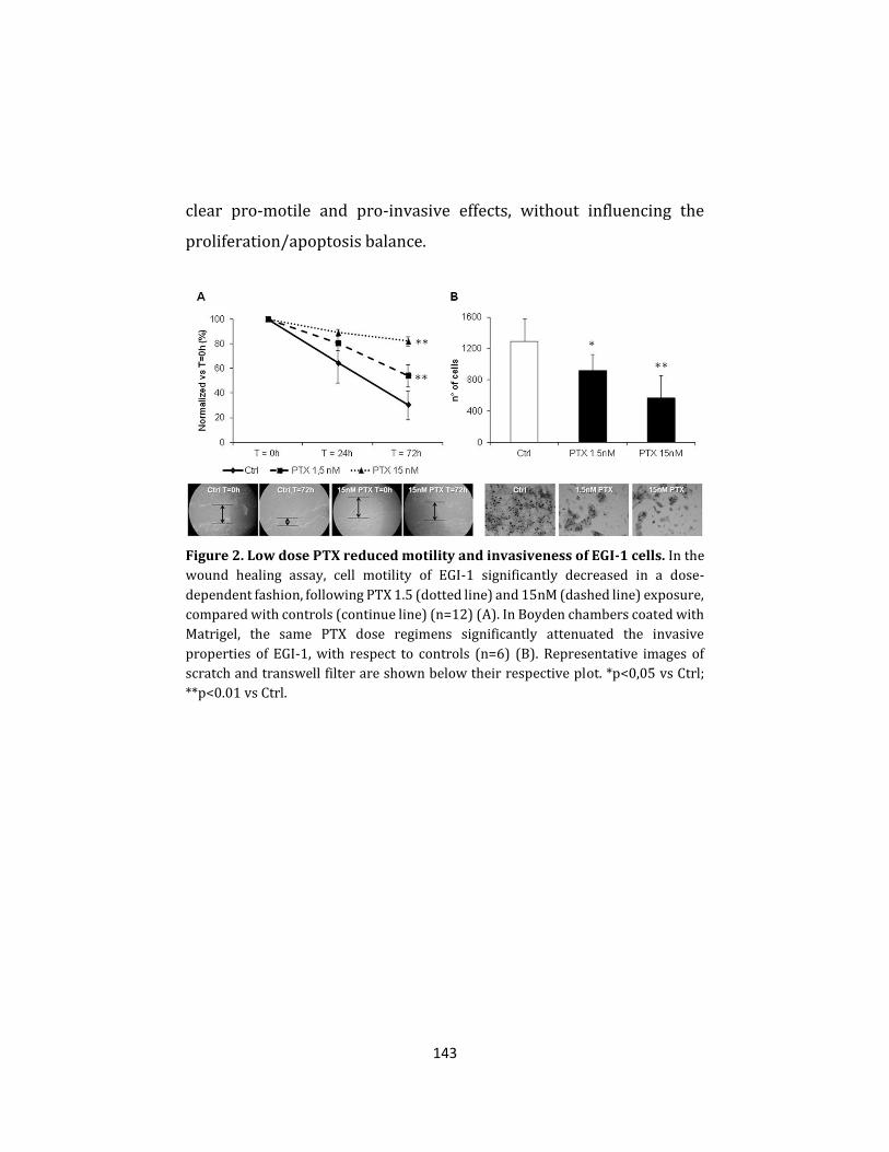
143
clear pro-motile and pro-invasive effects, without influencing the
proliferation/apoptosis balance.
Figure 2. Low dose PTX reduced motility and invasiveness of EGI-1 cells. In the
wound healing assay, cell motility of EGI-1 significantly decreased in a dose-
dependent fashion, following PTX 1.5 (dotted line) and 15nM (dashed line) exposure,
compared with controls (continue line) (n=12) (A). In Boyden chambers coated with
Matrigel, the same PTX dose regimens significantly attenuated the invasive
properties of EGI-1, with respect to controls (n=6) (B). Representative images of
scratch and transwell filter are shown below their respective plot. *p<0,05 vs Ctrl;
**p<0.01 vs Ctrl.

144
Figure 3. In contrast with high doses, low dose PTX did not affect cell
proliferation, viability, and apoptosis of EGI-1 cells. Effects of low dose PTX (1.5,
15nM) on cell proliferation (BRDU incorporation, A), viability (MTS assay, B), and
apoptosis (CC3 immunofluorescence, C) were evaluated in EGI-1 and compared with
effects of higher doses (150, 1500nM) and with untreated cells. In contrast with the
highest doses, these cell activities were not affected by low dose PTX (n=6 in all
experiments). **p<0.01 vs Ctrl.
Low dose PTX reduced Rho-A and Cdc-42 activation and MMP-9
secretion in EGI-1 cells. To better understand the mechanisms
promoting cell motility and invasiveness dependent upon S100A4
nuclearization, we turned to study the effects of low dose PTX on the
activity of small Rho GTPases, the expression of MT1–MMP and the
secretion of MMP-9. Small Rho GTPases are key effectors of cell
motility by inducing the formation of stress fibers (Rho-A),

145
lamellipodia (Rac-1) and filopodia (Cdc-42) [20]. MMP-9 on the other
hand, is a proteolytic enzyme secreted by many cancer cell types: it
potently stimulates matrix degradation, facilitating the invasive
migration of tumoral cells from the primary site of growth [21]. Its
activation depends upon the expression of membrane-anchored MMP,
particularly of MT1-MMP, whose expression at the surface of cancer
cells is critical for breaking the basement membrane [22]. Since EGI-1
phenocopied the behavior of primary CCA-TV3 cell line, the following
experiments were performed in EGI-1 only. As compared with
controls, EGI-1 treated with low dose PTX showed a significant
reduction in Rho-A (Fig. 4A) and Cdc-42 (Fig. 4B) GTP levels, but not
in Rac-1 (Supplementary Fig. 4). As evaluated by WB and
immunofluorescence on cultured EGI-1, MT1-MMP expression
significantly decreased after PTX treatment (inhibition of 40 and 46%
with 1.5, and of 50 and 49% with 15nM, for WB and
immunofluorescence, respectively) (Fig. 4C–D). Similarly, MMP-9
secretion and activation were inhibited by challenging CCA cell
cultures with PTX, as shown by ELISA (inhibition of 57% with 1.5, and
of 72% with 15nM) and gel zymography (Fig. 4E–F).

146
Figure 4. Low dose PTX inhibited Rho-A and Cdc-42 GTP levels, together with
MT1-MMP expression and MMP-9 secretion. By G-LISA assay, low dose PTX (1.5,
15nM) significantly reduced Rho-A (A) and Cdc-42 (B) GTP levels in EGI-1, with
respect to controls (n=3, in duplicate). Low dose PTX strongly inhibited MT1-MMP
expression (WB, C), specifically on its membrane localization (fluorescence intensity
profile, D) (n=3, for both experiments). Low dose PTX blunted both secretion (ELISA,
E) (n=5, in duplicate) and activation (gelatin zymography, F) of MMP-9. *p<0.05 vs
Ctrl, **p<0.01 vs Ctrl.
Low dose PTX did not induce cytoskeletal damage in EGI-1 cells.
S100A4 is normally associated to cytoskeletal fibers. To study if low
dose PTX altered cytoskeletal integrity, we performed phalloidin

147
fluorescence in dose-dependent experiments, ranging from 1.5–15nM
to 150–1500nM. In contrast with high dose PTX, which induced actin
fiber changes (shortening, thickening, fragmentation often leading to
dense coiling, accumulating in the perinuclear area) in 16% (150nM)
and 32% (1500nM) of cultured EGI-1, more than 90% of cells treated
with low dose PTX showed preserved cytoskeletal structure
(Supplementary Fig. 5). These data indicate that effects of low dose
PTX on cell motility and invasiveness are not due to a cytoskeletal
damage. Next, we sought to understand the mechanisms regulating the
nuclear import of S100A4.
PTX selectively reduced the SUMOylation fraction of S100A4, a
critical mechanism for cell invasiveness of EGI-1 cells. Post-
translational modification by small ubiquitin-like modifier (SUMO) of
target proteins is an important mechanism directing their intracellular
shuttling. We showed that EGI-1 contained much higher amounts of
SUMOylated S100A4 than TFK-1, a CCA cell line expressing S100A4
only in the cytoplasm, where instead un-SUMOylated S100A4 was
detected (Supplementary Fig. 6A). Furthermore, after treatment with
low dose PTX, EGI-1 showed a marked reduction selectively in the
SUMOylated fraction of S100A4 as compared with controls, whereas
the S100A4 unSUMOylated levels remained unchanged (Fig. 5A).
SUMO inhibition by PTX was not associated with decreased mRNA
expression levels of the three SUMO E components of the SUMOylating

148
complex (Supplementary Fig. 6B–D). To study if inhibition of
SUMOylation halted cell invasive capabilities, EGI-1 were treated with
GA, a specific natural inhibitor of the E1 subunit. Preliminary dose-
response experiments to assess toxicity levels of GA on EGI-1 cells
identified 1µM as the dose devoid of effects on cell viability (not
shown), and therefore used onwards. Consistent with our hypothesis,
GA significantly reduced cell motility of EGI-1 with respect to controls
(Fig. 5B), an effect associated with a significant reduction (37%) in the
nuclear expression of S100A4 of an extent comparable to PTX 1.5nM,
without affecting the cytoplasmic levels of S100A4 (Fig. 5C,D).

149
Figure 5. SUMOylation of S100A4 was modulated by low dose PTX and led
motility and S100A4 nuclear import in EGI-1 cells. By SUMOylation assay, EGI-1
exposed to PTX 1.5 and 15nM showed a stark reduction in the amount of SUMOylated
S100A4 compared with controls, without changes in the total amount of un-
SUMOylated S100A4 protein (flow through) (A). PTX effects were reproduced by
ginkolic acid (GA, 1µM), a specific SUMOylation inhibitor, which significantly reduced
EGI-1 cell migration (B, dotted line, n=12). This effect was associated with a
significant decrease in the nuclear S100A4 (C) compared with controls, whilst the
cytoplasmic fraction was unaffected (D). Below each column plot, representative
blots of S100A4 together with histone H3 (nuclear marker) and GAPDH (cytoplasmic
marker), are shown (n=6). *p<0.05 vs Ctrl.
PTX treatment reduced lung metastasization but not the tumor
growth at the site of injection in the experimental model of CCA.
Altogether, the in vitro data suggest that the SUMOylation-dependent
nuclear import of S100A4 is indeed a mechanistic determinant of the
invasive phenotype of CCA cells, which can be inhibited by PTX at nM
doses without altering the cytoskeletal integrity, as well as the
proliferation and/or apoptosis activities. To test in vivo whether
targeting nuclear S100A4 by low dose PTX is therapeutically relevant
to reduce CCA invasiveness, we moved to the experimental model of
CCA generated by EGI-1 cell xenotransplantation in the SCID mouse
[9,13]. To reproduce the small nM doses of PTX able to hamper S100A4
nuclear entry in CCA cells in vitro, a low dose metronomic regimen was
chosen for drug infusion in xenografted mice (LDM PTX). To assess
specific effects on cancer invasiveness, the treatment was started upon
successful EGI-1 cell engraftment as confirmed by bioluminescence
imaging (on average 29.75±4.53 days after intrasplenic injection).

150
Notably, before starting LDM PTX, the levels of photon emission were
comparable between the two groups. With respect to control animals,
LDM PTX did not significantly reduce neither photon emission from
the spleen through the treatment time (Supplementary Fig. 7A,B), nor
the size of the splenic tumor mass at the time of sacrifice
(Supplementary Fig. 7C). Immunohistochemistry in tissue sections
obtained from the tumor mass confirmed that LDM PTX was effective
in decreasing S100A4 expression in the nucleus of engrafted EGI-1
cells compared with controls (Fig. 6A), however, the expression of p-
Hist3 (proliferation marker) and CC3 (apoptosis marker) did not
significantly differ between LDM PTX-treated and control mice (Fig.
6B,C). These findings indicate that LDM PTX reproduces the down-
modulating effects on S100A4 nuclear expression by EGI-1 shown in
vitro, without causing a dysregulation in the proliferation/apoptosis
balance of CCA cells. In contrast, LDM PTX significantly halted
metastatic dissemination. Immunohistochemistry for human
mitochondria revealed that both ITC and MM in the lung were
significantly reduced by PTX treatment (Fig. 7A,B). These data further
prove the functional impact of S100A4 nuclear expression on CCA
biology as mechanism specifically driving hematogenous
metastasization, without stimulating tumorigenesis.

151
Figure 6. LDM PTX reduced nuclear S100A4 expression without affecting
proliferation and apoptosis of xenotransplanted EGI-1 cells, in vivo. Nuclear
immunoreactivity for S100A4 was significantly reduced in the tumor mass at the site
of injection (spleen) of LDM PTX-treated mice (n=10) with respect to vehicle-treated
controls (n=14) (A). Conversely, LDM PTX did not modify neither cell proliferation
(immunohistochemistry for p-Hist3, B) nor apoptosis (immunohistochemistry for
CC3, C) in the splenic mass compared with controls. Representative micrographs of
spleen sections immunostained for S100A4, p-Hist3 and CC3 are shown in the right
side of the plot (immunoperoxidase; A, M=100×; B, C, M=200×; insets, M=200×).
**p<0.01 vs Ctrl.

152
Figure 7. LDM PTX inhibited lung metastatic colonization of EGI-1 cells, in vivo.
A significant reduction in the number of both ITC (A) and MM (B) was found in
treated mice (n=10) with respect to controls (n=14). Representative micrographs of
ITC and MM (black arrows) derived from EGI-1 cell dissemination to the lung
parenchyma after xenograft in SCID mice undergoing LDM PTX and in controls,
identified by the specific immunoreactivity for human mitochondria, are shown
below their respective dot plot. *p<0.05 vs Ctrl; Immunoperoxidase; original
magnification, M=400× (ITC), M=200× (MM).
DISCUSSION
An unmet need in CCA, as in other malignancies whose dismal
prognosis relates to limited therapeutic approaches, is the
development of biomarkers able to identify patients most likely to take
advantage of curative treatments. Biomarkers may also represent
disease-relevant targets for therapeutic interventions [9]. Our
previous studies showed that nuclear expression of S100A4 in cancer
cells of resected CCA (nearly a half) identified a more invasive clinical
phenotype, characterized by increased metastasization and reduced

153
survival after surgery [9]. Of note, a worse prognosis after surgery was
still observed even when S100A4 nuclear expression was scattered,
limited to less than 30% of the neoplastic ducts [9]. The aim of the
present study was to understand the mechanisms by which nuclear
expression of S100A4 promotes cancer invasiveness, and to elucidate
the mechanism regulating nuclear translocation of S100A4. We also
wanted to understand if these mechanisms are putative target for
therapeutic intervention.
We initially found that PTX given in vitro at low doses (1.5 and
15nM) was able to effectively and selectively down-regulate S100A4
expression in the nucleus of CCA cells, leaving cytoplasmic expression
unaffected. Then, by combining in vitro and in vivo techniques, we
showed that: a) PTX-induced down-regulation of nuclear S100A4 was
associated with a reduction in motility and invasiveness of CCA cells,
in activity of Rho-A and Cdc-42, and in secretion of MMP-9; b) at the
doses able to down-regulate nuclear expression of S100A4, PTX did
not affect cell proliferation, apoptosis, and cytoskeletal architecture; c)
the nuclear translocation of S100A4 was regulated by SUMOylation, a
post-translational mechanism that was affected by PTX; d) in SCID
mice xenografted with human nuclear S100A4-expressing CCA cells,
down-regulation of nuclear S100A4 by LDM PTX was associated with
a reduction in hematogenous metastasization.
The biological functions of S100A4 are largely unknown. They
depend on its interacting partners, which are mainly located in the

154
cytoplasm, where S100A4 is commonly expressed. In the nucleus,
interacting partners have not been characterized yet, opening the
possibility that nuclear S100A4 may act independently as
transcription factor [7].
Earlier studies showed that in mouse melanoma cells, taxol at
conventional doses, reduced the total amount of S100A4, an effect
associated with maintenance in the G0 phase of the cells and increased
expression of p53 [10,11]. We initially found that in CCA cells, low
doses of PTX (1.5–15nM) induced a marked reduction in S100A4
selectively in the nucleus, without altering its cytoplasmic expression.
This effect, obtained in an established CCA cell line (EGI-1) was
reproduced in a primary CCA cell line derived from a patient
undergoing surgical resection. The nuclear down-regulation of
S100A4 was biologically relevant since it associated with a strong
inhibition of the motile and invasive properties displayed by CCA cells
in culture. Interestingly, at the same small doses, PTX did not exert
anti-proliferative and pro-apoptotic functions, nor it affected cell
viability or the integrity of the actin cytoskeletal filaments of cultured
CCA cells. All these cytotoxic effects were instead induced by PTX at
higher doses (150–1500nM), coupled with pronounced anti-
proliferative and pro-apoptotic activities, in line with the mechanism
of action supporting the current indications of PTX for the
chemotherapeutic treatment of several aggressive carcinomas, from
ovary, to breast and thyroid cancer [23,24]. The lack of pro-

155
proliferative stimuli when S100A4 translocates into the nucleus has
been recently shown also in colorectal cancer cells [25]. In this study,
although S100A4 translocated into the nucleus in a cell cycle-
dependent fashion, being most prominent in the G2/M phase,
lentiviral silencing of nuclear S100A4 did not induce changes in cell
proliferation. Our findings are consistent with these observations, and
confirm that nuclear S100A4 confers specific pro-invasive functions.
Among putative molecular players mediating the effects of
nuclear S100A4 on cell motility and invasiveness, we focused on the
small G proteins (GTPases) belonging to the Rho family and on matrix
metalloproteinases (MMPs). The small Rho GTPases are recognized as
key effectors able to activate invasion and metastasis programs
[20,26]. In EGI-1 cells, inhibition of S100A4 nuclear import by low dose
PTX significantly reduced the activation of Rho-A and Cdc-42. In cancer
cells, Cdc-42 is one of the factors involved in the formation of
specialized plasma-membrane actin-based microdomains combining
adhesive properties with matrix degrading activities, called
invadopodia, which support cancer invasion by dismantling the
basement membrane and then by invading the stromal environment
mostly composed of fibrillar type I collagen. A main component of
invadopodia is the transmembrane metalloproteinase MT1-MMP,
whose function is essential for the in situ to invasive carcinoma
transition in breast cancer [14]. MT1-MMP promotes the activation of
several soluble MMPs, such as MMP-2 and MMP-9. Our data indicate

156
that EGI-1 cells constitutively expressed MT1-MMP and MMP-9, but
not MMP-2, and when exposed to low dose PTX, they showed a
significant dose-dependent reduction in the surface expression of
MT1-MMP along with the ability to secrete MMP-9. Interestingly, the
interplay between Rho-A and Cdc-42 regulates the delivery and
accumulation of MMPs at the invading surface [27]. Furthermore, since
MMP-9 secretion can be also modulated by the activation of the Rho-
A/ROCK signaling [21], and the membrane translocation of MT1-MMP
is directly regulated by Cdc-42 [28], we suggest the cooperation of
Rho-A/MMP-9 and Cdc-42/MT1-MMP pathways to form invadopodia
activated by nuclear S100A4.
Cytoskeletal damages induced by PTX, a well-established
depolymerizing agent [29], could in theory influence the nuclear
translocation of S100A4. From this point of view, it is important to
underline that structural cytoskeleton alterations were absent with
low dose PTX, in contrast with what coherently observed with high
dose PTX. The mechanisms regulating S100A4 entry into the nucleus
remain enigmatic. Notably, S100A4 does not possess nuclear docking
sites or nuclear import sequences. In human chondrocytes,
nuclearization of S100A4 was induced by IL-1β through a
SUMOylation-dependent mechanism [30]. SUMOylation is a post-
translational mechanism similar to ubiquitination, operated by SUMO
proteins, and involved in several biological functions, including
protein stability, DNA repair, cell cycle regulation, apoptosis, nuclear

157
transport and gene transcription [31]. The SUMOylation processes are
catalyzed by three enzymes, SUMO E1, E2 and E3, acting as pacemaker
of the cascade reaction leading to the modification of their substrate
protein [32]. Components of the SUMOylation machinery have been
found deregulated in several human cancers, and are emerging as
relevant players in tumour invasiveness and in epithelial-
mesenchymal transition [33]. We found that EGI-1 (expressing
S100A4 the nucleus) contained higher amounts of SUMOylated
S100A4 with respect to TFK-1 (expressing S100A4 only in the
cytoplasm). Then, in EGI-1 treated with low dose PTX, down-
regulation of nuclear S100A4 was associated with a significant, dose-
dependent reduction in the S100A4 SUMOylated fraction, without
changes in the un-SUMOylated subset. These effects occurred without
affecting the expression levels of E1, E2, E3 subunits of the
SUMOylating complex, thus suggesting a functional inhibitory
mechanism, similar to GA. GA is a natural compound derived from
Gingko biloba that specifically inhibits the first step of the
SUMOylation reaction by directly binding E1 and inhibiting the
formation of the E1-SUMO intermediate [16]. Of note, also the
reduction in nuclear levels of S100A4 induced by GA significantly
inhibited EGI-1 cell motility, to an extent comparable to PTX. This
observation confirms the relevance of SUMOylation in mediating the
pro-oncogenic functions of S100A4 when translocated into the
nucleus.

158
To translate the results of the in vitro experiments, we turned
to an experimental model of CCA generated by xenotransplantation of
human EGI-1 cells into the spleen of SCID mice, as we performed in
previous studies [9,13]. Because of its short half-life [34], PTX was
administered to xenografted SCID mice with a LDM infusion.
Continuous delivery of PTX by LDM ensured the achievement of
constant low concentrations of PTX comparable with the small doses
used for the in vitro experiments [34]. To evaluate specific effects on
tumor invasion, LDM PTX was started only after human CCA cell
engraftment was confirmed by bioluminescence imaging. At the end of
treatment, histological evaluation of the splenic tumor showed in the
LDM PTX-treated group, a significant reduction in the amount of
nuclear S100A4-expressing EGI-1 cells. In line with what observed in
vitro, down-regulation of S100A4 was not associated with reduced
tumour cell proliferation (p-Hist3) or with increased apoptosis (CC3)
of CCA cells. Therefore, our experimental model is ideal to study if
pharmacologic targeting of nuclear S100A4 is a useful strategy to
inhibit invasiveness and metastasization. We found that LDM PTX did
not affect the growth of the tumor at the site of injection, but caused a
significant reduction in both ITC and MM in the lungs, where EGI-1
cells metastatise following a hematogenous route through the portal
vein and the hepatic veins. This finding is of great clinical value,
because the lung is the site most frequently involved in the
159
extrahepatic progression of CCA, fostered by a specific mechanism of
vascular encasement by tumor cells [5].
It is important to underline that PTX may exert additional
functions not related to modulation of S100A4 nuclear transport that
potentially may contribute to its anti-invasive effects. These are largely
dependent upon PTX ability to promote microtubule polymerization
and stabilization, which inhibits mitosis and leads to apoptosis. While
these mechanisms of action are well evident at the conventional doses,
at the much lower doses used in the current study, they do not seem to
occur. Recent studies performed in ovarian carcinoma cells indicate
that alternatively, PTX may inhibit the expression of other critical
molecular factors of tumor progression, such as hypoxia-inducible
factor-1α and vascular endothelial growth factor [35].
In conclusion, this study unveils a specific role of SUMOylation-
dependent nuclear import of S100A4 in CCA on hematogenous
metastasization. The small GTPases Cdc-42 and Rho-A, in concert with
MT1-MMP and MMP-9, are the molecular effectors mediating the pro-
invasive functions promoted by S100A4 nuclearization. Bearing in
mind the potential toxic effects of PTX in patients with overt cirrhosis
and cholestasis [36], these mechanisms represents a promising
therapeutic target aimed at preventing metastatic dissemination after
detection of the tumor.
Conflict of interest: All authors have no conflict of interest regarding this study to declare.

160
Financial support: Progetto di Ricerca Ateneo 2011 (Grant #CPD113799/11) to L. Fabris and M. Cadamuro. Grant Associazione Chirurgica Tarvisium to M. Massani, T. Stecca and N. Bassi. Grant Associazione Italiana Ricerca sul Cancro (AIRC) #IG14295 to S. Indraccolo. Projects CARIPLO 2011-0470 and PRIN 2009ARYX4T_005 to M. Strazzabosco. NIH Grants DK079005 to M. Strazzabosco, RO1DK 101528 to C. Spirli, and DK034989 Silvio O. Conte Digestive Diseases Research Core Centers to M. Strazzabosco and C. Spirli.
REFERENCES
1. Blechacz B, Gores GJ. Cholangiocarcinoma: advances in pathogenesis,
diagnosis, and treatment. Hepatology. 2008;48:308–321.
2. Khan SA, Thomas HC, Davidson BR, Taylor-Robinson SD.
Cholangiocarcinoma. Lancet. 2005;366:1303–1314.
3. Sempoux C, Jibara G, Ward SC, Fan C, Qin L, Roayaie S, et al. Intrahepatic
cholangiocarcinoma: new insights in pathology. Semin Liver Dis.
2011;31:49–60.
4. Valle J, Wasan H, Palmer DH, Cunningham D, Anthoney A, Maraveyas A, et
al. Cisplatin plus gemcitabine versus gemcitabine for biliary tract cancer. N
Engl J Med. 2010;362:1273–1281.
5. Fabris L, Alvaro D. The prognosis of peri-hilar cholangiocarcinoma after
radical treatments. Hepatology. 2012;56:800–802.
6. Boye K, Maelandsmo GM. S100A4 and metastasis: a small actor playing
many roles. Am J Pathol. 2010;176:528–535.
7. Saleem M, Kweon MH, Johnson JJ, Adhami VM, Elcheva I, Khan N, et al.
S100A4 accelerates tumorigenesis and invasion of human prostate cancer
through the transcriptional regulation of matrix metalloproteinase 9. Proc
Natl Acad Sci U S A. 2006;103:14825–14830.

161
8. Gongoll S, Peters G, Mengel M, Piso P, Klempnauer J, Kreipe H, et al.
Prognostic significance of calcium-binding protein S100A4 in colorectal
cancer. Gastroenterology. 2002;123:1478–1484.
9. Fabris L, Cadamuro M, Moserle L, Dziura J, Cong X, Sambado L, et al. Nuclear
expression of S100A4 calcium-binding protein increases
cholangiocarcinoma invasiveness and metastasization. Hepatology.
2011;54:890–899.
10. Lakshmi MS, Parker C, Sherbet GV. Metastasis associated MTS1 and NM23
genes affect tubulin polymerisation in B16 melanomas: a possible
mechanism of their regulation of metastatic behaviour of tumours.
Anticancer Res. 1993;13:299–303.
11. Parker C, Lakshmi MS, Piura B, Sherbet GV. Metastasis-associated mts1
gene expression correlates with increased p53 detection in the B16 murine
melanoma. DNA Cell Biol. 1994;13:343–351.
12. Horwitz SB. Taxol (paclitaxel): mechanisms of action. Ann Oncol.
1994;5:S3–S6.
13. Cadamuro M, Nardo G, Indraccolo S, Dall'olmo L, Sambado L, Moserle L,
et al. Platelet-derived growth factor-D and Rho GTPases regulate recruitment
of cancer-associated fibroblasts in cholangiocarcinoma. Hepatology.
2013;58:1042–1053.
14. Lodillinsky C, Infante E, Guichard A, Chaligné R, Fuhrmann L, Cyrta J, et al.
p63/MT1-MMP axis is required for in situ to invasive transition in basal-like
breast cancer. Oncogene. 2016;35:344–357.
15. Rozelle AL, Machesky LM, Yamamoto M, Driessens MH, Insall RH, Roth
MG, et al. Phosphatidylinositol 4,5-bisphosphate induces actin-based
movement of raft-enriched vesicles through WASP-Arp2/3. Curr Biol.
2000;10:311–320.

162
16. Fukuda I, Ito A, Hirai G, Nishimura S, Kawasaki H, Saitoh H, et al. Ginkgolic
acid inhibits protein SUMOylation by blocking formation of the E1-SUMO
intermediate. Chem Biol. 2009;16:133–140.
17. Hermanek P, Hutter RV, Sobin LH, Wittekind C. International Union
Against Cancer. Classification of isolated tumor cells and micrometastasis.
Cancer. 1999;86:2668–2673.
18. Amin BD, Hoda SA. Minimal metastatic disease in sentinel lymph nodes in
breast carcinoma: some modest proposals to refine criteria for "isolated
tumor cells". Adv Anat Pathol. 2006;13:185–189.
19. Zhang ZY, Ge HY. Micrometastasis in gastric cancer. Cancer Lett.
2013;336:34–45.
20. Raftopoulou M, Hall A. Cell migration: Rho GTPases lead the way.
Developmental Biology. 2004;265:23–32.
21. Turner NA, O'Regan DJ, Ball SG, Porter KE. Simvastatin inhibits MMP-9
secretion from human saphenous vein smooth muscle cells by inhibiting the
RhoA/ROCK pathway and reducing MMP-9 mRNA levels. FASEB J.
2005;19:804–806.
22. Gialeli C, Theocharis AD, Karamanos NK. Roles of matrix
metalloproteinases in cancer progression and their pharmacological
targeting. FEBS J. 2011;278:16–27.
23. Cresta S, Sessa C, Catapano CV, Gallerani E, Passalacqua D, Rinaldi A, et al.
Phase I study of bortezomib with weekly paclitaxel in patients with advanced
solid tumours. Eur J Cancer. 2008;44:1829–1834.
24. Ain KB, Egorin MJ, DeSimone PA. Treatment of anaplastic thyroid
carcinoma with paclitaxel: phase 2 trial using ninety-six-hour infusion.
Collaborative Anaplastic Thyroid Cancer Health Intervention Trials
(CATCHIT) Group. Thyroid. 2000;10:587–594.

163
25. Egeland EV, Boye K, Pettersen SJ, Haugen MH, Øyjord T, Malerød L, et al.
Enrichment of nuclear S100A4 during G2/M in colorectal cancer cells:
possible association with cyclin B1 and centrosomes. Clin Exp Metastasis.
2015;32:755–767.
26. Franco-Barraza J, Valdivia-Silva JE, Zamudio-Meza H, Castillo A, García-
Zepeda, Benítez-Bribiesca L, et al. Actin Cytoskeleton Participation in the
Onset of IL-1β Induction of an Invasive Mesenchymal-like Phenotype in
Epithelial MCF-7 Cells. Archives of Medical Research. 2010;41:170–181.
27. Spuul P, Ciufici P, Veillat V, Leclercq A, Daubon T, Kramer IJ, et al.
Importance of RhoGTPases in formation, characteristics, and functions of
invadosomes. Small GTPases. 2014;5:e28195.
28. Ispanovic E, Serio D, Haas TL. Cdc42 and RhoA have opposing roles in
regulating membrane type 1-matrix metalloproteinase localization and
matrix metalloproteinase-2 activation. Am J Physiol Cell Physiol.
2008;295:C600–C610.
29. Zhang Y, Zang H, Zang F, Liu S, Wang R, Sun Y, et al. Folate-targeted
paclitaxel-conjugated polymeric micelles inhibits pulmonary metastatic
hepatoma in experimental murine H22 metastasis model. Int J Nanomed.
2014;9:2019–2030.
30. Miranda KJ, Loeser RF, Yammani RR. SumoylSUMOylation and nuclear
translocation of S100A4 regulate IL-1beta-mediated production of matrix
metalloproteinase-13. J Biol Chem. 2010;285:31517–31524.
31. Pichler A, Melchior F. Ubiquitin-related modifier SUMO1 and
nucleocytoplasmic transport. Traffic. 2002;3:381–387.
32. Johnson ES. Protein modification by SUMO. Annu Rev Biochem.
2004;73:355–382.

164
33. Bogachek MV, De Andrade JP, Weigel RJ. Regulation of epithelial-
mesenchymal transition through SUMOylation of transcription factors.
Cancer Res. 2015;75:11–15.
34. Stearns ME. Taxol reduces circulating tumor cells to prevent bone
metastases in SCID mice. Invasion Metastasis. 1995;15:232–241.
35. Kim BR, Yoon K, Byun HJ, Seo SH, Lee SH, Rho SB. The anti-tumor activator
sMEK1 and paclitaxel additively decrease expression of HIF-1α and VEGF via
mTORC1-S6K/4E-BP-dependent signaling pathways. Oncotarget.
2014;5:6540–6551.
36. Xie JD, Huang Y, Chen DT, Pan JH, Bi BT, Feng KY, et al. Fentanyl Enhances
Hepatotoxicity of Paclitaxel via Inhibition of CYP3A4 and ABCB1 Transport
Activity in Mice. PLoS One. 2015;10:e0143701.
SUPPLEMENTARY MATERIALS AND METHODS
Cell apoptosis. Since in some treatments with rapid cytotoxicity,
reduction in mitochondrial activity does not affect cell survival and
therefore, analysis of cell viability by MTS might underestimate the
number of dead cells, cell apoptosis was assessed by
immunofluorescence for cleaved caspase 3 (CC3, 1:500, rabbit, Cell
Signaling) and then developed. Results were expressed as percentage
of cells with positive cytoplasmic staining for CC3 on the total number
of cells, in 20 random fields taken at 200x magnification, by two
independent observers (MC, LS) as previously described [9].

165
Cell migration (wound healing) assay. CCA cell lines were seeded in
a 6-well plate, grown until confluence, and then starved for 24h. Each
cell monolayer was scratched three times with a sterile p200 tip, and
three micrographs were taken at t=0h for each wound. Then, on the
same scratched area, micrographs were taken again at 24h and 72h to
measure the distance between the wound margins using LuciaG 5.0
software (Nikon), and expressed by normalizing each time point to
t=0h [9,13].
Membrane-type 1 (MT1) matrix metalloproteinase (MMP)
expression. To assess the membrane expression levels of MT1-MMP,
immunofluorescence staining was analyzed using the LuciaG 5.0
software (Nikon). After selecting a region of interest between two
adjacent tumor cells, the relative labeling intensity was evaluated on
either red (MT1-MMP, red line) or blue (DAPI, nuclei, blue line)
channels, and the higher peak of red fluorescence was measured
(Supplemental Fig. 4B).
Activity of Rho-A, Rac-1 and Cdc-42 GTPase. CCA cells were seeded
and cultured for three days to reach at least the 70% of confluence to
assess Rho-A and Rac-1 GTP levels, and the 20-30% of confluence for
Cdc-42 GTP levels. Then, confluent cells were exposed to PTX, and
proteins were extracted to perform the G-LISA assay according to the
supplier (Cytoskeleton Inc.).

166
MMP-9 secretion. Fifty-thousand cells were seeded on a 24-well plate
and then treated with PTX. Supernatant was harvested, stored at -
80°C, and ELISA performed to quantify both the pro and the active
MMP-9 forms (RayBiotech).
Gelatin zymography assay. One hundred µg of total protein lysate
was loaded on each well of a 10% Tris-Glycine + 0.1% Gelatin gel
(LifeTechnologies) and proteins were run for 90mins using a Tris-
Glycine Buffer. Gels were then incubated with renaturing buffer for
30min at room temperature (LifeTechnologies), and incubated in
developing buffer (LifeTechnologies) for 4h at 37°C. Finally, gels were
stained for 30min with Coomassie Brilliant blue (Bio-Rad) and
pictures were taken using the Gel Logic 100 Imaging System (Kodak).
Immunohistochemistry for S100A4, p-Hist3 and CC3 in SCID mice
spleen specimens. The sections derived from spleen were
immunostained for S100A4 (1:400), p-Hist3 (1:50, rabbit, Cell
Signaling), and CC3 (1:300), developed with DAB and H2O2 0.01% and
counterstained with Gill’s Haematoxylin N°2 (Sigma). All antibodies
were diluted in PBS 1M, and supplemented with 1% BSA and 0.05%
Tween20 (Sigma). Results were expressed as percentage of positive
cells/field at 200x in 10 randomly taken fields, as previously
performed [9]. Positive staining for S100A4 and p-Hist3 was
considered as expression of intense brown staining in the nucleus,

167
whereas positivity for CC3 was represented by granular brown
staining in the cytoplasm of EGI-1 cells.
SUPPLEMENTARY FIGURES
Supplementary Figure 1. Schedule of treatment with low dose metronomic PTX
and assessment by bioluminescence analysis in SCID mice xenografted with
EGI-1 cells. See text for detail.

168
Supplementary Figure 2. Low dose PTX reduced motility and invasiveness of
CCA-TV3 cells. Consistent with data obtained with EGI-1 cells, by wound-healing
assay, cell motility of CCA-TV3 cells significantly decreased, in a dose-dependent
fashion, following PTX 1.5 (dotted line) and 15nM (dashed line) exposure compared
with untreated controls (continue line) (n=4) (A). Similar to EGI-1 cells, the same
PTX dose regimens significantly blunted the invasive abilities of CCA-TV3 cells, with
respect to controls in Boyden chambers coated with Matrigel, (n=3) (B).
Representative images of scratch and transwell filter are shown below their
respective plot. *p<0,05 vs Ctrl; **p<0.01 vs Ctrl.

169
Supplementary Figure 3. In contrast with high doses, low dose PTX did not
affect cell proliferation, viability, and apoptosis of CCA-TV3 cells. Concomitant
effects of low dose PTX (1.5 and 15nM) on cell proliferation (BRDU incorporation,
A), viability (MTS assay, B), and apoptosis (CC3 immunofluorescence in cultured
cells, C) were also evaluated in CCA-TV3 cells and compared with the effects of higher
doses (150 and 1500nM) and with untreated controls. In contrast with high doses,
these cell activities were not affected by low dose PTX (n=3 in all experiments), in
accordance with what observed in EGI-1 cells. **p<0.01 vs Ctrl.

170
Supplementary Figure 4. Low dose PTX did not affect Rac-1 GTP levels, while it
reduced the MT1-MMP membrane expression. By G-LISA assay, low dose PTX
(1.5 and 15nM) did not modify Rac-1 GTP levels in EGI-1 cells, with respect to
untreated controls (A, n=3, in duplicate). In contrast, the same PTX doses
significantly blunted the expression of MT1-MMP (red) at the membrane level of EGI-
1 cells, as shown by immunofluorescence analysis of cultured cells; representative
plots are given above each micrograph. Red line depicts the fluorescence intensity
profile of MT1-MMP through the cell area, while blue line represents the intensity of
nuclear staining (DAPI); peak of red line corresponds to the lowest level of blue
staining, according to a membrane localization of MT1-MMP. Original magnification:
400x (B).

171
Supplementary Figure 5. Low dose PTX did not affect cytoskeletal integrity of
EGI-1 cells. By phalloidin staining, only a small percentage of EGI-1 cells challenged
with low dose PTX (1.5, 15nM), displayed structural alterations of the actin
filaments, which instead were observed in a larger subset of EGI-1 cells exposed to
high dose PTX (150, 1500nM). No differences in damaged cells between low dose
PTX and untreated cells could be observed (n=3, percentage of apoptotic cells
expressed as black area of the column) (A). Representative immunofluorescence for
FITC-conjugated phalloidin (green) in cultured cells treated with increasing doses of
PTX are shown below each column plot; perinuclear condensation, a hallmark of
cytoskeletal disaggregation, is clearly observed in single cells treated with PTX
150nM (white arrow), whereas cytoplasm shrinkage is evident in cells treated with
PTX 1500nM. Original magnification, M=400x (B). WB for β-tubulin in microtubule
fractions purified by ultracentrifugation shows lack of expression indicating
cytoskeletal damage in cells exposed to PTX 150 e PTX 1500nM (C).

172
Supplementary Figure 6. EGI-1 cells but not TFK-1 cells contained SUMOylated
S100A4; inhibitory effects of low dose PTX on SUMOylating complex did not
associate with any deregulation of SUMO E subunits. By SUMOylation assay,
SUMOylated S100A4 could be detected only in EGI-1 cells (expressing S100A4 in the
nucleus), whereas it was absent in TFK-1 cells, a CCA cell line where S100A4
expression is limited to the cytoplasm. Accordingly, un-SUMOylated S100A4 could
be detected in either cell lines, as shown by the band present in the flow through
fraction (A). By Real time PCR, mRNA expression levels of each SUMOylating complex
subunit in EGI-1 cells (B, E1; C, E2; D, E3) were not affected by treatment with low
dose PTX.

173
Supplementary Figure 7. LDM PTX did not affect the splenic tumor mass in SCID
mice xenotransplanted with EGI-1 cells, in vivo. SCID mice xenografted by
intrasplenic injection of EGI-1 cells transduced with a lentiviral vector encoding the
firefly luciferase gene were treated with LDM PTX or vehicle after confirming tumor
engraftment by bioluminescence imaging. No statistically significant differences
between LDM PTX treated mice and controls were observed both in the level of
photon emission (A, representative images taken at the beginning (T0) and at the
end (T2) of treatment; B, bioluminescence curve during the treatment) and in the
size of the splenic tumor mass measured at necroscopic examination (C).

174
CHAPTER 4
Platelet-derived growth factor D enables
cancer-associated fibroblasts to promote tumor
lymphangiogenesis in cholangiocarcinoma
Massimiliano Cadamuro1,2, Simone Brivio1, Marta Vismara1, Joachim
Mertens3,4, Anja Moncsek3,4, Chiara Milani1, Maria Cristina Malerba1,
Tommaso Stecca5, Marco Massani6, Valeria Mariotti7, Carlo Spirli8,
Romina Fiorotto8, Stefano Indraccolo9, Giorgia Nardo9, Mario
Strazzabosco#1,2,8, and Luca Fabris#2,7,8
1School of Medicine and Surgery, University of Milan-Bicocca, Monza, Italy 2International Center for Digestive Health, University of Milan-Bicocca, Monza, Italy 3Division of Gastroenterology and Hepatology, University Hospital Zürich, Zürich, Switzerland 4Swiss HPB Center, University Hospital Zürich, Zürich, Switzerland 5Department of Surgical Oncological and Gastroenterological Sciences, University of Padua, Padua, Italy 6Department of Surgery, IV unit, Regional Hospital Ca' Foncello, Treviso, Italy 7Department of Molecular Medicine, University of Padua, Padua, Italy 8Liver Center, School of Medicine Section of Digestive Diseases, Yale University, New Haven, Connecticut, USA 9Immunology and Molecular Oncology Unit, Veneto Institute of Oncology IRCCS, Padua, Italy #these authors contributed equally to this article
Unpublished data

175
ABSTRACT
Background and aims. In cholangiocarcinoma (CCA), early
metastatic spreading via lymphatic vessels often precludes curative
surgery. CCA invasiveness is fostered by the strong stromal reaction,
enriched in cancer-associated fibroblasts (CAF) and lymphatic
endothelial cells (LEC). Since platelet-derived growth factor (PDGF)-D
secreted by CCA cells recruits CAF, here we investigated its role in
promoting CCA-associated lymphangiogenesis. Methods. Human CCA
specimens were immunostained for D2-40 (LEC marker), α-SMA
(CAF), and for vascular endothelial growth factors (VEGF-A and VEGF-
C), and their cognate receptors VEGFR2 and VEGFR3
(lymphangiogenesis). VEGF-A and VEGF-C secretion (ELISA) was
evaluated in human fibroblasts challenged with PDGF-D, with or
without inhibitors of PDGFRβ (imatinib). Using human LEC exposed to
conditioned medium from PDGF-D-stimulated fibroblasts
(with/without imatinib), we assessed migration (Boyden chambers),
3D vascular assembly (AngioTool), trans-endothelial electric
resistance and trans-endothelial migration of CCA cells (EGI-1).
Results. In CCA specimens, CAF and LEC laid closely adjacent,
reciprocally expressing VEGF-A/VEGF-C (CAF), and VEGFR2/VEGFR3
(LEC). Upon stimulation with PDGF-D, fibroblasts secreted increased
levels of VEGF-A and VEGF-C. PDGF-D-stimulated fibroblasts induced
LEC recruitment and 3D vascular assembly, increased LEC monolayer
permeability, and promoted trans-endothelial migration of CCA cells,

176
all effects suppressed by imatinib. In a syngeneic rat model of CCA, CAF
depletion induced by navitoclax was associated to a markedly
decreased lymphatic vascularization. Conclusion. PDGF-D secreted by
CCA enables CAF to produce VEGF-A and VEGF-C, thus promoting the
expansion of the lymphatic vasculature and tumor cell intravasation,
likely responsible for the early metastatic dissemination observed in
CCA, which may eventually be blocked by inducing CAF apoptosis or
by inhibiting PDGFRβ signalling.
INTRODUCTION
Cholangiocarcinoma (CCA), originating from the intrahepatic or
extrahepatic bile ducts, is one of the epithelial malignancy with the
worst outcome worldwide [1]. Although the epidemiological impact of
CCA has become stronger in the last decade, effective treatments are
still scarce and limited to surgical resection and, in a few cases, liver
transplant [1-3]. However, only less than one third of patients are
actually eligible for curative surgery at the time of diagnosis, due to a
proclivity for early lymph node metastatization [1,4]. Although
mechanisms promoting CCA invasiveness are still unclear [5], the
lymphatic vasculature developing within the tumor
microenvironment provides an important initial route of metastatic
dissemination. Indeed, several lines of evidence indicate that the
expansion of the lymphatic bed correlates with both increased
metastatization and poor prognosis in CCA patients [6-8].

177
Tumor-associated lymphangiogenesis is driven by a number of
soluble mediators, including vascular endothelial growth factor
(VEGF)-A, VEGF-C, VEGF-D, angiopoietin (Ang)-1 and Ang-2, together
with their cognate receptors VEGFR2 (for VEGF-A, VEGF-C and VEGF-
D), VEGFR3 (for VEGF-C and VEGF-D), and Tie2 (for angiopoietins)
[9,10]. Although VEGF ligands can be expressed by the cancer cells
themselves, inflammatory cells and fibroblasts accumulating nearby
the tumor represent the main source of VEGF [11]. In fact, as in other
ductal carcinomas with pronounced invasiveness (e.g., breast and
pancreatic cancer) [12,13], the neoplastic growth of bile ducts occurs
in close contiguity with a rich stromal reaction, termed tumor reactive
stroma, mainly composed of cancer-associated fibroblasts (CAF),
tumor-associated macrophages, and lymphatic endothelial cells (LEC)
[14-16].
Within the tumor reactive stroma, a multitude of paracrine
signals is mutually exchanged between the cancer and stromal
compartment, aimed at fostering local invasiveness and metastatic
spread of the epithelial counterpart [5,11,17]. CAF are the most
represented cell type in the tumor reactive stroma. Recently, we
demonstrated that in CCA, they are locally recruited by malignant
cholangiocytes via the secretion of platelet-derived growth factor
(PDGF)-D [18]. In fact, PDGF-D is specifically produced by CCA cells
upon hypoxic stimulus, and can bind to its cognate receptor PDGFRβ
expressed by CAF [18]. The concept that CAF are essential drivers of

178
CCA aggressiveness has been highlighted by the finding that in a
syngeneic rat model of CCA, selective CAF depletion achieved by
navitoclax (i.e., a specific inhibitor of the anti-apoptotic proteins Bcl-2,
Bcl-w and Bcl-xL) markedly suppressed tumor growth and improved
host survival [19].
In the present study, we hypothesized that, in addition to
inducing CAF accumulation within the tumor stroma, PDGF-D may
stimulate their pro-lymphangiogenic abilities, eventually promoting
the chemotaxis of LEC, and their assembly in a proper vascular system
favoring CCA cell intravasation. Furthermore, we tested the hypothesis
that depletion of CAF by navitoclax would lead to a reduction in the
lymphatic vascularization of the tumor mass, in vivo.
MATERIALS AND METHODS
Cell lines. Human fibroblasts, obtained from liver explants of primary
sclerosing cholangitis, as previously published [18], and the
commercially available human lymphatic endothelial cells (LEC,
purchased from ScienCell™) and EGI-1 cells (PDGF-D expressing
extrahepatic CCA cell line, purchased from Deutsche Sammlung von
Mikroorganismen und Zellkulturen) were used. Phenotypic
characterization of cultured fibroblasts reproduced that of CAF, as
they expressed alpha-smooth muscle actin (α-SMA), fibroblast specific
protein-1 (FSP-1), vimentin and PDGFRβ, consistent with an activated
phenotype. See Supplemental Materials for details.

179
Human tissue samples and immunohistochemistry. Paraffin-
embedded histological samples of surgically resected intrahepatic CCA
(n=6) and hepatocellular carcinoma (HCC) (n=6) were obtained from
archival tissues from Ca’ Foncello Regional Hospital (Treviso, Italy).
Local regional ethical committee approval was obtained for tissue
collection. Tissue specimens were immunostained for D2-40
(lymphatic vessel marker) and CD34 (blood vessel marker), to
evaluate lymphatic microvessel density (LMVD) and blood
microvessel density (BMVD), respectively. In CCA slides, double
immunostaining for D2-40 and α-SMA (CAF marker) was performed to
evaluate the spatial relationship between CAF and LEC, whereas dual
immunofluorescence, with antibodies against VEGF-A, VEGF-C,
VEGFR2, or VEGFR3, matched with D2-40, α-SMA, or cytokeratin (K)19
antibodies, was performed to assess expression of lymphangiogenesis
growth factors and receptors. Further details are given in
Supplementary Methods and Supplementary Table 1.
Syngeneic rodent model of CCA. To evaluate whether targeting CAF
affects tumor lymphangiogenesis in vivo, we used the syngeneic rat
model of CCA, generated by intrahepatic transplantation of neoplastic
cholangiocytes (BDEneu rat cells) into Fischer 344 male rats, where
CAF were selectively depleted by navitoclax [19]. 8µm-cut cryostat
liver sections obtained from rats harboring syngeneic CCA, with (n=6)
and without (n=6) treatment with navitoclax, were studied by

180
immunohistochemistry and dual immunofluorescence for PDGF-D, α-
SMA, and Lyve-1 (LEC marker in rat) (same protocol as above) to
evaluate whether, in conditions of PDGF-D expression by CCA cells,
CAF reduction was associated to a decreased LMVD. See details in
Supplemental Materials.
Lymphangiogenic growth factors quantification. ELISA was
performed in fibroblast supernatant following exposure to
recombinant human (rh) PDGF-D (100ng/ml, 24h, R&D Systems) to
assess the secretion of lymphangiogenic growth factors VEGF-A, VEGF-
C, VEGF-D, Ang-1 or Ang-2. The intracellular pathway activated by
PDGF-D was dissected by assessing the secretory levels before and
after inhibition of PDGFRβ (imatinib mesylate, IM), and its
intracellular transducers, i.e., extracellular-regulated kinase (ERK)
(U0126) and c-Jun N-terminal kinase (JNK) (SP600125). See
Supplemental Material for details.
Western blotting (WB). WB was performed to assess the
directionality of the PDGF-D-induced cross talk involving CAF and LEC,
by evaluating PDGFRβ, VEGFR2 and VEGFR3 expression in LEC and
fibroblasts. Details of WB experiments are described in
Supplementary Methods and Supplementary Table 1.

181
In vitro assessment of lymphangiogenesis. The effects of
conditioned medium harvested from PDGF-D-stimulated fibroblasts
on LEC migration and 3D tube formation were studied as reported
in Supplemental Materials, with/without antagonism of PDGFRβ (on
fibroblasts), VEGFR2 and VEGFR3 (on LEC). VEGF-A and VEGF-C
served as positive controls.
In vitro assessment of lymphatic intravasation by CCA cells. Under
the same conditions reported above, we evaluated the
transendothelial resistance (TEER) of LEC monolayers, and the
transendothelial migration (TrEM) of EGI-1 cells transduced with a
lentiviral vector encoding EGFP reporter gene, as previously
performed [18]. See Supplementary Methods.
MTS assay. See Supplementary Methods.
Statistical Analysis. Results are shown as the mean ± standard
deviation. Statistical comparisons were performed using Student’s t-
test. A p value <0.05 was considered significant.
RESULTS
Lymphatic Microvascular Density (LMVD) rather than Blood
Microvascular Density (BMVD) is increased in CCA compared
with HCC. To evaluate the extent of tumor-associated

182
lymphangiogenesis and angiogenesis, we quantified the density of D2-
40+ lymphatic vessels (LMVD) and CD34+ blood vessels (BMVD) in
human specimens of CCA and HCC, a primary liver malignancy
characterized by a rich blood vascularization. With respect to HCC,
where high BMVD was associated with negligible LMVD, CCA showed
a marked increase in LMVD, but a lower BMVD (Fig. 1A,B). Specifically,
in CCA samples, LMVD was comparable to BMVD, thus confirming that
the expansion of the lymphatic vasculature is a defining feature of
desmoplastic CCA, in contrast with the scarcity of lymphatic vessels
observed in HCC.
Figure 1. In CCA, lymphatic microvascular density (LMVD) is much more
preponderant than in HCC, at variance with blood microvascular density
(BMVD). In human archival paraffin sections, LMVD was more extensively
represented in CCA with respect to HCC, as shown by IHC for D2-40 (lymphatic
endothelial cell marker) (A). On the contrary, BMVD, evaluated as number of CD34+
(blood endothelial cell marker) cells, was increased in HCC samples (B). Below the
plots, representative pictures of D2-40+ (A), and CD34+ (B) structures are shown for
CCA and HCC. n=6; *p<0.01; Original magnification: 200x.

183
In human CCA specimens, CAF and LEC are in close vicinity and
reciprocally express VEGF ligands and receptors. We then
investigated the spatial relationship between the prominent lymphatic
vasculature and CAF in CCA. Double immunostaining for α-SMA (CAF
marker) and D2-40 (LEC marker) revealed that CAF and LEC laid in
close vicinity within the tumor reactive stroma of human CCA (Fig. 2A).
By dual immunofluorescence, we further observed that CAF expressed
VEGF-A and VEGF-C, whereas their cognate receptors VEGFR2 and
VEGFR3 were reciprocally expressed by LEC (Fig. 2B-E). Some extra-
CAF immunostaining for VEGF-A and VEGF-C could be observed in
inflammatory cells populating the tumor stroma. However, in
neoplastic bile ducts, VEGF-A expression was scattered, whereas
VEGF-C was consistently negative (Fig. 2F,G). Overall, these data are
consistent with the hypothesis that a paracrine mechanism directed
from CAF to LEC is responsible for the development of a rich lymphatic
plexus within the tumor microenvironment.

184
Figure 2. In human CCA specimens, cancer-associated fibroblasts (CAF) and
lymphatic endothelial cells (LEC) are in close contiguity, and reciprocally

185
express VEGF ligands and receptors. Within the TRS of CCA, LEC were localized in
close proximity to CAF, as shown by dual IHC for D2-40 (blue) and α-SMA (brown)
(A, see inset for details). Moreover, α-SMA+ CAF (red) expressed VEGF-A and VEGF-
C (green) (B,D), while D2-40+ LEC (red) expressed VEGFR2 and VEGFR3 (green)
(C,E). K19+ neoplastic cholangiocytes (red) showed a patchy expression of VEGF-A
(green), whilst VEGF-C (green) was not expressed (F,G). Original magnification: A-D,
F-G, 200x; E, 400x. Inset in A: 400x.
PDGF-D stimulates the secretion of VEGF-A and VEGF-C, but not of
VEGF-D, Ang-1 and Ang-2 by human fibroblasts. To assess whether
PDGF‑D could provide fibroblasts with pro‑lymphangiogenic
functions, we challenged primary human fibroblasts with PDGF‑D, and
then we evaluated the secretion of paramount lymphangiogenic
growth factors, i.e., VEGF‑A, VEGF‑C, VEGF‑D, Ang‑1 and Ang‑2, by
ELISA. PDGF-D-treated fibroblasts showed a significant and marked
increased secretion of both VEGF-A and VEGF-C. Ang-1 was insensitive
to PDGF-D stimulation, and remained at much lower levels (Table 1).
Of note, VEGF-D and Ang-2 secretion was not detectable, no matter the
exposure to PDGF-D.
Ctrl PDGF-D
Ang-1 190.57 ± 84.68 162.27 ± 84.10
Ang-2 ND ND

186
VEGF-A 188.51 ± 87.56 1152.48 ± 297.11**
VEGF-C 696.47 ± 119.20 1715.30 ± 579.83**
VEGF-D ND ND
Table 1. Assessment of lymphangiogenic growth factors secreted in the
supernatant by cultured human fibroblasts exposed to PDGF-D. **p<0.01 vs
Ctrl; ND: not detectable.
ERK and JNK signaling mediates the PDGF-D-induced secretion of
VEGF-A and VEGF-C by fibroblasts. We next investigated the
signaling pathways regulating VEGF-A and VEGF-C secretion by
fibroblasts upon stimulation with PDGF-D. To this end, VEGF-A and
VEGF-C were assessed in supernatants harvested from fibroblasts
challenged with PDGF-D, with or without inhibitors of PDGFRβ
(imatinib mesylate), and of its downstream effectors ERK (U0126) and
JNK (SP600125). PDGFRβ antagonism, as well as ERK or JNK
inhibition, significantly counteracted the increase in VEGF-A and
VEGF-C secretion induced by PDGF-D, with comparable decreasing
effects (Fig. 3A,B). Overall, these data indicate that two distinct and
likely redundant signaling pathways are activated in fibroblasts upon
PDGF-D/PDGFRβ binding, and cooperate to promote VEGF-A and
VEGF-C secretion. These findings are in line with previous
observations performed in osteoblasts [20] and pericytes [21], with
PDGFRβ activation dependent upon PDGF-B stimulation.

187
Figure 3. PDGF-D-stimulated secretion of VEGF-A and VEGF-C by human
fibroblasts is dependent on ERK and JNK activation. While evaluating the
intracellular signaling mediating VEGF-A and VEGF-C secretion by fibroblasts, we
found that ERK and JNK inhibition abrogated the stimulatory effects of PDGF-D on
both VEGF-A (A) and VEGF-C (B) secretory levels, similarly to what observed with
imatinib mesylate (IM) (PDGFRβ inhibitor). n=6 *p<0.05 vs Ctrl; **p<0.01 vs Ctrl;
^p<0.05 vs PDGF-D; ^^p<0.01 vs PDGF-D.
The migration of LEC was potently stimulated by conditioned
medium (CM) collected from fibroblasts challenged with PDGF-D.
LEC migration is a prerequisite for tumor lymphangiogenesis [10]. To
understand whether PDGF-D endowed fibroblasts with the ability to
stimulate the chemotaxis of LEC, we studied LEC migration in Boyden
chambers. We found that CM harvested from fibroblasts treated with

188
PDGF-D significantly increased the migration of LEC compared to
controls, an effect completely abolished by challenging fibroblasts
with imatinib mesylate. The recruiting effect of the CM from PDGF-D-
stimulated fibroblasts on LEC was comparable to that exerted by
VEGF-A or VEGF-C (Fig. 4A,B). Of note, WB analysis confirmed that LEC
expressed VEGFR2 and VEGFR3, but not PDGFRβ, thus ruling out the
possibility that they were influenced by residual exogenous PDGF-D
potentially present in the CM (Supplementary Fig.1).
Figure 4. Upon PDGF-D stimulation, cultured human fibroblasts promote LEC
recruitment, in vitro. Conditioned medium (CM) from fibroblasts exposed to PDGF-
D, referred to as (PDGF-D) CM, potently stimulated LEC migration, an effect
prevented by PDGFRβ antagonism with imatinib mesylate (Im), referred to as

189
(PDGF-D+Im) CM (A). Representative pictures of Boyden Chamber inserts
(magnification = 200x) (B). n=6 **p<0.01 vs Ctrl; ^^p<0.01 vs (PDGF-D) CM.
Conditioned medium from PDGF-D-treated fibroblasts
stimulated tubulogenesis in three-dimensional cultures of LEC.
After showing the recruiting effect on LEC, we sought to evaluate
whether, upon PDGF-D stimulation, fibroblasts could also prompt LEC
to generate branched tubular structures. To this end, we generated 3D
cultures of LEC, we exposed them to CM from PDGF-D-treated
fibroblasts, and then we performed an AngioTool software-based
tubulization assay in order to evaluate the percentage of area covered
by vessels, the length of branches, and the number of junctions
between branches [22]. Compared to controls, CM from PDGF-D-
stimulated fibroblasts significantly increased all three morphometric
parameters, similarly to VEGF-A and VEGF-C. Importantly, these
effects were hampered by antagonizing either PDGFRβ on fibroblasts,
or VEGFR2 (by SU5416) and VEGFR3 (by SAR131675) on LEC (Fig. 5A-
D). Of note, SU5416 and SAR131675 were not cytotoxic on LEC at the
chosen experimental doses, as shown by a dose-response MTS assay
(Supplementary Fig. 2A,B). It must be borne in mind that VEGF-C can
act by binding to either VEGFR2 or VEGFR3, whereas VEGF-A only
binds to VEGFR2 [23]. Altogether, these data suggest that, following
PDGF-D stimulation, fibroblasts can potently promote
lymphangiogenesis.

190
Figure 5. Upon PDGF-D stimulation, cultured human fibroblasts exert multiple
lymphangiogenetic functions, inducing lumen formation, tubular branching

191
and tubular lengthening of cultured LEC, in vitro. Representative micrographs of
LEC tubulization and branching in a fibronectin/matrigel sandwich for each
treatment condition (A). CM from PDGF-D-treated fibroblasts, referred to as (PDGF-
D) CM, induced 3D-cultured LEC to increase significantly the vessel area (B), the
vessel length (C) and the number of junctions (D), with respect to controls. These
effects were significantly attenuated by PDGFRβ antagonism in PDGF-D-treated
fibroblasts, referred to as (PDGF-D + IM) CM, as well as by pre-treatment of LEC with
inhibitors of either VEGFR2, referred to as (PDGF-D) CM + αVEGFR2, or VEGFR3,
referred to as (PDGF-D) CM + αVEGFR3. n=minimum 4 experiments; **p<0.01 vs
Ctrl; ^p<0.05 vs (PDGF-D) CM; ^^p<0.01 vs (PDGF-D) CM. Original magnification:
200x.
The permeability of LEC monolayers was markedly enhanced by
conditioned medium from fibroblasts stimulated by PDGF-D. The
high permeability of the lymphatic vasculature caused by defective
tight junctions is conducive to tumor cell invasion [10,24]. Thus, to
evaluate the effect of PDGF-D-stimulated fibroblasts on the
permeability of lymphatic vessels, we allowed LEC to become
confluent, and then we measured the TEER across the endothelial
monolayer. We found that the TEER was significantly impaired by CM
from PDGF-D-stimulated fibroblasts (Fig. 6A), and this effect could be
prevented by treating fibroblasts with imatinib mesylate.
Interestingly, exogenous VEGF-C increased the permeability of LEC
monolayer, whereas VEGF-A did not. Consistently, VEGFR3 but not
VEGFR2 antagonism on LEC challenged with CM from PDGF-D-
stimulated fibroblasts restored the TEER to basal levels (Fig. 6A).
Overall, these results suggest that fibroblasts treated with PDGF-D can
dampen the integrity of the lymphatic endothelial barrier by secreting

192
VEGF-C, which is indeed capable of triggering the formation of
intercellular gaps between adjacent LEC [10].
Conditioned medium from PDGF-D-treated fibroblasts supported
the trans-endothelial migration of CCA cells across LEC
monolayers. After showing that the permeability of LEC monolayers
increased upon exposure to CM from PDGF-D-treated fibroblasts, we
sought to determine whether the migration of CCA cells through the
endothelial barrier was actually promoted as well. We seeded EGFP-
expressing EGI-1 cells on the top of a LEC monolayer concurrently
exposed to CM from PDGF-D-stimulated fibroblasts, and found that the
number of trans-LEC migrated CCA cells was considerably increased,
compared with controls. This effect was almost completely inhibited
by antagonizing either PDGFRβ on fibroblasts, or VEGFR3 (but not
VEGFR2) on LEC (Fig. 6B, and Supplementary Fig. 3).

193
Figure 6. Upon PDGF-D stimulation, cultured human fibroblasts reduce trans-
endothelial resistance of LEC monolayers (TEER) and stimulate trans-
endothelial migration (TrEM) of CCA cells (EGI-1-EGFP). CM from PDGF-D-
treated fibroblasts, referred to as (PDGF-D) CM, dramatically impaired the integrity
of the lymphatic endothelial barrier, more effectively than VEGF-C and VEGF-A. This
effect was only partially counteracted by the concomitant treatment with αVEGFR2,
referred to as (PDGF-D) CM + αVEGFR2, but completely abrogated by the
supplementation with αVEGFR3, referred to as (PDGF-D) CM + αVEGFR3, as well as
by PDGFRβ antagonism in PDGF-D-treated fibroblasts, referred to as (PDGF-D + IM)
CM (A). In TrEM experiments, CM from PDGF-D-treated fibroblasts enabled the CCA
cell line EGI-1-EGFP to more easily cross the LEC monolayer (similar to VEGF-A and

194
VEGF-C), an effect blunted by the treatment of PDGF-D-stimulated fibroblasts with
PDGFRβ antagonist, or by the treatment of LEC with αVEGFR3, but not with αVEGFR2
(B). n=minimum 7 experiments in duplicate *p<0.05 vs Ctrl, **p<0.01 vs Ctrl,
^p<0.05 vs (PDGF-D) CM, ^^p<0.01 vs (PDGF-D) CM.
Reduction of CAF by navitoclax markedly decreases the lymphatic
vascularization in a syngeneic rat model of CCA. To confirm in vivo
the strategic role played by CAF in directing tumor lymphangiogenesis,
we used a well-established syngeneic rat model of CCA [19,25,26],
featuring an intense expression of PDGF-D by CCA cells. Selective
apoptotic depletion of CAF induced by navitoclax was accompanied by
a significant decrease in the LMVD compared to control rats, without
affecting the expression of PDGF-D by tumoral cholangiocytes (Fig. 7
and Supplementary Fig. 4). These data pinpoints the concept that CAF
may represent a valuable target to hamper lymphatic dissemination in
CCA.
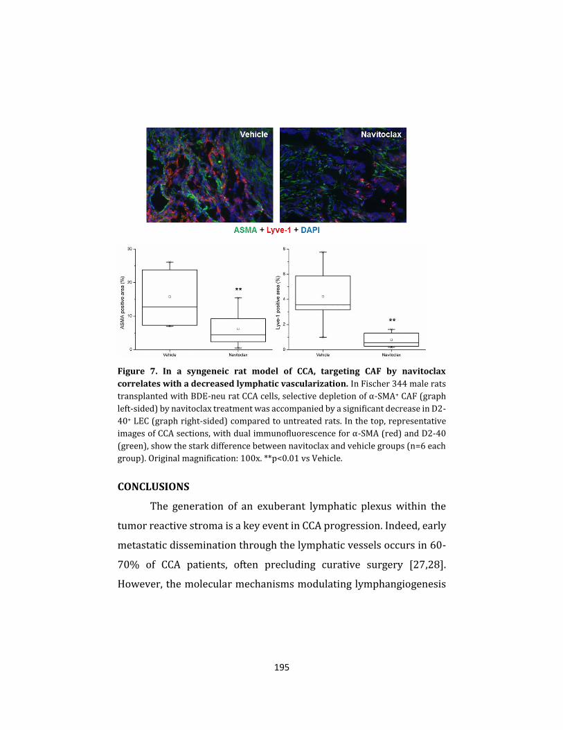
195
Figure 7. In a syngeneic rat model of CCA, targeting CAF by navitoclax
correlates with a decreased lymphatic vascularization. In Fischer 344 male rats
transplanted with BDE-neu rat CCA cells, selective depletion of α-SMA+ CAF (graph
left-sided) by navitoclax treatment was accompanied by a significant decrease in D2-
40+ LEC (graph right-sided) compared to untreated rats. In the top, representative
images of CCA sections, with dual immunofluorescence for α-SMA (red) and D2-40
(green), show the stark difference between navitoclax and vehicle groups (n=6 each
group). Original magnification: 100x. **p<0.01 vs Vehicle.
CONCLUSIONS
The generation of an exuberant lymphatic plexus within the
tumor reactive stroma is a key event in CCA progression. Indeed, early
metastatic dissemination through the lymphatic vessels occurs in 60-
70% of CCA patients, often precluding curative surgery [27,28].
However, the molecular mechanisms modulating lymphangiogenesis

196
in CCA are still poorly elucidated, and more generally, tumor-
associated lymphangiogenesis is still a major gap in knowledge in
cancer research.
Among the multiple factors mediating the mutual epithelial-
mesenchymal interactions typically fueling CCA aggressiveness, PDGF-
D is attracting considerable interest, owing to its key role in the
generation of the tumor reactive stroma, and its potential druggability
[14,18]. As shown in different settings, PDGF-D exerts a repertoire of
functions related to tumor-promotion, by fostering cancer cell
proliferation and invasiveness, fibroblast recruitment and activation,
and aberrant extracellular matrix deposition [18,29-31]. In this study,
we provide evidence that in CCA, cancer cell-derived PDGF-D (which
we previously showed to be up-regulated in malignant cholangiocytes
under hypoxic conditions) triggers a multi-step paracrine sequence
centered on CAF. This extensive paracrine signaling eventually leads
to a massive recruitment of LEC, as well as to their assembly in highly
branched and leaky tubular structures that can be easily invaded by
cancer cells for their metastatic dissemination. Therefore, this model
provides novel molecular insights into tumoral lymphangiogenesis in
CCA.
First, we observed that CCA samples displayed a striking
expansion of the lymphatic vascular bed compared to HCC, in sharp
contrast to blood vessels, which were instead, much less represented.
These findings lend strong support to the notion that the lymphatic

197
rather than the blood vasculature represents a preferential route for
CCA cells to escape from the primary site of growth. Moreover, we
found that the lymphatic structures thriving within CCA stroma were
closely surrounded by CAF. This strict physical proximity was most
likely related to the reciprocal expression of VEGF-A and VEGF-C
(which were negligibly expressed in malignant cholangiocytes) by
CAF, and of their cognate receptors VEGFR2 and VEGFR3 by LEC,
suggesting the existence of an intimate cross talk between the two cell
types. It is interesting to note that a similar pattern of expression was
found in other desmoplastic epithelial cancers, such as colorectal [32]
and ovarian [33] carcinomas.
In an effort to reproduce the biological interactions occurring
within the tumor microenvironment, we next challenged human
fibroblasts with recombinant PDGF-D. The modulatory effects of PDGF
family members (especially, PDGF-B) on the secretion of VEGF ligands
have been documented in various cell types, including hepatic stellate
cells [34] and pulmonary fibroblasts [35]. However, no study has ever
focused on the pro-lymphangiogenic properties of PDGF-D.
Nonetheless, it was previously shown that PDGFRβ inhibition reduced
VEGF, Ang-1, and Ang-2 mRNA expression in a mouse model of corneal
neovascularization [36]. In our study, we demonstrated that PDGF-D
potently elicits the secretion of VEGF-A and VEGF-C by fibroblasts,
without affecting the release of other lymphangiogenic growth factors,
such as VEGF-D, Ang-1, and Ang-2. In particular, the PDGF-D-
198
dependent production of both VEGF isoforms by fibroblasts was
mediated by two partially redundant pathways, namely ERK and JNK.
The latter are renowned downstream effectors of PDGFRβ activation,
well-known to regulate the secretion of several growth factors,
including VEGF, though their involvement has been investigated so far
more in epithelial than in mesenchymal cells. Overall, these data add
another piece to the puzzle of the pleiotropic functions played by CAF
in response to the PDGF-D signaling originating from CCA cells: CAF
are not only simply attracted, but are also empowered to direct tumor
lymphangiogenesis.
To further address this issue, we assessed the influence of CM
harvested from PDGF-D-stimulated fibroblasts on LEC motility,
vascular assembly and permeability. In order to rule out confounding
effects possibly related to the presence of residual exogenous PDGF-D,
we preliminarily evaluated the expression of PDGFRβ on LEC. We
found that LEC did not express PDGFRβ, while strongly expressing
VEGFR2 and VEGFR3. It is fair to note that these findings are in
contrast with data published in other studies. For instance, Cao et al.
reported PDGFRβ expression on LEC; however, they used LEC of
murine and rat origin, and moreover, they evaluated PDGFRβ
expression only at the mRNA and not at the protein level [37]. Other
studies in mice similarly described PDGFRβ expression on LEC, but
only on LEC from large vessels; conversely, PDGFRβ expression was
consistently negative on LEC lining the small vessels [38].

199
To evaluate the lymphangiogenic properties of CM from PDGF-
D-treated fibroblasts, we employed the classic Boyden chamber
system and the AngioTool software, which allows for a quick and
reproducible quantification of different vessel morphological and
spatial parameters, including vessel length, the percentage of area
covered by vessels, and the number of branch points [39]. We found
that cultured LEC exposed to CM from PDGF-D-stimulated fibroblasts
were chemotactically recruited to aptly assemble branched vascular
networks. Of note, the larger surface area covered by lymphatic
structures increases the likelihood of contact with tumor cells, thereby
favoring their access to lymphatics. This feature is regarded as a
readout of paramount importance for lymph node metastatization
[10,40].
In our model, enhanced lymphangiogenesis was dependent on
the sequential activation of PDGFRβ on fibroblasts, and of VEGFR2 or
VEGFR3 on LEC. In this regard, it is worth noting that the blockade of
PDGFRβ resulted in a stronger inhibition of tubulogenesis compared
to the specific antagonism of VEGFR2 or VEGFR3, thus suggesting that
additional soluble factors released from PDGF-D-stimulated
fibroblasts might act in concert with VEGF-A and VEGF-C to promote
tumor lymphangiogenesis in CCA.
Although the correlation between the expression of
lymphangiogenic growth factors and the propensity of the tumor to
develop lymph node metastasis has been described in different cancer

200
types [41-43], the molecular mechanisms driving the lymphatic
invasion of tumor cells remain elusive. Thus, to evaluate whether the
pro-lymphangiogenic functions of CAF could also favor the entry of
invasive cancer cells into the lymphatic vessels, we assessed the TEER
across LEC monolayers, and the trans-LEC monolayer migration of
CCA cells. Indeed, LEC monolayers challenged with CM from PDGF-D-
stimulated fibroblasts showed an increased permeability compared to
controls, in accordance with the observation that in colorectal cancer,
VEGF-C makes the lymphatic endothelial barrier looser by weakening
tight and adherent junctions [44]. Furthermore, CCA cells were able to
cross LEC monolayers more easily upon exposure to CM from PDGF-D-
stimulated fibroblasts. Of note, both effects were reverted by
antagonizing either PDGFRβ on fibroblasts, or VEGFR3, but not
VEGFR2, on LEC. This suggests that albeit VEGF-C and VEGF-A possess
partially overlapping functions in the initial establishment of the
lymphatic vascular network, the increase in endothelial permeability
is a specific effect of VEGF-C. Taken together, these data suggest that
PDGF-D is indeed a key factor secreted by CCA cells to recruit CAF and
then shape them into potent pro-lymphangiogenic players, an
essential step for permitting the lymphatic invasion and dissemination
of tumor cells.
To confirm that CAFs are central actors in tumor
lymphangiogenesis in CCA, we turned to a syngeneic rat model of CCA
[25,26]. This animal model is characterized by a highly desmoplastic
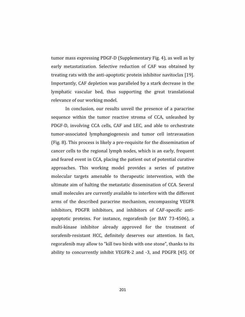
201
tumor mass expressing PDGF-D (Supplementary Fig. 4), as well as by
early metastatization. Selective reduction of CAF was obtained by
treating rats with the anti-apoptotic protein inhibitor navitoclax [19].
Importantly, CAF depletion was paralleled by a stark decrease in the
lymphatic vascular bed, thus supporting the great translational
relevance of our working model.
In conclusion, our results unveil the presence of a paracrine
sequence within the tumor reactive stroma of CCA, unleashed by
PDGF-D, involving CCA cells, CAF and LEC, and able to orchestrate
tumor-associated lymphangiogenesis and tumor cell intravasation
(Fig. 8). This process is likely a pre-requisite for the dissemination of
cancer cells to the regional lymph nodes, which is an early, frequent
and feared event in CCA, placing the patient out of potential curative
approaches. This working model provides a series of putative
molecular targets amenable to therapeutic intervention, with the
ultimate aim of halting the metastatic dissemination of CCA. Several
small molecules are currently available to interfere with the different
arms of the described paracrine mechanism, encompassing VEGFR
inhibitors, PDGFR inhibitors, and inhibitors of CAF-specific anti-
apoptotic proteins. For instance, regorafenib (or BAY 73-4506), a
multi-kinase inhibitor already approved for the treatment of
sorafenib-resistant HCC, definitely deserves our attention. In fact,
regorafenib may allow to “kill two birds with one stone”, thanks to its
ability to concurrently inhibit VEGFR-2 and -3, and PDGFR [45]. Of
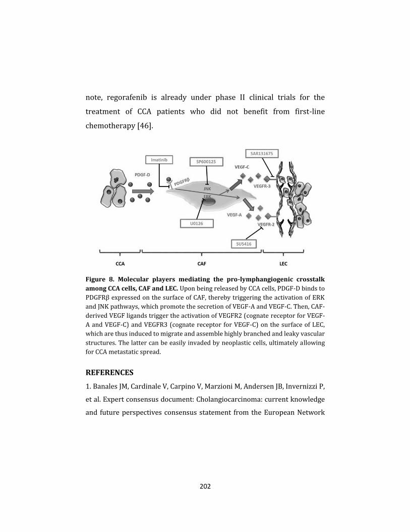
202
note, regorafenib is already under phase II clinical trials for the
treatment of CCA patients who did not benefit from first-line
chemotherapy [46].
Figure 8. Molecular players mediating the pro-lymphangiogenic crosstalk
among CCA cells, CAF and LEC. Upon being released by CCA cells, PDGF-D binds to
PDGFRβ expressed on the surface of CAF, thereby triggering the activation of ERK
and JNK pathways, which promote the secretion of VEGF-A and VEGF-C. Then, CAF-
derived VEGF ligands trigger the activation of VEGFR2 (cognate receptor for VEGF-
A and VEGF-C) and VEGFR3 (cognate receptor for VEGF-C) on the surface of LEC,
which are thus induced to migrate and assemble highly branched and leaky vascular
structures. The latter can be easily invaded by neoplastic cells, ultimately allowing
for CCA metastatic spread.
REFERENCES
1. Banales JM, Cardinale V, Carpino V, Marzioni M, Andersen JB, Invernizzi P,
et al. Expert consensus document: Cholangiocarcinoma: current knowledge
and future perspectives consensus statement from the European Network

203
for the Study of Cholangiocarcinoma (ENS-CCA). Nat Rev Gastroenterol
Hepatol 2016;13:261–280.
2. Razumilava N, Gores GJ. Cholangiocarcinoma. Lancet 2014;383:2168–
2179.
3. Erice O, Merino-Azpitarte M, Arbelaiz A, Gutierrez-Larranaga M, Jimenez-
Aguero R, Perugorria MJ, et al. Molecular Mechanisms of
Cholangiocarcinogenesis: New Potential Targets for Therapy. Curr Drug
Targets 2017;18:932-949.
4. DeOliveira ML, Cunningham SC, Cameron JL, Kamangar F, Winter JM,
Lillemoe KD, et al. Cholangiocarcinoma: thirty-one-year experience with 564
patients at a single institution. Ann Surg 2007;245:755-762.
5. Brivio S, Cadamuro M, Fabris L, Strazzabosco M. Molecular mechanisms
driving cholangiocarcinoma invasiveness: an overview. Gene Expr 2017;
Epub ahead of print.
6. Thelen A, Scholz A, Benckert C, Weichert W, Dietz E, Wiedenmann B, et al.
Tumor-associated lymphangiogenesis correlates with lymph node
metastases and prognosis in hilar cholangiocarcinoma. Ann Surg Oncol
2008;15:791–799.
7. Thelen A, Scholz A, Weichert W, Wiedenmann B, Neuhaus P, Gessner R, et
al. Tumor-associated angiogenesis and lymphangiogenesis correlate with
progression of intrahepatic cholangiocarcinoma. Am J Gastroenterol
2010;105:1123-1132.
8. Luo X, Yuan L, Wang Y, Ge R, Sun Y, Wei G. Survival Outcomes and
Prognostic Factors of surgical therapy for all potentially resectable
intrahepatic cholangiocarcinoma: a large single-center cohort study. J
Gastrointest Surg 2014;18:562-572.

204
9. Cao Y. Opinion: emerging mechanisms of tumor lymphangiogenesis and
lymphatic metastasis. Nat Rev Cancer 2005;5:735-743.
10. Stacker SA, Williams SP, Karnezis T, Shayan R, Fox SB, Achen MG.
Lymphangiogenesis and lymphatic vessel remodelling in cancer. Nat Rev
Cancer 2014;14:159–172.
11. Kalluri R, Zeisberg M. Fibroblasts in cancer. Nat Rev Cancer 2006;6:392-
401.
12. Qiao A, Gu F, Guo X, Zhang X, Fu L. Breast cancer-associated fibroblasts:
their roles in tumor initiation, progression and clinical applications. Front
Med 2016;10:33-40.
13. Pan B, Liao Q, Niu Z, Zhou L, Zhao Y. Cancer-associated fibroblasts in
pancreatic adenocarcinoma. Future Oncol 2015;11:2603-2610.
14. Cadamuro M, Morton SD, Strazzabosco M, Fabris L. Unveiling the role of
tumor reactive stroma in cholangiocarcinoma: an opportunity for new
therapeutic strategies. Transl Gastrointest Cancer 2013;2:130-144.
15. Cadamuro M, Stecca T, Brivio S, Mariotti V, Fiorotto R, Spirli C, et al. The
deleterious interplay between tumor epithelia and stroma in
cholangiocarcinoma. Biochim Biophys Acta 2017; Epub ahead of print.
16. Mertens JC, Rizvi S, Gores GJ. Targeting cholangiocarcinoma. Biochim
Biophys Acta 2017; Epub ahead of print.
17. Vaquero J, Guedj N, Clapéron A, Nguyen Ho-Bouldoires TH, Paradis V,
Fouassier L. Epithelial-mesenchymal transition in cholangiocarcinoma: From
clinical evidence to regulatory networks. J Hepatol. 2017;66:424-441.
18. Cadamuro M, Nardo G, Indraccolo S, Dall'olmo L, Sambado L, Moserle L,
et al. Platelet-Derived Growth Factor and Rho GTPases regulate recruitment
of Cancer-Associated Fibroblasts in Cholangiocarcinoma. Hepatology
2013;58:1042-1053.

205
19. Mertens JC, Fingas CD, Christensen JD, Smoot RL, Bronk SF, Werneburg
NW, et al. Therapeutic effects of deleting cancer-associated fibroblasts in
cholangiocarcinoma. Cancer Res 2013;73:897-907.
20. Tokuda H, Takai S, Hanai Y, Harada A, Matsushima-Nishiwaki R, Kato H,
et al. Potentiation by platelet-derived growth factor-BB of FGF-2-stimulated
VEGF release in osteoblasts. J Bone Miner Metab 2008;26:335-341.
21. Gaceb A, Özen I, Padel T, Barbariga M, Paul G. Pericytes secrete pro-
regenerative molecules in response to platelet-derived growth factor-BB. J
Cereb Blood Flow Metab 2017; Epub ahead of print.
22. Kazenwadel J, Secker GA, Betterman KL, Harvey NL. In Vitro Assays Using
Primary Embryonic Mouse Lymphatic Endothelial Cells Uncover Key Roles
for FGFR1 Signalling in Lymphangiogenesis. PLoS One 2012;7:e40497.
23. Ellis LM, Hicklin DJ. VEGF-targeted therapy: mechanisms of anti-tumour
activity. Nat Rev Cancer 2008;8:579-591.
24. Duong T, Koopman P, Francois M. Tumor lymphangiogenesis as a
potential therapeutic target. J Oncol 2012;2012:204946.
25. Lai GH, Zhang Z, Shen XN, Ward DJ, Dewitt JL, Holt SE, et al. ErbB-2/neu
transformed rat cholangiocytes recapitulate key cellular and molecular
features of human bile duct cancer. Gastroenterology 2005;129:2047-2057.
26. Sirica AE, Zhang Z, Lai GH, Asano T, Shen XN, Ward DJ, et al. A novel
"patient-like" model of cholangiocarcinoma progression based on bile duct
inoculation of tumorigenic rat cholangiocyte cell lines. Hepatology
2008;47:1178-1190.
27. Yamaguchi K, Chijiiwa K, Saiki S, Shimizu S, Takashima M, Tanaka M.
Carcinoma of the extrahepatic bile duct: mode of spread and its prognostic
implications. Hepatogastroenterology 1997;44:1256-1261.
206
28. Uenishi T, Hirohashi K, Kubo S, Yamamoto T, Yamazaki O, Kinoshita H.
Clinicopathological factors predicting outcomes after resection of mass-
forming intrahepatic cholangiocarcinoma. Br J Surg 2001;88:969–974.
29. Ustach CV, Taube ME, Hurst NJ Jr, Bhagat S, Bonfil RD, Cher ML, et al. A
potential oncogenic activity of platelet-derived growth factor d in prostate
cancer progression. Cancer Res 2004;64:1722-1729.
30. Xu L, Tong R, Cochran DM, Jain RK. Blocking platelet-derived growth
factor-D/platelet-derived growth factor receptor beta signaling inhibits
human renal cell carcinoma progression in an orthotopic mouse model.
Cancer Res 2005;65:5711-5719.
31. Borkham-Kamphorst E, van Roeyen CR, Ostendorf T, Floege J, Gressner
AM, Weiskirchen R. Pro-fibrogenic potential of PDGF-D in liver fibrosis. J Hep
2007;46:1064–1074.
32. Liang P, Hong JW, Ubukata H, Liu G, Katano M, Motohashi G, et al.
Myofibroblasts Correlate with Lymphatic Microvessel Density and Lymph
Node Metastasis in Early-stage Invasive Colorectal Carcinoma. Anticancer
Res 2005;25:2705-2712.
33. Zhang Y, Tang H, Cai J, Zhang T, Guo J, Feng D, et al. Ovarian cancer-
associated fibroblasts contribute to epithelial ovarian carcinoma metastasis
by promoting angiogenesis, lymphangiogenesis and tumor cell invasion.
Cancer Lett 2011;303:47-55.
34. Lu Y, Lin N, Chen Z, Xu R. Hypoxia-induced secretion of platelet-derived
growth factor-BB by hepatocellular carcinoma cells increases activated
hepatic stellate cell proliferation, migration and expression of vascular
endothelial growth factor-A. Mol Med Rep 2015;11:691-697.
35. Nauck M, Roth M, Tamm M, Eickelberg O, Wieland H, Stulz P, et al.
Induction of vascular endothelial growth factor by platelet-activating factor

207
and platelet-derived growth factor is downregulated by corticosteroids. Am
J Respir Cell Mol Biol 1997;16:398-406.
36. Dell S, Peters S, Müther P, Kociok N, Joussen AM. The Role of PDGF
Receptor Inhibitors and PI3-Kinase Signaling in the Pathogenesis of Corneal
Neovascularization. Invest Ophthalmol Vis Sci 2006;47:1928-1937.
37. Cao R, Björndahl MA, Religa P, Clasper S, Garvin S, Galter D, et al. PDGF-
BB induces intratumoral lymphangiogenesis and promotes lymphatic
metastasis. Cancer Cell 2004;6:333–345.
38. Kodama M, Kitadai Y, Sumida T, Ohnishi M, Ohara E, Tanaka M, et al.
Expression of platelet-derived growth factor (PDGF)-B and PDGF-receptor β
is associated with lymphatic metastasis in human gastric carcinoma. Cancer
Sci 2010;101:1984-1989.
39. Zudaire E, Gambardella L, Kurcz C, Vermeren S. A computational tool for
quantitative analysis of vascular networks. PLoS One 2011;6:e27385.
40. Leu AJ, Berk DA, Lymboussaki A, Alitalo K, Jain RK. Absence of functional
lymphatics within a murine sarcoma: a molecular and functional evaluation.
Cancer Res 2000;60:4324–4327.
41. Mandriota SJ, Jussila L, Jeltsch M, Compagni A, Baetens D, Prevo R, et al.
Vascular endothelial growth factor-C-mediated lymphangiogenesis
promotes tumour metastasis. EMBO J 2001;20:672-682.
42. Skobe M, Hawighorst T, Jackson DG, Prevo R, Janes L, Velasco P, et al.
Induction of tumor lymphangiogenesis by VEGF-C promotes breast cancer
metastasis. Nat Med 2001;7:192-198.
43. Karnezis T, Shayan R, Caesar C, Roufail S, Harris NC, Ardipradja K, et al.
VEGF-D promotes tumor metastasis by regulating prostaglandins produced
by the collecting lymphatic endothelium. Cancer Cell 2012;21:181-195.

208
44. Tacconi C, Correale C, Gandelli A, Spinelli A, Dejana E, D'Alessio S, et al.
Vascular endothelial growth factor C disrupts the endothelial lymphatic
barrier to promote colorectal cancer invasion. Gastroenterology
2015;148:1438-1451.e8.
45. Bruix J, Qin S, Merle P, Granito A, Huang YH, Bodoky G, et al. Regorafenib
for patients with hepatocellular carcinoma who progressed on sorafenib
treatment (RESORCE): a randomised, double-blind, placebo-controlled,
phase 3 trial. Lancet 2017;389:56-66.
46. Chong DQ, Zhu AX. The landscape of targeted therapies for
cholangiocarcinoma: current status and emerging targets. Oncotarget
2016;7:46750-46767.
SUPPLEMENTARY MATERIALS AND METHODS
Cell lines. Human fibroblasts were seeded on plastic 25cm2 flasks
(Falcon®) and cultured in Dulbecco's Modified Eagle's medium
(DMEM, Euroclone®) supplemented with 10% fetal bovine serum
(FBS) and 1% penicillin/streptomycin (Euroclone®). Human
lymphatic endothelial cells (LEC, purchased from ScienCell™) were
seeded in 25cm2 flasks (Falcon®) pre-coated with fibronectin 2µg/cm2
(ScienCell™), and cultured in Endothelial Cell Basal Medium (ECM,
ScienCell™) supplemented with 5% FBS (ScienCell™), 1%
penicillin/streptomycin (ScienCell™) and 1% endothelial cell growth
supplement (ECGS, ScienCell™). LEC were used until passage number
10. EGI-1 cells (PDGF-D expressing extrahepatic CCA cell line,
purchased from Deutsche Sammlung von Mikroorganismen und

209
Zellkulturen) were seeded in 25cm2 flasks (Falcon®) and cultured in
RPMI-1640 Medium (Gibco™) supplemented with 10% FBS and 1%
penicillin/streptomycin (Euroclone®). All cell lines were maintained
in an incubator at 37°C and 5% CO2 atmosphere (Galaxy S, RS Biotech).
Immunohistochemical staining. Briefly, after deparaffinization
using xylene, sections were rehydrated in absolute ethanol and then
washed in tap water; endogenous peroxidase activity was quenched
incubating slides with methanol containing 10% H2O2 for 30 minutes.
Antigen retrieval was performed by heating the slides in a steamer for
20 minutes in 10 mmol/L citrate buffer (pH 6) (Carlo Erba). Blocking
of the aspecifics was achieved using UltraVision protein block
(ThermoScientific) for 8 minutes. The sections were then incubated
overnight at 4°C with primary antibodies against CD34 or D2-40
diluted in PBS + BSA 1% (Sigma-Aldrich®). After rinsing with
phosphate-buffered saline (PBS) 1M supplemented with 0.05%
Tween20 (PBS-T; both Sigma), slides were incubated for 30 minutes
at room temperature with the specific horseradish peroxidase (HRP)-
conjugated secondary antibody (EnVision, DAKO). Specimens were
developed using 3,3-diaminobenzedine tetrahydrochloride 0.04
mg/mL (DAB, Sigma) with H2O2 0.01% diluted in PBS, counterstained
with Gill’s Hematoxylin N°2 (Sigma), and mounted using EuKitt (Bio-
optica). Following immunostaining, lymphatic microvascular density
(LMVD) and blood miscrovascular density (BMVD) were measured in

210
CCA and HCC samples using an Eclipse E800 microscope (Nikon).
Briefly, 10 randomly taken micrographs were collected from each
sample using a cooled digital camera (Nikon, DS-U1) and then
analyzed using LuciaG 5.0 software, as previously described [1].
Double immunohistochemical staining. To match two antibodies
developed in the same animal, we used a double immunohistochemical
approach. Following deparaffinization and hydratation, as above
described, CCA slides were incubated for 1h at room temperature with
antibody against α-SMA and, following incubation with an appropriate
HRP-conjugated secondary antibody, they were developed with DAB.
Slides were then washed with distilled water for 15 min and then with
PBS-T and incubated overnight at 4°C with antibody against D2-40.
Following washing with PBS-T and incubation with the secondary
antibody, staining was developed using TrueBlue (KPL), and then
mounted as above described.
Double immunofluorescent staining. Briefly, following
deparaffinization and hydratation, as above described, antigen
retrieval was performed using 10 mmol/L citrate buffer (pH 6).
Selected sections from CCA samples were incubated overnight at 4°C
with primary antibodies against VEGF-A, VEGF-C, VEGFR2 or VEGFR3,
together with antibodies against D2-40, α-SMA, or K19. Following
washing in PBS-T, slides were then incubated for 30 minutes at room

211
temperature with the respective secondary Alexa Fluor 488- or 594-
conjugated antibody (1:500, Life Technologies) and mounted with
VECTASHIELD® Mounting Medium with DAPI (Vector Laboratories).
ELISA. Fibroblasts were seeded into a 24-well plate, and once
confluent, were starved for 24h, before being exposed to recombinant
human (rh) PDGF-D (100ng/ml, 24h, R&D Systems). To quantify the
secretion of VEGF-A, VEGF-C, VEGF-D, Ang-1 or Ang-2, cultured
fibroblast supernatants were harvested, stored at -80°C, and then
analyzed by ELISA assay according to the supplier's instructions
(RayBiotech). Absorbance levels were detected with FLUO Star Omega
(BMG Labtech, Euroclone®). A calibration curve was generated for
each experiment, and concentration levels were expressed in relation
to the protein content of each experiment. In PDGF-D-stimulated
fibroblasts, secretory levels were measured with or without treatment
with imatinib mesylate (IM, PDGFRβ inhibitor, 1µM, 24h, Cayman
Chemical Company), U0126 (ERK inhibitor, 10µM, 24h, Sigma-
Aldrich®), and SP600125 (JNK inhibitor, 10µM, 24h, Sigma-Aldrich®),
in order to dissect the intracellular pathways activated downstream of
PDGFRβ, mediating the pro-lymphangiogenic functions of fibroblasts.
Western blotting. The expression of PDGFRβ, VEGFR2 and VEGFR3
was evaluated in untreated LEC and fibroblasts seeded into a 6-well
plate. Total protein content (CelLytic M, Sigma) was obtained from

212
fibroblasts and LEC according to the supplier's instructions. Protein
concentration was evaluated by Bradford assay (Sigma). Anti-PDGFRβ,
anti-VEGFR2, anti-VEGFR3, and anti-GAPDH were used as primary
antibodies. Equal concentrations of protein lysates were
electrophoresed on a 4–12% NuPAGE® Novex Bis-Tris gel (Life
Technologies) with MES (NuPage® Novex, Life Technologies in 500ml
di H2OmQ) for 40 minutes at 200 volt and 120 ampere and proteins
were transferred to a nitrocellulose membrane (Life Technologies) for
60 minutes at 30 volt and 170 ampere. The membrane was then
blocked with 5% non-fat dry milk (Euroclone) in Tris-buffered saline
(TBS) supplemented with 0.1% Tween-20 (TBS-T) for 1 hour and then
incubated overnight at 4°C with the primary antibody diluted in TBS-
T + nonfat dry milk 5%. The membrane was washed 3 times with TBS-
T before incubation with anti-rabbit (1:2000, Bio-Rad) or anti-mouse
(1:2000, Sigma) HRP-conjugated secondary antibodies for 1 hour.
Proteins were visualized using enhanced chemiluminescence
(SuperSignal West Pico, Thermo Scientific). Bands were quantified
with ImageJ and results were normalized versus reference proteins
(GAPDH).
LEC migration assay. To assess the migratory abilities of LEC elicited
by PDGF-D-stimulated fibroblasts, we used 6.5mm Transwell® with
8.0µm Pore Polycarbonate Membrane Insert (Corning), as previously
described [2,3]. 5x10⁴ LEC in ECM medium + 0,5% FBS were added to

213
the upper chamber, whereas the lower chamber was filled with DMEM
+ 0,5% FBS harvested from fibroblasts challenged with PDGF-D
(100ng/ml, 24h), with or without IM (1µM, 24h) (conditioned
medium, CM). DMEM + 0,5% FBS from untreated fibroblasts and
DMEM + 0.5% FBS supplemented with rh VEGF-A (100ng/ml, 24h,
R&D Systems) or VEGF-C (100ng/ml, 24h, R&D Systems) served as
negative and positive controls, respectively. After 24h, cells on the
upper surface of the membrane were removed with a cotton swab,
whereas cells adherent to the lower surface were fixed and stained
using a Diff-Quick Staining Set (Medion Diagnostics). Finally, 10
random fields of each membrane were photographed at 10x
magnification to count the number of clearly discernible nuclei.
Tube formation assay. To evaluate whether fibroblasts exposed to
PDGF-D could also direct LEC to get-together in proper vascular
structures, we performed a tube formation assay. LEC were re-
suspended in ECM + 0,5% FBS, and seeded into a 24-well plate
(Falcon®) at a density of 5x10⁴ cells/well. Each well was pre-coated
with fibronectin 2µg/cm2. After a 24h incubation, cultured LEC were
covered with matrigel (Corning®) diluted at a 1:1 ratio in ECM + 0,5%
FBS. Once matrigel solidified, cells were exposed to CM from
fibroblasts challenged with PDGF-D (100ng/ml, 24h), with or without
IM (1µM, 24h), and compared to cells exposed to medium from
untreated fibroblasts, as negative control. Treatments with rhVEGF-A
214
(100ng/ml, 24h) or rhVEGF-C (100ng/ml, 24h) were used as positive
controls. LEC exposed to CM from PDGF-D-treated fibroblasts were
also simultaneously treated with antagonists of VEGFR2 (SU5416,
1µM, Sigma) or VEGFR3 (SAR131675, 15nM, Selleckchem). After a 24h
exposure, analysis was performed in 5 random photos at 10x
magnification (obtained through the same tools reported above for
immunohistochemistry), by quantifying three parameters of
tubulogenesis: a) length of vascular branches, b) number of junctions
between branches, and c) luminal vascular area, using the AngioTool
software, as described elsewhere [4].
Evaluation of trans-endothelial resistance (TEER). The following
experiments were performed to understand whether, upon PDGF-D
stimulation, fibroblasts could favor the lymphatic intravasation of CCA
cells. To this end, 5x10⁴ LEC were seeded into a 24-well plate over a
0.4µm pore membrane (Corning) pre-coated with fibronectin
2µg/cm2. Once LEC reached confluence, they were treated as reported
for tube formation assay. Trans-endothelial electric resistance (TEER)
was then evaluated by a Epithelial Volt/Ohm Meter (EVOM, World
Precision Instruments) equipped with an STX2 electrode at different
time points (0, 30, 60, 120, 360 min). To confirm the integrity of LEC
monolayers, we ensured that TEER values at t=0 were higher than
1μΩ/μm2.

215
Evaluation of trans-endothelial migration (TrEM). 5x10⁴ LEC were
seeded over an 8.0µm pore polycarbonate membrane (Corning) pre-
coated with fibronectin 2µg/cm2. Once confluent, LEC were treated as
reported for tube formation assay, and 5x10⁴ EGI-1 cells were seeded
on the top of the endothelial monolayer. After 6h, cells on the upper
surface of the membrane were removed, while cells on the lower side
were fixed with ice-cold methanol and mounted with VECTASHIELD®
Mounting Medium with DAPI (Vector Laboratories). To discriminate
CCA cells from LEC, EGI-1 cells were originally transduced with a
lentiviral vector encoding EGFP reporter gene, as previously
performed [2,3]. The number of EGFP-positive cells was counted in 10
random pictures of each membrane taken at 10X magnification [5].
MTS assay. CellTiter 96® AQueous One Solution Cell Proliferation
Assay (MTS) (Promega) was used to assess whether SU5416 (0,1µM, 1
µM, and 5 µM) and SAR131675 (1nM, 15nM, and 30nM) could affect
LEC viability in response to a 24h treatment [6]. LEC were re-
suspended in 0,5% FBS culture medium, and seeded into a 96-well
plate at a density of 1x10⁴ cells/well. After a 24h incubation, cells were
treated with SU5416 or SAR131675 diluted in 0,5% FBS culture
medium. After treatment, 20µl of CellTiter 96® AQueous One Solution
Reagent (Promega) were added to each well, and the plate was
incubated at 37°C for 1 hour in a 5% CO2 atmosphere. 490nm

216
absorbance was evaluated using FLUO Star Omega (BMG Labtech,
Euroclone®). Each experiment was made in duplicate.
SUPPLEMENTARY FIGURES AND TABLES
Figure S1. Opposite pattern of expression of VEGFR2, VEGFR3 and PDGFRβ on
LEC compared with fibroblasts. In contrast to fibroblasts, LEC expressed VEGFR2
and VEGFR3 (A), whereas they did not express PDGFRβ (B), thereby indicating their
unresponsiveness to PDGF-D.
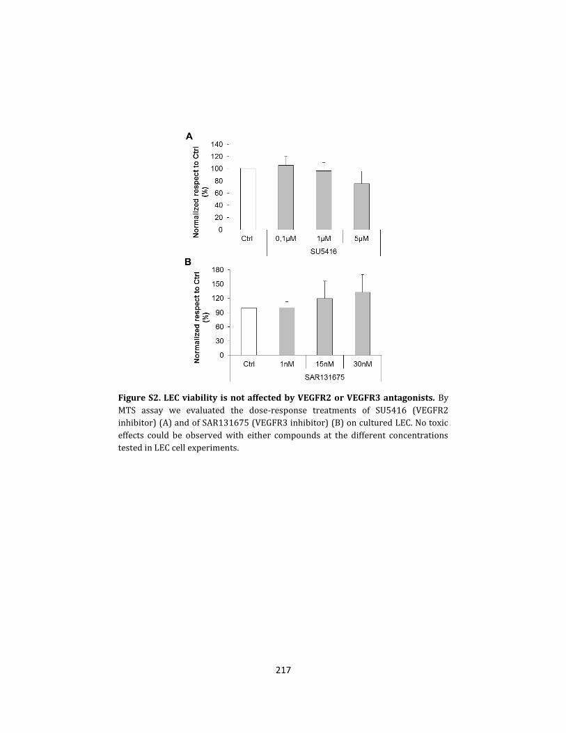
217
Figure S2. LEC viability is not affected by VEGFR2 or VEGFR3 antagonists. By
MTS assay we evaluated the dose-response treatments of SU5416 (VEGFR2
inhibitor) (A) and of SAR131675 (VEGFR3 inhibitor) (B) on cultured LEC. No toxic
effects could be observed with either compounds at the different concentrations
tested in LEC cell experiments.

218
Figure S3. Trans-endothelial migration of EGI-1-EGFP cells is potently induced
by CM from PDGF-D-stimulated fibroblasts. Representative micrographs of
Boyden chamber experiments evaluating the amount of EGI-1-EGFP cells (green)
crossing a LEC monolayer exposed to CM from fibroblasts challenged with PDGF-D,
alone, (PDGF-D) CM, or in combination with IM, (PDGF-D + IM) CM, in the
presence/absence of VEGFR2, (PDGF-D) CM + αVEGFR2, or VEGFR3, (PDGF-D) CM +
αVEGFR3, antagonists. Nuclear marker, DAPI (blue). Original magnification: 200x.

219
Figure S4. In a syngeneic rat model of CCA, targeting CAF by navitoclax did not
affect PDGF-D expression by tumoral cells. In Fischer 344 male rats transplanted
with BDE-neu rat CCA cells, treatment with navitoclax induced a selective depletion
of CAF, without affecting PDGF-D expression by tumoral cells. In the top,
representative images of CCA sections immunostained for PDGF-D, showing the
comparable levels of expression by PDGF-D+ tumoral areas between the two groups
(n=6 each group). Original magnification: 100x.
Antibody Isotype
(IHC/IF)
dilution;
antigen retrieval
(WB)
dilution Supplier
α-SMA
(clone
1A4)
Mouse
monoclonal
1:200; 10mmol/L
citrate pH 6 DAKO
Actin Rabbit
polyclonal 1:5000 Sigma-Aldrich
CD34
(clone
QBEnd/10)
Mouse
monoclonal
1:50; 10mmol/L
citrate pH 6
Leica
Biosystems
D2-40 Mouse
monoclonal
1:100; 10mmol/L
citrate pH 6 DAKO

220
GAPDH
(clone
0411)
Mouse
monoclonal 1:5000
Santa Cruz
Biotechnology
K19
(clone
RCK108)
Mouse
monoclonal 1:100 (frozen) DAKO
Lyve-1 Rabbit
polyclonal 1:100 (frozen) Acris
PDGF-D Rabbit
polyclonal 1:100 (frozen) Invitrogen
PDGFRβ Rabbit
polyclonal 1:1000 Cell Signaling
VEGF-A Rabbit
polyclonal
1:100; 10mmol/L
citrate pH 6
Santa Cruz
Biotechnology
VEGF-C Rabbit
polyclonal
1:100; 10mmol/L
citrate pH 6
Santa Cruz
Biotechnology
VEGFR2 Rabbit
polyclonal
1:100; 10mmol/L
citrate pH 6 1:1000 Cell Signaling
VEGFR3 Rabbit
polyclonal
1:100; 10mmol/L
citrate pH 6 1:500
Santa Cruz
Biotechnology
Supplementary Table 1. List of primary antibodies.
SUPPLEMENTARY REFERENCES
1. Fabris L, Cadamuro M, Fiorotto R, Roskams T, Spirlì C, Melero S, et al.
Effects of angiogenic factor overexpression by human and rodent
cholangiocytes in polycystic liver diseases. Hepatology 2006;43:1001-1012.
2. Fabris L, Cadamuro M, Moserle L, Dziura J, Cong X, Sambado L, et al. Nuclear
expression of S100A4 calcium-binding protein increases
cholangiocarcinoma invasiveness and metastasization. Hepatology
2011;54:890-899.
3. Cadamuro M, Nardo G, Indraccolo S, Dall'olmo L, Sambado L, Moserle L, et
al. Platelet-Derived Growth Factor and Rho GTPases regulate recruitment of

221
Cancer-Associated Fibroblasts in Cholangiocarcinoma. Hepatology
2013;58:1042-1053.
4. Kazenwadel J, Secker GA, Betterman KL, Harvey NL. In Vitro Assays Using
Primary Embryonic Mouse Lymphatic Endothelial Cells Uncover Key Roles
for FGFR1 Signalling in Lymphangiogenesis. PLoS One 2012;7:e40497.
5. Tacconi C, Correale C, Gandelli A, Spinelli A, Dejana E, D'Alessio S, et al.
Vascular endothelial growth factor C disrupts the endothelial lymphatic
barrier to promote colorectal cancer invasion. Gastroenterology
2015;148:1438-1451.e8.
6. Espagnolle N, Barron P, Mandron M, Blanc I, Bonnin J, Agnel M, et al.
Specific Inhibition of the VEGFR-3 Tyrosine Kinase by SAR131675 Reduces
Peripheral and Tumor Associated Immunosuppressive Myeloid Cells.
Cancers (Basel) 2014;6:472-490.

222
CHAPTER 5
Summary, conclusions and future perspectives

223
During my PhD studies, I sought to gain insights into the
molecular mechanisms underpinning the progression of
cholangiocarcinoma (CCA), particularly focusing on the deleterious
contribution of the tumor microenvironment, which is currently
recognized as a driving force behind the emergence of the hallmarks
of cancer [1-3]. Specifically, the studies presented here dissected the
tumor-stroma interplay from various points of view. On the one hand
(Chapters 2 and 3), we shed light on pro-oncogenic signaling systems
that are deleteriously fueled by the crosstalk between the tumor and
its stromal neighborhood, and provide CCA cells with enhanced
aggressiveness. On the other hand (Chapter 4), we highlighted the
capability of CCA cells to trigger a multi-step paracrine cascade that
involve different stromal components, and ultimately give rise to a
microenvironment conducive to tumor progression. Hopefully, a
comprehensive understanding of the dense, multifaceted paracrine
communications between cancer and reactive stromal cells in CCA will
considerably help to identify prognostically relevant molecular
signatures, as well as innovative therapeutic targets, with the ultimate
goal to improve the management of this devastating malignancy.
In the first study, we demonstrated that the pleiotropic
cytokine leukemia inhibitory factor (LIF) may strengthen the
chemoresistant phenotype of CCA cells, through autocrine and
paracrine mechanisms. Indeed, LIF is both overexpressed by CCA cells,
and abundantly produced by cancer-associated fibroblasts (CAFs) and

224
inflammatory cells populating the tumor microenvironment. In
addition, the cognate receptor for LIF is selectively up-regulated in
neoplastic bile ducts. At the functional level, LIF enables CCA cells to
escape from apoptosis induced by the combination of cisplatin and
gemcitabine (i.e., the first-line chemotherapy regimen for patients
with advanced CCA), an effect mediated by the up-regulation of the
anti-apoptotic protein myeloid cell leukemia (Mcl)-1. As already
discussed, the unbalance between anti-apoptotic and pro-apoptotic
members of the Bcl-2 family is a primary mechanism of
chemoresistance in several tumors, including CCA [4,5], and LIF seems
to play a relevant role in this context. In our model, Mcl-1 up-
regulation occurs downstream of the activation of the PI3K/Akt
pathway, which in turn has been widely recognized as a paramount
pro-oncogenic signaling network in human cancer [6]. It is important
to underline that both pro-inflammatory cytokines [7,8], and
intratumoral hypoxia [9] could be responsible for the overproduction
of LIF in CCA, in line with the notion that the tumor reactive stroma
(TRS) is an hectic microenvironment whose different (cellular and
non-cellular) components support each other in fueling tumor
progression.
In the second study, we showed that the nuclear import of the
calcium-binding protein S100A4 confers strong metastatic properties
upon CCA cells, an effect mediated by the activation of the small Rho
GTPases RhoA and Cdc42, the overproduction of matrix

225
metalloproteinase (MMP)-9, and the enhanced expression of
membrane-type (MT)1-MMP. Of note, RhoA and Cdc42 are key
effectors of cell motility [10,11], whereas the proteolytic activity of
MMP-9 and MT1-MMP is essential for massively degrading the
extracellular matrix, and thus enabling a pervasive migration [12].
Importantly, we unveiled that the translocation of S100A4 into the
nucleus of CCA cells is strictly dependent on its SUMOylation, a post-
translational modification that may alter the activity, subcellular
localization, or stability of several substrate proteins, mainly involved
in gene transcription and DNA repair [13]. In experimental models of
CCA, we finally proved that the pharmacological inhibition of S100A4
nuclearization by paclitaxel at nanomolar doses did not tackle tumor
growth, but effectively halted the hematogenous metastasization to
the lungs, which represent the preferential site of distant metastases
in CCA patients [14]. Although we did not provide direct evidence that
the nuclear expression of S100A4 in CCA cells is fostered by
microenvironmental signals, it is worth considering that the up-
regulation of epithelial-to-mesenchymal transition biomarkers,
including S100A4, is a classic readout of tumor-stroma interactions
[15] and moreover, in human chondrocytes, the SUMOylation-
dependent nuclear entry of S100A4 can be potently stimulated by the
pro-inflammatory cytokine interleukin 1β [16].
In the third study, we revealed that platelet-derived growth
factor (PDGF)-DD, which is overexpressed by CCA cells under severe

226
hypoxia and exert motogenic effects on CAFs [17], is also able to endow
fibroblasts with the ability to overproduce vascular endothelial
growth factor (VEGF)-A and VEGF-C, which may then stimulate the
chemotaxis of lymphatic endothelial cells (LECs), alongside their
assembly in highly branched tubular structures. Specifically, PDGF-DD
binds to its cognate receptor PDGFRβ on the surface of CAFs, thereby
activating ERK and JNK signaling cascades, whereas VEGF ligands bind
to VEGFR-2 (VEGF-A, VEGF-C) and VEGFR-3 (VEGF-C) on the surface
of LECs. Therefore, tumor-derived PDGF-DD not only plays a
prominent role in supporting fibroblast enrichment within the tumor
microenvironment [17], but also molds the behavior of CAFs so that
they can orchestrate lymphangiogenesis. As previously discussed, the
establishment of an abnormally rich lymphatic vasculature is a
histological hallmark of CCA, underlying its early metastatic spread
[1,18,19]. In line with the notion that VEGF-C may also impair the
integrity of lymphatic vessels by introducing intercellular gaps
[20,21], we additionally found that PDGF-DD-treated fibroblasts could
increase the permeability of LEC monolayers, ultimately favoring the
trans-endothelial migration of CCA cells. It is finally worth noting that
inflammatory cells represent an additional source of PDGF-DD [17]
and, alongside CCA cells, may even contribute to the biosynthesis of
VEGFs [22]. Overall, this further highlights that in CCA, the generation
of a pro-metastatic microenvironment is finely orchestrated by the
227
redundant and synergistic activities of the cancer cells and their
surrounding stromal cell populations.
Curative therapies for the management of CCA basically consist
of surgical resection by partial hepatectomy and orthotopic liver
transplantation. However, only a minority of patients can benefit from
these treatment options, since CCA is frequently diagnosed at
advanced stages [4]. Unfortunately, conventional chemotherapy is
traditionally ineffective and so far, clinical trials with molecular
targeted therapies failed to provide substantial survival benefits
[23,24]. This paucity of potentially curative treatments ultimately
results in a grim prognosis, which makes CCA a global health problem,
especially considering its increasing incidence in Western Countries
[4]. The development of effective pharmacological approaches to the
management of CCA has been limited not only by the pronounced
inter-tumor heterogeneity [24], but also by the fact that anti-cancer
therapies have been classically designed to target intrinsic traits of
neoplastic cells, without taking due account of the surrounding
microenvironment [2]. However, we and others have provided
evidence that reactive stromal cells may take a leading role in CCA
progression, by both directly fostering cancer cell survival and
invasiveness, and setting up a microenvironment conducive to
metastatic dissemination. Therefore, interfering with the epithelial-
mesenchymal crosstalk within CCA microenvironment might
substantially hinder tumor progression, by deadening a driving force

228
behind the emergence of aggressive tumor phenotypes. Basically, the
therapeutic targeting of the tumor stroma should seek to reduce the
amount of reactive stromal cells or alternatively, to quench the pro-
oncogenic pathways that they contribute to activate [2]. In this regard,
in the studies presented here, we shed light on a wide range of putative
novel targets potentially amenable to therapeutic modulation. For
instance, targeting the LIF/PI3K/Mcl-1 axis could represents a feasible
strategy to overcome CCA chemoresistance, whereas a selective
reduction in S100A4 nuclear import by low-dose paclitaxel seems to
be a promising approach to undermine CCA invasiveness. On the other
hand, the therapeutic interference with either PDGF-DD/PDGFRβ or
VEGF/VEGFR signaling systems should disrupt the crosstalk among
CCA cells, CAFs and LECs, eventually preventing cancer cells to
disseminate at distance through the lymphatic network. Interestingly,
small molecules targeting some of the above mentioned molecular
players are already approved for use or under clinical trials in other
types of cancer. These include, for example, MK2206 (Akt inhibitor),
MIK665 (Mcl-1 inhibitor), imatinib mesylate (PDGFRβ inhibitor), and
fruquintinib (VEGFRs inhibitor). However, further studies are
required to understand whether such TRS-oriented therapies could
actually be successful in treating CCA, either alone or in combination
with conventional anti-cancer treatments. It is also worth noting that
several molecules may serve as both therapeutic targets and
prognostic/predictive markers, and S100A4 is paradigmatic of this

229
concept. Indeed, our group previously found that nuclear expression
of S100A4 by neoplastic bile ducts was associated with poor outcome,
in terms of both metastasization and tumor-related death, thereby
representing a promising tool in clinical decision-making [25]. In
summary, the understanding of the dynamic and intricate interplay
between CCA cells and the wide web of neighboring stromal
components is undoubtedly an issue of great translational significance,
with a view to open novel therapeutic avenues in CCA, as well as in
other epithelial neoplasms characterized by an abundant stroma, such
as breast and pancreatic cancers.

230
References
1. Cadamuro M, Morton SD, Strazzabosco M, Fabris L. Unveiling the role
of tumor reactive stroma in cholangiocarcinoma: An opportunity for
new therapeutic strategies. Transl Gastrointest Cancer. 2013;2:130-
44.
2. Brivio S, Cadamuro M, Strazzabosco M, Fabris L. Tumor reactive
stroma in cholangiocarcinoma: The fuel behind cancer
aggressiveness. World J Hepatol. 2017;9:455-68.
3. Cadamuro M, Stecca T, Brivio S, Mariotti V, Fiorotto R, Spirli C,
Strazzabosco M, Fabris L. The deleterious interplay between tumor
epithelia and stroma in cholangiocarcinoma. Biochim Biophys Acta.
2017.
4. Banales JM, Cardinale V, Carpino G, Marzioni M, Andersen JB,
Invernizzi P, Lind GE, Folseraas T, Forbes SJ, Fouassier L, Geier A,
Calvisi DF, Mertens JC, Trauner M, Benedetti A, Maroni L, Vaquero J,
Macias RI, Raggi C, Perugorria MJ, Gaudio E, Boberg KM, Marin JJ,
Alvaro D. Expert consensus document: Cholangiocarcinoma: current
knowledge and future perspectives consensus statement from the
European Network for the Study of Cholangiocarcinoma (ENS-CCA).
Nat Rev Gastroenterol Hepatol. 2016;13:261-80.
5. Cadamuro M, Brivio S, Spirli C, Joplin RE, Strazzabosco M, Fabris L.
Autocrine and Paracrine Mechanisms Promoting Chemoresistance in
Cholangiocarcinoma. Int J Mol Sci. 2017;18.
6. Fruman DA, Rommel C. PI3K and cancer: lessons, challenges and
opportunities. Nat Rev Drug Discov. 2014;13:140-56.

231
7. Kamohara H, Ogawa M, Ishiko T, Sakamoto K, Baba H. Leukemia
inhibitory factor functions as a growth factor in pancreas carcinoma
cells: Involvement of regulation of LIF and its receptor expression. Int
J Oncol. 2007;30:977-83.
8. Albrengues J, Bourget I, Pons C, Butet V, Hofman P, Tartare-Deckert
S, Feral CC, Meneguzzi G, Gaggioli C. LIF mediates proinvasive
activation of stromal fibroblasts in cancer. Cell Rep. 2014;7:1664-78.
9. Kuphal S, Wallner S, Bosserhoff AK. Impact of LIF expression in
malignant melanoma. Exp Mol Pathol. 2013;95:156-65.
10. Ridley AJ. Rho GTPases and cell migration. J Cell Sci. 2001;114:2713-
22.
11. Ridley AJ. Rho GTPase signalling in cell migration. Curr Opin Cell Biol.
2015;36:103-12.
12. Brown GT, Murray GI. Current mechanistic insights into the roles of
matrix metalloproteinases in tumour invasion and metastasis. J
Pathol. 2015;237:273-81.
13. Monribot-Villanueva J, Zurita M, Vázquez M. Developmental
transcriptional regulation by SUMOylation, an evolving field. Genesis.
2017;55.
14. Fabris L, Alvaro D. The prognosis of perihilar cholangiocarcinoma
after radical treatments. Hepatology. 2012;56:800-2.
15. Brivio S, Cadamuro M, Fabris L, Strazzabosco M. Epithelial-to-
Mesenchymal Transition and Cancer Invasiveness: What Can We
Learn from Cholangiocarcinoma? J Clin Med. 2015;4:2028-41.
16. Miranda KJ, Loeser RF, Yammani RR. SUMOylation and nuclear
translocation of S100A4 regulate IL-1beta-mediated production of
matrix metalloproteinase-13. J Biol Chem. 2010;285:31517–31524.

232
17. Cadamuro M, Nardo G, Indraccolo S, Dall’olmo L, Sambado L, Moserle
L, Franceschet I, Colledan M, Massani M, Stecca T, Bassi N, Morton S,
Spirli C, Fiorotto R, Fabris L, Strazzabosco M. Platelet-derived growth
factor-D and Rho GTPases regulate recruitment of cancer-associated
fibroblasts in cholangiocarcinoma. Hepatology. 2013;58:1042-53.
18. Thelen A, Scholz A, Benckert C, Weichert W, Dietz E, Wiedenmann B,
Neuhaus P, Jonas S. Tumor-associated lymphangiogenesis correlates
with lymph node metastases and prognosis in hilar
cholangiocarcinoma. Ann Surg Oncol. 2008;15:791-9.
19. Thelen A, Scholz A, Weichert W, Wiedenmann B, Neuhaus P, Gessner
R, Benckert C, Jonas S. Tumor-associated angiogenesis and
lymphangiogenesis correlate with progression of intrahepatic
cholangiocarcinoma. Am J Gastroenterol. 2010;105:1123-32.
20. Duong T, Koopman P, Francois M. Tumor lymphangiogenesis as a
potential therapeutic target. J Oncol. 2012;2012:204946.
21. Stacker SA, Williams SP, Karnezis T, Shayan R, Fox SB, Achen MG.
Lymphangiogenesis and lymphatic vessel remodelling in cancer. Nat
Rev Cancer. 2014;14:159-72.
22. Leyva-Illades D, McMillin M, Quinn M, Demorrow S.
Cholangiocarcinoma pathogenesis: Role of the tumor
microenvironment. Transl Gastrointest Cancer. 2012;1:71-80.
23. Moeini A, Sia D, Bardeesy N, Mazzaferro V, Llovet JM. Molecular
Pathogenesis and Targeted Therapies for Intrahepatic
Cholangiocarcinoma. Clin Cancer Res. 2016;22:291-300.
24. Rizvi S, Gores GJ. Emerging molecular therapeutic targets for
cholangiocarcinoma. J Hepatol. 2017;67:632-44.

233
25. Fabris L, Cadamuro M, Moserle L, Dziura J, Cong X, Sambado L, Nardo
G, Sonzogni A, Colledan M, Furlanetto A, Bassi N, Massani M, Cillo U,
Mescoli C, Indraccolo S, Rugge M, Okolicsanyi L, Strazzabosco M.
Nuclear expression of S100A4 calcium-binding protein increases
cholangiocarcinoma invasiveness and metastasization. Hepatology.
2011;54:890-9.

234
Contribution to international publications
Research articles
Cadamuro M, Spagnuolo G, Sambado L, Indraccolo S, Nardo G,
Rosato A, Brivio S, Caslini C, Stecca T, Massani M, Bassi N,
Novelli E, Spirlì C, Fabris L, Strazzabosco M. Low Dose Paclitaxel
Reduces S100A4 Nuclear Import to Inhibit Invasion and
Hematogenous Metastasis of Cholangiocarcinoma. Cancer Res
2016; 76:4775-4784; doi: 10.1158/0008-5472.
Morton SD, Cadamuro M, Brivio S, Vismara M, Stecca T, Massani
M, Bassi N, Furlanetto A, Joplin RE, Floreani A, M, Fabris L,
Strazzabosco M. Leukemia Inhibitory Factor Protects
Cholangiocarcinoma Cells from Drug-Induced Apoptosis via a
PI3K/AKT-dependent Mcl-1 Activation. Oncotarget 2015;
6:26052-26064; doi: 10.18632/oncotarget.4482.
Review articles
Brivio S, Cadamuro M, Fabris L, Strazzabosco M. Molecular
mechanisms driving cholangiocarcinoma invasiveness: an
overview. Gene Expr 2017; Epub ahead of print; doi:
10.3727/105221617X15088670121925.
Cadamuro M, Stecca T, Brivio S, Mariotti V, Fiorotto R, Spirli C,
Strazzabosco M, Fabris L. The deleterious interplay between
tumor epithelia and stroma in cholangiocarcinoma. Biochim

235
Biophys Acta 2017; Epub ahead of print; doi:
10.1016/j.bbadis.2017.07.028.
Brivio S, Cadamuro M, Strazzabosco M, Fabris L. Tumor reactive
stroma in cholangiocarcinoma: The fuel behind cancer
aggressiveness. World J Hepatol 2017; 9:455-468; doi:
10.4254/wjh.v9.i9.455.
Cadamuro M, Brivio S, Spirli C, Joplin RE, Strazzabosco M, Fabris
L. Autocrine and Paracrine Mechanisms Promoting
Chemoresistance in Cholangiocarcinoma. Int J Mol Sci 2017; 18;
doi: 10.3390/ijms18010149.
Fabris L, Brivio S, Cadamuro M, Strazzabosco M. Revisiting
Epithelial-to-Mesenchymal Transition in Liver Fibrosis: Clues for
a Better Understanding of the ‘Reactive’ Biliary Epithelial
Phenotype. Stem Cells Int 2016; 2016:2953727; doi:
10.1155/2016/2953727.
Brivio S, Cadamuro M, Fabris L, Strazzabosco M. Epithelial-to-
Mesenchymal Transition and Cancer Invasiveness: What Can We
Learn from Cholangiocarcinoma? J Clin Med 2015; 4:2028-
2041; doi: 10.3390/jcm4121958.