microlepidogaster dimorpha , a new species of ... · neural da décima ou décima primeira...
TRANSCRIPT

79
Neotropical Ichthyology, 9(1):79-86, 2011Copyright © 2011 Sociedade Brasileira de Ictiologia
Microlepidogaster dimorpha, a new species of Hypoptopomatinae
(Siluriformes: Loricariidae) from the upper rio Paraná system
Fernanda de Oliveira Martins and Francisco Langeani
Microlepidogaster dimorpha, new species, is described from tributaries of rio Grande, upper rio Paraná system.Microlepidogaster dimorpha differs from M. perforatus and M. longicolla by having first dorsal-fin proximal radial attachedto the neural spine of seventh vertebra, with posterior portion contacting also the eighth centrum (vs. first dorsal-fin proximalradial attached to the neural spine of eighth or ninth vertebra in M. perforatus, and to the neural spine of tenth or eleventhvertebra in M. longicolla); 29-30 vertebrae (vs. 31 in M. perforatus and 31-33 in M. longicolla); 18-21 mid-dorsal plates (vs. 9-13 inM. perforatus, and 13-17 in M. longicolla); deeper caudal peduncle (10.0-11.4% in SL vs. 7.7-8.5% in M. perforatus, and 5.4-7.3%in M. longicolla); greater distance between dorsal-fin origin and anal-fin insertion (19.4-23.8% in SL vs. 16.4-18.8% in M.perforatus, and 14.7-16.2% in M. longicolla); and nostril width markedly wider in males than in females (vs. approximatelyequivalent in size for both sexes, slightly wider in males than in females in M. perforatus, and equivalent in size for both sexesin M. longicolla). Microlepidogaster dimorpha also differs from M. perforatus by presence of the iris operculum (vs. absence);median plate series complete to caudal peduncle end (vs. median plate series truncated, with last two plates of dorsal andventral series contacting in midline); greater head depth (43.4-53.1% vs. 40.7-42.3% in HL); greater orbital diameter (13.6-18.5%vs. 11.1-13.5% in HL); pelvic-fin first unbranched ray longer in males than in females (vs. equivalent in size in both sexes); andsupraneural without paired anterior processes (vs. processes present). Additionally, M. dimorpha can be distinguished fromM. longicolla by having anterior margin of snout with a paired rostral plate (vs. snout with small plates, naked in the anteriormargin); by pectoral-fin axillary slit present, even in adult specimens (vs. pectoral-fin axillary slit present only in juvenilespecimens); longer pectoral-fin unbranched ray (20.0-23.8% vs. 13.4-16.2% in SL in M. longicolla).
Microlepidogaster dimorpha, espécie nova, é descrita de tributários do rio Grande, drenagem do alto rio Paraná.Microlepidogaster dimorpha difere de M. perforatus e M. longicolla por apresentar o primeiro radial proximal da nadadeiradorsal contactando o espinho neural da sétima vértebra, com sua porção posterior contactando também o oitavo centrovertebral (vs. primeiro radial proximal contactando o espinho neural da oitava ou nona vértebra em M. perforatus e o espinhoneural da décima ou décima primeira vértebra em M. longicolla); 29-30 vértebras (vs. 31 em M. perforatus; e 31-33 emM. longicolla); 18-21 placas médio-dorsais (vs. 9-13 em M. perforatus, e 13-17 em M. longicolla); pedúnculo caudal alto (10,0-11,4% no comprimento-padrão vs. 7,7-8,5% em M. perforatus e 5,4-7,3% em M. longicolla); distância entre a origem danadadeira dorsal e a inserção da nadadeira anal 19,4-23,8% no comprimento-padrão (vs. 16,4-18,8% em M. perforatus e 14,7-16,2% in M. longicolla); e narina nitidamente mais larga em machos do que em fêmeas (vs. aproximadamente equivalentes emtamanho em ambos os sexos, ligeiramente mais larga em machos em M. perforatus, e equivalentes em tamanho em ambos ossexos em M. longicolla). Microlepidogaster dimorpha também difere de M. perforatus pelo opérculo da íris presente (vs.ausente); série de placas mediana completa até o fim do pedúnculo caudal (vs. série de placas mediana truncada, as duasúltimas placas das séries dorsal e ventral em contato); altura da cabeça 43,4-53,1% no comprimento da cabeça (vs. 40,7-42,3%);maior diâmetro orbital, 13,6-18,5% no comprimento da cabeça (vs. 11,1-13,5%); raio indiviso da nadadeira pélvica maior emmachos do que nas fêmeas (vs. equivalente em ambos os sexos); e ausência de processos anteriores pareados no supraneural(vs. presença). Adicionalmente, M. dimorpha pode ser distinguido de M. longicolla por apresentar a margem anterior dofocinho com uma placa rostral pareada (vs. focinho com pequenas placas, nu na margem anterior); fenda axilar da nadadeirapeitoral presente, inclusive em espécimes adultos (vs. fenda axilar da nadadeira peitoral presente apenas em espécimes juvenis);raio indiviso da nadadeira peitoral longo (20,0-23,8% no comprimento-padrão vs. 13,4-16,2% em M. longicolla).
Key words: Cascudinhos, Microlepidogaster perforatus, Neotropical Region, Sexual dimorphism, Taxonomy.
UNESP - Universidade Estadual Paulista, Instituto de Biociências, Letras e Ciência Exatas, Departamento de Zoologia e Botânica, Labora-tório de Ictiologia. Rua Cristóvão Colombo, 2265, 15054-000 São José do Rio Preto, SP, [email protected]

A new species of Hypoptopomatinae from the upper rio Paraná80
Introduction
Microlepidogaster, type species Microlepidogasterperforatus, was described by Eigenmann & Eigenmann (1889)as related to Otocinclus Cope, differing from that by havingthe ventral surface of body covered with minute granularplates, dorsal fin inserted far posterior to the ventrals, andtemporal plate perforate. However, these characters are notunequivocal to diagnose Microlepidogaster from otherhypoptopomatines, and in consequence, additional specieswere described inside the genus, as well as transferred fromother Hypoptopomatinae genera to Microlepidogaster.
Phylogenetic analyses performed by Schaefer (1991, 1997,1998) allowed a reevaluation and rearrangement of the genericcomposition of the Hypoptopomatinae. As a consequence,Schaefer (1998) restricted Microlepidogaster to its typespecies, M. perforatus, diagnosing it by the followingautapomorphies: levator crest absent from the hyomandibula;dorsal fin position shifted posteriorly (first dorsal-fin proximalradial attached to the neural spine of ninth vertebra);supraneural with paired anterior processes; median plateseries truncated; a median rostral plate present.
During samplings in the upper rio Paraná system, a newMicrolepidogaster was collected in headwaters of the rioGrande. This contribution aims to describe this new species,based on the possession of some of the derived traitsproposed by Schaefer (1998). We also make comments aboutthe current diagnosis for the genus, and describe a sexualdimorphism never reported to Hypoptopomatinae.
Material and Methods
Measurements were made with digital calipers, point-to-point, on the left side of the specimens and to the nearest 0.1mm, following Boeseman (1968), with modifications ofArmbruster & Page (1996), and Ribeiro et al. (2005). Additionalmeasurements were included: prepectoral length (from snouttip to pectoral-fin origin); prepelvic length (from snout tip topelvic-fin origin); head width (width in opercle region); dorsalto anal-fin length (from dorsal-fin origin to anal-fin origin);prenasal length (from snout tip to anterior edge of nostril);internasal length (distance between inner edges); nostril width(from outer to inner edge); suborbital depth (from inferioredge of eye to ventral margin of head). Plate counts andnomenclature followed Schaefer (1997). Plates were countedfrom both sides in cleared and stained (c&s) specimens,prepared according to Taylor & van Dyke (1985). Vertebraecounts included five from the Weberian apparatus, and thecompound caudal centrum was counted as a single element.Dorsal-fin ray counts included spinelet as first unbranchedray. Morphometric data were expressed as percents of standardlength (SL), except for measurements of the head, which weregiven as percents of head length (HL). Sex determination wasmade upon dissection of mature specimens. Museumabbreviations for specimens examined are listed in Fricke &Eschmeyer (2010), with the addition of the fish collection of
the Museu de Zoologia, Universidade Estadual de Londrina,Paraná State, Brazil (MZUEL).
Results
Microlepidogaster dimorpha, new speciesFigs. 1, 2b, and 3
Holotype. DZSJRP 10543, 37.6 mm SL, female, Brazil, MinasGerais, Uberaba, riacho Grotão at Fazenda Nossa Senhora da Abadia,unpaved road at BR-262, rio Grande drainage, 19º41’31”S47º42’57”W,12 May 2007, L. G. G. Silveira & F. Langeani.
Paratypes. All from Brazil, Minas Gerais, rio Grande drainage,Uberaba. DZSJRP 5564, 11, 25.8-35.1 mm SL (10, 25.8-35.1 mmSL), road Uberaba-Almeida Campos and Nova Ponte, rio Uberaba,19º39’40”S 47º49’23”W, 21 May 2003, J. P. Serra, F. Langeani, F.R. Carvalho & D. O. Tavares; DZSJRP 8750, 19, 2 c&s, 19.8-37.7mm SL (14, 25.0-37.7 mm SL), MCP 45866, 2, 26.7-30.8 mm SL,MNRJ 37848, 2, 25.5-26.8 mm SL, MZUSP 107585, 2, 27.8-30.2mm SL, same locality of holotype, 8 Sep 2006,F. Langeani, F. R.Carvalho, C. P. Ferreira, H. F. Chaves & F. O. Martin; DZSJRP12332, 17, 2 c&s, 20.3-34.1 mm SL (6, 29.7-37.6 mm SL), LBP4854, 4, 29.8-32.9 mm SL, MCP 41912, 4, 28.7-34.0 mm SL,collected with holotype.
Diagnosis. Microlepidogaster dimorpha differs from M.perforatus (Tables 3 and 4) and M. longicolla Calegari &Reis, 2010 by having first dorsal-fin proximal radial attachedto the neural spine of seventh vertebra, with posterior portioncontacting also the eighth centrum (vs. first dorsal-fin proximalradial attached to the neural spine of eighth or ninth vertebrain M. perforatus, and to the neural spine of tenth or eleventhvertebra in M. longicolla); 29-30 vertebrae (vs. 31 in M.perforatus; and 31-33 in M. longicolla); 18-21 mid-dorsalplates (vs. 9-13 in M. perforatus, and 13-17 in M. longicolla);deeper caudal peduncle (10.0-11.4% in SL vs. 7.7-8.5% in M.perforatus, and 5.4-7.3% in M. longicolla); greater distancebetween dorsal-fin origin and anal-fin insertion (19.4-23.8%in SL vs. 16.4-18.8% in M. perforatus, and 14.7-16.2% in M.longicolla); and nostril width markedly wider in males thanin females (vs. approximately equivalent in size for both sexes,slightly wider in males than in females in M. perforatus, andequivalent in size for both sexes in M. longicolla).Microlepidogaster dimorpha also differs from M. perforatusby having the iris operculum (vs. absent); median plate seriescomplete to caudal peduncle end (vs. median plate seriestruncated, with last two plates of dorsal and ventral seriescontacting in midline); greater head depth (43.4-53.1% vs. 40.7-42.3% in HL); greater orbital diameter (13.6-18.5% vs. 11.1-13.5% in HL); pelvic-fin first unbranched ray longer in malesthan in females (vs. equivalent in size in both sexes); andsupraneural without paired anterior processes (vs. processespresent). Additionally, M. dimorpha can be distinguishedfrom M. longicolla by having anterior margin of snout with apaired rostral plate (vs. snout with small plates, naked in theanterior margin); pectoral-fin axillary slit present, even in adultspecimens (vs. pectoral-fin axillary slit present only in juvenile

F. O. Martins & F. Langeani 81
specimens); longer pectoral-fin unbranched ray (20.0-23.8%in SL vs. 13.4-16.2% in M. longicolla).
Description. Morphometric and meristic data are given inTables 1-2. Dorsal body profile convex from tip of snout todorsal-fin origin; descending posteriorly at dorsal-fin base;almost straight from end of dorsal-fin base to caudal-finorigin. Ventral body profile almost straight; ascending
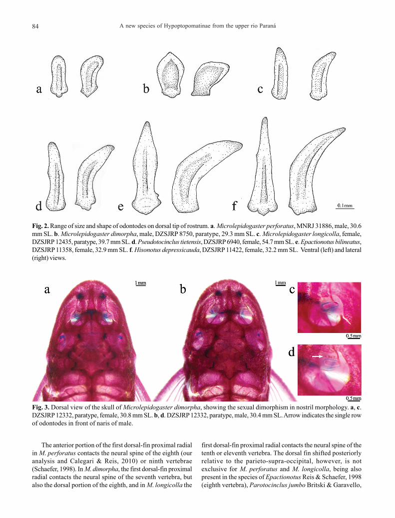
posteriorly from anus to end of anal-fin base; straight tocaudal-fin origin. Greatest body depth at dorsal-fin origin.Greatest body width at opercular region, gradually taperingtoward snout and caudal fin. Head profile rounded in dorsalview; rostral margins with thin plates, not deflected ventrally;anterior tip of rostrum with a median pair of plates; odontodesat anterior margin of snout small and leaf-shaped (Fig. 2b),some with edge slightly rounded. Odontodes equal in size
Fig. 1. Microlepidogaster dimorpha, holotype, DZSJRP 10543, 37.6 mm SL, female, riacho Grotão at Fazenda Nossa Senhorada Abadia, Uberaba, Minas Gerais, Brazil.

A new species of Hypoptopomatinae from the upper rio Paraná82
and uniformly distributed, not forming rows, on head andbody. Eyes small, dorsolaterally placed, not visible fromventral view. Iris operculum present. Infraorbital canalentering infraorbital series via compound pterotic.
Compound pterotic roughly quadrangular in shape, withoutelongate posterior extension; small to median roundishperforations on posteroventral margin. Caudal peduncleslightly flattened dorsally and ventrally, somewhatrectangular in transverse section.
Abdomen partially covered by small plates, approximatelysame size of pupil, irregularly arranged. Body entirely coveredwith bone plates, except on ventral part of head, regionoverlying opening of swim bladder capsule, and around anusand pelvic-fin origin.
Lips roundish, papillose; lower lip larger than upper lip;larger papillae near oral opening. Maxillary barbel reduced,free from oral disk. Teeth slender, bifid; median cusp largerand rounded, lateral smaller and pointed. Premaxillary teeth15-28 (mode = 23,24). Dentary teeth 12-25 (mode = 21).Premaxillary and dentary accessory teeth absent.
Dorsal fin ii, 6-7 (mode = 7), originating approximately atvertical through end of pelvic-fin base; its length surpassinganal-fin origin; spinelet small, somewhat rectangular, anteriorand posterior margins slightly straight; locking mechanismnon-functional. Anterior portion of compound supraneural-first dorsal-fin proximal radial contacting neural spine of 7th
vertebra; posteriorly contacting the 8th vertebra. Pectoral fini, 6-7 (mode = 6); originating immediately behind opercularopening and surpassing pelvic-fin origin. Cleithrum andcoracoid exposed and supporting odontodes laterally,covered by skin medially. Arrector fossae partially enclosedby ventral lamina of coracoid; opening extending laterallyapproximately halfway towards pectoral-fin base. Pectoralaxillary slit present, about 2.5 times in orbital diameter. Pelvicfin i, 5-6 (mode = 5); reaching anal-fin origin in males whendepressed. Anal fin i, 4-6 (mode = 5); originating at verticalthrough end of dorsal-fin base. Caudal fin i, 14, i; lobes equalin size; 5-6 (mode = 5) dorsal and 4-5 (mode = 5) ventralprocurrent rays. Adipose fin and azygous plates absent. Medianlateral plate series 24-27 plates, complete from compoundpterotic to caudal-fin base. Vertebrae 29-30 (mode = 29).
Table 1. Morphometric data for Microlepidogaster dimorpha.Holotype (H) and 30 paratypes, 16 females, and 14 males(DZSJRP 5564, 8750, 12332; MCP 45866; MNRJ 37848; MZUSP107585); range includes holotype. SD = Standard deviation.
Table 2. Frequency distribution of meristic data for Microlepidogaster dimorpha. Rays and teeth counts were made forholotype (DZSJRP 10543) and for 30 paratypes (DZSJRP 5564, 8750, 12332; MCP 45866; MNRJ 37848; MZUSP 107585). Plates(counted in both sides whenever possible), procurrent rays, and vertebrae counts were made for 4 c&s paratypes (DZSJRP8750, 12332).
Character H Minimum Maximum Mean SD Standard length (mm) 37.6 25.0 37.7 - -
Percents of Standard length Predorsal length 44.8 42.3 50.0 45.3 1.5 Preanal length 57.1 53.1 62.2 58.3 1.6 Prepectoral length 25.4 23.4 29.9 26.4 1.2 Prepelvic length 36.6 33.8 42.6 37.2 1.8 Postanal length 36.9 33.0 38.5 35.7 1.4 Thoracic length 16.1 13.8 18.5 15.9 1.1 Abdominal length 21.8 19.5 23.6 21.8 1.0 Caudal-peduncle depth 10.3 10.0 11.4 10.7 0.4 Head length 30.4 29.8 37.2 32.3 1.4 Head width 22.5 21.4 24.5 22.7 0.8 Head depth 14.4 14.1 16.7 15.2 0.7 Base of dorsal-fin length 12.9 10.6 14.3 12.6 0.8 Folded dorsal-fin length 22.4 20.3 25.2 23.3 1.1 Pectoral-fin unbranched ray length 21.6 20.0 23.8 22.1 1.0 Pelvic-fin unbranched ray length 16.4 15.4 20.9 18.3 1.8 Males - 18.3 20.9 19.9 0.8 Females 16.4 15.4 18.2 16.9 1.0 Snout-opercle length 24.9 23.8 28.8 25.7 0.9 Dorsal to anal-fin length 20.5 19.4 23.8 21.5 1.0
Percents of Head length Head width 74.1 62.7 75.9 70.3 2.9 Head depth 47.5 43.3 53.1 47.2 2.2 Snout length 51.4 48.3 54.4 51.1 1.5 Orbital diameter 17.3 13.6 18.5 16.5 1.3 Interorbital length 44.1 38.8 44.8 42.2 1.7 Barbel length 4.5 4.0 8.1 5.5 1.0 Prenasal length 34.5 28.0 35.8 32.4 1.9 Internasal length 16.3 9.2 17.0 12.8 1.8 Nostril width 7.9 7.9 15.4 11.2 2.4 Males - 11.5 15.4 13.5 1.2 Females 7.9 7.9 10.3 9.2 0.7 Suborbital depth 24.5 20.4 24.9 23.2 1.2
Character Frequency distribution Range Mode Dorsal plates 24(3); 25(4) 24-25 25 Mid-dorsal plates 18(1); 19(3); 20(1); 21(1) 18-21 19 Median plates 24(1); 25(2); 26(1); 27(1) 24-27 25 Mid-ventral plates 17(1); 18(2); 19(3); 20(1) 17-20 19 Ventral plates 20(1); 21(1); 22(4); 23(1) 20-23 22 Premaxillary teeth 15(1); 16(1); 17(2); 18(5); 19(7); 20(4); 21(3); 22(9); 23(10); 24(10); 25(2); 26(1); 27(4); 28(1) 15-28 23-24 Dentary teeth 12(1); 14(3); 15(4); 16(7); 17(5); 18(6); 19(6); 20(5); 21(9); 22(5); 23(2); 24(2); 25(1) 12-25 21 Dorsal-fin branched rays 6(1); 7(29) 6-7 7 Pectoral-fin branched rays 6(29); 7(1) 6-7 6 Pelvic-fin branched rays 5(29); 6(1) 5-6 5 Anal-fin branched rays 4(1); 5(28); 6(1) 4-6 5 Caudal-fin branched rays 14(30) - 14 Dorsal procurrent rays 5(2); 6(1) 5-6 5 Ventral procurrent rays 4(1); 5(2) 4-5 5 Vertebrae 29(3); 30(1) 29-30 29

F. O. Martins & F. Langeani 83
Color in alcohol. Most specimens with a dark brown coloralong the dorsal portion of head and dorsal midline of body.Four dorsal dark saddles, not extending laterally,inconspicuous in adults. Longitudinal clear stripe fromdorsal portion of snout to nostril. Lateroventral portion ofhead unpigmented. Lateral portion of body brown; aninconspicuous dark longitudinal stripe at median plateseries, extending from compound pterotic to verticalthrough end of depressed dorsal fin. Ventral surface ofbody light brown. Dorsal, anal, pectoral, and pelvic-finsmembrane hyaline; all rays with transverse dark bands.Caudal fin mostly dark brown, except for hyaline portionsat lobe tips, middle-rays extremities, and a circular area inthe middle of each lobe.
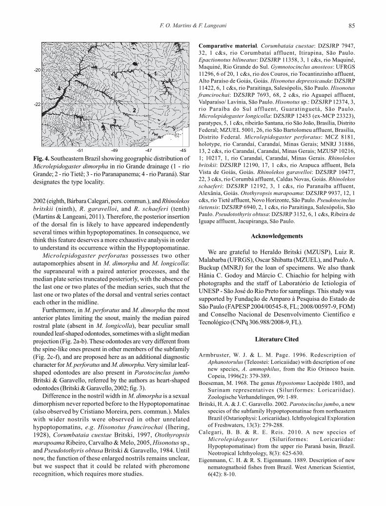
Sexual dimorphism. Males with a conspicuous urogenitalpapillae immediately posterior to anus (vs. absent in females);expanded flap of skin on dorsal surface of the first pelvic-finray and branched rays, less developed in the latter (vs. absentin females); pelvic fin reaching anal-fin origin, its firstunbranched ray 18.3-20.9% in SL (vs. pelvic fin not reachinganal-fin origin, its first unbranched ray 15.4-18.2% in SL infemales); males, usually with fewer teeth than females,probably related with their smaller size; males with widernostril width (11.5-15.4% of HL vs. 7.9-10.3% in females),with an anterior single row of 4 to 6 small odontodes
Table 3. Morphometric data for Microlepidogasterperforatus (MNRJ 31886, MZUSP 10216, 10217; n = 5). SD =Standard deviation.
Character Minimum Maximum Mean SD Standard length (mm) 29.93 32.76 - -
Percents of Standard length Predorsal length 45.5 47.8 46.7 1.0 Preanal length 54.8 58.4 57.2 1.4 Prepectoral length 25.5 27.5 26.5 0.8 Prepelvic length 35.2 36.6 35.9 0.6 Postanal length 35.5 39.5 37.1 1.6 Thoracic length 13.6 15.5 14.4 0.8 Abdominal length 19.3 23.2 20.8 1.6 Caudal-peduncle depth 7.7 8.5 8.1 0.3 Head lenght 33.2 34.7 34.1 0.6 Head width 21.0 23.2 22.4 0.9 Head depth 13.9 14.4 14.2 0.2 Base of dorsal-fin length 11.2 12.6 11.6 0.5 Folded dorsal-fin length 21.0 22.8 22.1 0.7 Pectoral-fin unbranched ray length
19.5 20.7 20.1 0.4
Pelvic-fin unbranched ray length 15.0 19.1 16.6 1.7 Snout-opercle length 24.1 26.5 25.7 0.9 Dorsal to anal-fin length 16.7 18.8 17.5 0.8
Percents of Head length Head width 60.6 68.0 65.6 3.0 Head depth 40.7 42.3 41.6 0.7 Snout length 47.5 51.9 49.2 1.9 Orbital diameter 11.1 13.5 12.6 1.0 Interorbital length 36.7 41.2 39.3 1.9 Barbel length 6.0 8.7 7.1 1.1 Prenasal length 32.2 35.3 33.6 1.4 Internasal length 10.0 12.7 11.6 1.0 Nostril width 11.2 12.4 11.9 0.62 Suborbital depth 20.4 21.7 21.0 0.5
Character Frequency distribution Range Mode Dorsal plates 25(1); 26(4); 27(1) 25-27 26 Mid-dorsal plates 9(1); 10(2); 11(1); 12(1); 13(1) 9-13 10 Median plates 21(2); 23(2); 25(2) 21-25 21/23/25 Mid-ventral plates 19(3); 20(3) 19-20 19/20 Ventral plates 20(2), 21(2); 22(2) 20-22 20/21/22 Premaxillary teeth 16(4); 17(4); 18(1); 19(1) 16-19 16/17 Dentary teeth 11(1); 12(2); 13(4); 14(1); 15(2) 11-15 13 Dorsal-fin branched rays 7(5) - 7 Pectoral-fin branched rays 5(1); 6(4) 5-6 6 Pelvic-fin branched rays 5(5) - 5 Anal-fin branched rays 5(5) - 5 Caudal-fin branched rays 14 (5) - 14 Dorsal procurrent rays 4(1); 5(2) 4-5 5 Ventral procurrent rays 4(3) - 4 Vertebrae 31 - 31
Table 4. Frequency distribution of meristics forMicrolepidogaster perforatus. Plates (counted in both sideswhenever possible), procurrent rays were made for 3 specimens,and vertebrae counts were made for 1 c&s specimen (MNRJ31886; MZUSP 10216, 10217).
associated with skin of the anterior portion of anterior nostril(vs. absent in females; Fig. 3).
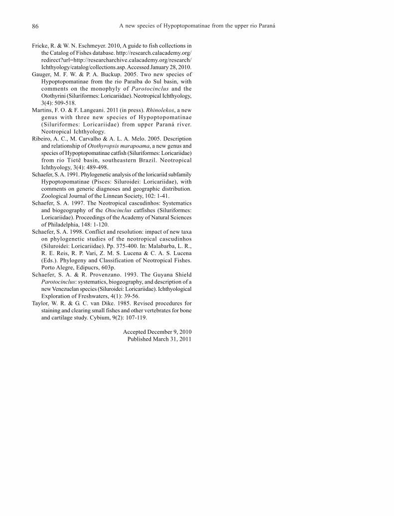
Distribution. The species is known from rio Uberaba andriacho Grotão, both tributaries of the rio Grande, upper rioParaná system, Minas Gerais, Brazil (Fig. 4).
Etymology. Epithet dimorpha from the Greek di, two, double,and morphe, form, in allusion to the accentuated sexualdimorphism presented by the species. A feminine adjective.
Discussion
According to Schaefer (1998), Microlepidogaster perforatusis diagnosed by five autapomorphies: levator crest absent(character 14:1); neural spine of seventh vertebra not contactingthe nuchal plate, and first dorsal-fin proximal radial articulatingwith the neural spine of the ninth vertebra (26: 1); supraneuralwith paired anterior processes (28: 1); median plate seriestruncated (33:1); median rostral plate, or paired plate, present (34:1).
Recently, a new species was proposed, M. longicollaCalegari & Reis, 2010, which was included in theMicrolepidogaster by sharing with M. perforatus the dorsalfin placed posteriorly, with the first pterygiophore articulatingwith the neural spine of the tenth or eleventh vertebral centrum(with the eighth or ninth in M. perforatus). Now,Microlepidogaster dimorpha was included in the genus bysharing with M. perforatus two other apomorphic characterstates of Schaefer (1998): levator crest extremely reduced insize and median rostral plate paired.
Schaefer (1998) considered the levator crest absent in M.perforatus; however Calegari & Reis (2010) identified a lowcrest in that species. Indeed, M. perforatus presents anextremely reduced crest in the hyomandibula, which is sharedwith M. dimorpha (vs. more developed in M. longicolla, as inthe plesiomorphic state).
A new species of Hypoptopomatinae from the upper rio Paraná84
The anterior portion of the first dorsal-fin proximal radialin M. perforatus contacts the neural spine of the eighth (ouranalysis and Calegari & Reis, 2010) or ninth vertebrae(Schaefer, 1998). In M. dimorpha, the first dorsal-fin proximalradial contacts the neural spine of the seventh vertebra, butalso the dorsal portion of the eighth, and in M. longicolla the
first dorsal-fin proximal radial contacts the neural spine of thetenth or eleventh vertebra. The dorsal fin shifted posteriorlyrelative to the parieto-supra-occipital, however, is notexclusive for M. perforatus and M. longicolla, being alsopresent in the species of Epactionotus Reis & Schaefer, 1998(eighth vertebra), Parotocinclus jumbo Britski & Garavello,
Fig. 3. Dorsal view of the skull of Microlepidogaster dimorpha, showing the sexual dimorphism in nostril morphology. a, c.DZSJRP 12332, paratype, female, 30.8 mm SL. b, d. DZSJRP 12332, paratype, male, 30.4 mm SL. Arrow indicates the single rowof odontodes in front of naris of male.
Fig. 2. Range of size and shape of odontodes on dorsal tip of rostrum. a. Microlepidogaster perforatus, MNRJ 31886, male, 30.6mm SL. b. Microlepidogaster dimorpha, male, DZSJRP 8750, paratype, 29.3 mm SL. c. Microlepidogaster longicolla, female,DZSJRP 12435, paratype, 39.7 mm SL. d. Pseudotocinclus tietensis, DZSJRP 6940, female, 54.7 mm SL. e. Epactionotus bilineatus,DZSJRP 11358, female, 32.9 mm SL. f. Hisonotus depressicauda, DZSJRP 11422, female, 32.2 mm SL. Ventral (left) and lateral(right) views.
F. O. Martins & F. Langeani 85
2002 (eighth, Bárbara Calegari, pers. commun.), and Rhinolekosbritskii (ninth), R. garavelloi, and R. schaeferi (tenth)(Martins & Langeani, 2011). Therefore, the posterior insertionof the dorsal fin is likely to have appeared independentlyseveral times within hypoptopomatines. In consequence, wethink this feature deserves a more exhaustive analysis in orderto understand its occurrence within the Hypoptopomatinae.
Microlepidogaster perforatus possesses two otherautapomorphies absent in M. dimorpha and M. longicolla:the supraneural with a paired anterior processes, and themedian plate series truncated posteriorly, with the absence ofthe last one or two plates of the median series, such that thelast one or two plates of the dorsal and ventral series contacteach other in the midline.
Furthermore, in M. perforatus and M. dimorpha the mostanterior plates limiting the snout, mainly the median pairedrostral plate (absent in M. longicolla), bear peculiar smallrounded leaf-shaped odontodes, sometimes with a slight medianprojection (Fig. 2a-b). These odontodes are very different fromthe spine-like ones present in other members of the subfamily(Fig. 2c-f), and are proposed here as an additional diagnosticcharacter for M. perforatus and M. dimorpha. Very similar leaf-shaped odontodes are also present in Parotocinclus jumboBritski & Garavello, referred by the authors as heart-shapedodontodes (Britski & Garavello, 2002; fig. 3).
Difference in the nostril width in M. dimorpha is a sexualdimorphism never reported before to the Hypoptopomatinae(also observed by Cristiano Moreira, pers. commun.). Maleswith wider nostrils were observed in other unrelatedhypoptopomatins, e.g. Hisonotus francirochai (Ihering,1928), Corumbataia cuestae Britski, 1997, Otothyropsismarapoama Ribeiro, Carvalho & Melo, 2005, Hisonotus sp.,and Pseudotothyris obtusa Britski & Garavello, 1984. Untilnow, the function of these enlarged nostrils remains unclear,but we suspect that it could be related with pheromonerecognition, which requires more studies.
Comparative material. Corumbataia cuestae: DZSJRP 7947,32, 1 c&s, rio Corumbataí affluent, Itirapina, São Paulo.Epactionotus bilineatus: DZSJRP 11358, 3, 1 c&s, rio Maquiné,Maquiné, Rio Grande do Sul. Gymnotocinclus anosteos: UFRGS11296, 6 of 20, 1 c&s, rio dos Couros, rio Tocantinzinho affluent,Alto Paraíso de Goiás, Goiás. Hisonotus depressicauda: DZSJRP11422, 6, 1 c&s, rio Paraitinga, Salesópolis, São Paulo. Hisonotusfrancirochai: DZSJRP 7693, 68, 2 c&s, rio Aguapeí affluent,Valparaíso/ Lavínia, São Paulo. Hisonotus sp.: DZSJRP 12374, 3,rio Paraíba do Sul affluent, Guaratinguetá, São Paulo.Microlepidogaster longicolla: DZSJRP 12453 (ex-MCP 23323),paratypes, 5, 1 c&s, ribeirão Santana, rio São João, Brasília, DistritoFederal; MZUEL 5001, 26, rio São Bartolomeu affluent, Brasília,Distrito Federal. Microlepidogaster perforatus: MCZ 8181,holotype, rio Carandaí, Carandaí, Minas Gerais; MNRJ 31886,13, 2 c&s, rio Carandaí, Carandaí, Minas Gerais; MZUSP 10216,1; 10217, 1, rio Carandaí, Carandaí, Minas Gerais. Rhinolekosbritskii: DZSJRP 12190, 17, 1 c&s, rio Arapuca affluent, BelaVista de Goiás, Goiás. Rhinolekos garavelloi: DZSJRP 10477,22, 3 c&s, rio Corumbá affluent, Caldas Novas, Goiás. Rhinolekosschaeferi: DZSJRP 12192, 3, 1 c&s, rio Paranaíba affluent,Alexânia, Goiás. Otothyropsis marapoama: DZSJRP 9937, 12, 1c&s, rio Tietê affluent, Novo Horizonte, São Paulo. Pseudotocinclustietensis: DZSJRP 6940, 2, 1 c&s, rio Paraitinga, Salesópolis, SãoPaulo. Pseudotothyris obtusa: DZSJRP 3152, 6, 1 c&s, Ribeira deIguape affluent, Jacupiranga, São Paulo.
Acknowledgements
We are grateful to Heraldo Britski (MZUSP), Luiz R.Malabarba (UFRGS), Oscar Shibatta (MZUEL), and Paulo A.Buckup (MNRJ) for the loan of specimens. We also thankHânia C. Godoy and Márcio C. Chiachio for helping withphotographs and the staff of Laboratório de Ictiologia ofUNESP - São José do Rio Preto for samplings. This study wassupported by Fundação de Amparo à Pesquisa do Estado deSão Paulo (FAPESP 2004/00545-8, FL; 2008/00597-9, FOM)and Conselho Nacional de Desenvolvimento Científico eTecnológico (CNPq 306.988/2008-9, FL).
Literature Cited
Armbruster, W. J. & L. M. Page. 1996. Redescription ofAphanotorulus (Teleostei: Loricaiidae) with description of onenew species, A. ammophilus, from the Rio Orinoco basin.Copeia, 1996(2): 379-389.
Boeseman, M. 1968. The genus Hypostomus Lacépède 1803, andSurinam representatives (Siluriformes: Loricariidae).Zoologische Verhandelingen, 99: 1-89.
Britski, H. A. & J. C. Garavello. 2002. Parotocinclus jumbo, a newspecies of the subfamily Hypoptopomatinae from northeasternBrazil (Ostariophysi: Loricariidae). Ichthyological Explorationof Freshwaters, 13(3): 279-288.
Calegari, B. B. & R. E. Reis. 2010. A new species ofMicrolepidogaster (Siluriformes: Loricariidae:Hypoptopomatinae) from the upper rio Paraná basin, Brazil.Neotropical Ichthyology, 8(3): 625-630.
Eigenmann, C. H. & R. S. Eigenmann. 1889. Description of newnematognathoid fishes from Brazil. West American Scientist,6(42): 8-10.
Fig. 4. Southeastern Brazil showing geographic distribution ofMicrolepidogaster dimorpha in rio Grande drainage (1 - rioGrande; 2 - rio Tietê; 3 - rio Paranapanema; 4 - rio Paraná). Stardesignates the type locality.
A new species of Hypoptopomatinae from the upper rio Paraná86
Fricke, R. & W. N. Eschmeyer. 2010, A guide to fish collections inthe Catalog of Fishes database. http://research.calacademy.org/redirect?url=http://researcharchive.calacademy.org/research/Ichthyology/catalog/collections.asp. Accessed January 28, 2010.
Gauger, M. F. W. & P. A. Buckup. 2005. Two new species ofHypoptopomatinae from the rio Paraíba do Sul basin, withcomments on the monophyly of Parotocinclus and theOtothyrini (Siluriformes: Loricariidae). Neotropical Ichthyology,3(4): 509-518.
Martins, F. O. & F. Langeani. 2011 (in press). Rhinolekos, a newgenus with three new species of Hypoptopomatinae(Siluriformes: Loricariidae) from upper Paraná river.Neotropical Ichthyology.
Ribeiro, A. C., M. Carvalho & A. L. A. Melo. 2005. Descriptionand relationship of Otothyropsis marapoama, a new genus andspecies of Hypoptopomatinae catfish (Siluriformes: Loricariidae)from rio Tietê basin, southeastern Brazil. NeotropicalIchthyology, 3(4): 489-498.
Schaefer, S. A. 1991. Phylogenetic analysis of the loricariid subfamilyHypoptopomatinae (Pisces: Siluroidei: Loricariidae), withcomments on generic diagnoses and geographic distribution.Zoological Journal of the Linnean Society, 102: 1-41.
Schaefer, S. A. 1997. The Neotropical cascudinhos: Systematicsand biogeography of the Otocinclus catfishes (Siluriformes:Loricariidae). Proceedings of the Academy of Natural Sciencesof Philadelphia, 148: 1-120.
Schaefer, S. A. 1998. Conflict and resolution: impact of new taxaon phylogenetic studies of the neotropical cascudinhos(Siluroidei: Loricariidae). Pp. 375-400. In: Malabarba, L. R.,R. E. Reis, R. P. Vari, Z. M. S. Lucena & C. A. S. Lucena(Eds.). Phylogeny and Classification of Neotropical Fishes.Porto Alegre, Edipucrs, 603p.
Schaefer, S. A. & R. Provenzano. 1993. The Guyana ShieldParotocinclus: systematics, biogeography, and description of anew Venezuelan species (Siluroidei: Loricariidae). IchthyologicalExploration of Freshwaters, 4(1): 39-56.
Taylor, W. R. & G. C. van Dike. 1985. Revised procedures forstaining and clearing small fishes and other vertebrates for boneand cartilage study. Cybium, 9(2): 107-119.
Accepted December 9, 2010Published March 31, 2011