madaka fish project
TRANSCRIPT

The life of Chester the fish
\

What Chester needs before hatching

What Chester Needs after hatching

Day 1 With Chester

Day 2 With Chester

Day 3 With Chester
Chester was really starting to grow big now. He was now in stage
thirty. You could really start to see the changes in his body now.
He was starting to develop blood vessels inside his body. You
could also see his heart now. This is a big jump for him. Pretty
soon he will actually have blood going through him.

Day 4 With Chester
The next day with Chester was a very interesting one. He
started having blood going through his body. I was very happy
to see this. I could now see more life in his body. As a parent I
was very excited to see this. He was now in stage 36 of
development. I was so excited for him to hatch soon!
Day 5 With Chester
This was the best day of my life Chester had finally hatched! I had
so many plans for him now. The first thing we were going to go do
was get him a new bigger bowl. Sydney was going to take him
since it was his choice. Once we got to the store he told her that he
didn’t care how big the bowl was as long as he was with Sydney
and I.

The Anatomy of Chester
• filament- Hair like sturcture coming out of the Medaka. The function of these is to protect the egg.
• chorion- This is a membrane that is found between the mother and Fetus. It is formed by layers and surrounds or protect the embryo.
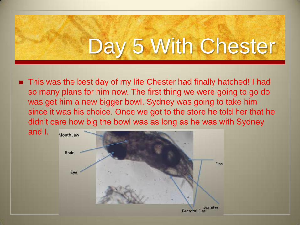
Mouth/ Jaws- The jaws form the opening fo the mouth. The mouth is the natural opening in which food passes into the body
• cytoplasm- This is enclosed within the plasma membrane. The cytoplasm contains many cell parts and is the location where many cell processes occur
The Anatomy of Chester
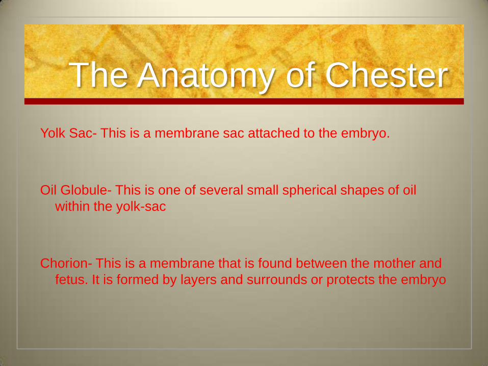
Yolk Sac- This is a membrane sac attached to the embryo.
Oil Globule- This is one of several small spherical shapes of oil
within the yolk-sac
Chorion- This is a membrane that is found between the mother and
fetus. It is formed by layers and surrounds or protects the embryo

The Anatomy of Chester
Heart- Is a mustcular organ responsible for pumping blood through
vessels which then transports the blood to the rest of the body
Eyes- Allows Medaka to see.
Pectoral Fins- These are paired fins located on each side of the
fish. The fins act as the forelimbs of the fish
Somites- These are distributed along the sides of the neural tube
that will eventually become dermis muscles and vertebrae.

Recources
Madaka Fish Book
http://ani.embl.de:8080/mepd/medakaStages/MedakaDevStages.ht
ml
http://animal-world.com/encyclo/fresh/Killifish/MoonlightMedaka.php