lights on for aminopeptidases in cystic kidney disease · of chikungunya virus from aedes aegypti...
TRANSCRIPT

commentaries
660 TheJournalofClinicalInvestigation http://www.jci.org Volume 120 Number 3 March 2010
8. Halstead SB. Chikungunya. In: Feign RD, Cherry J, Demmler-Harrison GJ, Kaplan SL, eds. Feign and Cherry’s Textbook of Pediatric Infectious Diseases. Phila-delphia, PA: Saunders Elsevier; 2004:2178–2184.
9. Enserink M. Epidemiology. Tropical dis-ease follows mosquitoes to Europe. Science. 2007;317(5844):1485.
10. Myers RM, Carey DE, Reuben R, Jesudass ES, De Ranitz C, Jadhav M. The 1964 epidemic of dengue-like fever in South India: isolation of chikungu-nya virus from human sera and from mosquitoes. Indian J Med Res. 1965;53(8):694–701.
11. Anderson CR, Singh KR, Sarkar JK. Isolation of chikungunya virus from Aedes aegypti fed on naturally infected humans in Calcutta. Curr Sci. 1965;34:579–580.
12. Simon F, et al. Chikungunya infection; an emerg-ing rheumatism among travelers returned from Indian Ocean Islands. Report of 47 cases. Medicine. 2007;86(3):123–137.
13. Schuffenecker I, et al. Genome microevolution of chikungunya viruses causing the Indian Ocean outbreak. PLoS Med. 2006;3(7):e263.
14. de Lamballerie X, Leroy E, Charrel RN, Tsetsarkin K, Higgs S, Gould EA. Chikungunya virus adapts to tiger mosquito via evolutionary convergence: a sign of things to come? Virol J. 2008;5:33.
15. Tsetsarkin KA, Vanlandingham DL, McGee CE, Higgs S. A single mutation in chikungunya virus affects vector specificity and epidemic potential. PLoS Pathog. 2007;3(12):e201.
16. Tsetsarkin KA, McGee CE, Volk SM, Vanlanding-ham DL, Weaver SC, Higgs S. Epistatic roles of E2 glycoprotein mutations in adaption of chikungunya virus to Aedes albopictus and Ae. aegypti mosquitoes. PLoS One. 2009;4(8):e6835.
17. Benedict MQ, Levine RS, Hawley WA, Lounibos LP. Spread of the tiger: global risk of invasion by the mosquito Aedes albopictus. Vector Borne Zoonotic Dis. 2007;7(1):76–85.
18. Gould EA, Higgs S. Impact of climate change and
other factors on emerging arbovirus diseases. Trans R Soc Trop Med Hyg. 2009;103(2):109–121.
19. Lidbury BA, et al. Macrophage-derived proinflamma-tory factors contribute to the development of arthri-tis and myositis after infection with an arthrogenic alphavirus. J Infect Dis. 2008;197(11):1585–1593.
20. Ziegler SA, Lu L, da Rosa AP, Xiao SY, Tesh RB. An animal model for studying the pathogenesis of chikungunya virus infection. Am J Trop Med Hyg. 2008;79(1):133–139.
21. Wang E, et al. Chimeric alphavirus vaccine candidates for chikungunya. Vaccine. 2008;26(39):5030–5039.
22. Couderc T, et al. A mouse model for Chikungunya: young age and inefficient type-I interferon signal-ing are risk factors for severe disease. PLoS Pathog. 2008;4(2):e29.
23. Taubitz W, et al. Chikungunya fever in travelers: clinical presentation and course. Clin Infect Dis. 2007;45(1):e1–e4.
24. Dubrulle M, Mousson L, Moutailler S, Vazeille M, Failloux AB. Chikungunya virus and Aedes mosqui-toes: saliva is infectious as soon as two days after oral infection. PLoS One. 2009;4(6):e5895.
25. Boppana VD, Thangamani S, Adler AJ, Wikel SK. SAAG-4 is a novel mosquito salivary protein that programmes host CD4 T cells to express IL-4. Para-site Immunol. 2009;31(6):287–295.
26. Schneider BS, Soong L, Girard YA, Campbell G, Mason P, Higgs S. Potentiation of West Nile encephalitis by mosquito feeding. Viral Immunol. 2006;19(1):74–82.
27. Binn LN, Harrison VR, Randall R. Patterns of vire-mia and antibody observed in rhesus monkeys inoculated with chikungunya and other serologi-cally related group A arboviruses. Am J Trop Med Hyg. 1967;16(6):782–785.
28. Paul SD, Singh KR. Experimental infection of Macaca radiata with Chikungunya virus and trans-mission of virus by mosquitoes. Indian J Med Res. 1968;56(6):802–811.
29. Chen C-I, et al. Development of a macaque model
for in utero chikungunya virus infection. In: Ameri-can Society of Tropical Medicine and Hygiene 58th Annual Meeting; November 18–22, 2009:Washing-ton, DC. Abstract 2997.
30. Akahata W, et al. A virus-like particle vaccine for epidemic Chikungunya virus protects non-human primates against infection [published online ahead of print January 28, 2010]. Nat Med. doi:10.1038/nm.2105.
31. Rulli NE, et al. Ross River virus: molecular and cel-lular aspects of disease pathogenesis. Pharmacol Ther. 2005;107(3):329–342.
32. McIntosh BM, Peterson HE, McGillivray GM, de Sousa J. Further studies on the chikungunya out-break in southern Rhodesia in 1962. I. Mosquitoes, wild primates and birds in relation to the epidemic. Ann Trop Med Parasitol. 1964;58:45–51.
33. McIntosh BM, Harwin RM, Paterson HE, Westwater ML. An epidemic of Chikungunya in South-Eastern Southern Rhodesia. Cent Afr J Med. 1963;43:351–359.
34. Inoue S, et al. Distribution of three arbovirus anti-bodies among monkeys (Macaca fascicularis) in the Philippines. J Med Primatol. 2003;32(2):89–94.
35. Marchette NJ, Rudnick A, Garcia R, MacVean DW. Alphaviruses in Peninusular Malaysia: I. Virus iso-lations and animal serology. Southeast Asian J Trop Med Public Health. 1978;9(3):317–329.
36. Peiris JS, Dittus WP, Ratnayake CB. Seroepide-miology of dengue and other arboviruses in a natural population of toque macaques (Macaca sinica) at Polonnaruwa, Sri Lanka. J Med Primatol. 1993;22(4):240–245.
37. Cawthon Lang KA. Primate factsheets: Long-tailed macaque (Macaca fascicularis) taxonomy, mor-phology, & ecology. http://pin.primate.wisc.edu/ factsheets/entry/long-tailed_macaque/taxon. Updat-ed January 6, 2006. Accessed January 28, 2010.
38. Chastel C. Chikungunya virus: its recent spread to the southern Indian Ocean and Reunion Island (2005-2006) [in French]. Bull Acad Natl Med. 2005;189(8):1827–1835.
Lights on for aminopeptidases in cystic kidney disease
Erwin P. Böttinger
Charles R. Bronfman Institute for Personalized Medicine, and Department of Medicine, Mount Sinai School of Medicine, New York, New York.
Whileeruditecellbiologistshaveformanydecadesdescribedsingularimmotileappendagesknownasprimaryciliatobepresentonmostcellsinourbodies,cilialfunction(s)longremainedanenigma.Drivenlargelybyaneverincreasingnumberofdiscoveriesofgeneticdefectsinprimaryciliaduringthepastdecade,ciliawerecatapultedfromalonglastingexistenceinobscurityintothebrightspotlightincellbiologyandmedicine.ThestudybyO’Tooleetal.inthisissueoftheJCIaddsanovel“enzymatic”facettotherapidlygrowinginformationabouttheselittlecellulartails,bydemonstrat-ingthatdefectsintheXPNPEP3gene,whichencodesmitochondrialandcytosolicsplicevariantsofX-prolylaminopeptidase3,cancausenephro-nophthisis-likeciliopathy.FuturestudiesareinordernowtoelucidatethecystogenicpathwaysaffectedbydisruptedenzymaticfunctionofXPNPEP3incilia-relatedcystogenicdiseases.
Based on a flood of recent evidence, prima-ry cilia are now heralded as sensory organ-elles for detection and transmission of a broad range of cues from the extracellular
Conflictofinterest: The author has declared that no conflict of interest exists.
Citationforthisarticle: J Clin Invest. 2010; 120(3):660–663. doi:10.1172/JCI42378.
environment of cells (1), including mechan-ical and chemical information as diverse as fluid flow in kidneys, mechanical bone deformation, and light and odorant detec-tion (2). By processing such physical and chemical information from the environ-ment into molecular signals in develop-ment and postnatal growth and homeosta-sis, cilia can affect cell differentiation and polarity and cell cycle control (3).
Genetic damage to primary cilia results in a spectrum of perplexing disorders with seemingly disparate manifestations, now classified as ciliopathies. A growing list of bona fide ciliopathies at present includes Bardet-Biedl syndrome (BBS), nephro-nophthisis (NPHP), and Senior-Loken syn-drome (SNLS), just to name a few. Common
commentaries
TheJournalofClinicalInvestigation http://www.jci.org Volume 120 Number 3 March 2010 661
clinical features of ciliopathies include the hallmark renal cysts as well as liver disease, laterality defects, polydactyly, cognitive dys-function, retinal degeneration, skeletal bone defects, and obesity among others (4).
Cilia are composed of a plasma mem-brane sheath, enveloping a microtubule-based axoneme that extends from the basal
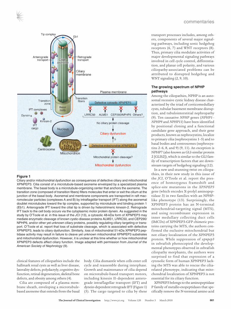
body. Cilia dismantle when cells enter cell cycle and reassemble during interphase. Growth and maintenance of cilia depend on microtubule-based transport motors, including kinesin II–dependent antero-grade intraflagellar transport (IFT) and dynein-dependent retrograde IFT (Figure 1) (5). The cargo targeted to cilia by these
transport processes includes, among oth-ers, components of several major signal-ing pathways, including sonic hedgehog receptors (6, 7) and WNT receptors (8). Thus, primary cilia modulate activities of major developmental signaling pathways involved in cell cycle control, differentia-tion, and planar cell polarity, and various ciliopathy-associated problems can be attributed to disrupted hedgehog and WNT signaling (2, 9, 10).
The growing spectrum of NPHP pathwaysAmong the ciliopathies, NPHP is an auto-somal recessive cystic kidney disease char-acterized by the triad of corticomedullary cysts, tubular basement membrane disrup-tion, and tubulointerstitial nephropathy (9). Ten causative NPHP genes (NPHP1–NPHP9 and NPHP11) have been identified by positional cloning and a functional candidate gene approach, and their gene products, known as nephrocystins, localize to primary cilia (nephrocystins 1–5) and to basal bodies and centrosomes (nephrocys-tins 2–6, 8, and 9) (9, 11). An exception is NPHP7 (also known as GLI-similar protein 2 [GLIS2]), which is similar to the GLI fam-ily of transcription factors that are down-stream targets of hedgehog signaling (12).
In a new and stunning twist on ciliopa-thies, in their new study in this issue of the JCI, O’Toole et al. report the pres-ence of homozygous frameshift and splice-site mutations in the XPNPEP3 gene (which encodes X-prolyl aminopep-tidase 3) in two families with an NPHP-like phenotype (13). Surprisingly, the XPNPEP3 protein has an N-terminal mitochondrial-targeting signal (MTS), and using recombinant expression in inner medullary collecting duct cells (IMCDs) of XPNPEP3-GFP chimeric pro-teins carrying the MTS, the authors con-firmed the exclusive mitochondrial but not ciliary localization of the XPNPEP3 protein. While suppression of xpnpep3 in zebrafish phenocopied the develop-mental phenotypes observed in zebrafish ciliopathy morphants, the authors were surprised to find that expression of a cytosolic form of human XPNPEP3 lack-ing the MTS was able to rescue the cilia-related phenotype, indicating that mito-chondrial localization of XPNPEP3 is not essential for its ciliary function.
XPNPEP3 belongs to the aminopeptidase P family of metallo-exopeptidases that spe-cifically remove the N-terminal amino acid
Figure 1Ciliary and/or mitochondrial dysfunction as consequences of defective ciliary and mitochondrial XPNPEP3. Cilia consist of a microtubule-based axoneme enveloped by a specialized plasma membrane. The basal body is a microtubule-organizing center that anchors the axoneme. The transition zone (composed of transition fibers) filters molecules that enter or exit the cilium at the junction of the basal body. Axonemal and membrane components are transported in raft mac-romolecular particles (complexes A and B) by intraflagellar transport (IFT) along the axonemal doublet microtubules toward the tip complex, supported by microtubule end binding protein 1 (Eb1). Anterograde IFT toward the cilial tip is driven by heterotrimeric kinesin-2. Retrograde IFT back to the cell body occurs via the cytoplasmic motor protein dynein. As suggested in the study by O’Toole et al. in this issue of the JCI (13), a cytosolic 48-kDa form of XPNPEP3 may mediate enzymatic cleavage of known cystic-disease proteins ALMS1, LRRC50, and CEP290/NPHP6, and/or other yet unknown ciliary proteins, possibly regulating ciliary targeting or trans-port. O’Toole et al. report that loss of substrate cleavage, which is associated with defective XPNPEP3, leads to ciliary dysfunction. Similarly, loss of mitochondrial 51-kDa XPNPEP3 pep-tidase activity may result in failure to cleave yet unknown mitochondrial XPNPEP3 substrates and mitochondrial dysfunction. However, it is unclear at this time whether or how mitochondrial XPNPEP3 defects affect ciliary function. Image adapted with permission from Journal of the American Society of Nephrology (9).

commentaries
662 TheJournalofClinicalInvestigation http://www.jci.org Volume 120 Number 3 March 2010
from peptides that have a proline residue in the second position after methionine (14). XPNPEP2 is a ubiquitous cell-surface mem-brane–bound peptidase that is thought to degrade peptides filtered by renal epithe-lium brush border membranes but may also be involved in bradykinin metabolism in endothelial cells (15). In contrast to XPNPEP2, physiological substrates or functions have not yet been identified for XPNPEP1 (which is a soluble cytosolic iso-form) or XPNPEP3 (which is known to occur in two alternative splice forms) (Figure 2) (14). In contrast with the mitochondrial form of XPNPEP3, the cytosolic variant lacks the MTS (Figure 2). Importantly, both splice variants are expressed in kidney (14).
Proof of principle for the XPNPEP3 enzymeHow then may a poorly characterized enzyme with mitochondrial localization in distal tubules and intercalated collect-ing duct cells in human kidney cause cystic renal disease similar to NPHP, a bona fide ciliopathy? O’Toole et al. examined mito-chondrial and respiratory chain functions in muscle biopsies and fibroblasts in affected individuals of the aforementioned kindreds (13). Mitochondriopathy with childhood end-stage renal disease and severe extrare-nal manifestations (seizures, cardiomyopa-thy, and pancreatitis) was observed in two affected individuals in the Turkish kindred, carrying the homozygous frameshift muta-tion p.N311LfsX5. However, mitochondrial function was normal in affected adults with moderate renal disease from the Finnish kindred, carrying the less severe splice-site mutation p.G453C in XPNPEP3. These find-
ings were inconsistent with the mitochon-drial dysfunction hypothesis, and together with the observation that a cytosolic form of XPNPEP3 was able to rescue cilia-related phenotypes in the zebrafish studies, these data suggest that the functional defect underlying the NPHP-like renal phenotype might be independent of mitochondria.
The authors offer no explanation for the apparent contradiction between mitochon-drial localization and cytosolic function of XPNPEP3 in NPHP-like phenotypes in their study but pursued another lead with acceptable results (13). They examined a pool of 426 likely ciliary protein sequences for candidate XPNPEP3 substrates and identified 51 putative candidates, includ-ing three proteins — centrosomal protein 290 kDa/NPHP6 (CEP290/NPHP6), Alstrom syndrome 1 (ALMS1), and leucine-rich repeat–containing 50 (LRRC50) — that are known to cause cystic disease (16–18). Indeed, synthetic peptides identical to the N-termini of all three substrate candidates were cleaved as predicted by the recombi-nant Escherichia coli ortholog of XPNPEP3, ecAPP. A key finding was the demonstration of the authors that an xpnpep3-mediated cleavage–resistant LRRC50 failed, while WT LRRC50 succeeded to rescue the cilia-related phenotype associated with endog-enous lrrc50 suppression in zebrafish. The authors rightfully concluded that loss of activity of XPNPEP3-mediated enzymatic cleavage of substrates with ciliary and cen-trosomal functions may explain some forms of NPHP-like cystic renal disease, but their provocative speculation about an emerging link between mitochondria and ciliary dys-function deserves further scrutiny.
Mitochondrial XPNPEP3: a red herring for ciliopathies?Is it possible that the mitochondrial local-ization of XPNPEP3 shown in this report (13) is a red herring when it comes to cili-opathy pathways? The answer may well be affirmative. XPNPEP3 localization stud-ies in the current report were performed exclusively using an XPNPEP3-GFP chime-ric construct containing the MTS, which, not surprisingly, localized exclusively to mitochondria when expressed in murine IMCD3 cells. However, two alternative splice variants of XPNPEP3 were previ-ously reported in kidney (14). The alterna-tive splice variant studied by O’Toole et al. lacks exon 3 and initiates translation of a 507–amino acid XPNPEP3 isoform in exon 1, encoding an N-terminal MTS, which is removed by peptidases after import to mitochondria, resulting in an approxi-mately 51-kDa mature mitochondrial enzyme (Figure 2). In contrast, exon 3 pres-ents a stop codon in-frame to the exon 1 AUG, resulting in premature termination and removal of the truncated protein, and expression of a 428–amino acid isoform of XPNPEP3 devoid of MTS, translated from an AUG site located in exon 4 (14). Indeed, after probing for endogenous XPNPEP3 in IMCD3 cell lysates, the authors describe, in addition to the 51-kDa mitochondrial form, a second discrete band of XPNPEP3 of approximately 48 kDa, which can be pre-dicted from the 428–amino acid cytosolic form of XPNPEP3 (13).
If the 48-kDa band is indeed the pre-dicted cytosolic form of XPNPEP3, then the mitochondrial link may well be a red herring, because the NPHP-like dis-ease observed in some individuals with XPNPEP3 mutations may be attributable to loss of proper cleavage of ciliary pro-teins by defective cytosolic XPNPEP3, as suggested by the overwhelming evidence from zebrafish studies (13). Of course, the same defect(s) in the mitochondrial form of XPNPEP3 may underlie some of the observed extrarenal manifestations, such as cardiomyopathy and seizures, which may be caused by mitochondrial dysfunc-tion, as demonstrated in one kindred, and independent of ciliary dysfunction.
Perhaps the most intriguing prospect of the exciting study by O’Toole et al. (13) is that the novel discovery of functional mutations in the XPNPEP3 aminopepti-dase in individuals presenting with NPHP-like ciliopathy may well lead to the further elucidation of two independent pathways
Figure 2Two alternative splice forms of XPNPEP3 exist in the kidney. If exon 3 of XPNPEP3 is absent, the XPNPEP3 gene product starts with amino acid sequence MPWI and contains an N-terminal MTS that targets the protein to mitochondria. Exon 3 contains a stop codon (stp) in-frame, resulting in premature termination and removal of the MTS-containing gene product. The alternative XPNPEP3, lacking MTS, is translated from exon 4 AUG, possesses the starting amino acid sequence MSLI, and is likely localized to the cytosol. Mitochondrial XPNPEP3, i.e., the exon 3–spliced form, was used in the localization studies reported in this issue by O’Toole et al. (13).

commentaries
TheJournalofClinicalInvestigation http://www.jci.org Volume 120 Number 3 March 2010 663
associated with a distinct phenotypic spec-trum: a cystogenic cilium-dependent path-way, dependent on defective cleavage of ciliary proteins by cytosolic XPNPEP3, and a mitochondriopathy, dependent on defec-tive cleavage of mitochondrial substrates by mitochondrial XPNPEP3. Wow! Future work is needed to define the mitochondrial versus ciliary substrates and roles for hith-erto rather obscure, spatially defined metal-lo-exopeptidases, encoded by a single novel human disease gene, XPNPEP3. Investiga-tors concerned with ciliopathies and mito-chondriopathies should stay tuned.
AcknowledgmentsThe author is supported by NIH grants 5 R 0 1 D K 0 5 6 0 7 7 , 5 R 0 1 D K 0 6 0 0 4 3 , 5R01DK073960, 3U01DK060995, and 1U01DK085688.
Address correspondence to: Erwin P. Böt-tinger, Charles R. Bronfman Institute for Personalized Medicine, and Depart-ment of Medicine, Mount Sinai School of Medicine, One Gustave L. Levy Pl.,
Box 1003, New York, NY 10029. Phone: 212.241.0800; Fax: 212.849.2643; E-mail: [email protected].
1. Pazour GJ, Witman GB. The vertebrate primary cilium is a sensory organelle. Curr Opin Cell Biol. 2003;15(1):105–110.
2. Gerdes JM, Davis EE, Katsanis N. The vertebrate primary cilium in development, homeostasis, and disease. Cell. 2009;137(1):32–45.
3. Singla V, Reiter JF. The primary cilium as the cell’s antenna: signaling at a sensory organelle. Science. 2006;313(5787):629–633.
4. Tobin JL, Beales PL. The nonmotile ciliopathies. Genet Med. 2009;11(6):386–402.
5. Scholey JM. Intraflagellar transport motors in cilia: moving along the cell’s antenna. J Cell Biol. 2008;180(1):23–29.
6. Rohatgi R, Milenkovic L, Scott MP. Patched1 regu-lates hedgehog signaling at the primary cilium. Science. 2007;317(5836):372–376.
7. Corbit KC, Aanstad P, Singla V, Norman AR, Stainier DY, Reiter JF. Vertebrate smooth-ened functions at the primary cilium. Nature. 2005;437(7061):1018–1021.
8. Gerdes JM, Katsanis N. Ciliary function and Wnt sig-nal modulation. Curr Top Dev Biol. 2008;85:175–195.
9. Hildebrandt F, Attanasio M, Otto E. Nephro-nophthisis: disease mechanisms of a ciliopathy. J Am Soc Nephrol. 2009;20(1):23–35.
10. Lancaster MA, et al. Impaired Wnt-beta-catenin signaling disrupts adult renal homeostasis and leads to cystic kidney ciliopathy. Nat Med.
2009;15(9):1046–1054. 11. Otto EA, et al. Hypomorphic mutations in meck-
elin (MKS3/TMEM67) cause nephronophthi-sis with liver fibrosis (NPHP11). J Med Genet. 2009;46(10):663–670.
12. Attanasio M, et al. Loss of GLIS2 causes neph-ronophthisis in humans and mice by increased apoptosis and f ibrosis. Nat Genet . 2007; 39(8):1018–1024.
13. O’Toole J, et al. Individuals with mutations in XPNPEP3, which encodes a mitochondrial protein, develop a nephronophthisis-like nephropathy. J Clin Invest. 2010;120(3):791–802.
14. Ersahin C, Szpaderska AM, Orawski AT, Simmons WH. Aminopeptidase P isozyme expression in human tis-sues and peripheral blood mononuclear cell fractions. Arch Biochem Biophys. 2005;435(2):303–310.
15. Kim KS, Kumar S, Simmons WH, Brown NJ. Inhi-bition of aminopeptidase P potentiates wheal response to bradykinin in angiotensin-converting enzyme inhibitor-treated humans. J Pharmacol Exp Ther. 2000;292(1):295–298.
16. Sayer JA, et al. The centrosomal protein nephro-cystin-6 is mutated in Joubert syndrome and acti-vates transcription factor ATF4. Nat Genet. 2006; 38(6):674–681.
17. Li G, et al. A role for Alstrom syndrome protein, alms1, in kidney ciliogenesis and cellular quiescence. PLoS Genet. 2007;3(1):e8.
18. Sullivan-Brown J, et al. Zebrafish mutations affecting cilia motility share similar cystic phe-notypes and suggest a mechanism of cyst forma-tion that differs from pkd2 morphants. Dev Biol. 2008;314(2):261–275.
Resisting arrest: a switch from angiogenesis to vasculogenesis in recurrent malignant gliomas
Jeffrey P. Greenfield,1,2 William S. Cobb,1,2 and David Lyden1,3
1Pediatric Brain Tumor Research and the Children’s Cancer and Blood Foundation Laboratories, Champalimaud Metastasis Programme, 2Department of Neurosurgery, and 3Department of Pediatrics, Weill Cornell Medical College and Memorial Sloan-Kettering Cancer Center, New York, New York.
Thecellularandmoleculareventsthatinitiateandpromotemalignantgliomadevelopmentarenotcompletelyunderstood.Thetreatmentmodali-tiesdesignedtopromoteitsdemiseareallultimatelyineffective,leadingtodiseaseprogression.InthisissueoftheJCI,Kioietal.demonstratethatvas-culogenesisandangiogenesispotentiallyplaydistinctrolesintheetiologyofprimaryandrecurrentmalignantgliomas,suggestingthatpatienttherapyshouldperhapsbetailoredspecificallyagainstthepredominantvasculaturepathwayatagivenspecificstageofgliomagenesis.
Formation of new blood vessels is an essen-tial component of malignant glioma devel-opment and progression. As the mecha-nisms underlying this crucial element of tumor growth are gradually elucidated, it has become increasingly evident that this vascularity arises through multiple mechanisms, depending on the stage of the
tumor and possibly the therapies utilized to combat tumor growth. With unregulated cell proliferation and growing tumor size, there is a need for greater oxygen supply to sustain this growth. As mounting cellular metabolism outstrips the oxygen supply made available via the existing vasculature, hypoxia ensues, triggering the process that has been dubbed the “angiogenic switch.” While the additional blood supply initially may be obtained simply by co-option of preexisting vessels, the increasing hypoxia eventually necessitates angiogenesis — the
sprouting of local vessels via proliferation of existing endothelial cells. Alternatively, vasculogenesis may occur as factors released from tumor cells increase recruitment of circulating endothelial precursor cells or bone marrow–derived hematopoietic cells, also resulting in the formation of new ves-sels to supply the tumor. Understanding the balance between angiogenesis and vas-culogenesis lies at the very core of elucidat-ing how tumors grow, and is crucial to the development of anti-angiogenic therapies. A better comprehension of this process may also allow for the design of new, more effec-tive therapies to target the pathways tumors employ to sustain their growth. In the cur-rent issue of the JCI, Kioi et al. (1) suggest that vasculogenesis — but not angiogenesis — is at the center of the revascularization that occurs during glioma recurrence. In clinical terms, their findings suggest that when gliomas recur after irradiation, as they
Conflictofinterest: The authors have declared that no conflict of interest exists.
Citationforthisarticle:J Clin Invest. 2010; 120(3):663–667. doi:10.1172/JCI42345.