lezione- reticolo endoplasmatico · muovere carichi cellulari (vescicole, mitocondri, lisosomi,...
TRANSCRIPT
Reticolo Endoplasmatico (ER) e
dintorni
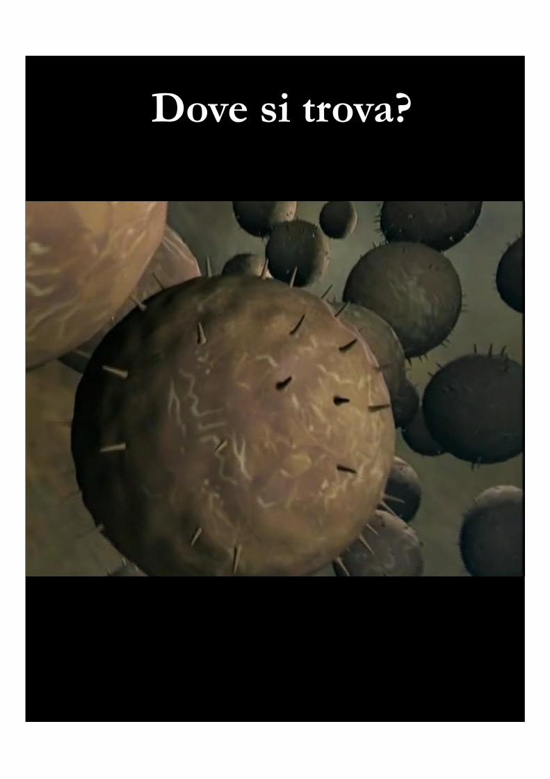
Dove si trova?


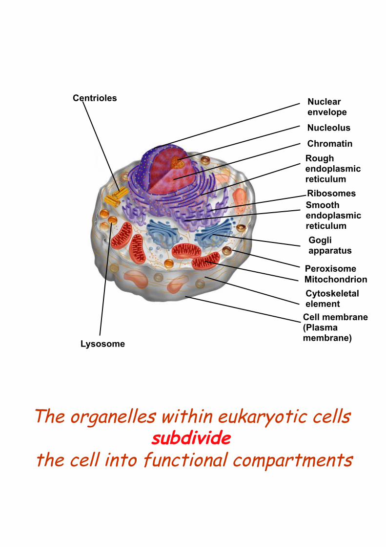
Centrioles
Lysosome
Nuclear envelope Nucleolus
Chromatin Rough endoplasmic reticulum Ribosomes Smooth endoplasmic reticulum Gogli apparatus
Peroxisome Mitochondrion Cytoskeletal element
Cell membrane (Plasma membrane)
The organelles within eukaryotic cells subdivide
the cell into functional compartments

The organelles within eukaryotic cells subdivide
the cell into functional compartments
• The endomembrane system is the site of biomolecule synthesis and considerable molecular movement
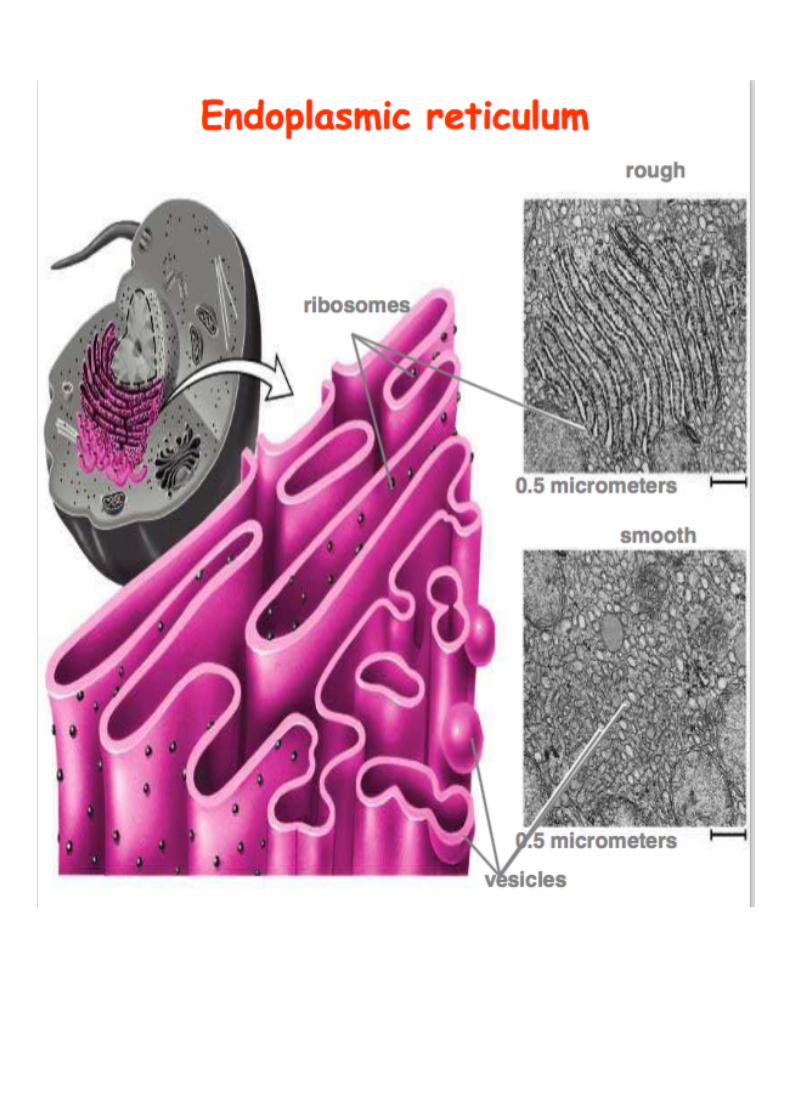
Rough ER (with ribosomes attached)
Smooth ER (no ribosomes attached)
Golgi apparatus

Intracellular comparment
Percentage total cell volume
Cytosol 54Mitochondria 22Rough ER 9Smooth ER + Golgi
6
Nucleus 6Peroxisomes 1Lysosomes 1Endosomes 1
THE ENDOPLASMIC
RETICULUM
is a mesh of interconnected membranes that serves a function
involving protein synthesis and transport

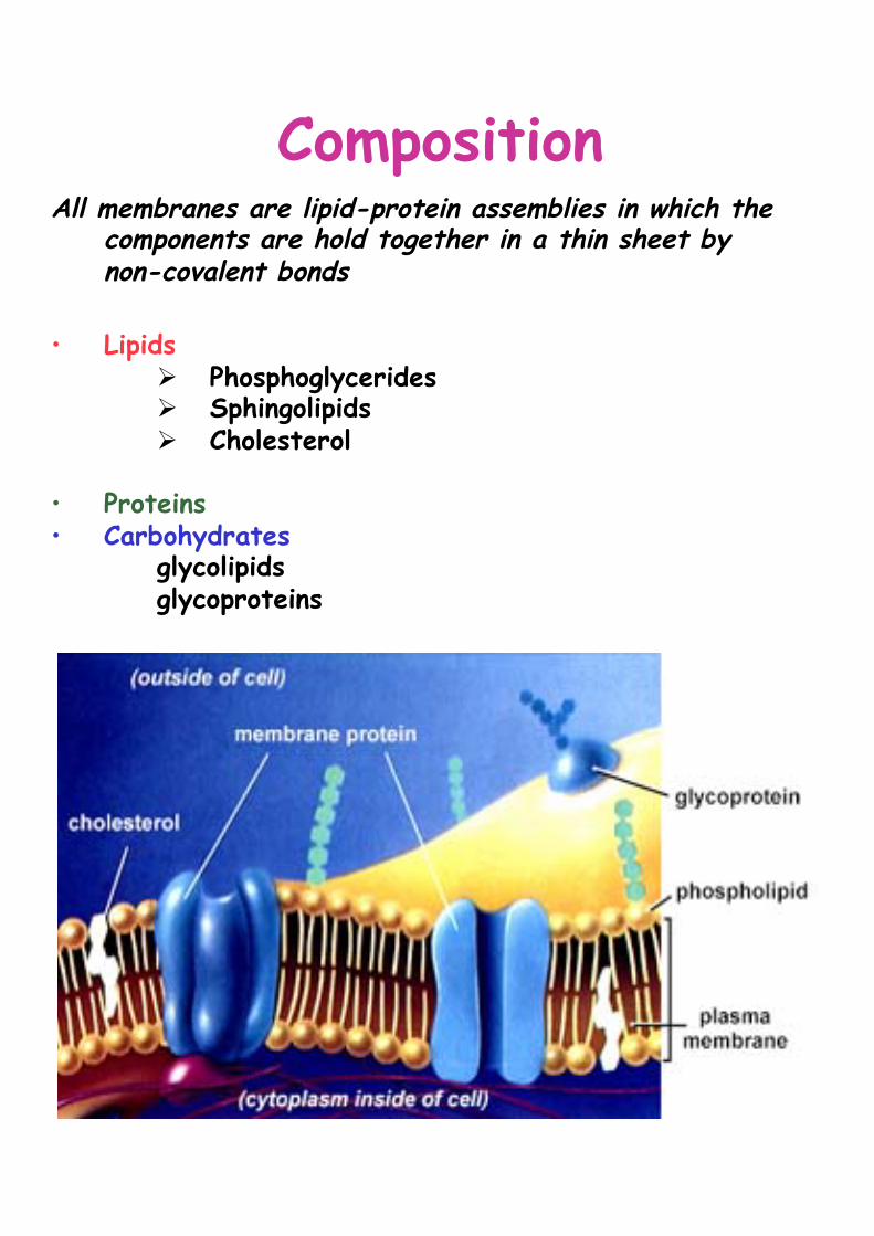
Composition All membranes are lipid-protein assemblies in which the
components are hold together in a thin sheet by non-covalent bonds
• Lipids
Ø Phosphoglycerides Ø Sphingolipids Ø Cholesterol
• Proteins • Carbohydrates
glycolipids glycoproteins

La composizione della membrana del RE


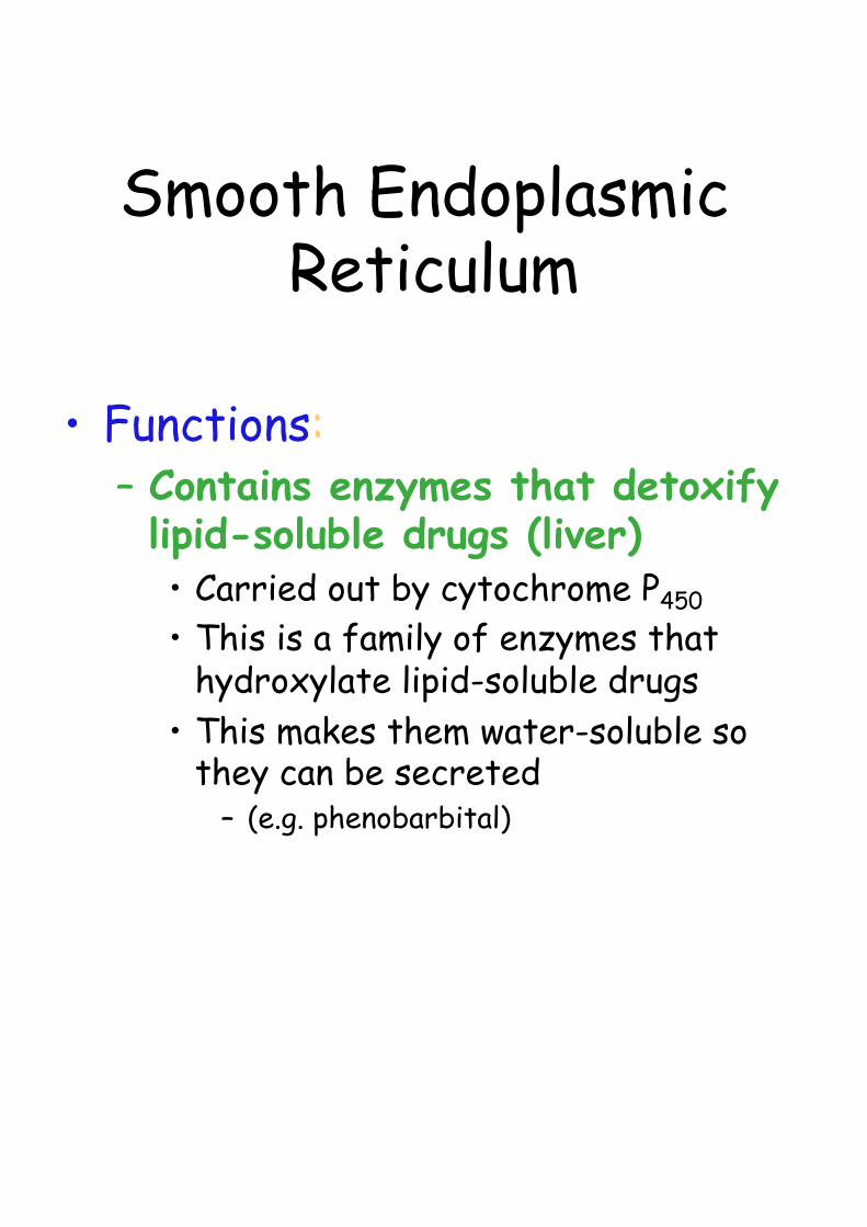
Smooth Endoplasmic Reticulum
• Functions: – Contains enzymes that detoxify lipid-soluble drugs (liver) • Carried out by cytochrome P450 • This is a family of enzymes that
hydroxylate lipid-soluble drugs • This makes them water-soluble so
they can be secreted – (e.g. phenobarbital)

Smooth Endoplasmic Reticulum
• Functions:
– Steroid hormones synthesis in endocrine cells of the gonads and of the adrenal glands
Smooth Endoplasmic Reticulum
• Functions: – Sequester Ca++ from the cytosol • The release of Ca++ into the cytosol
and its subsequent uptake into the ER are involved in many rapid responses to intracellular (muscle cells) extracellular signals
I RIBOSOMI
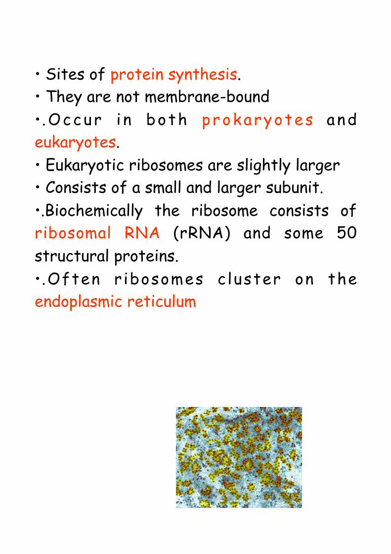
• Sites of protein synthesis. • They are not membrane-bound • .Occur i n both prokaryotes and eukaryotes. • Eukaryotic ribosomes are slightly larger • Consists of a small and larger subunit. • .Biochemically the ribosome consists of ribosomal RNA (rRNA) and some 50 structural proteins. • .Often r ibosomes c luster on the endoplasmic reticulum
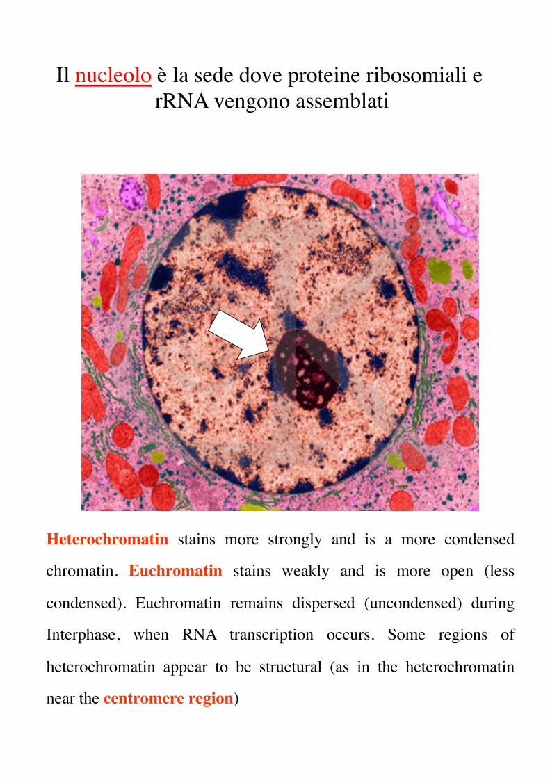
Heterochromatin stains more strongly and is a more condensed
chromatin. Euchromatin stains weakly and is more open (less
condensed). Euchromatin remains dispersed (uncondensed) during
Interphase, when RNA transcription occurs. Some regions of
heterochromatin appear to be structural (as in the heterochromatin
near the centromere region)
Il nucleolo è la sede dove proteine ribosomiali e rRNA vengono assemblati


Provenienza Ribosoma Subunita’ Composizione
rRNA 28S 60S RNA 5S
RNA 5.8SEucarioti 80S proteine 40S rRNA 18S
proteine
rRNA 23S 50S RNA 5SProcarioti 70S proteine
30S rRNA 16Sproteine
rRNA 16S 35S proteine
Mitocondri 55S 25S rRNA 12S
proteine

Types of proteins synthesised
1. On ER ribosomes • Proteins secreted by the cell
• Trans-membrane proteins
• Proteins of Golgi, lysosomes, endosomes, plant vacuoles
2. On free ribosomes
• Cytosol proteins (cytoskeleton, glycolysis enzymes)
• Peripheral proteins of the inner plasma membrane (spectrin, ankirin etc)
• Proteins of perossisomes, cloroplasts and mytocondria
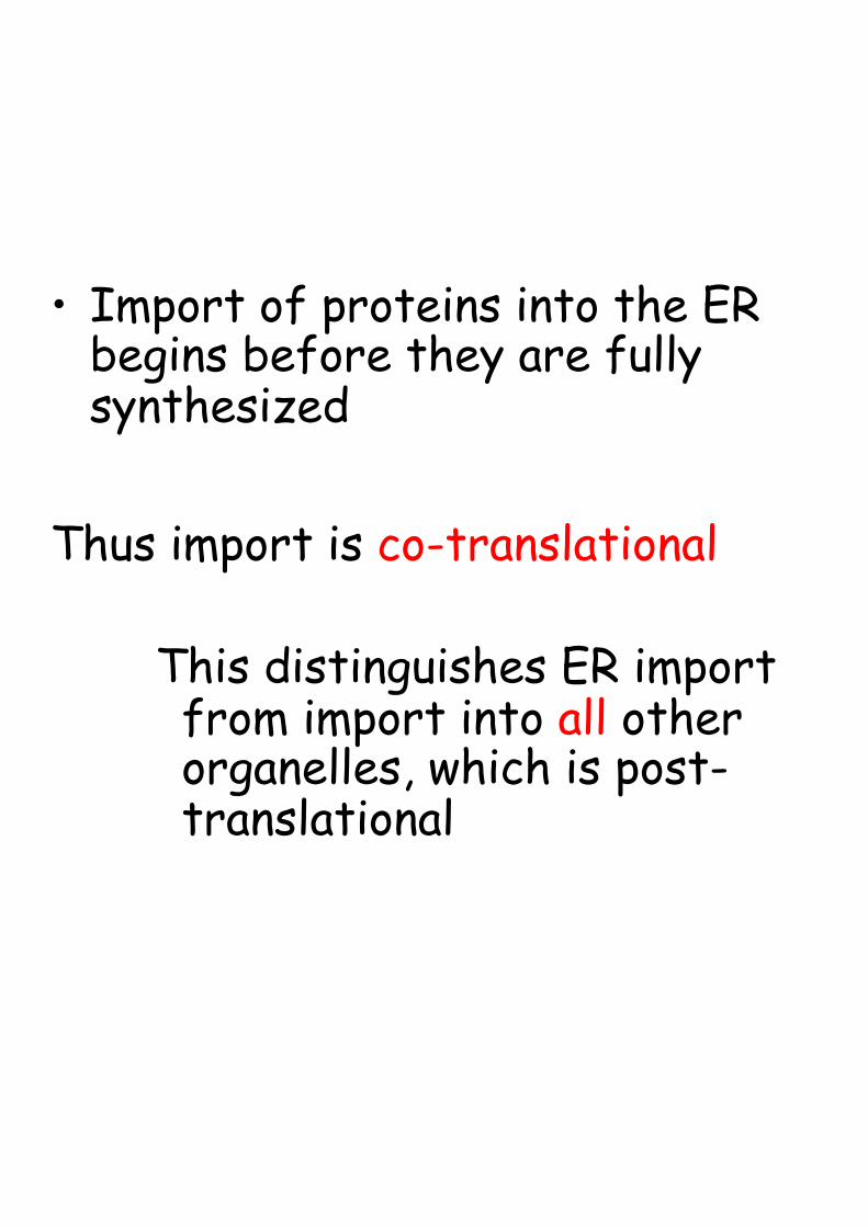
• Import of proteins into the ER begins before they are fully synthesized
Thus import is co-translational
This distinguishes ER import from import into all other organelles, which is post-translational


Sintesi proteica in ribosomi liberi

Three types of RNAThree types of RNA•Ribosomal RNA (rRNA)•Ribosomal RNA (rRNA)
•Messenger RNA (mRNA)•Messenger RNA (mRNA)
• Transfer RNA (tRNA)• Transfer RNA (tRNA)
combines with proteins to form the ribosomescombines with proteins to form the ribosomes
contains the instruction for the ordering of amino acids in proteinscontains the instruction for the ordering of amino acids in proteins
carries specific amino acids to ribosomescarries specific amino acids to ribosomes

• The genetic code is read in groups of three nucleotids, each group representing one amino acids. • Each trinucleotides sequence is called a codon. • A gene includes a series of codons that is read sequencially from a starting point at one end to a termination point at the other end.
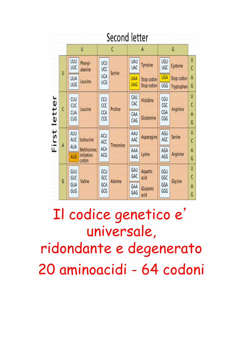
Il codice genetico e’ universale,
ridondante e degenerato 20 aminoacidi - 64 codoni

"for the discovery that proteins have intrinsic signals that govern their transport and localization in the cell"
The Nobel Prize in Physiology or Medicine 1999
Günter Blobel Rockefeller University New York, NY Howard Hughes Medical Institute Chevy Chase, MD, USA Born 1936 (in Waltersdorf/Silesia, Germany)

From mRNA to proteinsTransduction on ER-Ribosomes


Sintesi proteica in ribosomi sull’ER

Most of the membrane lipids are synthesised in the ER, with the exception of glycolipids and sfingomielin which synthesis begins in ER and is completed in the Golgi Differences in lipids composition in organelles are dependent to: • intrinsic capacity of organelles to modify lipids
• selection of phospholipids from vescicle membranes during budding
• protein-mediated phospholipids transport from ER to other organelles through the cytosol

Proteins destined for posttranslational import to the ER are synthesized on free ribosomes and maintained in an unfolded conformation by cytosolic chaperones. Their signal sequences are recognized by the Sec62/63 complex, which is associated with the Sec61 translocation channel in the ER membrane. The Sec63 protein is also associated with a chaperone protein (BiP), which acts as a molecular ratchet to drive protein translocation into the ER.
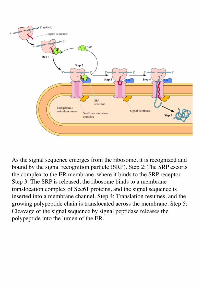
As the signal sequence emerges from the ribosome, it is recognized and bound by the signal recognition particle (SRP). Step 2: The SRP escorts the complex to the ER membrane, where it binds to the SRP receptor. Step 3: The SRP is released, the ribosome binds to a membrane translocation complex of Sec61 proteins, and the signal sequence is inserted into a membrane channel. Step 4: Translation resumes, and the growing polypeptide chain is translocated across the membrane. Step 5: Cleavage of the signal sequence by signal peptidase releases the polypeptide into the lumen of the ER.

Insertion of a membrane protein with a cleavable signal sequence and a single stop-transfer sequence
The signal sequence is cleaved as the polypeptide chain crosses the membrane, so the amino terminus of the polypeptide chain is exposed in the ER lumen. However, translocation of the polypeptide chain across the membrane is halted by a transmembrane stop-transfer sequence that closes the Sec61 translocation channel and exits the channel laterally to anchor the protein in the ER membrane. Continued translation results in a membrane-spanning protein with its carboxy terminus on the cytosolic side.

Insertion of a protein that spans the membrane multiple times
In this example, an internal signal sequence results in insertion of the polypeptide chain with its amino terminus on the cytosolic side of the membrane. A stop-transfer sequence then signals closure of the translocation channel, causing the polypeptide chain to form a loop within the lumen of the ER, and translation continues in the cytosol. A second internal signal sequence reopens the channel, triggering reinsertion of the polypeptide chain into the ER membrane and forming a loop in the cytosol. The process can be repeated many times, resulting in the insertion of proteins with multiple membrane-spanning regions.

Integral membrane proteins span the membrane via α-helical regions of 20 to 25 hydrophobic amino acids, which can be inserted in a variety of orientations. The two proteins at left and center each span the membrane once, but they differ in whether the amino (N) or carboxy (C) terminus is on the cytosolic side. On the right is an example of a protein that has multiple membrane-spanning regions.
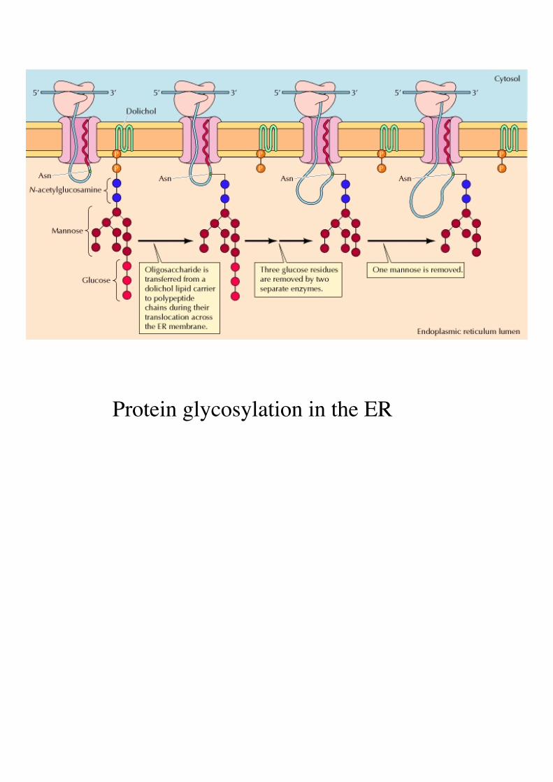
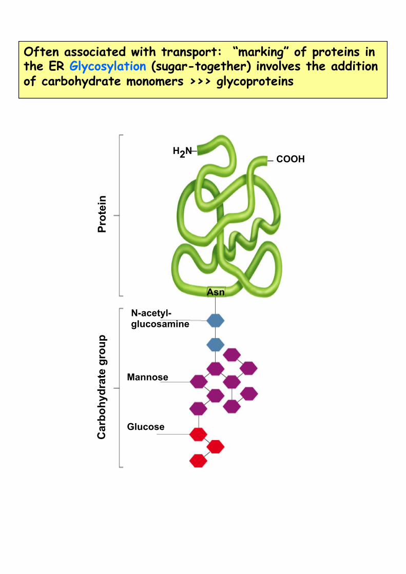
Protein glycosylation in the ER
Prot
ein
Car
bohy
drat
e gr
oup
N-acetyl-glucosamine
Mannose
Glucose
H2N COOH
Asn
Often associated with transport: “marking” of proteins in the ER Glycosylation (sugar-together) involves the addition of carbohydrate monomers >>> glycoproteins

Protein folding in the ER
The molecular chaperone BiP binds to polypeptide chains as they cross the ER membrane and facilitates protein folding and assembly within the ER.

Retrieval of resident ER proteins
Proteins destined to remain in the lumen of the ER are marked by the sequence Lys-Asp-Glu-Leu (KDEL) at their carboxy terminus. These proteins are exported from the ER to the Golgi in the nonselective bulk flow of proteins through the secretory pathway, but they are recognized by a receptor in the ER-Golgi intermediate compartment (ERGIC) or the Golgi apparatus and selectively returned to the ER.

The lumens of the endoplasmic reticulum and Golgi apparatus are topologically equivalent to the exterior of the cell. Consequently, those portions of polypeptide chains that are translocated into the ER are exposed on the cell surface following transport to the plasma membrane.
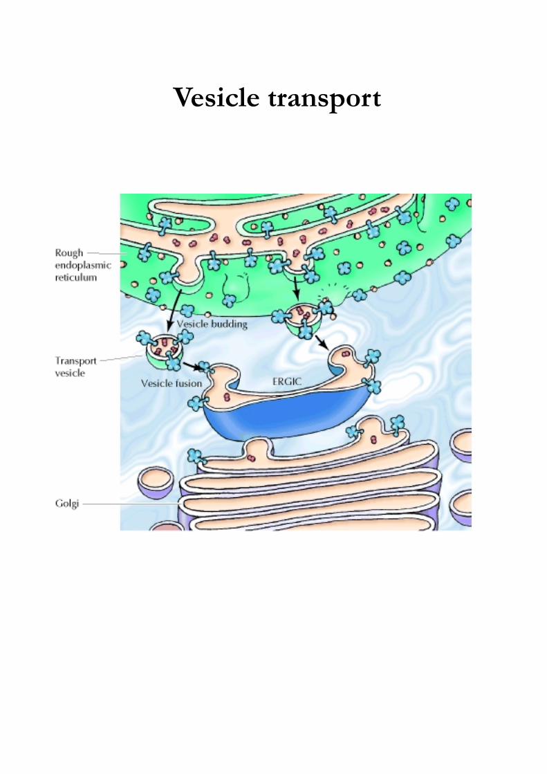
Vesicle transport
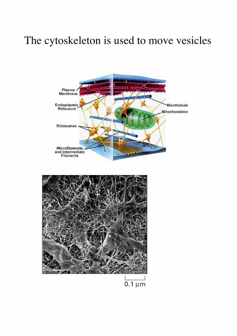
The cytoskeleton is used to move vesicles
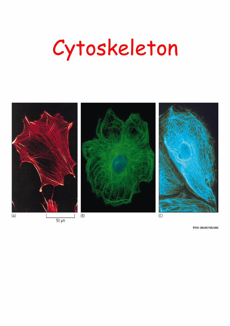
Cytoskeleton
actin microtubules intermediate
is the infrastructure on which most intracellular transport occurs
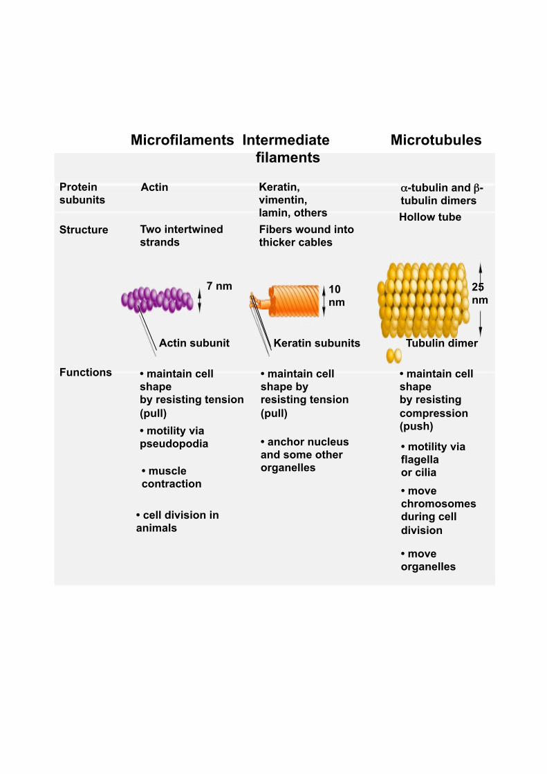
Microfilaments
Actin
Two intertwined strands
• maintain cell shape by resisting tension (pull)
Actin subunit
Protein subunits
Structure
Functions
• motility via pseudopodia
• muscle contraction
• cell division in animals
7 nm
Intermediate filaments
Keratin, vimentin, lamin, others Fibers wound into thicker cables
• maintain cell shape by resisting tension (pull)
• anchor nucleus and some other organelles
Microtubules
α-tubulin and β-tubulin dimers Hollow tube
Keratin subunits Tubulin dimer
• maintain cell shape by resisting compression (push)
• motility via flagella or cilia • move chromosomes during cell division
• move organelles
10 nm
25 nm

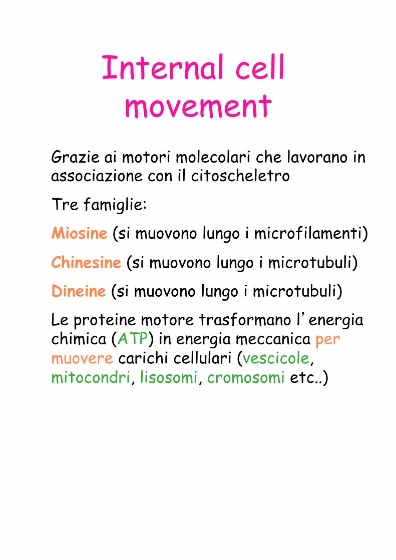
Internal cell movement
Grazie ai motori molecolari che lavorano in associazione con il citoscheletro
Tre famiglie:
Miosine (si muovono lungo i microfilamenti)
Chinesine (si muovono lungo i microtubuli)
Dineine (si muovono lungo i microtubuli)
Le proteine motore trasformano l’energia chimica (ATP) in energia meccanica per muovere carichi cellulari (vescicole, mitocondri, lisosomi, cromosomi etc..)

Structure of kinesin: one mechanism of movement
Tail
Stalk
Head

Transport vesicle
Kinesin
Microtubule
ATP ADP+Pi
Kinesin "walks" along a microtubule track
ATP ADP+Pi

Microtubule tracks Vesicles
Vesicle transport

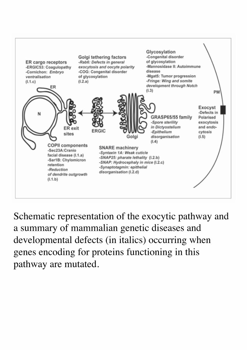
Schematic representation of the exocytic pathway and a summary of mammalian genetic diseases and developmental defects (in italics) occurring when genes encoding for proteins functioning in this pathway are mutated.