introduction to ekg monitoring - 360training.com to ekg monitoring ekg leads and traces the modern...
TRANSCRIPT

EKG Technician | Lesson 1: Introduction to EKG
© 2014 360training.com All Rights Reserved.
Introduction to EKG Monitoring EKG Leads and Traces
The modern EKG records cardiac electrical activity from different angles
of view. It records two basic electrical properties:
Depolarization: the spread of electrical current through heart
muscle, which can be demonstrated on the EKG as it occurs in
both the atria and the ventricles.
Repolarization: the return of the stimulated muscle to its resting state, ready for the next depolarization.
Repolarization can also be recorded on the EKG tracing.
The duration, amplitude, and direction of electrical activity within the heart are all evaluated on the EKG.
Duration is the time required to depolarize or repolarize cardiac muscle. Abnormal duration could signify
an electrical disturbance in the cardiac muscle.
Amplitude is determined in part by the size of the cardiac chambers and can be abnormal if those
chambers are enlarged, but amplitude can also be affected by a large body habitus of the patient.
The direction of the electrical activity depicts the overall vector direction of depolarization of the
ventricles.
The standard EKG consists of 12
different leads used to provide many
views of the heart from different
positions. On the EKG trace, 12 traces
correspond to each lead and additional
trace shows a longer version of Lead II.
Normal 12-Lead EKG Trace

EKG Technician | Lesson 1: Introduction to EKG
© 2014 360training.com All Rights Reserved.
Limb Leads
Six leads are called the limb leads. Leads I, II, and III are bipolar leads. Leads aVR, aVL, and aVF are unipolar
leads, which measure the heart’s electrical activity on the frontal plane (lying flat across the patient’s chest).
Limb Leads on a Normal 12-Lead EKG Trace
The three bipolar limb leads (I, II, III) are the original three leads of Einthoven’s triangle. They record impulse
between the following limbs:
Lead I: the right arm and left arm, where the negative pole is the right arm and the positive pole is the
left arm
Lead II: the right arm and left leg, where the right arm is the negative pole and the left leg is the
positive pole
Lead III: the left arm and left leg, where the left arm is the negative pole and the left leg is the
positive pole
Bipolar Limb Leads on a Normal 12-Lead EKG Trace
EKG Technician | Lesson 1: Introduction to EKG
© 2014 360training.com All Rights Reserved.
Bipolar leads each have a negative and positive end, or pole. The electrical
activity flows toward either a lead’s negative pole or positive pole.
For example, from the description of Lead I, the right arm is the negative pole
and the left arm is the positive pole. We will learn later that the heart’s normal
electrical activity spreads from right to left. Using this definition of Lead I, the
electrical activity, or impulse, will travel toward the patient’s left arm, or the
positive pole of Lead I. This will make Lead I appear upright on the EKG trace.
Look again at Lead I on the 12-lead EKG trace. Note that the tall parts of the
waves are above the baseline. We call this a positive deflection.
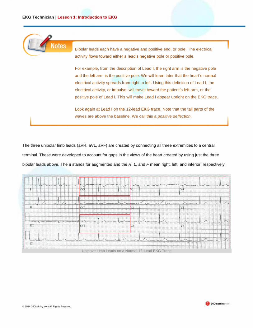
The three unipolar limb leads (aVR, aVL, aVF) are created by connecting all three extremities to a central
terminal. These were developed to account for gaps in the views of the heart created by using just the three
bipolar leads above. The a stands for augmented and the R, L, and F mean right, left, and inferior, respectively.
Unipolar Limb Leads on a Normal 12-Lead EKG Trace

EKG Technician | Lesson 1: Introduction to EKG
© 2014 360training.com All Rights Reserved.
Precordial (Chest) Leads
Six precordial, or chest, leads (V1–V6) provide views of the heart’s
electrical activity in the horizontal plane, similar to a flat plane crossing
through the patient’s chest.
Six Precordial Leads on a Normal 12-Lead EKG Trace
It is very important to understand that on the standard 12-lead EKG displayed
here, only 10 electrodes are actually attached to the patient—four limb and six
precordial.
Remember that if a wave of depolarization is moving toward the positive
electrode, the waveform is positive or upright on the EKG. If the wave of
depolarization is moving away from the positive electrode, the waveform is
negative or downward on the EKG.
Six Precordial Leads on a Man's
Chest