integumentary system human anatomy
TRANSCRIPT

Integumentary system
Human Anatomy
Assistant lecturer. Sawsan S. Hameed
Biology department
TIU
2021-2022

Outlines of the chapter
The integumentary system includes the skin, hair, oil and sweat glands, nails, and sensory receptors.
Thus, this chapter covers the following topics;
Functions of the integumentary system
Structure of the Skin and Subcutaneous tissue
Skin derivatives

Objectives of this lecture
You should be able to describe the following;
Functions of the integumentary system
Structure of the Skin
The layers of the skin
The difference between the integument layers
Introduction to integumentary system
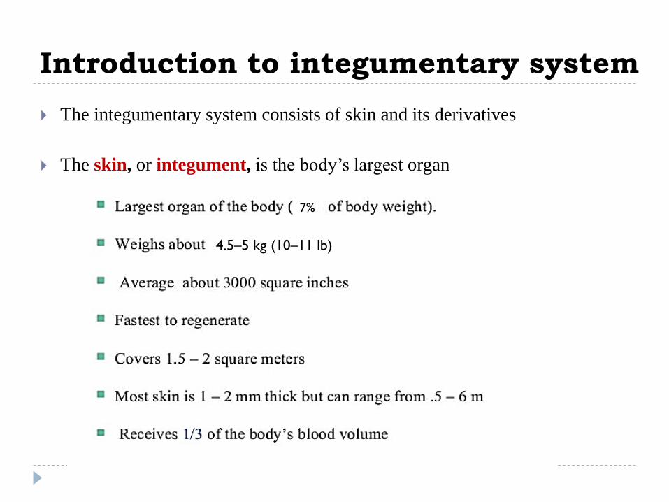
The integumentary system consists of skin and its derivatives
The skin, or integument, is the body’s largest organ
4.5–5 kg (10–11 lb)
7%

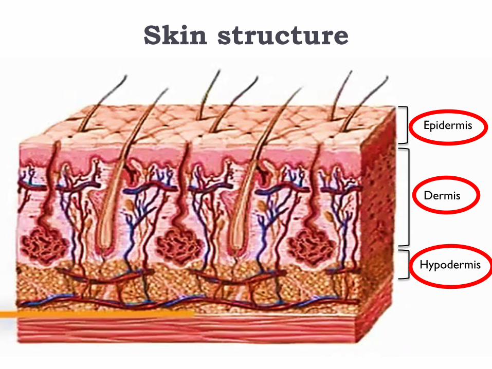
It consists of three major layers:
1) Stratified squamous epithelium called the Epidermis
2) Deeper connective tissue layer called the Dermis.
3) Connective tissue layer, called the Hypodermis,
Introduction to integumentary
system

Protection; external conditions
Regulating of body temperature
Excretion and secretion
Generate vitamin D
Formation of new cells from stratum germinativum
Sensory function; touch, pressure, pain, heat, and cold
Maintenance of the body form & store water, fat and glucose
Functions of integumentary system
Skin structure
Dermis
Hypodermis
Epidermis

Integumentary System
The epidermis subdivided into the following strata or layers
• Stratum corneum
• Stratum lucidum
• Stratum granulosum
• Stratum spinosum
• Stratum basale or
germinativum
Epidermis

Epidermis
Derived from the ectoderm, it
consists of a keratinized
stratified squamous epithelium
The superficial, thinner portion

Stratum corneum;
Dead cells contain keratin,
embedded in a lipid matrix
Stratum lucidum; Dead cells filled
with eleidin (intermediate form of
keratin) surrounded by an oily
substance of lamellar body.
Stratum granulosum; cells
contain keratin & grains protein
called keratohyalin and give the
layer its grainy appearance. Cells
also secret lamellar body.
Stratum spinosum; cells joined by
desmosomes, keratin fibers give the
cells the spiny appearance
Stratum basal; cells divide by
mitosis to add new cells to the
superficial strataAnchor down upper tissue to lower tissue
intracellular fibrous
protein

- About 8% of the epidermal cells are melanocytes (melano
= black), which produce the pigment melanin.
- Melanin is a yellow- red or brown-black pigment that
contributes to skin color and absorbs damaging ultraviolet
(UV) light.
- Once inside keratinocytes, the melanin granules cluster to
form a protective veil over the nucleus, on the side toward
the skin surface to protect the nuclear DNA from damage
by UV light.
- Melanin granules effectively protect keratinocytes but
melanocytes are particularly at risk to damage by UV light.
Cells that present in the basale layer


Present only in skin of fingertips, palms, and soles
In the middle of the epidermis, consists of 3-5 layers
of flattened keratinocytes that are undergoing
apoptosis.
Distinctive feature of cells is the presence dark staining
granules of a protein called keratohyalin and
membrane-enclosed lamellar granules present in the
keratinocytes which fuse with the plasma membrane
and release a lipid-rich secretion.

Also, keratinocytes which produce the protein keratin and lamellar granules
(contain a waterproof sealant).

Dermis
Derived from mesoderm consist of:
Collagen fibers for toughness
Elastin fibers for elasticity
Blood capillaries for nourishment
Cells present include fibroblasts, macrophages, adipocytes
Hair (protection & reception)
Sweat gland (excretion, temperature regulation)
Sebaceous gland
Sense receptors for (sensory neurons) called the Meissner corpuscles
The dermis can be divided into:
Thin superficial papillary region
Thick deeper reticular region.
Papillary Layer
Reticular Layer

Compare the composition of the papillary and reticular
regions of the dermis.
Papillary
Superficial portion of dermis (about one-fifth);
consists of areolar connective tissue with thin collagen and fine elastic fibers;
contains dermal ridges (dermal papillae) that house blood capillaries, corpuscles of touch, and free nerve endings
Reticular
Deeper portion of dermis (about four-fifths)
consists of dense irregular connective tissue with bundles of thick collagen and some coarse elastic fibers.
Spaces between fibers contain some adipose cells, hair follicles, nerves, sebaceous glands, and sudoriferous glands.

Hypodermis
Also called subcutaneous
Bind the skin to the
underlying tissue
The primary tissue is
Adipose (fat cells), for:
Storing energy
Padding
Thermal Insulation
The secondary tissue is
loos connective tissue

Integumentary System
Epidermis
DermisHypodermis
Granulosum Lucidum
SpinosumBasal
Corneum
Summary

Review:
Q & A List the layers of skin.
List the layers of the epidermis.
What is melanin?
Describe the protection nature of melanin.
How is the epidermis different from the dermis in terms of
composition, blood supply, and function?
Which epidermal layer includes stem cells that continually
undergo cell division?

Skin Derivatives
Hairs, or pili, are present on most skin surfaces except the palms,
palmar surfaces of the fingers, the soles, and plantar surfaces of the feet.
In adults, hair usually is most heavily distributed across the scalp, in the
eyebrows, in the axillae (armpits), and around the external genitalia.
Anatomy of a Hair
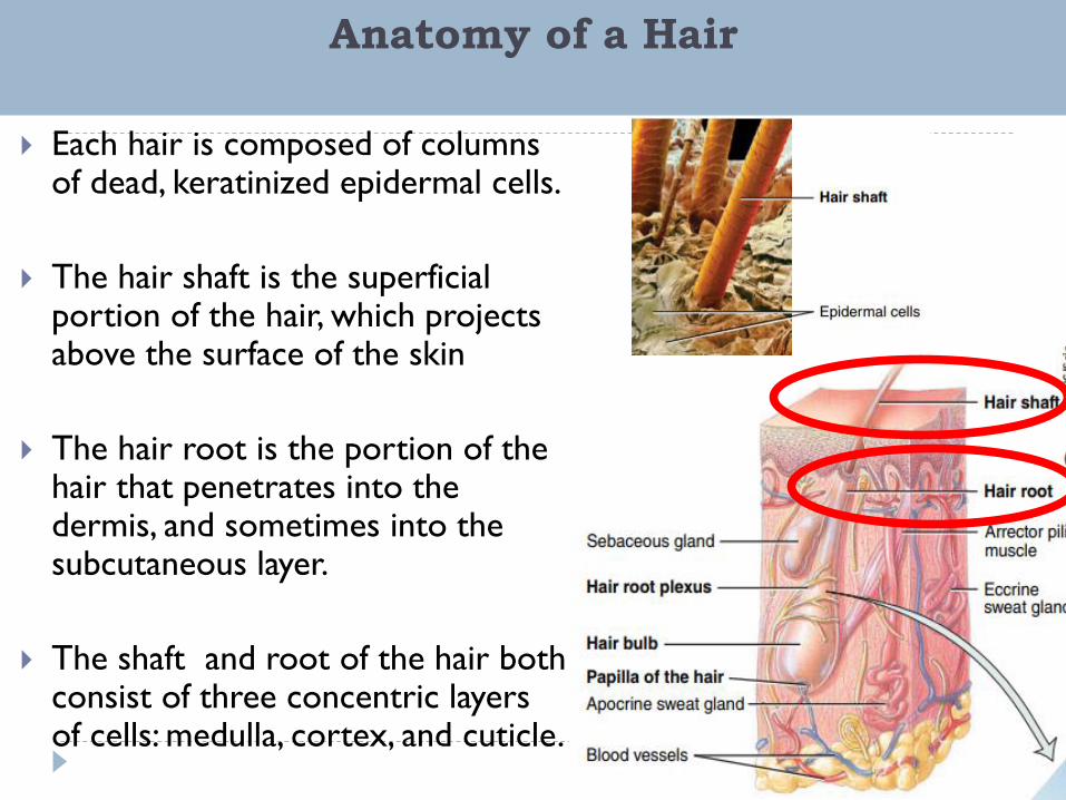
Each hair is composed of columns of dead, keratinized epidermal cells.
The hair shaft is the superficial portion of the hair, which projects above the surface of the skin
The hair root is the portion of the hair that penetrates into the dermis, and sometimes into the subcutaneous layer.
The shaft and root of the hair both consist of three concentric layers of cells: medulla, cortex, and cuticle.
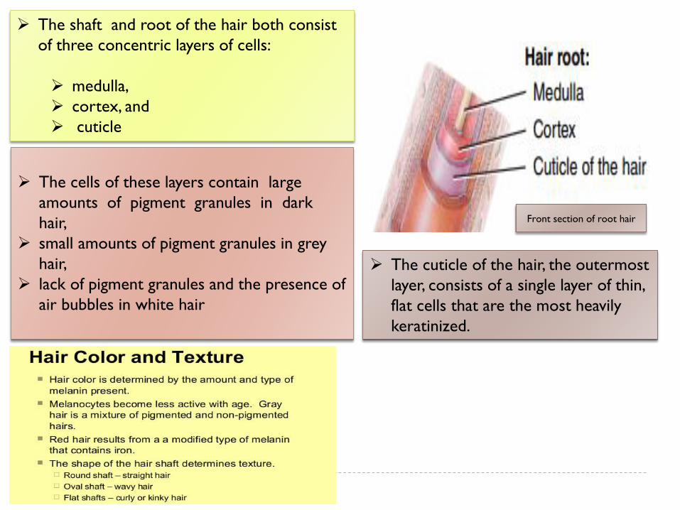
The cells of these layers contain large
amounts of pigment granules in dark
hair,
small amounts of pigment granules in grey
hair,
lack of pigment granules and the presence of
air bubbles in white hair
Front section of root hair
The shaft and root of the hair both consist
of three concentric layers of cells:
medulla,
cortex, and
cuticle
The cuticle of the hair, the outermost
layer, consists of a single layer of thin,
flat cells that are the most heavily
keratinized.

• The dense dermis
surrounding the hair
follicle is called the
dermal root sheath.
• Surrounding the root of
the hair is the hair
follicle, which is made up
of an external root
sheath and an internal
root sheath
• The external and internal
root sheath are referred
to as the epithelial
root sheath.
• The base of each hair follicle and its surrounding dermal root sheath
is an onion-shaped structure, the hair bulb contains the following;
• Papilla of the hair, which contains areolar connective tissue and many
blood vessels
• Hair matrix; Germinal layer of cells arise from the stratum basale,
the site of cell division. Hair matrix cells are responsible for the
growth of existing hairs and produce new hairs

Each nail consists of a nail body, a free edge, and a
nail root
The free edge is white because there are no
underlying capillaries.
The nail root is the portion of the nail that is buried
in a fold of skin.
The whitish, crescent-shaped area of the proximal
end of the nail body is called the lunula (little moon).
Anatomy of a nail

Anatomy of a nail

Skin Glands

References
For further reading please see:
Kenneth, S. S. (2017). Anatomy & physiology: The unity of form and
function. 8th edition. The McGraw−Hill Companies,. New york.
De Iuliis, G., & Pulerà, D. (2019). The dissection of vertebrates. 3rd
edition. Academic press. Elsevier, London.
Charles K. Weichert (2017). The Integumentary System. Elements of
chordate anatomy. 3rd edition. The McGraw−Hill Companies, New
york.
Murphrey, M. B., Miao, J. H., & Zito, P. M. (2018). Histology, stratum
corneum. In: StatPearls. StatPearls Publishing, Treasure Island (FL);
PMID.
Kardong, Kenneth V. (2019). Vertebrates : comparative
anatomy, function, evolution (8th edition). New York.