integumentary development - biology courses...
TRANSCRIPT
53
Integumentary DevelopmentThis chapter will introduce our foray into organogenesis as we examine the development of the integumentary system. This is an important organ system that forms about 8% of the total body mass and has a surface area in an average sized person of approximately 2.2 square meters. Prior to the lecture you should be able to:
• Describe all the anatomy of the integumentary system covered in the anatomy course.
After this lecture you should be able to:• List all the embryonic germ layers that contribute to the formation of
the integument.• Describe the processes that account for the development of the layers
of the skin and their substrata.• Explain the developmental processes that account for the
development of the glands and hair follicles.• Clearly describe tooth development and emergence.
The skin is largely ignored by the student in the dissecting room, just being incised, reflected, and cast aside as something which hides more interesting things underneath. Yet, it is that part of the body in which, par excellence, can be demonstrated, at all levels of observation, the relation between structure and function in biological organization.
Gray’ Anatomy, 38th British Edition
E m b r y o l o g y L e c t u r e M a n u a l b y M a r k N i e l s e n
54
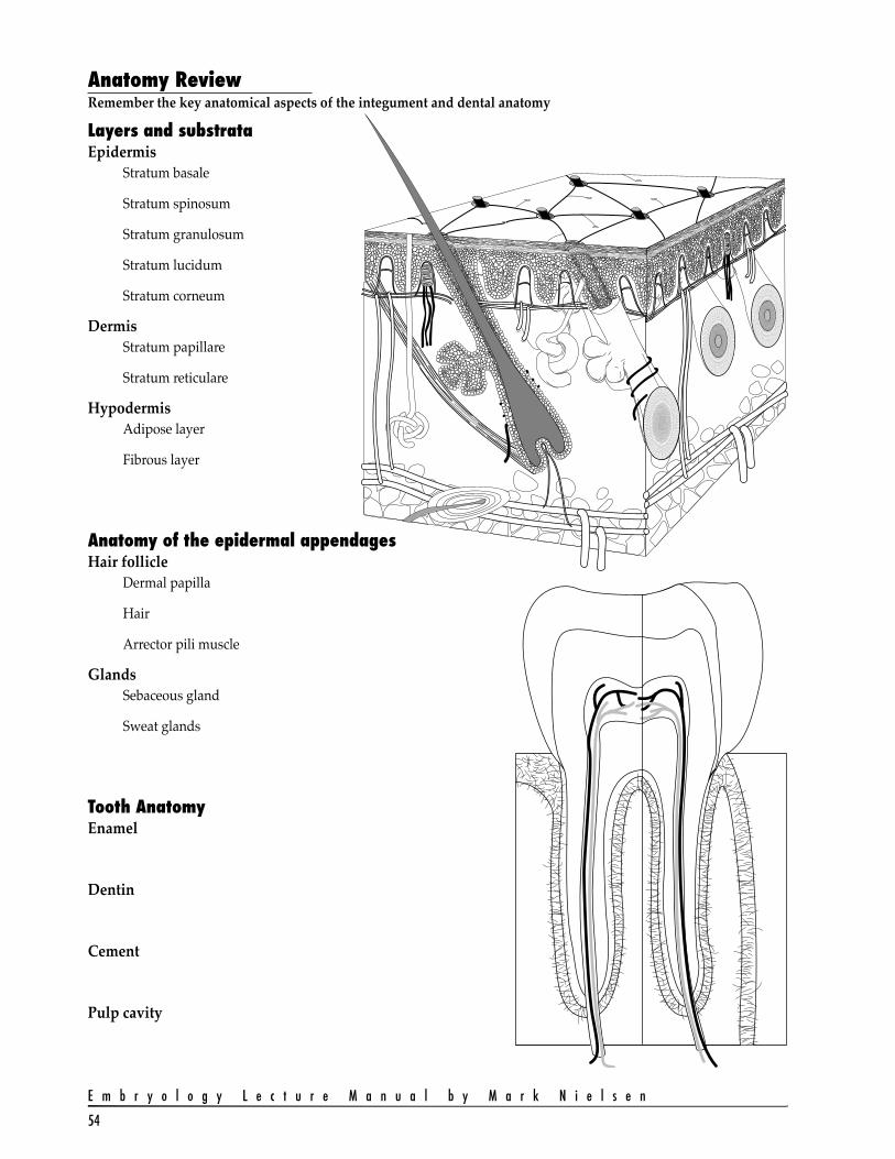
Anatomy ReviewRemember the key anatomical aspects of the integument and dental anatomy
Layers and substrataEpidermis
Stratum basale
Stratum spinosum
Stratum granulosum
Stratum lucidum
Stratum corneum
DermisStratum papillare
Stratum reticulare
HypodermisAdipose layer
Fibrous layer
Anatomy of the epidermal appendagesHair follicle
Dermal papilla
Hair
Arrector pili muscle
GlandsSebaceous gland
Sweat glands
Tooth AnatomyEnamel
Dentin
Cement
Pulp cavity
55
I n t e g u m e n t a r y D e v e l o p m e n t
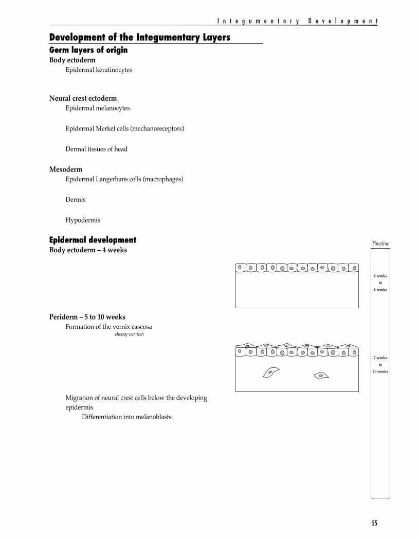
Development of the Integumentary LayersGerm layers of originBody ectoderm
Epidermal keratinocytes
Neural crest ectodermEpidermal melanocytes
Epidermal Merkel cells (mechanoreceptors)
Dermal tissues of head
MesodermEpidermal Langerhans cells (macrophages)
Dermis
Hypodermis
Epidermal developmentBody ectoderm – 4 weeks
Periderm – 5 to 10 weeksFormation of the vernix caseosa cheesy varnish
Migration of neural crest cells below the developing epidermis
Differentiation into melanoblasts
Timeline
4 weeksto
6 weeks
7 weeksto
10 weeks

E m b r y o l o g y L e c t u r e M a n u a l b y M a r k N i e l s e n
56
Stratified epithelium – 11 weeks to 21 weeksPeriderm
Intermediate layer
Basal layer Epidermal ridges
Melanoblasts migrate into epidermis to become melanocytes
Cornified layer replaces periderm - 22 weeksFormation of stratum corneum
Melanocyte activity
Mature EpidermisStratum basale
Stratum spinosum
Stratum granulosum
Stratum lucidum and corneum
Timeline
11 weeksto
21 weeks
22 weeksto
Neonate
57
I n t e g u m e n t a r y D e v e l o p m e n t
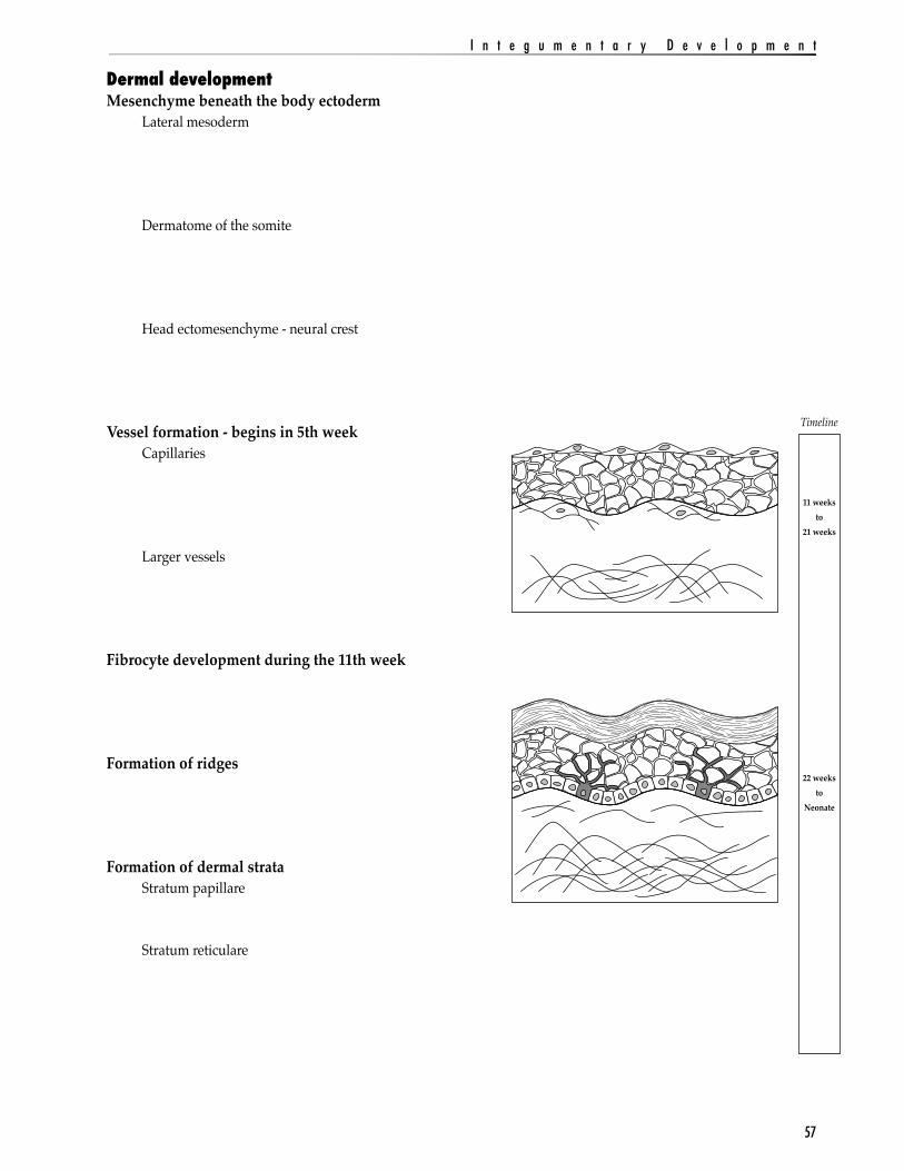
Dermal developmentMesenchyme beneath the body ectoderm
Lateral mesoderm
Dermatome of the somite
Head ectomesenchyme - neural crest
Vessel formation - begins in 5th weekCapillaries
Larger vessels
Fibrocyte development during the 11th week
Formation of ridges
Formation of dermal strataStratum papillare
Stratum reticulare
Timeline
11 weeksto
21 weeks
22 weeksto
Neonate
E m b r y o l o g y L e c t u r e M a n u a l b y M a r k N i e l s e n
58
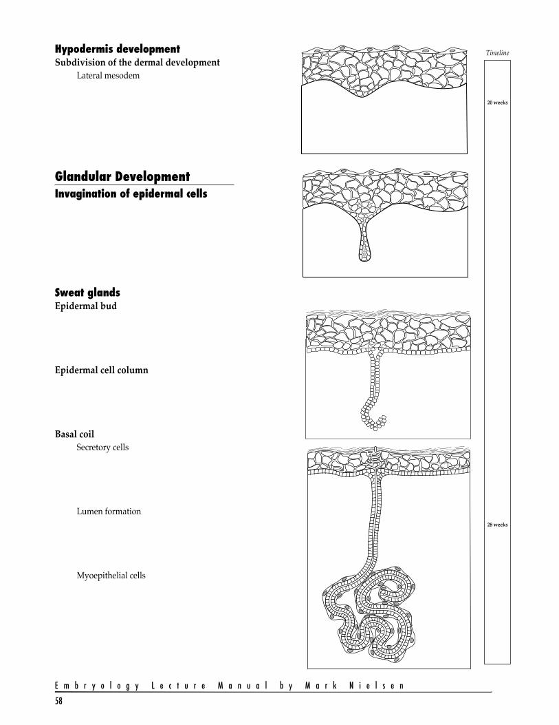
Hypodermis developmentSubdivision of the dermal development
Lateral mesodem
Glandular DevelopmentInvagination of epidermal cells
Sweat glandsEpidermal bud
Epidermal cell column
Basal coilSecretory cells
Lumen formation
Myoepithelial cells
Timeline
20 weeks
28 weeks
59
I n t e g u m e n t a r y D e v e l o p m e n t
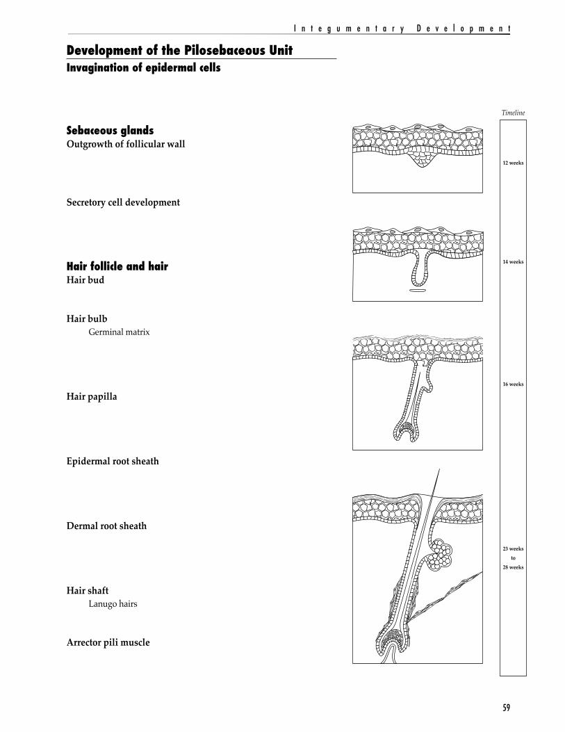
Development of the Pilosebaceous UnitInvagination of epidermal cells
Sebaceous glandsOutgrowth of follicular wall
Secretory cell development
Hair follicle and hairHair bud
Hair bulbGerminal matrix
Hair papilla
Epidermal root sheath
Dermal root sheath
Hair shaftLanugo hairs
Arrector pili muscle
Timeline
12 weeks
14 weeks
16 weeks
23 weeksto
28 weeks

E m b r y o l o g y L e c t u r e M a n u a l b y M a r k N i e l s e n
60
Mammary Gland DevelopmentMammary crests
Typical glandular formationInvagination process
Primary mammary bud
Secondary mammary buds
Mammary pit appears
Mammary glands emerge
Lactiferous duct
Areola of breast
Nipple formation
Timeline
12 weeks
20 weeks
30 weeks
Birth
61
I n t e g u m e n t a r y D e v e l o p m e n t
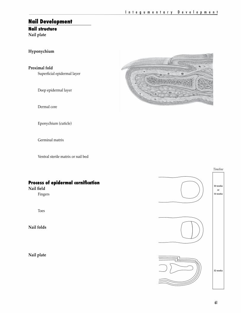
Nail DevelopmentNail structureNail plate
Hyponychium
Proximal foldSuperficial epidermal layer
Deep epidermal layer
Dermal core
Eponychium (cuticle)
Germinal matrix
Ventral sterile matrix or nail bed
Process of epidermal cornificationNail field
Fingers
Toes
Nail folds
Nail plate
Timeline
10 weeksor
14 weeks
32 weeks
E m b r y o l o g y L e c t u r e M a n u a l b y M a r k N i e l s e n
62
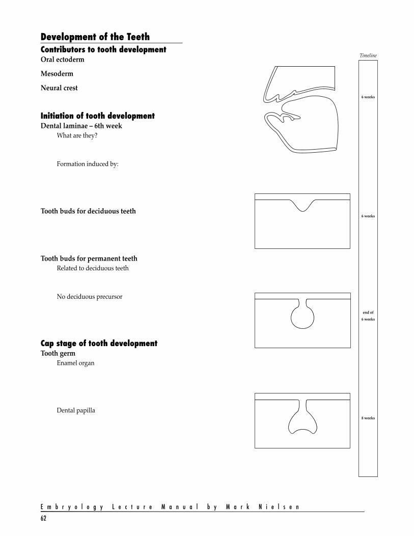
Development of the TeethContributors to tooth developmentOral ectoderm
Mesoderm
Neural crest
Initiation of tooth developmentDental laminae – 6th week
What are they?
Formation induced by:
Tooth buds for deciduous teeth
Tooth buds for permanent teethRelated to deciduous teeth
No deciduous precursor
Cap stage of tooth developmentTooth germ
Enamel organ
Dental papilla
Timeline
6 weeks
6 weeks
end of 6 weeks
8 weeks
63
I n t e g u m e n t a r y D e v e l o p m e n t
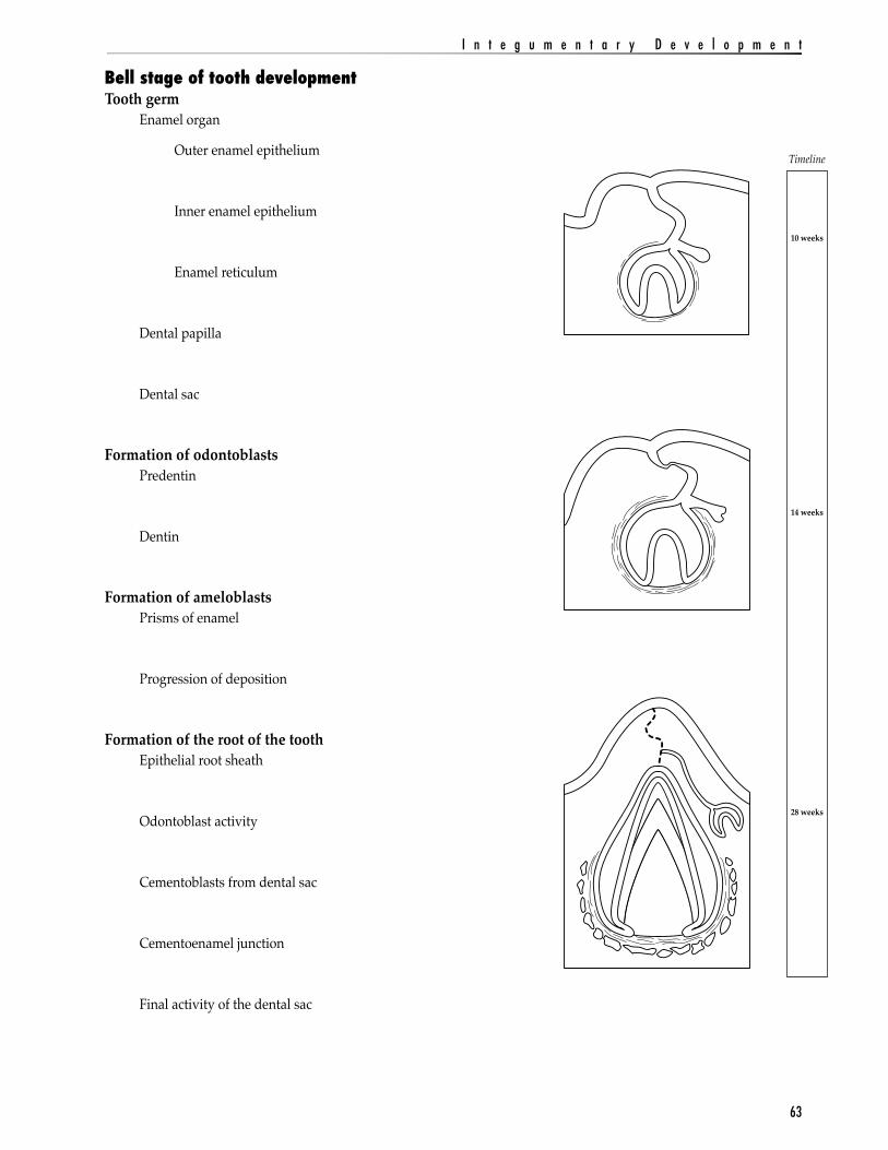
Bell stage of tooth developmentTooth germ
Enamel organ
Outer enamel epithelium
Inner enamel epithelium
Enamel reticulum
Dental papilla
Dental sac
Formation of odontoblastsPredentin
Dentin
Formation of ameloblastsPrisms of enamel
Progression of deposition
Formation of the root of the toothEpithelial root sheath
Odontoblast activity
Cementoblasts from dental sac
Cementoenamel junction
Final activity of the dental sac
Timeline
10 weeks
14 weeks
28 weeks
E m b r y o l o g y L e c t u r e M a n u a l b y M a r k N i e l s e n
64
Timeline
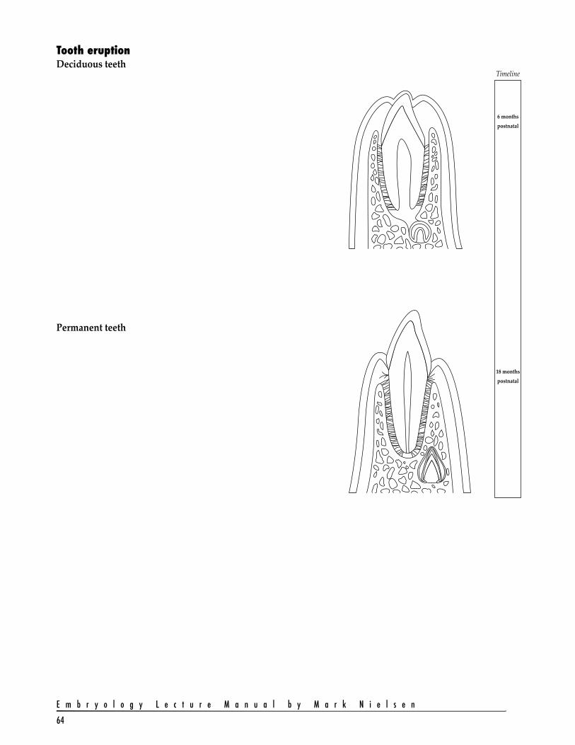
6 monthspostnatal
18 monthspostnatal
Tooth eruptionDeciduous teeth
Permanent teeth