immunopathologicalrolesofcytokines,chemokines
TRANSCRIPT

Hindawi Publishing CorporationClinical and Developmental ImmunologyVolume 2012, Article ID 715190, 14 pagesdoi:10.1155/2012/715190
Review Article
Immunopathological Roles of Cytokines, Chemokines,Signaling Molecules, and Pattern-Recognition Receptors inSystemic Lupus Erythematosus
Shui-Lian Yu,1 Woon-Pang Kuan,2 Chun-Kwok Wong,3 Edmund K. Li,1 and Lai-Shan Tam1
1 Department of Medicine and Therapeutics, Prince of Wales Hospital, The Chinese University of Hong Kong,30-32 Ngan Shing Street, Shatin, Hong Kong
2 Department of Rheumatology, Hospital Selayang, Lebuhraya Selayang-Kepong, 68100 Batu Caves, Malaysia3 Department of Chemical Pathology, Prince of Wales Hospital, The Chinese University of Hong Kong,30-32 Ngan Shing Street, Shatin, Hong Kong
Correspondence should be addressed to Lai-Shan Tam, [email protected]
Received 12 July 2011; Accepted 11 October 2011
Academic Editor: Philip Alex
Copyright © 2012 Shui-Lian Yu et al. This is an open access article distributed under the Creative Commons Attribution License,which permits unrestricted use, distribution, and reproduction in any medium, provided the original work is properly cited.
Systemic lupus erythematosus (SLE) is an autoimmune disease with unknown etiology affecting more than one million individualseach year. It is characterized by B- and T-cell hyperactivity and by defects in the clearance of apoptotic cells and immune complexes.Understanding the complex process involved and the interaction between various cytokines, chemokines, signaling molecules,and pattern-recognition receptors (PRRs) in the immune pathways will provide valuable information on the development ofnovel therapeutic targets for treating SLE. In this paper, we review the immunopathological roles of novel cytokines, chemokines,signaling molecules, PRRs, and their interactions in immunoregulatory networks and suggest how their disturbances may implicatepathological conditions in SLE.
1. Introduction
Systemic lupus erythematosus (SLE) is a prototypic systemicautoimmune disease which is characterized by a loss of toler-ance to nuclear antigens and various immunological abnor-malities, including dysregulated activation of both T and Blymphocytes and subsequent polyclonal activation of circula-ting B lymphocytes which produces a large quantity of auto-reactive antibodies and the formation of immune complexescausing tissue and organ damage [1]. This is a complexprocess involved interaction between various cytokines, che-mokines, signaling molecules, and pattern-recognition re-ceptors (PRRs) in the immune pathways. With the adventof new and advanced technique which include intracellularcytokine analysis by flow cytometry combined with multi-plex quantization of cytokine levels in recent years, it hadprovided us a reasonable understanding of the activationprofile of cytokine production and new insight in the im-mune and cellular mechanism in the pathogenesis of SLE,which further clarify the significance of the current body of
literatures. This had provided valuable information on thedevelopment of novel therapeutic targets for treating SLE.This article will focus on the recent advances of cytokines,chemokines, signaling molecules, and the role of PRRs inimmunopathogenesis in SLE.
2. Imbalance of Th1/Th2 Cytokines in SLE
Cytokines are a group of small peptides or glycoprotein pro-duced by a wide variety of cells with molecular weights be-tween 8 and 30 kDa. They had been shown to play anessential role in modulating the immune response againstforeign or self-antigens. These mediators have been classifiedaccording to their cellular source and effector functions, withthe paradigmatic T helper (Th)1 and Th2 cytokine familiesbest illustrating this division of function. Th1 cells arise inresponse to dendritic cells- (DCs-) derived interleukin- (IL-)12, produce tumor necrosis factor- (TNF-) α, interferon-(IFN-) γ, and are involved in mediating strong inflammatoryresponses to intracellular pathogens. IL-4-mediated Th2 cell

2 Clinical and Developmental Immunology
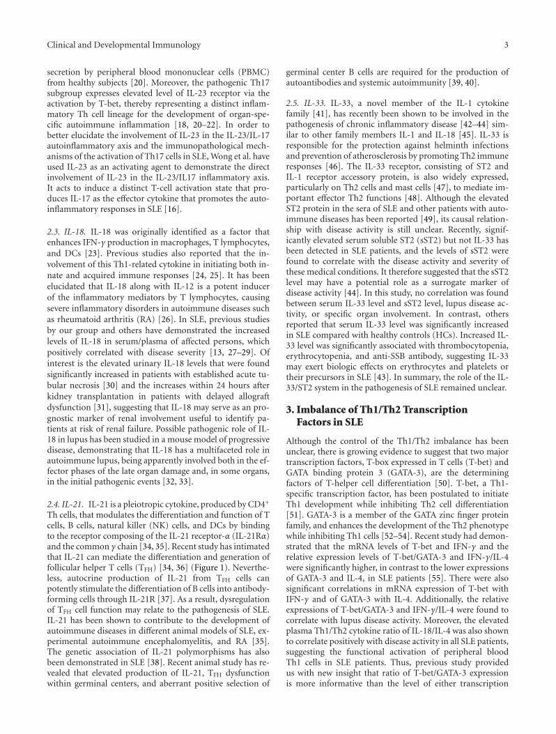
Th0
IL-2 IL-12IFN-γ TNF-α IL-4 IL-5
IL-25 IL-10IL-13
Th1
IFN-γ
IL-12 IL-4IL-10
Th2
Th17
IL-6IL-23
IL-21, 22, 24, 26IL-17AIL-17F
Function:antiviral,bacterial
immunity
Function:immunity toextracellular
parasites
Function:inflammation,auto-immunity
TFH
IL-21, IL-6, IL-10
Function:T cells help for
B cells
CXCR5CCR4CCR6
CrTh2CCR5CXCR3 Bcl-6
STAT-3STAT-3
STAT-6GATA-3
T-betSTAT-1
Naıve T cells
CXCR5
CCR7Primed T cells
IL-6IL-21CXCL13
Cytokines
Chemokinereceptors
Effectorcytokines
Functions
TranscriptionfactorsRORγT
IL-1β
Figure 1: Effector T-cell differentiation (Th1, Th2, Th17 and TFH), the expression of transcription factors, effector cytokines, chemokinereceptors, and T-cell functions.
differentiation results in cells that produce cytokines, includ-ing IL-4, IL-5, and IL-13, which mediate antibody responsesto extracellular pathogens (Figure 1).
The ratios of Th1 and Th2 cytokines have been inves-tigated to determine the cytokine homeostasis in order todetermine whether Th1 or Th2 predominance during thedevelopment of SLE [2, 3]. SLE was thought to be a Th2-po-larized disease because of the production of auto-antibod-ies specific for self-antigens [4]. However, significantly ele-vated cytokines for Th1 response including IL-12, TNF-α,and IFN-γ were also found in the plasma of SLE patients[5–8]. Th1 dominant immune responses have been generallyconsidered to be pathological in autoimmune disease via theinduction of inflammatory reaction. Recently, few cytokineswhich had been shown to be of great importance in patho-genesis of SLE had surfaced with advent of new technologyin detection, which enhances our understanding of their rolein SLE-related immune pathway. These cytokines, includingIL-12, IL-23, IL-18, IL-21, and IL-33, will be discussed below.
2.1. IL-12. IL-12 is a heterodimeric cytokine of 70 kDacomprising covalently linked p40 and p35 subunit whichhas been shown to be a central stimulator of Th1-relatedproinflammatory cytokine that induces IFN-γ in both innateand adaptive immunity [9, 10]. IL-12 had been suggested tobe associated with progression of severe glomerulonephritis[11]. Moreover, mRNA levels of p19, p40, and p35 of IL-12were found to be significantly higher in active SLE patientscompared with those patients with inactive disease [12].Accordingly, serum level of IL-12 was also found to be sig-nificantly elevated in SLE patients, and it is associated withthe increased level of Th1 cytokine IFN-γ but decreasedlevel of Th2 cytokine IL-13 [5, 13, 14]. Conversely, another
study reported the decreased ex vivo production of IL-12 from peripheral blood polymorphonuclear leukocytes(PMN) stimulated by lipopolysaccharide (LPS) in patientswith active SLE [15] using a different ELISA kit. Recently,the elevated plasma IL-12 concentration has been shown toexhibit positive correlation with systemic lupus erythemato-sus disease activity index (SLEDAI) in SLE patients with renalimpairment, supporting IL-12 could play a pathologicalrole in the development of autoinflammatory response inSLE patients with severe disease, probably through therecruitment of the effector leukocytes to the inflamed tissuefor orchestrating the immunoresponse at the site of inflam-mation [16].
2.2. IL-23. IL-23 is a novel heterodimeric cytokine composedof a unique p19 subunit, and a common p40 subunit sharedwith IL-12. IL-23 shares similar intracellular signal transduc-tion molecules with IL-12, therefore both cytokines exhibitsome overlapping function in promoting cellular immunity[17]. Different from IL-12, IL-23 does not promote thedevelopment of IFN-γ-producing Th1 cells, but is crucialfor the expansion of a pathogenic CD4+ T-cell populationcharacterized by the production of IL-17 and IL-22 [18, 19].Recent studies had shown that the mRNA levels of IL-23p19were significantly higher in active SLE patients when patientswere stratified into different disease activity groups, therebysuggesting that IL-23 should play a role in SLE disease ex-acerbation [12]. Moreover, the likely significance of IL-23in autoinflammatory responses was further supported by amore recent report indicated that Th1 transcription factorT-bet could upregulate IL-23 receptor expression and thedifferentiation of Th1 and Th17 cells in autoimmunity [20](Figure 1). IL-23 has been reported to enhance the IL-17
Clinical and Developmental Immunology 3
secretion by peripheral blood mononuclear cells (PBMC)from healthy subjects [20]. Moreover, the pathogenic Th17subgroup expresses elevated level of IL-23 receptor via theactivation by T-bet, thereby representing a distinct inflam-matory Th cell lineage for the development of organ-spe-cific autoimmune inflammation [18, 20–22]. In order tobetter elucidate the involvement of IL-23 in the IL-23/IL-17autoinflammatory axis and the immunopathological mech-anisms of the activation of Th17 cells in SLE, Wong et al. haveused IL-23 as an activating agent to demonstrate the directinvolvement of IL-23 in the IL-23/IL17 inflammatory axis.It acts to induce a distinct T-cell activation state that pro-duces IL-17 as the effector cytokine that promotes the auto-inflammatory responses in SLE [16].
2.3. IL-18. IL-18 was originally identified as a factor thatenhances IFN-γ production in macrophages, T lymphocytes,and DCs [23]. Previous studies also reported that the in-volvement of this Th1-related cytokine in initiating both in-nate and acquired immune responses [24, 25]. It has beenelucidated that IL-18 along with IL-12 is a potent inducerof the inflammatory mediators by T lymphocytes, causingsevere inflammatory disorders in autoimmune diseases suchas rheumatoid arthritis (RA) [26]. In SLE, previous studiesby our group and others have demonstrated the increasedlevels of IL-18 in serum/plasma of affected persons, whichpositively correlated with disease severity [13, 27–29]. Ofinterest is the elevated urinary IL-18 levels that were foundsignificantly increased in patients with established acute tu-bular necrosis [30] and the increases within 24 hours afterkidney transplantation in patients with delayed allograftdysfunction [31], suggesting that IL-18 may serve as an pro-gnostic marker of renal involvement useful to identify pa-tients at risk of renal failure. Possible pathogenic role of IL-18 in lupus has been studied in a mouse model of progressivedisease, demonstrating that IL-18 has a multifaceted role inautoimmune lupus, being apparently involved both in the ef-fector phases of the late organ damage and, in some organs,in the initial pathogenic events [32, 33].
2.4. IL-21. IL-21 is a pleiotropic cytokine, produced by CD4+
Th cells, that modulates the differentiation and function of Tcells, B cells, natural killer (NK) cells, and DCs by bindingto the receptor composing of the IL-21 receptor-α (IL-21Rα)and the common γ chain [34, 35]. Recent study has intimatedthat IL-21 can mediate the differentiation and generation offollicular helper T cells (TFH) [34, 36] (Figure 1). Neverthe-less, autocrine production of IL-21 from TFH cells canpotently stimulate the differentiation of B cells into antibody-forming cells through IL-21R [37]. As a result, dysregulationof TFH cell function may relate to the pathogenesis of SLE.IL-21 has been shown to contribute to the development ofautoimmune diseases in different animal models of SLE, ex-perimental autoimmune encephalomyelitis, and RA [35].The genetic association of IL-21 polymorphisms has alsobeen demonstrated in SLE [38]. Recent animal study has re-vealed that elevated production of IL-21, TFH dysfunctionwithin germinal centers, and aberrant positive selection of
germinal center B cells are required for the production ofautoantibodies and systemic autoimmunity [39, 40].
2.5. IL-33. IL-33, a novel member of the IL-1 cytokinefamily [41], has recently been shown to be involved in thepathogenesis of chronic inflammatory disease [42–44] sim-ilar to other family members IL-1 and IL-18 [45]. IL-33 isresponsible for the protection against helminth infectionsand prevention of atherosclerosis by promoting Th2 immuneresponses [46]. The IL-33 receptor, consisting of ST2 andIL-1 receptor accessory protein, is also widely expressed,particularly on Th2 cells and mast cells [47], to mediate im-portant effector Th2 functions [48]. Although the elevatedST2 protein in the sera of SLE and other patients with auto-immune diseases has been reported [49], its causal relation-ship with disease activity is still unclear. Recently, signif-icantly elevated serum soluble ST2 (sST2) but not IL-33 hasbeen detected in SLE patients, and the levels of sST2 werefound to correlate with the disease activity and severity ofthese medical conditions. It therefore suggested that the sST2level may have a potential role as a surrogate marker ofdisease activity [44]. In this study, no correlation was foundbetween serum IL-33 level and sST2 level, lupus disease ac-tivity, or specific organ involvement. In contrast, othersreported that serum IL-33 level was significantly increasedin SLE compared with healthy controls (HCs). Increased IL-33 level was significantly associated with thrombocytopenia,erythrocytopenia, and anti-SSB antibody, suggesting IL-33may exert biologic effects on erythrocytes and platelets ortheir precursors in SLE [43]. In summary, the role of the IL-33/ST2 system in the pathogenesis of SLE remained unclear.
3. Imbalance of Th1/Th2 TranscriptionFactors in SLE
Although the control of the Th1/Th2 imbalance has beenunclear, there is growing evidence to suggest that two majortranscription factors, T-box expressed in T cells (T-bet) andGATA binding protein 3 (GATA-3), are the determiningfactors of T-helper cell differentiation [50]. T-bet, a Th1-specific transcription factor, has been postulated to initiateTh1 development while inhibiting Th2 cell differentiation[51]. GATA-3 is a member of the GATA zinc finger proteinfamily, and enhances the development of the Th2 phenotypewhile inhibiting Th1 cells [52–54]. Recent study had demon-strated that the mRNA levels of T-bet and IFN-γ and therelative expression levels of T-bet/GATA-3 and IFN-γ/IL-4were significantly higher, in contrast to the lower expressionsof GATA-3 and IL-4, in SLE patients [55]. There were alsosignificant correlations in mRNA expression of T-bet withIFN-γ and of GATA-3 with IL-4. Additionally, the relativeexpressions of T-bet/GATA-3 and IFN-γ/IL-4 were found tocorrelate with lupus disease activity. Moreover, the elevatedplasma Th1/Th2 cytokine ratio of IL-18/IL-4 was also shownto correlate positively with disease activity in all SLE patients,suggesting the functional activation of peripheral bloodTh1 cells in SLE patients. Thus, previous study providedus with new insight that ratio of T-bet/GATA-3 expressionis more informative than the level of either transcription
4 Clinical and Developmental Immunology
factor alone, which may be disproportionately affected bythe changes in their coexpression in cell populations. The T-bet/GATA-3 expression ratio not only enhances our under-standing of Th1/Th2 polarization, it may also serve as a sup-plementary tool for further assessment of Th1/Th2 statusand development of SLE disease activity (Figure 1).
4. Th17-Mediated Inflammation of SLE
Apart from Th1 and Th2 cells, there is a novel subset of IL-17 producing effector T helper cells, called Th17 cells, whosedysregulation is thought to participate in the pathogenesis ofSLE [56, 57]. Transforming growth factor (TGF)-β, IL-6, IL-21, and IL-23 have been implicated for Th17 formation [58,59]. Other proteins involved in their differentiation are signaltransducer and activator of transcription 3 (STAT3) andthe retinoic-acid-receptor-related orphan receptors alpha(RORα) and gamma (RORγ) [58]. Moreover, effector cytok-ines associated with this cell type are IL-17, IL-21, and IL-22[60] (Figure 1). We herein highlighted some of the biologicaleffects of IL-17 implication for Th17-mediated inflammationof SLE.
IL-17 is a type I 17-kDa transmembrane protein thatcomprises six members and five receptors mostly producedby activated T cells [61]. It is a pleiotropic proinflammatorycytokine that enhances T-cell priming and stimulates epi-thelial, endothelial, and fibroblastic cells to produce multipleproinflammatory mediators, including IL-1, IL-6, TNF-α,and chemokines [62]. Additionally, IL-17 also exerts itseffects through the recruitment of monocytes and neutro-phils by increasing the local production of chemokines(IL-8, monocyte chemoattractant protein-1, growth-relatedoncogene protein-α), the facilitation of T-cell infiltrationand activation by stimulating the expression of intercellularadhesion molecule-1 by T cells as well as the amplificationof the immune response by inducing the production of IL-6,prostaglandin E2, granulocyte-macrophage colony-stimulat-ing factor, and granulocyte colony-stimulating factor [63].Lastly, this cytokine synergizes with other cytokines, inparticular with IL-1β, TNF-α, and IFN-γ [63].
Wong et al. have demonstrated that SLE patients havehigher plasma/serum levels of IL-17 than HCs [13, 16, 56],which positively associated with SLE disease activity [16].Accordingly, the frequency of IL-17-producing T cells is in-creased in peripheral blood of SLE patients [16, 64]. Sig-nificant levels of IL-17 and IFN-γ were detected in T cellsfrom SLE patients [64]. Additionally, overproduction oftotal immunoglobulin G (IgG), antidouble stranded DNA,and IL-6 by PBMC of patients with lupus nephritis wasobserved upon the stimulation with IL-17 [65], suggestinga potential role of IL-17 in human lupus progression. Onthe other hand, no elevation of IL-17 was found in serum ofcohort of Japanese lupus patients [66]. Most recent evidencesuggested that the ability of regulatory T cells (Tregs) to ex-press IFN-γ and IL-17 was impaired in SLE patients, whereasthe proportion of Tregs was similar between SLE patientsand HCs [67]. Additionally, studies in mice support theconcept that IL-17 and Th17 cells may be involved inthe development of lupus nephritis [56, 68]. For instance,
IL-17 was recently found to be critical for the formation ofautoreactive germinal centres in autoimmune BXD2 mice, astrain that develops a lupus-like syndrome [69]. In a spon-taneous mouse model of lupus, the New Zealand Black(NZB) mice, stimulation of splenocytes with nucleosomes asan autoantigen results in the activation of large numbers ofIL-17-secreting T cells [70]. Upon adoptive transfer to naıverecipient mice, IL-23-dependent IL-17 producing CD4+ ef-fector T-cell subset Th17 can invade the target organ andpromote the development of organ-specific autoimmune in-flammation. Consistently, Wong et al. also found that theproinflammatory cytokine IL-23 and IL-12 can promote thedisease severity by activating pathogenic Th1 and Th17 cellsvia the induction of downstream Th1 chemokine CXCL10and inflammatory cytokine IL-17 in SLE, demonstrating thatthe IL-23/IL-17 axis of inflammation and related moleculesmay arise as a therapeutic target for treating autoimmunedisease.
5. Chemokines in SLE
Chemokines in itself refer to a group of smaller cytokines(mass between 8 to 12 kDa) with chemotactic properties,which are classified into four families according to the loca-tion of cysteine residues. The four chemokine groups are CC,C, CXC, and CX3C, where C is a cysteine and X is any aminoacid residue [71]. These small molecules have had well-defined roles in directing cell migration necessary for theinitiation of T cell immune response, attraction of appropri-ate effector cells to sites of inflammation, and regulation ofdifferential recruitment of T helper (Th1 and Th2) lympho-cytes [72–74]. There has been growing evidence suggestingthat infiltration of T lymphocytes and other leucocytesinto the sites of inflammation plays a critical role in organinvolvement in SLE [75]. Recent studies have also shownthat chemokines and their receptors are intimately involvedin regulating organ-specific leucocyte trafficking and inflam-mation, suggesting their important roles in the pathophys-iology of autoimmune diseases such as RA, multiple sclerosis,and SLE [76–78]. Chemokine CXCL13 in emerging studieshad consolidated the important role of these chemokinesin pathogenesis of SLE. Other chemokines that will bebriefly discussed in this article mainly include CC and CXCchemokines which had been shown to play some roles inSLE disease.
5.1. CXCL13. CXCL13/B lymphocyte chemoattractant(BLC) is a small cytokine belonging to the CXC chemokinefamily that is produced by cells in the omentum, peritonealmacrophages, and DCs [79, 80], which is selectively chem-otactic for B cells including both the B1 and B2 subsetsby interacting with specific chemokine receptor CXCR5[79, 81]. The accumulation of B1 cells in the peritoneal cavityand spleen are responsible for the body cavity immunity andthe production of autoantibody for the development of auto-immune disease in the murine model [79, 82, 83]. Elevatedlevels of B1 cells have been documented in patients withautoimmune disorders such as Sjogren’s syndrome andRA [84, 85]. Previous studies using murine model of SLE

Clinical and Developmental Immunology 5
showed that CXCL13 is highly produced by CD11b+ CD11c+
DCs in the target organs including thymus and kidney forthe chemoattraction of B1 cells into target organ [83, 86–88].Therefore, the elevated expression of CXCL13 by myeloiddendritic cells (mDCs) in the target organs may play a cru-cial role in breaking the immune tolerance in the thymusleading to the activation of self-reactive CD4+ Th cells andthe recruitment of autoantibody producing B cells in the de-velopment of murine lupus [83, 87, 88]. In addition to that,studies have revealed that CXCL13 can induce the traffickingof distinct CXCR5+ T cells designated as TFH which arespecifically involved in high-affinity IgG production in ger-minal centers developed within B-cell follicles of secondarylymphoid tissues including lymph nodes, spleen, and tonsils[36, 89–91]. CD4+ TFH cells, located at B-cell follicles,provide a T helper function to B cells and represents one ofthe most numerous and important subsets of effector T cellsin lymphoid tissue [37, 92]. Several studies demonstratedthat B-cell chemokine CXCL13 is ectopically and highly ex-pressed in thymus and kidney in murine model for SLE.Studies on humans also demonstrated that serum CXCL13level was significantly elevated in SLE patients and theelevation correlated significantly with SLE disease activity[93, 94]. As anti-TNF-α treatment was found to be able toreduce the plasma level of CXCL13 in RA patients [95], ithad been postulated that serum level of CXCL 13 can act asa disease activity marker for both RA and SLE patients.
5.2. CC Chemokines. Monocyte chemoattractant protein-1(MCP-1/CCL2) is a prototype CC chemokine, which canattract monocytes, T cells, NK cells, and basophils [96, 97].An increase of serum MCP-1/CCL2 was observed with theprogression of disease activity in SLE patients compared toHCs [98]. Further investigation reported that cerebral spinalfluids (CSF) MCP-1/CCL2 levels were significantly higher inneuropsychiatric syndromes of systemic lupus erythemato-sus (NPSLE) patients than those non-NPSLE patients [99].Regulated upon activation, normal T-cell-expressed andsecreted (RANTES)/CCL5 is another CC chemokine whichattracts monocytes, memory T cells, and NK cells [100]. In-creased plasma RANTES/CCL5 concentrations were foundin SLE patients more than in controls, and correlated signifi-cantly with SLEDAI score [101]. Moreover, the expression ofmiR-125a was found to contribute to the elevated expressionof RANTES/CCL5 in SLE [102]. In addition to that, studiesfrom animal models and patients with lupus nephritis dem-onstrated that inflammatory chemokines, especially CCL2and CCL5, are detectable in kidney tissues and urine beforeother signs of inflammation [103–106]. With this finding,urine chemokines had been proposed as a possibility toserve as biomarkers for renal SLE flare [107], suggesting thatthe reduced plasma concentration of these circulating che-mokines in lupus patients with renal involvement may resultfrom a protein leakage in the urine.
5.3. CXC Chemokines. Interferon-gamma inducible protein-10 (IP-10)/CXCL10 and monokine induced by gamma-inter-feron (MIG)/CXCL9, the prototype of the CXC family,have chemotactic activity mainly for activated Th1 cells and
are involved in the pathogenesis of various Th1-dominantautoimmune diseases [71, 108]. Their synthesis and expres-sion from neutrophils, macrophages, and other immune cellsare induced by IFN-γ, and this response is suppressed byIL-10 and IL-4 [71, 109]. Th1 cells and IFN-γ had beenshown to be important for cell-mediated inflammation indeveloping autoimmune disease such as SLE [5], thus impli-cated that these chemokines might have an important rolein pathogenesis of SLE. Furthermore, several studies haveshown that levels of IP-10/CXCL10 and MIG/CXCL9 weresignificantly elevated in active SLE [98, 110, 111]. Moreover,Okamoto et al. reported that IP-10/CXCL10 was upregulatedin the central nervous system (CNS) fluid of NPSLE [112,113], suggesting that IP-10/MCP-1 ratio in CSF is a usefuldiagnostic marker of NPSLE [112]. On the other hand, CXCchemokines CXCL8 and CXCL1 are potent chemoattractantsand activators of T cells, neutrophils, thereby enhancing theirproinflammatory and proangiogenic activities [114]. Theyalso stimulate neutrophil degranulation to release reactiveoxygen radicals, thereby inducing an acute inflammatoryreaction [115, 116]. They had also been shown to be sig-nificantly elevated in serum of patient with active lupus, andthe elevation was associated with disease activity [111].
6. Intracellular Signaling Pathways in SLE
Signal transduction refers to an ordered biochemical processby which a signal or stimulus is transferred within a singlecell. This cascade begins with binding of extracellular signal-ing molecules to cell surface receptors, triggering an initialstimulus that propagated into the cytoplasm. Nowadays, themost well-known and established signal transduction path-way that has been identified is mitogen-activated proteinkinase (MAPK) pathway. MAPKs are serine and threonineprotein kinases that can be activated by phosphorylationin response to extracellular stimuli, such as mitogens,growth factors, cytokines, and osmotic stress [117, 118]. Nu-clear translocation of activated MAPKs can induce and trans-activate transcription factors including nuclear factor- (NF-)κB and activator protein 1, which facilitate the modulation ofgene transcription in cellular activation, proliferation, apop-tosis, and the expression of cytokines, chemokines, adhe-sion molecules, and metalloproteinases [117, 118]. Threemain distinct MAPKs, p42/p44 extracellular signal-regulatedkinase (ERK), c-Jun NH2-terminal protein kinase (JNK),and p38 MAPK, have been identified in mammalian cells.The activation of NF-κB, JNK, and p38 MAPK plays crucialroles in cytokine-mediated signaling pathways regulating therelease of chemokines and the expression of adhesion mole-cules of eosinophils and Th cells [119–121]. Activation ofp38 MAPK has been shown to be crucial for B-cell activationleading to Ig production, and p38 MAPK regulates the pro-duction of a number of cytokines, including IL-6 that pro-motes the differentiation and survival of plasma cells [122].Moreover, B-cell-activating factor of the TNF family, anessential factor for B-cell activation and differentiation, wasregulated through JNK and p38 MAPK [123]. Furthermore,nuclear factor of activated T cells (NFAT), a downstream

6 Clinical and Developmental Immunology
transcription factor of the ERK and JNK pathways, is essen-tial for T and B lymphocyte activation and differentiation[124], and specific anti-NFAT drug therapy has been shownto be pharmacologic armamentarium against RA, inflamma-tory arthropathies, and related autoimmune disorders [125].
7. Interaction between Cytokines, Chemokines,and Signaling Molecules in SLE
As mentioned before, immunopathogenesis of SLE is a com-plex process that involved the interaction and synergisticeffect of various cytokines, chemokines, and signaling mole-cules which perpetuate the disease activity in SLE. Thissection below will highlight the recent update on the inter-action between all these agents in promoting the disease ac-tivity in SLE.
7.1. Role of IL-18 and Chemokines. The potential role of IL-18 and chemokines in the exacerbation of SLE disease hadbeen highlighted in a study, which provided valuable infor-mation on the development of SLE disease markers [111]. Inthis study, plasma concentration of CXCL10, CCL5, CXCL9,CXCL8, CXCL1, and CCL2 was significantly elevated in SLEpatients and the elevation was correlated significantly withdisease activity. Furthermore, plasma concentration of IL-18 was found to be correlated positively with production ofCXCL10, CXCL9, CXCL1, and CXCL8 in SLE patients, it wasalso shown to be a potent costimulus for the induction ofthese chemokine release from activated PBMC as there wasa significant increase in ex vivo production of these inflam-matory chemokines when their PBMC were cultured in thepresence of IL-18.
This enhances our knowledge that successful delivery ofthe appropriate population of leucocytes to sites of acute in-flammation will depend on the repertoire of inducible che-mokines synthesized locally, and the temporal expression ofchemokine receptors on the leucocytes. Meanwhile, the che-mokine expressions are influenced by proinflammatory cyto-kines, mainly IL-18, to present in the local environment ofthe cells at the time of stimulation. Furthermore, inflam-matory activities of IL-18, together with the induction ofTh1 cytokine IFN-γ and the activation of Th cells, naturalkiller cells (NK), and cytotoxic T lymphocytes-inflammatorychemokines, may even enhance the Th1-mediated inflam-matory process, the activation of NK and T cells, and themigration of macrophages for initiating and perpetuating theTh1 immune response in SLE. In summary, the correlationof raised plasma concentration and ex vivo production ofinflammatory chemokines with disease activity, and theirassociation with IL-18, supports that the chemotaxis ofTh1/Th2 lymphocytes and neutrophils is important in SLEpathogenesis.
7.2. Role of CXCL13 and IL-21. A recent study [93] hadshown that CXCL13 and IL-21 may relate with the immuno-pathogenesis mediated by the function of TFH cells in SLE asserum level of all these cytokines were found to be signif-icantly elevated in lupus patient with the increase inCXCL13 concentration correlated positively and significantly
with SLEDAI score. Furthermore, cell surface expression ofCXCR5 on Th and B cells and IL-21R on B cells was found tobe significantly lower in SLE patients, which indicated thatmost differentiated TFH cells migrate out from circulationinto lymphoid organ upon activation during the disease de-velopment of SLE. This piece of information suggests thatthe elevated production of CXCL13, BAFF, and IL-21 maybe associated with the function of TFH for the immu-nopathogenesis in SLE, and CXCL13 may serve as a potentialdisease marker of SLE.
7.3. Role of IL-23, IL-17, IL-18, Th17, and CXCL10. Thepathogenic role of IL-23/IL-17 autoinflammatory axis in SLEhad been elucidated in a recent study [16]. First, parallellyelevated plasma IL-12, IL-17, and CXCL10 concentrationsexhibited positive correlation with the SLEDAI in their lupuspatients with renal impairment, which supported that thesecytokines cascade could play a pathological role in the devel-opment of autoinflammatory response in SLE patients withsevere disease, through the recruitment of the effector leuko-cytes into the inflamed tissue for orchestrating the immun-oresponse at the site of inflammation. Second, when usingIL-23 as activator, the CD3 and CD28 costimulated PBMCresponded with an aberrant ex vivo production of IL-17,which provided robust evidence on the direct involvementof IL-23 in the IL-23/IL-17 inflammatory axis, which acts toinduce a distinct T-cell activation state that produces IL-17as the effector cytokine that promotes the autoinflammatoryresponses in SLE. Third, ex vivo production of IL-12, IL-23,and IL-17 from PMBC was significantly enhanced by the pre-sence of IL-18 which indicated that the expressions of inflam-matory cytokines IL-12, IL-23, and IL-17 and activation ofTh17 cells are in part influenced by proinflammatory cyto-kine IL-18 present in the local environment of the cellsduring stimulation. IL-23-mediated activation of IL-17-pro-ducing Th cells in SLE patients may closely be influencedby IL-18 activation, which orchestrates the inflammation ofSLE. In conclusion, proinflammatory cytokine IL-18 and IL-12 family cytokines IL-12 and IL-23 can promote the diseaseseverity by activating pathogenic Th1 and Th17 cells viathe induction of downstream Th1 chemokine CXCL10 andinflammatory cytokine IL-17 in SLE.
7.4. Role of MAPK, IL-18, and CXCL10. As for the roles ofMAPK transduction pathway in pathogenesis of SLE, highlyabnormal ERK and NF-κB activities in T lymphocytes oflupus patients had been reported [126, 127]. The lyn kinasedeficiency in B lymphocytes and decreased ras-MAPK in Tlymphocytes had also been demonstrated in SLE patients[128–130]. A recent study had further consolidated the factsthat p38 MAPK and JNK are the key signaling moleculesin regulating the inflammation-mediated hyperactivity of Tand B lymphocytes in SLE [131]. In this study, the basal ex-pressions of p38 MAPK in CD4+ T lymphocytes, CD8+ Tlymphocytes, and B lymphocytes had been shown to be sig-nificantly higher in SLE patients, and the expression ofphospho-p38 MAPK in CD4+ T lymphocytes, CD8+ T lym-phocytes, and B lymphocytes, and phospho-JNK in CD8+ T
Clinical and Developmental Immunology 7
lymphocytes and B lymphocytes was also significantly ele-vated in SLE patients upon the activation by IL-18, exhibitingsignificant correlation with the plasma concentrations ofTh1 chemokine CXCL10. Furthermore, the expression ofphospho-JNK in IL-18-activated CD8+ T lymphocytes andthe relative percentage (%) fold increase of the expressionof phospho-JNK upon IL-18 activation in B lymphocyteswere significantly correlated with SLE disease activity index.Therefore, the inflammation-mediated activation of JNK andp38 MAPK signaling pathways in T and B lymphocytes canbe the underlying intracellular mechanism causing lympho-cyte hyperactivity in SLE.
8. Pattern-Recognition Receptors in SLE:Friend or Foe
An infectious etiology of SLE has been a longstanding hypo-thesis [132–134] and with the discovery of PRRs in SLE,the role of bacteria and viruses in the pathogenesis of SLEhas been invigorated. PRRs can alert and activate the innateimmune system through recognizing the conserved mole-cular patterns to distinguish extrinsic pathogen-associatedmolecular patterns (PAMPs) and endogenous danger-asso-ciated molecular patterns (DAMPs). Several PRRs partic-ipated in the recognition of viral components, such as geno-mic DNA and RNA, in a replication-independent way. Ad-ditionally, cells express intracellular RNA helicases that func-tion as PRRs of actively replicating viruses [135]. These PRRsare also essential in establishing antiviral immunity by trig-gering type I interferon responses.
8.1. Toll-Like Receptor in SLE. Being the most studied PRRs,Toll-like receptors- (TLRs-) mediated intracellular signalingis a crucial link between innate and adaptive immunity [136],which principally sense structurally conserved molecularmotifs called PAMPs for triggering NF-κB, p38 MAPK,JNK, and the IFN pathways, which results in the translo-cation of transcription factors, cytokine modulation, andIFN-stimulated gene regulation leading to inflammatoryresponses [137]. The stimulation of TLR by PAMPs is animportant prerequisite for the induction of various autoim-mune diseases [138]. To date, at least 10 human TLRs havebeen identified, and the functions of human TLR1-9 havebeen characterized [138, 139]. Cell surface TLRs (TLR-1, 2,4, 5, and 6) are designed for the engagement of extracellularpathogens, whereas the intracellular TLRs (TLR-3, 7, 8, and9) are against intracellular pathogen-derived products [140].
Animal studies of SLE have indicated that TLRs areimportant in the pathogenesis of lupus mouse. For instance,in myeloid differentiation primary response gene (MyD) 88-deficient MRL/MpJ-Fas(lpr) (MRL/lpr) mice, both MyD88-dependent and -independent innate signals were found toplay a crucial role in the development of autoimmune neph-ritis [141]. Treatment of lupus-prone mice with a dualinhibitor of TLR-7 and TLR-9 was found to lead to the reduc-tion of autoantibody production and amelioration of diseasesymptoms [142]. Lupus-prone mice deficient in TLR-7 alsofailed to generate antibodies against RNA-containing anti-gens such as Smith, which decreased lymphocyte activation
and serum IgG [143]. Conversely, the absence of TLR-9can exacerbate the disease activity by the activation of lym-phocytes and plasmacytoid dendritic cells (pDCs), inducingthe subsequent increase of serum IgG and IFN-γ [143].Emerging evidence revealed that TLR-9 was involved in class-switching to pathogenic autoantibody production in SLE[144, 145]. Accordingly, patients with active SLE had beenshown to have upregulated expression of TLR-9 in peripheralblood memory and plasma B lymphocytes, suggesting thatendogenous nucleic acids released during apoptosis maystimulate B lymphocytes via TLR-9 and contribute to SLEpathogenesis [146]. Upregulated expression of TLR-7 andTLR-9 mRNA, together with IFN-γ mRNA in PBMC, mayalso contribute to the pathogenesis of human lupus [147].Consistently, other study also revealed that PBMCs of SLEpatients with a higher expression of TLRs are more proneto be activated by diverse TLR ligands when compared toHCs [147, 148], suggesting that the innate immune responsefor extracellular pathogens and self-originated DNA playsimmunopathological roles via TLR activation in SLE.
Recent study by our group found that antagonist-medi-ated diminished intracellular TLRs might act as potent ac-tivators of innate immune responses involved in the higherprevalence of human papillomavirus infection (HPV) in SLE[149]. TLR antagonist, such as hydroxychloroquine, mightdecrease the expression of intracellular TLRs in SLE patients,thereby increasing the risk of acquiring HPV infection.Moreover, high-risk HPV infections may play a predominantrole in further downregulating the expression of intracellularTLR in SLE patients with HPV infection resulting in a high-er prevalence of persistent infection, suggesting that theavoidance of stimulation and downregulation of the innateimmune system, which might permit persistence of HPV inSLE, is evidently part of an immune evasion strategy used byoncogenic HPV establishing of persistence infection [149].
8.2. Nucleotide-Binding Oligomerization Domain Containing2 in SLE. In contrast to the well-elucidated membrane-bound TLRs, cytoplasmic nucleotide binding oligomerisa-tion domain (NOD) receptors are a new family of PRRsfor the recognition of extracellular PAMPs [150, 151]. TwoNOD-like receptor (NLR) proteins, namely, NOD1 andNOD2, can participate in the signaling events triggered byhost recognition of specific motifs of bacterial peptidogly-cans (PGNs) and, upon activation, induce the production ofproinflammatory mediators [150]. NOD1 recognizes prod-ucts from gram-negative bacteria (diaminopimelic acids),whereas NOD2 senses muramyl dipeptide (MDP), a pep-tidoglycan derived peptide from gram-negative as well asgram-positive bacteria [152]. It has been shown that NLRscomplement and synergize with TLRs in innate immuneresponses [153–156]. NLRs are associated with Crohn’s dis-ease and inflammatory arthritis [155–157]. However, theprecise mechanisms by which NOD-mediated recognition ofPGNs in the pathogenesis of inflammatory diseases are stillunclear. Apart from the putative link between the geneticvariants of NOD2 and SLE [158–163], little is known aboutthe expression and function of NOD2 in SLE [164]. Ourrecent study demonstrated an overexpression of NOD2 in

8 Clinical and Developmental Immunology
monocyte of immunosuppressant naıve SLE patients withlonger process might lead to activation of PBMCs to produceproinflammatory cytokines, implicating the innate immuneresponse for extracellular pathogens in immunopathologicalmechanisms in SLE [165]. Conversely, immunosuppressivetherapy may downregulate the expression of NOD2 in CD8+
T, monocytes, mDCs, and pDCs in SLE which subsequent-ly reduce regulatory cytokine IL-10, allowing for an aberrantinflammatory response contributing towards the regulationof immunopathological mechanisms of SLE, at the expenseof increasing risk of bacterial infection [165]. NOD1 expres-sion in PBMC subsets of SLE patients and HCs could not bedetected using flow cytometry [165].
Recently, increased prevalence of HPV and tuberculosis(TB) in SLE has been reported by our group and others [166–169]. Whether the immune evasion strategy, specific bacte-ria, or virus could escape PRRs recognition, establishment ofpersistent infection in SLE playing a significant part in host-pathogenic interaction need further considerations. Furtherelucidation of the infectious process and immune responseagainst infections and exploration of the efficacy of agonistsas therapeutic tools for eliminating infected cells in SLE willbe worth investigation.
9. Conclusion
The understanding of the immunopathologic mechanismsof SLE has been gradually evolving with budding studies onassessing the activation of monocytes, T, and B lymphocytesupon stimulation of various stimuli and also underlyingintracellular signaling mechanisms. This further enhancedour current and limited knowledge regarding the cellularmechanism and pathway in the immunopathogenesis of SLE,which had shed light on developing potential and novel ther-apies in treating this chronic immunological disorder. Ther-apeutic inhibitors of the pathways of JNK or p38MAPK [170,171] and antibodies against IL-21, CXCL13 [172, 173], andTLR [174, 175] have been shown to exhibit some promis-ing beneficial effects. Hopefully, with the advent of more ad-vanced technology and emergence of more studies, our un-derstanding for this elusive disease can be further strength-ened in the future.
Acknowledgments
Work in the authors’ laboratories is funded by the ChineseUniversity of Hong Kong Direct Grant, Research GrantsCouncil, and Health and Health Services Research Fund.
References
[1] L. D. Heinlen, M. T. McClain, J. Merrill et al., “Clinicalcriteria for systemic lupus erythematosus precede diagnosis,and associated autoantibodies are present before clinicalsymptoms,” Arthritis and Rheumatism, vol. 56, no. 7, pp.2344–2351, 2007.
[2] J. F. Viallard, J. L. Pellegrin, V. Ranchin et al., “Th1 (IL-2,interferon-gamma (IFN-γ)) and Th2 (IL-10, IL-4) cytokineproduction by peripheral blood mononuclear cells (PBMC)
from patients with systemic lupus erythematosus (SLE),”Clinical and Experimental Immunology, vol. 115, no. 1, pp.189–195, 1999.
[3] K. Miyake, M. Akahoshi, and H. Nakashima, “Th subsetbalance in lupus nephritis,” Journal of Biomedicine and Bio-technology, vol. 2011, Article ID 980286, 2011.
[4] C. Mohan, S. Adams, V. Stanik, and S. K. Datta, “Nucleo-some: a major immunogen for pathogenic autoantibody-in-ducing T cells of lupus,” Journal of Experimental Medicine,vol. 177, no. 5, pp. 1367–1381, 1993.
[5] Y. Tokano, S. Morimoto, H. Kaneko et al., “Levels of IL-12in the sera of patients with systemic lupus erythematosus(SLE)—relation to Th1- and Th2-derived cytokines,” Clinicaland Experimental Immunology, vol. 116, no. 1, pp. 169–173,1999.
[6] E. M. Davas, A. Tsirogianni, I. Kappou, D. Karamitsos, I.Economidou, and P. C. Dantis, “Serum IL-6, TNFα, p55srTNFα, p75 srTNFα, srIL-2α levels and disease activity insystemic lupus erythematosus,” Clinical Rheumatology, vol.18, no. 1, pp. 17–22, 1999.
[7] M. Al-Janadi, S. Al-Balla, A. Al-Dalaan, and S. Raziuddin,“Cytokine profile in systemic lupus erythematosus, rheuma-toid arthritis, and other rheumatic diseases,” Journal ofClinical Immunology, vol. 13, no. 1, pp. 58–67, 1993.
[8] S. A. Apostolidis, L. A. Lieberman, K. Kis-Toth, J. C. Crispin,and G. C. Tsokos, “The dysregulation of cytokine networksin systemic lupus erythematosus,” Journal of Interferon andCytokine Research, vol. 31, no. 10, pp. 769–779, 2011.
[9] C. L. Langrish, B. S. McKenzie, N. J. Wilson, R. De WaalMalefyt, R. A. Kastelein, and D. J. Cua, “IL-12 and IL-23:master regulators of innate and adaptive immunity,” Im-munological Reviews, vol. 202, pp. 96–105, 2004.
[10] G. Trinchieri, “Interleukin-12 and the regulation of innateresistance and adaptive immunity,” Nature Reviews Immunol-ogy, vol. 3, no. 2, pp. 133–146, 2003.
[11] A. R. Kitching, A. L. Turner, G. R. A. Wilson et al., “IL-12p40and IL-18 in crescentic glomerulonephritis: IL-12p40 is thekey Th1-defining cytokine chain, whereas IL-18 promoteslocal inflammation and leukocyte recruitment,” Journal of theAmerican Society of Nephrology, vol. 16, no. 7, pp. 2023–2033,2005.
[12] X. Huang, J. Hua, N. Shen, and S. Chen, “Dysregulated ex-pression of interleukin-23 and interleukin-12 subunits in sys-temic lupus erythematosus patients,” Modern Rheumatology,vol. 17, no. 3, pp. 220–223, 2007.
[13] C. K. Wong, C. Y. Ho, E. K. Li, and C. W. K. Lam, “Elevationof proinflammatory cytokine (IL-18, IL-17, IL-12) and Th2cytokine (IL-4) concentrations in patients with systemiclupus erythematosus,” Lupus, vol. 9, no. 8, pp. 589–593, 2000.
[14] S. Aggarwal, N. Ghilardi, M. H. Xie, F. J. De Sauvage, andA. L. Gurney, “Interleukin-23 promotes a distinct CD4 Tcell activation state characterized by the production of inter-leukin-17,” Journal of Biological Chemistry, vol. 278, no. 3, pp.1910–1914, 2003.
[15] C. Y. Tsai, T. H. Wu, C. L. Yu, Y. Y. Tsai, and C. T. Chou,“Decreased IL-12 production by polymorphonuclear leuko-cytes in patients with active systemic lupus erythematosus,”Immunological Investigations, vol. 31, no. 3-4, pp. 177–189,2002.
[16] C. K. Wong, L. C. W. Lit, L. S. Tam, E. K. M. Li, P. T. Y. Wong,and C. W. K. Lam, “Hyperproduction of IL-23 and IL-17 inpatients with systemic lupus erythematosus: implications forTh17-mediated inflammation in auto-immunity,” ClinicalImmunology, vol. 127, no. 3, pp. 385–393, 2008.

Clinical and Developmental Immunology 9
[17] C. S. R. Lankford and D. M. Frucht, “A unique role forIL-23 in promoting cellular immunity,” Journal of LeukocyteBiology, vol. 73, no. 1, pp. 49–56, 2003.
[18] Z. Chen, C. M. Tato, L. Muul, A. Laurence, and J. J. O’Shea,“Distinct regulation of interleukin-17 in human T helperlymphocytes,” Arthritis and Rheumatism, vol. 56, no. 9, pp.2936–2946, 2007.
[19] M. A. Hoeve, N. D. L. Savage, T. de Boer et al., “Divergenteffects of IL-12 and IL-23 on the production of IL-17 byhuman T cells,” European Journal of Immunology, vol. 36, no.3, pp. 661–670, 2006.
[20] A. R. Gocke, P. D. Cravens, L. H. Ben et al., “T-bet regulatesthe fate of Th1 and Th17 lymphocytes in autoimmunity,”Journal of Immunology, vol. 178, no. 3, pp. 1341–1348, 2007.
[21] J. Furuzawa-Carballeda, M. I. Vargas-Rojas, and A. R. Cabral,“Autoimmune inflammation from the Th17 perspective,”Autoimmunity Reviews, vol. 6, no. 3, pp. 169–175, 2007.
[22] E. Bettelli, M. Oukka, and V. K. Kuchroo, “TH-17 cells in thecircle of immunity and autoimmunity,” Nature Immunology,vol. 8, no. 4, pp. 345–350, 2007.
[23] C. A. Dinarello, “IL-18: a TH1-inducing, proinflammatorycytokine and new member of the IL-1 family,” Journal of Al-lergy and Clinical Immunology, vol. 103, no. 1, pp. 11–24,1999.
[24] T. Hoshino, R. H. Wiltrout, and H. A. Young, “IL-18 is apotent coinducer of IL-13 in NK and T cells: a new potentialrole for IL-18 in modulating the immune response,” Journalof Immunology, vol. 162, no. 9, pp. 5070–5077, 1999.
[25] T. Yoshimoto, H. Mizutani, H. Tsutsui et al., “IL-I 8 inductionof IgE: dependence on CD4+ T cells, IL-4 and STAT6,” NatureImmunology, vol. 1, no. 2, pp. 132–137, 2000.
[26] T. A. Fehniger, M. H. Shah, M. J. Turner et al., “Differentialcytokine and chemokine gene expression by human NK cellsfollowing activation with IL-18 or IL-15 in combination withIL-12: implications for the innate immune response,” Journalof Immunology, vol. 162, no. 8, pp. 4511–4520, 1999.
[27] C. K. Wong, E. K. Li, C. Y. Ho, and C. W. K. Lam, “Elevationof plasma interleukin-18 concentration is correlated withdisease activity in systemic lupus erythematosus,” Rheuma-tology, vol. 39, no. 10, pp. 1078–1081, 2000.
[28] P. Amerio, A. Frezzolini, D. Abeni et al., “Increased IL-18 inpatients with systemic lupus erythematosus: relations withTh-1, Th-2, pro-inflammatory cytokines and disease activity.IL-18 is a marker of disease activity but does not correlatewith pro-inflammatory cytokines,” Clinical and ExperimentalRheumatology, vol. 20, no. 4, pp. 535–538, 2002.
[29] K. Shibatomi, H. Ida, S. Yamasaki et al., “A novel role forinterleukin-18 in human natural killer cell death: high serumlevels and low natural killer cell numbers in patients withsystemic autoimmune diseases,” Arthritis and Rheumatism,vol. 44, no. 4, pp. 884–892, 2001.
[30] C. R. Parikh, A. Jani, V. Y. Melnikov, S. Faubel, and C. L.Edelstein, “Urinary interleukin-18 is a marker of humanacute tubular necrosis,” American Journal of Kidney Diseases,vol. 43, no. 3, pp. 405–414, 2004.
[31] C. R. Parikh, A. Jani, J. Mishra et al., “Urine NGAL and IL-18 are predictive biomarkers for delayed graft function fol-lowing kidney transplantation,” American Journal of Trans-plantation, vol. 6, no. 7, pp. 1639–1645, 2006.
[32] F. Favilli, C. Anzilotti, L. Martinelli et al., “IL-18 activityin systemic lupus erythematosus,” Annals of the New YorkAcademy of Sciences, vol. 1173, pp. 301–309, 2009.
[33] D. Neumann, E. Del Giudice, A. Ciaramella, D. Boraschi,and P. Bossu, “Lymphocytes from autoimmune MRL lpr/lpr
mice are hyperresponsive to IL-18 and overexpress the IL-18receptor accessory chain,” Journal of Immunology, vol. 166,no. 6, pp. 3757–3762, 2001.
[34] R. I. Nurieva, Y. Chung, D. Hwang et al., “Generation ofT follicular helper cells is mediated by interleukin-21 butindependent of T Helper 1, 2, or 17 cell lineages,” Immunity,vol. 29, no. 1, pp. 138–149, 2008.
[35] R. Spolski and W. J. Leonard, “Interleukin-21: basic biologyand implications for cancer and autoimmunity,” Annual Re-view of Immunology, vol. 26, pp. 57–79, 2008.
[36] A. Vogelzang, H. M. McGuire, D. Yu, J. Sprent, C. R. Mackay,and C. King, “A fundamental role for interleukin-21 in thegeneration of T follicular helper cells,” Immunity, vol. 29, no.1, pp. 127–137, 2008.
[37] C. King, S. G. Tangye, and C. R. Mackay, “T follicular helper(TFH) cells in normal and dysregulated immune responses,”Annual Review of Immunology, vol. 26, pp. 741–766, 2008.
[38] A. H. Sawalha, K. M. Kaufman, J. A. Kelly et al., “Genetic as-sociation of interleukin-21 polymorphisms with systemiclupus erythematosus,” Annals of the Rheumatic Diseases, vol.67, no. 4, pp. 458–461, 2008.
[39] J. A. Bubier, T. J. Sproule, O. Foreman et al., “A critical rolefor IL-21 receptor signaling in the pathogenesis of systemiclupus erythematosus in BXSB-Yaa mice,” Proceedings of theNational Academy of Sciences of the United States of America,vol. 106, no. 5, pp. 1518–1523, 2009.
[40] M. A. Linterman, R. J. Rigby, R. K. Wong et al., “Follicularhelper T cells are required for systemic autoimmunity,” Jour-nal of Experimental Medicine, vol. 206, no. 3, pp. 561–576,2009.
[41] C. A. Dinarello, “Interleukin-1,” Cytokine and Growth FactorReviews, vol. 8, no. 4, pp. 253–265, 1997.
[42] D. Xu, H. R. Jiang, P. Kewin et al., “IL-33 exacerbates antigen-induced arthritis by activating mast cells,” Proceedings of theNational Academy of Sciences of the United States of America,vol. 105, no. 31, pp. 10913–10918, 2008.
[43] Z. Yang, Y. Liang, W. Xi, C. Li, and R. Zhong, “Associationof increased serum IL-33 levels with clinical and laboratorycharacteristics of systemic lupus erythematosus in Chinesepopulation,” Clinical and Experimental Medicine, vol. 11, no.2, pp. 75–80, 2011.
[44] M. Y. Mok, F. P. Huang, W. K. Ip et al., “Serum levels of IL-33and soluble ST2 and their association with disease activity insystemic lupus erythematosus,” Rheumatology, vol. 49, no. 3,Article ID kep402, pp. 520–527, 2009.
[45] H. E. Barksby, S. R. Lea, P. M. Preshaw, and J. J. Taylor,“The expanding family of interleukin-1 cytokines and theirrole in destructive inflammatory disorders,” Clinical andExperimental Immunology, vol. 149, no. 2, pp. 217–225, 2007.
[46] F. Y. Liew, N. I. Pitman, and I. B. McInnes, “Disease-as-sociated functions of IL-33: the new kid in the IL-1 family,”Nature Reviews Immunology, vol. 10, no. 2, pp. 103–110,2010.
[47] J. Schmitz, A. Owyang, E. Oldham et al., “IL-33, aninterleukin-1-like cytokine that signals via the IL-1 receptor-related protein ST2 and induces T helper type 2-associatedcytokines,” Immunity, vol. 23, no. 5, pp. 479–490, 2005.
[48] M. Lohning, A. Stroehmann, A. J. Coyle et al., “T1/ST2 ispreferentially expressed on murine Th2 cells, independentof interleukin 4, interleukin 5, and interleukin 10, and im-portant for Th2 effector function,” Proceedings of the NationalAcademy of Sciences of the United States of America, vol. 95,no. 12, pp. 6930–6935, 1998.
[49] K. Kuroiwa, T. Arai, H. Okazaki, S. Minota, and S. I.Tominaga, “Identification of human ST2 protein in the sera

10 Clinical and Developmental Immunology
of patients with autoimmune diseases,” Biochemical and Bio-physical Research Communications, vol. 284, no. 5, pp. 1104–1108, 2001.
[50] R. W. Y. Chan, F. M. M. Lai, E. K. M. Li et al., “Imbalanceof Th1/Th2 transcription factors in patients with lupusnephritis,” Rheumatology, vol. 45, no. 8, pp. 951–957, 2006.
[51] S. J. Szabo, S. T. Kim, G. L. Costa, X. Zhang, C. G. Fathman,and L. H. Glimcher, “A novel transcription factor, T-bet, di-rects Th1 lineage commitment,” Cell, vol. 100, no. 6, pp. 655–669, 2000.
[52] D. H. Zhang, L. Yang, and A. Ray, “Cutting edge: differentialresponsiveness of the IL-5 and IL-4 genes to transcriptionfactor GATA-3,” Journal of Immunology, vol. 161, no. 8, pp.3817–3821, 1998.
[53] W. Ouyang, M. Lohning, Z. Gao et al., “Stat6-independentGATA-3 autoactivation directs IL-4-independent Th2 devel-opment and commitment,” Immunity, vol. 12, no. 1, pp. 27–37, 2000.
[54] G. R. Lee, P. E. Fields, and R. A. Flavell, “Regulation of IL-4gene expression by distal regulatory elements and GATA-3 atthe chromatin level,” Immunity, vol. 14, no. 4, pp. 447–459,2001.
[55] L. C. W. Lit, C. K. Wong, E. K. M. Li, L. S. Tam, C. W. K. Lam,and Y. M. D. Lo, “Elevated gene expression of Th1/Th2 as-sociated transcription factors is correlated with disease ac-tivity in patients with systemic lupus erythematosus,” Journalof Rheumatology, vol. 34, no. 1, pp. 89–96, 2007.
[56] L. A. Garrett-Sinha, S. John, and S. L. Gaffen, “IL-17 andthe Th17 lineage in systemic lupus erythematosus,” CurrentOpinion in Rheumatology, vol. 20, no. 5, pp. 519–525, 2008.
[57] E. Lubberts, “IL-17/Th17 targeting: on the road to preventchronic destructive arthritis?” Cytokine, vol. 41, no. 2, pp. 84–91, 2008.
[58] C. Dong, “TH17 cells in development: an updated view oftheir molecular identity and genetic programming,” NatureReviews Immunology, vol. 8, no. 5, pp. 337–348, 2008.
[59] N. Manel, D. Unutmaz, and D. R. Littman, “The differen-tiation of human TH17 cells requires transforming growthfactor-β and induction of the nuclear receptor RORγt,” Na-ture Immunology, vol. 9, no. 6, pp. 641–649, 2008.
[60] W. Ouyang, J. K. Kolls, and Y. Zheng, “The biological func-tions of T Helper 17 cell effector cytokines in inflammation,”Immunity, vol. 28, no. 4, pp. 454–467, 2008.
[61] E. Rouvier, M. F. Luciani, M. G. Mattei, F. Denizot, andP. Golstein, “CTLA-8, cloned from an activated T cell,bearing AU-rich messenger RNA instability sequences, andhomologous to a herpesvirus Saimiri gene,” Journal ofImmunology, vol. 150, no. 12, pp. 5445–5456, 1993.
[62] J. K. Kolls and A. Linden, “Interleukin-17 family membersand inflammation,” Immunity, vol. 21, no. 4, pp. 467–476,2004.
[63] A. Nalbandian, J. C. Crispın, and G. C. Tsokos, “Interleukin-17 and systemic lupus erythematosus: current concepts,”Clinical and Experimental Immunology, vol. 157, no. 2, pp.209–215, 2009.
[64] J. C. Crispın, M. Oukka, G. Bayliss et al., “Expanded doublenegative T cells in patients with systemic lupus erythemato-sus produce IL-17 and infiltrate the kidneys,” Journal of Im-munology, vol. 181, no. 12, pp. 8761–8766, 2008.
[65] G. Dong, R. Ye, W. Shi et al., “IL-17 induces autoantibodyoverproduction and peripheral blood mononuclear cell over-expression of IL-6 in lupus nephritis patients,” Chinese Med-ical Journal, vol. 116, no. 4, pp. 543–548, 2003.
[66] K. Kurasawa, K. Hirose, H. Sano et al., “Increased inter-leukin-17 production in patients with systemic sclerosis,”
Arthritis and Rheumatism, vol. 43, no. 11, pp. 2455–2463,2000.
[67] S. Dolff, M. Bijl, M. G. Huitema, P. C. Limburg, C. G.M.Kallenberg, and W. H. Abdulahad, “Disturbed Th1, Th2,Th17 and Treg balance in patients with systemic lupus ery-thematosus,” Clinical Immunology, vol. 141, no. 2, pp. 197–207, 2011.
[68] A. B. Pernis, “Th17 cells in rheumatoid arthritis and systemiclupus erythematosus,” Journal of Internal Medicine, vol. 265,no. 6, pp. 644–652, 2009.
[69] H. C. Hsu, P. A. Yang, J. Wang et al., “Interleukin 17-pro-ducing T helper cells and interleukin 17 orchestrate autore-active germinal center development in autoimmune BXD2mice,” Nature Immunology, vol. 9, no. 2, pp. 166–175, 2008.
[70] H. K. Kang, M. Liu, and S. K. Datta, “Low-dose peptidetolerance therapy of lupus generates plasmacytoid dendriticcells that cause expansion of autoantigen-specific regulatoryT cells and contraction of inflammatory Th17 cells,” Journalof Immunology, vol. 178, no. 12, pp. 7849–7858, 2007.
[71] A. Zlotnik and O. Yoshie, “Chemokines: a new classificationsystem and their role in immunity,” Immunity, vol. 12, no. 2,pp. 121–127, 2000.
[72] F. Sallusto and A. Lanzavecchia, “Understanding dendriticcell and T-lymphocyte traffic through the analysis ofchemokine receptor expression,” Immunological Reviews, vol.177, pp. 134–140, 2000.
[73] B. A. Premack and T. J. Schall, “Chemokine receptors: gate-ways to inflammation and infection,” Nature Medicine, vol. 2,no. 11, pp. 1174–1178, 1996.
[74] F. Sallusto, “The role of chemokines and chemokine receptorsin T cell priming and Th1/Th2-mediated responses,” Haema-tologica, vol. 84, pp. 28–31, 1999.
[75] R. W. Hoffman, “T cells in the pathogenesis of systemic lupuserythematosus,” Frontiers in Bioscience, vol. 6, pp. D1369–D1378, 2001.
[76] K. J. Katschke Jr., J. B. Rottman, J. H. Ruth et al., “Differentialexpression of chemokine receptors on peripheral blood, syn-ovial fluid, and synovial tissue monocytes/macrophages inrheumatoid arthritis,” Arthritis and Rheumatism, vol. 44, no.5, pp. 1022–1032, 2001.
[77] C. L. Galligan, W. Matsuyama, A. Matsukawa et al., “Up-regulated expression and activation of the orphan chemokinereceptor, CCRL2, in rheumatoid arthritis,” Arthritis andRheumatism, vol. 50, no. 6, pp. 1806–1814, 2004.
[78] H. Bartosik-Psujek, E. Belniak, K. Mitosek-Szewczyk, B.Dobosz, and Z. Stelmasiak, “Interleukin-8 and RANTESlevels in patients with relapsing-remitting multiple sclerosis(RR-MS) treated with cladribine,” Acta Neurologica Scandi-navica, vol. 109, no. 6, pp. 390–392, 2004.
[79] K. M. Ansel, R. B. S. Harris, and J. G. Cyster, “CXCL13 isrequired for B1 cell homing, natural antibody production,and body cavity immunity,” Immunity, vol. 16, no. 1, pp. 67–76, 2002.
[80] J. L. M. Vissers, F. C. Hartgers, E. Lindhout, C. G. Figdor,and G. J. Adema, “BLC (CXCL13) is expressed by differentdendritic cell subsets in vitro and in vivo,” European Journalof Immunology, vol. 31, no. 5, pp. 1544–1549, 2001.
[81] D. F. Legler, M. Loetscher, R. S. Roos, I. Clark-Lewis, M.Baggiolini, and B. Moser, “B cell-attracting chemokine 1,a human CXC chemokine expressed in lymphoid tissues,selectively attracts B lymphocytes via BLR1/CXCR5,” Journalof Experimental Medicine, vol. 187, no. 4, pp. 655–660, 1998.
[82] K. Hayakawa and R. R. Hardy, “Development and functionof B-1 cells,” Current Opinion in Immunology, vol. 12, no. 3,pp. 346–354, 2000.

Clinical and Developmental Immunology 11
[83] T. Sato, S. Ishikawa, K. Akadegawa et al., “Aberrant B1 cellmigration into the thymus results in activation of CD4 T cellsthrough its potent antigen-presenting activity in the devel-opment of murine lupus,” European Journal of Immunology,vol. 34, no. 12, pp. 3346–3358, 2004.
[84] M. Dauphinee, Z. Tovar, and N. Talal, “B cells expressingCD5 are increased in Sjogren’s syndrome,” Arthritis andRheumatism, vol. 31, no. 5, pp. 642–647, 1988.
[85] C. Plater-Zyberk, R. N. Maini, K. Lam, T. D. Kennedy, andG. Janossy, “A rheumatoid arthritis B cell subset expresses aphenotype similar to that in chronic lymphocytic leukemia,”Arthritis and Rheumatism, vol. 28, no. 9, pp. 971–976, 1985.
[86] T. Ito, S. Ishikawa, T. Sato et al., “Defective B1 cell homing tothe peritoneal cavity and preferential recruitment of B1 cellsin the target organs in a murine model for systemic lupuserythematosus,” Journal of Immunology, vol. 172, no. 6, pp.3628–3634, 2004.
[87] S. Ishikawa, T. Sato, M. Abe et al., “Aberrant high expres-sion of B lymphocyte chemokine (BLC/CXCL13) byC11b+CD11c+ dendritic cells in murine lupus and prefer-ential chemotaxis of B1 cells towards BLC,” Journal of Ex-perimental Medicine, vol. 193, no. 12, pp. 1393–1402, 2001.
[88] S. Ishikawa and K. Matsushima, “Aberrant B1 cell traffickingin a murine model for lupus,” Frontiers in Bioscience, vol. 12,no. 5, pp. 1790–1803, 2007.
[89] P. Schaerli, K. Willimann, A. B. Lang, M. Lipp, P. Loetscher,and B. Moser, “CXC chemokine receptor 5 expression definesfollicular homing T cells with B cell helper function,” Journalof Experimental Medicine, vol. 192, no. 11, pp. 1553–1562,2000.
[90] D. Breitfeld, L. Ohl, E. Kremmer et al., “Follicular B helperT cells express CXC chemokine receptor 5, localize to B cellfollicles, and support immunoglobulin production,” Journalof Experimental Medicine, vol. 192, no. 11, pp. 1545–1551,2000.
[91] N. Fazilleau, L. Mark, L. J. McHeyzer-Williams, and M. G.McHeyzer-Williams, “Follicular Helper T cells: lineage andlocation,” Immunity, vol. 30, no. 3, pp. 324–335, 2009.
[92] C. G. Vinuesa, S. G. Tangye, B. Moser, and C. R. Mackay,“Follicular B helper T cells in antibody responses and auto-immunity,” Nature Reviews Immunology, vol. 5, no. 11, pp.853–865, 2005.
[93] C. K. Wong, P. T. Y. Wong, L. S. Tam, E. K. Li, D. P. Chen, andC. W. K. Lam, “Elevated production of B Cell ChemokineCXCL13 is correlated with systemic lupus erythematosusdisease activity,” Journal of Clinical Immunology, vol. 30, no.1, pp. 45–52, 2010.
[94] L. Schiffer, P. Kumpers, A. M. Davalos-Misslitz et al., “B-cell-attracting chemokine CXCL13 as a marker of disease activityand renal involvement in systemic lupus erythematosus(SLE),” Nephrology Dialysis Transplantation, vol. 24, no. 12,pp. 3708–3712, 2009.
[95] I. Rioja, F. J. Hughes, C. H. Sharp et al., “Potential novelbiomarkers of disease activity in rheumatoid arthritis pa-tients: CXCL13, CCL23, transforming growth factor α,tumor necrosis factor receptor superfamily member 9, andmacrophage colony-stimulating factor,” Arthritis and Rheu-matism, vol. 58, no. 8, pp. 2257–2267, 2008.
[96] P. Loetscher, M. Seitz, I. Clark-Lewis, M. Baggiolini, andB. Moser, “Monocyte chemotactic proteins MCP-1, MCP-2,and MCP-3 are major attractants for human CD4+ and CD8+
T lymphocytes,” FASEB Journal, vol. 8, no. 13, pp. 1055–1060,1994.
[97] H. Nakajima, M. Kobayashi, R. B. Pollard, and F. Suzuki,“Monocyte chemoattractant protein-1 enhances HSV-in-duced encephalomyelitis by stimulating Th2 responses,” Jour-nal of Leukocyte Biology, vol. 70, no. 3, pp. 374–380, 2001.
[98] H. Kaneko, H. Ogasawara, T. Naito et al., “Circulating levelsof β-chemokines in systemic lupus erythematosus,” Journalof Rheumatology, vol. 26, no. 3, pp. 568–573, 1999.
[99] N. Iikuni, H. Okamoto, T. Yoshio et al., “Raised monocytechemotactic protein-1 (MCP-1)/CCL2 in cerebrospinal fluidof patients with neuropsychiatric lupus,” Annals of theRheumatic Diseases, vol. 65, no. 2, pp. 253–256, 2006.
[100] T. J. Schall, K. Bacon, K. J. Toy, and D. V. Goeddel, “Selectiveattraction of monocytes and T lymphocytes of the memoryphenotype by cytokine RANTES,” Nature, vol. 347, no. 6294,pp. 669–671, 1990.
[101] L. C. W. Lit, C. K. Wong, L. S. Tam, E. K. M. Li, and C. W. K.Lam, “Raised plasma concentration and ex vivo productionof inflammatory chemokines in patients with systemic lupuserythematosus,” Annals of the Rheumatic Diseases, vol. 65, no.2, pp. 209–215, 2006.
[102] X. Zhao, Y. Tang, B. Qu et al., “MicroRNA-125a contributesto elevated inflammatory chemokine RANTES levels viatargeting KLF13 in systemic lupus erythematosus,” Arthritisand Rheumatism, vol. 62, no. 11, pp. 3425–3435, 2010.
[103] V. Eis, B. Luckow, V. Vielhauer et al., “Chemokine receptorCCR1 but not CCR5 mediates leukocyte recruitment andsubsequent renal fibrosis after unilateral ureteral obstruc-tion,” Journal of the American Society of Nephrology, vol. 15,no. 2, pp. 337–347, 2004.
[104] K. J. Moore, T. Wada, S. D. Barbee, and V. R. Kelley, “Genetransfer of RANTES elicits autoimmune renal injury in MRL-Faslpr mice,” Kidney International, vol. 53, no. 6, pp. 1631–1641, 1998.
[105] R. W. Y. Chan, L. S. Tam, E. K. M. Li et al., “Inflammatorycytokine gene expression in the urinary sediment of patientswith lupus nephritis,” Arthritis and Rheumatism, vol. 48, no.5, pp. 1326–1331, 2003.
[106] B. H. Rovin, H. Song, D. J. Birmingham, L. A. Hebert, C. Y.Yu, and H. N. Nagaraja, “Urine chemokines as biomarkers ofhuman systemic lupus erythematosus activity,” Journal of theAmerican Society of Nephrology, vol. 16, no. 2, pp. 467–473,2005.
[107] M. Baggiolini, B. Dewald, and B. Moser, “Interleukin-8 andrelated chemotactic cytokines—CXC and CC chemokines,”Advances in Immunology, vol. 55, pp. 97–179, 1994.
[108] E. Y. Lee, Z. H. Lee, and Y. W. Song, “CXCL10 and autoim-mune diseases,” Autoimmunity Reviews, vol. 8, no. 5, pp. 379–383, 2009.
[109] S. Gasperini, M. Marchi, F. Calzetti et al., “Gene expressionand production of the monokine induced by IFN-γ (MIG),IFN-inducible T cell α chemoattractant (I-TAC), and IFN-γ-inducible protein-10 (IP-10) chemokines by human neu-trophils,” Journal of Immunology, vol. 162, no. 8, pp. 4928–4937, 1999.
[110] S. Narumi, T. Takeuchi, Y. Kobayashi, and K. Konishi, “Serumlevels of IFN-inducible protein-10 relating to the activity ofsystemic lupus erythematosus,” Cytokine, vol. 12, no. 10, pp.1561–1565, 2000.
[111] L. C. W. Lit, C. K. Wong, L. S. Tam, E. K. M. Li, and C. W. K.Lam, “Raised plasma concentration and ex vivo productionof inflammatory chemokines in patients with systemic lupuserythematosus,” Annals of the Rheumatic Diseases, vol. 65, no.2, pp. 209–215, 2006.

12 Clinical and Developmental Immunology
[112] H. Okamoto, N. Iikuni, S. Kamitsuji, T. Yoshio, S. Minota,and N. Kamatani, “IP-10/MCP-1 ratio in CSF is an use-ful diagnostic marker of neuropsychiatric lupus patients,”Rheumatology, vol. 45, no. 2, pp. 232–234, 2006.
[113] H. Okamoto, Y. Katsumata, K. Nishimura, and N. Kamatani,“Interferon-inducible protein 10/CXCL10 is increased in thecerebrospinal fluid of patients with central nervous systemlupus,” Arthritis and Rheumatism, vol. 50, no. 11, pp. 3731–3732, 2004.
[114] T. Matsumiya, T. Imaizumi, H. Itaya et al., “Productionof growth related oncogene protein-α in human umbilicalvein endothelial cells stimulated with soluble interleukin-6receptor-α: role of signal transducers, janus kinase 2 andmitogen-activated kinase kinase,” Life Sciences, vol. 70, no. 26,pp. 3179–3190, 2002.
[115] P. Pantelidis, A. M. Southcott, C. M. Black, and R. M. DuBois, “Up-regulation of IL-8 secretion by alveolar macro-phages from patients with fibrosing alveolitis: a subpopu-lation analysis,” Clinical and Experimental Immunology, vol.108, no. 1, pp. 95–104, 1997.
[116] M. B. Bolster, A. Ludwicka, S. E. Sutherland, C. Strange, andR. M. Silver, “Cytokine concentrations in bronchoalveolarlavage fluid of patients with systemic sclerosis,” Arthritis andRheumatism, vol. 40, no. 4, pp. 743–751, 1997.
[117] C. K. Wong, W. K. Ip, and C. W. K. Lam, “Biochemical as-sessment of intracellular signal transduction pathways in eos-inophils: implications for pharmacotherapy,” Critical Reviewsin Clinical Laboratory Sciences, vol. 41, no. 1, pp. 79–113,2004.
[118] L. Chang and M. Karin, “Mammalian MAP kinase signallingcascades,” Nature, vol. 410, no. 6824, pp. 37–40, 2001.
[119] P. F. Y. Cheung, C. K. Wong, W. K. Ip, and C. W. K. Lam,“IL-25 regulates the expression of adhesion molecules oneosinophils: mechanism of eosinophilia in allergic inflamma-tion,” Allergy, vol. 61, no. 7, pp. 878–885, 2006.
[120] C. K. Wong, P. F. Y. Cheung, W. K. Ip, and C. W. K.Lam, “Interleukin-25-induced chemokines and interleukin-6release from eosinophils is mediated by p38 mitogen-activat-ed protein kinase, c-Jun N-terminal kinase, and nuclearfactor-κB,” American Journal of Respiratory Cell and Molec-ular Biology, vol. 33, no. 2, pp. 186–194, 2005.
[121] C. K. Wong, P. W. Li, and C. W. K. Lam, “Intracellular JNK,p38 MAPK and NF-κB regulate IL-25 induced release ofcytokines and chemokines from costimulated T helper lym-phocytes,” Immunology Letters, vol. 112, no. 2, pp. 82–91,2007.
[122] G. A. Bishop, Y. Hsing, B. S. Hostager, S. V. Jalukar, L. M.Ramirez, and M. A. Tomai, “Molecular mechanisms of Blymphocyte activation by the immune response modifier R-848,” Journal of Immunology, vol. 165, no. 10, pp. 5552–5557,2000.
[123] K. Yoshimoto, Y. Takahashi, M. Ogasawara et al., “Aberrantexpression of BAFF in T cells of systemic lupus erythemato-sus, which is recapitulated by a human T cell line, Loucy,” In-ternational Immunology, vol. 18, no. 7, pp. 1189–1196, 2006.
[124] S. L. Peng, A. J. Gerth, A. M. Ranger, and L. H. Glimcher,“NFATc1 and NFATc2 together control both T and B cellactivation and differentiation,” Immunity, vol. 14, no. 1, pp.13–20, 2001.
[125] F. Pessler, L. Dai, R. Q. Cron, and H. R. Schumacher, “NFATtranscription factors - New players in the pathogenesis ofinflammatory arthropathies?” Autoimmunity Reviews, vol. 5,no. 2, pp. 106–110, 2006.
[126] H. K. Wong, G. M. Kammer, G. Dennis, and G. C.Tsokos, “Abnormal NF-κB activity in T lymphocytes from
patients with systemic lupus erythematosus is associatedwith decreased p65-RelA protein expression,” Journal ofImmunology, vol. 163, no. 3, pp. 1682–1689, 1999.
[127] S. Gorjestani, V. Rider, B. F. Kimler, C. Greenwell, and N. I.Abdou, “Extracellular signal-regulated kinase 1/2 signallingin SLE T cells is influenced by oestrogen and disease activity,”Lupus, vol. 17, no. 6, pp. 548–554, 2008.
[128] S. N. C. Liossis, E. E. Solomou, M. A. Dimopoulos, P.Panayiotidis, M. M. Mavrikakis, and P. P. Sfikakis, “B-cell kinase lyn deficiency in patients with systemic lupuserythematosus,” Journal of Investigative Medicine, vol. 49, no.2, pp. 157–165, 2001.
[129] E. C. Jury, P. S. Kabouridis, A. Abba, R. A. Mageed, and D. A.Isenberg, “Increased ubiquitination and reduced expressionof LCK in T lymphocytes from patients with systemic lupuserythematosus,” Arthritis and Rheumatism, vol. 48, no. 5, pp.1343–1354, 2003.
[130] C. Deng, M. J. Kaplan, J. Yang et al., “Decreased ras-mitogen-activated protein kinase signaling may cause DNAhypomethylation in T lymphocytes from lupus patients,”Arthritis and Rheumatism, vol. 44, no. 2, pp. 397–407, 2001.
[131] C. K. Wong, P. T. Y. Wong, L. S. Tam, E. K. Li, D. P.Chen, and C. W. K. Lam, “Activation profile of intracellularmitogen-activated protein kinases in peripheral lymphocytesof patients with systemic lupus erythematosus,” Journal ofClinical Immunology, vol. 29, no. 6, pp. 738–746, 2009.
[132] R. Verdolini, L. Bugatti, M. Giangiacomi, M. Nicolini, G.Filosa, and R. Cerio, “Systemic lupus erythematosus induc-ed by Epstein-Barr virus infection,” British Journal of Derma-tology, vol. 146, no. 5, pp. 877–881, 2002.
[133] H. Tomita, M. Yamada, I. Sekigawa, T. Yoshiike, N. Iida, andH. Hashimoto, “Systemic lupus erythematosus-like autoim-mune abnormalities induced by bacterial infection,” Clinicaland Experimental Rheumatology, vol. 21, no. 4, pp. 497–499,2003.
[134] M. R. Lerner, N. C. Andrews, G. Miller, and J. A. Steitz, “Twosmall RNAs encoded by Epstein-Barr virus and complexedwith protein are precipitated by antibodies from patientswith systemic lupus erythematosus,” Proceedings of the Na-tional Academy of Sciences of the United States of America, vol.78, no. 2, pp. 805–809, 1981.
[135] T. Kawai and S. Akira, “Innate immune recognition of viralinfection,” Nature Immunology, vol. 7, no. 2, pp. 131–137,2006.
[136] D. Werling and T. W. Jungi, “TOLL-like receptors linkinginnate and adaptive immune response,” Veterinary Immunol-ogy and Immunopathology, vol. 91, no. 1, pp. 1–12, 2003.
[137] M. Yamamoto, S. Sato, H. Hemmi et al., “Role of adaptorTRIF in the MyD88-independent toll-like receptor signalingpathway,” Science, vol. 301, no. 5633, pp. 640–643, 2003.
[138] M. Fischer and M. Ehlers, “Toll-like receptors in autoimmu-nity,” Annals of the New York Academy of Sciences, vol. 1143,pp. 21–34, 2008.
[139] L. A. J. O’Neill, “The interleukin-1 receptor/Toll-like receptorsuperfamily: 10 Years of progress,” Immunological Reviews,vol. 226, no. 1, pp. 10–18, 2008.
[140] K. Takeda, T. Kaisho, and S. Akira, “Toll-like receptors,” An-nual Review of Immunology, vol. 21, pp. 335–376, 2003.
[141] A. Sadanaga, H. Nakashima, M. Akahoshi et al., “Protectionagainst autoimmune nephritis in MyD88-deficient MRL/lprmice,” Arthritis and Rheumatism, vol. 56, no. 5, pp. 1618–1628, 2007.
[142] R. D. Pawar, A. Ramanjaneyulu, O. P. Kulkarni, M. Lech, S.Segerer, and H. J. Anders, “Inhibition of Toll-like receptor-7

Clinical and Developmental Immunology 13
(TLR-7) or TLR-7 plus TLR-9 attenuates glomerulonephritisand lung injury in experimental lupus,” Journal of the Amer-ican Society of Nephrology, vol. 18, no. 6, pp. 1721–1731, 2007.
[143] S. R. Christensen, J. Shupe, K. Nickerson, M. Kashgarian,R. Flavell, and M. J. Shlomchik, “Toll-like receptor 7 andTLR9 dictate autoantibody specificity and have opposinginflammatory and regulatory roles in a murine model oflupus,” Immunity, vol. 25, no. 3, pp. 417–428, 2006.
[144] M. Ehlers, H. Fukuyama, T. L. McGaha, A. Aderem, and J. V.Ravetch, “TLR9/MyD88 signaling is required for class switch-ing to pathogenic IgG2a and 2b autoantibodies in SLE,” Jour-nal of Experimental Medicine, vol. 203, no. 3, pp. 553–561,2006.
[145] H. Poeck, M. Wagner, J. Battiany et al., “Plasmacytoid den-dritic cells, antigen, and CpG-C license human B cells forplasma cell differentiation and immunoglobulin productionin the absence of T-cell help,” Blood, vol. 103, no. 8, pp. 3058–3064, 2004.
[146] E. D. Papadimitraki, C. Choulaki, E. Koutala et al., “Expan-sion of toll-like receptor 9-expressing B cells in active sys-temic lupus erythematosus: implications for the inductionand maintenance of the autoimmune process,” Arthritis andRheumatism, vol. 54, no. 11, pp. 3601–3611, 2006.
[147] A. Komatsuda, H. Wakui, K. Iwamoto et al., “Up-regulatedexpression of Toll-like receptors mRNAs in peripheral bloodmononuclear cells from patients with systemic lupus erythe-matosus,” Clinical and Experimental Immunology, vol. 152,no. 3, pp. 482–487, 2008.
[148] C. K. Wong, P. T. Wong, L. S. Tam, E. K. Li, D. P. Chen,and C. W. Lam, “Activation profile of Toll-like receptors ofperipheral blood lymphocytes in patients with systemic lupuserythematosus,” Clinical and Experimental Immunology, vol.159, no. 1, pp. 11–22, 2010.
[149] P. K. C. S. L. Yu, C. K. Wong, C. C. Szeto, and S. C. Ho,“Antagonist-mediated down-regulation of the expression ofintracellular toll-like receptors increases the prevalence ofhuman papillomavirus infection in systemic lupus erythe-matosus. Abstract of the European league against rheuma-tism annual congress,” Annals of the Rheumatic Diseases, vol.70, supplement 3, p. 529, 2011.
[150] N. Inohara, M. Chamaillard, C. McDonald, and G. Nunez,“NOD-LRR proteins: role in host-microbial interactions andinflammatory disease,” Annual Review of Biochemistry, vol.74, pp. 355–383, 2005.
[151] T. A. Kufer, J. H. Fritz, and D. J. Philpott, “NACHT-LRR pro-teins (NLRs) in bacterial infection and immunity,” Trends inMicrobiology, vol. 13, no. 8, pp. 381–388, 2005.
[152] D. V. Koval’chuk, M. V. Khoreva, and A. S. Nikonova, “Re-cognition receptors of innate immunity (NLR, RLr, andCLR),” Zhurnal Mikrobiologii, Epidemiologii, i Immunobi-ologii, no. 1, pp. 93–100, 2011.
[153] M. Fukata, A. S. Vamadevan, and M. T. Abreu, “Toll-likereceptors (TLRs) and Nod-like receptors (NLRs) in inflam-matory disorders,” Seminars in Immunology, vol. 21, no. 4,pp. 242–253, 2009.
[154] T. D. Kanneganti, M. Lamkanfi, and G. Nunez, “IntracellularNOD-like receptors in host defense and disease,” Immunity,vol. 27, no. 4, pp. 549–559, 2007.
[155] C. Ospelt, F. Brentano, A. Jungel et al., “Expression, reg-ulation, and signaling of the pattern-recognition receptornucleotide-binding oligomerization domain 2 in rheumatoidarthritis synovial fibroblasts,” Arthritis and Rheumatism, vol.60, no. 2, pp. 355–363, 2009.
[156] L. A. B. Joosten, B. Heinhuis, S. Abdollahi-Roodsaz et al.,“Differential function of the NACHT-LRR (NLR) membersNod1 and Nod2 in arthritis,” Proceedings of the NationalAcademy of Sciences of the United States of America, vol. 105,no. 26, pp. 9017–9022, 2008.
[157] W. Strober, P. J. Murray, A. Kitani, and T. Watanabe,“Signalling pathways and molecular interactions of NOD1and NOD2,” Nature Reviews Immunology, vol. 6, no. 1, pp.9–20, 2006.
[158] I. Ferreiros-Vidal, J. Garcia-Meijide, P. Carreira et al., “Thethree most common CARD15 mutations associated withCrohn’s disease and the chromosome 16 susceptibility locusfor systemic lupus erythematosus,” Rheumatology, vol. 42, no.4, pp. 570–574, 2003.
[159] O. Hitotsumatsu, R. C. Ahmad, R. Tavares et al., “The ubi-quitin-editing enzyme A20 restricts nucleotide-binding oli-gomerization domain containing 2-triggered signals,” Immu-nity, vol. 28, no. 3, pp. 381–390, 2008.
[160] R. R. Graham, C. Cotsapas, L. Davies et al., “Genetic variantsnear TNFAIP3 on 6q23 are associated with systemic lupuserythematosus,” Nature Genetics, vol. 40, no. 9, pp. 1059–1061, 2008.
[161] S. L. Musone, K. E. Taylor, T. T. Lu et al., “Multiple polymor-phisms in the TNFAIP3 region are independently associatedwith systemic lupus erythematosus,” Nature Genetics, vol. 40,no. 9, pp. 1062–1064, 2008.
[162] J. W. Han, H. F. Zheng, Y. Cui et al., “Genome-wide as-sociation study in a Chinese Han population identifies ninenew susceptibility loci for systemic lupus erythematosus,”Nature Genetics, vol. 41, no. 11, pp. 1234–1237, 2009.
[163] I. Adrianto, F. Wen, A. Templeton et al., “Association of afunctional variant downstream of TNFAIP3 with systemiclupus erythematosus,” Nature Genetics, vol. 43, no. 3, pp.253–258, 2011.
[164] A. J. van Beelen, Z. Zelinkova, E. W. Taanman-Kueter et al.,“Stimulation of the intracellular bacterial sensor NOD2 pro-grams dendritic cells to promote interleukin-17 productionin human memory T cells,” Immunity, vol. 27, no. 4, pp. 660–669, 2007.
[165] S. L. Yu, C. K. Wong, P. T. Wong et al., “Down-regulatedNOD2 by immunosuppressants in peripheral blood cells inpatients with SLE reduces the muramyl dipeptide-inducedIL-10 production,” PLoS One, vol. 6, no. 8, Article ID e23855,2011.
[166] L. S. Tam, P. K. S. Chan, S. C. Ho et al., “Risk factors for squa-mous intraepithelial lesions in systemic lupus erythematosus:a prospective cohort study,” Arthritis Care and Research, vol.63, no. 2, pp. 269–276, 2011.
[167] L. S. Tam, E. K. Li, S. M. Wong, and C. C. Szeto, “Risk factorsand clinical features for tuberculosis among patients withsystemic lupus erythematosus in Hong Kong,” ScandinavianJournal of Rheumatology, vol. 31, no. 5, pp. 296–300, 2002.
[168] L. S. Tam, A. Y. Chan, P. K. Chan, A. R. Chang, and E. K.Li, “Increased prevalence of squamous intraepithelial lesionsin systemic lupus erythematosus: association with humanpapillomavirus infection,” Arthritis and Rheumatism, vol. 50,no. 11, pp. 3619–3625, 2004.
[169] L. S. Tam, P. K. S. Chan, S. C. Ho et al., “Natural history ofcervical papilloma virus infection in systemic lupus erythe-matosus—a prospective cohort study,” Journal of Rheumatol-ogy, vol. 37, no. 2, pp. 330–340, 2010.
[170] S. E. Sweeney and G. S. Firestein, “Signal transduction inrheumatoid arthritis,” Current Opinion in Rheumatology, vol.16, no. 3, pp. 231–237, 2004.

14 Clinical and Developmental Immunology
[171] M. J. Thiel, C. J. Schaefer, M. E. Lesch et al., “Centralrole of the MEK/ERK MAP kinase pathway in a mousemodel of rheumatoid arthritis: potential proinflammatorymechanisms,” Arthritis and Rheumatism, vol. 56, no. 10, pp.3347–3357, 2007.
[172] J. A. Bubier, S. M. Bennett, T. J. Sproule et al., “Treatmentof BXSB-Yaa mice with IL-21R-Fc fusion protein minimallyattenuates systemic lupus erythematosus,” Annals of the NewYork Academy of Sciences, vol. 1110, pp. 590–601, 2007.
[173] B. Zheng, Z. Ozen, X. Zhang et al., “CXCL13 neutralizationreduces the severity of collagen-induced arthritis,” Arthritisand Rheumatism, vol. 52, no. 2, pp. 620–626, 2005.
[174] W. U. Kim, A. Sreih, and R. Bucala, “Toll-like receptors in sys-temic lupus erythematosus; prospects for therapeutic inter-vention,” Autoimmunity Reviews, vol. 8, no. 3, pp. 204–208,2009.
[175] F. J. Barrat and R. L. Coffman, “Development of TLR inhib-itors for the treatment of autoimmune diseases,” Immunolog-ical Reviews, vol. 223, no. 1, pp. 271–283, 2008.

Submit your manuscripts athttp://www.hindawi.com
Stem CellsInternational
Hindawi Publishing Corporationhttp://www.hindawi.com Volume 2014
Hindawi Publishing Corporationhttp://www.hindawi.com Volume 2014
MEDIATORSINFLAMMATION
of
Hindawi Publishing Corporationhttp://www.hindawi.com Volume 2014
Behavioural Neurology
EndocrinologyInternational Journal of
Hindawi Publishing Corporationhttp://www.hindawi.com Volume 2014
Hindawi Publishing Corporationhttp://www.hindawi.com Volume 2014
Disease Markers
Hindawi Publishing Corporationhttp://www.hindawi.com Volume 2014
BioMed Research International
OncologyJournal of
Hindawi Publishing Corporationhttp://www.hindawi.com Volume 2014
Hindawi Publishing Corporationhttp://www.hindawi.com Volume 2014
Oxidative Medicine and Cellular Longevity
Hindawi Publishing Corporationhttp://www.hindawi.com Volume 2014
PPAR Research
The Scientific World JournalHindawi Publishing Corporation http://www.hindawi.com Volume 2014
Immunology ResearchHindawi Publishing Corporationhttp://www.hindawi.com Volume 2014
Journal of
ObesityJournal of
Hindawi Publishing Corporationhttp://www.hindawi.com Volume 2014
Hindawi Publishing Corporationhttp://www.hindawi.com Volume 2014
Computational and Mathematical Methods in Medicine
OphthalmologyJournal of
Hindawi Publishing Corporationhttp://www.hindawi.com Volume 2014
Diabetes ResearchJournal of
Hindawi Publishing Corporationhttp://www.hindawi.com Volume 2014
Hindawi Publishing Corporationhttp://www.hindawi.com Volume 2014
Research and TreatmentAIDS
Hindawi Publishing Corporationhttp://www.hindawi.com Volume 2014
Gastroenterology Research and Practice
Hindawi Publishing Corporationhttp://www.hindawi.com Volume 2014
Parkinson’s Disease
Evidence-Based Complementary and Alternative Medicine
Volume 2014Hindawi Publishing Corporationhttp://www.hindawi.com