chemokines in cancer development and progression and …
TRANSCRIPT
Review ArticleChemokines in Cancer Development and Progression and TheirPotential as Targeting Molecules for Cancer Treatment
Naofumi Mukaida,1,2 So-ichiro Sasaki,1,2 and Tomohisa Baba1
1 Division of Molecular Bioregulation, Cancer Research Institute, Kanazawa University, Kakuma-machi, Kanazawa 920-1192, Japan2 Japan Science and Technology Agency, Core Research for Evolutional Science and Technology, Chiyoda-ku, Tokyo 102-0075, Japan
Correspondence should be addressed to Naofumi Mukaida; [email protected]
Received 14 November 2013; Accepted 2 March 2014; Published 22 May 2014
Academic Editor: Ryu-Ichiro Hata
Copyright © 2014 Naofumi Mukaida et al. This is an open access article distributed under the Creative Commons AttributionLicense, which permits unrestricted use, distribution, and reproduction in any medium, provided the original work is properlycited.
Chemokines were initially identified as bioactive substances, which control the trafficking of inflammatory cells includinggranulocytes and monocytes/macrophages. Moreover, chemokines have profound impacts on other types of cells associated withinflammatory responses, such as endothelial cells and fibroblasts. These observations would implicate chemokines as masterregulators in various inflammatory responses. Subsequent studies have further revealed that chemokines can regulate themovementof a wide variety of immune cells including lymphocytes, natural killer cells, and dendritic cells in both physiological andpathological conditions. These features endow chemokines with crucial roles in immune responses. Furthermore, increasingevidence points to the vital effects of several chemokines on the proliferative and invasive properties of cancer cells. It is widelyacknowledged that cancer develops and progresses to invade and metastasize in continuous interaction with noncancerous cellspresent in cancer tissues, such as macrophages, lymphocytes, fibroblasts, and endothelial cells. The capacity of chemokines toregulate both cancerous and noncancerous cells highlights their crucial roles in cancer development and progression. Here, we willdiscuss the roles of chemokines in carcinogenesis and the possibility of chemokine targeting therapy for the treatment of cancer.
1. Introduction
Chemokines are heparin-binding proteins with 4 cysteineresidues in the conserved positions [1]. Two intermoleculardisulfide bonds are formed between the first and thirdcysteines and between the second and fourth cysteines.These bonds lead to the formation of triple-stranded 𝛽-sheet structures, while the carboxyl-terminal region formsa 𝛼-helix form [2]. This accounts for their similar three-dimensional structure despite their low overall sequencesimilarities. Chemokines exert their biological activities bybinding their corresponding receptors, which belong to G-protein coupled receptor (GPCR) with 7-span transmem-brane portions [1]. Thus, the target cell specificity of eachchemokine is determined by the expression pattern of itscognate receptor (Table 1). Moreover, chemokines can bindto proteoglycans and glycosaminoglycans with a high avidity,because the carboxyl-terminal region is capable of bindingheparin. Consequently, most chemokines are produced as
secretory proteins, but upon their secretion, they are immobi-lized on endothelium cells and/or in extracellular matrix byinteracting with proteoglycans and glycosaminoglycans [2].The immobilization facilitates the generation of a concentra-tion gradient, which is important for inducing the target cellsto migrate in a directed way.
Chemokines are structurally divided into 4 subgroups,namely, CXC, CC, CX3C, and C [1]. The first 2 cysteinesare separated by 1 and 3 amino acids in CXC and CX3Cchemokines, respectively, while the first 2 cysteines areadjacent in CC chemokines. The C chemokines lacks thesecond and fourth cysteines [1]. The CXC chemokines arefurther grouped based on the presence or the absence of a3-amino acid sequence, glutamic acid-leucine-arginine (theELR motif), immediately preceding the CXC sequence [3].Chemokines can be functionally classified as inflammatory,homeostatic, or both, based on their expression patterns[4]. Various types of inflammatory stimuli induce abun-dantly the expression of inflammatory chemokines to induce
Hindawi Publishing CorporationMediators of InflammationVolume 2014, Article ID 170381, 15 pageshttp://dx.doi.org/10.1155/2014/170381
2 Mediators of Inflammation
Table 1: The human chemokine system.
Chemokine receptor Chemokines Receptor expression inLeukocytes Epithelium Endothelium
CXCR1 CXCL6, 8 PMN + −
CXCR2 CXCL1, 2, 3, 5, 6, 7, 8 PMN + +CXCR3 CXCL4, 9, 10, 11 Th1, NK − +CXCR4 CXCL12 Widespread + +CXCR5 CXCL13 B − −
CXCR6 CXCL16 Activated T + −
CXCR7 (ACKR3) CXCL12, CXCL11 Widespread + +Unknown CXCL14 (acts on monocytes)CCR1 CCL3, 4, 5, 7, 14, 15, 16, 23 Mo, M𝜙, iDC, NK + +
CCR2 CCL2, 7, 8, 12, 13 Mo, M𝜙, iDC, NKactivated T, B + +
CCR3 CCL5, 7, 11, 13, 15, 24, 26, 28 Eo, Ba, Th2 − +CCR4 CCL2, 3, 5, 17, 22 iDC, Th2, NK, T, M𝜙 − −
CCR5 CCL3, 4, 5, 8 Mo, M𝜙, NK, Th1activated T + −
CCR6 CCL20 iDC, activated T, B + −
CCR7 CCL19, 21 mDC, M𝜙, naıve Tactivated T + −
CCR8 CCL1, 4, 17 Mo, iDC, Th2, Treg − −
CCR9 CCL25 T + −
CCR10 CCL27, 28 Activated T, Treg + −
Unknown CCL18 (acts on mDC and naıve T)CX3CR1 CX3CL1 Mo, iDC, NK, Th1 + −
XCR1 XCL1, 2 T, NK − −
Miscellaneous Scavenger receptors for chemokinesDuffy antigen (ACKR1) CCL2, 5, 11, 13, 14
CXCL1, 2, 3, 7, 8D6 (ACKR2) CCL2, 3, 4, 5, 7, 8, 12
CCL13, 14, 17, 22CCRRL1 (ACKR4) CCL19, CCL21, CCL25Leukocyte anonyms are as follows. Ba: basophil, Eo: eosinophil, iDC: immature dendritic cell, mDC: mature dendritic cell, Mo: monocyte, M𝜙: macrophage,NK: natural killer cell, Th1: type I helper T cell, Th2: type II helper T cell, and Treg: regulatory T cell.
the infiltration of inflammatory cells such as granulocytesand monocytes/macrophages. Representative inflammatorychemokines are CXC chemokines with ELRmotif and CCL2.On the contrary, homeostatic chemokines are expressedconstitutively in specific tissues or cells. They have a crucialrole in organogenesis of various organs including lymphnodes, arising from their key roles in stem cell migration.Moreover, most homeostatic chemokines can control themovement of lymphocytes and dendritic cells and eventuallyadaptive immunity.
The human and mouse genomes contain over 44 and38 different chemokine genes, respectively [5]. There is adifference in gene numberswith some ambiguities of ortholo-gous relationship between the human and mouse chemokinefamily. These observations would indicate species-specificexpansions and contractions in chemokine genes, resultingfrom their rapid evolution. A prominent difference is found
in one major chemokine, CXCL8, and its receptors, CXCR1and CXCR2. Mice and rats do not possess a homolog ofthe CXCL8/IL-8 gene, which is present in other speciesincluding humans, rabbits, cats, and dogs [5]. Moreover,the CXCR1 and CXCR2 genes encode functional receptorproteins in humans, whereas there still remains a question onthe presence of functional CXCR1 gene in mice or rats [6].Furthermore, humans and mice exhibit different expressionpatterns also in other chemokine receptors such as CCR1 [7].These observations should be taken into consideration whenthe findings obtained with mouse models are extrapolatedinto human conditions.
Here, we will review the potential roles of chemokinesin tumor development and progression by focusing on theireffects on noncancerous and cancerous cells. We will furtherdiscuss the potential of chemokine targeting therapy forcancer treatment.
Mediators of Inflammation 3
2. Effects on Noncancerous Cells
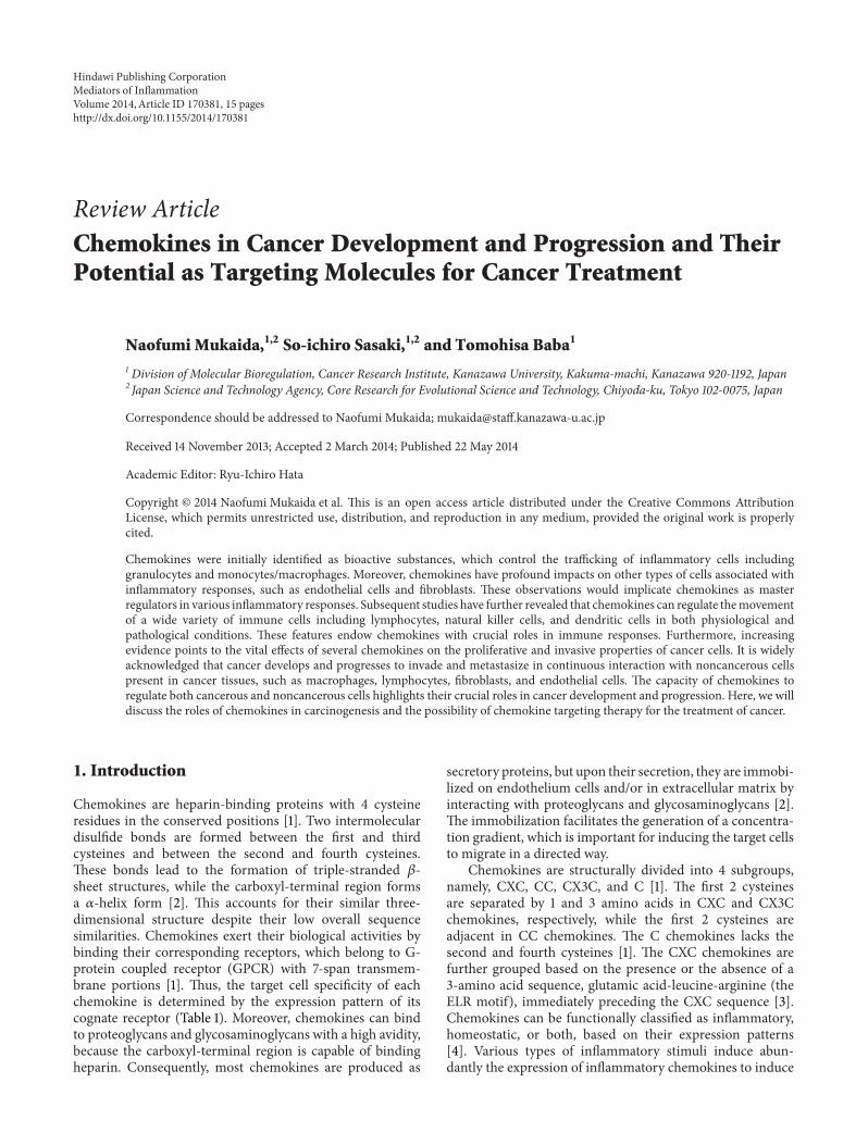
2.1. Leukocytes. Since the first description by Virchow morethan a century ago, it is widely acknowledged that leukocytesare present in both the tumor areas and the tumor-supportingstroma [8]. Moreover, leukocytes might account for upto 50% of the tumor mass, the most predominant subsetbeing macrophages. Tumor-associated macrophages (TAMs)are derived mostly from circulating monocytes which areattracted into tumor sites by locally produced chemotacticfactors, such as CCL2, CCL5, CCL7, CCL8, and CXCL12,and macrophage colony stimulating factor (M-CSF) [8].Among these chemotactic factors, CCL2 is presumed toplay an important role in TAM recruitment [8, 9]. Repeateddextran sodium sulfate (DSS) solution ingestion causes thedevelopment of multiple colonic tumors in mice, whichreceived a prior administration of azoxymethane (AOM).The resultant colonic tumors contain a large number ofmonocytes/macrophages expressing cyclooxygenase (COX)-2, an enzyme crucially involved in colon carcinogenesis [10].Abundant CCL2 is detected in colon tissues, and CCL2blockade reduces the infiltration of CCR2-positive COX-2expressing monocytes/macrophages and eventually colonictumor development and progression [10].
TAMs produce various growth factors such as vascularendothelial growth factor (VEGF) and fibroblast growthfactor (FGF) in addition to prostaglandin [8, 9]. Monocytesare recruited by CCL2 to pulmonary metastatic sites ofmurine breast cancer and promote the extravasation of tumorcells, a necessary step formetastasis, in a process that requiresmonocyte-derived VEGF [11]. Moreover, TAMs exhibit theproperties of M2-polarized macrophages and are capable ofproducing immunosuppressive molecules including IL-10,transforming growth factor (TGF)-𝛽, and arginase [12].Theseproperties endow TAMs with an immunosuppressive capac-ity. Thus, TAMs can promote tumor progression throughthe production of growth factors and the suppression ofantitumor immunity (Figure 1).
Tumor tissues contain myeloid-derived suppressor cells(MDSCs), which can suppress adaptive immunity. MDSCsare characterized by the coexpression of the myeloid-celllineage differentiation antigen Gr-1 and CD11b in mice, whilethey are defined as CD14−CD11b+ cells or as cells that expressthe common myeloid marker CD33 but lack the expressionof mature myeloid and lymphoid markers in humans [13].MDSCs contain abundantly immunosuppressive enzymes,arginase 1 and inducible NO synthetase (iNOS), and produceimmunosuppressive cytokines such as TGF-𝛽1 and IL-10,thereby inhibiting the T-cell response [13]. CCL2 recruitsMDSCs in several types of mouse cancer, including Lewislung carcinoma, meth A sarcoma, melanoma, and lymphoma[14]. However, CCR2 deficiency results in the conversion oftheMDSCphenotype frommacrophage lineage to neutrophillineage without affecting tumor growth [15]. MDSCs, partic-ularly granulocytic ones, express CXCR2 and are plentifullypresent in colonic tumors developed after the combinedtreatment of AOM and DSS [16]. CXCR2 ligands, such asCXCL1, CXCL2, and CXCL5, are present abundantly in thecolon tumor tissues and the loss of CXCR2 dramatically
CCL2,CCL7,CCL8CCL3,CCL4,CCL5
CXCL12
CXCL1,CXCL2,CXCL5CCL2,CCL7,CCL8
CCL5CXCL12
CCR2CCR5
CXCR4
CXCR2CCR2CCR5
CXCR4
Tumor-associated
macrophages(TAMs)
Myeloid-derived
suppressorcells (MDSCs)
Immune responsesGrowth factorproduction
Figure 1: Chemokines acting on TAMs and MDSCs.
suppresses tumorigenesis through inhibiting MDSC infiltra-tion [16] (Figure 1).
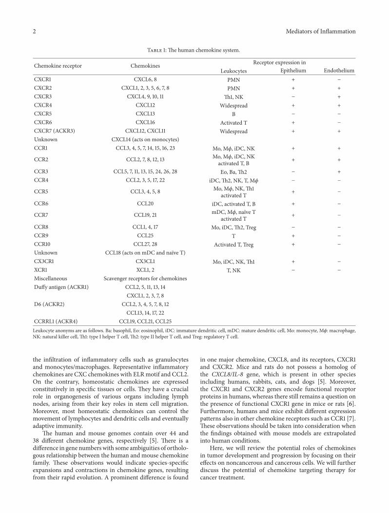
Regulatory T (Treg) cells have a crucial role in themaintenance of immunological self-tolerance [17]. A largenumber of Treg cells often infiltrate into tumors and systemicremoval of Treg cells enhances natural as well as vaccine-induced antitumor T-cell immunity. Treg cells express CCR4,and its ligand, CCL22, regulates intratumoral Treg infiltrationin various tumors [17]. Hypoxia induces the expression ofanother chemokine, CCL28, in tumor sites. CCL28 promotesangiogenesis and recruits Treg cells, thereby also propagatingimmune tolerance [18] (Figure 2).
Bindea and colleagues demonstrated that T follicularhelper (Tfh) cells and B cells infiltrated tumor sites of humancolorectal cancer patients, along with tumor progression [19].Moreover, the numbers of B cells were associated with pro-longed survival. Furthermore, when colon cancer cells wereendoscopically injected into the colon submucosa, CXCL13injection reduced tumor formation, whereas the deficiencyin CXCR5 gene, a receptor for CXCL13, accentuated tumorformation [19]. Thus, the CXCL13/CXCR5 axis might bepivotal factors for the Tfh/B cell infiltration into tumor sitesand subsequent tumor formation.
Antitumor responses are attributable to tumor infiltrat-ing lymphocytes (TILs), particularly cytotoxic T lympho-cytes (CTLs) [20]. CTLs can specifically recognize tumor-associated antigens (TAAs) and attack tumor cells in humans
4 Mediators of Inflammation
CXCL9,CXCL10,CXCL11CCL3,CCL4,CCL5
CCL2,CCL8,CCL12,CCL13CCL19,CCL21
CX3CL1
CXCR3CCR1,CCR5
CCR2CX3CR1
Cytotoxic Tlymphocytes (CTLs)
CXCR3
Natural killer(NK) cells
Antitumorresponses
RegulatoryT cell(Treg)
CCR4CCR10
CCL22CCL28
CXCL9,CXCL10,CXCL11
Figure 2: Effects of chemokines on antitumor responses.
CCR7lymph nodes
Antigen capturecapacity
Antigen presentationcapacity
MHC class II expression
Costimulatory moleculeexpression
Responding chemokines
High
High
High
High
Low
Low
Low
Low
CCL19,CCL21CXCL12
CCL3,CCL4,CCL5CCL2,CCL7,CCL8
CCL22,CCL1,CCL17CXCL12
CCR2
CCR1
CCR4
CCR5
CCR6 CCR8 CXCR4
Immature dendritic cells Mature dendritic cells
CCR7
CCR7
CXCR4
Migration to
Figure 3: Chemokines acting on dendritic cells at different maturation stages.
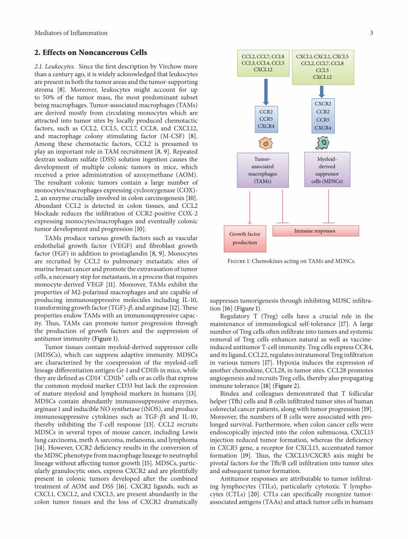
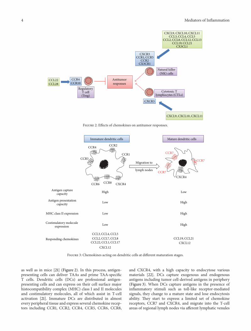
as well as in mice [21] (Figure 2). In this process, antigen-presenting cells can deliver TAAs and prime TAA-specificT cells. Dendritic cells (DCs) are professional antigen-presenting cells and can express on their cell surface majorhistocompatibility complex (MHC) class I and II moleculesand costimulatory molecules, all of which assist in T-cellactivation [21]. Immature DCs are distributed in almostevery peripheral tissue and express several chemokine recep-tors including CCR1, CCR2, CCR4, CCR5, CCR6, CCR8,
and CXCR4, with a high capacity to endocytose variousmaterials [22]. DCs capture exogenous and endogenousantigens including tumor cell-derived antigens in periphery(Figure 3). When DCs capture antigens in the presence ofinflammatory stimuli such as toll-like receptor-mediatedsignals, they change to a mature state and lose endocytosisability. They start to express a limited set of chemokinereceptors, CCR7 and CXCR4, and migrate into the T-cellareas of regional lymph nodes via afferent lymphatic venules
Mediators of Inflammation 5
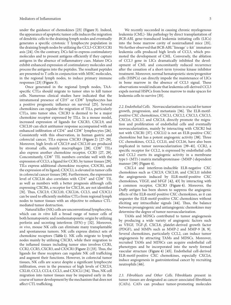
under the guidance of chemokines [23] (Figure 3). Indeed,the appearance of apoptotic tumor cells induces themigrationof dendritic cells to the draining lymph nodes and eventuallygenerates a specific cytotoxic T lymphocyte population inthe draining lymph nodes by utilizing the CCL3-CCR5/CCR1axis [24]. On the contrary, DCs fail to express costimulatorymolecules and to present antigens efficiently if they captureantigens in the absence of inflammatory cues. Mature DCsexhibit enhanced expression of costimulatory molecules andprocess the antigens into the peptides. The resultant peptidesare presented to T cells in conjunction with MHCmolecules,in the regional lymph nodes, to induce primary immuneresponses [23] (Figure 3).
Once generated in the regional lymph nodes, TAA-specific CTLs should migrate to tumor sites to kill tumorcells. Numerous clinical studies have indicated that theintratumoral presence of CD3+ or CD8+ lymphocytes hasa positive prognostic influence on survival [25]. Severalchemokines can regulate the migration of TILs, particularlyCTLs, into tumor sites. CXCR3 is deemed to be a majorchemokine receptor expressed by TILs. In a mouse model,increased expression of ligands for CXCR3, CXCL9, andCXCL10 can elicit antitumor response accompanied with anenhanced infiltration of CD4+ and CD8+ lymphocytes [26].Consistently with this observation, in human gastric andcolorectal cancer, TILs express CXCR3 (Figure 2) [27–29].Moreover, high levels of CXCL9 and CXCL10 are producedby stromal cells, mainly macrophages [28]. CD8+ TILsalso express another chemokine receptor, CCR5 [27, 29].Concomitantly, CD8+ TIL numbers correlate well with theexpression of CCL5, a ligand for CCR5, by tumor tissues [29].TILs express additional chemokine receptor, CX3CR1, andthe expression of its ligand, CX3CL1, is elevated in tumor cellsin colorectal cancer tissues [30]. Furthermore, the expressionlevel of CXCL16 also correlates with CD4+ and CD8+ TILas well as numbers with a better prognosis although cellsexpressing CXCR6, a receptor for CXCL16, are not identified[31]. Thus, CXCL9, CXCL10, CXCL16, CCL5, and CX3CL1can be used to efficiently mobilize CTLs from regional lymphnodes to tumor tissues with an objective to enhance CTL-mediated tumor destruction.
Natural killer (NK) cells are unconventional lymphocytes,which can in vitro kill a broad range of tumor cells ofboth hematopoietic and nonhematopoietic origin by utilizingperforin and secreting interferon (IFN)-𝛾 [32]. Moreover,in vivo, mouse NK cells can eliminate many transplantableand spontaneous tumors. NK cells express distinct sets ofchemokine receptors (Table 1). NK cells migrate to lymphnodes mainly by utilizing CXCR3, while their migration tothe inflamed tissues including tumor sites involves CCR1,CCR2, CCR5, CXCR3, and CX3CR1 (Figure 2) [33].Thus, theligands for these receptors can regulate NK cell traffickingand augment their functions. However, in colorectal tumortissues, NK cells are scarce despite a significant lymphocyteinfiltration, even in the presence of high levels of CXCL9,CXL10, CCL3, CCL4, CCL5, and CX3CL1 [34].Thus, NK cellmigration into tumor tissues may be impaired early in thecourse of tumor development by themechanism that does notaffect CTL trafficking.
We recently succeeded in causing chronic myelogenousleukemia (CML)- like pathology by direct transplantation ofBCR-ABL gene-transduced leukemia initiating cells (LICs)into the bone marrow cavity of nonirradiated mice [35].We further observed that BCR-ABL+lineage−c-kit− immatureleukemia cells produced high levels of CCL3, which pro-moted the development of CML. Conversely, the ablationof CCL3 gene in LICs dramatically inhibited the devel-opment of CML and concomitantly reduced recurrenceafter the cessation of a short-term tyrosine kinase inhibitortreatment. Moreover, normal hematopoietic stem/progenitorcells (HSPCs) can directly impede the maintenance of LICsin bone marrow in the absence of CCL3 signal. Theseobservations would indicate that leukemia cell-derived CCL3expels normal HSPCs from bone marrow to make spaces forleukemia cells to survive [35].
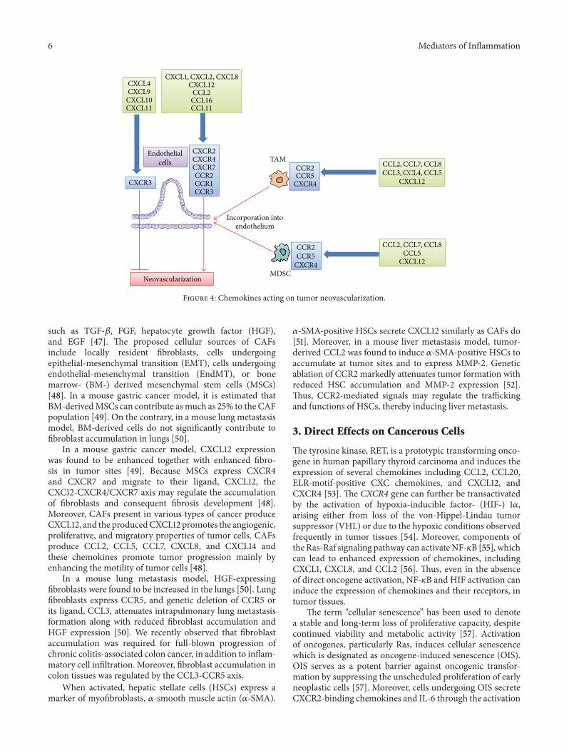
2.2. Endothelial Cells. Neovascularization is crucial for tumorgrowth, progression, and metastasis [36]. The ELR-motif-positive CXC chemokines, CXCL1, CXCL2, CXCL3, CXCL5,CXCL6, CXCL7, and CXCL8, directly promote the migra-tion and proliferation of endothelial cells and eventuallyneovascularization, mainly by interacting with CXCR2 butnot with CXCR1 [37]. CXCL12 is not an ELR-positive CXCchemokine but has a potent angiogenic activity [38]. ThreeCC chemokines, CCL2, CCL11, and CCL16, have also beenimplicated in tumor neovascularization [39–41]. CCR2, aspecific receptor for CCL2, is expressed by endothelial cellsand CCL2 exerts its angiogenic activity in a membranetype 1- (MT1-) matrix metalloproteinase- (MMP-) dependentmanner [39] (Figure 4).
CXCL4 and interferon-inducible ELR-negative CXCchemokines such as CXCL9, CXCL10, and CXCL11 inhibitthe angiogenesis induced by ELR-motif-positive CXCchemokines, VEGF, and bFGF [42, 43] by interacting witha common receptor, CXCR3 (Figure 4). Moreover, theDuffy antigen has been shown to suppress the angiogeniceffects of the ELR-motif-positive CXC chemokines, as it cansequester the ELR-motif-positive CXC chemokines withouteliciting any intracellular signals [44]. Thus, the balancebetween proangiogenic and antiangiogenic chemokines maydetermine the degree of tumor neovascularization.
TAMs and MDSCs contributed to tumor angiogenesisby producing a wide variety of angiogenic factors suchas VEGF, TGF-𝛽, CXCL8, platelet-derived growth factor(PDGF), and MMPs such as MMP-2 and MMP-9 [8, 9].Several chemokines, particularly CCL2, can induce tumorangiogenesis by attracting TAMs and MDSCs. Moreover,recruited TAMs and MDSCs can acquire endothelial cellphenotypes and be incorporated into the newly formedvascular structure (Figure 4) [45]. Endothelial cell-derivedELR-motif-positive CXC chemokines, especially CXCL6,induce angiogenesis in gastrointestinal cancer by recruitingneutrophils [46].
2.3. Fibroblasts and Other Cells. Fibroblasts present intumor tissues are designated as cancer-associated fibroblasts(CAFs). CAFs can produce tumor-promoting molecules
6 Mediators of Inflammation
CXCL1,CXCL2,CXCL8CXCL12
CCL2CCL16CCL11
CXCL4CXCL9
CXCL10CXCL11
Endothelialcells
CXCR2CXCR4CXCR7CCR2CCR1CCR3
CXCR3
CCR2CCR5
CXCR4
TAM CCL2,CCL7,CCL8CCL3,CCL4,CCL5
CXCL12
CCL2,CCL7,CCL8CCL5
CXCL12
Neovascularization
CCR2CCR5
CXCR4MDSC
Incorporation intoendothelium
Figure 4: Chemokines acting on tumor neovascularization.
such as TGF-𝛽, FGF, hepatocyte growth factor (HGF),and EGF [47]. The proposed cellular sources of CAFsinclude locally resident fibroblasts, cells undergoingepithelial-mesenchymal transition (EMT), cells undergoingendothelial-mesenchymal transition (EndMT), or bonemarrow- (BM-) derived mesenchymal stem cells (MSCs)[48]. In a mouse gastric cancer model, it is estimated thatBM-derivedMSCs can contribute asmuch as 25% to the CAFpopulation [49]. On the contrary, in a mouse lung metastasismodel, BM-derived cells do not significantly contribute tofibroblast accumulation in lungs [50].
In a mouse gastric cancer model, CXCL12 expressionwas found to be enhanced together with enhanced fibro-sis in tumor sites [49]. Because MSCs express CXCR4and CXCR7 and migrate to their ligand, CXCL12, theCXC12-CXCR4/CXCR7 axis may regulate the accumulationof fibroblasts and consequent fibrosis development [48].Moreover, CAFs present in various types of cancer produceCXCL12, and the producedCXCL12 promotes the angiogenic,proliferative, and migratory properties of tumor cells. CAFsproduce CCL2, CCL5, CCL7, CXCL8, and CXCL14 andthese chemokines promote tumor progression mainly byenhancing the motility of tumor cells [48].
In a mouse lung metastasis model, HGF-expressingfibroblasts were found to be increased in the lungs [50]. Lungfibroblasts express CCR5, and genetic deletion of CCR5 orits ligand, CCL3, attenuates intrapulmonary lung metastasisformation along with reduced fibroblast accumulation andHGF expression [50]. We recently observed that fibroblastaccumulation was required for full-blown progression ofchronic colitis-associated colon cancer, in addition to inflam-matory cell infiltration. Moreover, fibroblast accumulation incolon tissues was regulated by the CCL3-CCR5 axis.
When activated, hepatic stellate cells (HSCs) express amarker of myofibroblasts, 𝛼-smooth muscle actin (𝛼-SMA).
𝛼-SMA-positive HSCs secrete CXCL12 similarly as CAFs do[51]. Moreover, in a mouse liver metastasis model, tumor-derived CCL2 was found to induce 𝛼-SMA-positive HSCs toaccumulate at tumor sites and to express MMP-2. Geneticablation of CCR2 markedly attenuates tumor formation withreduced HSC accumulation and MMP-2 expression [52].Thus, CCR2-mediated signals may regulate the traffickingand functions of HSCs, thereby inducing liver metastasis.
3. Direct Effects on Cancerous Cells
The tyrosine kinase, RET, is a prototypic transforming onco-gene in human papillary thyroid carcinoma and induces theexpression of several chemokines including CCL2, CCL20,ELR-motif-positive CXC chemokines, and CXCL12, andCXCR4 [53]. The CXCR4 gene can further be transactivatedby the activation of hypoxia-inducible factor- (HIF-) 1𝛼,arising either from loss of the von-Hippel-Lindau tumorsuppressor (VHL) or due to the hypoxic conditions observedfrequently in tumor tissues [54]. Moreover, components ofthe Ras-Raf signaling pathway can activateNF-𝜅B [55], whichcan lead to enhanced expression of chemokines, includingCXCL1, CXCL8, and CCL2 [56]. Thus, even in the absenceof direct oncogene activation, NF-𝜅B and HIF activation caninduce the expression of chemokines and their receptors, intumor tissues.
The term “cellular senescence” has been used to denotea stable and long-term loss of proliferative capacity, despitecontinued viability and metabolic activity [57]. Activationof oncogenes, particularly Ras, induces cellular senescencewhich is designated as oncogene-induced senescence (OIS).OIS serves as a potent barrier against oncogenic transfor-mation by suppressing the unscheduled proliferation of earlyneoplastic cells [57]. Moreover, cells undergoing OIS secreteCXCR2-binding chemokines and IL-6 through the activation
Mediators of Inflammation 7
CXCL12CXCL1,CXCL5,CXCL8
CXCL12CCL27,CCL28CCL19,CCL19
CXCL12CCL20
CXCL16
CXCL12CXCL1,CXCL5,CXCL8
CCL19,CCL21CCL25
CXCL1,CXCL8
CXCR4CXCR1,CXCR2
CXCR4CCR6
CXCR6
CXCR4CCR10CCR7
CXCR4CXCR1,CXCR2
CCR7CCR9
CXCR2
EMTProliferation
Apoptosis
Motility Senescence
Tumor growth Invasionmetastasis Tumorigenesis
Figure 5: Effects of chemokines on tumor cells.
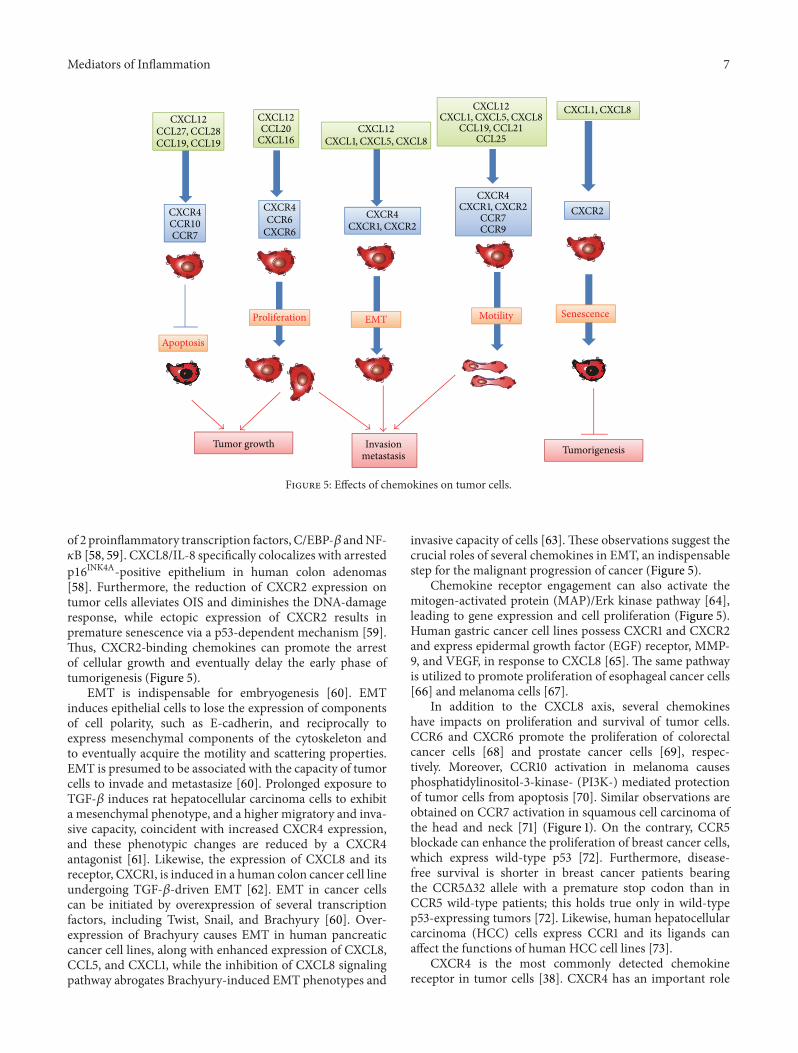
of 2 proinflammatory transcription factors, C/EBP-𝛽 andNF-𝜅B [58, 59]. CXCL8/IL-8 specifically colocalizes with arrestedp16INK4A-positive epithelium in human colon adenomas[58]. Furthermore, the reduction of CXCR2 expression ontumor cells alleviates OIS and diminishes the DNA-damageresponse, while ectopic expression of CXCR2 results inpremature senescence via a p53-dependent mechanism [59].Thus, CXCR2-binding chemokines can promote the arrestof cellular growth and eventually delay the early phase oftumorigenesis (Figure 5).
EMT is indispensable for embryogenesis [60]. EMTinduces epithelial cells to lose the expression of componentsof cell polarity, such as E-cadherin, and reciprocally toexpress mesenchymal components of the cytoskeleton andto eventually acquire the motility and scattering properties.EMT is presumed to be associated with the capacity of tumorcells to invade and metastasize [60]. Prolonged exposure toTGF-𝛽 induces rat hepatocellular carcinoma cells to exhibita mesenchymal phenotype, and a higher migratory and inva-sive capacity, coincident with increased CXCR4 expression,and these phenotypic changes are reduced by a CXCR4antagonist [61]. Likewise, the expression of CXCL8 and itsreceptor, CXCR1, is induced in a human colon cancer cell lineundergoing TGF-𝛽-driven EMT [62]. EMT in cancer cellscan be initiated by overexpression of several transcriptionfactors, including Twist, Snail, and Brachyury [60]. Over-expression of Brachyury causes EMT in human pancreaticcancer cell lines, along with enhanced expression of CXCL8,CCL5, and CXCL1, while the inhibition of CXCL8 signalingpathway abrogates Brachyury-induced EMT phenotypes and
invasive capacity of cells [63]. These observations suggest thecrucial roles of several chemokines in EMT, an indispensablestep for the malignant progression of cancer (Figure 5).
Chemokine receptor engagement can also activate themitogen-activated protein (MAP)/Erk kinase pathway [64],leading to gene expression and cell proliferation (Figure 5).Human gastric cancer cell lines possess CXCR1 and CXCR2and express epidermal growth factor (EGF) receptor, MMP-9, and VEGF, in response to CXCL8 [65]. The same pathwayis utilized to promote proliferation of esophageal cancer cells[66] and melanoma cells [67].
In addition to the CXCL8 axis, several chemokineshave impacts on proliferation and survival of tumor cells.CCR6 and CXCR6 promote the proliferation of colorectalcancer cells [68] and prostate cancer cells [69], respec-tively. Moreover, CCR10 activation in melanoma causesphosphatidylinositol-3-kinase- (PI3K-) mediated protectionof tumor cells from apoptosis [70]. Similar observations areobtained on CCR7 activation in squamous cell carcinoma ofthe head and neck [71] (Figure 1). On the contrary, CCR5blockade can enhance the proliferation of breast cancer cells,which express wild-type p53 [72]. Furthermore, disease-free survival is shorter in breast cancer patients bearingthe CCR5Δ32 allele with a premature stop codon than inCCR5 wild-type patients; this holds true only in wild-typep53-expressing tumors [72]. Likewise, human hepatocellularcarcinoma (HCC) cells express CCR1 and its ligands canaffect the functions of human HCC cell lines [73].
CXCR4 is the most commonly detected chemokinereceptor in tumor cells [38]. CXCR4 has an important role
8 Mediators of Inflammation
in the proliferation of various cancer cells including ovarian,glioma, melanoma, lung, renal, and thyroid cancer cells [38,74]. The CXCL12/CXCR4 axis delivers surviving signals tohepatocellular carcinoma cells, ovarian carcinoma cells, andchronic leukemia cells, while CXCR4 blockade induces theapoptosis of these malignant cells [38, 61, 75] (Figure 5).CXCL12 expression correlates well with lower apoptosis inhuman myelodysplastic syndrome [76].
Chemokines can regulate the migration of tumor cells.CXCR4-expressing cells canmigrate in vitro towards CXCL12[77]. CCR7, CCR9, CXCR1, and CXCR2 are also detectedin tumor cells and their ligands can induce the chemo-taxis of the corresponding receptor-expressing cells [78, 79].These chemokines can serve as inducers of invasion withinthe primary tumor and dissemination to distant organs(Figure 5). Moreover, human pancreatic ductal adenocarci-noma (PDAC) cells express CX3CR1 andCX3CL1 abundantlyand the CX3CR1/CX3CL1 axis can regulate intraneural inva-sion of PDAC [80]. Adult T-cell leukemia (ATL) cells expressfrequently CCR4 and can migrate in vitro to CCL17 andCCL22, ligands for CCR4 [81].TheCCL17/CCL22-CCR4 axismay account for the frequent infiltration of ATL into skinand lymph nodes, where CCL17 and CCL22 are abundantlyexpressed.
Circulating tumor cells (CTCs) are presumed to be asource ofmetastasizing tumor cells, but they can also colonizeat their original site [82]. This process, which is called astumor self-seeding, can accelerate tumor growth and angio-genesis. CTCs produce ELR-motif-positive CXC chemokinesincluding CXCL8 and CXCL1, and these chemokines eventu-ally promote self-seeding [82].
Several models have been proposed to explain the molec-ular mechanisms underlying the enhancement and regula-tion of metastasis by the chemokines. Specific chemokinereceptor-expressing tumor cells may migrate to organs withhigh expression levels of the corresponding chemokinesalong a concentration gradient [77]. This hypothesis mayexplain the tissue tropism observed in certain types ofcancer, but there is little evidence to indicate the presenceof chemokine concentration gradients between primary andmetastatic sites. A transcellular CCR7 ligand gradient canbe created when cancer cells produce CCR7 ligands underflow conditions and the resultant gradient can be the basisof lymphatic metastasis [83]. Thus, cancer cells themselvesmay actively promote their own metastasis and tropism byproducing chemokines. Another plausible explanation is thatthe arrival of tumor cells in a specific organ is passive andthat chemokine receptor expression provides tumor cellswith an advantage to survive and grow in a different ligand-rich metastatic microenvironment [84]. Moreover, CXCL12and a CCR7 ligand, CCL21, can induce the resistance ofcancer cells to anoikis, which is a major hindrance to themetastatic spread of various types of cancer, by regulatingproapoptotic Bmf and antiapoptotic Bcl-xL proteins [85].Thus, chemokines may accelerate metastasis by promotingtumor cell proliferation or preventing tumor cell death.
Thus, chemokines can prevent tumorigenesis in the earlyphase by inducing cellular senescence while they can also
promote invasion and metastasis by inducing EMT andenhancing the motility and survival of tumor cells (Figure 5).
4. Potential of Chemokine Targeting Therapyas Cancer Treatment
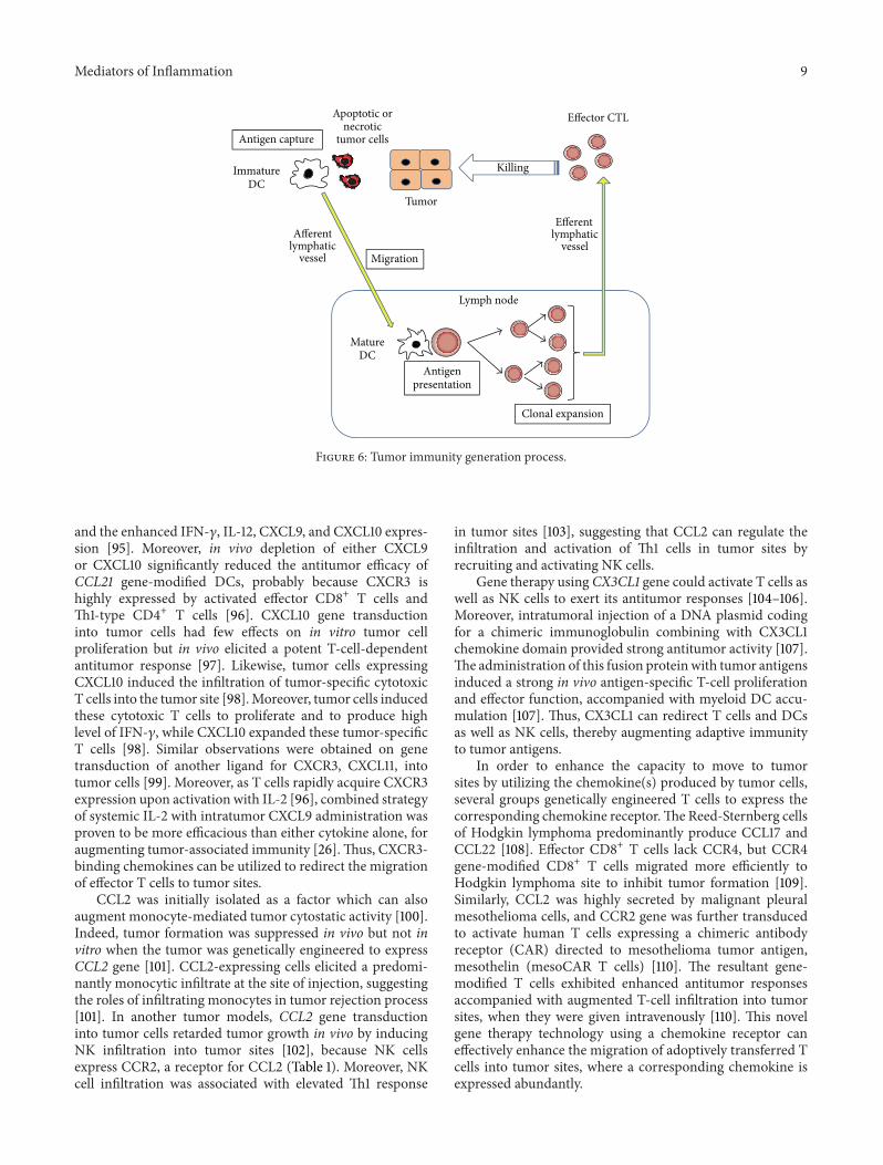
4.1. Chemokine-Mediated Enhancement in Tumor Immunity.Theestablishment of tumor immunity is amultistage process:migration of DCs to tumor sites, capture of TAAs by DCs,migration of DCs to regional lymph nodes, antigen presen-tation to effector cells by DCs in regional lymph nodes, andmigration of effector cells to tumor sites (Figure 6) [21, 22].Chemokines have profound impacts on tumor immunity,particularly migration steps.
Immature dendritic cells move to the tumor tissues tophagocytose apoptotic tumor cells, capture tumor-associatedantigens (TAAs), and migrate to draining lymph nodes,where DCs present antigens to induce specific CTLs [21–23]. Tumor-infiltrating DCs expressed CCR1 and CCR5,and a ligand for these receptors, CCL3, was abundantlydetected in mouse bearing HCC [24]. Mirroring the capacityof CCL3 to mobilize CCR1- or CCR5-expressing immatureDCs to peripheral blood from bone marrow [86], systemicadministration of CCL3 increased the numbers of DCsin peripheral blood and tumor tissues and concomitantlyaugmented antitumor effects after radiofrequency ablation ofmurine HCCs [87]. Thus, CCL3 may be effective to enhancetumor immunity by attracting immature DCs to dying tumorcells.
CCL19 and CCL21, ligands for CCR7, can regulate DCmigration to lymph nodes for antigen presentation to naıveT cells, which also utilize CCL19 and/or CCL21 to enter T-cell zone [88], and they can additionally attract NK cellsto the lymph node. As a consequence, when CCL21 wasinjected into a regional lymph node of SV40-transgenic micethat developed bilateral multifocal lung adenocarcinomas, itincreased CD4+ and CD8+ lymphocytes as well as DCs atlymph nodes and tumor sites and eventually led to a markedreduction in tumor burdens with enhanced survival [89].Similar results were also obtained when CCL19 was injectedintranodally into SV40-transgenic mice [90].
Low clinical efficacy of DC-based vaccines can beexplained by a very limited capacity of ex vivo generated DCto move from the injected sites to draining lymph nodes [91].In order to circumvent these problems, DCs are geneticallymodified to express CCL19 and CCL21. Indeed, intratumoralinjection ofCCL21 gene-modifiedDCs resulted inmore effec-tive tumor growth inhibition than unmodified control DCs[92], together with intratumoral accumulation of DCs and Tcells [93]. Moreover, even when CCL21 gene-modified DCswere pulsed with tumor lysates and subsequently injectedsubcutaneously to tumor-free sites in tumor-bearing mice,it elicited a good antitumor response [92]. These promisingpreclinical results have led to ongoing phase I clinical trials[94].
Intratumoral administration of CCL21 gene-modifiedDCs reduced a tumor burden in spontaneous murine lungcarcinoma, accompanied with extensive T-cell infiltration
Mediators of Inflammation 9
Antigen capture
ImmatureDC
Apoptotic ornecrotic
tumor cells
Tumor
Killing
Effector CTL
Afferentlymphatic
vessel
Efferentlymphatic
vessel
Lymph node
MatureDC
Antigenpresentation
Clonal expansion
Migration
Figure 6: Tumor immunity generation process.
and the enhanced IFN-𝛾, IL-12, CXCL9, and CXCL10 expres-sion [95]. Moreover, in vivo depletion of either CXCL9or CXCL10 significantly reduced the antitumor efficacy ofCCL21 gene-modified DCs, probably because CXCR3 ishighly expressed by activated effector CD8+ T cells andTh1-type CD4+ T cells [96]. CXCL10 gene transductioninto tumor cells had few effects on in vitro tumor cellproliferation but in vivo elicited a potent T-cell-dependentantitumor response [97]. Likewise, tumor cells expressingCXCL10 induced the infiltration of tumor-specific cytotoxicT cells into the tumor site [98].Moreover, tumor cells inducedthese cytotoxic T cells to proliferate and to produce highlevel of IFN-𝛾, while CXCL10 expanded these tumor-specificT cells [98]. Similar observations were obtained on genetransduction of another ligand for CXCR3, CXCL11, intotumor cells [99]. Moreover, as T cells rapidly acquire CXCR3expression upon activation with IL-2 [96], combined strategyof systemic IL-2 with intratumor CXCL9 administration wasproven to be more efficacious than either cytokine alone, foraugmenting tumor-associated immunity [26].Thus, CXCR3-binding chemokines can be utilized to redirect the migrationof effector T cells to tumor sites.
CCL2 was initially isolated as a factor which can alsoaugment monocyte-mediated tumor cytostatic activity [100].Indeed, tumor formation was suppressed in vivo but not invitro when the tumor was genetically engineered to expressCCL2 gene [101]. CCL2-expressing cells elicited a predomi-nantly monocytic infiltrate at the site of injection, suggestingthe roles of infiltrating monocytes in tumor rejection process[101]. In another tumor models, CCL2 gene transductioninto tumor cells retarded tumor growth in vivo by inducingNK infiltration into tumor sites [102], because NK cellsexpress CCR2, a receptor for CCL2 (Table 1). Moreover, NKcell infiltration was associated with elevated Th1 response
in tumor sites [103], suggesting that CCL2 can regulate theinfiltration and activation of Th1 cells in tumor sites byrecruiting and activating NK cells.
Gene therapy using CX3CL1 gene could activate T cells aswell as NK cells to exert its antitumor responses [104–106].Moreover, intratumoral injection of a DNA plasmid codingfor a chimeric immunoglobulin combining with CX3CL1chemokine domain provided strong antitumor activity [107].The administration of this fusion proteinwith tumor antigensinduced a strong in vivo antigen-specific T-cell proliferationand effector function, accompanied with myeloid DC accu-mulation [107]. Thus, CX3CL1 can redirect T cells and DCsas well as NK cells, thereby augmenting adaptive immunityto tumor antigens.
In order to enhance the capacity to move to tumorsites by utilizing the chemokine(s) produced by tumor cells,several groups genetically engineered T cells to express thecorresponding chemokine receptor.The Reed-Sternberg cellsof Hodgkin lymphoma predominantly produce CCL17 andCCL22 [108]. Effector CD8+ T cells lack CCR4, but CCR4gene-modified CD8+ T cells migrated more efficiently toHodgkin lymphoma site to inhibit tumor formation [109].Similarly, CCL2 was highly secreted by malignant pleuralmesothelioma cells, and CCR2 gene was further transducedto activate human T cells expressing a chimeric antibodyreceptor (CAR) directed to mesothelioma tumor antigen,mesothelin (mesoCAR T cells) [110]. The resultant gene-modified T cells exhibited enhanced antitumor responsesaccompanied with augmented T-cell infiltration into tumorsites, when they were given intravenously [110]. This novelgene therapy technology using a chemokine receptor caneffectively enhance the migration of adoptively transferred Tcells into tumor sites, where a corresponding chemokine isexpressed abundantly.
10 Mediators of Inflammation
4.2. Reversal of Suppressor Cell-Mediated Immune Suppres-sion. Tumor immunity can frequently induce immune sup-pressive mechanisms to reduce the “immunity to self ” bythe action of several negative immunoregulatory receptorssuch as cytotoxic T lymphocyte antigen-4 (CTLA-4) and theprogrammed death receptor-1- (PD-1-) PD ligand-1 (PD-L1)axis. Consequently, the antagonizing monoclonal antibodiesto CTLA-4, PD-1, or PD-L1 are effective against varioustypes of cancer even at advanced stages [111, 112]. Theseobservations indicate that reversal of immune suppressioncan be effective to enhance tumor immunity.
Tumor tissues contain the leukocytes that can diminishtumor immunity. The most predominant subset is TAMs[8, 12]. TAMs can promote tumor progression by induc-ing angiogenesis and suppression of adaptive and innateantitumor immunity (Figure 1). Circulating monocytes aremostly the precursor of these TAMs and are attracted intotumor sites, by several chemokines, particularly CCL2 [8,12]. Indeed, systemic delivery of neutralizing anti-CCL2antibody attenuated tumor burdens in human prostatecancer-bearing mice although its effects on TAMs have notbeen examined [113]. Moreover, CCL2 blockade reducedCCR2-expressing TAM infiltration and eventually tumorformation in chronic colitis-associated carcinogenesis [10].Furthermore, CCL2 also recruited monocytes to pulmonarymetastatic sites of murine breast cancer to promote theextravasation of tumor cells, a prerequisite step formetastasis,in monocyte-derived VEGF-dependent manner [11]. CCL2blockade markedly reduced lung metastasis together withreduced monocyte/macrophage infiltration.
Another type of immune suppressor cells is MDSCs witha strong ability to suppress various T-cell functions [13].CCL2 recruits MDSCs in several types of mouse cancer [14].Moreover, CCL2-mediated MDSC accumulation can nega-tively regulate the entry of adoptively transferred activatedCD8+ cells into tumor sites [114]. CCR2 deficiency, however,caused conversion of theMDSC phenotype to neutrophil lin-eage without affecting tumor growth [15], probably becauseMDSC contains a subset of immature neutrophils [115].On the contrary, CXCR2 blockade reduced the infiltrationof CXCR2-expressing granulocytic MDSCs and eventuallytumor growth in chronic colitis-associated colon cancer [16].
Adult T-cell leukemia (ATL) cells are characterized byrobust expression of CCR4 and canmigrate in vitro to CCL17andCCL22, ligands for CCR4 [81]. Humanized defucosylatedmonoclonal antibody to CCR4 has been obtained. The resul-tant antibody can exert more potent antibody-dependentcytotoxicity (ADCC) [116] and is capable of removing CCR4-expressing ATL cells in peripheral blood and bone marrowmainly by ADCC.
A large number of Treg cells often infiltrate into tumorsand systemic removal of Treg cells enhances natural as wellas vaccine-induced antitumor T-cell immunity [17]. Treg cellsexpress CCR4 and its ligand, CCL22, mainly regulates intra-tumoral Treg infiltration in various tumors [17] (Figure 2).Indeed, intratumoral CCL22 expression correlated well withFoxp3 expression in colorectal carcinoma tissues [117]. Inline with these observations, anti-CCR4 antibody treatment
depletes Tregs and eventually evokes CD8+ T-cell responseagainst TAAs [118]. Furthermore, CCL2 is also involved inTreg accumulation and, as a consequence, anti-CCL2 anti-body augmented cancer immunotherapy against non-smallcell lung cancer in mice with reduced intratumoral Tregs andincreased numbers of intratumoral antigen-specific activatedCD8+ cells, when it was administered in combination witha tumor vaccine [119]. These observations illustrate thattargeting these chemokines can reduce intratumoral Tregcells, resulting in the enhancement of tumor immunity.
Recently, CCR1-expressing CD34+ immature myeloidcells have been detected in murine intestinal tumors withSMAD4 deficiency [120]. These cells expressed abundantlyMMP-9 and MMP-2 and were involved in invasion. More-over, a CCR1 antagonist suppressed colon cancer liver metas-tasis by blocking accumulation of CD34+ immature myeloidcells [121].
4.3. Other Strategies of AntitumorTherapy Targeting Chemok-ines. Neovascularization is crucial for tumor growth, pro-gression, and metastasis [36]. The ELR motif-positive CXCchemokines, CXCL1, CXCL2, CXCL3, CXCL5, CXCL6,CXCL7, and CXCL8, can directly promote the migration andproliferation of endothelial cells and eventually neovascular-ization [37] (Figure 4). Indeed, the administration of anti-CXCL8 reduced the tumor sizes of humannon-small cell lungcancer cells which are injected into severe combined immunedeficient (SCID) mice in advance [122]. The reduction intumor size was associated with a decline in tumor-associatedvascular density and was accompanied by a decrease inspontaneous lung metastasis. CXCL12 [123] and three CCchemokines, CCL2, CCL11, and CCL16, have also beenimplicated in tumor neovascularization [39–41, 45].However,it still remains elusive on the efficacy of targeting thesechemokines for the control of tumor neovascularization.
CXCL4, CXCL9, CXCL10, and CXCL11 inhibit the angio-genesis induced by ELR motif-positive CXC chemokines,VEGF, and bFGF [42, 43]. Targeted expression of CXCL9 orintratumoral CXCL9 administration retarded in vivo tumorgrowth by inhibiting tumor-derived angiogenesis [26, 124].Thus, these chemokines can be effective for tumor therapyby inhibiting neovascularization as well as inducing CXCR3-expressing cytotoxic T-cell infiltration.
5. Concluding Remarks
Cancer development and progression are profoundly affectedby inflammatory and immune responses. Inflammatoryresponses consist of leukocyte infiltration, neovasculariza-tion, and fibrosis, while immune responses were exertedby immune cells such as lymphocytes and dendritic cells.Chemokines have great impacts on the cells involved inboth inflammatory and immune responses.Moreover, severalchemokines have direct effects on the proliferative andinvasive properties of tumor cells. Consequently, chemokinesplay crucial roles in tumor development and progressionby acting on cancerous and noncancerous cells. However,it is embarrassing that the same chemokine can induce
Mediators of Inflammation 11
tumor progression as well as protection against a tumor, in acontext-dependent manner. Given the multifactorial roles ofchemokines in carcinogenesis, the elucidation of their roleswill further advance our understanding of the pathophysio-logical processes of tumor development and progression andwill subsequently pave a novel way to develop a novel type ofanticancer treatment by targeting chemokines.
Conflict of Interests
The authors declare that there is no conflict of interestsregarding the publication of this paper.
Acknowledgment
This work is partly supported by the grant-in-aid fromthe Ministry of Education, Culture, Sports, Science andTechnology of the Japanese Government.
References
[1] B. Moser, M. Wolf, A. Walz, and P. Loetscher, “Chemokines:multiple levels of leukocytemigration control,”Trends in Immu-nology, vol. 25, no. 2, pp. 75–84, 2004.
[2] E. J. Fernandez and E. Lolis, “Structure, function, and inhibitionof chemokines,” Annual Review of Pharmacology and Toxicol-ogy, vol. 42, pp. 469–499, 2002.
[3] J. Vandercappellen, J. van Damme, and S. Struyf, “The role ofCXC chemokines and their receptors in cancer,” Cancer Letters,vol. 267, no. 2, pp. 226–244, 2008.
[4] A. Zlotnik and O. Yoshie, “The chemokine superfamily revis-ited,” Immunity, vol. 36, no. 5, pp. 705–716, 2012.
[5] H. Nomiyama, N. Osada, and O. Yoshie, “The evolution ofmammalian chemokine genes,” Cytokine and Growth FactorReviews, vol. 21, no. 4, pp. 253–262, 2010.
[6] B. Moepps, E. Nuesseler, M. Braun, and P. Gierschik, “A homo-log of the human chemokine receptor CXCR1 is expressed inthe mouse,” Molecular Immunology, vol. 43, no. 7, pp. 897–914,2006.
[7] S.-B. Su, N. Mukaida, J. Wang, H. Nomura, and K. Matsushima,“Preparation of specific antagonizing polyclonal antibodies toa C-C chemokine receptor, CCR1 and determination of itsdistribution of various types of leukocytes,” Journal of LeukocyteBiology, vol. 60, no. 5, pp. 658–666, 1996.
[8] A. Sica, P. Allavena, and A. Mantovani, “Cancer related inflam-mation: the macrophage connection,” Cancer Letters, vol. 267,no. 2, pp. 204–215, 2008.
[9] G. Lazennec and A. Richmond, “Chemokines and chemokinereceptors: new insights into cancer-related inflammation,”Trends in Molecular Medicine, vol. 16, no. 3, pp. 133–144, 2010.
[10] B. K. Popivanova, F. I. Kostadinova, K. Furuichi et al., “Blockadeof a chemokine, CCL2, reduces chronic colitis-associated car-cinogenesis in mice,” Cancer Research, vol. 69, no. 19, pp. 7884–7892, 2009.
[11] B. Z. Qian, J. Li, H. Zhang et al., “CCL2 recruits inflammatorymonocytes to facilitate breast-tumour metastasis,” Nature, vol.475, no. 7355, pp. 222–225, 2011.
[12] A. Mantovani, S. Sozzani, M. Locati, P. Allavena, and A. Sica,“Macrophage polarization: tumor-associated macrophages as a
paradigm for polarizedM2mononuclear phagocytes,”Trends inImmunology, vol. 23, no. 11, pp. 549–555, 2002.
[13] D. I. Gabrilovich and S. Nagaraj, “Myeloid-derived suppressorcells as regulators of the immune system,” Nature ReviewsImmunology, vol. 9, no. 3, pp. 162–174, 2009.
[14] B. Huang, Z. Lei, J. Zhao et al., “CCL2/CCR2 pathway mediatesrecruitment of myeloid suppressor cells to cancers,” CancerLetters, vol. 252, no. 1, pp. 86–92, 2007.
[15] Y. Sawanobori, S. Ueha, M. Kurachi et al., “Chemokine-mediated rapid turnover of myeloid-derived suppressor cellsin tumor-bearing mice,” Blood, vol. 111, no. 12, pp. 5457–5466,2008.
[16] H. Katoh, D. Wang, T. Daikoku, H. Sun, S. K. Dey, and R.N. DuBois, “CXCR2-expressing myeloid-derived suppressorcells are essential to promote colitis-associated tumorigenesis,”Cancer Cell, vol. 24, no. 3, pp. 631–644, 2013.
[17] H. Nishikawa and S. Sakaguchi, “Regulatory T cells in tumorimmunity,” International Journal of Cancer, vol. 127, no. 4, pp.759–767, 2010.
[18] A. Facciabene, X. Peng, I. S. Hagemann et al., “Tumour hypoxiapromotes tolerance and angiogenesis via CCL28 and Treg cells,”Nature, vol. 475, no. 7355, pp. 226–230, 2011.
[19] G. Bindea, B. Miecnik, M. Tosolini et al., “Spaciotemporaldynamics of intratumoral immune cells reveals the immunelandscape in human cancer,” Immunity, vol. 39, no. 4, pp. 782–795, 2013.
[20] H. J. Steer, R. A. Lake, A. K. Nowak, and B. W. Robinson, “Har-nessing the immune response to treat cancer,”Oncogene, vol. 29,no. 48, pp. 6301–6313, 2010.
[21] K. L. Knutson and M. L. Disis, “Tumor antigen-specific Thelper cells in cancer immunity and immunotherapy,” CancerImmunology and Immunotherapy, vol. 54, no. 8, pp. 721–728,2005.
[22] K. Palucka and J. Banchereau, “Cancer immunotherapy viadendritic cells,” Nature Reviews Cancer, vol. 12, no. 4, pp. 265–277, 2012.
[23] S. Sozzani, “Dendritic cell trafficking: more than justchemokines,” Cytokine and Growth Factor Reviews, vol.16, no. 6, pp. 581–592, 2005.
[24] N. Iida, Y. Nakamoto, T. Baba et al., “Tumor cell apoptosisinduces tumor-specific immunity in a CC chemokine receptor1- and 5-dependent manner in mice,” Journal of LeukocyteBiology, vol. 84, no. 4, pp. 1001–1010, 2008.
[25] M. J. Gooden, G. H. de Bock, N. Leffers, T. Daemen, and H.W. Nijman, “The prognostic influence of tumour-infiltratinglymphocytes in cancer: a systematic reviewwithmeta-analysis,”The British Journal of Cancer, vol. 105, no. 1, pp. 93–103, 2011.
[26] J. Pan, M. D. Burdick, J. A. Belperio et al., “CXCR3/CXCR3ligand biological axis impairs RENCA tumor growth by amechanism of immunoangiostasis,” Journal of Immunology, vol.176, no. 3, pp. 1456–1464, 2006.
[27] H. Musha, H. Ohtani, T. Mizoi et al., “Selective infiltrationof CCR5+CXCR3+ T lymphocytes in human colorectal carci-noma,” International Journal of Cancer, vol. 116, no. 6, pp. 949–956, 2005.
[28] H. Ohtani, Z. Jin, S. Takegawa, T. Nakayama, and O. Yoshie,“Abundant expression of CXCL9 (MIG) by stromal cells thatinclude dendritic cells and accumulation of CXCR3+ T cells inlymphocyte-rich gastric carcinoma,” Journal of Pathology, vol.217, no. 1, pp. 21–31, 2009.
12 Mediators of Inflammation
[29] R.Muthuswamy, E. Berk, B. F. Junecko et al., “NF-𝜅B hyperacti-vation in tumor tissues allows tumor-selective reprogrammingof the chemokine microenvironment to enhance the recruit-ment of cytolytic T effector cells,” Cancer Research, vol. 72, no.15, pp. 3735–3743, 2012.
[30] M. Ohta, F. Tanaka, H. Yamaguchi, N. Sadanaga, H. Inoue, andM. Mori, “The high expression of fractalkine results in a betterprognosis for colorectal cancer patients,” International Journalof Oncology, vol. 26, no. 1, pp. 41–47, 2005.
[31] S.Hojo, K.Koizumi, K. Tsuneyama et al., “High-level expressionof chemokine CXCL16 by tumor cells correlates with a goodprognosis and increased tumor-infiltrating lymphocytes incolorectal cancer,” Cancer Research, vol. 67, no. 10, pp. 4725–4731, 2007.
[32] E. Vivier, E. Tomasello, M. Baratin, T. Walzer, and S. Ugolini,“Functions of natural killer cells,” Nature Immunology, vol. 9,no. 5, pp. 503–510, 2008.
[33] T.Walzer and E. Vivier, “G-protein-coupled receptors in controlof natural killer cell migration,” Trends in Immunology, vol. 32,no. 10, pp. 486–492, 2011.
[34] N. Halama, M. Braun, C. Kahlert et al., “Natural killer cellsare scarce in colorectal carcinoma tissue despite high levels ofchemokines and cytokines,”Clinical Cancer Research, vol. 17, no.4, pp. 678–689, 2011.
[35] T. Baba, L. Naka, S. Morishita, N. Komatsu, A. Hirao, and N.Mukaida, “MIP-1𝛼/CCL3-mediated maintenance of leukemiainitiating cells in the initiation process of chronic leukemia,”Journal of ExperimentalMedicine, vol. 210, no. 12, pp. 2661–2673,2013.
[36] I. J. Fidler and L. M. Ellis, “The implications of angiogenesis forthe biology and therapy of cancer metastasis,” Cell, vol. 79, no.2, pp. 185–188, 1994.
[37] E. C. Keeley, B. Mehrad, and R. M. Strieter, “Chemokinesas mediators of tumor angiogenesis and neovascularization,”Experimental Cell Research, vol. 317, no. 5, pp. 685–690, 2011.
[38] B. A. Teicher and S. P. Fricker, “CXCL12 (SDF-1)/CXCR4 path-way in cancer,”Clinical Cancer Research, vol. 16, no. 11, pp. 2927–2931, 2010.
[39] B. G. Galvez, L. Genıs, S. Matıas-Roman et al., “Membranetype 1-matrix metalloproteinase is regulated by chemokinesmonocyte-chemoattractant protein-1/CCL2 and interleukin-8/CXCL8 in endothelial cells during angiogenesis,”The Journalof Biological Chemistry, vol. 280, no. 2, pp. 1292–1298, 2005.
[40] R. Salcedo, H. A. Young, M. L. Ponce et al., “Eotaxin (CCL11)induces in vivo angiogenic responses by human CCR3+ endo-thelial cells,” Journal of Immunology, vol. 166, no. 12, pp. 7571–7578, 2001.
[41] M. Strasly, G. Doronzo, P. Cappello et al., “CCL16 activates anangiogenic program in vascular endothelial cells,” Blood, vol.103, no. 1, pp. 40–49, 2004.
[42] T. E. Maione, G. S. Gray, J. Petro et al., “Inhibition of angio-genesis by recombinant human platelet factor-4 and relatedpeptides,” Science, vol. 247, no. 4938, pp. 77–79, 1990.
[43] P. Romagnani, F. Annunziato, L. Lasagni et al., “Cell cycle-dependent expression of CXC chemokine receptor 3 byendothelial cells mediates angiostatic activity,” Journal of Clini-cal Investigation, vol. 107, no. 1, pp. 53–63, 2001.
[44] J. Du, J. Luan, H. Liu et al., “Potential role for Duffy antigenchemokine-binding protein in angiogenesis and maintenanceof homeostasis in response to stress,” Journal of LeukocyteBiology, vol. 71, no. 1, pp. 141–153, 2002.
[45] J. Rehman, J. Li, C. M. Orschell, and K. L. March, “Periph-eral blood “endothelial progenitor cells” are derived frommonocyte/macrophages and secrete angiogenic growth factors,”Circulation, vol. 107, no. 8, pp. 1164–1169, 2003.
[46] K. Gijsbers, M. Gouwy, S. Struyf et al., “GCP-2/CXCL6synergizes with other endothelial cell-derived chemokines inneutrophil mobilization and is associated with angiogenesis ingastrointestinal tumors,” Experimental Cell Research, vol. 303,no. 2, pp. 331–342, 2005.
[47] K. Rasanen and A. Vaheri, “Activation of fibroblasts in cancerstroma,” Experimental Cell Research, vol. 316, no. 17, pp. 2713–2722, 2010.
[48] P. Mishra, D. Banerjee, and A. Ben-Baruch, “Chemokines atthe crossroads of tumor-fibroblast interactions that promotemalignancy,” Journal of Leukocyte Biology, vol. 89, no. 1, pp. 31–39, 2011.
[49] X. Guo, H. Oshima, T. Kitmura, M. M. Taketo, and M.Oshima, “Stromal fibroblasts activated by tumor cells promoteangiogenesis in mouse gastric cancer,”The Journal of BiologicalChemistry, vol. 283, no. 28, pp. 19864–19871, 2008.
[50] Y. Wu, Y. Y. Li, K. Matsushima, T. Baba, and N. Mukaida,“CCL3-CCR5 axis regulates intratumoral accumulation ofleukocytes andfibroblasts andpromotes angiogenesis inmurinelung metastasis process,” Journal of Immunology, vol. 181, no. 9,pp. 6384–6393, 2008.
[51] R. Matsusue, H. Kubo, S. Hisamori et al., “Hepatic stellate cellspromote liver metastasis of colon cancer cells by the action ofSDF-1/CXCR4 axis,” Annals of Surgical Oncology, vol. 16, no. 9,pp. 2645–2653, 2009.
[52] X. Yang, P. Lu, Y. Ishida, W. A. Kuziel, C. Fujii, and N.Mukaida, “Attenuated liver tumor formation in the absenceof CCR2 with a concomitant reduction in the accumulationof hepatic stellate cells, macrophages and neovascularization,”International Journal of Cancer, vol. 118, no. 2, pp. 335–345, 2006.
[53] M. G. Borrello, L. Alberti, A. Fischer et al., “Induction of aproinflammatory program in normal human thyrocytes by theRET/PTC1 oncogene,” Proceedings of the National Academy ofSciences of the United States of America, vol. 102, no. 41, pp.14825–14830, 2005.
[54] T. Schioppa, B. Uranchimeg, A. Saccani et al., “Regulationof the chemokine receptor CXCR4 by hypoxia,” Journal ofExperimental Medicine, vol. 198, no. 9, pp. 1391–1402, 2003.
[55] Y. Ben-Neriah andM. Karin, “Inflammation meets cancer, withNF-𝜅B as the matchmaker,” Nature Immunology, vol. 12, no. 8,pp. 715–723, 2011.
[56] G. L.Hold andE.M. El-Omar, “Genetic aspects of inflammationand cancer,” Biochemical Journal, vol. 410, no. 2, pp. 225–235,2008.
[57] T. Kuilman, C. Michaloglou, W. J. Mooi, and D. S. Peeper, “Theessence of senescence,” Genes and Development, vol. 24, no. 22,pp. 2463–2479, 2010.
[58] T. Kuilman, C. Michaloglou, L. C. Vredeveld et al., “Oncogene-induced senescence relayed by an interleukin-dependentinflammatory network,”Cell, vol. 133, no. 6, pp. 1019–1031, 2008.
[59] J. C. Acosta, A. O’Loghlen, A. Banito et al., “Chemokine signal-ing via theCXCR2 receptor reinforces senescence,”Cell, vol. 133,no. 6, pp. 1006–1018, 2008.
[60] J. P. Thiery, H. Acloque, R. Y. Huang, and M. A. Nieto,“Epithelial-mesenchymal transitions in development and dis-ease,” Cell, vol. 139, no. 5, pp. 871–890, 2009.
Mediators of Inflammation 13
[61] E. Bertran, L. Caja, E. Navarro et al., “Role of CXCR4/SDF-1𝛼 in the migratory phenotype of hepatoma cells that haveundergone epithelial-mesenchymal transition in response tothe transforming growth factor-𝛽,” Cellular Signalling, vol. 21,no. 11, pp. 1595–1606, 2009.
[62] R. C. Bates, M. J. Deleo III, and A.M.Mercurio, “The epithelial-mesenchymal transition of colon carcinoma involves expressionof IL-8 and CXCR-1-mediated chemotaxis,” Experimental CellResearch, vol. 299, no. 2, pp. 315–324, 2004.
[63] R. I. Fernando,M.D. Castillo,M. Litzinger, D.H.Hamilton, andC. Palena, “IL-8 signaling plays a critical role in the epithelial-mesenchymal transition of human carcinoma cells,” CancerResearch, vol. 71, no. 15, pp. 5296–5306, 2011.
[64] P. Allavena, G. Germano, F. Marchesi, and A. Mantovani,“Chemokines in cancer related inflammation,” ExperimentalCell Research, vol. 317, no. 5, pp. 664–673, 2011.
[65] Y. Kitadai, K. Haruma, N. Mukaida et al., “Regulation ofdisease-progression genes in human gastric carcinoma cells byinterleukin 8,” Clinical Cancer Research, vol. 6, no. 7, pp. 2735–2740, 2000.
[66] B.Wang, D. T. Hendricks, F. Wamunyokoli, andM. I. Parker, “Agrowth-related oncogene/CXC chemokine receptor 2 autocrineloop contributes to cellular proliferation in esophageal cancer,”Cancer Research, vol. 66, no. 6, pp. 3071–3077, 2006.
[67] S. Singh, K. C. Nannuru, A. Sadanandam, M. L. Varney, andR. K. Singh, “CXCR1 and CXCR2 enhances human melanomatumourigenesis, growth and invasion,” The British Journal ofCancer, vol. 100, no. 10, pp. 1638–1646, 2009.
[68] P. Ghadjar, C. Rubie, D. M. Aebersold, and U. Keilholz, “ThechemokineCCL20 and its receptorCCR6 inhumanmalignancywith focus on colorectal cancer,” International Journal of Cancer,vol. 125, no. 4, pp. 741–745, 2009.
[69] M. Darash-Yahana, J. W. Gillespie, S. M. Hewitt et al., “Thechemokine CXCL16 and its receptor, CXCR6, as markers andpromoters of inflammation-associated cancers,” PLoS ONE, vol.4, no. 8, Article ID e6695, 2009.
[70] T. Murakami, A. R. Cardones, S. E. Finkelstein et al., “Immuneevasion bymurinemelanomamediated throughCC chemokinereceptor-10,” Journal of Experimental Medicine, vol. 198, no. 9,pp. 1337–1347, 2003.
[71] J. Wang, R. R. Seethala, Q. Zhang et al., “Autocrine andparacrine chemokine receptor 7 activation in head and neckcancer: implications for therapy,” Journal of the National CancerInstitute, vol. 100, no. 7, pp. 502–512, 2008.
[72] S. Manes, E. Mira, R. Colomer et al., “CCR5 expressioninfluences the progression of human breast cancer in a p53-dependent manner,” Journal of Experimental Medicine, vol. 198,no. 9, pp. 1381–1389, 2003.
[73] P. Lu, Y. Nakamoto, Y. Nemoto-Sasaki et al., “Potential inter-action between CCR1 and its ligand, CCL3, induced byendogenously produced interleukin-1 in human hepatomas,”TheAmerican Journal of Pathology, vol. 162, no. 4, pp. 1249–1258,2003.
[74] E. Righi, S. Kashiwagi, J. Yuan et al., “CXCL12/CXCR4 blockadeinduces multimodal antitumor effects that prolong survival inan immunocompetent mouse model of ovarian cancer,” CancerResearch, vol. 71, no. 16, pp. 5522–5534, 2011.
[75] D. Messmer, J.-F. Fecteau, M. O’Hayre, I. S. Bharati, T. M.Handel, and T. J. Kipps, “Chronic lymphocytic leukemia cellsreceive RAF-dependent survival signals in response to CXCL12that are sensitive to inhibition by sorafenib,” Blood, vol. 117, no.3, pp. 882–889, 2011.
[76] Y. Zhang, H. Zhao, D. Zhao et al., “SDF-1/CXCR4 axis inmyelodysplastic syndromes: correlation with angiogenesis andapoptosis,” Leukemia Research, vol. 36, no. 3, pp. 281–286, 2012.
[77] A. Muller, B. Homey, H. Soto et al., “Involvement of chemokinereceptors in breast cancermetastasis,”Nature, vol. 410, no. 6824,pp. 50–56, 2001.
[78] F. F. Amersi, A. M. Terando, Y. Goto et al., “Activation ofCCR9/CCL25 in cutaneous melanoma mediates preferentialmetastasis to the small intestine,” Clinical Cancer Research, vol.14, no. 3, pp. 638–645, 2008.
[79] D. J. Waugh and C. Wilson, “The interleukin-8 pathway incancer,” Clinical Cancer Research, vol. 14, no. 21, pp. 6735–6741,2008.
[80] F. Marchesi, L. Piemonti, G. Fedele et al., “The chemokinereceptor CX3CR1 is involved in the neural tropism and malig-nant behavior of pancreatic ductal adenocarcinoma,” CancerResearch, vol. 68, no. 21, pp. 9060–9069, 2008.
[81] O. Yoshie, R. Fujisawa, T. Nakayama et al., “Frequent expressionof CCR4 in adult T-cell leukemia and human T-cell leukemiavirus type 1-transformed T cells,” Blood, vol. 99, no. 5, pp. 1505–1511, 2002.
[82] M. Y. Kim, T.Oskarsson, S. Acharyya et al., “Tumor self-seedingby circulating cancer cells,” Cell, vol. 139, no. 7, pp. 1315–1326,2009.
[83] J. D. Shields, M. E. Fleury, C. Yong, A. A. Tomei, G. J. Randolph,and M. A. Swartz, “Autologous chemotaxis as a mechanismof tumor cell homing to lymphatics via interstitial flow andautocrine CCR7 singaling,” Cancer Cell, vol. 11, no. 6, pp. 526–538, 2007.
[84] X. H. Zhang, Q.Wang,W. Gerald et al., “Latent bone metastasisin breast cancer tied to Src-dependent survival signals,” CancerCell, vol. 16, no. 1, pp. 67–78, 2009.
[85] M. Kochetkova, S. Kumar, and S. R. McColl, “Chemokinereceptors CXCR4 and CCR7 promote metastasis by preventinganoikis in cancer cells,” Cell Death and Differentiation, vol. 16,no. 5, pp. 664–673, 2009.
[86] Y. Zhang, H. Yoneyama, Y. Wang et al., “Mobilization ofdendritic cell precursors into the circulation by administrationofMIP-1 𝛼 in mice,” Journal of the National Cancer Institute, vol.96, no. 3, pp. 201–209, 2004.
[87] N. Iida, Y. Nakamoto, T. Baba et al., “Antitumor effect afterradiofrequency ablation of murine hepatoma is augmentedby an active variant of CC chemokine ligand 3/macrophageinflammatory protein-1𝛼,” Cancer Research, vol. 70, no. 16, pp.6556–6565, 2010.
[88] R. Forster, A. Schubel, D. Breitfeld et al., “CCR7 coordinates theprimary immune response by establishing functional microen-vironments in secondary lymphoid organs,” Cell, vol. 99, no. 1,pp. 23–33, 1999.
[89] S. Sharma,M. Stolina, L. Zhu et al., “Secondary lymphoid organchemokine reduces pulmonary tumor burden in spontaneousmurine bronchoalveolar cell carcinoma,” Cancer Research, vol.61, no. 17, pp. 6406–6412, 2001.
[90] S. Hillinger, S. C. Yang, R. K. Batra et al., “CCL19 reducestumour burden in amodel of advanced lung cancer,”TheBritishJournal of Cancer, vol. 94, no. 7, pp. 1029–1034, 2006.
[91] A. E. Chang, B. G. Redman, J. R. Whitfield et al., “A phase Itrial of tumor lysate-pulsed dendritic cells in the treatment ofadvanced cancer,” Clinical Cancer Research, vol. 8, no. 4, pp.1021–1032, 2002.
14 Mediators of Inflammation
[92] C. J. Kirk, D. Hartigan-O’Connor, B. J. Nickoloff et al., “Tcell-dependent antitumor immunity mediated by secondarylymphoid tissue chemokine: augmentation of dendritic cell-based immunotherapy,” Cancer Research, vol. 61, no. 5, pp.2062–2070, 2001.
[93] C. J. Kirk, D. Hartigan-O’Connor, and J. J. Mule, “The dynamicsof the T-cell antitumor response: chemokine-secreting den-dritic cells can prime tumor-reactive T cells extranodally,”Cancer Research, vol. 61, no. 24, pp. 8794–8802, 2001.
[94] F. Baratelli, H. Takedatsu, S. Hazra et al., “Pre-clinical charac-terization of GMP grade CCL21-gene modified dendritic cellsfor application in a phase I trial in non-small cell lung cancer,”Journal of Translational Medicine, vol. 6, article 38, 2008.
[95] S. C. Yang, S.Hillinger, K. Riedl et al., “Intratumoral administra-tion of dendritic cells overexpressing CCL21 generates systemicantitumor responses and confers tumor immunity,” ClinicalCancer Research, vol. 10, no. 8, pp. 2891–2901, 2004.
[96] J. R. Groom and A. D. Luster, “CXCR3 in T cell function,”Experimental Cell Research, vol. 317, no. 5, pp. 620–631, 2011.
[97] A. D. Luster and P. Leder, “IP-10, a -C-X-C- chemokine, elicits apotent thymus-dependent antitumor response in vivo,” Journalof Experimental Medicine, vol. 178, no. 3, pp. 1057–1065, 1993.
[98] X. Yang, Y. Chu, Y. Wang, R. Zhang, and S. Xiong, “Targeted invivo expression of IFN-𝛾-inducible protein 10 induces specificantitumor activity,” Journal of Leukocyte Biology, vol. 80, no. 6,pp. 1434–1444, 2006.
[99] P. J. Hensbergen, P. G. Wijnands, M. W. Schreurs, R. J.Scheper, R. Willemze, and C. P. Tensen, “The CXCR3 target-ing chemokine CXCL11 has potent antitumor activity in vivoinvolving attraction of CD8+ T lymphocytes but not inhibitionof angiogenesis,” Journal of Immunotherapy, vol. 28, no. 4, pp.343–351, 2005.
[100] K. Matsushima, C. G. Larsen, G. C. DuBois, and J. J. Oppen-heim, “Purification and characterization of a novel mono-cyte chemotactic and activating factor produced by a humanmyelomonocytic cell line,” Journal of Experimental Medicine,vol. 169, no. 4, pp. 1485–1490, 1989.
[101] B. J. Rollins andM. E. Sunday, “Suppression of tumor formationin vivo by expression of the JE gene in malignant cells,”Molecular and Cellular Biology, vol. 11, no. 6, pp. 3125–3131, 1991.
[102] H. Nokihara, H. Yanagawa, Y. Nishioka et al., “Natural killercell-dependent suppression of systemic spread of human lungadenocarcinoma cells by monocyte chemoattractant protein-1gene transfection in severe combined immunodeficient mice,”Cancer Research, vol. 60, no. 24, pp. 7002–7007, 2000.
[103] T. Tsuchiyama, Y. Nakamoto, Y. Sakai et al., “Prolonged,NK cell-mediated antitumor effects of suicide gene therapycombined with monocyte chemoattractant protein-1 againsthepatocellular carcinoma,” Journal of Immunology, vol. 178, no.1, pp. 574–583, 2007.
[104] E. Lavergne, B. Combadiere, O. Bonduelle et al., “Fractalkinemediates natural killer-dependent antitumor responses in vivo,”Cancer Research, vol. 63, no. 21, pp. 7468–7474, 2003.
[105] L. Tang, H. D. Hu, P. Hu et al., “Gene therapy with CX3CL1/Fractalkine induces antitumor immunity to regress effectivelymouse hepatocellular carcinoma,” Gene Therapy, vol. 14, no. 16,pp. 1226–1234, 2007.
[106] Y. Zeng, N. Huebener, S. Fest et al., “Fractalkine (CX3CL1)-and interleukin-2-enriched neuroblastoma microenvironmentinduces eradication of metastases mediated by T cells andnatural killer cells,”Cancer Research, vol. 67, no. 5, pp. 2331–2338,2007.
[107] M. Iga, A. Boissonnas, B. Mahe, O. Bonduelle, C. Combadiere,and B. Combadiere, “Single CX3CL1-Ig DNA administrationenhances T cell priming in vivo,” Vaccine, vol. 25, no. 23, pp.4554–4563, 2007.
[108] A. van den Berg, L. Visser, and S. Poppema, “High expressionof the CC chemokine TARC in Reed-Sternberg cells: a possibleexplanation for the characteristic T-cell infiltrate in Hodgkin’slymphoma,” The American Journal of Pathology, vol. 154, no. 6,pp. 1685–1691, 1999.
[109] A. di Stasi, B. de Angelis, C. M. Rooney et al., “T lymphocytescoexpressing CCR4 and a chimeric antigen receptor targetingCD30 have improved homing and antitumor activity in aHodgkin tumor model,” Blood, vol. 113, no. 25, pp. 6392–6402,2009.
[110] E. K. Moon, C. Carpenito, J. Sun et al., “Expression of afunctional CCR2 receptor enhances tumor localization andtumor eradication by retargeted human T cells expressing amesothelin-specific chimeric antibody receptor,” Clinical Can-cer Research, vol. 17, no. 14, pp. 4719–4730, 2011.
[111] A. A. Sarnaik and J. S. Weber, “Recent advances using anti-CTLA-4 for the treatment of melanoma,” Cancer Journal, vol.15, no. 3, pp. 169–173, 2009.
[112] A. Ribas, “Tumor immunotherapy directed at PD-1,” The NewEngland Journal of Medicine, vol. 366, no. 26, pp. 2517–2519,2012.
[113] R. D. Loberg, C. Ying, M. Craig et al., “Targeting CCL2 withsystemic delivery of neutralizing antibodies induces prostatecancer tumor regression in vivo,” Cancer Research, vol. 67, no.19, pp. 9417–9424, 2007.
[114] A. M. Lesokhin, T. M. Hohl, S. Kitano et al., “MonocyticCCR2+ myeloid-derived suppressor cells promote immuneescape by limiting activated CD8 T-cell infiltration into thetumor microenvironment,” Cancer Research, vol. 72, no. 4, pp.876–886, 2012.
[115] S. Brandau, S. Trellakis, K. Bruderek et al., “Myeloid-derivedsuppressor cells in the peripheral blood of cancer patients con-tain a subset of immature neutrophils with impaired migratoryproperties,” Journal of Leukocyte Biology, vol. 89, no. 2, pp. 311–317, 2011.
[116] T. Ishida and R. Ueda, “Antibody therapy for adult T-cellleukemia-lymphoma,” International Journal of Hematology, vol.94, no. 5, pp. 443–452, 2011.
[117] T. J. Curiel, G. Coukos, L. Zou et al., “Specific recruitmentof regulatory T cells in ovarian carcinoma fosters immuneprivilege and predicts reduced survival,” Nature Medicine, vol.10, no. 9, pp. 942–949, 2004.
[118] D. Sugiyama, H. Nishikawa, Y. Maeda et al., “Anti-CCR4 mAbselectively depletes effector-type FoxP3+CD4+ regulatory Tcells, evoking antitumor responses in humans,” Proceeding ofNational Academy of Science of the United States of America, vol.110, no. 44, pp. 17945–17950, 2013.
[119] Z. G. Fridlender, G. Buchlis, V. Kapoor et al., “CCL2 blockadeaugments cancer immunotherapy,”Cancer Research, vol. 70, no.1, pp. 109–118, 2010.
[120] T. Kitamura, K. Kometani, H. Hashida et al., “SMAD4-deficientintestinal tumors recruit CCR1+ myeloid cells that promoteinvasion,” Nature Genetics, vol. 39, no. 4, pp. 467–475, 2007.
[121] T. Kitamura, T. Fujishita, P. Loetscher et al., “Inactivation ofchemokine (C-C motif) receptor 1 (CCR1) suppresses coloncancer liver metastasis by blocking accumulation of immaturemyeloid cells in amouse model,” Proceedings of the National
Mediators of Inflammation 15
Academy of Sciences of the United States of America, vol. 107, no.29, pp. 13063–13068, 2010.
[122] D. A. Arenberg, S. L. Kunkel, P. J. Polverini, M. Glass, M. D.Burdick, and R.M. Strieter, “Inhibition of interleukin-8 reducestumorigenesis of human non-small cell lung cancer in SCIDmice,” Journal of Clinical Investigation, vol. 97, no. 12, pp. 2792–2802, 1996.
[123] I. Kryczek, S. Wei, E. Keller, R. Liu, and W. Zou, “Stroma-derived factor (SDF-1/CXCL12) and human tumor pathogen-esis,” The American Journal of Physiology—Cell Physiology, vol.292, no. 3, pp. C987–C995, 2007.
[124] C. L. Addison, D. A. Arenberg, S. B. Morris et al., “TheCXC chemokine, monokine induced by interferon-𝛾, inhibitsnon-small cell lung carcinoma tumor growth and metastasis,”Human Gene Therapy, vol. 11, no. 2, pp. 247–261, 2000.