identificationofsmallmoleculeproliferatingcellnuclear ... ·...
TRANSCRIPT

Identification of Small Molecule Proliferating Cell NuclearAntigen (PCNA) Inhibitor That Disrupts Interactions withPIP-box Proteins and Inhibits DNA Replication*□S
Received for publication, February 14, 2012, and in revised form, March 1, 2012 Published, JBC Papers in Press, March 1, 2012, DOI 10.1074/jbc.M112.353201
Chandanamali Punchihewa‡1, Akira Inoue‡1, Asami Hishiki§, Yoshihiro Fujikawa¶, Michele Connelly‡,Benjamin Evison‡, Youming Shao�, Richard Heath�, Isao Kuraoka**, Patrick Rodrigues‡‡, Hiroshi Hashimoto§,Masanobu Kawanishi¶, Mamoru Sato§, Takashi Yagi¶, and Naoaki Fujii‡2
From the ‡Department of Chemical Biology and Therapeutics, �Protein Production Facility, and ‡‡Macromolecular Synthesis, St.Jude Children’s Research Hospital, Memphis, Tennessee 38105, the §Department of Supramolecular Biology, Graduate School ofIntegrated Science, Yokohama City University, Yokohama 230-0045, Japan, the ¶Laboratory of Molecular and Cellular Genetics,Department of Biology, Graduate School of Science, Osaka Prefecture University, Sakai 599-8570, Japan, and the **BiologicalChemistry Group, Division of Chemistry, Graduate School of Engineering Science, Osaka University, Toyonaka 560-8531, Japan
Background: PCNA is a multifunctional component of DNA replication and repair machinery.Results: A novel small molecule inhibitor of the PCNA protein-protein interaction inhibited DNA replication, induced DNAreplication stress, and increased cisplatin-mediated DNA damage response in cells.Conclusion: The biochemical PCNA inhibitor can inhibit PCNA functions essential for cells.Significance: Inhibition of the PCNA protein-protein interaction can be a new strategy to sensitize cancer cells tochemotherapy.
We have discovered that 3,3�,5-triiodothyronine (T3)inhibits binding of a PIP-box sequence peptide to proliferat-ing cell nuclear antigen (PCNA) protein by competing for thesame binding site, as evidenced by the co-crystal structure ofthe PCNA-T3 complex at 2.1 Å resolution. Based on thisobservation, we have designed a novel, non-peptide smallmolecule PCNA inhibitor, T2 amino alcohol (T2AA), a T3derivative that lacks thyroid hormone activity. T2AA inhib-ited interaction of PCNA/PIP-box peptide with an IC50 of �1�M and also PCNA and full-length p21 protein, the tightestPCNA ligand protein known to date. T2AA abolished inter-action of PCNA andDNA polymerase � in cellular chromatin.De novo DNA synthesis was inhibited by T2AA, and the cellswere arrested in S-phase. T2AA inhibited growth of cancercells with induction of early apoptosis. Concurrently, Chk1and RPA32 in the chromatin are phosphorylated, suggestingthat T2AA causes DNA replication stress by stalling DNAreplication forks. T2AA significantly inhibited translesionDNA synthesis on a cisplatin-cross-linked template in cells.When cells were treated with a combination of cisplatin and
T2AA, a significant increase in phospho(Ser139)histoneH2AX induction and cell growth inhibition was observed.
Numerous cancer drug discovery programs have targetedspecific pathways mediated by oncogene products. Often theseagents are not therapeutically efficacious in diverse cancers,presumably because tumors can be supported by multiplemechanisms (e.g. not “addictive” to the pathway targeted). Thisleads to failure of proof-of-concept validation in the cancerdrug discovery programs. Such therapeutics have been provensuccessful for cancers if they are addictive to the pathway (1).However, there is a high risk of resistance based onmutation ofthe pathway components targeted, as seen in mutations ofBCR-ABL by imatinib (2) and of SMO by vismodegib (3). Incontrast, therapeutics targeting non-oncogenic mediators thatubiquitously support cancers, such as histone deacetylase, heat-shock proteins, ubiquitin ligase, spliceosome, and proteasome,have been successful (4).PCNA3 is an essential DNA clamp loader, which acts as a
scaffold protein that organizes numerous components forDNAreplication, DNAdamage repair, chromatin formation, and cellcycle progression (5). Several translesion DNA synthesis (TLS)DNA polymerases also interact with PCNA (6, 7) and engage inreplication repair of DNA damage induced by chemotherapyagents (8), therefore promoting chemotherapy resistance.
* This work was supported by the American Lebanese Syrian AssociatedCharities, American Cancer Society Research Scholar Grant RSG CDD-120969 (to A. I. and N. F.), Grant-in-aid for Scientific Research for YoungScientists B 19710058 from the Ministry of Education, Culture, Sports, Sci-ence and Technology, Japan (to M. K.), and KAKENHI and the TargetedProteins Research Program from MEXT, Japan (to A. H., H. H., and M. S.).
□S This article contains supplemental Materials and Methods, Table S1, andReferences.
The atomic coordinates and structure factors (code 3VKX) have been deposited inthe Protein Data Bank, Research Collaboratory for Structural Bioinformatics,Rutgers University, New Brunswick, NJ (http://www.rcsb.org/).
1 Both authors contributed equally to this work.2 To whom correspondence should be addressed: Dept. of Chemical Biology
and Therapeutics, St Jude Children’s Research Hospital, 262 Danny ThomasPl., Memphis, TN 38105. Tel.: 901-595-5854; Fax: 901-595-5715; E-mail:[email protected].
3 The abbreviations used are: PCNA, proliferating cell nuclear antigen; CSK,cytoskeletal; �H2AX, phospho(Ser139)histone H2AX; NER, nucleotide exci-sion repair; PIP-box, PCNA-interacting protein box; T2AA, T2 amino alco-hol ((S)-4-(4-(2-amino-3-hydroxypropyl)-2,6-diiodophenoxy)phenol); T3,3,3�,5-triiodothyronine; TLS, translesion DNA synthesis; TR, thyroid hor-mone receptor; X-gal, 5-bromo-4-chloro-3-indolyl-�-D-galactopyranoside;FP, fluorescence polarization; IDCL, interdomain connecting loop; Pol�,Pol�, and Pol�, polymerase �, �, and �, respectively.
THE JOURNAL OF BIOLOGICAL CHEMISTRY VOL. 287, NO. 17, pp. 14289 –14300, April 20, 2012© 2012 by The American Society for Biochemistry and Molecular Biology, Inc. Published in the U.S.A.
APRIL 20, 2012 • VOLUME 287 • NUMBER 17 JOURNAL OF BIOLOGICAL CHEMISTRY 14289
by guest on March 14, 2019
http://ww
w.jbc.org/
Dow
nloaded from

Given its diverse functions, PCNA is regarded as one of theessential non-oncogenic mediators supporting cancer growth(9); therefore, inhibitors of PCNA could be useful for cancertherapeutics. Although post-translational modifications, suchas ubiquitination, emerged as important for PCNA functions(10), the majority of PCNA-interacting components bind to acavity of PCNAwith a short sequence motif called a PIP-box ina very similarmanner (5). Thus, the PCNA/PIP-box interactionis absolutely essential for many PCNA functions, and conse-quently compounds inhibiting this interaction can be expectedto inhibit PCNA functions. In this study, we have created asmall molecular inhibitor of the PCNA/PIP-box interactionand investigated its pharmacological effects in cells from a che-motherapeutic viewpoint.
EXPERIMENTAL PROCEDURES
Antibodies, Plasmids, andCell Cultures—The following anti-bodies were used per the manufacturers’ recommendation:anti-PCNA PC10mousemAb (Cell Signaling 2586), anti-Pol�3rabbit (Sigma Prestige Antibodies HPA039627), anti-BrdU B44mouse mAb (BD Biosciences Immunocytometry Systems347580), anti-phospho(Ser33)RPA32 (replication protein A32-kDa subunit) rabbit (Bethyl Laboratories A300-246A), anti-phospho(Ser345)Chk1 (checkpoint kinase 1) 133D3 rabbit (CellSignaling 2348), Anti-phospho(Ser139)histone H2AX JBW301mouse (Millipore 05-636), IgG-HRP-conjugated secondaryantibodies (Cell Signaling), and Alexa Fluor-conjugated sec-ondary antibodies (Invitrogen). Sources for plasmids in thisstudy are given in the supplemental material. U2OS and HeLacells were obtained from American Type Culture Collection(ATCC, Manassas, VA) and cultured in Dulbecco’s modifiedEagle’s medium (DMEM) containing 10% FBS. XP2OS(SV40)cells were cultured in RPMI1640 medium containing 10% FBS.All cells were maintained at 37 °C in a humidified, 5% carbondioxide incubator.High-throughput Screening and Fluorescence Polarization
Assay—Screening for PCNA inhibitors was carried out using afluorescence polarization (FP) assay in a solution consisting of100 nM PCNAprotein and 10 nMN-terminal 5-carboxyfluores-cein-labeled PL-peptide (SAVLQKKITDYFHPKK) (11) in FPbuffer (35 mM HEPES, pH 7.4, 10% glycerol, and 0.01% TritonX-100) for 38,035 compounds. Briefly, 20 �l of the assay solu-tion was transferred into each well of a black 384-well plateusing Wellmate (Matrix). Twenty nanoliters of the test com-pounds in DMSO solution were pin-transferred (V&P Scien-tific) by Biomek (BeckmanCoulter) into the PCNA-PL solutionin triplicate to give a final drug concentration of 10 �M in eachwell. The negative control in each plate was DMSO, whereasthe positive control was unlabeled PL-peptide as a self-compet-itor. After 30 min, the fluorescence was read using an EnVisionplate reader (PerkinElmer Life Sciences) with 485-nm excita-tion (20-nmbandpass) and 535-nmemission (20-nmbandpass)filters and the supplied 510-nm dichroicmirror. FP values werecalculated in millipolarization units. All data processing wasperformed using in-house programs in the Pipeline Pilot(Accelrys, version 7.0.1). The quality of the primary screen wasassessed by Z-prime and other screening quality metrics. 91compounds (0.24% hit rate) showing�40% decrease in fluores-
cence polarization were further validated by establishing doseresponse (10 �M to 0.5 �M final concentration) in triplicateusing a freshly prepared batch of each compound in at least twodifferent FP buffers. Among compounds giving regenerativesaturating sigmoidal dose-response curves, T3 was selected forvalidation in this study.Crystallization and Structure Determination of PCNA in
Complex with T3—Human PCNAwas overexpressed and puri-fied by a procedure similar to that previously reported (12).Crystals of PCNA-T3 complex were obtained by sitting-dropvapor diffusion methods by mixing of equal volumes of proteinsolution (9 mg/ml PCNA, 1 mM T3, 9 mM HEPES-NaOH, pH7.4, 90mMNaCl, and 10%DMSO) and a reservoir solution (100mM sodium cacodylate, pH 6.5, 200mMNaCl, and 2.0 M ammo-nium sulfate). Cubic crystals were obtained in several days at293 K. Prior to x-ray diffraction experiments, crystals weretransferred to the reservoir solution containing 20% ethyleneglycol for cryoprotection. X-ray diffraction data were collectedunder N2 gas steam (100 K) at Photon Factory beamline BL-5A.Diffraction data were processed using the program HKL2000.The structure of the PCNA-T3 complex was solved by themolecular replacement method with the program MOLREP.Model building was performed with the program COOT.Structure refinement was performed with the programs CNSand REFMAC. The geometries of the final structure were vali-dated with the program PROCHECK. Data collection andrefinement statistics are given in Table 1. References for theprograms are listed in the supplemental material.Preparation of T2 Amino Alcohol (T2AA) Compound—To a
mixture of dry tetrahydrofuran (5.5 ml), dry 1,4-dioxane (5.0ml), and 2 M lithium borohydride tetrahydrofuran solution(3.0 ml), chlorotrimethylsilane (1.53 ml) was added in drop-wise manner with cooling by an ice bath under nitrogen. Themixture was stirred at room temperature for 15 min andcooled again in an ice bath, and 3,5-diiodothyronine(Toronto Research Chemicals) (1.05 g) was added at once.The mixture was stirred overnight, allowing it to reach theambient temperature. The mixture has been heterogeneousthroughout, in which the absence of 3,5-diiodothyroninewas ensured by TLC and LC-MS. The reaction mixture wascarefully added to ice water, adjusted to pH �9, and
TABLE 1Protein crystallography data collection and refinement statisticsValues in parentheses are for the highest resolution shell. Ramachandran statisticsindicate the fraction of residues in the most favored/allowed/disallowed regions,respectively.
Data collectionSpace group I213a � b � c (Å) 143.1Resolution range (Å) 50.0–2.10 (2.18–2.10)Observed reflections 164,205Unique reflections 28,025Rmerge 0.066 (0.392)Completeness (%) 98.0 (92.9)�I�/��I� 11.0 (2.0)
RefinementResolution range (Å) 20.0–2.10R/Rfree 0.205/0.252Root mean square bond distances(Å)/angles (degrees)
0.011/1.331
Ramachandran statistics (%) 95.1/4.9/0Protein Data Bank code 3VKX
Nonpeptide Small Molecule PCNA Inhibitor
14290 JOURNAL OF BIOLOGICAL CHEMISTRY VOLUME 287 • NUMBER 17 • APRIL 20, 2012
by guest on March 14, 2019
http://ww
w.jbc.org/
Dow
nloaded from

extracted by ethyl acetate. After drying the organic phaseover anhydrous sodium sulfate and evaporation underreduced pressure, the crude material was solidified and suc-cessively washed with a mixture of n-hexane and diethylether (4:1) to isolate T2AA as white crystals (0.91 g, 89%yield), which were further purified by recrystallization froma mixture of diethyl ether and 1,4-dioxane (8:1) and dried invacuo. 1H NMR (400 MHz, DMSO) � 9.08 (broad s, 1H), 7.76(s, 2H), 6.66 (d, J � 8.0 Hz, 2H), 6.52 (d, J � 8.0 Hz, 2H), 4.61(broad s, 1H), 3.32–3.24 (m, 2H), 2.85 - 2.78 (m, 1H), 2.65(dd, J � 13.4, 5.0 Hz, 1H), 2.35 (dd, J � 13.3, 8.2 Hz, 1H), 1.48(broad s, 2H). MS (ESI�) m/z, 511.79.Preparation of Recombinant Proteins and TR Reporter Assay—
See the supplemental material.Pull-down Assay—A His protein interaction pull-down kit
(Thermo Scientific) was used, in which a 4:1 mixture of TBSand lysis buffer provided in the kit was used as the bufferthroughout. For immobilizing p21, 100�g of p21His6 was incu-bated overnight at 4 °C with 25 �l (50% suspension) of pre-washed cobalt chelate resin in 500 �l of the buffer and washedwith the buffer four times. Separately, PCNA (500 nM) in 500�lof the buffer was mixed with the indicated final concentrationof T2AA and transferred to the p21-immobilized resin. Afterthe mixture was incubated overnight at 4 °C, the resin waswashed with buffer four times, and the bound proteins wereeluted with 100 �l of the buffer containing 400 mM imidazole.Each 10 �l of the eluted samples was analyzed by SDS-PAGEwith Oriole fluorescent gel stain (Bio-Rad) and a FluorChemimager (Alpha Innotech).Immunoblotting—The cells treated as indicatedwerewashed
twice with ice-cold PBS, collected in a spin tube, and lysed withradioimmune precipitation assay buffer supplemented withHalt protease inhibitormixture andHalt phosphatase inhibitormixture (Thermo Scientific) using approximately twice the vol-ume of the cell pellet on ice for 0.5 h. Protein concentrationwasdetermined by a BCA assay (Thermo Scientific) according tothe manufacturer’s recommendation. Normalized amounts ofsamples were loaded on SDS-PAGE as indicated and electro-transferred to a PVDFmembrane. The membrane was blockedwith SuperBlock buffer (Thermo Scientific) and incubatedwiththe indicated primary antibodies in SuperBlock at 4 °C over-night, rinsed with TBS, 0.05% Tween 20 three times for 10min,incubated with the corresponding secondary IgG-HRP conju-gate in SuperBlock at room temperature for 1 h, and rinsedwithTBS, 0.05% Tween 20 for 15min. Proteins probed on themem-brane were visualized by chemiluminescence using WestPicoreagent (Thermo Scientific) and developed on BioMaxMR film(Eastman Kodak Co.).Cell Growth Assay—The cells were treated with drugs as
indicated in a black (transparent flat bottom) 384-well or96-well plate. Cellular viability was determined by using Ala-mar Blue reagent (Invitrogen) on an EnVision plate readeraccording to the manufacturer’s recommendations and nor-malizedwith signal fromwells of DMSO treatment� 100% andof no cells � 0%.Chromatin Immunostaining—Cells grown on a coverslip
were incubated with the indicated concentrations of T2AA for24 h at 37 °C, rinsed once with ice-cold PBS, pre-extracted with
ice-cold CSK buffer (100 mM sodium chloride, 3 mM magne-sium chloride, 10mMHEPES, pH 7.4, 300mM sucrose) contain-ing 0.3% Triton X-100, Halt protease inhibitor mixture andHalt phosphatase inhibitor mixture (Thermo Scientific) for 5min on ice, and rinsed once with the CSK buffer to removeTriton X-100 from the specimens. The cells were fixed with 4%paraformaldehyde in PBS for 20 min at room temperature,rinsed three times with PBS for 5 min each, permeabilized withice-cold methanol for 10 min at �20 °C, and rinsed once withPBS for 5 min. The specimens were blocked with PBS contain-ing 5% FBS and 0.03% Triton X-100 for 60 min, applied anti-PCNA antibody (1:3200) and anti-Pol�3 antibody (2.9 �g/ml)or anti-phospho(Ser33)RPA32 antibody (4.0�g/ml) in antibodybuffer (PBS containing 1% FBS and 0.03% Triton X-100), incu-bated overnight in a humid chamber at 4 °C, and rinsed withPBS three times for 5min each at room temperature. The spec-imens were incubated with Alexa Fluor 555-conjugated goatanti-mouse IgG (Invitrogen) antibody (1:500) in the antibodybuffer for 2 h in a humidified chamber at room temperature inthe dark, rinsed once with PBS for 5 min, incubated with AlexaFluor 488-conjugated goat anti-rabbit IgG (Invitrogen) anti-body (1:500) in the antibody buffer for 2 h in a humidifiedchamber at room temperature in the dark, and rinsed threetimes with PBS for 5 min. The coverslips were mounted inVectaShield containingDAPI at 1�g/ml (Vector Laboratories).Immunofluorescence imaging for chromatinwas performed ona C1Si microscope (Nikon) with a Cascade 512B photomulti-plier (Photometrics). Images were processed using EZC1(Nikon) and Photoshop (Adobe) software. Phospho-Chk1 and�H2AX staining was performed in the same manner exceptusing anti-phospho(Ser345)Chk1 antibody (1:50) or anti-phospho(Ser139)histone H2AX antibody (1:500) and omittingthe pre-extraction and methanol treatment steps.DNA Replication Assay—DNA replication in U2OS and/or
HeLa cells were analyzed by the rates of bromodeoxyuridine(BrdU) incorporation by amethod reported previously (13). Forimmunofluorescence, cells were propagated at a subconfluentdensity onto a coverslip in a 6-well culture plate the day beforethe assay. Cells were treatedwithT2AA for 24 h at the indicatedconcentrations.During the last 15min in the incubation period,BrdUwas added at a concentration of 10�M. Cells werewashedwith PBS and fixed with 4% paraformaldehyde in PBS for 15min at room temperature. After permeabilization with 0.2%Triton X-100 in PBS for 15 min, cells were treated with 4 N
hydrochloric acid for 10 min and extensively rinsed with PBS.After blocking cells with 3% FBS in PBS for 20 min, anti-BrdUmonoclonal antibody (1 �g/ml; clone 44, BD Pharmingen) wasadded to the cells and incubated at room temperature for 1 h.Cells were rinsed with PBS three times for 5 min each. Anti-mouse IgG conjugated with Alexa Fluor 555 (Invitrogen) wasadded and incubated at room temperature for 30 min. Cellswere rinsed with PBS three times for 5 min each and mountedin VectaShield containing DAPI (1 �g/ml). Fluorescencemicroscopy was done on an E800 microscope (Nikon) with aDXM1120 digital camera (Nikon). Images were processedusing Photoshop (Adobe). For quantitative analyses, BrdUincorporation was measured with the APC BrdU Flow Kit (BDPharmingen) following the manufacturer’s instructions. Cells
Nonpeptide Small Molecule PCNA Inhibitor
APRIL 20, 2012 • VOLUME 287 • NUMBER 17 JOURNAL OF BIOLOGICAL CHEMISTRY 14291
by guest on March 14, 2019
http://ww
w.jbc.org/
Dow
nloaded from

were pulsed with 20 �M BrdU 15 min before harvesting. Thepopulations of BrdU-positive cells (a) andmean BrdU intensityin S-phase cells (b) were recorded. Total BrdU incorporationwas defined as a b.Flow Cytometry—For cell cycle analysis, cells were cultured
at 1 106 cells/well of 6-well plates in 2 ml of medium, treatedwith drugs as indicated, trypsin-lifted, washed once with PBS,suspended in propidium iodide (PI) solution (0.05 mg/ml), andtreated with ribonuclease A (2 �g/ml; Calbiochem) at roomtemperature for 30 min. For apoptosis analysis, cells were cul-tured at 1 105 cells/well of 12-well plates in 1 ml of mediumand treated with drugs as indicated. Dead cells detached in themedium were recovered by centrifuge, and live cells attachedon the wells were lifted by trypsin. Both dead and live cells werecombined and washed once with PBS, and the number of cellsin each sample was adjusted to 1–3 105. The cells werestained with Annexin-V-FITC reagent (Roche Applied Sci-ence) according to the manufacturer’s recommendation, fol-lowed by staining with PI. For �H2AX staining, cells were cul-tured at 4 105 cells/well of 12-well plates in 1 ml of medium,treated with drugs as indicated, trypsin-lifted and stained byusing the FlowCellect H2A.X DNADamage kit (FCCH025142,Millipore) according to the manufacturer’s recommendations.All samples were filtered through 40-�m nylon mesh prior torunning flow cytometry. The flow data were acquired using anLSRII flow cytometer equipped with 405-, 488-, 561-, and640-nm lasers (BD Biosciences).TLS Assay in Mammalian Cultured Cells—The cisplatin-
modified plasmid was constructed, and the TLS assay was per-formed as previously described (14) with some modifications.Briefly, two oligonucleotides, 5�-GGGAGATCTGGAAGGA-TCTG-3� and 5�-AATTCAGATCCTTCCAGATCTCCC-3�,were annealed and inserted between the sites of EcoRV andEcoRI in pUCSV40H. The resultant plasmid was designated aspUCSV40H�BglII. A 13-mer oligonucleotide modified with a1,3-intrastrand d(GpTpG) platinum cross-link (Pt-GTG) wasprepared as described previously (15). This oligonucleotide hasthe cross-link at the underlined site of 5�-CCTTCGTGCT-CCC-3�. An unmodified 13-mer oligonucleotide was used forthe construction of control plasmid. These oligonucleotideswere purified with HPLC and PAGE. To form a gapped duplexplasmid, pUCSV40H and pUCSV40H�BglII were digestedwith EcoRV and ScaI, respectively, mixed, and incubated asdescribed previously (14). The gapped duplex plasmid wasincubated with a 2-fold molar excess of the purified 13-meroligonucleotide, which is complementary to the gap sequenceexcept for a bulge in the middle in the presence of ATP (1 mM)and T4 DNA ligase (New England Biolabs, Ipswich, MA) at16 °C for 6 min. The covalently closed circle plasmid was puri-fied with equilibrium centrifugation on a CsCl gradient. Theplasmids with and without the cisplatin adduct are hereafterreferred to as Pt-GTG and Mock-GTG, respectively.For cellular TLS assay, nucleotide excision repair (NER)-de-
ficient XP2OS(SV40) cells (16) were used to avoid eliminationof the cross-link by NER. The cells were transfected with Pt-GTG orMock-GTG plasmid (150 ng) using a Qiagen EffecteneReagent kit (Qiagen GmbH, Hilden, Germany) as describedpreviously (14). Twenty-four hours after the transfection, the
cells were exposed to 5 �M T2AA or 0.25% DMSO (as solventcontrol) for 48 h. The plasmids were extracted from the cells byusing the QIAprep spin miniprep kit (Qiagen), digested withthe restriction endonuclease DpnI (New England Biolabs) toremove the unreplicated plasmids, and introduced into Esche-richia coli strain DH5�. The E. coli was plated onto LB agarplates containing ampicillin, X-gal, and isopropyl-�-D-thioga-lactopyranoside. In this system, if the cisplatin-modified strandwas replicated as a template (i.e. TLS), the reading frame of thelacZ gene of progeny plasmids would be functional; hence,E. coli colonies became blue. If the opposite strand was repli-cated (i.e. damage-induced strand loss), progeny plasmidswould have a dysfunctional reading frame of the lacZ gene;hence, E. coli colonies showed a white color. Therefore, TLSfrequency (%) was defined as (number of blue colonies/numberof total colonies) 100.
RESULTS
T3 Binds to PCNA Cavity That Interacts with PIP-boxSequences—We have established a high-throughput screeningprotocol to find agents that inhibit biochemical PCNA/PIP-boxinteractions. This method uses conventional FP for screeningchemical libraries by competing the binding of recombinantPCNA protein and fluorescently tagged PL-peptide that pos-sesses a PIP-box sequence, which was previously optimized forhigh PCNA affinity (IC50 of �100 nM in self-competition; datanot shown) (11). We have screened �38,000 compounds,including FDA-approved drugs and those with known biologi-cal activity, and then carefully verified initial hits. After elimi-nating nonspecific compounds by establishing dose-responsecurves for the hit compounds with at least two independentexperiments, we have ensured that T3 (Fig. 1A) is a true inhib-itor of PCNA/PL-peptide binding at IC50 of�3�M (Fig. 1C). Tofurther determine if T3 is binding to the bona fide PIP-boxinteraction site of PCNA or an allosteric site to trigger confor-mational change that leads to abolishing PCNA/PL-peptideinteraction, we have solved the co-crystal structure of thePCNA-T3 complex (Fig. 2A; the detailed interactions of T3with PCNAamino acids are depicted in Fig. 2C). T3 binds to thesame PCNA cavity that PIP-box protein sequences tightly bind,by inducing an additional binding cavity with allostericmovement of the interdomain connecting loop (IDCL) ofPCNA. This IDCL movement around the cavity is unique tothe PCNA-T3 complex and is not observed in other PCNAcomplex structures reported to date (Fig. 2B). This suggeststhat IDCL has a role in accommodating ligands with addi-tional interactions. Indeed, 3,3�-diiodothyronine, a T3 deriv-ative, showed remarkably lower affinity than that of T3 (datanot shown). This is presumably because the 5-iodine atom ismissing, and thus it disables the induction of the IDCLmovement.T2AA, T3Derivative LackingTRActivity, Inhibits PCNAPro-
tein-Protein Interaction—T3 is a very potent thyroid hormone,and this property precludes its use as a PCNA inhibitor. Thecarboxylic acid and 3�-iodine of T3 is essential for its thyroidhormone activity (17). In the PCNA-T3 crystal structure, the3�-iodine atom of T3 is positioned out from the cavity (Fig. 2A),suggesting that it is dispensable for binding. Therefore, we have
Nonpeptide Small Molecule PCNA Inhibitor
14292 JOURNAL OF BIOLOGICAL CHEMISTRY VOLUME 287 • NUMBER 17 • APRIL 20, 2012
by guest on March 14, 2019
http://ww
w.jbc.org/
Dow
nloaded from

created a T3 derivative compound in which the carboxylic acidof T3 is replaced by an alcohol and the 3�-iodine is removed.The new compound T2AA (Fig. 1A) showed almost no thyroidhormone activity in a TR reporter assay (Fig. 1B) but inhibitedthe PCNA/PL-peptide binding at an IC50 value of �1 �M, aslightly better potency than that of T3 (Fig. 1C).We next investigated if T2AA can inhibit PCNA interaction
with a full-length protein. p21 is a protein containing a PIP-boxwith the highest affinity to PCNA known to date (5). Thus, ifT2AA can inhibit PCNA-p21 interaction, theoretically T2AAcan perturb any PCNA/PIP-box protein interactions in cells. Apull-down assay was carried out by using recombinant p21 pro-tein immobilized on cobalt beads. PCNAproteinwas incubatedwith it in the presence of T3 or T2AA. They diminished theintensity of the PCNA band on the immobilized p21 dose-de-pendently (Fig. 1D). This result suggests that T2AA theoreti-cally can inhibit any PCNAprotein-protein interactions knownto date once it reaches PCNA in the cells. Given the fact thatPCNA-p21 interaction is mediated also at the IDCL region inaddition to the PIP-box (18), the ability of T2AA to eliminatethis interaction is noteworthy and demonstrates that the PIP-box interaction is the primary determinant for the PCNA-pro-tein interaction.
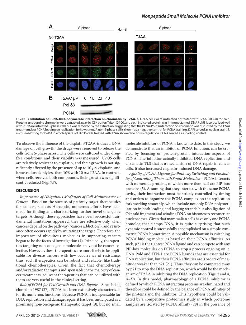
T2AA Inhibits PCNA-DNA Polymerase � Interaction on Rep-lication Foci in Cells but Does Not Remove Chromatin-boundPCNA—Upon observing that T2AA disrupts PCNA-p21 inter-action in vitro, we investigated if T2AA inhibits PCNA interac-tions in cells, in the same way that p21 does to control the cellcycle (19). DNA polymerase � is recruited to the DNA replica-tion fork by PCNA (20) to synthesize the lagging strand, inwhich the subunit 3 (Pol�3) directly binds to PCNA with itsPIP-box (21). Affinity of the Pol�3 PIP-box to PCNA is muchsmaller than that of p21, which possibly allows dynamic repli-cation regulation, such as polymerase switching and replicationinhibition by p21 (22). Therefore, theoretically T2AA shoulddisrupt PCNA-Pol�3 interaction if enough concentration isachieved on the replication fork. To verify this, we have imagedPCNA and Pol�3 in chromatin of S-phase cells upon treatmentof T2AA. Cells were treated by T2AA, pre-extracted to removeproteins unbound to chromatin, and immunostained. PCNAand Pol�3 are clearly colocalized in the absence of T2AA.How-ever, when cells were treated with T2AA, Pol�3 was exclusivelywashed out from the chromatin, but PCNA was not (Fig. 3A).Therefore, T2AA dissociated Pol�3 from PCNA but did notdissociate PCNA from the replication fork. On the other hand,expressions of Pol�3 protein in whole cell lysate were notreduced (Fig. 3B), showing that the elimination of chromatinPol�3 by T2AA is not due to its down-regulation.T2AA Inhibits DNA Replication in Cancer Cells and Their
Proliferation—Because T2AA disrupted PCNA-Pol�3 interac-tion on chromatin, next we investigated if T2AA could achieverelevant functional effects in cells. DNA polymerase � synthe-sizes de novo DNA strands, which is measurable by nucleotideincorporation. When cells were treated with BrdU under titra-tion of T2AA, T2AA inhibited the BrdU incorporation signifi-cantly in a dose-dependent manner (Figs. 4, A–C), which isconsistent with the elimination of Pol�3 from chromatin. Byperforming the assays in a time-dependent manner after theaddition or removal of T2AA, pharmacological action of T2AAappeared fairly rapid and was reversible. The BrdU incorpora-tions were �90% inhibited within 2 h of T2AA treatment and�70% recovered after 2 h of T2AA release (Fig. 4D), which was�100% recovered after 18 h (data not shown). Parallel to theinhibition of DNA synthesis, T2AA arrested cells at S-phase(Fig. 4E), similarly to other agents that inhibit DNA replication/synthesis, such as aphidicolin and hydroxyurea. Further, whencells were culturedwithT2AA, growth of bothU2OS (p53WT)and HeLa cells (p53 destroyed by E6) was inhibited with a sim-ilar efficacy (Fig. 4F), suggesting that growth inhibition byT2AA is p53-independent. The growth inhibition was accom-panied by increased cellular populations in early apoptosis butnot necrosis (Fig. 4G), suggesting that T2AA induced thegrowth inhibition not by nonspecific toxicity.T2AA Induces DNA Replication Stress in Cells by Stalling
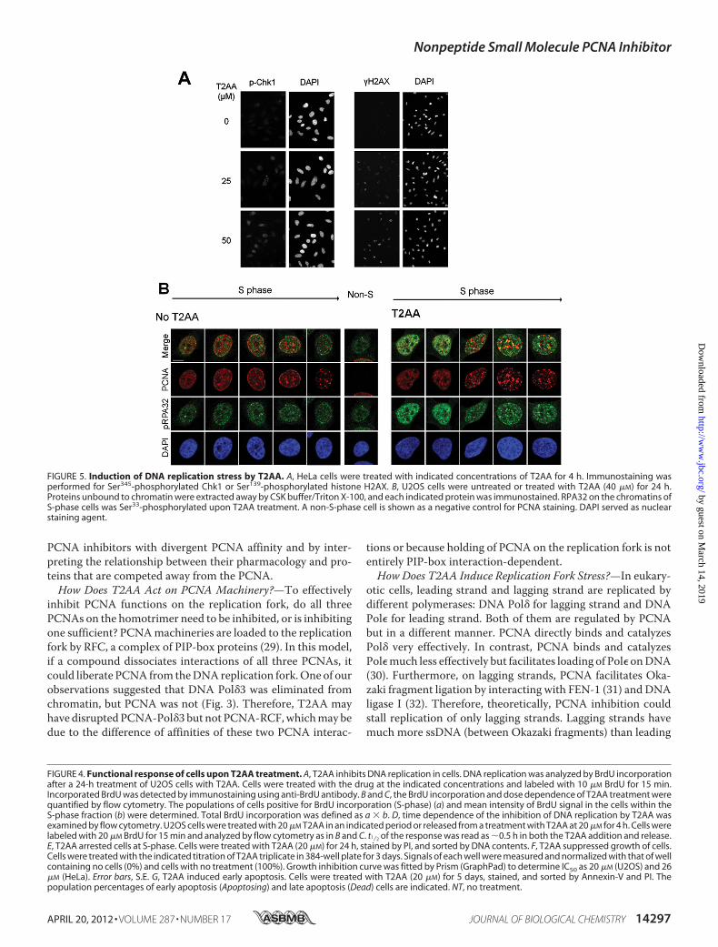
Single-stranded DNA—Because T2AA inhibits DNA replica-tion, next we investigated if T2AA actually induces DNA repli-cation stress. When DNA replication is arrested in S-phase,cells contain unreplicated single-stranded DNA (ssDNA) inchromatin and subsequently activate the ATR-Chk1 pathway,leading to histone H2AX phosphorylation (23). Chk1 andH2AX were phosphorylated upon T2AA treatment (Fig. 5A).
FIGURE 1. Inhibition of PCNA protein-protein interaction by T3 and T2AA.A, chemical structure of T3 and T2AA. T2AA lacks 3�-iodine and carboxylic acidof T3 that are essential for the thyroid hormone activity of T3. B, thyroid hor-mone activity of T3 and T2AA. HepG2 cells were transiently transfected withan expression vector, CMV-TR�, and a firefly luciferase reporter vector thatcontains thyroid hormone-responsive element, and were stimulated by titra-tion of T3 (circle) or T2AA (square). T3 fully activated TR�-TRE reporter atEC50 10 nM, and T2AA showed almost no activation. Error bars, S.E. C, inhi-bition of PCNA/PIP-box peptide interaction by T3 and T2AA. Fluorescentpolarization value (in millipolarization units (mP)) was measured for titra-tion of T3 (circle) or T2AA (square) in triplicate in a mixture of PCNA protein(100 nM) and fluorescein-tagged-SAVLQKKITDYFHPKK peptide (10 nM)that contains a PIP-box motif (underlined). Error bars, S.E. The inhibitioncurve was fitted by Prism (GraphPad) to determine IC50 as 3 �M (T3) and 1�M (T2AA). D, inhibition of PCNA/full-length p21 interaction by T3 andT2AA. p21-His6 protein was immobilized on cobalt beads and incubatedovernight with PCNA (500 nM) containing the indicated concentration ofT3 or T2AA. PCNA bound on the p21-immobilized beads was analyzed bySDS-PAGE/Oriole Orange staining.
Nonpeptide Small Molecule PCNA Inhibitor
APRIL 20, 2012 • VOLUME 287 • NUMBER 17 JOURNAL OF BIOLOGICAL CHEMISTRY 14293
by guest on March 14, 2019
http://ww
w.jbc.org/
Dow
nloaded from

ATR phosphorylated RPA32 that was accumulated on ssDNA,and such RPA32 phosphorylation was observed in cells treatedwith aphidicolin or hydroxyurea (24). To verify this for T2AA,we have investigated the RPA32 phosphorylation in chromatinby immunostaining. After 24 h of treatment, T2AA increasedthe immunofluorescence of phospho-RPA32 in nuclei (Fig. 5B).The phospho-RPA32 foci were not colocalizedwell with PCNAfoci, which is consistent with a previous observation that phos-phorylation of RPA32 prevents RPA association to replicationforks (25) where PCNA should exist. These observations sug-gest that T2AA-mediated DNA replication arrest activates theATR-Chk1 pathway and accumulates stalled ssDNA.T2AA Inhibits TLS in Cells—One of the important roles of
PCNA is polymerase switching from regular replication poly-merases (such as DNA Pol�) to translesion synthesis poly-merases (such as DNA Pol�) when the replication fork meets adamaged site on the DNA template (such as a cisplatin intras-trand cross-link, the major reaction product of DNA and cis-platin) (26). Because T2AA inhibited DNA synthesis in cells(Fig. 4A), we investigated if T2AA also can inhibit TLS that isPCNA-dependent (7), at least in part. To verify this in cells, weused a cellular TLS assay that uses replications of a plasmid
DNA containing an intrastrand cisplatin cross-link (Pt-GTG)in a coding region of the lacZ gene (Fig. 6A), in which anotherplasmid lacking the cross-link serves as a control for non-TLSplasmid replication (14). To avoid removal of the cross-link byNER, we used XP2OS(SV40), an NER-deficient XPA cell line(16). The cells were transfected with the plasmid in the pres-ence of T2AA. The replicated plasmids in the cells were recov-ered and analyzed for the TLS event by E. coli transformation/colony selection on X-gal plates (Fig. 6, B and C). T2AAsignificantly reduced the occurrence of TLS compared withthat of DMSO control (14.2 versus 18.5% (i.e. 23% reduction);Fig. 6D).T2AA Increases Cellular DNA Damage Induced by Cisplatin—
BecauseT2AA inhibited theTLS in cells, nextwe investigated ifT2AAcould actually increase cellularDNAdamage upon treat-ment with cisplatin. U2OS cells were treated with T2AA, cis-platin, or both, and then the DNA damage response was meas-ured by �H2AX staining. As expected, cisplatin significantlyinduced �H2AX, whereas T2AA induced �H2AX only a little.In contrast, when both T2AA and cisplatin were addedtogether, the �H2AX level was further increased much higherthan the level combined from each single treatment (Fig. 7A).
FIGURE 2. Co-crystal structure of PCNA-T3 complex. A, close-up view of PCNA-T3 interaction site (Protein Data Bank code 3VKX). PCNA is shown as a graysurfaced model. T3 (Fig. 2A) is shown as a stick model, in which carbon is blue, oxygen is red, nitrogen is indigo, iodine is violet, and hydrogen is omitted. Watermolecules packed in the crystal were omitted. The 5-iodine interacts with the IDCL loop, inducing an extra cavity (yellow circle). The 3�-iodine (orange circle) jutsout from the PCNA interaction interface. B, superimposition of PCNA structures bound to T3 (green), p21 peptide (cyan, magenta, and yellow) (Protein Data Bankcode 1AXC), and DNA Pol� peptide (pink, white, and orange) (Protein Data Bank code 2ZVK). The averaged root mean square deviation value is 0.84 Å forcorresponding C� atoms. All PCNA structures except that of the PCNA-T3 complex are structurally very similar, whereas the PCNA-T3 complex possessessignificant perturbation of IDCL and the region of Asp41–Val45. C, stereo diagram of detailed interaction between T3 (cyan) and PCNA (white). T3 and residuesof PCNA involved in the interaction with T3 are shown by sticks and transparent spheres. A hydrogen bond is shown by orange dots.
Nonpeptide Small Molecule PCNA Inhibitor
14294 JOURNAL OF BIOLOGICAL CHEMISTRY VOLUME 287 • NUMBER 17 • APRIL 20, 2012
by guest on March 14, 2019
http://ww
w.jbc.org/
Dow
nloaded from
To observe the influence of the cisplatin/T2AA-induced DNAdamage on cell growth, the drugs were removed to release thecells from S-phase arrest. The cells were cultured under drug-free conditions, and their viability was measured. U2OS cellsare relatively resistant to cisplatin, and their growth is not sig-nificantly affected by the presence of up to 10 �M cisplatin, anditwas reduced only less than 10%with 10�MT2AA. In contrast,when cells received both compounds, their growth was signifi-cantly reduced (Fig. 7B).
DISCUSSION
Importance of Ubiquitous Mediators of Cell Maintenance inCancer—Based on the success of pathway target therapeuticsfor cancers, such as Herceptin, numerous efforts have beenmade for finding and characterizing further novel oncogenictargets. Although these approaches have been successful, fun-damental limitations appeared; they are effective only whencancers depend on the pathway (“cancer addiction”), and resist-ance often occurs rapidly bymutating the target. Therefore, theimportance of ubiquitous molecules in supporting cancersbegan to be the focus of investigation (4). Principally, therapeu-tics targeting non-oncogenic molecules may not be cancer-se-lective. However, these therapeutics aremore likely to be appli-cable for diverse cancers with low occurrence of resistance;thus, such therapeutics can be robust and reliable, like tradi-tional chemotherapies. Given the fact that chemotherapyand/or radiation therapy is indispensable in themajority of can-cer treatments, adjuvant therapeutics that can be utilized withthem are very useful in the clinical setting.Role of PCNA for Cell Growth and DNA Repair—Since being
cloned in 1987 (27), PCNA has been extensively characterizedfor its numerous functions. Because PCNA is indispensable forDNA replication and damage repair, it has been anticipated as apromising non-oncogenic therapeutic target (9), but no small
molecule inhibitor of PCNA is known to date. In this study, wedemonstrate that an inhibitor of PCNA functions can be cre-ated by focusing on protein-protein interaction aspects ofPCNA. The inhibitor actually inhibited DNA replication andenzymatic TLS that is a mechanism of DNA repair in cancercells. It also increased cisplatin-induced DNA damage.Affinity of PCNALigands for Pathway Switching and Possibil-
ity of Controlling Themwith SmallMolecules—PCNA interactswith numerous proteins, of which more than half are PIP-boxproteins (5). Assuming that they interact with the same PCNAcavity, their interaction must be strictly controlled by timingand orders to organize the PCNA complex on the replicationfork working smoothly, which include not only DNA polymer-ization in both leading and lagging strands but also ligation ofOkazaki fragment andwindingDNAonhistones to reconstructnucleosomes.Given thatmammalian cells have only one PCNAhomolog that clamps DNA, it is quite surprising that suchdynamic control is successfully accomplished on a simple sym-metric PCNA homotrimer. A possible mechanism is switchingPCNA binding molecules based on their PCNA affinities. Assuch, p21 is the tightest PCNA ligand and can competewith anyPIP-box molecules on PCNA to stop a process ongoing on it.DNA Pol� and FEN-1 are PCNA ligands that are essential forDNA replication, but their PCNA affinities are 3 orders ofmag-nitude weaker than p21 (21). Thus, they can be competed awayby p21 to stop the DNA replication, which would be the mech-anism of T2AA in inhibiting the DNA replication (Figs. 3 and 4,A–D). In this model, pharmacology of a PCNA inhibitor isdefined bywhichPCNA interacting proteins are eliminated andtherefore could be defined by the balance of PCNA affinities ofthe proteins and the inhibitor. This hypothesis could be vali-dated by a competitive proteomics study in which proteomesamples are isolated by PCNA affinity (28) in the presence of
FIGURE 3. Inhibition of PCNA-DNA polymerase interaction on chromatin by T2AA. A, U2OS cells were untreated or treated with T2AA (20 �M) for 24 h.Proteins unbound to chromatin were extracted away by CSK buffer/Triton X-100, and each indicated protein was immunostained. DNA Pol�3 is colocalized wellwith PCNA in untreated S-phase cells but was removed by the extraction, suggesting that the PCNA-Pol�3 interaction on chromatin was disrupted by the T2AAtreatment, but PCNA loading on replication forks was not. A non-S-phase cell is shown as a negative control for PCNA staining. DAPI served as nuclear stain. B,immunoblotting for Pol�3 in whole lysates of U2OS cells treated with T2AA showed no down-regulation. PCNA served as a loading control.
Nonpeptide Small Molecule PCNA Inhibitor
APRIL 20, 2012 • VOLUME 287 • NUMBER 17 JOURNAL OF BIOLOGICAL CHEMISTRY 14295
by guest on March 14, 2019
http://ww
w.jbc.org/
Dow
nloaded from

Nonpeptide Small Molecule PCNA Inhibitor
14296 JOURNAL OF BIOLOGICAL CHEMISTRY VOLUME 287 • NUMBER 17 • APRIL 20, 2012
by guest on March 14, 2019
http://ww
w.jbc.org/
Dow
nloaded from
PCNA inhibitors with divergent PCNA affinity and by inter-preting the relationship between their pharmacology and pro-teins that are competed away from the PCNA.How Does T2AA Act on PCNA Machinery?—To effectively
inhibit PCNA functions on the replication fork, do all threePCNAs on the homotrimer need to be inhibited, or is inhibitingone sufficient? PCNAmachineries are loaded to the replicationfork by RFC, a complex of PIP-box proteins (29). In this model,if a compound dissociates interactions of all three PCNAs, itcould liberate PCNA from theDNAreplication fork.One of ourobservations suggested that DNA Pol�3 was eliminated fromchromatin, but PCNA was not (Fig. 3). Therefore, T2AA mayhave disruptedPCNA-Pol�3 but not PCNA-RCF,whichmay bedue to the difference of affinities of these two PCNA interac-
tions or because holding of PCNA on the replication fork is notentirely PIP-box interaction-dependent.How Does T2AA Induce Replication Fork Stress?—In eukary-
otic cells, leading strand and lagging strand are replicated bydifferent polymerases: DNA Pol� for lagging strand and DNAPol� for leading strand. Both of them are regulated by PCNAbut in a different manner. PCNA directly binds and catalyzesPol� very effectively. In contrast, PCNA binds and catalyzesPol�much less effectively but facilitates loading of Pol� onDNA(30). Furthermore, on lagging strands, PCNA facilitates Oka-zaki fragment ligation by interacting with FEN-1 (31) and DNAligase I (32). Therefore, theoretically, PCNA inhibition couldstall replication of only lagging strands. Lagging strands havemuch more ssDNA (between Okazaki fragments) than leading
FIGURE 4. Functional response of cells upon T2AA treatment. A, T2AA inhibits DNA replication in cells. DNA replication was analyzed by BrdU incorporationafter a 24-h treatment of U2OS cells with T2AA. Cells were treated with the drug at the indicated concentrations and labeled with 10 �M BrdU for 15 min.Incorporated BrdU was detected by immunostaining using anti-BrdU antibody. B and C, the BrdU incorporation and dose dependence of T2AA treatment werequantified by flow cytometry. The populations of cells positive for BrdU incorporation (S-phase) (a) and mean intensity of BrdU signal in the cells within theS-phase fraction (b) were determined. Total BrdU incorporation was defined as a b. D, time dependence of the inhibition of DNA replication by T2AA wasexamined by flow cytometry. U2OS cells were treated with 20 �M T2AA in an indicated period or released from a treatment with T2AA at 20 �M for 4 h. Cells werelabeled with 20 �M BrdU for 15 min and analyzed by flow cytometry as in B and C. t1⁄2 of the response was read as �0.5 h in both the T2AA addition and release.E, T2AA arrested cells at S-phase. Cells were treated with T2AA (20 �M) for 24 h, stained by PI, and sorted by DNA contents. F, T2AA suppressed growth of cells.Cells were treated with the indicated titration of T2AA triplicate in 384-well plate for 3 days. Signals of each well were measured and normalized with that of wellcontaining no cells (0%) and cells with no treatment (100%). Growth inhibition curve was fitted by Prism (GraphPad) to determine IC50 as 20 �M (U2OS) and 26�M (HeLa). Error bars, S.E. G, T2AA induced early apoptosis. Cells were treated with T2AA (20 �M) for 5 days, stained, and sorted by Annexin-V and PI. Thepopulation percentages of early apoptosis (Apoptosing) and late apoptosis (Dead) cells are indicated. NT, no treatment.
FIGURE 5. Induction of DNA replication stress by T2AA. A, HeLa cells were treated with indicated concentrations of T2AA for 4 h. Immunostaining wasperformed for Ser345-phosphorylated Chk1 or Ser139-phosphorylated histone H2AX. B, U2OS cells were untreated or treated with T2AA (40 �M) for 24 h.Proteins unbound to chromatin were extracted away by CSK buffer/Triton X-100, and each indicated protein was immunostained. RPA32 on the chromatins ofS-phase cells was Ser33-phosphorylated upon T2AA treatment. A non-S-phase cell is shown as a negative control for PCNA staining. DAPI served as nuclearstaining agent.
Nonpeptide Small Molecule PCNA Inhibitor
APRIL 20, 2012 • VOLUME 287 • NUMBER 17 JOURNAL OF BIOLOGICAL CHEMISTRY 14297
by guest on March 14, 2019
http://ww
w.jbc.org/
Dow
nloaded from

strands have; therefore, T2AA could have induced high num-bers of the unreplicated ssDNA that were detected by RPA32phosphorylation (Fig. 5B).Replication Fork Stress and Lethality of T2AA Treatments—
PCNA is incomparably multifunctional, and its inhibitioncould thus cause promiscuous effects in regard to cell growth.However, the actual effect of T2AA treatment was not entirelycytotoxic (Fig. 4H). When cells are faced with DNA replicationstress, they attempt to remove the stalled site by transientlyinducing double strand breaks, in which BLM helicase, Mus81endonuclease, and ATR kinase are needed (33). PCNA is disso-
ciated from chromatin during this process (34), suggesting thatreplication forks are disassembled and therefore T2AA wouldno longer be effective. Replication stress induced by T2AAcould become lethal when this process is inhibited, such aswhen co-administrating it with an ATR inhibitor.Mechanistically, PCNA inhibitors can be selective to repli-
cating cells and valuable as long as they are cooperative withcytotoxic therapies. Induction of DNA stress in actively repli-cating cells generates many stalled replication forks whereDNA is disassembled from nucleosomes. The formed linkerDNA portion is unprotected from the histone core and could
FIGURE 6. Inhibition of TLS across the cisplatin Pt-GTG cross-link in mammalian cultured cells by T2AA. Shown are the structure of the cisplatin-cross-linked Pt-GTG plasmid (A) and experimental protocol used in this study (B). C, predicted sequences of replicated plasmid and phenotypes of colonies on theX-gal LB plates; DISL, damage induced strand loss. D, T2AA inhibited TLS across the cisplatin-cross-linked Pt-GTG adduct. Relative values of the TLS frequency(percentages) across Pt-GTG adduct in XPA cells treated with DMSO (18.5 � 3.0%) or T2AA (14.2 � 3.6%) are shown, which is the ratio of the TLS frequency ofthe modified plasmid to that of the non-modified plasmid (i.e. (TLS frequency of Pt-GTG plasmid/TLS frequency of Mock-GTG plasmid) 100). Data aremeans with S.D. values (error bars) from at least three independent experiments. The value significantly decreased (*, p 0.01) from that of the DMSOcontrol. Statistical comparison was carried out using Student’s t test for one-tailed comparison. Actual numbers of the colony counting are given insupplemental Table S1.
FIGURE 7. Increasing cisplatin-induced DNA damage by T2AA. A, T2AA increased cisplatin-induced DNA damage response. U2OS cells were treated with theindicated combination of T2AA and cisplatin for 18 h, stained for �H2AX, and analyzed by flow cytometry. B, growth of cells treated with cisplatin and T2AA.U2OS cells were treated with the indicated titration of cisplatin with T2AA for 18 h, replated in fresh medium to remove the drugs, and cultured for 3 days.Viability of the cells was measured by Alamar Blue reagent, and signals were normalized with those of no cell well (0%) and cells with no drugs (100%). NT, notreatment. Error bars, S.D.
Nonpeptide Small Molecule PCNA Inhibitor
14298 JOURNAL OF BIOLOGICAL CHEMISTRY VOLUME 287 • NUMBER 17 • APRIL 20, 2012
by guest on March 14, 2019
http://ww
w.jbc.org/
Dow
nloaded from

thus be liable against DNA damage inducers, such as cisplatin.In fact, cisplatin and other alkylation agents react with DNApreferentially in the linker portion rather than in the nucleo-some region in vitro (35). T2AA could thus have sensitized cellsfor inducing cisplatin-mediated DNA damage (Fig. 7A).Another possible mechanism is that T2AA could also inhibitTLS that makes cells exempt from DNA damage, which wasverified in the TLS inhibition (Fig. 6D). The majority of DNAdamage induced by cisplatin is intrastrand cross-links that arenot highly toxic to cells if they are allowed to be repaired (36).The �H2AX induction observed in cells co-treated by cisplatinand T2AA (Fig. 7A) could represent such cross-links. It wasshown that cells are not expected to die unless they retain�H2AX foci 24 h after cisplatin treatment (37). Thus, investi-gating if T2AA also prevents cisplatin-induced DNA damagerepair that is facilitated by PCNA (such as NER) and if T2AAstabilizes the �H2AX foci long enough could be valuable forvalidating lethality of cisplatin/T2AAco-treatment. This is verylikely because T2AA inhibits interaction of PCNA and Pol�3 inchromatin (Fig. 3A), and both of themare absolutely essential inthe late step of NER (gap-filling synthesis) (38). Actually, per-turbation of this step reportedly induced �H2AX (39), whichcould be themechanism of the �H2AXup-regulation by cispla-tin/T2AA co-treatment.Future Investigation—The PCNA-T3 co-crystal structure is
useful to rationally design new leads with improved PCNAaffinity. The observation that IDCL and theAsp41–Val45 regionof PCNA are specifically perturbed upon T2AA association(Fig. 2B) indicates that they are key interaction sites to dynam-ically adopt a small molecule. Lead optimization efforts underthis hypothesis are ongoing to produce new compounds withhigher PCNA affinity. Such compounds will be evaluated inanimal tumor models for chemotherapy sensitization, whichthus will validate a new strategy for sensitizing cancer cells tocisplatin treatment by targeting PCNA. T2AA could function-ally inhibit other processes that PCNA coordinates, such asorigin refiring (40), chromatin assembly (41), epigenetic inher-itance (42) and sister chromatid cohesion (43, 44). Functionalcharacterization of T2AA in these processes will elucidate theirfunctional significance from the chemotherapeutic viewpoint.
Acknowledgments—We thank David Smithson, Armand Guigemde,Heather Ross, and Enas Ahmed for high-throughput screening; AndyLemoff, Cynthia Jeffries, and Bing Yan for analytical chemistry; Mar-celo Actis and Anand Mayasundari for synthetic chemistry support;Robert Cassell for peptide synthesis; Scott Perry, Dana Lucas, Ann-Marie Hamilton-Easton, and Richard Ashmun for flow cytometry;Jennifer Peters and Victoria Frohlich for light microscopy; the beam-line staff of SPring-8 andPhoton Factory Japan for the crystallographydata collection; and Youngsoo Lee and Peter McKinnon for scientificadvice. Toshiki Tsurimoto, Kip Guy, and Richard Kriwacki kindlyprovided plasmids for this study (see supplemental material).
REFERENCES1. Weinstein, I. B., and Joe, A. K. (2006) Mechanisms of disease. Oncogene
addiction. A rationale formolecular targeting in cancer therapy.Nat. Clin.Pract. Oncol. 3, 448–457
2. Gorre, M. E., Mohammed, M., Ellwood, K., Hsu, N., Paquette, R., Rao,
P. N., and Sawyers, C. L. (2001) Clinical resistance to STI-571 cancertherapy caused by BCR-ABL genemutation or amplification. Science 293,876–880
3. Yauch, R. L., Dijkgraaf, G. J., Alicke, B., Januario, T., Ahn, C. P., Holcomb,T., Pujara, K., Stinson, J., Callahan, C. A., Tang, T., Bazan, J. F., Kan, Z.,Seshagiri, S., Hann, C. L., Gould, S. E., Low, J. A., Rudin, C. M., and deSauvage, F. J. (2009) Smoothenedmutation confers resistance to a Hedge-hog pathway inhibitor in medulloblastoma. Science 326, 572–574
4. Luo, J., Solimini, N. L., and Elledge, S. J. (2009) Principles of cancer ther-apy. Oncogene and non-oncogene addiction. Cell 136, 823–837
5. Moldovan, G. L., Pfander, B., and Jentsch, S. (2007) PCNA, the maestro ofthe replication fork. Cell 129, 665–679
6. Haracska, L., Johnson, R. E., Unk, I., Phillips, B., Hurwitz, J., Prakash, L.,and Prakash, S. (2001) Physical and functional interactions of humanDNApolymerase � with PCNA.Mol. Cell Biol. 21, 7199–7206
7. Hishiki, A., Hashimoto, H., Hanafusa, T., Kamei, K., Ohashi, E., Shimizu,T., Ohmori, H., and Sato, M. (2009) Structural basis for novel interactionsbetween human translesion synthesis polymerases and proliferating cellnuclear antigen. J. Biol. Chem. 284, 10552–10560
8. Waters, L. S., Minesinger, B. K., Wiltrout, M. E., D’Souza, S., Woodruff,R. V., and Walker, G. C. (2009) Eukaryotic translesion polymerases andtheir roles and regulation in DNA damage tolerance.Microbiol. Mol. Biol.Rev. 73, 134–154
9. Stoimenov, I., and Helleday, T. (2009) PCNA on the crossroad of cancer.Biochem. Soc. Trans. 37, 605–613
10. Acharya, N., Yoon, J. H., Gali, H., Unk, I., Haracska, L., Johnson, R. E.,Hurwitz, J., Prakash, L., and Prakash, S. (2008) Roles of PCNA-binding andubiquitin-binding domains in human DNA polymerase eta in translesionDNA synthesis. Proc. Natl. Acad. Sci. U.S.A. 105, 17724–17729
11. Kontopidis, G., Wu, S. Y., Zheleva, D. I., Taylor, P., McInnes, C., Lane,D. P., Fischer, P. M., and Walkinshaw, M. D. (2005) Structural and bio-chemical studies of human proliferating cell nuclear antigen complexesprovide a rationale for cyclin association and inhibitor design. Proc. Natl.Acad. Sci. U.S.A. 102, 1871–1876
12. Hishiki, A., Shimizu, T., Serizawa, A., Ohmori, H., Sato, M., andHashimoto, H. (2008) Crystallographic study of G178S mutant of humanproliferating cell nuclear antigen. Acta Crystallogr. Sect. F Struct. Biol.Cryst. Commun. 64, 819–821
13. Taddei, A., Roche, D., Sibarita, J. B., Turner, B. M., and Almouzni, G.(1999) Duplication and maintenance of heterochromatin domains. J. CellBiol. 147, 1153–1166
14. Sawai, T., Kawanishi, M., Takamura-Enya, T., and Yagi, T. (2009) Estab-lishment of a method for analyzing translesion synthesis across a singlebulky adduct in human cells. Genes Environ. 31, 24–30
15. Shivji,M. K.,Moggs, J. G., Kuraoka, I., andWood, R.D. (2006)Assaying forthe dual incisions of nucleotide excision repair using DNAwith a lesion ata specific site.Methods Mol. Biol. 314, 435–456
16. Kawanishi, M., Enya, T., Suzuki, H., Takebe, H., Matsui, S., and Yagi, T.(1998)Mutagenic specificity of a derivative of 3-nitrobenzanthrone in thesupF shuttle vector plasmids. Chem. Res. Toxicol. 11, 1468–1473
17. Leeson, P. D., Ellis, D., Emmett, J. C., Shah, V. P., Showell, G. A., andUnderwood, A. H. (1988) Thyroid hormone analogues. Synthesis of 3�-substituted 3,5-diiodo-L-thyronines and quantitative structure-activitystudies of in vitro and in vivo thyromimetic activities in rat liver and heart.J. Med. Chem. 31, 37–54
18. Gulbis, J.M., Kelman, Z., Hurwitz, J., O’Donnell,M., andKuriyan, J. (1996)Structure of the C-terminal region of p21WAF1/CIP1 complexed with hu-man PCNA. Cell 87, 297–306
19. Chen, J., Jackson, P. K., Kirschner, M. W., and Dutta, A. (1995) Separatedomains of p21 involved in the inhibition of Cdk kinase and PCNA. Na-ture 374, 386–388
20. Bravo, R., Frank, R., Blundell, P. A., and Macdonald-Bravo, H. (1987) Cy-clin/PCNA is the auxiliary protein of DNA polymerase-�. Nature 326,515–517
21. Bruning, J. B., and Shamoo, Y. (2004) Structural and thermodynamic anal-ysis of human PCNA with peptides derived from DNA polymerase-� p66subunit and flap endonuclease-1. Structure. 12, 2209–2219
22. Li, R., Waga, S., Hannon, G. J., Beach, D., and Stillman, B. (1994) Differ-
Nonpeptide Small Molecule PCNA Inhibitor
APRIL 20, 2012 • VOLUME 287 • NUMBER 17 JOURNAL OF BIOLOGICAL CHEMISTRY 14299
by guest on March 14, 2019
http://ww
w.jbc.org/
Dow
nloaded from

ential effects by the p21 CDK inhibitor on PCNA-dependent DNA repli-cation and repair. Nature 371, 534–537
23. Flynn, R. L., and Zou, L. (2011) ATR: a master conductor of cellular re-sponses to DNA replication stress. Trends Biochem. Sci. 36, 133–140
24. Vassin, V.M., Anantha, R.W., Sokolova, E., Kanner, S., and Borowiec, J. A.(2009) Human RPA phosphorylation by ATR stimulates DNA synthesisand prevents ssDNA accumulation during DNA replication stress. J. CellSci. 122, 4070–4080
25. Vassin, V. M., Wold, M. S., and Borowiec, J. A. (2004) Replication proteinA (RPA) phosphorylation prevents RPA association with replication cen-ters.Mol. Cell Biol. 24, 1930–1943
26. Lehmann,A. R. (2006)Translesion synthesis inmammalian cells.Exp. CellRes. 312, 2673–2676
27. Matsumoto, K.,Moriuchi, T., Koji, T., andNakane, P. K. (1987)Molecularcloning of cDNA coding for rat proliferating cell nuclear antigen (PCNA)/cyclin. EMBO J. 6, 637–642
28. Ohta, S., Shiomi, Y., Sugimoto, K., Obuse, C., and Tsurimoto, T. (2002) Aproteomics approach to identify proliferating cell nuclear antigen(PCNA)-binding proteins in human cell lysates. Identification of the hu-man CHL12/RFCs2–5 complex as a novel PCNA-binding protein. J. Biol.Chem. 277, 40362–40367
29. Bowman, G. D., O’Donnell, M., and Kuriyan, J. (2004) Structural analysisof a eukaryotic sliding DNA clamp-clamp loader complex. Nature 429,724–730
30. Chilkova, O., Stenlund, P., Isoz, I., Stith, C. M., Grabowski, P., Lundström,E. B., Burgers, P. M., and Johansson, E. (2007) The eukaryotic leading andlagging strand DNA polymerases are loaded onto primer-ends via sepa-rate mechanisms but have comparable processivity in the presence ofPCNA. Nucleic Acids Res. 35, 6588–6597
31. Wu, X., Li, J., Li, X., Hsieh, C. L., Burgers, P. M., and Lieber, M. R. (1996)Processing of branched DNA intermediates by a complex of humanFEN-1 and PCNA. Nucleic Acids Res. 24, 2036–2043
32. Levin, D. S., McKenna, A. E., Motycka, T. A., Matsumoto, Y., and Tom-kinson, A. E. (2000) Interaction between PCNAandDNA ligase I is criticalfor joining of Okazaki fragments and long-patch base-excision repair.Curr. Biol. 10, 919–922
33. Hanada, K., Budzowska, M., Davies, S. L., van Drunen, E., Onizawa, H.,Beverloo, H. B., Maas, A., Essers, J., Hickson, I. D., and Kanaar, R. (2007)The structure-specific endonucleaseMus81 contributes to replication re-start by generating double strand DNA breaks. Nat. Struct. Mol. Biol. 14,
1096–110434. Shimura, T., Torres,M. J.,Martin,M.M., Rao,V.A., Pommier, Y., Katsura,
M., Miyagawa, K., and Aladjem, M. I. (2008) Bloom’s syndrome helicaseandMus81 are required to induce transient double strand DNA breaks inresponse to DNA replication stress. J. Mol. Biol. 375, 1152–1164
35. Galea, A. M., andMurray, V. (2010) The influence of chromatin structureon DNA damage induced by nitrogen mustard and cisplatin analogues.Chem. Biol. Drug Des. 75, 578–589
36. Siddik, Z. H. (2003) Cisplatin. Mode of cytotoxic action and molecularbasis of resistance. Oncogene 22, 7265–7279
37. Banáth, J. P., Klokov, D., MacPhail, S. H., Banuelos, C. A., and Olive, P. L.(2010) Residual �H2AX foci as an indication of lethal DNA lesions. BMCCancer. 10, 4
38. Ogi, T., Limsirichaikul, S., Overmeer, R. M., Volker, M., Takenaka, K.,Cloney, R., Nakazawa, Y., Niimi, A., Miki, Y., Jaspers, N. G., Mullenders,L.H., Yamashita, S., Fousteri,M. I., and Lehmann,A. R. (2010)ThreeDNApolymerases, recruited by different mechanisms, carry out NER repairsynthesis in human cells.Mol. Cell 37, 714–727
39. Matsumoto, M., Yaginuma, K., Igarashi, A., Imura, M., Hasegawa, M.,Iwabuchi, K., Date, T., Mori, T., Ishizaki, K., Yamashita, K., Inobe, M., andMatsunaga, T. (2007) Perturbed gap-filling synthesis in nucleotide exci-sion repair causes histone H2AX phosphorylation in human quiescentcells. J. Cell Sci. 120, 1104–1112
40. Arias, E. E., and Walter, J. C. (2006) PCNA functions as a molecular plat-form to trigger Cdt1 destruction and prevent re-replication.Nat. Cell Biol.8, 84–90
41. Shibahara, K., and Stillman, B. (1999) Replication-dependent marking ofDNA by PCNA facilitates CAF-1-coupled inheritance of chromatin. Cell96, 575–585
42. Iida, T., Suetake, I., Tajima, S., Morioka, H., Ohta, S., Obuse, C., and Tsu-rimoto, T. (2002) PCNA clamp facilitates action of DNA cytosine meth-yltransferase 1 on hemimethylated DNA. Genes Cells 7, 997–1007
43. Moldovan, G. L., Pfander, B., and Jentsch, S. (2006) PCNA controls estab-lishment of sister chromatid cohesion during S phase. Mol. Cell 23,723–732
44. Inoue, A., Li, T., Roby, S. K., Valentine, M. B., Inoue, M., Boyd, K., Kidd,V. J., and Lahti, J. M. (2007) Loss of ChlR1 helicase inmouse causes lethal-ity due to the accumulation of aneuploid cells generated by cohesion de-fects and placental malformation. Cell Cycle 6, 1646–1654
Nonpeptide Small Molecule PCNA Inhibitor
14300 JOURNAL OF BIOLOGICAL CHEMISTRY VOLUME 287 • NUMBER 17 • APRIL 20, 2012
by guest on March 14, 2019
http://ww
w.jbc.org/
Dow
nloaded from

Naoaki FujiiRodrigues, Hiroshi Hashimoto, Masanobu Kawanishi, Mamoru Sato, Takashi Yagi and
Connelly, Benjamin Evison, Youming Shao, Richard Heath, Isao Kuraoka, Patrick Chandanamali Punchihewa, Akira Inoue, Asami Hishiki, Yoshihiro Fujikawa, Michele
ReplicationInhibitor That Disrupts Interactions with PIP-box Proteins and Inhibits DNA Identification of Small Molecule Proliferating Cell Nuclear Antigen (PCNA)
doi: 10.1074/jbc.M112.353201 originally published online March 1, 20122012, 287:14289-14300.J. Biol. Chem.
10.1074/jbc.M112.353201Access the most updated version of this article at doi:
Alerts:
When a correction for this article is posted•
When this article is cited•
to choose from all of JBC's e-mail alertsClick here
Supplemental material:
http://www.jbc.org/content/suppl/2012/03/01/M112.353201.DC1
http://www.jbc.org/content/287/17/14289.full.html#ref-list-1
This article cites 44 references, 12 of which can be accessed free at
by guest on March 14, 2019
http://ww
w.jbc.org/
Dow
nloaded from