pcna expression in cutaneous keratinous neoplasms and verruca
TRANSCRIPT
American Journal of Pathology, Vol. 141, No. 1, July 1992Copyrigbt C American Association of Pathologists
PCNA Expression in CutaneousKeratinous Neoplasms andVerruca Vulgaris
Neal S. Penneys, Maria Bogaert,Ulrike Serfling, and Manuel SistoFrom the Department ofDernatology and CutaneousSurgery, University ofMiami School ofMedicine,Miami, Florida
Using an antibody to PCNA and a standard immu-nohistochemical system, the authors examined nor-
mal epidermis and cutaneous neoplasias for expres-sion of PCNA, a protein associated with DNA poly-merase delta and DNA replication In squamous cellcarcinoma in situ (SCCI), a unique expression ofPCNA, which frequently involved the nuclei of allkeratinocytes within the lesion, wasfound Heavieststaining was in the uppermost layers of the epider-mis. PCNA expression ended abruptly at the histo-logic margin of the lesion Because SCCI can be as-
sociated with the presence ofhuman papillomavirus(HPV) DNA, the authors evaluated PCNA expressionin verruca vulgaris and found a pattern similar tothat in SCCI. Assuming thatPCNA expression in thesetwo lesions is related to cell division, the authorshypothesize that the mechanisms that control prolif-eration in SCCI may be similar to those operative inverruca vulgaris. (AmJPathol 1992, 141:139-142)
Cell kinetic information may aid in tumor classification,contribute to the understanding of tumor behavior, andserve a predictive role in assessing the risk of recurrenceor spread. Immunohistochemical methods of assessingcell proliferation have advantages in that they maintaincellular and tissue architecture. What has limited this ap-plication has been the absence of a useful antibody re-agent that functions in conventionally fixed and pro-cessed tissues.
PCNA is a highly conserved acidic nuclear proteinwhose synthesis is increased in late G1 and in Sphase.1' PCNA therefore correlates with the proliferativestate of the cell. The recent availability of monoclonal an-tibodies against PCNA that function in routinely fixed and
processed tissues has permitted the assessment of theproliferative capacity of a variety of lesions,"' which aredifficult to obtain as fresh or unfixed tissues. In this report,we note that the pattern of expression of PCNA in squa-mous cell carcinoma in situ (SCCI) is unique among avaried group of cutaneous neoplasms.
Materials and Methods
Histologic slides and paraffin blocks were retrieved fromthe dermatopathology files of the division of dermatopa-thology, University of Miami School of Medicine. All tis-sues had been formalin-fixed and routinely processedduring 1989, 1990, and 1991. All cases were reviewedand 6-,m sections were obtained for immunohistochem-ical assay. The following conditions were studied: normalskin (10 cases), actinic keratosis (10 cases), squamouscell carcinoma (10 cases), expanding keratoacanthoma(10 cases), regressing keratoacanthoma (5 cases), basalcell carcinoma (10 cases), Bowen's disease (nongenitaland excluding digital lesions) (30 cases), Bowen's dis-ease (genital, 10 cases), verruca vulgaris (10 cases), andseborrheic keratosis (10 cases).
Standard immunohistochemical methods were usedincluding the avidin-biotin-alkaline phosphatase system(Vectastain, Vector Laboratories, Burlingame, CA) and amonoclonal antibody to PCNA (Dako Corp., Carpenteria,CA). Sections were deparaffinized with xylene and hy-drated through a series of alcohols. Endogenous perox-idase was inhibited by treatment with 0.6% hydrogenperoxide in methanol for 20 minutes. Each slide wastreated sequentially with normal goat serum, monoclonalantibody to PCNA at a dilution of 1:1 0, biotinylated rabbitanti-mouse IgG (1:250), and an alkaline phosphatase bi-otin-avidin complex (1:500) and developed using alkalinephosphatase substrate kit from Vector Laboratories. Ap-propriate controls were included in all assays.
Accepted for publication January 17, 1992.Address reprint requests to Dr. Neal S. Penneys, Department of Der-
matology, St. Louis University Medical Center, 1402 S. Grand Boulevard,St. Louis, MO 63104.
139

140 Penneys et alAJP July 1992, Vol. 141, No. 1
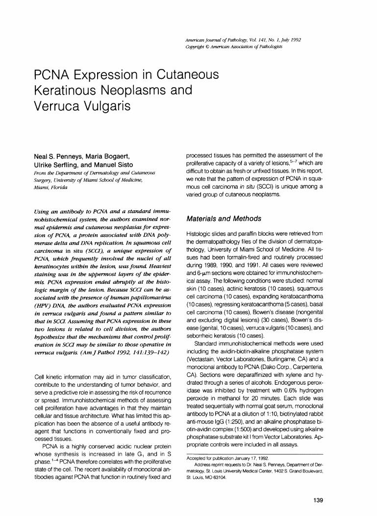
Figure 1. Top left: Increased numbers of basalar keratinocytes contain intranuclear PCNA in actinic keratosis (X200).Figure 2. Top right: In expanding keratoacanthoma, a dramatic increase in PCNA expression isfound in the basilar layers oftumor masses(x 100).Figure 3. Middle left: In SCCI, diffuse labeling of keratinocytic nuclei is present (X40). Middle right: At higher magnification, intensegranular and diffuse expression ofPCNA is seen in the nucleoplasm (x400).Figure 4. Bottom left: In verruca vulgaris, the pattern ofPCNA elaboration is similar to that in SCCI, affecting primarily the upper layersof the epidermis (x40). Bottom right: At higher magnification, widespread expression of PCNA is seen to end abruptly at the histologicmargin of the verruca. A rare positive basal cell is labeled in normal epidermis adjacent to the verruca (x200).
Results
Normal Skin
In normal epidermis, rare basal cells contained immuno-reactive product. Although not quantified, the pattern ofPCNA expression appeared to be similar to that foundwith other proliferation markers such as K167, bromode-oxyuridine, and tritiated thymidine.
Cutaneous NeoplasmsIncreased labeling of keratinocytic nuclei was found inthe basal cell layer in actinic keratosis (Figure 1). In squa-
mous cell carcinoma, variable numbers of cells withPCNA-positive nuclei were scattered in tumor masses.On the other hand, PCNA expression in expanding kera-toacanthoma was found in cells composing the basal celllayer, frequently affecting all cells in this region, with littlelabeling within the more differentiated tumor masses (Fig-ure 2). In regressing keratoacanthoma, only focal label-ing was seen in the basal cell layer. In basal cell carci-noma, there were areas with increased nuclear stainingbut these were focal and limited to the basal cell layers oftumor aggregates. In SCCI, intense PCNA staining al-ways occurred in nuclei of keratinocytes in the upper epi-dermal layers. Frequently, keratinocytes at all epidermallevels in SCCI contained PCNA-reactive material in their

PCNA Expression in Cutaneous Keratinous Neoplasms and Verruca Vulgaris 141AJPJulv 1992, Vol. 141, No. 1
nuclei (Figure 3). PCNA expression ended abruptly at thehistologic margin of the lesion.
Verruca Vulgar/s and Seborrheic Keratosis
A pattern similar to but less intense than that seen in SCCIwas found in the majority of verrucae. Strongest expres-sion was found in the upper layers of the epidermis (Fig-ure 4). In some areas, all epidermal layers containedPCNA-positive keratinocytes. In seborrheic keratosis,there was rare PCNA positivity in keratinocytes, usually inthe lower layers of the acanthotic epidermal masses.
Discussion
PCNA was initially described by Miyachi et al8 who usedautoantibodies found in the sera of patients with systemiclupus erythematosus to identify a nuclear protein. Bravoand Cellis then described a 36-kd protein that appearedto be identical to PCNA.9 Subsequently, PCNA was iden-tified as an auxiliary protein of DNA polymerase delta,10an enzyme that most likely functions in the catalysisof eukaryotic leading strand synthesis and in DNA re-pair.' 1-12 The synthesis of PCNA within the cell correlatessomewhat with the proliferative state of the cell. However,PCNA is present in variable amounts in the cell duringquiescent states. Studies by Bravo et al have demon-strated two populations of PCNA, one probably associ-ated with the sites of replication and another that is ho-mogeneously spread in the nucleoplasm.13
In this study, we used a commercially available mono-clonal antibody to PCNA and archival tissues that wereprocessed in a routine manner after variable fixationtimes in formalin solution. Acceptable technical resultswere obtained. Reaction product was found overlying thenucleus in two patterns: 1) a granular pattern most likelyrepresenting binding of antibody to DNA replication sites,and 2) a diffuse pattern overlying the entire nucleoplasm.
Expression of PCNA seemed to correlate with pat-terns previously described in the skin using other labelingmethods that do not work in paraffin-embedded tissues.In normal epidermis, an occasional cell in the basal celllayer labeled with antibody to PCNA. In actinic keratosis,there was variable increase in the number of labeled cellsin the lower epidermal layers. In invasive lesions of squa-mous cell carcinoma, there was a variably increasednumber of labeled keratinocytes scattered within the tu-mor masses. An interesting pattern was seen in kerato-acanthoma, a rapidly enlarging lesion with a character-istic architecture. In expanding keratoacanthoma, prom-inent labeling of nuclei by antibodies to PCNA was seen
in the basal cell layer of the tumor mass with little labelingwithin tumor aggregates, whereas minimal basilar label-ing was observed in regressing keratoacanthoma. Thesepatterns were distinctly different from that of squamouscell carcinoma. Basal cell carcinomas had low levels oflabeling; when present, they were in the palisaded layerat the periphery of tumor aggregates.
Both genital and nongenital SCCI had diffuse labelingof keratinocytes, frequently at all epidermal levels, withheaviest labeling occurring in the upper layers of the stra-tum malphighii and stratum granulosum, extending to thestratum corneum. Both granular and diffuse nuclearstaining patterns were found within this lesion. In someinstances, a band of decreased labeling separated theintensely stained upper levels from labeling in the basalcell region. This pattern differed from that of all other cu-taneous neoplastic disorders. Knowing that SCCI can berelated to human papilloma virus infection in certain bodysites, we then examined verruca vulgaris and found apattern of PCNA expression similar to but less intensethan that in SCCI; there was heaviest immunostaining inthe upper ayers of the epidermis. As a control for verrucavulgaris, we examined the benign hyperplastic lesion,seborrheic keratosis, and found a different pattern ofPCNA expression. In this lesion, PCNA was found in ke-ratinocytes in the lower layers of epidermal masses andat low levels of expression.
The pattern of PCNA expression is unique in SCCIwhen compared with other keratinocytic atypia. This dif-ference in pattern may reflect a different pathogenesis forSCCI. The expression of PCNA within nuclei of kerati-nocytes throughout SCCI raises several interesting pos-sibilities. Although it is clear that PCNA synthesis in non-neoplastic cells is correlated with DNA synthesis, it is dif-ficult to accept that the elaboration of PCNA in SCCI isrelated entirely to cell division. Although mitotic activityoccurs in the upper layers of the epidermis in SCCI, thenumber of cells actively in division is low and the expres-sion of PCNA is high. A similar lack of correlation of PCNAexpression with cellular proliferation has been describedin other forms of neoplasia.7 PCNA could serve morethan one function in the nucleoplasm of the keratinocyte;another possible explanation is that PCNA is necessarybut not sufficient for cell division. The abrupt cessation ofPCNA expression at the histologic margin of these le-sions suggests that PCNA is not induced by an extracel-lular diffusible factor and implies that local intranuclearalteration(s) results in its elaboration. Based on our ob-servation of colocalization of PCNA expression in HPV-associated verrucae and SCCI, we propose that similarproliferative mechanisms may be functioning in both le-sions. Lastly, if PCNA expression in SCCI truly representsthe proliferating pool of keratinocytes in this lesion, thenthe low level of invasion associated with SCCI might be

142 Penneys et alAJPJu4l 1992, Vol. 141, No. 1
explained.14 The majority of PCNA-positive cells are notnear basement membrane. Division in the upper layers ofthe epidermis would tend to thicken such a lesion and notto disrupt the integrity of the basement membrane region.
References1. Bravo R, Celis JF: A search for differential polypeptide syn-
thesis throughout the cell cycle of HeLa cells. J Cell Biol1980, 84:795-802
2. Bravo R, Fey SJ, Bellatin J, Mose Larsen P, Arevalo J, CelisJE: Identification of a nuclear and of a cytoplasmic polypep-tide whose relative proportions are sensitive to exchanges inthe rate of cell proliferation. Exp Cell Res 1981,136:311-319
3. Celis JE, Celis A: Individual nuclei in polykaryons can controlcyclin distribution and DNA synthesis. EMBO J 1985, 4:1187-1192
4. Kurki P, Vanderlaan M, Dolbeare F, Gray J, Tan EM: Expres-sion of proliferating cell nuclear antigen (PCNA/cyclin) dur-ing the cell cycle. Exp Cell Res 1986,166:209-219
5. Takahashi H, Strutton GM, Parsons PG: Determination ofproliferating fractions in malignant melanomas by anti-PCNA/cyclin monoclonal antibody. Histopathology 1991,18:221-227
6. Garcia RL, Coltrera MD, Gown AM: Analysis of proliferativegrade using anti-PCNA/cyclin monoclonal antibodies infixed, embedded tissues. Am J Pathol 1989, 134:733-739
7. Hall PA, Levison DA, Woods AL, Yu C, Kellock DB, WatkinsJA, Barnes DM, Gillett CE, Camplejohn R, Dover R, Wa-seem NH, Lane DP: Proliferating cell nuclear antigen(PCNA) immunolocalization in paraffin sections: an index ofcell proliferation with evidence of deregulated expression insome neoplasms. J Pathol 1990, 162:285-294
8. Miyachi K, Fritzler MJ, Tan EM: Autoantibody to a nuclearantigen in proliferating cells. J Immunol 1978, 121:2228-2234
9. Bravo R, Celis JE: A search for differential polypeptide syn-thesis throughout the cell cycle of Hela cells. J Cell Biol1980, 84:795-802
10. Prelich G, Tan C-K, Kostura M, Mathews MB, So AG,Downey KM, Stillman B: Functional identity of proliferatingcell nuclear antigen and a DNA polymerase-S auxiliary pro-tein. Nature 1987, 326:517-520
11. So AG, Downey KM: Mammalian DNA polymerase alphaand delta: Current status in DNA replication. Biochemistry1988, 27:4591-4595
12. Nishida C, Reinhard P, Linn S: DNA repair synthesis in hu-man fibroblasts requires DNA polymerase delta. J BiolChem 1988, 263:501-510
13. Bravo R, Macdonald-Bravo H: Existence of two populationsof cyclin/proliferating cell nuclear antigen during the cell cy-cle: Association with DNA replication sites. J Cell Biol 1987,105:1549-1554
14. Kao GF: Carcinoma arising in Bowen's disease. Arch Der-matol 1986, 122:1124-1126