identification of thermally changed fibres
TRANSCRIPT
Forensic Science International85 (1997) 51–63
Identification of thermally changed fibres
Jolanta Wa̧sInstitute of Forensic Research, ul. Westerplatte 9, 31-033 Cracow, Poland
Received 26 March 1996; accepted 18 October 1996
Abstract
An attempt to identify fibres after their thermal alteration (e.g. melting, decomposition,burning, incineration) is described. The study provides conclusive results that FT-IRmicrospectroscopy, SEM/EDX, petrographic microscopy and X-ray diffraction can besuccessfully applied in forensic examination for identification as well as differentiationbetween thermally changed fibres. 1997 Elsevier Science Ireland Ltd.
Keywords: Fibres; Thermal changes; Organic and inorganic compounds; FT-IR; SEM/EDX; Petrographic (polarising) microscopy; X-Ray diffraction; Identification
1. Introduction
Nowadays, the examination of altered (damaged) fibres has become more andmore frequent. The changes can be mechanical as a result of physical stress, orchemical as a result of treatment with substances such as acids, alkalis and othercorrosive chemicals [1]. Also, the biological covering can have a destructiveinfluence on the state of a fibre [2].
Thermally changed fibres are also the subject of study. Usually, they can befound in cases of arson, fire-raising, conflagration, etc. in which the effect of hightemperatures can be observed. They can also come from the garments of peopleinvolved in road accidents. Unidentified burned or incinerated substances andpartially burnt fibres, threads or fabrics are often the evidence obtained forforensic examination. For an examiner who has to analyse such damagedmaterial, the greatest problem is how to determine the original kind of fibres andto establish the conditions in which the fibres underwent the thermal change. Theexaminer must base his conclusions on a detailed study of the solid products from0379-0738/97/$17.00 1997 Elsevier Science Ireland Ltd. All rights reservedPII S0379-0738( 96 )02075-0
52 J. Wa̧s / Forensic Science International 85 (1997) 51 –63
thermally changed fibres using effective analytical methods. Because only a smallamount of material is usually available in forensic examination, techniques whichdo not destroy the material are preferable.
When the fibre is heated, its morphology, physical properties and chemicalcomposition may change [3]. The nature of these changes is influenced by suchfactors as: the range of applied temperatures, time and rate of heating, thecomposition of the atmosphere surrounding the fibre and its change duringheating.
The investigations presented here were carried out in order to test thepossibility of identification of fibres after their thermal treatment. Thermallychanged fibres have rarely been subjected to basic examination. In 1933,Laudermilk [4] extensively analysed the ash of wool, cotton, silk and man-madefibres by means of optical microscopes, determining the size and shape of variousfragments and their elementary composition. In 1976, Bhattacharya and Guha [5]examined the morphology of the ash obtained from coir fibres by opticalmicroscopy.
2. Materials and methods
The scope of the research included about 30 kinds of textile natural andman-made fibres, without dyestuff, improving substances and other chemicalswhich could be introduced during manufacturing processes. Only in the case ofsynthetic fibres was titanium dioxide incorporated in the polymer to improve thetexture of fibres. A representative group of fibres was selected for examinationfrom the textiles obtained.
The following methods of thermal treatment were used to simulate thephenomena taking place in fibres at raised temperatures: fibre melting (decompo-sition) in a heating cell (maximum 3008C), fibre burning in a gas burner and fibreincineration in a muffle furnace (maximum 8008C). The material obtained wasexamined with the following analytical methods:
2.1. Infrared spectroscopy
Infrared spectroscopy using a microscope and diamond anvil optical cell wasused for the analysis of chemical composition of melted, reduced, burned andincinerated fibres.
Spectroscopic measurements were carried out in the spectral range 4000–80071 71cm (but for some incinerated fibres, the range 4000–400 cm was used), using
a Bio-Rad Digilab Division UMA 300A microscope, connected to an FT-IRspectrometer.
Samples of all original fibres, decomposed and burned thermoset fibres wereanalysed using a Miniature Diamond Anvil Optical Cell of type IIA diamonds,manufactured by High Pressure Diamond Optics, Inc.
Samples of melted and burned thermoplastic fibres and all incinerated fibres

J. Wa̧s / Forensic Science International 85 (1997) 51 –63 53
were crushed with potassium bromide in an agate mortar and then pressed into adisc.
2.2. Scanning electron microscopy with energy dispersive X-ray microanalysis(SEM /EDX)
Scanning electron microscopy was used for the analysis of morphologicalstructure of incinerated fibres, and energy dispersive X-ray microanalysis for thechemical analysis of incinerated fibres.
The examination of morphological structure and the chemical composition ofthermally changed fibre specimens was carried out using a Philips XL 30 scanningmicroscope connected to a Link ISIS energy dispersive X-ray spectrometer(Oxford Instruments).
The surfaces of incinerated fibres were observed by the registration of lowenergy ‘secondary’ electron signals at magnifications from 10008 to 50008.
2.3. Petrographic microscopy
Petrographic (polarising) microscopy was used for the optical analysis of thechemical species in incinerated fibres. The Jena-Pol (C. Zeiss, Jena) petrographic(polarising) microscope was used.
The powdered samples of incinerated fibres were cemented onto a glassmicroscope slide with Canada balsam adhesive of known refractive index. Themicroscopic observations were conducted in transmitted light, in two stages:under one polarising filter and under two crossed polars.
2.4. X-Ray diffraction
X-Ray diffraction on powders was used for the analysis of chemical species inincinerated fibres. The examination of incinerated fibres was carried out using aPhilips PW 1050/80 diffractometer (CoKa radiation). Incinerated fibres were firstpulverized and then placed on the silicon surface of the goniometer. X-Raydiffraction spectra were collected in the 2u range from 12 to 908. The identifica-tion of crystalline phases was carried out using the programme XRAYAN, whichincluded the ASTM structure data.
(Preliminary results of this examination were presented at the 13th IAFS¨Meeting in Dusseldorf [6].)
3. Results and discussion
The results obtained in this work are described below according to the methodsused.

54 J. Wa̧s / Forensic Science International 85 (1997) 51 –63
3.1. Infrared spectroscopy
The full set of results with this method are presented elsewhere [7]. In thisstudy, the most typical spectra of thermally changed Sudanese cotton andPolana fibres are presented in Fig. 1 and Fig. 2, respectively.
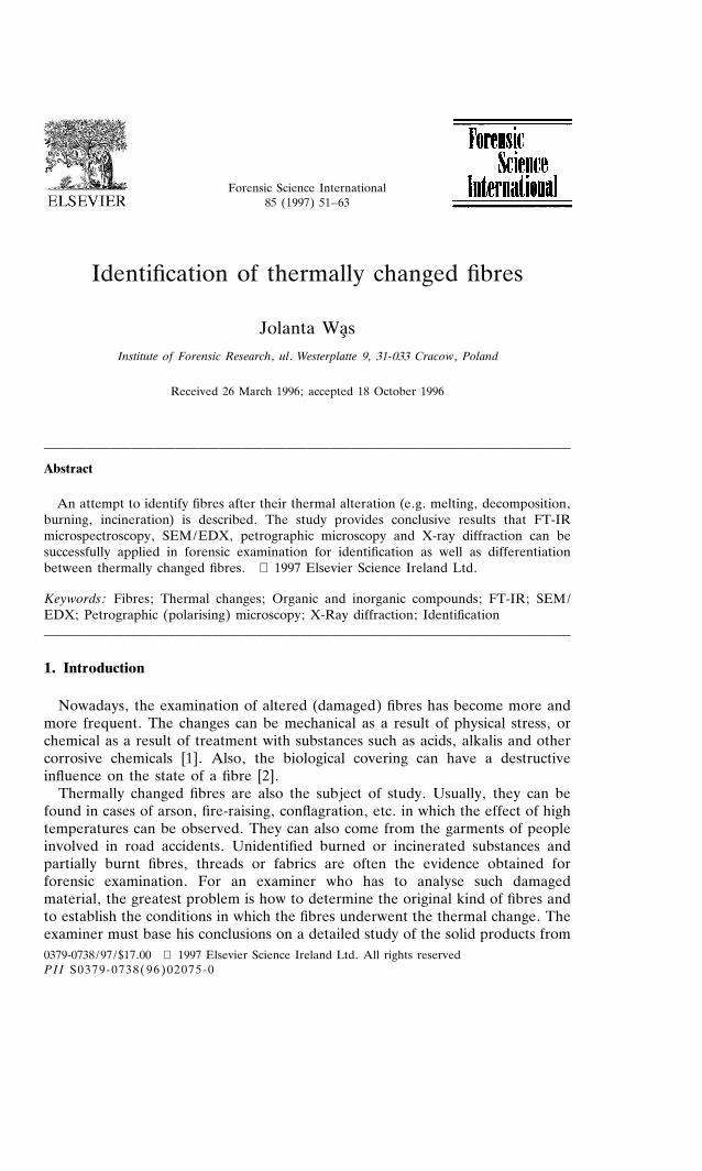
The analysis of FT-IR spectra from melted fibres showed no difference in thenumber and location of characteristic bands in comparison to the spectra from theoriginal fibres.
In the case of spectra from decomposed and burned fibres, the absorptionbands characteristic of original fibres exist beside the bands characteristic ofthermally degraded fibrous material.
The analysis of spectra from incinerated fibres can identify the chemical speciesin this material; however, infrared techniques cannot be considered conclusive inthe identification of mineral samples.
Fig. 1. FT-IR spectra of original (a), burned (b, d, e), decomposed (c) and incinerated (f) Sudanesecotton.
J. Wa̧s / Forensic Science International 85 (1997) 51 –63 55
Fig. 2. FT-IR spectra of original (a), burned (b), melted (c) and incinerated (d) Polana.
3.2. Scanning electron microscopy with energy dispersive X-ray microanalysis
Selected results from the observation of surface morphologies of incineratedfibres are given in Fig. 3 Fig. 4 Fig. 5 Fig. 6 Fig. 7 Fig. 8 Fig. 9 Fig. 10. A morecomplete catalogue of SEM images from incinerated fibres has been presented[8].
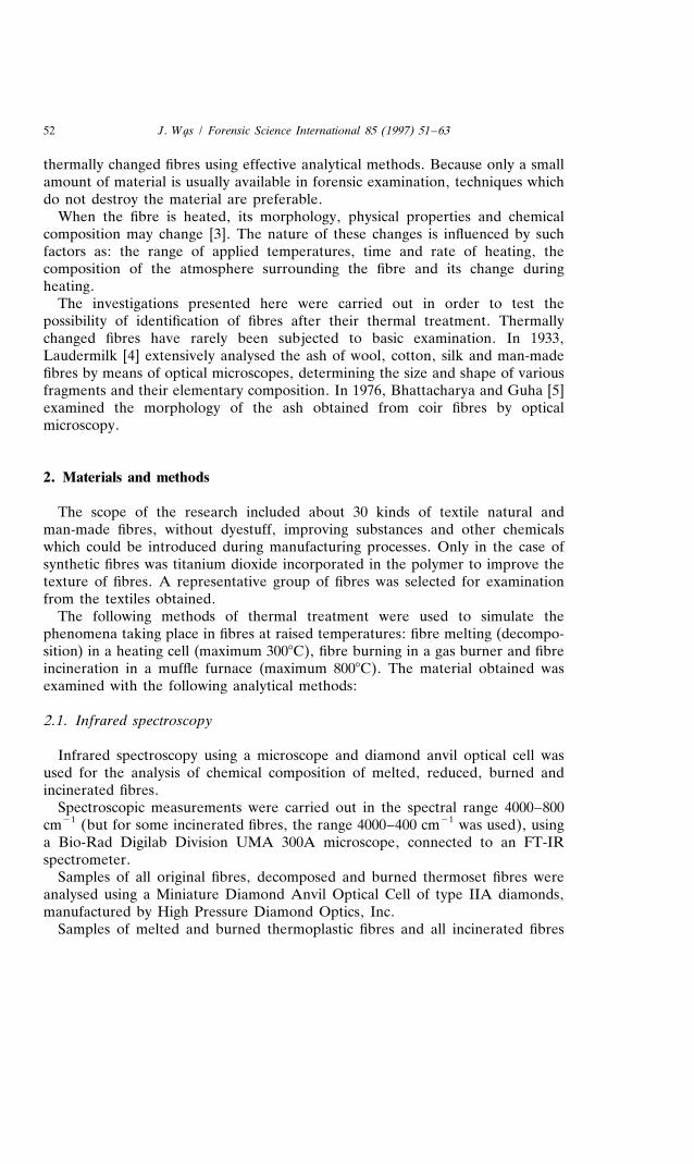
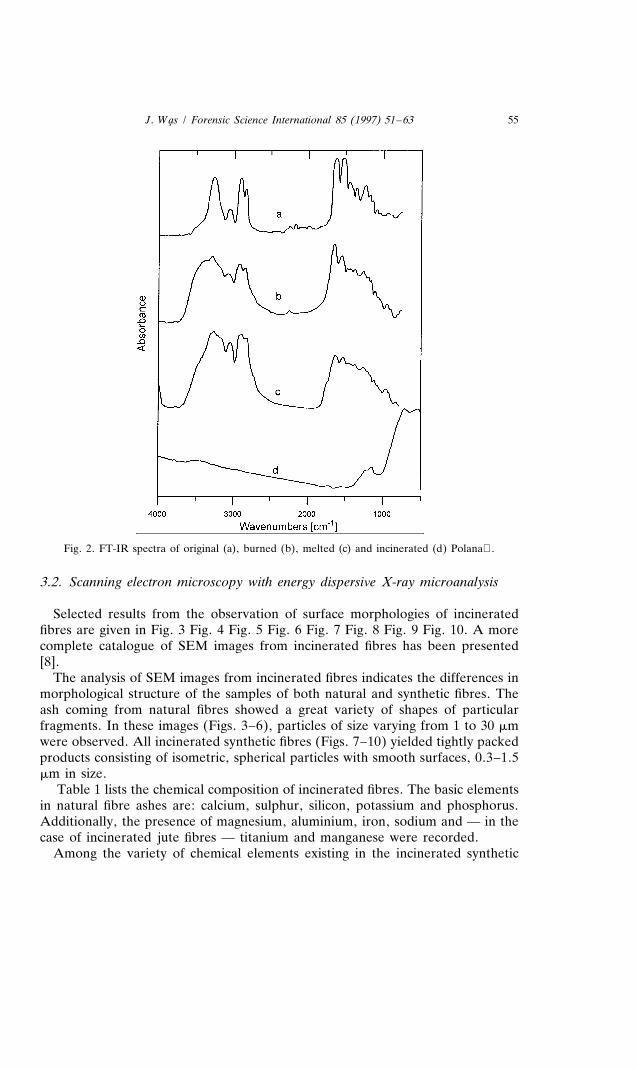
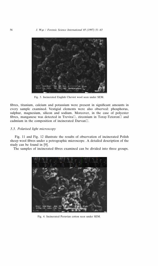
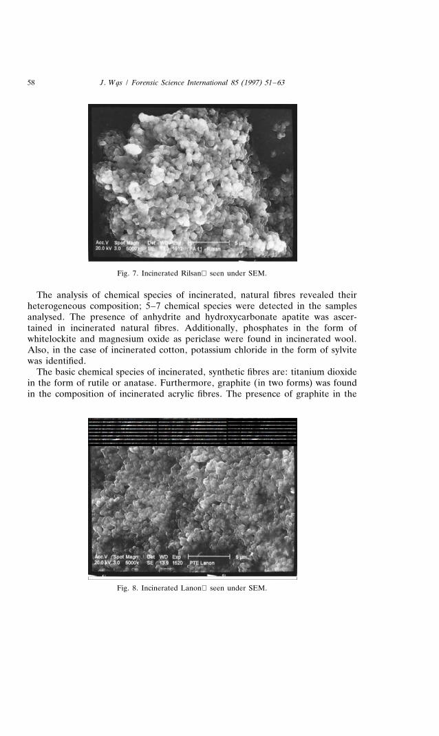
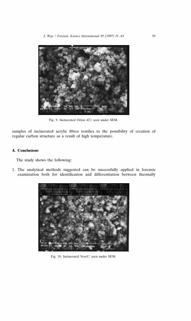
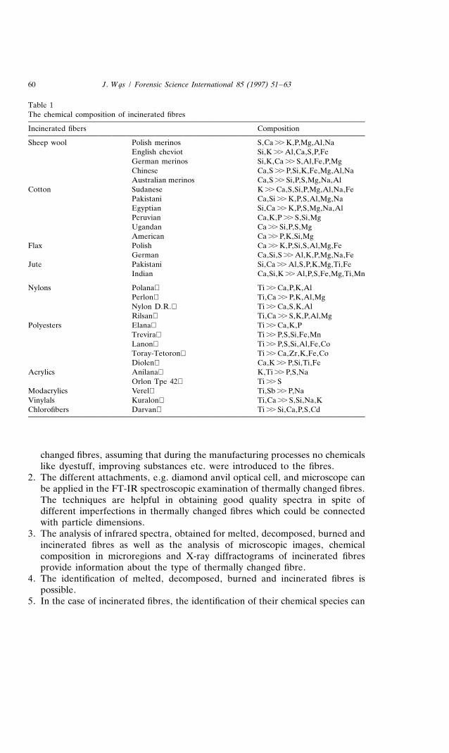
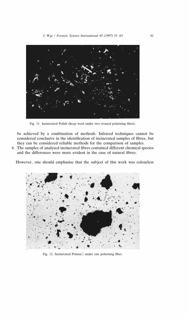
The analysis of SEM images from incinerated fibres indicates the differences inmorphological structure of the samples of both natural and synthetic fibres. Theash coming from natural fibres showed a great variety of shapes of particularfragments. In these images (Figs. 3–6), particles of size varying from 1 to 30 mmwere observed. All incinerated synthetic fibres (Figs. 7–10) yielded tightly packedproducts consisting of isometric, spherical particles with smooth surfaces, 0.3–1.5mm in size.
Table 1 lists the chemical composition of incinerated fibres. The basic elementsin natural fibre ashes are: calcium, sulphur, silicon, potassium and phosphorus.Additionally, the presence of magnesium, aluminium, iron, sodium and — in thecase of incinerated jute fibres — titanium and manganese were recorded.
Among the variety of chemical elements existing in the incinerated synthetic
56 J. Wa̧s / Forensic Science International 85 (1997) 51 –63
Fig. 3. Incinerated English Cheviot wool seen under SEM.
fibres, titanium, calcium and potassium were present in significant amounts inevery sample examined. Vestigial elements were also observed: phosphorus,sulphur, magnesium, silicon and sodium. Moreover, in the case of polyesterfibres, manganese was detected in Trevira, zirconium in Toray-Tetoron andcadmium in the composition of incinerated Darvan.
3.3. Polarised light microscopy
Fig. 11 and Fig. 12 illustrate the results of observation of incinerated Polishsheep wool fibres under a petrographic microscope. A detailed description of thestudy can be found in [9].
The samples of incinerated fibres examined can be divided into three groups.
Fig. 4. Incinerated Peruvian cotton seen under SEM.

J. Wa̧s / Forensic Science International 85 (1997) 51 –63 57
Fig. 5. Incinerated Polish flax seen under SEM.
The first group includes the samples of incinerated natural fibres which aretransparent and optically anisotropic. The second group comprises incineratedpolyamide and polyester fibres. They are weakly transparent substances, opticallyisotropic and create opaque concentrations (aggregates) with irregular borders.The incinerated acrylic fibres belong to the third group. They are non-transparentand generally form random shapes.
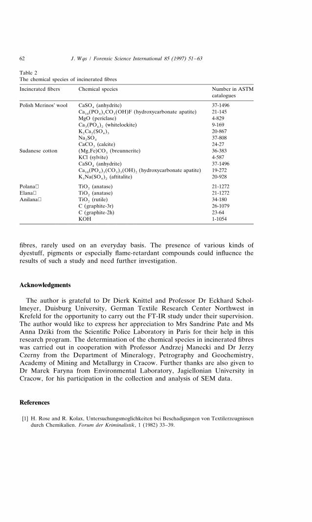
3.4. X-Ray diffraction
Selected results from the analysis of these spectra are presented in Table 2 andhave been published in [10].
Fig. 6. Incinerated Indian jute seen under SEM.
58 J. Wa̧s / Forensic Science International 85 (1997) 51 –63
Fig. 7. Incinerated Rilsan seen under SEM.
The analysis of chemical species of incinerated, natural fibres revealed theirheterogeneous composition; 5–7 chemical species were detected in the samplesanalysed. The presence of anhydrite and hydroxycarbonate apatite was ascer-tained in incinerated natural fibres. Additionally, phosphates in the form ofwhitelockite and magnesium oxide as periclase were found in incinerated wool.Also, in the case of incinerated cotton, potassium chloride in the form of sylvitewas identified.
The basic chemical species of incinerated, synthetic fibres are: titanium dioxidein the form of rutile or anatase. Furthermore, graphite (in two forms) was foundin the composition of incinerated acrylic fibres. The presence of graphite in the
Fig. 8. Incinerated Lanon seen under SEM.
J. Wa̧s / Forensic Science International 85 (1997) 51 –63 59
Fig. 9. Incinerated Orlon 42 seen under SEM.
samples of incinerated acrylic fibres testifies to the possibility of creation ofregular carbon structure as a result of high temperature.
4. Conclusions
The study shows the following:
1. The analytical methods suggested can be successfully applied in forensicexamination both for identification and differentiation between thermally
Fig. 10. Incinerated Verel seen under SEM.
60 J. Wa̧s / Forensic Science International 85 (1997) 51 –63
Table 1The chemical composition of incinerated fibres
Incinerated fibers Composition
Sheep wool Polish merinos S,Ca .. K,P,Mg,Al,NaEnglish cheviot Si,K .. Al,Ca,S,P,FeGerman merinos Si,K,Ca .. S,Al,Fe,P,MgChinese Ca,S .. P,Si,K,Fe,Mg,Al,NaAustralian merinos Ca,S .. Si,P,S,Mg,Na,Al
Cotton Sudanese K .. Ca,S,Si,P,Mg,Al,Na,FePakistani Ca,Si .. K,P,S,Al,Mg,NaEgyptian Si,Ca .. K,P,S,Mg,Na,AlPeruvian Ca,K,P .. S,Si,MgUgandan Ca .. Si,P,S,MgAmerican Ca .. P,K,Si,Mg
Flax Polish Ca .. K,P,Si,S,Al,Mg,FeGerman Ca,Si,S .. Al,K,P,Mg,Na,Fe
Jute Pakistani Si,Ca .. Al,S,P,K,Mg,Ti,FeIndian Ca,Si,K .. Al,P,S,Fe,Mg,Ti,Mn
Nylons Polana Ti .. Ca,P,K,AlPerlon Ti,Ca .. P,K,Al,MgNylon D.R. Ti .. Ca,S,K,AlRilsan Ti,Ca .. S,K,P,Al,Mg
Polyesters Elana Ti .. Ca,K,PTrevira Ti .. P,S,Si,Fe,MnLanon Ti .. P,S,Si,Al,Fe,CoToray-Tetoron Ti .. Ca,Zr,K,Fe,CoDiolen Ca,K .. P,Si,Ti,Fe
Acrylics Anilana K,Ti .. P,S,NaOrlon Tpe 42 Ti .. S
Modacrylics Verel Ti,Sb .. P,NaVinylals Kuralon Ti,Ca .. S,Si,Na,KChlorofibers Darvan Ti .. Si,Ca,P,S,Cd
changed fibres, assuming that during the manufacturing processes no chemicalslike dyestuff, improving substances etc. were introduced to the fibres.
2. The different attachments, e.g. diamond anvil optical cell, and microscope canbe applied in the FT-IR spectroscopic examination of thermally changed fibres.The techniques are helpful in obtaining good quality spectra in spite ofdifferent imperfections in thermally changed fibres which could be connectedwith particle dimensions.
3. The analysis of infrared spectra, obtained for melted, decomposed, burned andincinerated fibres as well as the analysis of microscopic images, chemicalcomposition in microregions and X-ray diffractograms of incinerated fibresprovide information about the type of thermally changed fibre.
4. The identification of melted, decomposed, burned and incinerated fibres ispossible.
5. In the case of incinerated fibres, the identification of their chemical species can
J. Wa̧s / Forensic Science International 85 (1997) 51 –63 61
Fig. 11. Incinerated Polish sheep wool under two crossed polarising filters.
be achieved by a combination of methods. Infrared techniques cannot beconsidered conclusive in the identification of incinerated samples of fibres, butthey can be considered reliable methods for the comparison of samples.
6. The samples of analysed incinerated fibres contained different chemical speciesand the differences were more evident in the case of natural fibres.
However, one should emphasise that the subject of this work was colourless
Fig. 12. Incinerated Polana under one polarising filter.
62 J. Wa̧s / Forensic Science International 85 (1997) 51 –63
Table 2The chemical species of incinerated fibres
Incinerated fibers Chemical species Number in ASTMcatalogues
Polish Merinos’ wool CaSO (anhydrite) 37-14964
Ca (PO ) CO (OH)F (hydroxycarbonate apatite) 21-14510 4 5 3
MgO (periclase) 4-829Ca (PO ) (whitelockite) 9-1693 4 2
K Ca (SO ) 20-8672 2 4 3
Na SO 37-8082 4
CaCO (calcite) 24-273
Sudanese cotton (Mg,Fe)CO (breunnerite) 36-3833
KCl (sylvite) 4-587CaSO (anhydrite) 37-14964
Ca (PO ) (CO ) (OH) (hydroxycarbonate apatite) 19-27210 4 3 3 3 2
K Na(SO ) (aftitalite) 20-9283 4 2
Polana TiO (anatase) 21-12722
Elana TiO (anatase) 21-12722
Anilana TiO (rutile) 34-1802
C (graphite-3r) 26-1079C (graphite-2h) 23-64KOH 1-1054
fibres, rarely used on an everyday basis. The presence of various kinds ofdyestuff, pigments or especially flame-retardant compounds could influence theresults of such a study and need further investigation.
Acknowledgments
The author is grateful to Dr Dierk Knittel and Professor Dr Eckhard Schol-lmeyer, Duisburg University, German Textile Research Center Northwest inKrefeld for the opportunity to carry out the FT-IR study under their supervision.The author would like to express her appreciation to Mrs Sandrine Pate and MsAnna Dziki from the Scientific Police Laboratory in Paris for their help in thisresearch program. The determination of the chemical species in incinerated fibreswas carried out in cooperation with Professor Andrzej Manecki and Dr JerzyCzerny from the Department of Mineralogy, Petrography and Geochemistry,Academy of Mining and Metallurgy in Cracow. Further thanks are also given toDr Marek Faryna from Environmental Laboratory, Jagiellonian University inCracow, for his participation in the collection and analysis of SEM data.
References
[1] H. Rose and R. Kolax, Untersuchungsmoglichkeiten bei Beschadigungen von Textilerzeugnissendurch Chemikalien. Forum der Kriminalistik, 1 (1982) 33–39.

J. Wa̧s / Forensic Science International 85 (1997) 51 –63 63
[2] G. Buschle-Doller et al., Enzymatic hydrolysis of cotton linen and viskore rayon fabric. TextileRes. J., 64 (1994) 270–279.
[3] Sh.W. Shalaby, Thermoplastic polymers, In Thermal Characterization of Polymeric Material,Academic Press, Orlando, 1981.
[4] J.D. Laudermilk, The identification of cloth ash. J. Criminal Law Criminol., 24 (1933) 503–516.[5] J. Bhattacharya and G. Guha, Examination of textile ash in coir fibers. Indian Textile J., 12
(1976) 73–75.[6] J. Was, The analysis of thermally changed fibres for forensic examination. 13th IAFS Meeting,
¨Dusseldorf Tagungsband S. A 217 (1993).[7] J. Was, D. Knittel and E. Schollmeyer, The use of FT-IR microspectroscopy for the identification
of thermally changed fibers. J. Forensic Sci., 41(6) (1996) 1005–1011.[8] J. Was, M. Faryna and A. Wlochowicz, The use of SEM/EDX for the identification of
incinerated fibers. J. Polym. Sci., submitted.[9] J. Was, A. Manecki and A. Wlochowicz, The possibility of identifying ashed fibres by analyses of
microscopic pictures obtained with optical methods. Probl. Forensic Sci., XXXIII (1996) 28–38.[10] J. Was, J. Czerny and A. Wlochowicz, The identification of incinerated fibers by means of x-ray
powder diffraction method. Fire and Materials, submitted.