how enzymes work - george mason universitymason.gmu.edu/~bbishop1/chem 660...
TRANSCRIPT
How Enzymes Work
1
Enzyme Catalysis
• Frequently the interaction between protein and ligand(s) is followed by covalent changes in the ligand(s). The protein is a catalyst for the chemical reaction and is unaltered once the reaction is complete.
• In such cases the protein is known as the enzyme and the ligand(s) its substrate(s).
• In some cases, the catalyzed reaction may be coupled to other processes:
• Mechanical movement/work.
• Molecular transport.
• Other chemical reactions.... etc.
• Enzyme catalysis has been studied extensively throughout the 20th century and continues to be an area of considerable interest.
• Begin with introduction to classical enzyme kinetics
• Then focus on enzyme structural properties and catalytic mechanisms enzymes.
2
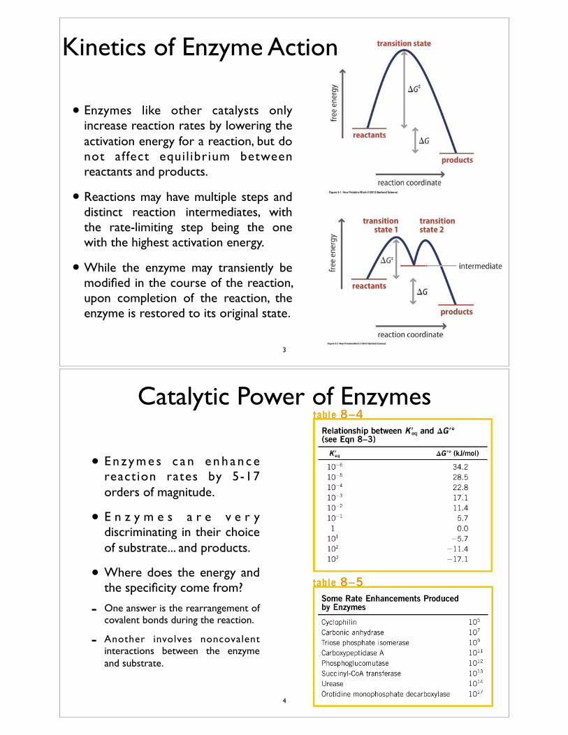
• Enzymes like other catalysts only increase reaction rates by lowering the activation energy for a reaction, but do not affect equi l ibrium between reactants and products.
• Reactions may have multiple steps and distinct reaction intermediates, with the rate-limiting step being the one with the highest activation energy.
• While the enzyme may transiently be modified in the course of the reaction, upon completion of the reaction, the enzyme is restored to its original state.
Kinetics of Enzyme Action
3
Catalytic Power of Enzymes
• E n z y m e s c a n e n h a n c e reaction rates by 5-17 orders of magnitude.
• E n z y m e s a r e v e r y discriminating in their choice of substrate... and products.
• Where does the energy and the specificity come from?
- One answer is the rearrangement of covalent bonds during the reaction.
- Another involves noncovalent interactions between the enzyme and substrate.
4
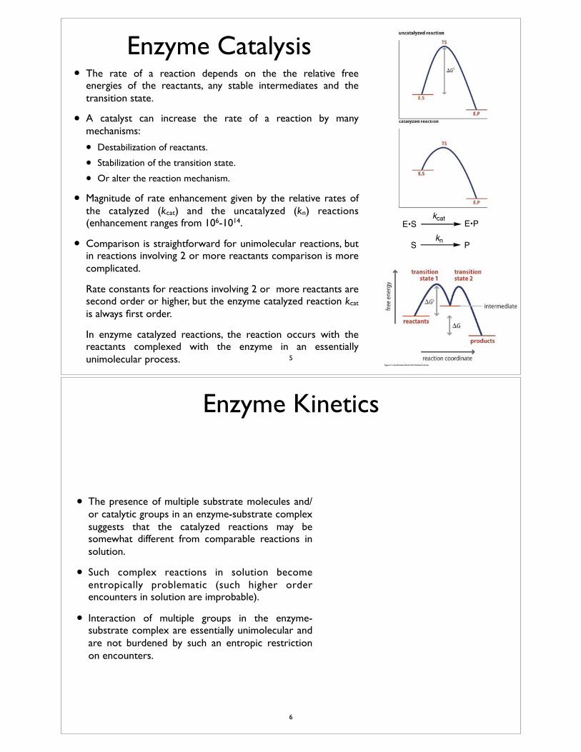
Enzyme Catalysis• The rate of a reaction depends on the the relative free
energies of the reactants, any stable intermediates and the transition state.
• A catalyst can increase the rate of a reaction by many mechanisms:
• Destabilization of reactants.
• Stabilization of the transition state.
• Or alter the reaction mechanism.
• Magnitude of rate enhancement given by the relative rates of the catalyzed (kcat) and the uncatalyzed (kn) reactions (enhancement ranges from 106-1014.
• Comparison is straightforward for unimolecular reactions, but in reactions involving 2 or more reactants comparison is more complicated.
• Rate constants for reactions involving 2 or more reactants are second order or higher, but the enzyme catalyzed reaction kcat is always first order.
• In enzyme catalyzed reactions, the reaction occurs with the reactants complexed with the enzyme in an essentially unimolecular process.
E•S E•P
S Pkn
kcat
5
Enzyme Kinetics
• The presence of multiple substrate molecules and/or catalytic groups in an enzyme-substrate complex suggests that the catalyzed reactions may be somewhat different from comparable reactions in solution.
• Such complex reactions in solution become entropically problematic (such higher order encounters in solution are improbable).
• Interaction of multiple groups in the enzyme-substrate complex are essentially unimolecular and are not burdened by such an entropic restriction on encounters.
6
Enzyme Kinetics
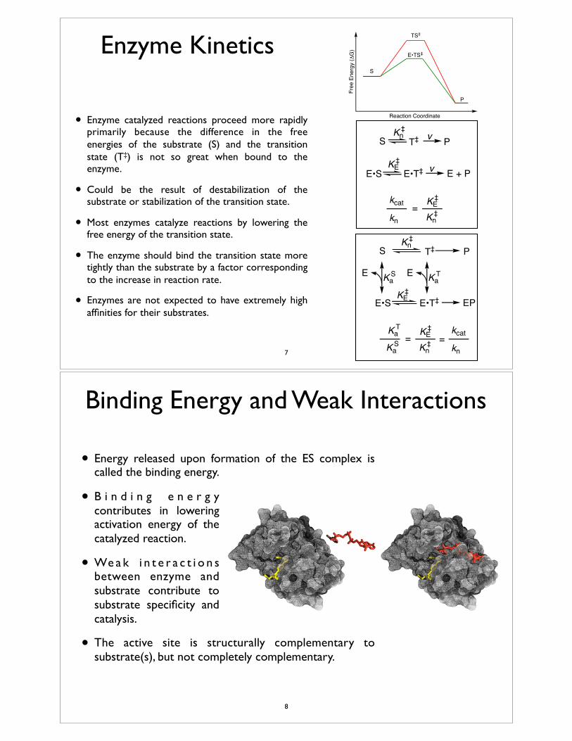
• Enzyme catalyzed reactions proceed more rapidly primarily because the difference in the free energies of the substrate (S) and the transition state (T‡) is not so great when bound to the enzyme.
• Could be the result of destabilization of the substrate or stabilization of the transition state.
• Most enzymes catalyze reactions by lowering the free energy of the transition state.
• The enzyme should bind the transition state more tightly than the substrate by a factor corresponding to the increase in reaction rate.
• Enzymes are not expected to have extremely high affinities for their substrates.
E•S
S PKn
T‡‡
KE‡
E•T‡ E + Pv
v
kcat
kn=
Kn‡
KE‡
E•S
S PKn
T‡‡
KE‡
E•T‡ EP
E KaS E Ka
T
KaS
KaT
=Kn
‡KE
‡ kcat
kn=
S
P
TS‡
E•TS‡
Free
Ene
rgy
(ΔG
)
Reaction Coordinate
7
Binding Energy and Weak Interactions
• Energy released upon formation of the ES complex is called the binding energy.
• B i n d i n g e n e r g y contributes in lowering activation energy of the catalyzed reaction.
• We a k i n t e r a c t i o n s between enzyme and substrate contribute to substrate specificity and catalysis.
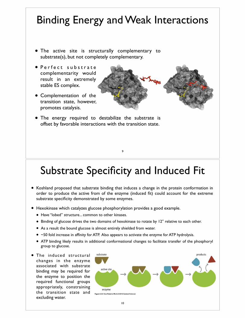
• The active site is structurally complementary to substrate(s), but not completely complementary.
8
Binding Energy and Weak Interactions
• The active site is structurally complementary to substrate(s), but not completely complementary.
• P e r f e c t s u b s t r a t e complementarity would result in an extremely stable ES complex.
• Complementation of the transition state, however, promotes catalysis.
• The energy required to destabilize the substrate is offset by favorable interactions with the transition state.
9
Substrate Specificity and Induced Fit
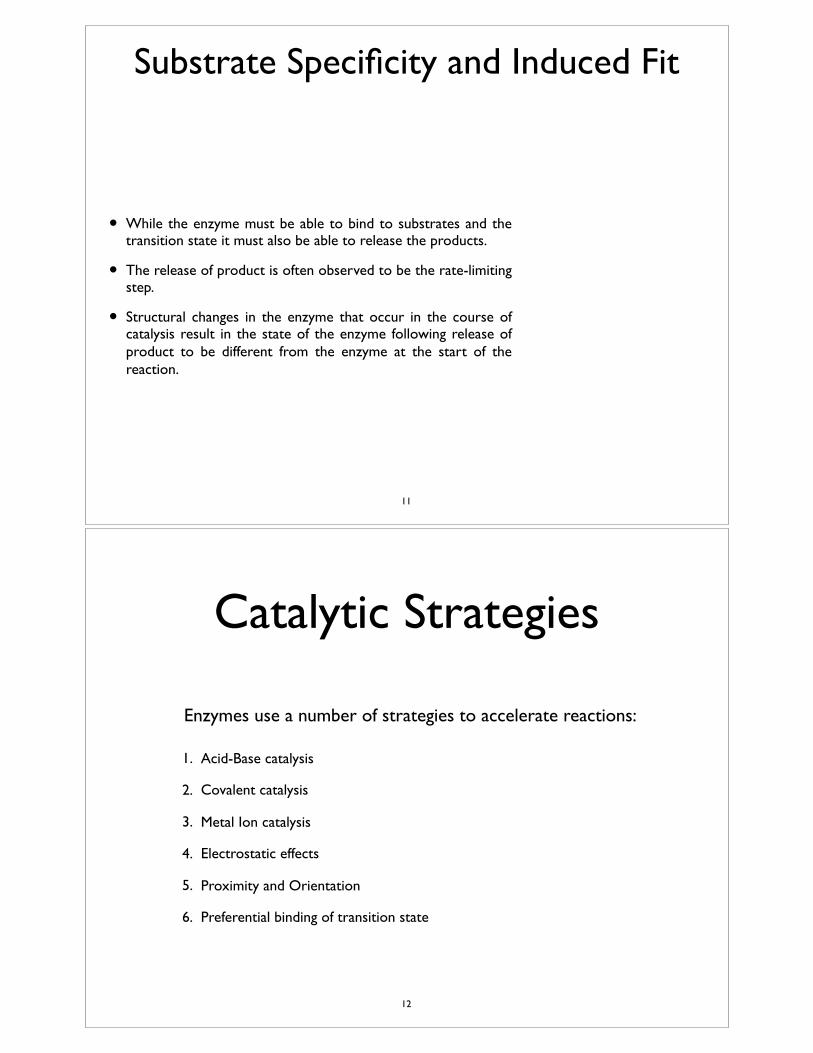
• Koshland proposed that substrate binding that induces a change in the protein conformation in order to produce the active from of the enzyme (induced fit) could account for the extreme substrate specificity demonstrated by some enzymes.
• Hexokinase which catalyzes glucose phosphorylation provides a good example.
• Have “lobed” structure... common to other kinases.
• Binding of glucose drives the two domains of hexokinase to rotate by 12° relative to each other.
• As a result the bound glucose is almost entirely shielded from water.
• ~50 fold increase in affinity for ATP. Also appears to activate the enzyme for ATP hydrolysis.
• ATP binding likely results in additional conformational changes to facilitate transfer of the phosphoryl group to glucose.
• The induced structural changes in the enzyme associated with substrate binding may be required for the enzyme to position the required functional groups appropriately, constraining the transition state and excluding water.
• 10
Substrate Specificity and Induced Fit
• While the enzyme must be able to bind to substrates and the transition state it must also be able to release the products.
• The release of product is often observed to be the rate-limiting step.
• Structural changes in the enzyme that occur in the course of catalysis result in the state of the enzyme following release of product to be different from the enzyme at the start of the reaction.
11
Catalytic Strategies
• Enzymes use a number of strategies to accelerate reactions:
1. Acid-Base catalysis
2. Covalent catalysis
3. Metal Ion catalysis
4. Electrostatic effects
5. Proximity and Orientation
6. Preferential binding of transition state
12
Reactions on the Enzyme (catalytic mechanisms)
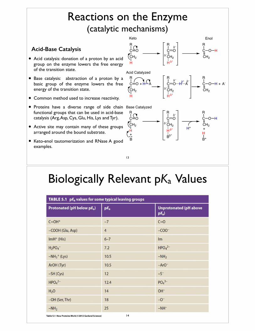
Acid-Base Catalysis
• Acid catalysis: donation of a proton by an acid group on the enzyme lowers the free energy of the transition state.
• Base catalysis: abstraction of a proton by a basic group of the enzyme lowers the free energy of the transition state.
• Common method used to increase reactivity.
• Proteins have a diverse range of side chain functional groups that can be used in acid-base catalysis (Arg, Asp, Cys, Glu, His, Lys and Tyr).
• Active site may contain many of these groups arranged around the bound substrate.
• Keto-enol tautomerization and RNase A good examples.
RCCH2
O
H
RCCH2
O
H
RCCH2
Oδ−
δ+
δ−H
Keto Enol
RCCH2
O
H
RCCH2
O
H
RCCH2
Oδ−
δ+
δ−HH A H A
δ−δ++ + A-
RCCH2
O
H
RCCH2
O
H
RCCH2
Oδ−
δ+
δ−H
Acid Catalyzed
Base Catalyzed
B+ B
HB+
δ+
H+ +
13
Biologically Relevant pKa Values
14
Reactions on the Enzyme (catalytic mechanisms)
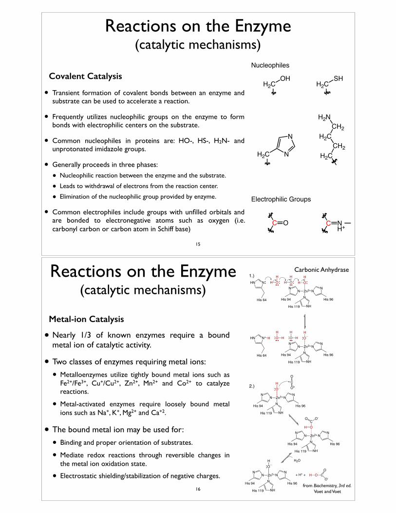
Covalent Catalysis
• Transient formation of covalent bonds between an enzyme and substrate can be used to accelerate a reaction.
• Frequently utilizes nucleophilic groups on the enzyme to form bonds with electrophilic centers on the substrate.
• Common nucleophiles in proteins are: HO-, HS-, H2N- and unprotonated imidazole groups.
• Generally proceeds in three phases:
• Nucleophilic reaction between the enzyme and the substrate.
• Leads to withdrawal of electrons from the reaction center.
• Elimination of the nucleophilic group provided by enzyme.
• Common electrophiles include groups with unfilled orbitals and are bonded to electronegative atoms such as oxygen (i.e. carbonyl carbon or carbon atom in Schiff base)
OHH2C
SHH2C
H2C H2CCH2
H2CCH2
H2N
N
N
C O C NH+
Nucleophiles
Electrophilic Groups
15
Reactions on the Enzyme (catalytic mechanisms)
Metal-ion Catalysis
• Nearly 1/3 of known enzymes require a bound metal ion of catalytic activity.
• Two classes of enzymes requiring metal ions:
• Metalloenzymes utilize tightly bound metal ions such as Fe2+/Fe3+, Cu+/Cu2+, Zn2+, Mn2+ and Co2+ to catalyze reactions.
• Metal-activated enzymes require loosely bound metal ions such as Na+, K+, Mg2+ and Ca+2.
• The bound metal ion may be used for:
• Binding and proper orientation of substrates.
• Mediate redox reactions through reversible changes in the metal ion oxidation state.
• Electrostatic shielding/stabilization of negative charges.
Zn2+
OH
HN
N
NH
N
NN
NHN H OH
H OH
His 64
His 119
His 94 His 96
Zn2+
OH
NN
NH
N
NN
N+HN OH
H OH
His 64
His 119
His 94 His 96
H H
Zn2+
OH
NN
NH
N
NN
His 119
His 94 His 96
CO
O
Zn2+
OC
NN
NH
N
NN
His 119
His 94 His 96
H
O O-
Zn2+
OH
NN
NH
N
NN
His 119
His 94 His 96
+ H+ + H O CO
O-
H2O
2.)
1.)Carbonic Anhydrase
from Biochemistry, 3rd ed. Voet and Voet16
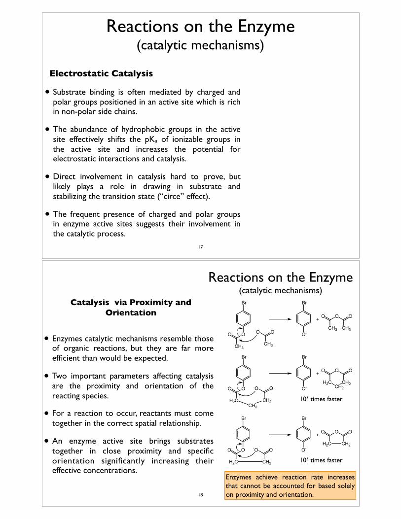
Reactions on the Enzyme (catalytic mechanisms)
Electrostatic Catalysis
• Substrate binding is often mediated by charged and polar groups positioned in an active site which is rich in non-polar side chains.
• The abundance of hydrophobic groups in the active site effectively shifts the pKa of ionizable groups in the active site and increases the potential for electrostatic interactions and catalysis.
• Direct involvement in catalysis hard to prove, but likely plays a role in drawing in substrate and stabilizing the transition state (“circe” effect).
• The frequent presence of charged and polar groups in enzyme active sites suggests their involvement in the catalytic process.
17
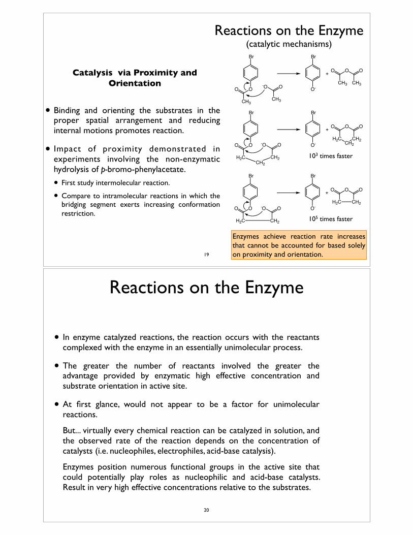
Reactions on the Enzyme (catalytic mechanisms)
Catalysis via Proximity and Orientation
• Enzymes catalytic mechanisms resemble those of organic reactions, but they are far more efficient than would be expected.
• Two important parameters affecting catalysis are the proximity and orientation of the reacting species.
• For a reaction to occur, reactants must come together in the correct spatial relationship.
• An enzyme active site brings substrates together in close proximity and specific orientation significantly increasing their effective concentrations.
O O
CH3
Br
-O O
CH3
O-
Br
+ O
CH3 CH3
OO
O O
H2C
Br
-O O
CH2
O-
Br
+ O
H2C CH2
OO
CH2
CH2
O O
H2C
Br
-O O
CH2
O-
Br
+ O
H2C CH2
OO
103 times faster
105 times faster
Enzymes achieve reaction rate increases that cannot be accounted for based solely on proximity and orientation.18
Reactions on the Enzyme (catalytic mechanisms)
Catalysis via Proximity and Orientation
• Binding and orienting the substrates in the proper spatial arrangement and reducing internal motions promotes reaction.
• Impact of proximity demonstrated in experiments involving the non-enzymatic hydrolysis of p-bromo-phenylacetate.
• First study intermolecular reaction.
• Compare to intramolecular reactions in which the bridging segment exerts increasing conformation restriction.
Enzymes achieve reaction rate increases that cannot be accounted for based solely on proximity and orientation.
O O
CH3
Br
-O O
CH3
O-
Br
+ O
CH3 CH3
OO
O O
H2C
Br
-O O
CH2
O-
Br
+ O
H2C CH2
OO
CH2
CH2
O O
H2C
Br
-O O
CH2
O-
Br
+ O
H2C CH2
OO
103 times faster
105 times faster
19
Reactions on the Enzyme
• In enzyme catalyzed reactions, the reaction occurs with the reactants complexed with the enzyme in an essentially unimolecular process.
• The greater the number of reactants involved the greater the advantage provided by enzymatic high effective concentration and substrate orientation in active site.
• At first glance, would not appear to be a factor for unimolecular reactions.
• But... virtually every chemical reaction can be catalyzed in solution, and the observed rate of the reaction depends on the concentration of catalysts (i.e. nucleophiles, electrophiles, acid-base catalysis).
• Enzymes position numerous functional groups in the active site that could potentially play roles as nucleophilic and acid-base catalysts. Result in very high effective concentrations relative to the substrates.
20
Reactions on the Enzyme
• Relative to the uncatalyzed reactions:
• Acid-base and covalent catalytic mechanisms can be estimated to increase reaction rates by one or two orders of magnitude.
• Orientation and proximity can account for up to 108 fold increase in reaction rate (often much less).
• Some enzymes achieve 1014-1017 fold increases in rates of reaction.
21
Reactions on the Enzyme
Transition State Binding
• One of the most important catalytic mechanisms employed by enzymes is preferential binding and stabilization of the reaction transition state.
• Enzymes believed to strain/distort substrates into transition state geometries through binding sites that optimally bind the transition state.
• By this mechanism, a 106 fold rate increase would require a 106 fold enhancement in transition state binding relative to substrate (~8.1 Kcal/mol at RT).
• Very large rate enhancements could be achieved with the formation of a few new bonds/interactions between the transition state and the enzyme.
22
Reactions on the EnzymeTransition State Binding
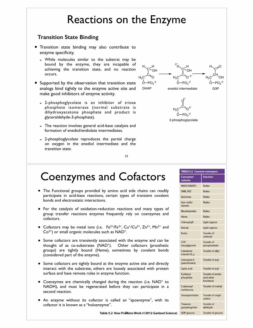
• Transition state binding may also contribute to enzyme specificity.
- While molecules similar to the substrat may be bound by the enzyme, they are incapable of achieving the transition state, and no reaction occurs.
• Supported by the observation that transition state analogs bind tightly to the enzyme active site and make good inhibitors of enzyme activity.
- 2-phosphoglycolate is an inhibitor of triose phosphate isomerase (normal substrate is dihydroxyacetone phosphate and product is glyceraldehyde-3-phosphate).
- The reaction involves general acid-base catalysis and formation of enediol/endiolate intermediates.
- 2-phosphoglycolate reproduces the partial charge on oxygen in the enediol intermediate and the transition state.
CO
HC
H2CO
OHH
PO3-2
CO
HC
H2CO
OHH
PO3-2
CHOH
OC
H2CO
H
PO3-2
CO
O
H2CO PO3
-2
δ−
δ+
δ−
δ−
2-phosphoglycolate
DHAP G3Penediol intermediate
23
• The Functional groups provided by amino acid side chains can readily participate in acid-base reactions, certain types of transient covalent bonds and electrostatic interactions.
• For the catalysis of oxidation-reduction reactions and many types of group transfer reactions enzymes frequently rely on coenzymes and cofactors.
• Cofactors may be metal ions (i.e. Fe2+/Fe3+, Cu+/Cu2+, Zn2+, Mn2+ and Co2+) or small organic molecules such as NAD+.
• Some cofactors are transiently associated with the enzyme and can be thought of as co-substrates (NAD+). Other cofactors (prosthetic groups) are tightly bound (Heme), sometimes by covalent bonds (considered part of the enzyme).
• Some cofactors are tightly bound at the enzyme active site and directly interact with the substrate, others are loosely associated with protein surface and have remote roles in enzyme function.
• Coenzymes are chemically changed during the reaction (i.e. NAD+ to NADH), and must be regenerated before they can participate in a second reaction.
• An enzyme without its cofactor is called an “apoenzyme”, with its cofactor it is known as a “holoenzyme”.
Coenzymes and Cofactors
24
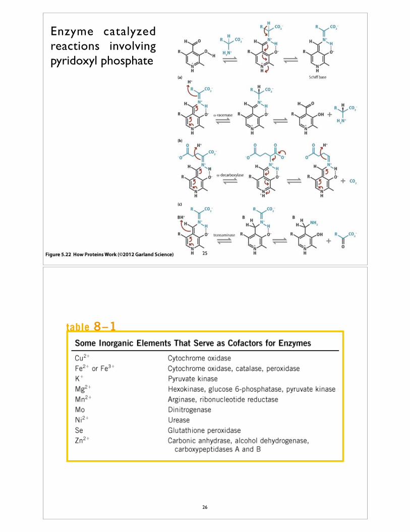
Enzyme catalyzed reactions involving pyridoxyl phosphate
25
26
Enzyme Inhibition and Regulation
• An organism must be able to regulate the catalytic activity of its enzymes.
• Two general schemes:★ Controlling enzyme availability by controlling expression
and degradation.
★ Controlling activity:
✤ Reversible covalent modification of enzymes resulting in inactivation (often phosphorylation of Ser residues).
✤ Expression and storage of enzymes in nonfunctional proforms (zymogens) that require proteolytic processing for activation.
✤ Direct control of enzyme activity through the use of inhibitors and enhancers that directly affect the enzyme.
27
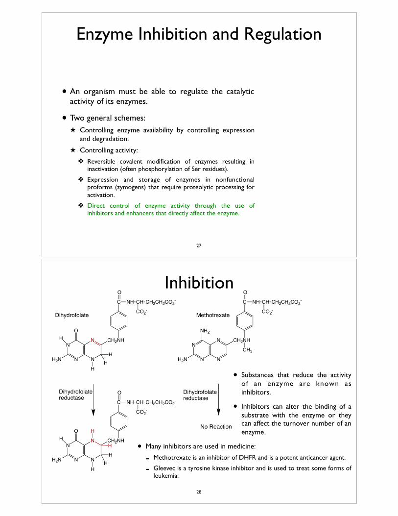
Inhibition
• Substances that reduce the activity of an enzyme are known as inhibitors.
• Inhibitors can alter the binding of a substrate with the enzyme or they can affect the turnover number of an enzyme.
• Many inhibitors are used in medicine:
- Methotrexate is an inhibitor of DHFR and is a potent anticancer agent.
- Gleevec is a tyrosine kinase inhibitor and is used to treat some forms of leukemia.
N
N N
N CH2
NH2
H2N
NH
CH3
C
O
NH CH
CO2-
CH2CH2CO2-
N
N N
N CH2
O
H2N
NH
C
O
NH CH
CO2-
CH2CH2CO2-
H
HH
H
Dihydrofolate Methotrexate
N
N N
N CH2
O
H2N
NH
C
O
NH CH
CO2-
CH2CH2CO2-
H
HH
H
Dihydrofolate reductase
Dihydrofolate reductase
H
H
No Reaction
28
Enzyme Kinetics
29
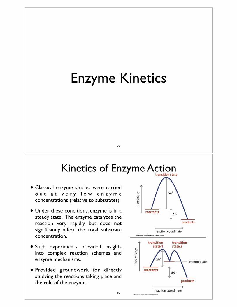
Kinetics of Enzyme Action
• Classical enzyme studies were carried o u t a t v e r y l o w e n z y m e concentrations (relative to substrates).
• Under these conditions, enzyme is in a steady state. The enzyme catalyzes the reaction very rapidly, but does not significantly affect the total substrate concentration.
• Such experiments provided insights into complex reaction schemes and enzyme mechanisms.
• Provided groundwork for directly studying the reactions taking place and the role of the enzyme.
30
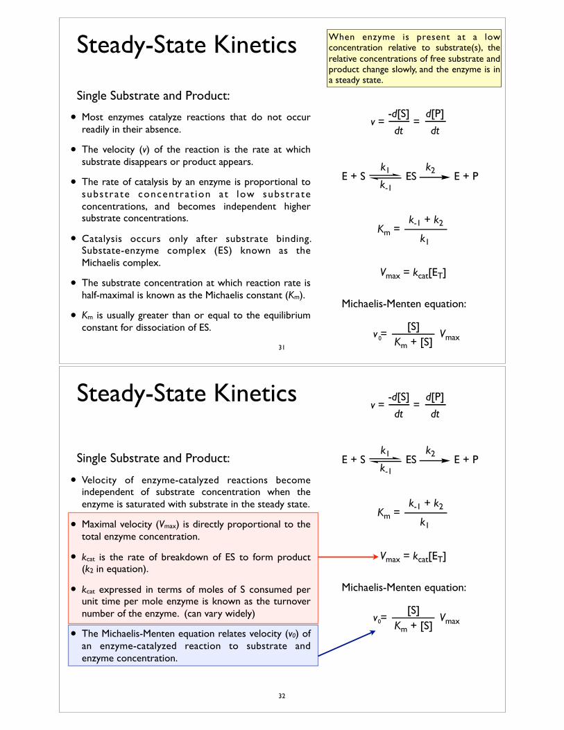
Steady-State Kinetics
Single Substrate and Product:
• Most enzymes catalyze reactions that do not occur readily in their absence.
• The velocity (v) of the reaction is the rate at which substrate disappears or product appears.
• The rate of catalysis by an enzyme is proportional to substrate concentrat ion at low substrate concentrations, and becomes independent higher substrate concentrations.
• Catalysis occurs only after substrate binding. Substate-enzyme complex (ES) known as the Michaelis complex.
• The substrate concentration at which reaction rate is half-maximal is known as the Michaelis constant (Km).
• Km is usually greater than or equal to the equilibrium constant for dissociation of ES.
When enzyme is present at a low concentration relative to substrate(s), the relative concentrations of free substrate and product change slowly, and the enzyme is in a steady state.
v =-d[S]
=dt
d[P]
dt
E + S ES E + Pk1k-1
k2
Km =k1
k-1 + k2
Vmax = kcat[ET]
Michaelis-Menten equation:
v = Vmax[S]
Km + [S]0
31
Steady-State Kinetics
Single Substrate and Product:
• Velocity of enzyme-catalyzed reactions become independent of substrate concentration when the enzyme is saturated with substrate in the steady state.
• Maximal velocity (Vmax) is directly proportional to the total enzyme concentration.
• kcat is the rate of breakdown of ES to form product (k2 in equation).
• kcat expressed in terms of moles of S consumed per unit time per mole enzyme is known as the turnover number of the enzyme. (can vary widely)
• The Michaelis-Menten equation relates velocity (v0) of an enzyme-catalyzed reaction to substrate and enzyme concentration.
v =-d[S]
=dt
d[P]
dt
E + S ES E + Pk1k-1
k2
Km =k1
k-1 + k2
Vmax = kcat[ET]
Michaelis-Menten equation:
v = Vmax[S]
Km + [S]0
32
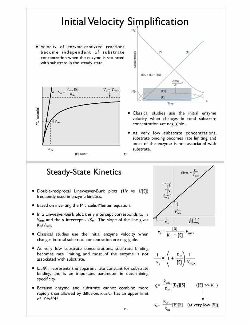
Initial Velocity Simplification
• Velocity of enzyme-catalyzed reactions become independent o f subs t ra te concentration when the enzyme is saturated with substrate in the steady state.
• Classical studies use the initial enzyme velocity when changes in total substrate concentration are negligible.
• At very low substrate concentrations, substrate binding becomes rate limiting, and most of the enzyme is not associated with substrate.
33
Steady-State Kinetics
Michaelis-Menten equation:
v = Vmax[S]
Km + [S]
v Vmax=
11 +
1
[S]
Km
v =Km
kcat[ET][S] ([S] << Km)
v =Km
kcat[E][S] (at very low [S])
• Double-reciprocal Lineweaver-Burk plots (1/v vs 1/[S]) frequently used in enzyme kinetics.
• Based on inverting the Michaelis-Menten equation.
• In a Linweaver-Burk plot, the y intercept corresponds to 1/Vmax and the x intercept -1/Km. The slope of the line gives Km/Vmax.
• Classical studies use the initial enzyme velocity when changes in total substrate concentration are negligible.
• At very low substrate concentrations, substrate binding becomes rate limiting, and most of the enzyme is not associated with substrate.
• kcat/Km represents the apparent rate constant for substrate binding, and is an important parameter in determining specificity.
• Because enzyme and substrate cannot combine more rapidly than allowed by diffusion, kcat/Km has an upper limit of 109s-1M-1.
0
0
0
0
34
Steady-State KineticsE + S E•S E•P E + P
Km
KmP
kcatf
rkcat
= Keq =kcat / Km
kcat / KmP
f
r
S
S [P]eq
[S]eq
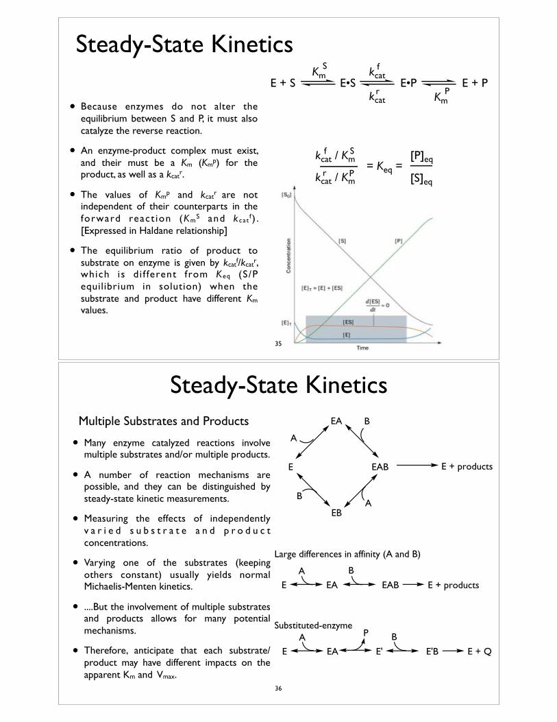
• Because enzymes do not alter the equilibrium between S and P, it must also catalyze the reverse reaction.
• An enzyme-product complex must exist, and their must be a Km (Kmp) for the product, as well as a kcatr.
• The values of Kmp and kcatr are not independent of their counterparts in the forward react ion (KmS and k cat f ) . [Expressed in Haldane relationship]
• The equilibrium ratio of product to substrate on enzyme is given by kcatf/kcatr, which is d i f ferent from Keq (S/P equilibrium in solution) when the substrate and product have different Km values.
35
Steady-State KineticsMultiple Substrates and Products
• Many enzyme catalyzed reactions involve multiple substrates and/or multiple products.
• A number of reaction mechanisms are possible, and they can be distinguished by steady-state kinetic measurements.
• Measuring the effects of independently v a r i e d s u b s t r a t e a n d p r o d u c t concentrations.
• Varying one of the substrates (keeping others constant) usually yields normal Michaelis-Menten kinetics.
• ....But the involvement of multiple substrates and products allows for many potential mechanisms.
• Therefore, anticipate that each substrate/product may have different impacts on the apparent Km and Vmax.
E
EA
EB
EAB E + products
A
B
BA
E EAB E + productsEAA B
E E'B E + QEAA B
E'
PSubstituted-enzyme
Large differences in affinity (A and B)
36
Steady-State Kinetics
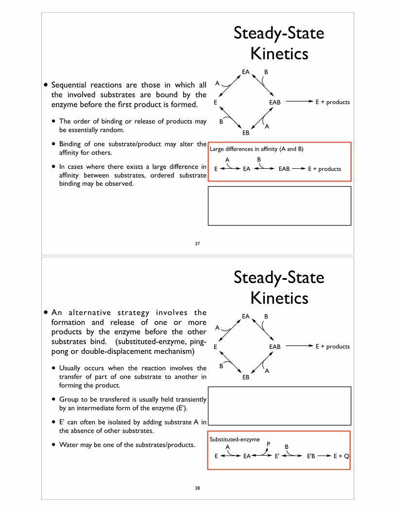
• Sequential reactions are those in which all the involved substrates are bound by the enzyme before the first product is formed.
• The order of binding or release of products may be essentially random.
• Binding of one substrate/product may alter the affinity for others.
• In cases where there exists a large difference in affinity between substrates, ordered substrate binding may be observed.
E
EA
EB
EAB E + products
A
B
BA
E EAB E + productsEAA B
E E'B E + QEAA B
E'
PSubstituted-enzyme
Large differences in affinity (A and B)
37
Steady-State Kinetics
• An alternative strategy involves the formation and release of one or more products by the enzyme before the other substrates bind. (substituted-enzyme, ping-pong or double-displacement mechanism)
• Usually occurs when the reaction involves the transfer of part of one substrate to another in forming the product.
• Group to be transfered is usually held transiently by an intermediate form of the enzyme (E’).
• E’ can often be isolated by adding substrate A in the absence of other substrates.
• Water may be one of the substrates/products.
E
EA
EB
EAB E + products
A
B
BA
E EAB E + productsEAA B
E E'B E + QEAA B
E'
PSubstituted-enzyme
Large differences in affinity (A and B)
38
Example: Serine Proteases
39
Serine Proteases• Proteolytic enzymes that cleave peptide bonds (endopeptidases):
• Chymotrypsin cleaves peptide bonds C-terminal to amino acids with aromatic side chains (Trp, Phe and Tyr).
• Trypsin cleaves peptide bonds C-terminal to amino acids with basic side chains (Arg and Lys).
• Elastase cleaves peptide bonds C-terminal to amino acids with small hydrophobic side chains (Gly, Ala and Val).
• Enhance bond hydrolysis at least 109 fold.
• Water ultimately is added across the peptide bond, but a covalent acyl-enzyme intermediate is formed.
• The reaction can be separated into two parts, formation of the acyl-enzyme and hydrolysis by water.
40
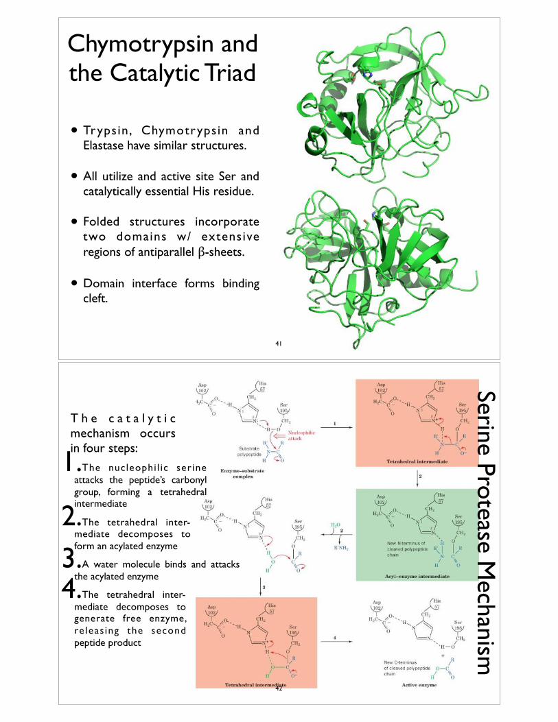
Chymotrypsin and the Catalytic Triad
• Trypsin, Chymotrypsin and Elastase have similar structures.
• All utilize and active site Ser and catalytically essential His residue.
• Folded structures incorporate two domains w/ extensive regions of antiparallel β-sheets.
• Domain interface forms binding cleft.
41
Serine Protease Mechanism
T h e c a t a l y t i c mechanism occurs in four steps:
1.The nucleophi l ic serine attacks the peptide’s carbonyl group, forming a tetrahedral intermediate
2.The tetrahedral inter-mediate decomposes to form an acylated enzyme
3.A water molecule binds and attacks the acylated enzyme
4.The tetrahedral inter-mediate decomposes to generate free enzyme, re leas ing the second peptide product
42
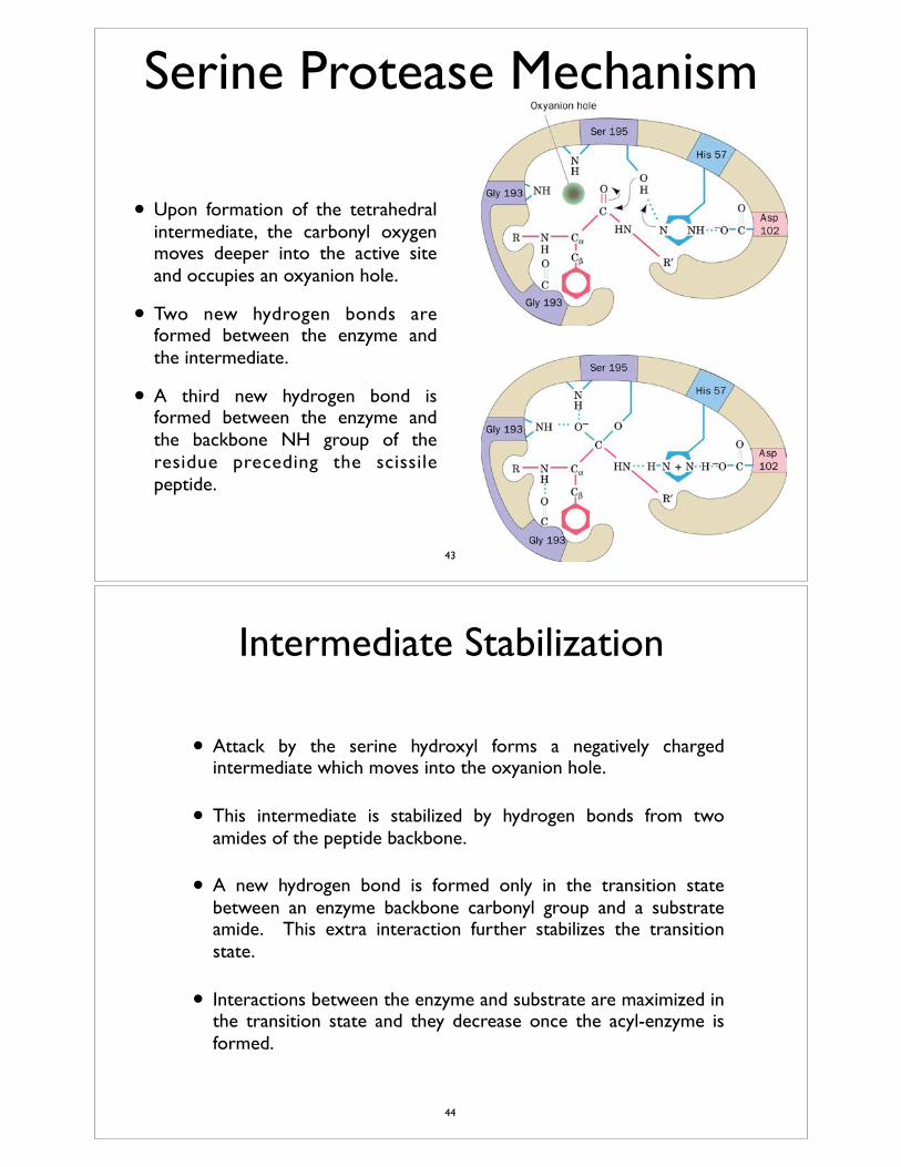
• Upon formation of the tetrahedral intermediate, the carbonyl oxygen moves deeper into the active site and occupies an oxyanion hole.
• Two new hydrogen bonds are formed between the enzyme and the intermediate.
• A third new hydrogen bond is formed between the enzyme and the backbone NH group of the residue preceding the scissile peptide.
Serine Protease Mechanism
43
Intermediate Stabilization
• Attack by the serine hydroxyl forms a negatively charged intermediate which moves into the oxyanion hole.
• This intermediate is stabilized by hydrogen bonds from two amides of the peptide backbone.
• A new hydrogen bond is formed only in the transition state between an enzyme backbone carbonyl group and a substrate amide. This extra interaction further stabilizes the transition state.
• Interactions between the enzyme and substrate are maximized in the transition state and they decrease once the acyl-enzyme is formed.
44
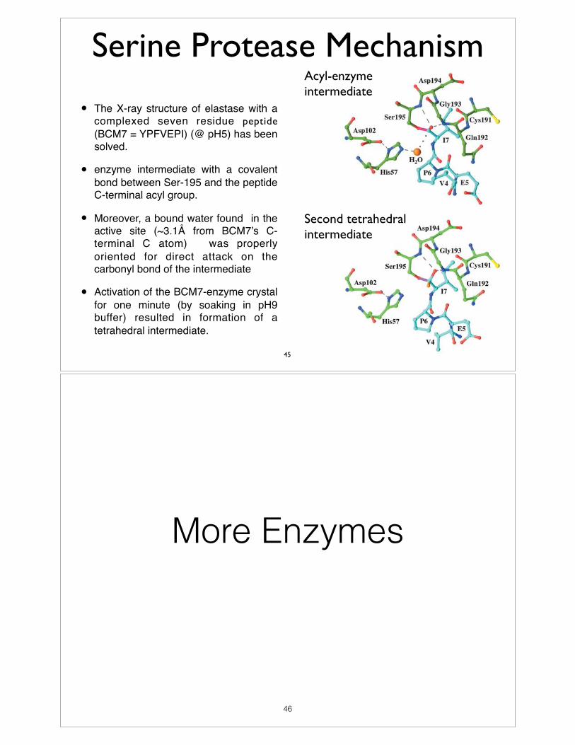
• The X-ray structure of elastase with a complexed seven residue peptide (BCM7 = YPFVEPI) (@ pH5) has been solved.
• enzyme intermediate with a covalent bond between Ser-195 and the peptide C-terminal acyl group.
• Moreover, a bound water found in the active site (~3.1Å from BCM7’s C-terminal C atom) was properly oriented for direct attack on the carbonyl bond of the intermediate
• Activation of the BCM7-enzyme crystal for one minute (by soaking in pH9 buffer) resulted in formation of a tetrahedral intermediate.
Serine Protease Mechanism
Second tetrahedral intermediate
Acyl-enzyme intermediate
45
More Enzymes
46
Aminoacyl tRNA Synthetase
• Attachment of amino acids to the correct t-RNA molecule is an essential step for translation and protein synthesis.
• Aminoacyl tRNA synthetases generate activated amino acids and mediate their transfer to the 3’ end of the acceptor arm of the appropriate t-RNA molecule. (Acceptor arm contain a conserved -CCA-3’ sequence)
• They are amino acid specific and pair amino acid with correct tRNA molecule.
• An error in this process could be disastrous. Therefore, the error rate for most aminoacyl tRNA synthetases is extremely low.
• Aminoacyl tRNA synthetases link amino acids to the 3’ end of the acceptor arm of tRNA molecules. Characterized by a conserved -CCA-OH 3’ sequence.
• Aminoacyl t-RNA synthetases are grouped into two families (Class I and Class II).
• Class I synthetases utilize a Rossmann fold motif in binding ATP/AA-AMP. Class II enzymes utilize an antiparallel β-sheet.
• Most Class I synthetases are monomeric (Tyr tRNA synthetase is an exception). Class II synthetases more commonly dimeric.
• Class I enzymes bind at the minor groove of the acceptor stem. Class II synthetases bind the major groove.
Amino acid + tRNA + ATP Aminoacyl-tRNA + AMP + PPi
47
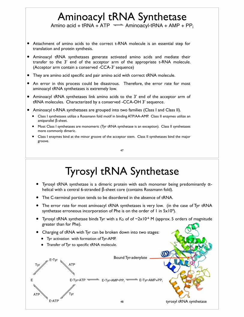
Tyrosyl tRNA Synthetase
E
E•Tyr
E•ATP
E•Tyr•ATP E•Tyr•AMP•PPi E•Tyr•AMP+PPi
Tyr
ATP
ATP
Tyr
• Tyrosyl tRNA synthetase is a dimeric protein with each monomer being predominantly α-helical with a central 6-stranded β-sheet core (contains Rossmann fold).
• The C-terminal portion tends to be disordered in the absence of tRNA.
• The error rate for most aminoacyl tRNA synthetases is very low. (in the case of Tyr tRNA synthetase erroneous incorporation of Phe is on the order of 1 in 5x10⁴).
• Tyrosyl tRNA synthetase binds Tyr with a KD of of ~2x10⁻⁶ M (approx. 5 orders of magnitude greater than for Phe).
• Charging of tRNA with Tyr can be broken down into two stages:
• Tyr activation with formation of Tyr-AMP.
• Transfer of Tyr to specific tRNA molecule.
tyrosyl tRNA synthetase
Bound Tyr-adenylate
48
Tyrosyl tRNA Synthetase
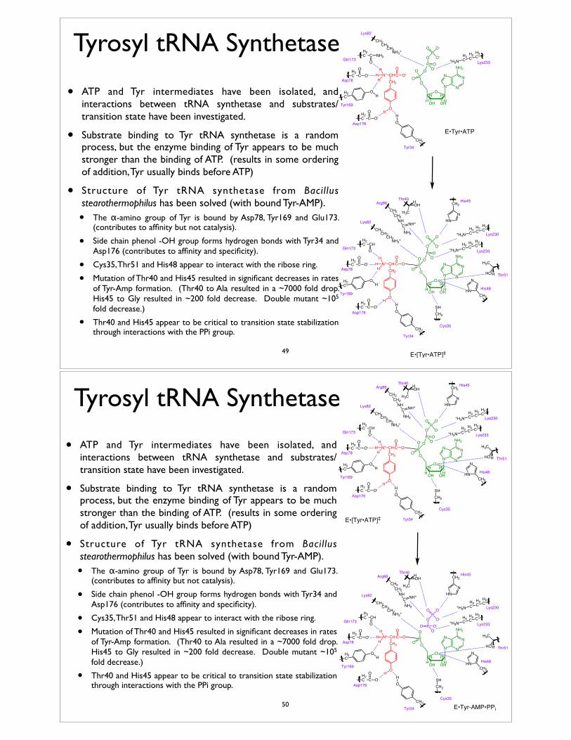
• ATP and Tyr intermediates have been isolated, and interactions between tRNA synthetase and substrates/transition state have been investigated.
• Substrate binding to Tyr tRNA synthetase is a random process, but the enzyme binding of Tyr appears to be much stronger than the binding of ATP. (results in some ordering of addition, Tyr usually binds before ATP)
• Structure of Tyr tRNA synthetase from Bacil lus stearothermophilus has been solved (with bound Tyr-AMP).
• The α-amino group of Tyr is bound by Asp78, Tyr169 and Glu173. (contributes to affinity but not catalysis).
• Side chain phenol -OH group forms hydrogen bonds with Tyr34 and Asp176 (contributes to affinity and specificity).
• Cys35, Thr51 and His48 appear to interact with the ribose ring.
• Mutation of Thr40 and His45 resulted in significant decreases in rates of Tyr-Amp formation. (Thr40 to Ala resulted in a ~7000 fold drop. His45 to Gly resulted in ~200 fold decrease. Double mutant ~105 fold decrease.)
• Thr40 and His45 appear to be critical to transition state stabilization through interactions with the PPi group.
N
NN
N
NH2
O
OHOH
HHHH
OPOP
O
-O O-O
OPO-
O O-
N+ CH CCH2
O-O
O
H2CH2
CH2C+H3N Lys233
H2C C O-
O
H2C C NH2
O
CH2
OH
H
HHHH2
C C O-O
H2C
OH
CH2CH2CH2NH3+
Tyr34
Asp176
Tyr169
Asp78
Lys82
Gln173
N
NN
N
NH2
O
OHOH
HHHH
OPOP
O
O- O-O
OPO-
O O-
N+ CH CCH2
OO
O
H2CH2
CH2C+H3N
H2CH2
CH2C+H3N
CH2
NHN
Lys233
Lys230
His45
H2C C O-
O
H2C CH
O
HO
H3C
H
CH2
OH
H
HHHH2
C C O-O
H2C
OH
CH2HN
N
His48
OHH3C
H
CH2CH2CH2NH3+
CH2CH2NHCNH2
NH+
CH2
SH
Cys35
Tyr34
Asp176
Tyr169
Asp78Thr51
Thr40
Lys82
Arg86
Gln173
E•Tyr•ATP
E•[Tyr•ATP]‡49
Tyrosyl tRNA Synthetase
• ATP and Tyr intermediates have been isolated, and interactions between tRNA synthetase and substrates/transition state have been investigated.
• Substrate binding to Tyr tRNA synthetase is a random process, but the enzyme binding of Tyr appears to be much stronger than the binding of ATP. (results in some ordering of addition, Tyr usually binds before ATP)
• Structure of Tyr tRNA synthetase from Bacil lus stearothermophilus has been solved (with bound Tyr-AMP).
• The α-amino group of Tyr is bound by Asp78, Tyr169 and Glu173. (contributes to affinity but not catalysis).
• Side chain phenol -OH group forms hydrogen bonds with Tyr34 and Asp176 (contributes to affinity and specificity).
• Cys35, Thr51 and His48 appear to interact with the ribose ring.
• Mutation of Thr40 and His45 resulted in significant decreases in rates of Tyr-Amp formation. (Thr40 to Ala resulted in a ~7000 fold drop. His45 to Gly resulted in ~200 fold decrease. Double mutant ~105 fold decrease.)
• Thr40 and His45 appear to be critical to transition state stabilization through interactions with the PPi group.
N
NN
N
NH2
O
OHOH
HHHH
OPOP
O
O- O-O
OPO-
O O-
N+ CH CCH2
OO
O
H2CH2
CH2C+H3N
H2CH2
CH2C+H3N
CH2
NHN
Lys233
Lys230
His45
H2C C O-
O
H2C CH
O
HO
H3C
H
CH2
OH
H
HHHH2
C C O-O
H2C
OH
CH2HN
N
His48
OHH3C
H
CH2CH2CH2NH3+
CH2CH2NHCNH2
NH+
CH2
SH
Cys35
Tyr34
Asp176
Tyr169
Asp78Thr51
Thr40
Lys82
Arg86
Gln173
N
NN
N
NH2
O
OHOH
HHHH
OP
O P
O
O- O-O-
OPO-
O O-
N+ CH CCH2
OO
O
H2CH2
CH2C+H3N
H2CH2
CH2C+H3N
CH2
NHN
Lys233
Lys230
His45
H2C C O-
O
H2C CH
O
HO
H3C
H
CH2
OH
H
HHHH2
C C O-O
H2C
OH
CH2HN
N
His48
OHH3C
H
CH2CH2CH2NH3+
CH2CH2NHCNH2
NH+
CH2
SH
Cys35
Tyr34
Asp176
Tyr169
Asp78Thr51
Thr40
Lys82
Arg86
Gln173
E•[Tyr•ATP]‡
E•Tyr-AMP•PPi50
Tyrosyl tRNA Synthetase• Structure of Tyr tRNA synthetase from Bacillus
stearothermophilus has been solved (with bound Tyr-AMP).
• Enzyme binding pocket is designed to constrain the substrate molecules in an extended geometry which lowers the activation energy for the formation of the Tyr-AMP complex intermediate. (true for aminoacyl tRNA synthetases in general).
• Involves two motifs with sequences of His-Ile-Gly-His (HIGH) and Met-Ser-Lys (MSK)-(characteristic in Class I aminoacyl tRNA synthetases).
• These groups interact with carboxyl group of the bound amino acid and the α-phosphate group of ATP.
• The phosphate group acts as a leaving group.
• Less detail is known about the transfer of the amino acid to tRNA.
• Crystal structures of other Class I (and Class II) synthetases provide insights.
• Suggest that the tRNA is bound in a tight complex, positioning the acceptor arm (C74C75A76) in close proximity of the ATP in the active site.
• The arrangement observed in structure of Gln tRNA synthetase with bound Gln-adenylate analogs and tRNA.
Glutaminyl tRNA synthetase with bound tRNA and ATP
Bound tRNA
Bound ATP
51
Tyrosyl tRNA Synthetase
• High fidelity of tRNA aminoacylation not only the result of specificity in amino acid binding.
• In the case of Tyr tRNA synthetase the binding preferential binding Tyr (over Phe) can account for much of the specificity exhibited by the enzyme (the enzyme binds Tyr 1x10³ times more tightly than it does Phe).
• Depending on the substrate, there are limits to the specificity than can be achieved solely through preferred substrate binding.
• In charging tRNA, Ile tRNA synthetase must differentiate between Ile and Val, which differ by only a methylene.
• Ile tRNA synthetase binds Ile with ~100-200 fold greater affinity than it does Val.
• Would translate into an error rate of 2-5%, but the actual error rate is only 0.03%.
• Specificity is improved by incorporating an editing mechanism, which results in hydrolysis of the incorrect adenylate directly or after transfer to the tRNA.
• The rate of hydrolysis of the correct adenylate or charged-tRNA is much slower.
52
53
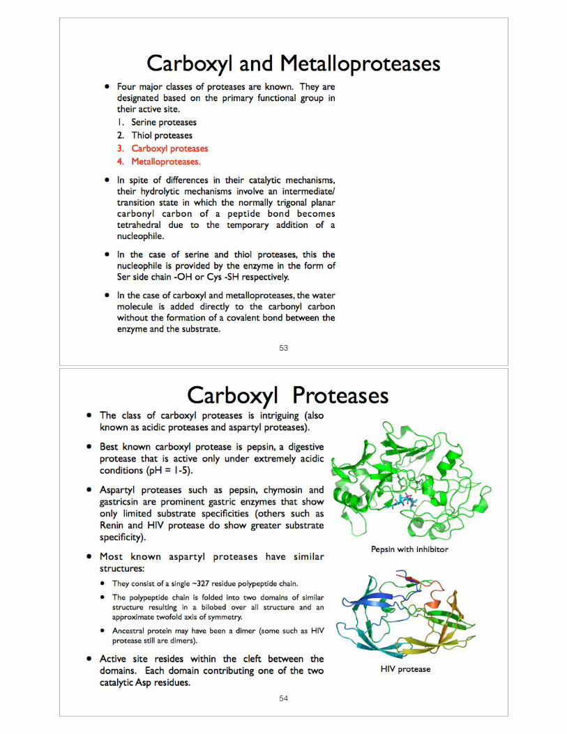
54
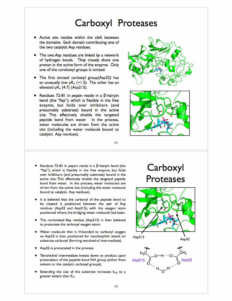
55
56
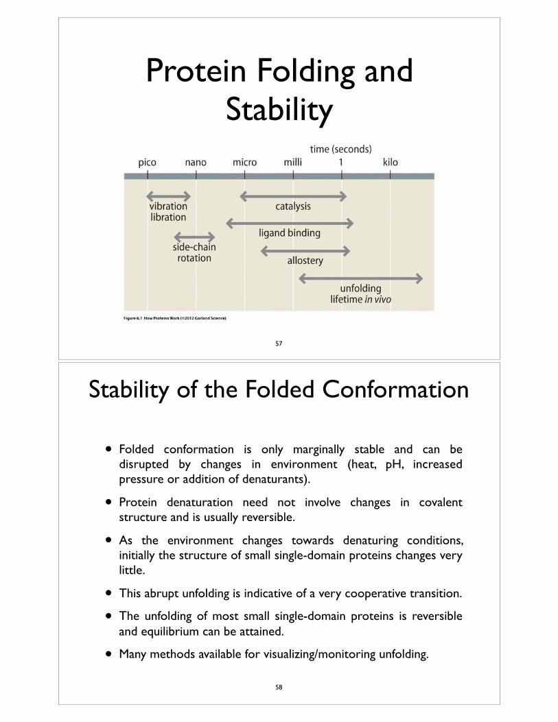
Protein Folding and Stability
57
Stability of the Folded Conformation
• Folded conformation is only marginally stable and can be disrupted by changes in environment (heat, pH, increased pressure or addition of denaturants).
• Protein denaturation need not involve changes in covalent structure and is usually reversible.
• As the environment changes towards denaturing conditions, initially the structure of small single-domain proteins changes very little.
• This abrupt unfolding is indicative of a very cooperative transition.
• The unfolding of most small single-domain proteins is reversible and equilibrium can be attained.
• Many methods available for visualizing/monitoring unfolding.
58
Structure1320
Figure 1. Design of Ala2Ile2-6
(a) An !-helical wheel diagram (looking down the long axis) of the heptadrepeat of Rop. The “a” (yellow) and “d” (red) residues form the hydrophobiccore and are the residues mutated in the repacked protein Ala2Ile2-6. Twolayers of the core are shown. Helices from protomer A are designated 1 and2, and the protomer B helices are labeled 1" and 2". Arrows indicate thedirection of the polypeptide chain from the N terminus to the C terminus.(b) Sequence alignment of Rop and Ala2Ile2-6 with the residue number placedabove every tenth residue. The “a” and “d” residues are colored to matchthe diagram. To create Ala2Ile2-6, residues in the “a” and “d” positions ofRop were changed to alanine and isoleucine, respectively. The outermostlayer at each end of the four-helix bundle, consisting of residues 5, 29, 31,and 56, were not changed. Residue 56, in the “e” position of the heptadrepeat, acts as a “d” residue by packing its side chain into the appropriatecore position. Figure 2. Thermodynamic Comparison of Rop and Ala2Ile2-6
(a) Thermal stability profile (#G versus T) and representative thermal denatur-ations (inset) of Rop (solid circles) and Ala2Ile2-6 (open circles).(b) Calculated values of #H (squares) and -T#S (circles) as a function oflost RNA binding activity, but which has enhanced, nativeliketemperature for Rop (solid) and Ala2Ile2-6 (open).thermal stability.
Results and Discussionchain volume and hydrophobicity, any structural and thermo-dynamic differences between Ala2Leu2-6 and Ala2Ile2-6 are,Design and Initial Characterization
Rop variants were generated by replacing the core residues therefore, primarily a consequence of the different side chainstereochemistries of leucine and isoleucine.(Figure 1a) of the heptad repeat with a regular pattern of hy-
drophobic amino acids. One of the first and most conservative The initial characterization of Ala2Ile2-6 demonstrated that itis highly helical with a circular dichroism (CD) spectrum similarmutants created was Ala2Leu2-6 [9], in which the middle six
layers of the hydrophobic core incorporate alanines in the “a” to that of wild-type Rop. Furthermore, Ala2Ile2-6 maintainednativelike thermodynamic properties illustrated, for example,positions and leucines in the “d” positions. Ala2Leu2-6 has
nativelike structural and thermodynamic properties, binds RNA by a cooperative and reversible thermal unfolding transition(Figure 2a, inset) accompanied by a large change in the heatwith wild-type affinity, and has a significantly higher melting
temperature than that of wild-type Rop. To investigate the capacity (Table 1). Ala2Ile2-6 was shown to be a dimer by bothsedimentation equilibrium centrifugation and multiangle laserimportance of side chain geometry in packing the core of Rop,
we created Ala2Ile2-6 with alanine in the “a” positions and iso- light scattering measurements (data not shown). However, theelectromobility shift assay for protein-RNA interaction [25]leucine in the “d” positions of the middle six layers of the core
(Figure 1b). Because isoleucine and leucine share similar side demonstrated that Ala2Ile2-6 had completely lost the ability to
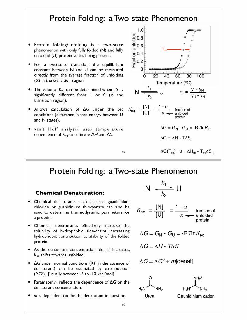
Protein Folding: a Two-state Phenomenon
• Protein folding/unfolding is a two-state phenomenon with only fully folded (N) and fully unfolded (U) protein states being present.
• For a two-state transition, the equilibrium constant between N and U can be measured directly from the average fraction of unfolding (α) in the transition region.
• The value of Keq can be determined when α is significantly different from 1 or 0 (in the transition region).
• Allows calculation of ΔG under the set conditions (difference in free energy between U and N states).
• van’t Hof f analys is : uses temperature dependence of Keq to estimate ΔH and ΔS.
Tm
Keq =[U][N] = 1 - α
α
ΔG = GN - GU = -RTlnKeq
N Uk1
k2
fraction of unfolded protein
ΔG = ΔH - TΔS
ΔG(Tm)= 0 = ΔHm - TmΔSm
α =yU - yN
y - yN
59
Protein Folding: a Two-state Phenomenon
Chemical Denaturation:• Chemical denaturants such as urea, guanidinium
chloride or guanidinium thiocyanate can also be used to determine thermodynamic parameters for a protein.
• Chemical denaturants effectively increase the solubility of hydrophobic side-chains, decreasing hydrophobic contribution to stability of the folded protein.
• As the denaturant concentration [denat] increases, Keq shifts towards unfolded.
• ΔG under normal conditions (RT in the absence of denaturant) can be estimated by extrapolation (ΔG0). [usually between -5 to -10 kcal/mol]
• Parameter m reflects the dependence of ΔG on the denaturant concentration.
• m is dependent on the the denaturant in question.
Keq =[U][N] = 1 - α
α
ΔG = GN - GU = -RTlnKeq
N U
ΔG = ΔG0 + m[denat]
k1
k2
fraction of unfolded protein
ΔG = ΔH - TΔS
NH2H2N
O
NH2H2N
NH2+
Urea Gaunidinium cation60
The Unfolded State
• Many proteins under strongly denaturing conditions have been shown to have properties consistent with random coil conformations.
• If interactions between different parts of the polypeptide are preferred over interactions with solvent, then the chain tends to be more compact and less disordered than expected for a random coil.
• While the physical properties of unfolded states produced under different unfolding conditions may differ, they are energetically indistinguishable.
• Difficult to characterize the unfolded state of a protein because many conformations are possible and may be populated.
• The molten-globule state: under certain conditions proteins have been known to demonstrate properties consistent with a molten globule state.
61
Mechanism of Protein Folding
Folding Pathways:
• How does a protein fold into its native conformation?
• A protein cannot randomly explore all of the conformational possibilities until it achieves its native conformation.✦ Levinthal Paradox: a 100 residue peptide sampling 1013 conformations per
second would take 1085 sec to fold. (Universe is estimated to ~20 billion years or ~6x10¹⁷ seconds old.)
• Therefore, proteins must employ an ordered pathway or set of pathways which ultimately allow the protein to achieve its native fold.
• There is the possibility that the observed folded state may not be the conformation with the lowest possible free energy, but is the most stable of the kinetically accessible conformations.
• Proteins fold to a significant degree within 1 millisecond.
62
Kinetic Analysis of Complex Reactions
Kinetics of Unfolding
• Protein unfolding is almost always observed to be an all or none process.
• Native protein represents a relatively conformationally homogeneous population, and unfolding generally proceeds with a single kinetic phase and a single rate constant. (no lag phase).
Kinetics of Refolding
• Kinetic complexity is a hallmark of protein folding. Starting with the conformational heterogeneity of the unfolded population.
• Heterogeneity includes cis-trans isomerization of peptide bonds (slow process).
• In an unfolded population with the native cis-trans isomers, refolding generally occurs with a single rate constant in spite of the conformational heterogeneity of the unfolded state.
63
Protein Folding Events
• Initial folding events (burst phase): [milliseconds]❖ For many small-single domain proteins, much of the secondary structure is
established.❖ Much of the driving force attributed to hydrophobic collapse. (hydrophobic
groups coalesce and expel water)
❖ Initial collapsed state is molten globular.❖ Side chains are extensively disordered.
• Intermediate folding events: [~5-1000 milliseconds]❖ Secondary structure stabilizes and native-like tertiary structure appears.
❖ Side chains are still mobile.
• Final folding events: [≤ several seconds]❖ Protein achieves native structure.
❖ Complex motions allow the protein to attain relatively rigid packing and hydrogen bonding.
❖ Remaining interior water molecules are expelled from the core.
64
Hierarchal Protein Folding
• The folding process begins with the formation of marginally stable local order/structure.
• These structure elements then interact (locally) to form intermediates of increasing complexity.
• Process continues ultimately yielding the native protein.
• Evidence supporting the premise of hierarchal protein folding:✤ Many peptide fragments excised from proteins will assume their native conformation.
✤ Observed folding intermediates are consistent with a hierarchal folding process.
✤ Helix boundaries are fixed by their primary sequence, not so much by 3-D interactions.
✤ Secondary structure can be predicted with reasonable accuracy, even when long-range interactions are not accounted for or are suppressed.
• Sequence information defining a specific fold is both distributed throughout the polypeptide chain and is highly overdetermined.
65
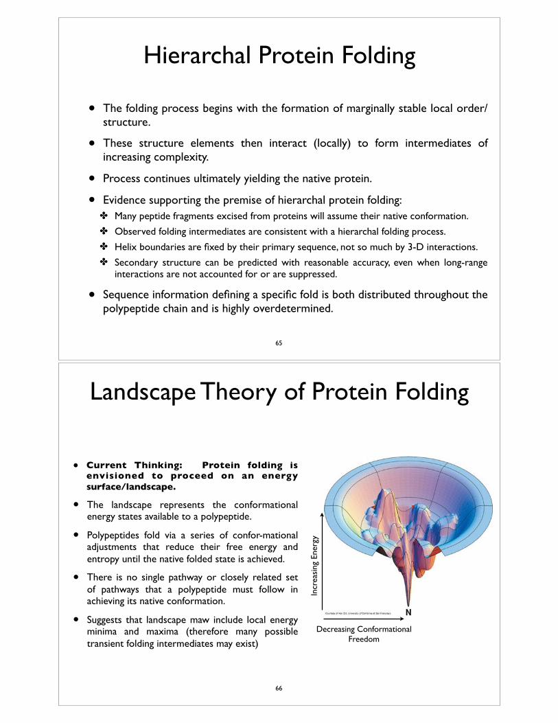
Landscape Theory of Protein Folding
• Current Thinking: Protein folding is envisioned to proceed on an energy surface/landscape.
• The landscape represents the conformational energy states available to a polypeptide.
• Polypeptides fold via a series of confor-mational adjustments that reduce their free energy and entropy until the native folded state is achieved.
• There is no single pathway or closely related set of pathways that a polypeptide must follow in achieving its native conformation.
• Suggests that landscape maw include local energy minima and maxima (therefore many possible transient folding intermediates may exist)
Incr
easi
ng E
nerg
y
Decreasing Conformational Freedom
66
Folding of Multidomain and Multimeric Proteins
• Large proteins may be composed of multiple domains or polypeptide chains.
• Independent domains unfold and refold like single-domain proteins, which can lead to complex unfolding curves for proteins. (in such cases, domains may unfold under different conditions)
• Can also be varying degrees of interaction between the domain. Interactions between domains can effect folding.
• Where the isolated domains are stable, folding of the intact multidomain protein appears to occur by initial folding of the domains, followed by association of the domains.
• Domain association is often the slowest step in the folding process. (domains may not be folded entirely correctly or because small adjustments are required for interaction between the domains.)
• When association is slow step, an intermediate can accumulate where domains are folded but impaired. May lead to intermolecular interactions and precipitation.
67
• Large proteins may be composed of multiple domains or polypeptide chains.
• Folding of oligomeric proteins has similar considerations because the polypeptide chains involved often incorporate multiple domains.
• Oligomerization necessitates specific interactions between the polypeptide monomers.
• Polypeptide monomers generally fold to nearly their final conformations before oligomeric association. The specific interactions likely requires that the polypeptide monomers have a folded conformation in order to provide the interaction sites.
• Rate-limiting step may be either intramolecular folding or associtation of the monomers.
• Final adjustments of the structure appears to have significant energy barrier (it is a relatively slow process)
Folding of Multidomain and Multimeric Proteins
68
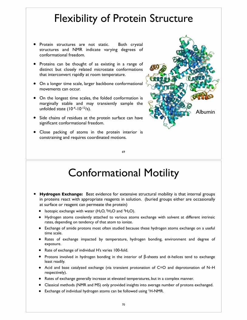
Flexibility of Protein Structure
• Protein structures are not static. Both crystal structures and NMR indicate varying degrees of conformational freedom.
• Proteins can be thought of as existing in a range of distinct but closely related microstate conformations that interconvert rapidly at room temperature.
• On a longer time scale, larger backbone conformational movements can occur.
• On the longest time scales, the folded conformation is marginally stable and may transiently sample the unfolded state (10-4-10-12/s).
• Side chains of residues at the protein surface can have significant conformational freedom.
• Close packing of atoms in the protein interior is constraining and requires coordinated motions.
Albumin
69
Conformational Motility
• Hydrogen Exchange: Best evidence for extensive structural mobility is that internal groups in proteins react with appropriate reagents in solution. (buried groups either are occasionally at surface or reagent can permeate the protein)
• Isotopic exchange with water (H2O, 2H2O and 3H2O).
• Hydrogen atoms covalently attached to various atoms exchange with solvent at different intrinsic rates, depending on tendency of that atom to ionize.
• Exchange of amide protons most often studied because these hydrogen atoms exchange on a useful time scale.
• Rates of exchange impacted by temperature, hydrogen bonding, environment and degree of exposure.
• Rate of exchange of individual H’s varies 100-fold.
• Protons involved in hydrogen bonding in the interior of β-sheets and α-helices tend to exchange least readily.
• Acid and base catalyzed exchange (via transient protonation of C=O and deprotonation of N–H respectively).
• Rates of exchange generally increase at elevated temperatures, but in a complex manner.
• Classical methods (NMR and MS) only provided insights into average number of protons exchanged.
• Exchange of individual hydrogen atoms can be followed using 1H-NMR.
70
Conformational Motility
Hydrogen exchange continued...
• While proteins may sample unfolded state, this is not likely responsible for the exchange of buried hydrogen atoms. (not all interior hydrogens exchange with same rate)
• Local unfolding or “breathing” is often used to explain the exchange of interior hydrogen atoms. The hypothetical open form is unstable and transient.
• Under most conditions, proteins demonstrate exchange rates consistent with kex<k-1 and rate = K1kex.
• Alternative explanation involves rare instances of diffusion of solvent into the interior sites in the protein. Supported by exchange in proteins in crystalline state.
• Both require some degree of backbone conformational flexibility.
• Available information suggests that different site in folded proteins likely exchange with solvent by a wide range of different processes, depending on the protein and conditions.
openfolded H-exchangedk1
k-1
kex k1
k-1K1 =
71
Fluorescent Quenching
• Fluorescence of aromatic groups is instantly quenched by close physical interaction with some small molecules such as O2, I- and acrylamide.
• Aromatic side chains are quenched by diffusion-controlled encounters with such molecules.
• Many internal groups are also quenched, only slightly less efficiently with O2.
• Charged and polar quenchers (I- and acrylamide) are less efficient and likely only act when the protein is in an open conformation.
• Detailed interpretations are complicated by energy transfer between fluorescent groups within the protein, by varied quantum yields and possible localization of quenchers to specific sites in the protein.
72
Rotations of Side Chains
• Side chains on the protein surface and terminal methyl groups of side chains in the interior tend to have mobilities comparable to those in unfolded proteins (rotating on 10-11-10-8 s time scale).
• Slower motion of interior groups masked by rotation of the entire protein molecule.
• Can study motion of aromatic rings by 1H-NMR.
• Most proteins have Phe and Tyr side chains that give average spectra, suggesting that they rotate on the order of 180° flip 104/s even when buried.
• Buried Trp and His residues do not appear to flip.
73