henry a. hanni, karl schrnetzer, and heinz-jiirgen
TRANSCRIPT

By Henry A. Hanni, Karl Schrnetzer, and Heinz-Jiirgen Bernhardt
Greek manufacturer 1. a) A. Douros intro- duced a new flux synthetic ruby in early 1993. Grown bv spontaneous nucleation in a , A
lead-based solvent, the Douros synthetic ruby occurs as rhombohedra1 single crystals and twinned tabular crystals, as well as in clus- ters. The tabular crystals typically have intense red cores that gradually decrease in sat~uation to a near-colorless outermost layer; the rhombohedra1 crystals have a deep red body and a thin near-colorless laver on the rhombohedra1 faces, with umbrella-like growth patterns in some areas. Some samples contain distinctive inclusions of yellow resid- ual flux with spherical bubbles. Chemically, the crystals are heavily zoned, and EDXRF analyses revealed variable trace amounts of Ti, Fe, Cr, and Ga, as well as some Pb, SEM- EDS identified the flux particles as lead bear- ing. Microprobe analyses showed high Cr val- ues in the crystal cores; in certain growth zones, Fe replaces Cr in the outermost layers. If inclusions or typical growth structures are not present, chemical composition appears to be the best means of separating these DOLUOS synthetic rubies from natural rubies.
ABOUT THE AUTHORS Dr. Hanni is director of SSEF Swiss Gemological Institute, Zurich, and associate professor of gemol- ogy at Base1 University, Switzerland. Dr. Schrnetzer is a research scientist residing in Petershausen near Munich, Germany. Dr. Bernhardt is a research sci- entist at the Institute for Mineralogy of Ruhr- University, Bochum, Germany. Acknowledgements appear at end of article. Gems & Gemology, Vol. 30, No. 2, pp. 72-86. 0 7994 Gem&@ Institute of America
ecently, a new type of flux-grown synthetic ruby entered the gem market (figure 1). Manufactured by
J. & A. Douros Created Gems in Piraeus, Greece, the Douros synthetic ruby poses new challenges for gemolo- gists. Like the Ramaura flux-grown synthetic rubies intro- duced in 1982 (Bosshart, 1983; Kane, 1983), stones faceted from the Douros laboratory-grown crystals may create con- siderable difficulties for identification. Standard gemological tests are inadequate for clean or only slightly included sam- ples. Even with advanced tests such as U.V.-visible spec- trophotometry, EDXRF chemical analysis, or identification of growth planes, the present material may cause difficul- ties in identification (see, e.g., Hanni and Bosshart, 1993; Hanni and Schmetzer, 1993; and Smith and Bosshart, 1993).
This article reports on our examination of several crys- tals and faceted samples of the Douros material. To help establish criteria by which the Douros synthetic rubies can be separated from their natural counterparts, we will describe in detail the mineralogical, gemological, and chem- ical characteristics of this new flux-grown synthetic ruby.
PRODUCTION Brothers John and Angelos Douros run a small family busi- ness that manufactures synthetic crystals. A physicist and an electrical engineer, respectively, they previously special- ized in refining precious metals. With furnaces they built themselves, the Douros brothers use controlled sponta- neous nucleation and slow cooling techniques to produce synthetic rubies by unseeded flux growth (J , Douros, pers. comm., 1993).
After some years of experimentation, in January 1993 they presented the material to the public for the first time, at the Athens Jewellery Fair. Simultaneously, they reported on their new synthetic ruby in the Greek jewelry magazine Chrysotechni (Douros and Douros, 1993) .
At present, the Douros brothers use two furnaces in
72 Synthetic Rubies by Douros GEMS & GEMOLOGY Summer 1994

Synthetic Rubies by Douros
Figure 1. This 44.74-ct rhombohedra1 crystal and the accompanying
1 cut stones (from left to r igh t4 .93 , 2.55, 2.14, and 3.51 ct) are represen- tative of some of the Douros flux-grown syn- thetic rubies that have been produced to date.
, Samples courtesy of John
GEMS & GEMOLOGY Summer 1994
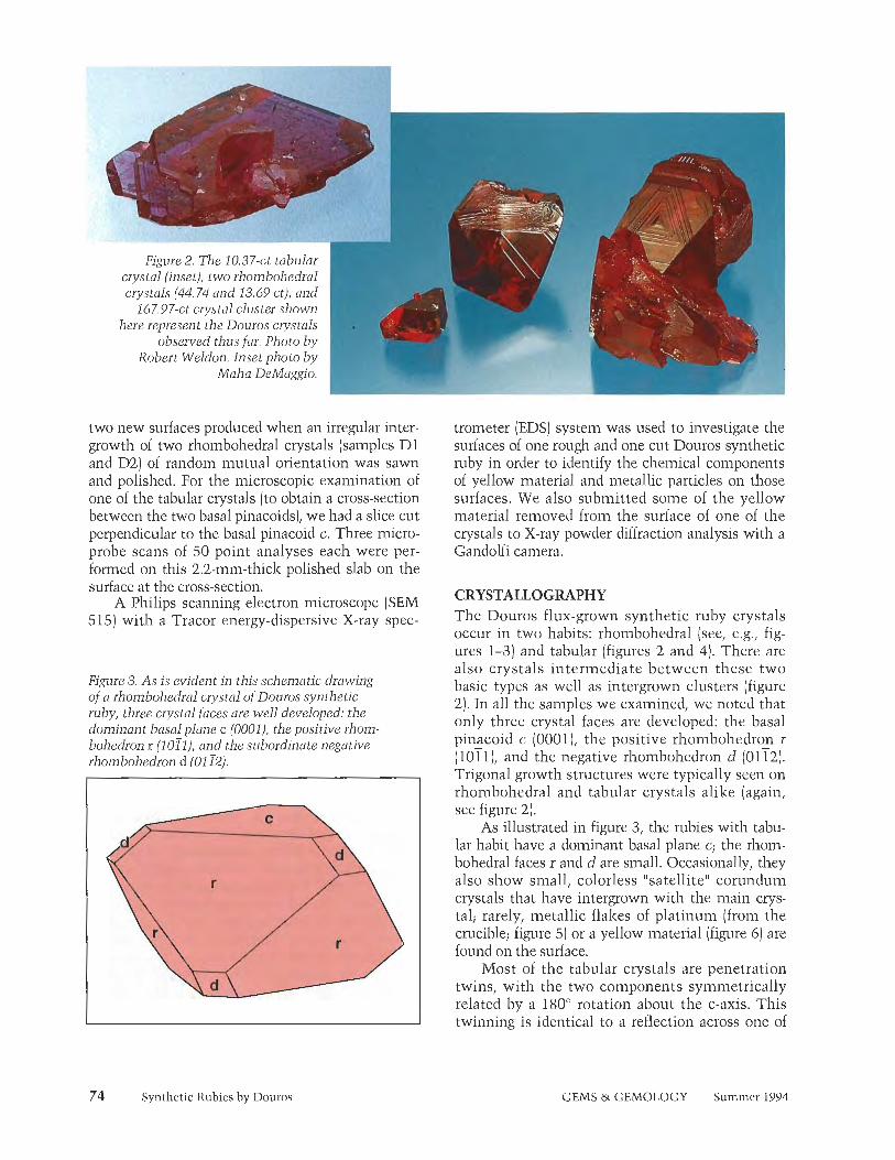
crystals (44.74 and 13.69 ct), anc 167.97-ct crystal cluster s h o w
here represent the Douros crystals rn observed thus far. Photo bj
Robert Weldon. Inset photo bj Maha DeMaggio. rn
two new surfaces produced when an irregular inter- growth of two rhombohedral crystals (samples Dl and D2) of random mutual orientation was sawn and polished. For the microscopic examination of one of the tabular crystals [to obtain a cross-section between the two basal pinacoids), we had a slice cut perpendicular to the basal pinacoid c. Three micro- probe scans of 50 point analyses each were per- formed on this 2.2-mm-thick polished slab on the surface at the cross-section.
A Philips scanning electron microscope (SEM 515) with a Tracer energy-dispersive X-ray spec-
Figure 3. As is evident in this schematic drawing of a rhombohedral crystal of Douros synthetic ruby, three crystal faces are well developed: the dominant basal plane c (0001), the positive rhom- bohedron r (1011). and the subordinate negative rhombohedron d (01 12).
Synthetic Rubies by Douros
trometer (EDS) system was used to investigate the surfaces of one rough and one cut Douros synthetic ruby in order to identify the chemical components of yellow material and metallic particles on those surfaces. We also submitted some of the yellow material removed from the surface of one of the crystals to X-ray powder diffraction analysis with a Gandolfi camera.
CRYSTALLOGRAPHY The Douros flux-grown synthetic ruby crystals occur in two habits: rhombohedral (see, e.g., fig- ures 1-3) and tabular (figures 2 and 4). There are also crystals intermediate between these two basic types as well as intergrown clusters (figure 2). In all the samples we examined, we noted that only three crystal faces are developed: the basal pinacoid c (0001], the positive rhombohedron r (1011), and the negative rhombohedron d (0112). Trigonal growth structures were typically seen on rhombohedral and tabular crystals alike (again, see figure 21.
As illustrated in figure 3, the rubies with tabu- lar habit have a dominant basal plane c; the rhom- bohedral faces r and d are small. Occasionally, they also show small, colorless "satellite" corundum crystals that have intergrown with the main crys- tal; rarely, metallic flakes of platinum (from the crucible; figure 5) or a yellow material (figure 6) are found on the surface.
Most of the tabular crystals are penetration twins, with the two components symmetrically related by a 180Â rotation about the c-axis. This twinning is identical to a reflection across one of
GEMS & GEMOLOGY Summer 1994

the first-order hexagonal prism faces (1010) or across the basal pinacoid (0001). The two coinpo- nents of the twinned individuals are in contact along four twin boundaries, which are oriented par- allel to the twin planes (1010) and (0001), respec- tively (figure 4; see, e.g., Schmetzer et al., 1994).
The rhombohedral crystals are more equidi- mensional and thus better suited for cutting. They are basically formed by dominant basal and rhoin- bohedral faces, c and r, with somewhat smaller d faces (again, see figure 3). We did not see prism faces on any of our samples, but we did see small hexagonal dipyrainids n (2243) on one crystal. We did not observe twinning in any of the rhombohe- dral crystals.
These two habits of Douros synthetic rubies are similar to those described for Ramaura flux- grown synthetic rubies (Kane, 1983; Schmetzer, 1986a and b]. The penetration twinning seen in the Douros synthetic rubies is identical to that com- monly seen in Ramaura synthetic rubies (as described in Schmetzer et al., 1994), but different from twinning in other flux-grown synthetic rubies, such as those produced by Chathain Created Gems. However, whereas we observed twinning only in tabular Douros crystals, most twinned Ramaura synthetic ruby crystals observed up to now are of rhombohedral habit.
Figure 4. This schematic drawing of a tabular Douros synthetic ruby crystal shows the dominant c planes and the subordinate r and d faces. The sample illustrated is actually a penetration twin, which is frequently encountered in this material. The second crystal i s denoted by the darker color and its relative faces marked c', r', and d'.
COLOR AND COLOR ZONING The samples ranged from saturated red to purplish red and reddish purple (see, e.g., figures 1 and 2), depending on the amounts of chromium, titanium, and iron present in each. The different concentra- tions of color-causing trace elements also produced geometrically bounded color zoning within the crys- tals. In the tabular crystals only, we noted: (1) near- colorless to light red outennost zones confined to r and d faces (figure 7); (2) near-colorless to light red triangular growth sectors confined to twin bound- aries (figure 8); and (3) purple to bluish purple color bands running parallel to both rhombohedral faces r and d (figure 9). In both the rhombohedral and tabu-
Figure 5. A small satellite crystal and a shiny grain of platinum (from the crucible) can be seen on the surface (basal face c) of this tabular crys- tal. The crazed orange area is enclosed flux that lies below the surface. Magnified 50x. Figure 6. On the surfaces of some of the crystals,
we saw pits filled with a yellow polycrystalline 1 material, Magnified 6x.
Synthetic Rubies by Douros GEMS & GEMOLOGY Summer 1994 75
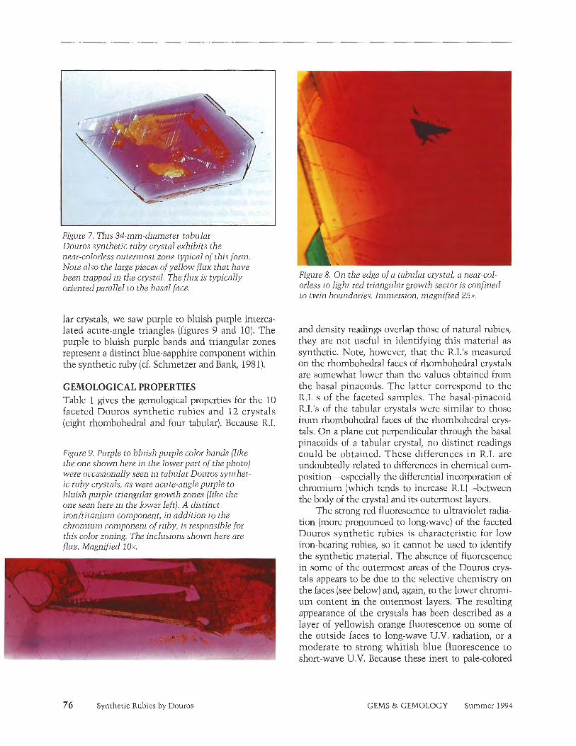
Figure 7. This 34-mm-diameter tabular Douros synthetic ruby crystal exhibits the near-colorless outermost zone typical of this form, Note also the large pieces of yellow flux that have been trapped in the crystal. The flux is typically oriented parallel to the basal face.
lar crystals, we saw purple to bluish purple interca- lated acute-angle triangles (figures 9 and 10). The purple to bluish purple bands and triangular zones represent a distinct blue-sapphire component within the synthetic ruby (cf. Schmetzer and Bank, 1981).
GEMOLOGICAL PROPERTIES Table 1 gives the gemological properties for the 10 faceted Douros synthetic rubies and 12 crystals (eight rhombohedral and four tabular). Because R.I.
Figure 9. Purple to bluish purple color bands (like the one shown here in the lower part of the photo) were occasionally seen in tabular Douros synthet- ic ruby crystals, as were acute-angle purple to bluish purple triangular growth zones (like the one seen here in the lower left). A distinct ironltitanium component, in addition to the chromium component of ruby, is responsible for this color zoning. The inclusions shown here are flux. Magnified l ox .
76 Synthetic Rubies by Douros
Figure 8. On the edge of a tabular crystal, a near-col- orless to light red triangular growth sector is confined to twin boundaries. Immersion, magnified 25x,
and density readings overlap those of natural rubies, they are not useful in identifying this material as synthetic. Note, however, that the R.I.'s measured on the rhombohedral faces of rhombohedral crystals are somewhat lower than the values obtained from the basal pinacoids. The latter correspond to the R.I.'s of the faceted samples. The basal-pinacoid R.I.'s of the tabular crystals were similar to those from rhombohedral faces of the rhombohedral crys- tals. On a plane cut perpendicular through the basal pinacoids of a tabular crystal, no distinct readings could be obtained. These differences in R.I. are undoubtedly related to differences in chemical com- position-especially the differential incorporation of chromium (which tends to increase R.1.J-between the body of the crystal and its outermost layers.
The strong red fluorescence to ultraviolet radia- tion (more pronounced to long-wave) of the faceted Douros synthetic rubies is characteristic for low iron-bearing rubies, so it cannot be used to identify the synthetic material. The absence of fluorescence in some of the outermost areas of the Douros crys- tals appears to be due to the selective chemistry on the faces (see below) and, again, to the lower chromi- um content in the outermost layers. The resulting appearance of the crystals has been described as a layer of yellowish orange fluorescence on some of the outside faces to long-wave U.V. radiation, or a moderate to strong whitish blue fluorescence to short-wave U.V. Because these inert to pale-colored
GEMS & GEMOLOGY Summer 1994

portions of the crystals are generally removed during cutting, the fluorescence of the faceted Douros syn- thetic rubies is typically a uniform red.
MICROSCOPIC CHARACTERISTICS Structural Properties (Growth Features and Twinning). The internal growth features of both the rough and the faceted Douros samples correspond to the external morphology of the crystals. We iden- tified internal growth planes parallel to all three macroscopically observed crystal faces c, r, and d. Although similar growth planes are also found in Ramaura and Chatham flux-grown synthetic rubies (Schmetzer, 1987), there are three types of growth patterns that appear to be most characteristic of rhombohedral Douros synthetic rubies (table 2).
In most of the rhombohedra1 crystals, we recog- nized a growth pattern that is evident as areas of different colors with sharply defined boundaries. The main body color between the basal planes is intense red, whereas the thin outermost layers con- fined to the r and d faces are near-colorless to light red (figure 11). These outer layers represent the lat- est growth stage.
All of the rhombohedral crystals showed faces parallel to the negative rhombohedron d. Such faces were prominent (although smaller than c and r) on the actual surface of the crystals, which represents the latest growth period. In earlier stages of growth i.e., more toward the center of the crystal) the d
Figure 10. Acute-angle color zoning was also seen in rhombohedra1 Doums synthetic ruby crystals. Compare the appearance of the zone in this sam- ple with that in the tabular Douros crystal in fig- ure 9. Again, the blvish purple color is due to (Fe,Ti)-rich zones within the ruby crystal. Immersion, magnified 40x.
faces were smaller. In the earliest growth stage, in the center of the crystal, growth parallel to d was absent, and the surface of the crystal at that stage was defined just by r and c faces. When a rhombo- hedral Douros synthetic ruby crystal was viewed with immersion in the microscope, in most cases the area confined to d growth showed a distinct, curved, umbrella-like outline, because this part of the crystal has a higher saturation of red compared to neighboring zones (figure 12). In some cases, the
TABLE 1. Gemological properties of Douros synthetic rubies.3
Property Rhombohedra1 crystals Faceted samples Tabular crystals
Density(g/cma) 3.993-4.010 Refractive indices Basal pinacoid c:
n,, 1.771 - 1.773 n, 1.763 - 1.765 Pos. rhombohedron r: no 1.768 - 1.770 no 1.760- 1.762
Pleochroism llc 1c
Fluorescence LWUV
swuv
Yellowish red to red Purplish red
Intense orangy red, inert in the surface layer confined to rand d Moderate red, inert in the surface layer confined to r and d
3.997 - 4.01 5 4.023 - 4.029 Basal pinacoid c:
no 1.772 - 1.774 no 1.768 - 1.771 ne 1.762 - 1.764 nn 1.760 - 1.763
Plane cut and polished perpendicular to c: no distinct reading
Yellowish red to red Yellowish red to red Purplish red Purplish red
Intense red
Moderate red
Intense orangy red, inert in the surface layer confined to c , r, and d Moderate red, inert in the surface layer confined to c, r, and d
"As recorded for eight rhombohedral crystals, 10 faceted samples, and four tabular crystals.
Synthetic Rubies by Douros GEMS & GEMOLOGY Summer 1994 77

Figure 11. The thin outermost layers confined to Figure 12. The growth sector formed by cl-face the rand d faces that form the sharp edge of this growth (at the bottom of the picture) in this rhotnbohedrcil crystal are near-colorless to light rhombohedra1 crystal is defined by a distinc- red. Note that thew is no colorless layer on the Live umbrella-like boundary toward the neigh- basal faces (here, upper right and upper left). boring r and r' face growth areas. Immersion, Immersion, magnified 25x. magnified 40x.
d-face growth zones were confined to the outer bluish purple triangles that lie in the red core areas of the crystal; the intermediate and central (again, see figure 10). These areas undoubtedly con- areas showed only growth parallel to c and r faces tain a distinct blue sapphire (Fe, Ti) component in (figure 13). addition to the Cr content responsible for the ruby
Also in the rhombohedral crystals, as noted ear- coloration. lier, we occasionally saw acute-angle purple to In the tabular crystals, growth patterns includ-
TABLE 2. Crystallography, structural properties, and color zoning in Douros synthetic ruby crystals."
Property Rhombohedra1 crystals
Faces Basal pinacoid c (0001) Dominant Pos. rhombohedron r (1 01 1) Dominant Neg. rhombohedron d (01 12) Subordinate Hexagonal dipyramid (2243) Extremely small (in one sample)
Twinning Not observed Growth patterns, color, color zoning c faces
r faces
d faces
Growth patterns present from the center to the surface; red; no color zoning, no boundary
Growth patterns present from the center to the surface; red; no color zoning from the center to a distinct boundary near the surface, with the outermost zone almost colorless
Growth patterns not present in the center, but begin to develop toward the surface; darker red (umbrella-type) growth structure; no color zoning from the begin- ning to a distinct boundary near the surface, with the outermost zone almost colorless
Undetermined faces Intercalated acute-angle purple to bluish purple triangles present
Â¥'A observed in eight d-iombohedral and four tabular crystals examined.
Tabular crystals
Dominant Subordinate Subordinate Not observed Penetration twinning
Growth patterns present from the center to the surface: red in the center, continuous decrease in saturation to- ward the surface, and alrnosl colorless in the outermost layer Growth patterns present from the center to the surface; red in the center, continuous decrease in saturation toward the surface, and almost colorless in the outermost zone Growth patterns present from the center to the surface; dark red in the center, with a continuous decrease in sat- uration toward the surface; almost colorless in the outermost zone
Intercalated acute-angle purple to bluish purple triangles present
78 Synthetic Rubies by Douros GEMS & GEMOLOGY Summer 1994

ed both regular and irregular distributions of color (again, see table 2). As a rule, there was no color zoning in the cores, and the outermost areas tended to be light red or near-colorless (again, see figure 7). The slice cut parallel to the c-axis of a twinned tab- ular crystal (on which microprobe analysis was also done) exhibits a rather complex growth pattern (fig- ure 14). From the upper to the lower surface (i.e., from the c to the -c face), growth zones parallel to c, r, d, and -c can be seen. Both of the growth zones parallel to c and -c are colorless or light red on the surface. The color saturation increases toward the center of the crystal, which is saturated red. Growth areas confined to faces r and d are intense red in the center of the crystal, but they decrease in saturation in a direction parallel to the length of the slab, that is, toward the surfaces formed by the r and d faces. The outer zones of these areas are light red, merging to near-colorless next to the surface of the crystal. In the slice examined, the d growth zones are a more intense red than the adjacent r zones. This feature was also found in the rhombo- hedral crystals examined.
Purple -to bluish purple acute-angle zones were also observed in some of the tabular crystals; bands of similar color were seen in planes parallel to the rhombohedra1 faces (again, see figure 9). These rep- resent areas rich in Fe and Ti.
Growth patterns of penetration twins on tabu-
Figure 13. This rhombohedral Douros synthetic ruby crystal also revealed an umbrella-like growth sector confined to d (intense red) as well as r and r' (light red), but the central part of the crystal shows only r and r' growth. Immersion,
, - ignified 50x.
Synthetic Rubies by Douros
Figure 14. As illustrated in the line drawing, this slice from a tabular crystal cut parallel to the c- axis shows the large c (upper) and -c (lower) faces, as well as the r and'd faces that form the edge of the crystal. Internal growth zoning is accentuated here due to differences in trace-element chemistry. The darkest red zone is confined to the d-growth area. Immersion, magnified 25x. The positions of the two paths of micropmbe point analyses (A and C) are shown in the schematic drawing. (Path B was located between paths A and C.)
lar crystals may be extremely complex in areas con- fined to one of the four boundaries of the synthetic ruby. In general, the differences are so subtle that these patterns are not useful to discriminate between natural and synthetic rubies. Therefore, synthetic stones faceted from these areas cannot be separated from natural ruby on the basis of growth structures alone.
We did not see colorless outer zones in any faceted samples, which indicates that this character- istic growth feature is removed during cutting. In two or three faceted samples, we saw part of the umbrella-type growth pattern, which indicates that they had been cut from crystals with rhombohedral habit. In one case, we found an irregularly outlined d growth zone in a "distorted" umbrella pattern (figure 15). Note that these zones become smaller toward the interior of the crystal. Therefore, it is possible that some Douros synthetic rubies cut from the cores of rhombohedral crystals might also lack any distinctive structural patterns. See Schmetzer (1986a
GEMS & GEMOLOGY Summer 1994

Figure 15. Structural characteristics are not always easy to recognize. Note this "distorted" umbrella pattern found in a faceted Douros syn- thetic ruby. These peculiar growth structures, with r, d, and r ' sectors, were observed in immer- sion at 50x magnification.
and b) and Kiefert and Schmetzer (1991) for more on the method to identify growth planes.
Inclusions. Although many of the faceted samples appeared to be free of distinctive inclusions even at 40x magnification, some showed evidence of resid- ual flux similar to that seen in other flux-grown synthetic rubies. Such inclusions may occur as larg- er, individual pieces of flux or as "veils" formed by many tiny droplets of flux. The former actually consist of coarse, rounded to elongated cavities that have been filled with a typically yellow substance and contain one or more voids or bubbles. Note that, in small amounts, the flux appears near-color- less; when present in larger volumes, the flux not only looks yellow but also reveals a distinctive crazed appearance and forms a mosaic-like pattern (figure 16). Occasionally, the flux inclusions are ori- ented parallel to the basal pinacoids, especially in twinned tabular crystals. In rhombohedral crystals, we have seen flat flux-containing inclusions orient- ed parallel to the rhombohedral faces.
Synthetic Rubies by Douros
Figure 16. These pieces of yellow flux seen near the surface of a Douros synthetic ruby crystal are actually crazed residual fillings. Note the mosaic- like pattern of the flux, as well as the rounded voids or bubbles. Magnified 25x.
We saw rounded bubbles in nearly all of these flux inclusions (figure 17). Burch (1984) described similar two-phase inclusions for Kashan synthetic rubies. Most coarse flux is, as a rule, encountered near the surface of the crystals; that is, i t was enclosed during the latest growth period.
The veil type of flux inclusion seen in the Douros synthetic rubies we examined (figure 18) looks very similar to patterns seen in heat- (and 'borax-") treated natural ruby, which now represent most of the natural rubies encountered in the trade. Some of the flux particles are transparent, usually containing a bubble (figure 19).
Figure 17, This large piece of flux in a Douros syn- thetic ruby has a distinctive mosaic pattern and contains two bubbles. The fine lines-or "craz- ing"--in the solid part of the inclusion help distin- guish such inclusions from two-phase inclusions that might be seen in a natural ruby. Immersion, magnified 25x.
GEMS & GEMOLOGY Summer 1994

For the most part, the Douros faceted synthetic rubies we examined were very clean, only slight11 included, or the inclusions present resembled those seen in natural rubies (figure 20). They did not have platinum platelets or blades.
Optical Spectroscopy. The absorption spectra wt observed with gemological desk-model spectro scopes do not differ from those of natural or syn thetic rubies that have no (or little) iron content That is, the Douros synthetic rubies show the well known absorption lines in the red and blue regions of the spectrum, as well as the typical red fluores- cence lines due to chromium.
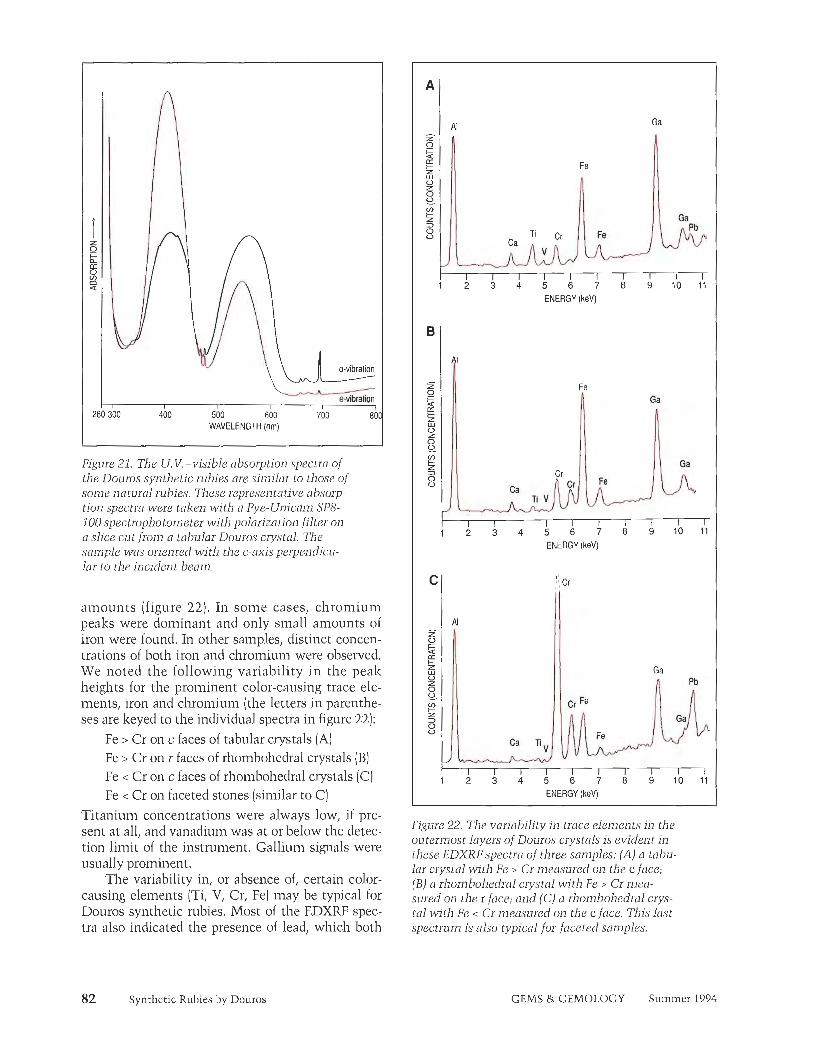
The spectrophotometer results in the ultravio- let-visible range are also characteristic for rubies with Cr-only spectra (figure 21). Absorption inini- ma for a typical sample lie at 328 and 471 nm for the o-vibration and at 328 and 481 nin for the e- vibration. Absorption maxima (i.e., broad bands) are centered at 410 and 560 nm for the o-vibration and 400 and 545 nm for the e-vibration. General absorp- tion in the ultraviolet region is at 290 (el and 280 nm (o), respectively. There were no significant dif- ferences in spectra among the various crystals or faceted samples.
When we calculated the spectral characteristics as proposed by Bosshart (1982), we determined typi- cal values of lo and ^./W as 32816.5 for the o-vibra- tion and 32615.8 for the e-vibration. These values overlap those for both Knischlza synthetic rubies
Figure 18. This veil-like inclusion in a faceted Douros synthetic ruby looks very similar to the networks of interconnecting channels seen in heat- (and "borax-") treated natural rubies, Magnified 5 0 x .
Figure 19. These small flux inclusions, in parallel alignment, are transparent. Note that each con- tains a prominent bubble, which appears in high relief against the flux. The flux itself shows low relief against the ruby, which indicates that its refractive indexis close to that of the ruby. Magnified 50x.
and natural rubies from Mogolz. The minor spread of the plotting points within the Douros material is probably due to'the trace-element variations encountered among different samples. Therefore, for the material examined so far, this method will not provide adequate distinction from natural rubies.
Chemistry. The EDXRF spectra showed the pres- ence of chromium, titanium, and iron in varying
Figure 20. The presence of a fissure composed of tiny droplets, like that seen in this faceted Douros synthetic ruby, is of no help to a gemologist work- ing only with a microscope. Such a pattern could also be seen in a natural ruby. Magnified 50x.
Synthetic Rubies by Douros GEMS & GEMOLOGY Summer 1994
A
a-vibration 1 I I f
280 300 1
400 500 600 700 80 WAVELENGTH (nm)
Figure 21. The U. V.-visible absorption spectra of the Douros synthetic rubies are similar to those of some natural rubies. These representative absorp- tion spectra were taken with a Pye-Unicam SP8- 700 spectrophotometer with polarization filter on a slice cut from a tab~tlar Douros crystal. The sample was oriented with the c-axis perpendicu- lar to the incident beam.
amounts (figure 22). In some cases, chromium peaks were dominant and only small amounts of iron were found. In other samples, distinct concen- trations of both iron and chromium were observed. We noted the following variability in the peak heights for the prominent color-causing trace ele- ments, iron and chromium (the letters in parenthe- ses are keyed to the individual spectra in figure 22):
Fe > Cr on c faces of tabular crystals (A) Fe > Cr on r faces of rhombohedra1 crystals (B) Fe < Cr on c faces of rhombohedra1 crystals (C) Fe < Cr on faceted stones (similar to C)
Titanium concentrations were always low, if pre- sent at all, and vanadium was at or below the detec- tion limit of the instrument. Gallium signals were usually prominent.
The variability in, or absence of, certain color- causing elements (Ti, V, Cr, Fe) may be typical for Douros synthetic rubies. Most of the EDXRF spec- tra also indicated the presence of lead, which both
Synthetic Rubies by Douros
ENERGY (keV)
I I I I I I I I I I 1 2 3 4 5 6 7 8 9 1 0 1 1
ENERGY (keV)
r I I I I I I I I I 1 2 3 4 5 6 7 8 9 1 0 1 1
ENERGY (keV)
Figure 22. The variability in trace elements in the outermost layers of Douros crystals is evident in these EDXRFspectra of three san~ples: (A) a tabu- lar crystal with Fe > Cr measured on the c face; (B) a rhombohedra1 crystal with Fe > Cr mea- sured on the r face; and (C) a rhombohedral crys- la1 with Fe < Cr measured on the c face. This last spectrum is also typical for faceted samples.
GEMS & GEMOLOGY Summer 1994

SEM-EDS and X-ray diffraction analyses (see below) showed to be a component of the flux used in the growth process. Even in those cases where flux inclusions cannot be seen with high magnification, EDXRF analysis sometimes reveals the presence of Pb peaks as evidence of s~~binicroscopic particles of flux.
EDXRF analysis did not reveal traces of either platinum or iridium (from the crucible) or of lan- thanum or bismuth. The latter are typical compo- nents of the flux used to grow Rainaura synthetic rubies (Schmetzer, 1986a; Sclimetzer et al., 1994).
The quantitative data provided by the inicro- probe analyses (see Dunn, 1977) revealed only a limited chemical variation within each of five of the six rhombohedra1 rubies analyzed (three crys- tals and three faceted samples; table 3). The faceted samples (A, B, and C), as well as the intergrown crystals (D l and D2), showed distinctly more chromiun~ than iron. In contrast, iron was found to surpass chron~ium (present in very small amounts) on the rhombohedra1 face r of rough sample El which was a typical "ruby" red color and had an ordinary chromium absorption spectrum. This indi- cates a selective compositional zoning with respect to different crystal faces, as well as between late- stage growth layers and the cores, in the rhonibohe- dral crystals. It is consistent with the color zoning observed in the crystals.
Sample D l showed a distinct zoning with respect to chromium, in the form of a composition- al discontinuity between the two parts of the crys- tal. We recorded C r f i values between 0.65 and
0.55 wt.% in the main part, and only 0.18 to 0.10 wt.% in the narrow outermost layer. There was no con~parable zoning for iron. This zoning of chromi- urn is consistent with the observation of dark red cores and lighter red or nearly colorless rims in the rhombohedra1 crystals (figure 11). Since the surface layers are typically thin, they are usually removed during cutting.
The results of microprobe analysis of the slice cut from a tabular crystal are given in table 4. The central part of the crystal (again, see figure 14) con- sists of two areas of different chromium content. In both areas, which are designated "upper core" and lower core" in figure 14 and table 4, iron content is low. However, we found no distinct pattern to the distribution of iron and cl~romium within either core. In contrast, we measured a high variation in chromiun~ and iron contents in the upper and lower surface layers: In both, chroniium progressively decreased-and iron progressively increased--from the core side to the surface side. A s~ibstit~itional exchange of Cr by Fe in these growth zones is evi- dent. We could not detect a systematic variation of titanium in die same scans. We also found that the chromium content decreases from the center (scan A) to the outer zone (scan C) of the crystal (table 4). The data obtained from scan B are intermediate between those of scans A and C.
Scanning Electron Microscopy and X-Ray Diffraction Analysis. SEM-EDS analysis of the residual yellow flux exposed at the surface of some faceted samples showed characteristic X-ray fluo-
TABLE 3. Electron microprobe analyses of five Douros synthetic rubies.3
Oxide Faceted Faceted Faceted Inlergrov~lh ol two Rhornbohedral (4.21 cl) (1.31 ct) (1.18~1) rhombohedra1 crystals (3.48 ct) crystal
(7.57 ct) Crystal D l Crystal D2 rlace
Core Surface layer Core
A1203 97.23-98.04 98.12-98.63 97.56-98.29 98.12-98.42 98.68-98.08 97.83-98.64 97.95-100.29 TiO, 0.00- 0.03 0.01- 0.05 0.00- 0.03 0.00- 0.02 0.00- 0.02 0.00- 0.02 0.01- 0.07 C r A 0.79- 0.96 0.60- 0.70 0.77- 0.99 0.55- 0.65 0.10- 0.18 0.47- 075 0.01- 0.04 Fe203 0.03- 0.07 0.04- 0.09 0.09- 0.16 0.03- 0.07 0.05- 0.08 0.03- 0.08 0.13- 0.34 MnO 0.00- 0.02 0.00- 0.02 0.00- 0.01 0.00- 0.02 0.00- 0.01 0.00- 0.02 0.00- 0.01
Â¥'Oxide in vit. %, ranges 01 30point analyses performed on each sample. Vanadium was belo~i lhe detection limil; gallium was not measured.
Synthetic Rubies by Douros GEMS & GEMOLOGY Summer 1994 83

rescence lines for lead (figure 23). A silvery metal grain on the surface of one crystal (again, see figure 5) proved to be platinum, probably from the cru- cible.
An X-ray powder diffraction pattern was pre- pared from some of the yellow material removed from the surface of one of the crystals (see, e.g., fig- ure 6). The pattern revealed that the material was lead nitrate, PbN03, which undoubtedly formed from the lead-bearing flux on the surface of the crystal when it was separated from the flux in nitric acid, HN03.
DISCUSSION The present investigation indicates that the new Douros synthetic rubies occur in essentially two types of crystals-(1) rhombohedral and (2) tabu- lar-which differ in habit, structural properties, and compositional characteristics. These two types probably result from the different temperature con- ditions that occur during the slow cooling that is part of the crystallization process from a flux. The ideal early growth conditions favor the formation of a rhombohedral crystal habit with c and r faces. When supersaturation decreases, and when the temperature starts to drop, new physical and chem- ical conditions favor the formation of d faces; the
development of tabular crystals seems to reflect these later formation conditions. Also, at the end of the growth period, there is less of the dominant color-causing element chromium and more iron, which leads to the formation of near-colorless sur- face layers.
EDXRF results support this scenario. Spectra taken on the basal plane of tabular Douros synthetic ruby crystals show small chromium pealzs and high iron pealzs. This agrees with the microprobe analy- ses talzen on a cross-section, as shown in table 4. The complex chemical zoning observed across the tabular crystal is also responsible for the fact that no distinct reading could be talzen on this plane with the refractometer. The low-chromium/high-iron content near the surfaces of tabular crystal faces not only explains the near-colorless to light red zones seen along the outermost layers, but also explains the lower intensity or even absence of U.V. fluores- cence in these areas.
The EDXRF spectra of rhombohedral crystals revealed a difference in outer-layer composition between the intense red basal faces (Cr > Fe] and the almost-colorless to light red rhombohedral faces (Fe > Cr). Microprobe analyses of the outer layers of two rhombohedral samples (sample E and the sur- face layer of sample Dl, in table 3) also revealed dis-
TABLE 4. Chemical zoning in a tabular crystal of Douros synthetic ruby.3
Oxide Upper layer, Upper core, Lower core, Lower layer, confined to c confined to r confined to d confined to -c
Scan A
TiO, cr203
Fez03
MnO
Scan C
A1203 Ti02 Cr,0.3
MnO
12 point analyses
100.21 -99,72 0.00- 0.02 0.04- 1.22b
surface -> core 0.31 - 0.11~
surface <- core 0.00- 0.01
7 point analyses
98.10-99.35 0.00- 0.02 0.04 - 0.90b
surface -> core 0.23- 0 , l l c
surface <- core 0.00- 0.01
17 point analyses
98.66-99.55 0.00- 0.02 1.00- 1.12
0.09- 0.15
0.00 - 0.02
23 point analyses
98.63-99.75 0.00- 0.02 0.67 - 0.81
0.13- 0.15
0.00- 0.02
8 point analyses
98.36-99.73 0.00- 0.02 1.15- 1.43
0.08- 0.14
0.00 - 0.02
12 point analyses
98.42-99.35 0.00 - 0.01 0.92 - 1.03
0.11 - 0.15
0.00 - 0.02
13 point analyses
98.55-99.93 0.00- 0.02 1.07- 0 . 0 3
core <- surface 0.15- 0.32':
core -> surface 0.00- 0.02
8 point analyses
98.67-99.52 0.00- 0.02 0.88- 0.02b
core <- surface 0.10- 0 .27~
core -> surface 0.00- 0.01
Oxides in wt.%; ranges of electron microprobe analyses; scans along the c-axis between the upper and lower basal pinacoids; thickness of the tabular crystal is 2.2 mm (see text for further details of sample preparation). Vanadium was below detection limit; gallium was not measured. Continuous decrease in chromium from core to surface layer in c face areas. 'Continuous increase in iron from core to surface layer in c face areas.
84 Synthetic Rubies by Douros GEMS â‚ GEMOLOGY Summer 1994

Fiere 23. This scanning electron micrograph shows the corner of a faceted ruby with a yellow material that was analyzed by SEM-EDS.
The resulting spectrum indicates that lead (Pb) is
the main component in this flux panicle.
Livetine: 60 Deadtime: 42%
tinctly lower chromium and higher iron concentra- tions in these regions, as compared to sample D2 and analyses of the faceted samples. (Note that only the core area of D2 was analyzed, because the speci- men had been cut such that there was no distinct outer layer on the new surface.)
Because EDXRF analyses were performed on the table facets of the faceted rubies, and there is no gen- eral rule for the crystallographic orientation of the tables, we also examined random orientations and cutting angles of the crystals. We found that the cut Douros synthetic rubies usually exhibited the same chemical characteristics as the cores (i.e., from which the colorless outer layers were removed) of rhombohedra1 material. This is consistent with our n~icroscopic observations. Microprobe analyses of three faceted samples (A, B, and C of table 3)) as compared to analyses of the core of rhombohedra1 crystal D l and crystal D2, confirmed the high chromium and low iron concentrations.
With careful microscopic examination, we saw color zoning in all of the rhombohedra1 Douros crystals in the form of thin colorless outer layers on the d and r faces. R.I. differences between the r and c faces (table 1) and the different reactions to ultra- violet radiation of these external layers agree with the results of X-ray fluorescence and microprobe investigations. The zones with less-intense U.V. fluorescence were also mentioned by Smith and Bosshart (1 993) for rhombohedral crystals.
SEPARATION OF DOUROS SYNTHETIC RUBY FROM NATURAL RUBY Faceted Douros synthetic rubies may show a num- ber of characteristic features that distinguish them from natural rubies if they are inspected carefully. Although yellow flux inclusions (e.g., figure 16) are probably the easiest means of identification, such
large flux inclusions were relatively rare in the faceted stones we examined. Because the flux con- sists of a lead compound, however, Pb may be iden- tified by EDXRF as a trace element in the bulk composition. Lead is not found in natural rubies (except possibly in fissures or cavities, if a stone was polished on a lead wheel), and few natural rubies contain inclusions that resemble the typical large flux inclusions.
Structural cl~aracteristics-such as the unlbrel- la-shaped internal d growth zones (figure 15) or the near-colorless surface layers (figure 7)-cannot be expected in each sample. These properties are con- fined to the intermediate and outer growth areas, which would probably be at least partly removed by the bruting or cutting process. The characteristic twinning observed in tabular crystals is not present in cut stones from rhombohedra1 rough.
As demonstrated above, trace-element ratios may vary within a sample, depending on the orien- tation of the crystal measured. We usually found distinct amounts of Ti, Fe, and Ga, in addition to Cr, but V, if present, was below the detection limit of EDXRF.
Natural rubies usually have crystal faces differ- ent from those observed in the Douros synthetic ruby. They often show intercalated fine twin lamel- lae in one, two, or three spatial directions, with characteristic intersection lines. We did not see such structural features in the Douros material. Most Douros synthetic crystals did reveal growth planes confined to color zoning that do not occur in natural material in this form.
Even after heat treatment, mineral inclusions in ruby may exhibit crystal shapes that will help characterize the host stone as natural. Rutile nee- dles, or the traces that remain after heat treatment, were not present in the Douros synthetic rubies we
Synthetic Rubies by Douros GEMS & GEMOLOGY Summer 1994

examined. However, "veils" or fissures such as those shown in figure 20 could very well be con- fused with similar-appearing features that are com- monly seen in natural ruby.
CONCLUSION The Douros flux-grown synthetic ruby has been in the marketplace since January 1993. The manufac- turers, John and Angelos Douros, report that they have been producing approximately 2,000 ct per month. The Douros product most closely resem- bles the Ramaura flux-grown synthetic ruby, with characteristics that are not seen in synthetic rubies by other manufacturers.
Most of the gemological properties of the Douros material overlap those of natural rubies. It is most easily identified as a synthetic when there
are large inclusions of flux, which are readily recog- nized by their coarseness) yellow color, crazed appearance, and bubbles. If no characteristic flux inclusions are seen, a con~bination of chemical analysis (to reveal the presence of Pb and compare the contents of V, Ti, Cr, Fe, and Ga with those typ- ically encountered in natural rubies) and immersion microscopy (to reveal characteristic crystal-growth patterns and color zoning) should provide the evi- dence to distinguish this new synthetic ruby from its natural counterpart.
Acknowledgments: The authors are grateful to Prof. Ft. Guggenheim and Mr. D. Mathys, of the Laboratory for Scanning Electron Microscopy o f Base1 University, for their help in analyzing the flux. Dr. 0. Medenbach, of the Institute for Mineralogy of Bochum University, helped with crystal drawings and X-ray diffraction analysis. Except where noted, all of the photomicrographs are by the authors.
REFERENCES Bosshart G. (1982) Distinction of natural and synthetic rubies by
ultraviolet spcctrophoto~nctry. Journal of Gen~nwiogy, Vol. 8, pp. 145-160.
Bosshart G. (1983) Ramaura-cine neue Rubinsynthese (erste Unters~~chui-igsergebi-i isse). Zeitschrift der D e ~ ~ t s c h e n Ceinmologischei~ Gesellschfi, Vol. 32, pp. 164-1 71.
Burch C.R. (1984) Some observations on a fiashan synthctic ruby. fozirnal of Geinmology, Vol. 19, pp. 54-61.
Douros J., Douros A. (1993) Cultivatccl ruby from Greek produc- tion. Cluysotechni, Vol. 4, No. 45, p. 56.
D u ~ m P. (1977) The use of the electron microprobe in gcinology. Journal of Gemmology, Vol. 15, No. 5, pp. 248-258.
I-Iinni H.A., Bosshart G. (1993) Flux synthetic ruby alleged European production. ICA Early Warning Flash, Laboratory Alert No, 71, June 8, 1993.
Hanni H.A., Schmetzer K. (1993) First results on a new flux- grown synthetic ruby (Douros) produced in Greece. In Abstracts of the 24th International Gemolofical Conference, Paris, France, 2-1.5 October 1993, Association Franqaise de Geminologie, Paris, 1993.
Kane R.E. (1983) T h e Ramaura syn the t i c ruby. Gems a)
Gemology, Vol. 19, No. 3, pp. 130-148. ICiefert L., Schn-ictzer I<. (1991) The microscopic determination of
structural properties for the characterization of optical uniaxi- al natural and synthctic gemstones. Part 1: General considera- tions and description of the method. ] o m 1 of Genwwiogy,
Vol. 22, pp. 344-354. Muhlmeistcr S., Devouard B. (19911 Determining the natural or
synthctic origin of rubies using energy-dispersive X-ray fluo- rescence (EDXRF). In A. S. Keller, Ed., lJroceedings of the International Gendogical Symposium 1991, Geniological Institute of America, Santa Monica, CA, pp. 139-140.
Schmetzer I<. (1986a) Nutiirliche und synthetische R~~bine- Eigenschdften iincl Bestimmung. Schweizcrbart, Stuttgart.
Schmctzer I<. 11986b) An improved sample holder and its use in the distinction of natural and synthetic ruby as well as natu- ral and synthetic amethyst. ]own~il of Gemmology, Vol. 20, pp. 20-33.
Schmctzer K. 11987) On twinning in natural and synthetic flux- grown ruby. /ournal of Gen~mology, Vol. 20, pp. 294-305.
Schmctzer I<., Bank H. (1981): The color of natural corundum. Neues jahrbuch fur Mineralogie Monatshefte, Vol. 198 1, pp. 59-68.
Scl-imetzer K., Smith C.P., Bosshart G., Medenbach 0. (1994) T w i n n i n g in Ramaura syn thc t i c rubies . loiirnal of Ceinmology, Vol. 24, pp. 87-93.
Smith C.P., Bosshart G. (1993) New flux-grown synthetic rubies from Greece. lewelSiam, Vol. 4, No. 4, pp. 106-1 14; No. 5, p. 16.
Stcrn W.B., Hiinni H.A. (1982) Energy dispersive X-ray spectrom- e t ry : A non-destructive tool in gemology. l o w 1 o f Gemmology, Vol. 18, pp. 285-296.
86 Synthetic Rubies by Douros GEMS & GEMOLOGY Summer 1994