functional reconstitutional of the human epidermal growth factor
TRANSCRIPT
Functional Reconstitutional of the Human Epidermal Growth Factor Receptor System in Xenopus Oocytes Lee K. O p r e s k o a n d H . S teven Wiley
Cell Biology and Immunology Division, Department of Pathology, University of Utah Medical Center, Salt Lake City, Utah 84132
Abstract. We have expressed the human EGF recep- tor (hEGF-R) in Xenopus oocytes by injecting mRNA synthesized in vitro using SP6 vectors containing receptor cDNAs. Each oocyte could express over 1 × 10 ~° receptors of a single affinity class and these were able to bind and rapidly internalize EGE Occupancy resulted in receptor tyrosine autophosphorylation, downregulation, and release of intracellular calcium. Occupied receptors also rapidly induced meiotic matu- ration in stage VI oocytes. Receptors lacking tyrosine kinase activity bound EGF normally, but did not downregulate or induce any biological responses. The rate of oocyte maturation was proportional to hEGF-R occupancy and was significantly faster than progesterone-induced maturation at nanomolar EGF
concentrations. Mutant hEGF-R truncated at residue 973 displayed identical phenotypes in both mammalian cells and oocytes in that they were defective in their ability to release intracellular calcium, undergo ligand induced internalization and receptor downregulation. However, these receptors were fully capable of induc- ing oocyte maturation. The remarkable retention of specific biological activities of different hEGF-R in the context of oocytes suggests that this receptor system interacts with generally available cellular components that have been conserved during evolution. In addi- tion, it suggests that cell surface tyrosine kinase activ- ity may play an important role in regulating resump- tion of the cell cycle.
central question in biology is the mechanism by which growth factors regulate entry into the cell cycle. Polypeptide mitogens such as EGF bind to specific
surface receptors which possess intrinsic tyrosine kinase ac- tivity (Carpenter, 1985; Cohen, 1983). Phosphorylation of cellular substrates on tyrosine residues leads to a multitude of responses such as ion transport (Chen et ai., 1987), induc- tion of receptor internalization (Glenney et al., 1988), gene induction, and eventually cell division (Carpenter, 1985; Chen et al., 1987). Although receptor tyrosine kinase activ- ity is necessary for the mitogenic action of EGF, it has been difficult to identify those events and cellular substrates directly involved in regulating the cell cycle. Partly this is due to the plethora of responses to growth factors and the asynchronous movement of mammalian cells through the cell cycle. It is thus difficult to identify a particular biochemi- cal event as being crucial to either a specific response or to a responsive subpopulation of cells. Indeed, experimental systems which do not suffer from these drawbacks, such as yeast and Xenopus oocytes, have proven to be more amenable to studies on the biochemical regulation of the cell cycle (Mailer, 1987; Simanis et al., 1987).
Recent studies in Xenopus oocytes have identified the homologue of the yeast cdc2 gene product as a central com- ponent in the progesterone-mediated resumption of mei- osis (Dunphy et al., 1988; Gautier et al., 1988). This protein (p34 ~a~2) is a component of the maturation promoting factor
(MPF) ~ that appears during meiosis and has serine kinase activity (Labbe et al., 1989; Simanis et al., 1987). Numer- ous studies have focused on the regulation of p34 cdc2 by other proteins (Draetta et al., 1989; Dunphy and Newport, 1989) and p34 cat2 has been identified as a major substrate of tyrosine kinases in mammalian cells (Draetta et ai., 1988). Despite a growing body of evidence on how p34 cd~2 is regu- lated in cycling cells, little is known regarding the mecha- nisms of its initial activation (Mailer, 1988). Although most studies have concluded that phosphorylation of p34 cdc2 on tyrosine residues inhibits its activity (Dunphy and Newport, 1989; Morla et al., 1989), tyrosine kinase activity has been shown to facilitate oocyte maturation (Mailer, 1987; Morgan et al., 1986). Unfortunately, oocytes lack sufficient numbers of receptors possessing tyrosine kinase activity for biochem- ical analyses (Mailer and Koontz, 1981). In addition, direct microinjection of tyrosine kinases such as the insulin recep- tor (Mailer, 1987) and src kinase (Spivack et al., 1984) into oocytes does not trigger maturation. Thus the relationship between activation of growth factor receptor tyrosine kinase activity and the p34 c~c2 pathway remains obscure.
Since oocytes express foreign proteins when injected with a suitable mRNA (Dawid and Sargent, 1988), we sought to
1. Abbreviations used in this paper: GVBD, germinal vesicle breakdown; hEGF-R, human epidermal growth factor receptor; Kin +, tyrosine kinase active; Kin-, tyrosine kinase inactive; MPF, maturation promoting factor.
© The Rockefeller University Press, 0021-9525/90/10/1661/11 $2.00 The Journal of Cell Biology, Volume 111, October 1990 1661-1671 1661
on March 23, 2018jcb.rupress.org Downloaded from http://doi.org/10.1083/jcb.111.4.1661Published Online: 1 October, 1990 | Supp Info:

develop a method for the expression of high levels of human EGF receptors (hEGF-R) in oocytes. Since a number of cDNA vectors containing site-directed mutants of the hEGF-R are available (Chen et al., 1987), this approach allows ma- nipulation of the receptor system at the molecular level. By using SP6 vectors to direct the in vitro synthesis of large amounts of mRNA for oocyte injection (Krieg and Melton, 1987), we obtained the expression of over 1 × 10 l° recep- tors per oocyte. Significantly, we found that the hEGF-R can function normally in oocytes and its tyrosine kinase activity can trigger meiosis.
Materials and Methods
Oocyte Isolation and Culture Xenopus laevis were purchased from the South African Snake Farm (Fish Hock, Cape Province) and maintained as previously described (Opresko and Karpf, 1987). Animals were injected with 500 IU hCG at least 2 wk before oocyte isolation by manual dissection as previously described (Opresko and Karpf, 1987). Oocytes were maintained in 50% L-15 medium containing 5 % calf serum and 1% BSA (Wallace et al., 1980) for 18 h before injection with mRNA. The oocytes were injected with "~20 ng mRNA in 10-20 nl of water and maintained in the above medium for 40 h before evalu- ation. All injections were performed with a custom pneumatic injector calibrated by video microscopy.
Construction of the hEGF-R Vector The pLOLB vector was constructed from the pSP64 (polyA) vector (Promega Biotec, Madison, WI) by replacing the Eco RI linearization site with a Not I site. The pXER vector containing the full-length cDNA for the hEGF-R as well as the M TM and c'973 truncation mutations were ob- tained from Drs. Gordon Gill and Michael Rosenfeld (University of Califor- nia, San Diego, CA). The hEGF-R cDNAs were excised by cutting with Hind III, end-filling with Kienow fragment, and then cutting with Xba I. The cDNAs were cloned into the pLOLB vector using the Xba I and Sma I sites, yielding the pOBER vector. The SP6 RNA polymerase transcription initiation site was 95 nucleotides upstream of the hEGF-R coding region which terminated 310 nucleotides from the polyA tail. This plasmid was lin- earized with Not I and mRNA was transcribed using SP6 RNA polymerase (Krieg and Melton, 1987). To obtain capped mRNA, diguanosine triphos- phate was present in a fivefold molar excess to rGTP. The newly synthesized mRNA was treated with RNAse-free DNAse, extracted with chloroform/ phenol, precipitated and resuspended in DEPC-treated water to a concen- tration of 0.5-1.5 #g/#l and frozen in small aliquots. The concentration of mRNA was determined spectrophotometrically after G-50 column chroma- tography.
Receptor Labeling The hEGF-R was biosynthetically labeled by incubating mRNA-injected oo- cytes for 48 h in bicarbonate-free, DME with 10% normal methionine and cysteine, 10% dialyzed calf serum, and 20 mM Hepes buffer, pH 7.6 (Wallace and Misulovin, 1978). This medium was diluted 1:1 with 20 mM Hepes buffer and contained 1 mCi/ml 35S-Translabel (ICN Radiochemi- cais, Irvine, CA). Groups of five oocytes were homogenized in 1 ml of 12.5 mM CHAPS, 10 mM NaC1, 1 mM EDTA 10 mM Tris-C1, pH 8.0 and 100/zg/ml each aprotinin, leupeptin, chymostatin, and pepstatin, and 3 mM PMSE The insoluble residue was removed by centrifugation and the labeled hEGF-R was immunoprecipitated with the 528 mouse monoclonal antibody (Gill et al., 1984). Mouse B82 cells expressing high levels of the tyrosine kinase active (Kin +) hEGF-R (Glenney et al., 1988) were labeled overnight as described above using normal strength DME and 50 #Ci/ml of aSS_ Translabel. Solubilized immunoprecipitates were subjected to SDS gel electrophoresis using a 7.5% gel. Gels were treated with EN3HANCE be- fore fluorography. Molecular weights of the labeled bands were determined using prestained molecular weight markers (Bio-Rad Laboratories, Rich- mond, CA).
The presence of phosphotyrosine in the hEGF-R was determined by Western blot analysis. Groups of oocytes injected 40 h previously with the indicated mRNA were incubated either with or without EGE After a 30-
rain extraction at 0°C with the CHAPS solution described above containing 10 mM NaF and 0.1 mM Na3VO4, SDS and DTT were added to 1% and 12 mM respectively. Samples were boiled and subjected to SDS gel elec- trophoresis using 5-15% gradient gels. The proteins were transferred to nitrocellulose using a PolyBlot apparatus (Fisher Scientific Co., Pittsburgh, PA) and probed with 125I-labeled antiphosphotyrosine monoclonal anti- body PY20 (ICN Biochemicals Inc., Cleveland, OH) as previously de- scribed (Glenney et al., 1988).
Histone Kinase Assay
The assay for the activation of oocyte histone H1 kinase was based on the protocol previously described (Labbe et al., 1988). Groups of three oocytes were rinsed once and homogenized in 20 #l of 50 mM/3-glyceropbosphate, 10 mM MgC12, 7.5 mM EGTA, 1 mM DTT, 20 mM Hepes, pH 7.5 and 100/~g/ml each aprotinin, leupeptin, chymostatin, and pepstatin. Insoluble material was removed by centrifugation for 1 min at 8,000 rpm in a tabletop swinging bucket microfuge (Savant Instruments, Inc., Hicksville, NY) maintained at 4°C. Homogenates (15 #l) were mixed with 5 #l of histone reaction mixture (5 mg/ml lysine-rich histories (HmS from Sigma Chemi- cal Co., St. Louis, MO), 15 mM MgC12, 1 mM ATP, and 50 #Ci/ml [3,-32P] ATP from Amersham Corp., Arlington Heights, IL). After 10 rain, the reaction was terminated by adding 40 #1 of 2% SDS, 2%/~-mereapto- ethanol, and boiling for 2 min. The samples were subjected to SDS gel elec- trophoresis using 12% gels followed by staining, drying, and autoradiogra-
A ~vu i Sca i ssp IAa t ii
F s ~ / h e l Hind Ill Amp r ~ Sal I
a l
Not I ~
B 10"
6 . 0 "
× 6.0" E
u_
~ 4.0" ku
m 2.0"
0.0-
0 ° C 22 °C I I I I
8 o
o
8 o o
i o0 H
Water Ovary Ovary Ovary injected #1 #2 #3
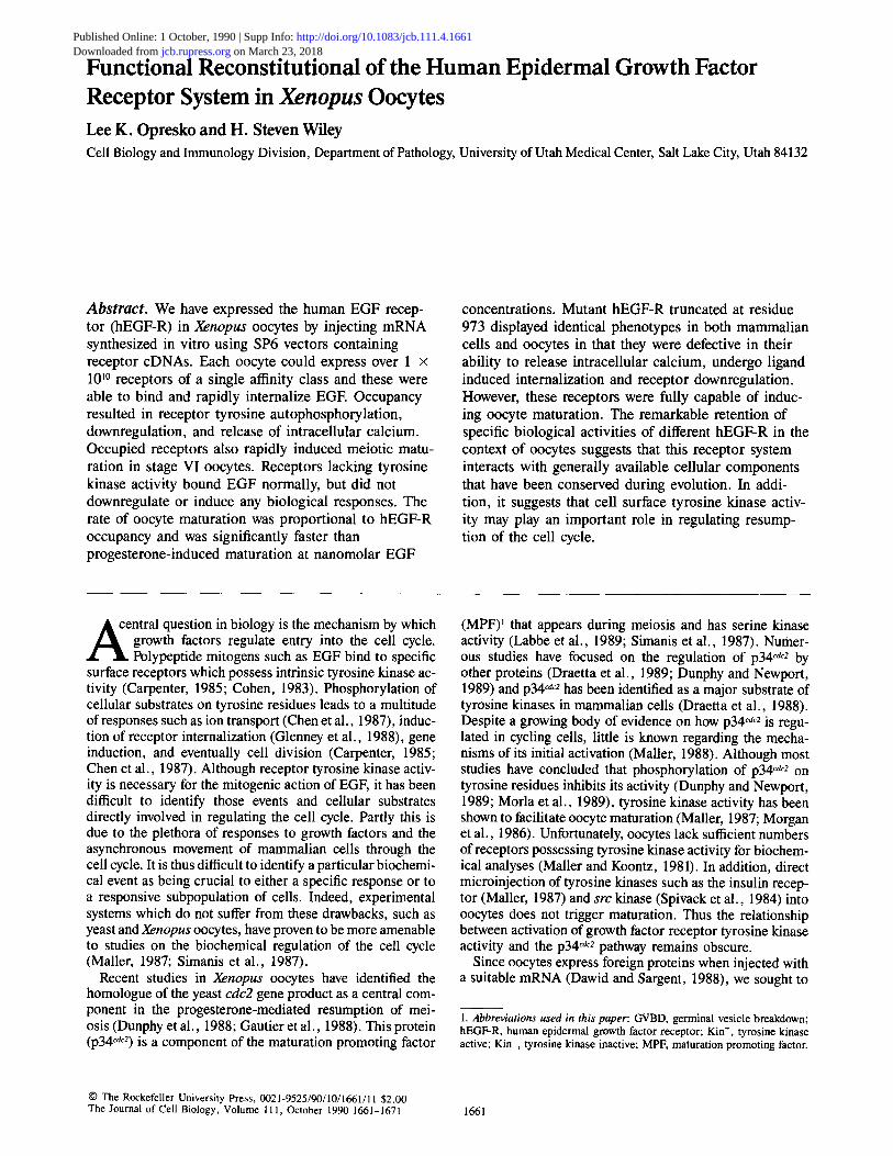
Figure I. Oocytes injected with mRNA encoding the hEGF-R will bind EGE (A) Map of the pOBER vector used to generate mRNA in vitro for oocyte injections. (B) Binding of t2SI-EGF to oocytes at both 0 and 20°C. Oocytes were injected with 20 ng mRNA or water 40 h before exposure to radiolabeled ligand, t2SI-EGF (6.7 x 10⁸ cpm/nmol) was added at a concentration of 1.7 x 10 -8 M. Binding at 0°C was done for 2 h and uptake at 20°C was done for 1 h. Each point represents an individual oocyte.
The Journal of Cell Biology, Volume 111, 1990 1662

x
LU
5.0
4.°A /t 3.o O /
ii°oi ! I/i 0.0 ,
20 40 60 Time (h)
6.0
% x
o 4.0
t~
LL
P, zo e n
B o\
O N ~ KD=3.6x 10⁻⁸M
0.0 80 0.0 510 10 15
Bound (molecules x cell'1 x 1 0 "9 )
Figure 2. Surface expression of the hEGF- R. (.4) Groups of oocytes were injected with either water (zx) or with 20 ng of mRNA encoding the wild-type hEGF-R and at the indicated times were evaluated at 0°C for their ability to bind 125I-EGF at a concen- tration of 3.3 x 10 -8 M. Data is shown for oocytes of 1.0-mm diameter (o) and 1.3- mm diameter (El) -t- the standard error of the mean. (B) Scatchard analysis of ~25I- EGF binding to oocytes at 0°C. Oocytes in- jected with 20 ng mRNA 40 h earlier were brought to equilibrium with :5I-EGF con- centrations ranging from 5 x 10 -1° to 1.6 × 10 -7 M. Each point represents 10 oo- cytes and receptor affinity was estimated by linear regression.
phy. Quantitation of the amount of label in the bands was determined using a densitometer (model 620; Bio-Rad Laboratories). Exposure times of the autoradiographs were adjusted to remain in the linear range of the den- sitometer as determined by parallel samples of known specific activities.
EGF Binding to Oocytes 1 h before binding, the cells were placed into fresh 50% L-15 solution con- taining 125 ~g/ml sodium ipodate which inhibits the deiodination activity of oocytes (Opresko et al., 1980). 1251-EGF was prepared as previously de- scribed (Wiley and Cunningham, 1982). Scatchard analyses (Scatchard, 1949) were performed at 0°C to prevent internalization. Oocytes were brought to equilibrium (4-6 h) with 125I-EGF concentrations ranging from 5 x 10 -~° to 1.6 × 10 -7 M. The labeled EGF was not diluted with unla- beled EGF to avoid potential problems associated with differential binding between labeled and unlabeled ligand (Wiley, 1985). A minimum of 10 oo- cytes were used to evaluate binding at each 125I-EGF concentration, Nonspecific binding was determined in parallel using water-injected oo- cytes and was always <10 % of total binding. To allow internalization, oo- cytes were incubated at 20°C for the indicated times. After the incubation, cells were washed in three changes of solution O-R2 (Wallace et al., 1973) containing 1 mg/ml BSA at 0°C. The relative amount of surface-associated and internalized 125I-EGF was determined by acid stripping groups of two cells as described previously (Wiley and Cunningham, 1982). Oocytes were solubilized in groups of two using 100 #1 formic acid before counting. The data were corrected for nonspecific binding and converted to internalization plots as previously described (Opresko and Wiley, 1987b). Steady-state binding of EGF was also conducted at 20°C for 2 h using 125I-EGF con- centrations ranging from 5 × 10 -1° to 1.6 × 10 -7 M as previously de- scribed (Knauer et al., 1984; Wiley and Cunningham, 1981).
Calcium Efflux Measurements Oocytes injected with the appropriate mRNA 40 h earlier were incubated overnight with 50/zCi/ml of 4SCa (Amersham Corp.) in standard culture medium. Before the efflux measurements, the oocytes were rinsed rapidly in three changes of solution OR-2 and placed in the upper chamber of an equilibrium dialysis flow cell (Womack and Colowick, 1973). The oocytes were supported on nylon mesh and the upper chamber (volume 0.5 ml) was sealed with parafilm. The lower flow chamber (100 /zl) was constantly stirred with a small magnetic bar and solution OR-2 was pumped through at a flow rate of 1.5 ml/min. Fractions were collected at 1-min intervals and the amount of radioactivity assessed by scintillation counting. The amount of 45Ca remaining in the oocytes after the experiment was released by de- tergent and counted.
Results
Expression of High Levels of EGF Receptors in Oocytes Efforts to induce the synthesis of the hEGF-R by injecting total polyA mRNA from A431 cells (Simmen et al., 1984)
did not result in any observable ~25I-EGF binding by oo- cytes. We therefore obtained the pXER expression vector containing the cDNA encoding the full-length hEGF-R (Lin et al., 1986). Direct microinjection of this vector into the oo- cyte nucleus resulted in a significant, but highly variable binding of ~25I-EGF to recipient cells (data not shown). To improve the reproducibility of hEGF-R expression, we con- structed the vector shown in Fig. 1 A. This vector supports the in vitro synthesis of hEGF-R mRNA containing a 30- residue polyA tail. Injection of this polyA mRNA resulted in very efficient synthesis of hEGF-R by recipient oocytes. Shown in Fig. 1 B is the net binding of ~:SI-EGF to in- dividual oocytes at both 0 and 22°C. Binding of ~25I-EGF to mRNA-injected oocytes at 0°C, a temperature that prevents receptor internalization, was usually 10-fold greater than water-injected controls. However, a significant increase in ~25I-EGF binding was observed at 22°C, probably as a con- sequence of ligand internalization. To separate the contribu- tion of surface receptor expression from receptor internali- zation, we examined t25I-EGF binding at 0°C and then analyzed the ability of the expressed receptors to internalize ligand.
Groups of oocytes were injected with either water or mRNA and cultured for different lengths of time. The ability of oocytes to oocytes to bind ~25I-EGF at 0°C in both stage IV/V (1.0 mm) and VI (1.3 mm) oocytes was then evaluated. As shown in Fig. 2 A, significant ~25I-EGF binding could be observed by 24 h and this continued to increase for the entire period of the experiment. The 1.0-mm-diam oocytes accu- mulated surface hEGF-R at a rate of ~ 7 × 10⁷ h -I between 24- and 72-h postinjection while the accumulation rate ofoo- cytes 1.3-mm diameter was ~2.3 x 10⁸ h -~. Within this period, there was no sign of an upper limit in the number of receptors observed on the oocyte surface. Even after correct- ing for differences in oocyte volume and surface area, stage VI oocytes were still more efficient in expressing surface hEGF-R, consistent with previous observations that protein synthesis rates in these cells are significantly higher than in smaller oocytes (Wasserman et al., 1984).
The hEGF-R expressed by stage VI oocytes were of a sin- gle affinity class. Shown in Fig. 2 B is a Scatchard plot of ~25I-EGF binding to mRNA-injected oocytes at 0°C. Non- specific binding to water-injected oocytes at 0°C usually ranged between 5 and 10% and equilibrium binding was
Opresko and Wiley Reconstitution of EGF Receptor System in Oocytes 1663

Figure 3. The human EGF receptor ex- pressed by oocytes is full length and dis- plays EGF-induced autophosphorylation. (A) Immunoprecipitation of metabolically labeled hEGF receptor. Oocytes were in- jected with mRNA encoding either the nor- mal hEGF-R (Kin +) or the hEGF-R in which lysine at position 721 was altered to a methionine by site-directed mutagenesis (Kin-). Metabolically labeled hEGF-R from mouse B82 cells were used as a con- trol. After 40-h labeling, the receptor was immunoprecipitated using the 528 mono- clonal antibody. (B) Western blot analysis of EGF-induced tyrosine phosphorylation in oocytes. Stage V oocytes injected 40 h previously with the indicated mRNA were incubated either with (4-) or without ( - ) 1.7 x 10 -8 M EGF for 5 min followed by extraction and Western blot analysis using ~25I-labeled antiphosphotyrosine monoclo- nal PY20 (Glenney et al., 1988).
achieved by 4 h (data not shown). Although normal human fibroblasts also express receptors of a single affinity class (Carpenter et al., 1975; Knauer et al., 1984), the calculated equilibrium affinity for the hEGF-R in oocytes was 3.6 x 10 -s M, which is '~10-fold lower than that observed in hu- man fibroblasts (Wiley et al., 1989). In the experiments shown in Fig. 2 B, each oocyte expressed ,~1.5 × 101° hEGF-R, or '~100-fold the number of endogenous insulin/IGF-1 recep- tors (Mailer and Koontz, 1981).
The absolute number of surface hEGF-R expressed by oo- cytes depended both on the amount of injected mRNA, the particular batch of oocytes, the oocyte stage, and the time al- lowed for synthesis. Typically, this number ranged between 3 × 10⁹ and 3 × 101° receptors/oocyte. However, the sur- face area of a 1.0-mm oocyte is approximately that of 50,000 fibroblasts (Opresko and Wiley, 1987a), translating to a sur- face receptor density equivalent to between 0.6 and 6 × 10⁵ for a typical mammalian cell. Although this receptor density is high, it falls within the range reported for transfected mammalian cells and is substantially less than that observed for A431 cells (Chen et al., 1987; Haigler et al., 1979). In addition, the number of endogenous vitellogenin receptors
expressed on the oocyte surface is between 0.3 and 3 × 10" (Opresko and Wiley, 1987a). Thus the surface densi- ties of hEGF-R expressed by oocytes is high, but well within the capacity of the oocyte plasma membrane.
We also synthesized mRNA from a hEGF-R cDNA that contains methionine rather than lysine at residue 721 (Chen et al., 1987). Receptors made from this mutant cDNA lack intrinsic tyrosine kinase activity, but are unaltered in their ability to bind EGF (Chen et al., 1987). Oocytes injected with mRNA derived from the "wild-type" (Kin +) and M TM
(tyrosine kinase inactive [Kin-]) constructions synthesized equivalent numbers of receptors as a function of injected mRNA and both bound EGF with the same affinity (data not shown).
To determine whether these receptors were normally pro- cessed, oocytes were incubated with [35S]methionine/cys- teine followed by immunoprecipitation with a monoclonal antibody to the hEGF-R (Gill et al., 1984). As shown in Fig. 3 A, a protein was immunoprecipitated with the same mobil- ity as the hEGF-R synthesized by transfected mammalian cells. The additional bands found in the oocyte immunopre- cipitate are nonspecifically adsorbed, sulfated yolk proteins
The Journal of Cell Biology, Volume 111, 1990 1664

8.0 I1"
60 A
/ / g 2.0 v • Surface
60 . . . . . '
~ B I n s i d e
i 4.0 ~ ta~ O []
2.0 j Surface
o n g . " o - - - - o -
0.0 . , . , . , . ,
0 20 40 60 80 T i m e (rain)
6.0- o O"
Kin+
d
o o E 0.0 2'.0 410 6.0
Integral surface binding (molecules x rain x 10 -9)
Figure 4. Internalization and down-regulation of the kinase positive and kinase negative hEGF-R by oocytes. (A) Binding and internali- zation of Kin* (• , m) receptors. Oocytes were incubated with 3.3 × 10 -8 M t25I-EGF (5.7 × 10⁸ cpm/nmol) and at the indicated times groups of eight oocytes were rinsed and the relative amount of surface-associated and internalized ~25I-EGF determined by acid stripping. (B) Binding and internalization of Kin- (o, []) receptors. Same experiment described in A, but the oocytes were injected with mRNA derived from the Kin- hEGF-R. (C) Inter- nalization plots of the Kin + (•) and Kin- (o) receptors. The ki- netics of internalization was determined as described above using 1.7 × 10 -9 M 125I-EGF and the data were transformed into inter- nalization plots. The slope of these plots is proportional to the specific internalization rate of the receptor, yielding a value of 0. 21 min -1 for the Kin + receptor, and 0.11 min -t for the Kin- receptor.
The EGF Receptor Is Fully Functional in Oocytes and Induces Meiotic Maturation
Our ability to synthesize hEGF-R in oocytes allowed us to test which functional aspects of the receptor are intrinsically dependent on tyrosine kinase activity. For example, the Kin- hEGF-R in mammalian cells is unable to transmit a biological signal (Chen et al., 1987) or to trigger receptor downregulation (Glenney et al., 1988). Kin- receptors do not undergo downregulation because of their lack of ligand- induced internalization. We therefore examined the relative ability of Kin + and Kin- hEGF-R to induce these events in the oocytes. To determine whether both Kin + and Kin- hEGF-R undergo endocytosis and downregulation, we in- cubated oocytes with a high concentration of ~2q-EGE The extent of internalization and surface binding was then quanti- tated by acid stripping (Wiley and Cunningham, 1982). As shown in Fig. 4, both Kin + and Kin- receptors internalized EGE However, the specific internalization rate of the Kin + receptor was about twice that of its Kin- counterpart (Fig.
and are also found in sham-injected oocytes (data not shown). The molecular weight of the labeled band was 170,000, in- dicating that the hEGF-R is fully glycosylated by oocytes (Soderquist et al., 1988).
The tyrosine kinase activity of the hEGF-R in oocytes was determined by Western blot analysis using an anti-phospho- tyrosine (anti-pTyr) monoclonal antibody (Glenney et al., 1988). As seen in Fig. 3 B, only oocytes injected with mRNA encoding the Kin + hEGF-R showed an EGF-mediated ap- pearance of a new tyrosine-phosphorylated protein. The mo- lecular weight of the EGF-induced band was 170,000, indi- cating that it is the autophosphorylated hEGF-R (Downward et al., 1984). These results demonstrate that oocytes can syn- thesize a fully glycosylated and enzymatically functional EGF receptor.
Figure 5. Occupied EGF receptors can induce meiotic maturation in oocytes. (A) Kinetics of EGF and progesterone-induced matura- tion. Oocytes injected with mRNA encoding the Kin + (e) or Kin- (o) hEGF-R were exposed to 1.7 x 10 -8 M EGE Alternatively, water-injected oocytes were exposed to 10 #g/ml progesterone ([]). At the indicated times, they were scored for the appearance of the characteristic white spot at the apex of the animal hemisphere, indicating GVBD. (B) Effect of EGF concentration on the rate of maturation. Groups of oocytes (n = 7) expressing the Kin + hEGF-R were treated with the indicated concentrations of EGE The time of GVBD was then scored as indicated in A. The results are shown as the mean time of GVBD + the standard deviation. Control oocytes were treated with 10/,g/ml progesterone.
Opresko and Wiley Reconstitution of EGF Receptor System in Oocytes 1665

4 C). Significantly, surface binding dropped to less than one- third the initial values by 80 min in cells expressing Kin + receptors while surface binding for the Kin- receptor re- mained constant. This indicates that Kin + receptors are undergoing ligand-induced internalization and downregula- tion, consistent with the observations of higher internali- zation rates for the Kin + receptor. To confirm downregula- tion of Kin + receptors, we conducted a "steady state" analysis of surface binding (Wiley and Cunningham, 1981) at 22°C to allow for endocytosis (Wallace et al., 1973). Oocytes were incubated with increasing concentrations of t25I-EGF and allowed to approach a steady state of surface binding. The maximum extent of surface binding was then determined by extrapolation and compared to equilibrium binding at 0°C. The results of this experiment showed that oocytes that dis- played an average of 2.4 x 101° receptors at 0°C (before EGF exposure) had a maximum of 8.5 × 10⁹ surface recep- tors at steady state binding. This confirms that oocytes lose approximately two-thirds of their surface receptors by down- regulation. The absence of downregulation of Kin- recep- tors does not seem to be due to differences in their ability to recycle relative to Kin + receptors. EGF internalized by either receptor is transferred to a light endosomal compart- ment and eventually to yolk platelets (data not shown). This indicates that intrinsic kinase activity induces hEGF-R down- regulation at the level of internalization in oocytes as it does in mammalian cells (Chen et al., 1987; Glenney et al., 1988).
To determine whether the hEGF-R could induce a biologi- cal response in oocytes, we examined the effect of EGF on meiotic maturation. It has already been established that in- cubating oocytes with high concentrations of insulin and IGF-1 can facilitate and sometimes induce oocyte maturation (LeGoascogne et al., 1984; Mailer and Koontz, 1981). How- ever, since microinjected tyrosine kinases such as the src protein or insulin receptor cannot induce maturation (Mailer, 1987; Spivack et al., 1984), a direct relationship between tyrosine kinase activity and maturation is uncertain. We therefore compared the relative ability of EGF to induce maturation in oocytes expressing either the Kin + or Kin- hEGF-R. As shown in Fig. 5 A, EGF was very effective in
inducing maturation in oocytes that expressed the Kin + re- ceptor. However, treatment of oocytes expressing Kin- re- ceptors with EGF was without effect, even after 24-h treat- ment. In addition, neither water-injected nor uninjected oocytes responded to EGE Germinal vesicle breakdown (GVBD) in EGF-treated oocytes was confirmed by section- ing of fixed oocytes. Cytoplasm from EGF-matured oocytes could also induce maturation in recipient oocytes with the same kinetics as cytoplasm from progesterone-stimulated oocytes (data not shown), confirming the presence of MPF (Wasserman and Masui, 1975a).
We were quite surprised to find that EGF could induce maturation significantly faster than progesterone (Fig. 5 B). However, the rate of EGF-induced maturation was highly de- pendent on both the concentration of EGF and the level of hEGF-R expression at the oocyte surface. As shown in Fig. 5 B, reducing the concentration of EGF resulted in a de- crease in the rate of maturation to below that observed for progesterone. When the level of surface hEGF-R expression was decreased to below 109 per oocyte, the maximum rate of EGF-stimulated maturation also fell below that typically induced by progesterone, but was still much faster than insulin-induced maturation (data not shown). However, we occasionally found batches of oocytes in which progesterone induced maturation nearly as fast as that induced by EGE Therefore, the mechanism(s) which regulates the sensitivity of oocytes to progesterone does not necessarily affect their sensitivity to EGE Nevertheless, the intrinsic tyrosine ki- nase activity of the EGF receptor is essential for the induc- tion of maturation and EGF can stimulate oocyte responses at physiological ligand concentrations.
Activation of the MPF/p34 c~2 Complex
Progesterone-stimulated maturation involves the activation of the MPF/p34 ~dc2 complex which contains a serine/threo- nine histone H1 kinase activity (Arion et al., 1988; Labbe et al., 1989). The activation of the MPF/p34 cdc2 complex is thought to be essential for subsequent events in meiosis. However, the rapid maturation stimulated by EGF and its de-
EGF in oocytes expressing the Kin + (e) or Kin- (o) hEGF-R or in response to 10/zg/ml progesterone (D). (bottom) under the same conditions described in the top panel.
Figure 6. Activation ofhistone kinase activity in response to EGE (A) Autoradiograph of in vitro phosphorylation of his- tones by oocyte extracts. Con- trol oocytes were treated with progesterone (top), whereas oocytes injected with mRNA encoding the Kin + (center) or Kin- (bottom) hEGF-R were exposed to 1.7 x 10 -s M EGE At the indicated times, oocyte extracts were evaluated for their ability to catalyze histone phos- phorylation. Shown is the histone region of the autora- diograph. (B) Densitometric quantitation of histone phos- phorylation (top) induced by Kinetics of oocyte maturation
The Journal of Cell Biology, Volume 111, 1990 1666

2.0
• 1.5
x
~ 1.0
8 ~ 0.5
0.0
S . 0 - % .
× 4 . 0 -
,,, 3.0.
• - 2.0.
E 1.0.
0Z 0.0
A = .______-~--~
/
10 2 0 3 0
Time (min)
B
/
40
5.0 10 15 20
Integral surface binding (molecules x min' lx 10 -10)
Figure 7. I n t e r n a l i z a t i o n a n d d o w n r e g u l a t i o n o f c ' 9 7 3 h E G F - R in
oocytes. Stage VI oocytes were injected with 20 ng of mRNA en- coding the wild-type hEGF-R (e) and receptors truncated at resi- due 973 (n). After 40-h culture, oocytes were evaluated at 22°C for their ability to approach steady state surface binding (A) as well as internalize ~25I-EGF (B) using a ~25I-EGF concentration of 1.7 X 10 -9 M .
pendence on tyrosine kinase activity is difficult to reconcile with the observation that activation of the p34 cdc2 complex is correlated with removal of its tyrosine phosphate (Dunphy and Newport, 1989; Gautier et al., 1989; Morla et al., 1989). To determine the relationship between hEGF-R tyro- sine kinase activity and the activation of the MPF/p34 cdc2 complex, we measured histone H1 kinase activity in both progesterone and EGF-treated oocytes. As shown in Fig. 6, progesterone induced a dramatic increase in histone kinase activity prior to GVBD. These results are essentially identi- cal to those reported by other investigators (Labbe et al., 1988). In oocytes expressing the Kin + hEGF-R, the addi- tion of EGF resulted in a very similar activation in histone kinase activity (Fig. 6). Significantly, there was no measur- able increase in this activity for several hours after the addi- tion of EGE As expected, oocytes expressing the Kin- hEGF-R showed no response to the addition of EGF (Fig. 6). However, these cells did respond normally to the addition of progesterone (data not shown). The delay between the activation of the hEGF-R and the activation of the MPF/ p34 cdc2 complex indicates either that internalization of the ligand- receptor complex is required, or that the activation is indirect.
Expression of an Internalization-defective hEGF-R in Oocytes
When we first examined the effect of EGF on oocyte matura- tion, we noted that oocytes expressing the Kin + hEGF-R appeared morphologically normal until '~6-8 h after EGF addition. After this time, oocytes became mottled and then visibly degenerated. This is similar to what has been ob- served for maturation induced by both H-ras injection (Birchmeier et al., 1985), high concentrations of divalent
cations and ionophores (Wasserman and Masui, 1975b), and other nonphysiological agents that can affect intracellular calcium distribution (Smith, 1989). Although the hEGF-R can release intracellular calcium in mammalian cells (Sawyer and Cohen, 1981), progesterone treatment does not induce calcium release in oocytes (Cork et al., 1987). Therefore, we felt that this additional response could be responsible for the eventual oocyte degeneration. To test this hypothesis, we made use of a recently described mutant of the hEGF-R that lacks the carboxyl-terminal domain distal to residue 973 (c'973 hEGF-R). This receptor has full kinase activity, but is defective in its ability to release intracellular calcium and to undergo ligand-induced internalization (Chen et al., 1989). Significantly, the c'973 receptors are fully capable of inducing mitosis in mammalian cells, suggesting that cal- cium release and induced internalization are not required for induction of cell division. We were thus interested to deter- mine whether these receptors would induce meiotic matura- tion in oocytes without causing oocyte degeneration.
We inserted the c'973 hEGF-R into our SP6 vector and synthesized the appropriate mRNA. When injected into oo- cytes, this mRNA directed the synthesis of a 150,000-kD hEGF-R protein, as determined by [35S]methionine/cysteine labeling followed by immunoprecipitation (data not shown). This is the expected size of the c'973 hEGF-R. When in- jected with the same amount of mRNA, ~25I-EGF binding at 0°C to oocytes expressing the c'973 receptor was indistin- guishable from those expressing the wild-type receptor (data not shown). We also obsezved that the addition of EGF did not result in autophosphorylation of the receptor molecule, confirming that all of the autophosphorylation sites had been removed by the c'973 truncation.
Since the c'973 receptor is internalization defective and
+EGF Rinse + l o n o r n y d n
2o t t Wild-Type
8.0
v t~
0 4.0
0m0 ' ' I I . . . . I I ' ' o 8.0
I I
t i c'973
4.0
0 .0 i ~ I i i i J i 0 10 20 30 40
T i m e (m in )
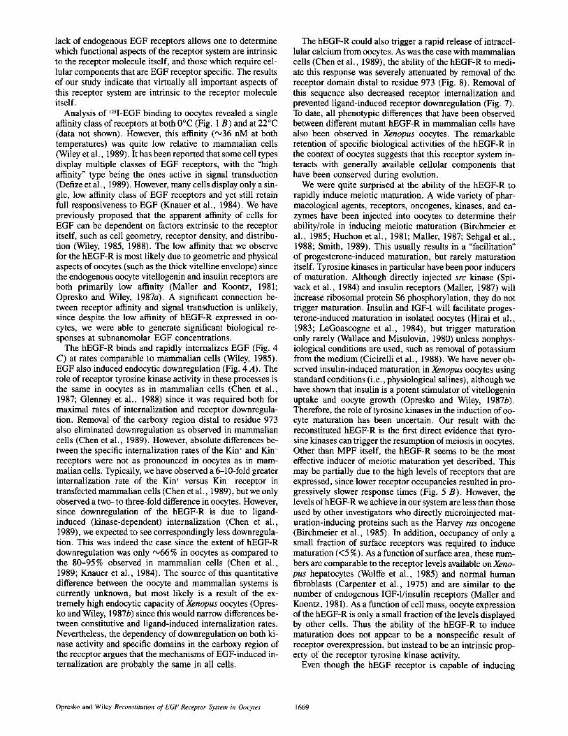
Figure 8. EGF-induced release of intracellular calcium by oocytes expressing wild-type and c'973 receptors. Groups of 50 oocytes in- jected 40 h previously with 20 ng of mRNA encoding the appropri- ate receptor were preloaded overnight with 45Ca and placed in a continuous flow chamber. Calcium released in the perfusion medium was then monitored as described in Materials and Methods. At the indicated time, perfusion medium containing 1.7 x 10 -9 M EGF was used, followed by a change to initial buffer (rinse) followed by buffer containing 10 ~M ionomycin.
Opresko and Wiley Reconstitution of EGF Receptor System in Oocytes 1667

does not undergo downregulation in mammalian cells, we were interested in confirming this receptor attribute in oo- cytes. As shown in Fig. 7 B, internalization plot analysis dem- onstrated that the specific internalization rate of the c'973 receptors was significantly lower than the K ÷ hEGF-R. On the average, the internalization rate of the c'973 was 0.03 min -~, or between 35 and 40% of the rate observed in par- allel sets of oocytes expressing the wild-type K ÷ hEGF-R. As expected, surface binding of ~25I-EGF to oocytes express- ing the wild-type hEGF-R displayed a time-dependent de- crease at 20°C as a consequence of downregulation while surface binding to oocytes expressing the c'973 hEGF-R reached a constant steady state binding (Fig. 7 A). Binding at 0°C confirmed that treatment of oocytes expressing c'973 hEGF-R with high concentrations of EGF for 2 h at 20°C did not decrease the initial number of surface receptors (data not shown). We conclude that the internalization/downregu- lation defective phenotype of the c'973 receptor in mam- malian cells is preserved in Xenopus oocytes.
To determine the relative ability of the different hEGF-R to release intracellular calcium stores in oocytes, cells ex- pressing the appropriate receptor were loaded overnight with 45Ca, rinsed, and placed in a continuous flow chamber. The
Figure 9. EGF-induced maturation of oocytes expressing the c'973 hEGF-R. (A) Uninjected oocytes treated for 8 h with 10 /zg/ml progesterone. (B) Oocytes were injected with 20 ng of mRNA en- coding the c'973 hEGF-R and 40 h later were treated with 1.7 × 10 -9 M EGF for 8 h. The EGF-treated oocytes matured at 3 h while the progesterone-treated cells matured at 4 h.
release of 45Ca from the oocytes was then evaluated after adding EGF to the perfusate. As shown in Fig. 8, the addition of EGF to oocytes expressing the wild-type hEGF-R resulted in a rapid release of preloaded 45Ca. Water-injected oocytes or those expressing Kin- receptors displayed no response to the addition of EGF (data not shown). In contrast to the situ- ation with the wild-type receptor, EGF addition to oocytes expressing the c'973 receptor resulted in very little release of 45Ca. However, oocytes could release calcium in response to ionomycin (Fig. 8). The relative ability of EGF to induce calcium release in oocytes expressing either wild-type or c'973 hEGF-R is essentially identical to what has been ob- served in mammalian cells (Chen et al., 1989).
Once we had confirmed that the cX)73 hEGF-R preserved their phenotype in the Xenopus oocyte system, we examined the question of whether they could induce meiotic matura- tion. We found that the addition of EGF to oocytes express- ing c'973 hEGF-R indeed resulted in rapid meiotic matura- tion. However, the rate of maturation we observed was not significantly different than what we had observed after stim- ulation of oocytes expressing the wild-type receptor. In addi- tion, the number of occupied receptors required for induc- tion of maturation was similar between the wild-type and c'973 receptors (data not shown). This indicates that the sig- naling mechanism required for the induction of maturation is not significantly different between the two receptor con- structions. However, as shown in Fig, 9, the morphology of oocytes matured in response to the c'973 hEGF-R was in- distinguishable from that displayed by oocytes that matured in response to progesterone. Prolonged incubations with EGF did not result in any visible abnormalities or morpho- logical changes, other than those normally associated with maturation. Thus the removal of the internalization/calcium domain from the hEGF-R results in a receptor which is fully capable of inducing meiotic maturation, but which does not trigger the negative side effects observed with the wild-type receptor.
Discussion
Our results show that the human EGF receptor system can be functionally reconstituted in Xenopus oocytes. Other investi- gators have previously reported the synthesis of hEGF-R by oocytes after injection of mRNA derived from A431 cells (Simmen et al., 1984), but insufficient numbers were ob- tained for biological or biochemical studies. Our approach of synthesizing mRNAs in vitro from cDNA vectors allowed us to obtain sufficient amounts of mRNA to cause high levels of receptor expression. Indeed, the levels of surface receptor expression we observe (over 10 ~° per oocyte) approach that of many transfected mammalian cells (Chen et al., 1989).
The Xenopus oocyte is particularly useful for investigating the EGF receptor because they constitute a large, syn- chronized cell population with well-defined, stage-specific responses to growth factors (Maller, 1987; Taylor and Smith, 1987; Wallace and Misulovin, 1978). They also lack endoge- nous EGF receptors. The lack of ~25I-EGF binding to oo- cytes that we and other investigators have observed is not simply due to their inability to bind mammalian EGF since the ligand can readily bind to Xenopus hepatocytes at nanomolar concentrations as well as induce DNA synthesis and receptor autophosphorylation (Wolffe et al., 1985). The
The Journal of Cell Biology, Volume 111, 1990 1668
lack of endogenous EGF receptors allows one to determine which functional aspects of the receptor system are intrinsic to the receptor molecule itself, and those which require cel- lular components that are EGF receptor specific. The results of our study indicate that virtually all important aspects of this receptor system are intrinsic to the receptor molecule itself.
Analysis of 125I-EGF binding to oocytes revealed a single affinity class of receptors at both 0°C (Fig. 1 B) and at 22°C (data not shown). However, this affinity (~36 nM at both temperatures) was quite low relative to mammalian cells (Wiley et al., 1989). It has been reported that some cell types display multiple classes of EGF receptors, with the "high affinity" type being the ones active in signal transduction (Defize et al., 1989). However, many cells display only a sin- gle, low affinity class of EGF receptors and yet still retain full responsiveness to EGF (Knauer et al., 1984). We have previously proposed that the apparent affinity of cells for EGF can be dependent on factors extrinsic to the receptor itself, such as cell geometry, receptor density, and distribu- tion (Wiley, 1985, 1988). The low affinity that we observe for the hEGF-R is most likely due to geometric and physical aspects of oocytes (such as the thick vitelline envelope) since the endogenous oocyte vitellogenin and insulin receptors are both primarily low affinity (Mailer and Koontz, 1981; Opresko and Wiley, 1987a). A significant connection be- tween receptor affinity and signal transduction is unlikely, since despite the low affinity of hEGF-R expressed in oo- cytes, we were able to generate significant biological re- sponses at subnanomolar EGF concentrations.
The hEGF-R binds and rapidly internalizes EGF (Fig. 4 C) at rates comparable to mammalian cells (Wiley, 1985). EGF also induced endocytic downregulation (Fig. 4 A). The role of receptor tyrosine kinase activity in these processes is the same in oocytes as in mammalian cells (Chen et al., 1987; Glenney et al., 1988) since it was required both for maximal rates of internalization and receptor downregula- tion. Removal of the carboxy region distal to residue 973 also eliminated downregulation as observed in mammalian cells (Chen et al., 1989). However, absolute differences be- tween the specific internalization rates of the Kin + and Kin- receptors were not as pronounced in oocytes as in mam- malian cells. Typically, we have observed a 6-10-fold greater internalization rate of the Kin + versus Kin- receptor in transfected mammalian cells (Chen et al., 1989), but we only observed a two- to three-fold difference in oocytes. However, since downregulation of the hEGF-R is due to ligand- induced (kinase-dependent) internalization (Chen et al., 1989), we expected to see correspondingly less downregula- tion. This was indeed the case since the extent of hEGF-R downregulation was only '~66 % in oocytes as compared to the 80-95% observed in mammalian cells (Chen et al., 1989; Knauer et al., 1984). The source of this quantitative difference between the oocyte and mammalian systems is currently unknown, but most likely is a result of the ex- tremely high endocytic capacity of Xenopus oocytes (Opres- ko and Wiley, 1987b) since this would narrow differences be- tween constitutive and ligand-induced internalization rates. Nevertheless, the dependency of downregulation on both ki- nase activity and specific domains in the carboxy region of the receptor argues that the mechanisms of EGF-induced in- ternalization are probably the same in all cells.
The hEGF-R could also trigger a rapid release of intracel- lular calcium from oocytes. As was the case with mammalian cells (Chen et al., 1989), the ability of the hEGF-R to medi- ate this response was severely attenuated by removal of the receptor domain distal to residue 973 (Fig. 8). Removal of this sequence also decreased receptor internalization and prevented ligand-induced receptor downregulation (Fig. 7). To date, all phenotypic differences that have been observed between different mutant hEGF-R in mammalian cells have also been observed in Xenopus oocytes. The remarkable retention of specific biological activities of the hEGF-R in the context of oocytes suggests that this receptor system in- teracts with generally available cellular components that have been conserved during evolution.
We were quite surprised at the ability of the hEGF-R to rapidly induce meiotic maturation. A wide variety of phar- macological agents, receptors, oncogenes, kinases, and en- zymes have been injected into oocytes to determine their ability/role in inducing meiotic maturation (Birchmeier et al., 1985; Huchon et al., 1981; Mailer, 1987; Sehgal et al., 1988; Smith, 1989). This usually results in a "facilitation" of progesterone-induced maturation, but rarely maturation itself. Tyrosine kinases in particular have been poor inducers of maturation. Although directly injected src kinase (Spi- vack et al., 1984) and insulin receptors (Mailer, 1987) will increase ribosomal protein $6 phosphorylation, they do not trigger maturation. Insulin and IGF-1 will facilitate proges- terone-induced maturation in isolated oocytes (Hirai et al., 1983; LeGoascogne et al., 1984), but trigger maturation only rarely (Wallace and Misulovin, 1980) unless nonphys- iological conditions are used, such as removal of potassium from the medium (Cicirelli et al., 1988). We have never ob- served insulin-induced maturation in Xenopus oocytes using standard conditions (i.e., physiological salines), although we have shown that insulin is a potent stimulator of vitellogenin uptake and oocyte growth (Opresko and Wiley, 1987b). Therefore, the role of tyrosine kinases in the induction of oo- cyte maturation has been uncertain. Our result with the reconstituted hEGF-R is the first direct evidence that tyro- sine kinases can trigger the resumption of meiosis in oocytes. Other than MPF itself, the hEGF-R seems to be the most effective inducer of meiotic maturation yet described. This may be partially due to the high levels of receptors that are expressed, since lower receptor occupancies resulted in pro- gressively slower response times (Fig. 5 B). However, the levels of hEGF-R we achieve in our system are less than those used by other investigators who directly microinjected mat- uration-inducing proteins such as the Harvey ras oncogene (Birchmeier et al., 1985). In addition, occupancy of only a small fraction of surface receptors was required to induce maturation (<5 %). As a function of surface area, these num- bers are comparable to the receptor levels available on Xeno- pus hepatocytes (Wolffe et al., 1985) and normal human fibroblasts (Carpenter et al., 1975) and are similar to the number of endogenous IGF-1/insulin receptors (Mailer and Koontz, 1981). As a function of cell mass, oocyte expression of the hEGF-R is only a small fraction of the levels displayed by other cells. Thus the ability of the hEGF-R to induce maturation does not appear to be a nonspecific result of receptor overexpression, but instead to be an intrinsic prop- erty of the receptor tyrosine kinase activity.
Even though the hEGF receptor is capable of inducing
Opresko and Wiley Reconstitution of EGF Receptor System in Oocytes 1669

maturation faster than progesterone, it does not appear to directly activate the MPF/p34 cdc2 complex. We were unable to induce maturation in the smaller stage IV oocytes that were unresponsive to progesterone (data not shown) al- though the hEGF-R was fully capable of undergoing EGF- induced autophosphorylation (Fig. 3 B). Activated MPF is capable of inducing GVBD in these cells (Taylor and Smith, 1987). The fastest response time we ever observed for EGF- induced maturation (2-3 h) is similar to the fastest times ob- served for progesterone-induced maturation (Fig. 6 B; also see Reynhout et al., 1975), but slower than the response time observed for MPF (Wasserman and Masui, 1975a; also data not shown). Finally, we observed a several hour lag time be- tween the addition of EGF and activation of histone H1 ki- nase activity (Fig. 6), but no significant lag between the addi- tion of ligand and autophosphorylation of the EGF receptor (Fig. 3 B). Injection of oocytes with MPF induces an almost immediate activation of histone HI kinase activity (Labbe et al., 1988). Together these data indicate that the hEGF-R mediates its action through an unidentified intermediate step(s).
It is not yet certain whether internalization of the hEGF-R is necessary to induce maturation, but our data support the hypothesis that it acts to phosphorylate a substrate at the cell surface. Although the c'973 hEGF-R was only internalized at one-third the rate of its wild-type counterpart, it was fully capable of inducing maturation. Indeed, a requirement for activated receptors to reside at the oocyte surface would ex- plain why previous efforts to demonstrate a role for tyrosine kinases in maturation have failed, since those studies directly microinjected kinases into the oocyte cytoplasm (Mailer, 1987; Spivack et al., 1984). Direct microinjection of pro- gesterone also fails to induce maturation (Smith and Ecker, 1971), again suggesting that maturation triggered by hor- mone receptors may require action at the cell surface.
The ability of the hEGF-R to induce meiotic maturation in oocytes suggests that the fundamental processes regulat- ing the eucaryotic cell cycle are highly conserved during evo- lution. Our results also point to a central role for tyrosine kinases in both the activation and regulation of this process. However, it is noteworthy that maturation induced by mutant hEGF-R that are deficient in triggering some biological re- sponses, such as calcium release, more closely resembles that produced by physiological agents. Since progesterone treatment does not release intracellular calcium in oocytes (Cork et al., 1987), this is an "inappropriate" hormonal re- sponse. The ability of wild-type hEGF-R to trigger responses typical of its normal context suggests that the response ma- chinery is similar in all cells and that different receptors function to orchestrate different responses. Correct induc- tion of a final cellular response may require selective stimu- lation of the machinery. Indeed, it has been suggested that normal oocyte maturation occurs through both an inhibition and stimulation of selective signal transduction pathways (Smith, 1989). Thus qualitative as well as quantitative changes in growth factor receptors may be necessary to induce nor- mal cell division in an otherwise inappropriate context. Be- cause of the availability of a series of hEGF-R mutations with defined alterations in autophosphorylation, substrate spe- cificity, ability to stimulate ion transport and receptor inter- nalization (Chen et al., 1989), the oocyte is a good system in which to investigate these types of problems.
We thank Larry Blyn and David Low for their assistance and advice in preparing the pOBER vectors, Gordon Gill and Michael Rosenfeld for their generous gift of the pXER vectors and 528 antibodies, and John McParlane for expert technical assistance.
This work was supported by National Institutes of Health (NIH) grant DK33602 and by an NIH Research Career Development Award to H. S. Wiley.
Received for publication 12 April 1990 and in revised form 18 June 1990.
References
Arion, D., L. Meijer, L. Brizuela, and D. Beach. 1988. cdc2 is a component of the M phase-specific histone H1 kinase: evidence for identity with MPF. Cell. 55:371-378.
Birchmeier, C., D. Broek, and M. Wigler. 1985. Ras proteins can induce meio- sis in Xenopus oocytes. Cell. 43:615-621.
Carpenter, G. 1985. Epidermal growth factor: biology and receptor metabo- lism. J. Cell. Sci. Suppl. 3:1-9.
Carpenter, G., K. J. Lembach, M. M. Morrison, and S. Cohen. 1975. Charac- terization of the binding of ~25I-labeled epidermal growth factor to human fibroblasts. J. Biol. Chem. 250:4297-304.
Chen, W. S., C. S. Lazar, M. Poenie, R. Y. Tsien, G. N. Gill, and M. G. Rosenfeld. 1987. Requirement for intrinsic protein tyrosine kinase in the im- mediate and late actions of the EGF receptor. Nature (Lond.). 328:820-823.
Chen, W. S., C. S. Lazar, K. A. Lund, J. B. Welsh, C. P. Chang, G. M. Wal- ton, C. J. Der, H. S. Wiley, G. N. Gill, and M. G. Rosenfeld. 1989. Func- tional independence of the epidermal growth factor receptor from a domain required for ligand-induced internalization and calcium regulation. Cell. 59:33-43.
Cicirelli, M. F., S. L. Pelech, and E. G. Krebs. 1988. Insulin and progesterone activate a common synthetic ribosomal protein $6 peptide kinase in Xenopus oocytes. FEBS (Fed. Eur. Biochem. Soc.) Lett. 241:195-201.
Cohen, S. 1983. The receptor for epidermal growth factor functions as a tyrosyl-specific kinase. Prog. Nucleic Acid Res. Mol. Biol. 29:245-247.
Cork, R. J., M. F. Cicirelli, and K. R. Robinson. 1987. A rise in cytosolic cal- cium is not necessary for maturation of Xenopus laevis oocytes. Dev. Biol. 121:41-47.
Dawid, I. B., and T. D. Sargent. 1988. Xenopus laevis in developmental and molecular biology. Science (Wash. DC). 240:1443-1448.
Defize, L. H. K., J. Boonstra, J. Meisenhelder, W. Kruijer, L. G. J. Tertoolen, B. C. Tilly, T. Hunter, P. M. P. van Bergen en Henegouwen, W. H. Moolenaar, and S. W. de Laat. 1989. Signal transduction by epidermal growth factor occurs through the subclass of high affinity receptors. J. Cell Biol. 109:2495-2507.
Downward, J., P. Parker, and M. D. Waterfield. 1984. Autophosphorylation sites on the epidermal growth factor receptor. Nature (Lond.). 311:483-485.
Draetta, G., H. Piwnica-Worms, D. Morrison, B. Druker, T. Roberts, and D. Beach. 1988. Human cdc2 protein kinase is a major cell-cycle regulated tyro- sine kinase substrate. Nature (Lond.). 336:738-744.
Draetta, G., F. Luca, J. Westendorf, L. Brizuela, J. Ruderman, and D. Beach. 1989.Cdc2 protein kinase is complexed with both cyclin A and B: evidence for proteolytic inactivation of MPF. Cell. 56:829-838.
Dunphy, W. G., and J. W. Newport. 1989. Fission yeast p13 blocks mitotic activation and tyrosine dephosphorylation of the Xenopus cdc2 protein ki- nase. Cell. 58:181-191.
Dunphy, W. G., L. Brizuela, D. Beach, and J. Newport. 1988. The Xenopus cdc2 protein is a component of MPF, a cytoplasmic regulator of mitosis. Cell. 54:423-431.
Gautier, J., C. Norbury, M. Lohka, P. Nurse, and J. Mailer. 1988. Purified maturation-promoting factor contains the product of a Xenopus homolog of the fission yeast cell cycle control gene cdc2 ÷. Cell. 54:433-439.
Gautier, J., T. Matsukawa, P. Nurse, and J. Mailer. 1989. Dephosphorylation and activation of Xenopus p34 cdc2 protein kinase during the cell cycle. Na- ture (Lond.). 339:626-629.
Gill, G. N., T. Kawamoto, C. Cochet, A. Le, J. D. Sato, H. Masui, C. McLeod, and J. Mendelsohn. 1984. Monoclonal anti-epidermal growth fac- tor receptor antibodies which are inhibitors of epidermal growth factor bind- ing and antagonists of epidermal growth factor binding are antagonists of epidermal growth factor-stimulated tyrosine protein kinase activities. J. Biol. Chem. 259:7755-7760.
Glenney, J. R. J., W. S. Chen, C. S. Lazar, G. M. Walton, L. M. Zokas, M. G. Rosenfeld, and G. N. Gill. 1988. Ligand-induced endocytosis of the EGF receptor is blocked by mutational inactivation and by microinjection of anti- phosphotyrosine antibodies. Cell. 52:675-684.
Haigler, H. T., J. A. McKanna, and S. Cohen. 1979. Direct visualization of the binding and internalization of a ferritin conjugate of epidermal growth factor in human carcinoma cells A-431. J. Cell Biol. 81:382-395.
Hirai, S., C. Le Goascogne, and E. E. Baulieu. 1983. Induction of germinal vesicle breakdown in Xenopus laevis oocytes: response of denuded oocytes to progesterone and insulin. Dev. Biol. 100:214-221.
Huchon, D., R. Ozon, E. H. Fischer, and J. G. Demaille. 1981. The pure inhib- itor of cAMP-dependent protein kinase initiates Xenopus laevis meiotic
The Journal of Cell Biology, Volume 111, 1990 1670

maturation. A 4-step scheme for meiotic maturation. Mol. Cell. Endocrinol. 22:211-222.
Knauer, D. J., H. S. Wiley, and D. D. Cunningham. 1984. Relationship be- tween epidermal growth factor receptor occupancy and mitogenic response. Quantitative analysis using a steady state model system. J. Biol. Chem. 259:5623-5631.
Krieg, P. A., and D. A. Melton. 1987. In vitro RNA synthesis with SP6 RNA polymerase. Methods Enzymol. 155:397-415.
Labbe, J. C., A. Picard, E. Karsenti, and M. Doree. 1988. An M-phase-specific protein kinase of Xenopus oocytes: partial purification and possible mecha- nism of its periodic activation. Dev. Biol. 127:157-169.
Labbe, J. C., A. Picard, G. Peaucellier, J. C. Cavadore, P. Nurse, and M. Doree. 1989. Purification of MPF from starfish: identification as the H 1 his- tone kinase p34 cd~2 and a possible mechanism for its periodic activation. Cell. 57:253-263.
LeGoascogne, C., S. Hirai, and E. E. Baulieu. 1984. Induction of germinal vesicle breakdown in Xenopus laevis oocytes: synergistic action of progesterone and insulin. J. Endocrinol. 101:7-12.
Lin, C. R., W. S. Chen, C. S. Lazar, C. D. Carpenter, G. N. Gill, R. M. Evans, and M. G. Rosenfeld. 1986. Protein kinase C phosphorylation at Thr 654 of the unoccupied EGF receptor and EGF binding regulate functional receptor loss by independent mechanisms. Cell. 44:839-848.
Mailer, J. L. 1987. Mitogenic signalling and protein phosphorylation in Xeno- pus oocytes. J. Cyclic Nucleotide Protein Phosphorylation Res. 11:543-555.
Mailer, J. L. 1988. Oocyte maturation in amphibians and the regulation of mei- osis and mitosis. Prog. Clin. Biol. Res. 267:259-274.
Mailer, J. L., and J. W. Koontz. 1981. A study of the induction of cell division in amphibian oocytes by insulin. Dev. Biol. 85:309-316.
Morgan, D. O., L. Ho, L. J. Korn, and R. A. Roth. 1986. Insulin action is blocked by a monoclonal antibody that inhibits the insulin receptor kinase. Proc. Natl. Acad. Sci. USA. 83:328-332.
Morla, A. O., G. Draetta, D. Beach, and J. Y. J. Wang. 1989. Reversible tyro- sine phosphorylation of cdc2: dephosphorylation accompanies activation during entry into mitosis. Cell. 58:193-203.
Opresko, L. K., and R. A. Karpf. 1987. Specific proteolysis regulates fusion between endocytic compartments in Xenopus oocytes. Cell. 51:557-568.
Opresko, L. K., and H. S. Wiley. 1987a. Receptor-mediated endocytosis in Xenopus oocytes. I. Characterization of the vitellogenin receptor system. J. Biol. Chem. 262:4109-4115.
Opresko, L. K., and H. S. Wiley. 1987b. Receptor-mediated endocytosis in Xenopus oocytes. II. Evidence for two novel mechanisms of hormonal regu- lation. J. Biol. Chem. 262:4116-4123.
Opresko, L., H. S. Wiley, and R. A. Wallace. 1980. Proteins iodinated by the chloramine, T method appear to be degraded at an abnormally rapid rate after endocytosis. Proc. Natl. Acad. Sci. USA. 77:1556-1560.
Reynhout, J. K., C. Taddei, L. D. Smith, and M. J. LaMarca. 1975. Response of large oocytes of Xenopus laevis to progesterone in vitro in relation to oo- cyte size and time after previous HCG-induced ovulation. Dev. Biol. 44: 375-379.
Sawyer, S. T., and S. Cohen. 1981. Enhancement of calcium uptake and phos- phatidylinositol turnover by epidermal growth factor in A-431 cells. Bio- chemistry. 20:6280-6286.
Scatchard, G. 1949. The attraction of proteins for small molecules and ions. Ann. NY Acad. Sci. 51:660-672.
Sehgal, A., D. A. Wall, and M. V. Chao. 1988. Efficient processing and ex- pression of human nerve growth factor receptors in Xenopus laevis oocytes: effects on maturation. Mol. Cell. Biol. 8:2242-2246.
Simanis, V., J. Hayles, and P. Nurse. 1987. Control over the onset of DNA synthesis in fission yeast. Philos. Trans. R. Soc. Lond. B. Biol. Sci. 317: 507-516.
Simmen, F. A., T. Z. Schulz, D. R. Headon, D. A. Wright, G. Carpenter, and B. W. O'Malley. 1984. Translation in Xenopus oocytes of messenger RNA from A431 cells for human epidermal growth factor receptor proteins. DNA (NY). 3:393-399.
Smith, D. L. 1989. The induction of oocyte maturation: transmembrane signal- ing events and regulation of the cell cycle. Development (Camb.). 107: 685-699.
Smith, L. D., and R. E. Ecker. 1971. The interaction of steroids with Rana pipiens oocytes in the induction of maturation. Dev. Biol. 25:232-247.
Soderquist, A. M., C. Stoscheck, and G. Carpenter. 1988. Similarities in glycosylation and transport between the secreted and plasma membrane forms of the epidermal growth factor receptor in A-431 cells. J. Cell. Phys- iol. 136:447-454.
Spivack, J. G., R. L. Erikson, and J. Mailer. 1984. Microinjection of pp60 v-~r~ into Xenopus oocytes increases phosphorylation of ribosomal protein $6 and accelerates the rate of progesterone-induced meiotic maturation. Mol. Cell. Biol. 4:1631-1634.
Taylor, M. A., and L. D. Smith. 1987. Induction of maturation in small Xeno- pus laevis oocytes. Dev. Biol. 121:111-118.
Wallace, R. A., and Z. Misulovin. 1978. Long-term growth and differentiation of Xenopus oocytes in a defined medium. Proc. Natl. Acad. Sci. USA. 75:5534-5538.
Wallace, R. A., and Z. Misulovin. 1980. The role of zinc and follicle cells in insulin-mediated meiotic maturation of Xenopus laevis oocytes. Science (Wash. DC). 210:928-930.
Wallace, R. A., D. W. Jared, J. N. Dumont, and M. W. Sega. 1973. Protein incorporation by isolated amphibian oocytes. III. Optimum incubation condi- tions. J. Exp. Zool. 184:321-333.
Wallace, R. A., Z. Misulovin, and H. S. Wiley. 1980. Growth of anuran oo- cytes in serum-supplemented medium. Reprod. Nutr. Dev. 20(3A):699-708.
Wasserman, W. J., and Y. Masui. 1975a. Effects of cycloheximide on a cyto- plasmic factor initiating meiotic maturation in Xenopus oocytes. Exp. Cell Res. 91:381-388.
Wasserman, W. J., and Y. Masui. 1975b. Initiation of meiotic maturation in Xenopus laevis oocytes by the combination of divalent cations and ionophore A23187. J. Exp. Zool. 193:369-375.
Wasserman, W. J., J. G. Houle, and D. Samuel. 1984. The maturation response of stage IV, V, and VI Xenopus oocytes to progesterone stimulation in vitro. Dev. Biol. 105:315-324.
Wiley, H. S. 1985. Receptors as models for the mechanisms of membrane pro- tein turnover and dynamics. Curr. Tops. Membr. Trans. 24:369-412.
Wiley, H. S. 1988. Anomalous binding of epidermal growth factor to A431 cells is due to the effect of high receptor densities and a saturable endocytic system. J. Cell Biol. 107:801-810.
Wiley, H. S., and D. D. Cunningham. 1981. A steady state model for analyzing the cellular binding, internalization and degradation of polypeptide ligands. Cell. 25:433-440.
Wiley, H. S., and D. D. Cunningham. 1982. The endocytic rate constant: a cellular parameter for quantitating receptor-mediated endocytosis. J. Biol. Chem. 257:4222-4229.
Wiley, H. S., B. J. Walsh, and K. A. Lund. 1989. Global modulation of the epidermal growth factor receptor is triggered by occupancy of only a few receptors: evidence for a binary regulatory system in normal human fibro- blasts. J. Biol. Chem. 264:18912-18920.
Wolffe, A. P., R. I. Bersimbaev, and J. R. Tata. 1985. Inhibition by estradiol of binding and mitogenic effect of epidermal growth factor in primary cul- tures of Xenopus hepatocytes. Mol. Cell. Endocrinol. 40:167-173.
Womack, F. C., and S. P. Colowick. 1973. Rapid measurement of binding of ligands by rate of dialysis. Methods Enzymol. 27:464-471.
Opresko and Wiley Reconstitution of EGF Receptor System in Oocytes 1671