the epidermal growth factor receptor ligand amphiregulin
TRANSCRIPT

The Epidermal Growth Factor Receptor LigandAmphiregulin Participates in the Development of
Mouse Liver FibrosisMaria J. Perugorria,1* M. Ujue Latasa,1* Alexandra Nicou,1 Hugo Cartagena-Lirola,1 Josefa Castillo,1 Saioa Goni,1
Umberto Vespasiani-Gentilucci,2 Maria G. Zagami,2 Sophie Lotersztajn,3,4,5 Jesus Prieto,1,6 Carmen Berasain,1† and
Matias A. Avila1†
The hepatic wound-healing response to chronic noxious stimuli may lead to liver fibrosis, acondition characterized by excessive deposition of extracellular matrix. Fibrogenic cells,including hepatic stellate cells and myofibroblasts, are activated in response to a variety ofcytokines, growth factors, and inflammatory mediators. The involvement of members of theepidermal growth factor family in this process has been suggested. Amphiregulin (AR) is anepidermal growth factor receptor (EGFR) ligand specifically induced upon liver injury.Here, we have addressed the in vivo role of AR in experimental liver fibrosis. To this end,liver fibrosis was induced in AR�/� and AR�/� mice by chronic CCl4 administration.Histological and molecular markers of hepatic fibrogenesis were measured. Additionally, theresponse of cultured human and mouse liver fibrogenic cells to AR was evaluated. Weobserved that AR was expressed in isolated Kupffer cells and liver fibrogenic cells in responseto inflamatory stimuli and platelet-derived growth factor, respectively. We demonstrate thatthe expression of �-smooth muscle actin and collagen deposition were markedly reduced inAR�/� mice compared to AR�/� animals. AR�/� mice also showed reduced expression oftissue inhibitor of metalloproteinases-1 and connective tissue growth factor, two genes thatresponded to AR treatment in cultured fibrogenic cells. AR also stimulated cell proliferationand exerted a potent antiapoptotic effect on isolated fibrogenic cells. Conclusion: Theseresults indicate that among the different EGFR ligands, AR plays a specific role in liverfibrosis. AR may contribute to the expression of fibrogenic mediators, as well as to the growthand survival of fibrogenic cells. Additionally, our data lend further support to the role of theEGFR system in hepatic fibrogenesis. (HEPATOLOGY 2008;48:1251-1260.)
Abbreviations: AR, amphiregulin; CHX, cycloheximide; CM, conditioned medium; CTGF, connective tissue growth factor; ECM, extracellular matrix; EGFR,epidermal growth factor receptor; ELISA, enzyme-linked immunosorbent assay; ERK1/2, extracellular regulated kinase 1/2; FAK focal adhesion kinase; FasL, Fas ligand;FBS, fetal bovine serum; HSCs, hepatic stellate cells; IL, interleukin; JNK, jun N-terminal kinase; KC, Kupffer cells; LPS, bacterial lipopolysaccharide; MFB, myofibro-blasts; MEK1, mitogen-activated protein kinase/extracellular signal-regulated kinase kinase-1; MMP, matrix metalloproteinase; PDGF, platelet-derived growth factor;PI-3K, phosphatidylinositol 3-kinase; �SMA, �-smooth muscle actin; TACE, tumor necrosis factor-� converting enzyme; TGF, transforming growth factor; TIMP, tissueinhibitor of metalloproteinases; TNF�, tumor necrosis factor-�.
From the 1Division of Hepatology and Gene Therapy, CIMA, University of Navarra, Pamplona, Spain, 2University Campus Bio-Medico of Rome, Rome, Italy, 3InstitutNational de la Sante et de la Recherche Medicale, Unite 841, Creteil, France, 4Universite Paris 12, Faculte de Medecine, Creteil, France, 5Assistance Publique-Hopitauxde Paris, Groupe Hospitalier Henri Mondor-Albert Chenevier, Service d’Hepatologie et de Gastroenterologie, Creteil, France, and 6Centro de Investigacıon Biomedica enRed de Enfermedades Hepaticas y Digestivas, University Clinic, Pamplona, Spain.
Received February 25, 2008; accepted May 15, 2008.*These authors contributed equally to this work.†Equal senior authors.Address reprint requests to: Dr. Matias A. Avila or Dr. Carmen Berasain, Division of Hepatology and Gene Therapy, CIMA, University of Navarra, Avenida Pio XII,
n55, 31008 Pamplona, Spain. E-mail: [email protected] (M.A.A.) and [email protected] (C.B.); fax: 34-948-194717.Copyright © 2008 by the American Association for the Study of Liver Diseases.Published online in Wiley InterScience (www.interscience.wiley.com).DOI 10.1002/hep.22437Potential conflict of interest: Nothing to report.Additional Supporting Information may be found in the online version of this article.
1251

During chronic liver injury, the persistent wound-healing response of the organ may result in theprogressive substitution of normal hepatic pa-
renchyma by fibrous scar tissue, with the consequent al-teration of the normal tissue architecture.1,2 In asignificant number of patients, chronic liver injuryprogresses to liver cirrhosis characterized by massive dep-osition of extracellular matrix (ECM), the formation ofnodules of regenerating hepatocytes, and a profound im-pairment of liver function. Current understanding of thepathogenesis of liver fibrosis indicates that ECM accumu-lation results from the disruption of normal matrix ho-meostasis in favor of net deposition of fibrillar collagen.1-3
Improving our knowledge of the mechanisms involved inhepatic fibrogenesis will increase the possibilities of ther-apeutic intervention.
The ECM produced in chronic liver injury originatesfrom myofibroblastic cells (MFBs) deriving from distinct cellpopulations, including hepatic stellate cells (HSCs) and por-tal fibroblasts.4-7 Activation of these matrix-producing cellsoccurs upon tissue injury through a complex interplayamong different cell types. The profibrogenic mediators canbe produced by hepatocytes, Kupffer cells (KCs) or endothe-lial cells, as well as by infiltrating nonhepatic cells, and act onMFBs in a paracrine fashion.3 Additionally, activated MFBs
are capable of autocrine stimulation mediated by the con-comitant expression of activating factors and their receptors.8
These factors include reactive oxygen species, inflammatorycytokines such as interleukin-1� (IL-1�), IL-6, monocytechemotactic protein type 1, and tumor necrosis factor-�(TNF�), vasoactive cytokines, and adipokines.3,4,8,9 Growthfactors like platelet-derived growth factor (PDGF), connec-tive tissue growth factor (CTGF), and transforming growthfactor-� (TGF�) are known to play central roles in the pro-liferation, survival, and acquisition of the myofibroblasticphenotype of fibrogenic cells.1,2,4,8-10
Activation of the epidermal growth factor receptor(EGFR) on ECM-producing cells has also been recog-nized to contribute to their phenotypic transforma-tion.10-14 EGFR ligands such as EGF and TGF� arereleased during liver injury and inflammation,15 and invitro experiments have shown that these factors can stim-ulate the proliferation and migratory properties of fibro-genic cells.10,12-14 Besides EGF and TGF�, EGFR may beactivated by heparin-binding EGF, epiregulin, betacellu-lin, and amphiregulin (AR).15 To the best of our knowl-edge, the relative contribution of the different EGFRligands to liver fibrogenesis in vivo has not been evaluatedso far. The expression of AR is hardly detectable in thehealthy liver; however, it is readily induced upon acute
Fig. 1. Expression of AR during CCl4-induced liver fibrogenesis in mice and in isolated liver cells. (A) Hepatic AR mRNA levels in AR�/� miceafter CCl4 treatment (*P � 0.05 versus controls). Mice were sacrificed 1 or 4 days after the last CCl4 injection. Inset shows Sirius Red–stained liversections of (a) control mice or mice treated for (b) 4 or (c) 6 weeks with CCl4. (B) AR mRNA levels in control isolated mouse KCs or cells treatedfor 3 hours with LPS (100 ng/mL), TNF� (20 ng/mL), or IL-1� (2 ng/mL) (*P � 0.05 versus control). (C) AR mRNA levels in control LX-2 cells andmouse MFBs, or cells treated with EGF (100 ng/mL) or PDGF (10 ng/mL) for 6 hours (*P � 0.05 versus control). (D) TACE mRNA and (E) Westernblot analysis of TACE protein in untreated LX-2 cells, mouse MFBs, and mouse KCs. Data are means � SEM of three independent cultures. (F) ELISAanalysis of AR protein in the 24-hour CM of LX-2 cells treated with EGF (100 ng/mL) or PDGF (10 ng/mL).
1252 PERUGORRIA, LATASA, ET AL. HEPATOLOGY, October 2008

injury and inflammation and remains elevated in cirrho-sis.15-17 Here, we have evaluated whether AR participatesin hepatic fibrogenesis. Our data show that AR gene ex-pression is up-regulated during CCl4-induced liver fibro-genesis and that AR-deficient mice develop significantlyless collagen accumulation, suggesting that this EGFRligand plays a nonredundant role in hepatic fibrosis.
Materials and Methods
Experimental Model of Fibrosis and HistologicalAnalyses. Male AR�/� and AR�/� littermates (20 g)(n � 4-5 per condition and time-point) were used.16 Liverfibrosis was induced by intraperitoneal injection of 0.6�L/g of body weight of CCl4 twice a week. The CCl4 wasdiluted in olive oil (1:4), and control mice received thesame volume of vehicle. Animals were treated for 4 or 6weeks and were sacrificed 1 or 4 days after the last injec-
tion. Animals received humane care according to Na-tional Institutes of Health guidelines (NIH publication86-23, revised 1985). Details about histological analysesand immunostaining are described in SupplementaryMaterials and Methods.
Isolation, Culture, and Treatment of Mouse KCs,HSCs, Hepatic MFBs, and Human LX-2 Cells. Detailsare described in Supplementary Materials and Methods.
Measurement of Apoptosis. Cell death enzyme-linked immunosorbent assays (ELISAs) were performedusing the Cell Death Detection Assay (Roche).17
RNA Isolation and Gene Expression Analyses.RNA was extracted as described.17 Real-time polymerasechain reaction (PCR) was performed using an iCycler(Bio-Rad Laboratories, Hercules, CA) and the iQ SYBRGreen Supermix (Bio-Rad).17 Gene expression was deter-mined using the �CT calculation.17
Fig. 2. CCl4-induced liver fibrosis in AR�/� and AR�/� mice. (A) Sirius Red-stained liver sections from AR�/� and AR�/� mice treated withCCl4 for 4 or 6 weeks and killed 1 or 4 days after the last CCl4 injection. (B) Quantification of fibrosis as function of mean percentage of stainedarea. Results are expressed relative to staining found in AR�/� mice killed 1 day after 4 and 6 weeks of CCl4 treatment, which was given the arbitraryvalue of 100% (*P � 0.05). (C) Serum aspartate aminotransferase and alanine aminotransferase levels in AR�/� and AR�/� mice treated withCCl4 for 4 or 6 weeks and killed 1 day after the last CCl4 injection.
HEPATOLOGY, Vol. 48, No. 4, 2008 PERUGORRIA, LATASA, ET AL. 1253

Immunoblotting, AR ELISA, and Assay of HepaticTGF�. Details are described in Supplementary Materi-als and Methods.
Statistical Analysis. Data are the means � standarderror of the mean (SEM). Unless otherwise stated, exper-iments were performed at least three times in duplicate.Statistical significance was estimated with the Mann-Whitney test. A P value of �0.05 was considered signif-icant.
Results
AR Is Expressed During Mouse Liver Fibrogenesis.After 4 weeks of CCl4 treatment, the presence of fibroussepta was already evident, progressing to extensive collag-enous networks by 6 weeks (Fig. 1A). AR gene expressionwas significantly induced after 4 and 6 weeks of CCl4administration (Fig. 1A). We observed that AR is ex-pressed in isolated mouse KCs, and that it is up-regulatedupon treatment with lipopolysaccharide (LPS), TNF�, or
IL-1� (Fig. 1B). Additionally, we also show that AR isexpressed in primary mouse MFBs and human HSCs(LX-2) in response to EGF or PDGF (Fig. 1C). AR pro-tein is produced as a membrane-anchored precursor thatis released from the cell by the tumor necrosis factor-�converting enzyme (TACE/ADAM17).18 Expression ofTACE messenger RNA (mRNA) was observed in primarymouse MFBs and KCs, as well as in LX-2 cells (Fig. 1D).TACE mRNA levels correlated with the presence ofTACE protein, detected as the full-length precursor (pro-TACE) and the mature form as described for other celltypes18 (Fig. 1E). In agreement with these observations,AR protein was detected by ELISA in the cellular matrixof LX-2 cells stimulated with EGF or PDGF (Fig. 1F).
CCl4-Induced Fibrosis Is Attenuated in AR�/�Mice. To evaluate the potential contribution of AR toliver fibrosis, AR�/� and AR�/� mice were treatedwith CCl4 for 4 and 6 weeks. Animals were sacrificed at1 or 4 days (recovery phase), after the last CCl4 injec-tion. Sirius Red staining of liver sections revealed thepresence of fibrosis in AR�/� mice at 4 weeks of treat-ment, with evidence of bridging fibrosis after 6 weeks(Fig. 2A). Livers from AR�/� mice showed significantattenuation of the fibrogenic response. Morphometricquantification of the Sirius Red–stained areas con-firmed the reduced accumulation of cross-linked colla-gen in AR�/� mice and also showed an attenuatedrecovery reaction (Fig. 2B). To assess whether the re-duced fibrogenic response in AR�/� mice could bedue to diminished liver injury we measured circulatingaspartate aminotransferase and alanine aminotransfer-ase levels. After 4 weeks of CCl4 administrationtransaminase levels in AR�/� animals were slightlyhigher than in AR�/� mice, but this tendency was notappreciated at 6 weeks (Fig. 2C). Accordingly, hema-toxylin & eosin staining revealed similar histologicalcharacteristics (hepatocellular damage and inflamma-tory infiltration) in the two genotypes (not shown).Additionally, no significant differences were observedbetween both strains in the hepatic expression ofCYP2E1, the key enzyme in CCl4 bioactivation19 (notshown).
Expression of Fibrogenic Markers and Mediators inAR�/� and AR�/� Mice After CCl4 Treatment.Immunohistochemical detection of �-smooth muscle ac-tin (�SMA) showed that CCl4-treated AR�/� mice con-tained a smaller number of fibrogenic cells than AR�/�mice (Fig. 3A). This was validated by Western blot anal-ysis of liver �SMA protein (Fig. 3B). Accordingly, �SMAmRNA levels were also higher in CCl4-treated AR�/�than in AR�/� mice, whereas untreated mice had simi-larly low basal levels of �SMA expression (not shown).
Fig. 3. Liver �SMA expression in AR�/� and AR�/� mice under-going CCl4-induced liver fibrosis. (A) �SMA immunostaining in liversections from control and CCl4-treated AR�/� and AR�/� mice.Images correspond to animals killed 1 day after 4 and 6 weeks oftreatment. (B) Western blot analysis of �SMA protein in total liverextracts from AR�/� and AR�/� mice killed 1 day after 6 weeks oftreatment.
1254 PERUGORRIA, LATASA, ET AL. HEPATOLOGY, October 2008

We also observed comparable basal levels of glial fibrillaryacidic protein and vimentin mRNAs (data not shown),both markers of HSCs,20 indicating that apparently therewere no basal differences in the number or activation stateof ECM-producing cells between both genotypes.
In agreement with histological data, expression of�1(I)procollagen mRNA was reduced in AR-deficientmice (Fig. 4A). In rodents, expression of the interstitialcollagenase matrix metalloproteinase 13 (MMP13) andits inhibitor tissue inhibitor of metalloproteinase 1(TIMP1) is induced during fibrogenesis.21 We observedthat CCl4-treated AR�/� mice displayed enhancedTIMP1 mRNA levels compared to AR�/� mice (Fig.4B). Similarly, the expression of MMP13 was higher in
AR�/� animals at earlier stages of fibrogenesis (4 weeks)(Fig. 4C). CTGF expression was also higher in AR�/�than in AR�/� mice (Fig. 4D). The expression of EGF,TGF�, and heparin-binding EGF in the livers of CCl4-treated AR�/� and AR�/� mice was not different (notshown). However, TGF� protein levels were significantlyhigher in AR�/� than in AR�/� mice when tested at 4weeks of CCl4 treatment (430 � 15 versus 250 � 20pg/mg of liver protein lysate, P � 0.05).
Direct Effects of AR on ECM-Producing Cells. Hu-man LX-2 HSCs, primary mouse MFBs, or quiescentmouse HSCs were treated with AR and the expression ofkey genes involved in liver fibrosis was measured. ARinduced the expression of TIMP1 and also that of CTGF
Fig. 4. Quantitative real-time PCR analysis of the expression of fibrosis-related genes in the livers of AR�/� and AR�/� mice after 4 or 6 weeksof CCl4-treatment. (A) Expression of �1(I)procollagen mRNA. (B) Expression of TIMP1 mRNA. (C) Expression of MMP13 mRNA. (D) Expression of CTGFmRNA. The mRNA levels are expressed in arbitrary units. *P � 0.05 versus values in AR�/� mice.
HEPATOLOGY, Vol. 48, No. 4, 2008 PERUGORRIA, LATASA, ET AL. 1255

(Fig. 5A-C). Interestingly, AR was also able to stimulateits own expression in ECM-producing cells (Fig. 5A-C).We further examined the effects of AR on freshly isolatedmouse HSCs, and observed the specific activation of
EGFR and downstream signaling through mitogen-acti-vated protein kinase/extracellular signal-regulated kinasekinase-1 (MEK)/extracellular regulated kinase 1/2(ERK1/2) and Akt (Fig. 6A). In these cells, AR stimulatedthe expression of cell proliferation–related early-responsegenes, such as c-fos and Egr-1, in an EGFR/MEK/ERK1/2-dependent manner (Fig. 6B). Accordingly, we observedthat AR stimulated cell growth (Fig. 6C) and DNA syn-thesis in mouse HSCs. Treatment for 48 hours with AR at50 nmol/L elicited a 147% increase versus controls in[3H]thymidine incorporation into DNA, whereasPDGF-BB treatment at 20 ng/mL resulted in a 142%increase, values that are consistent with previous observa-tions in mouse HSCs.22
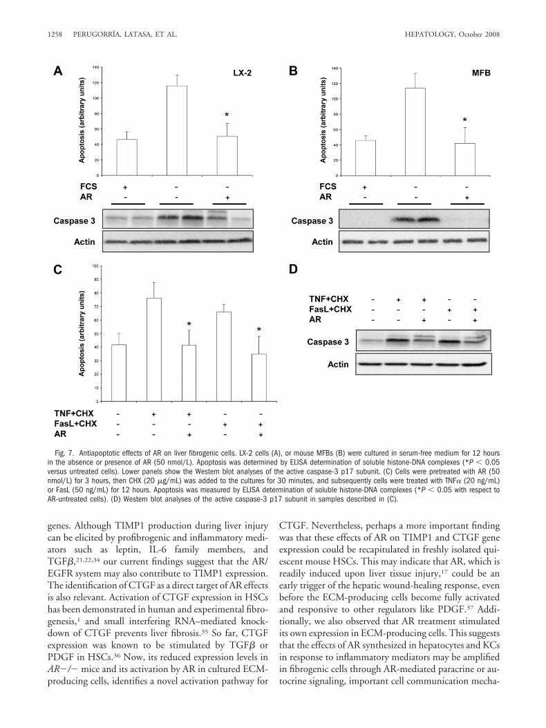
Survival of activated ECM-producing cells is essentialfor the progression of liver fibrosis.23 Therefore, we eval-uated the effects of AR on apoptosis induced by serumwithdrawal in LX-2 cells and mouse MFBs. AR signifi-cantly suppressed apoptosis in both cell types (Fig. 7A,B).In concordance with the antiapoptotic effect of AR, weobserved that production of the active caspase-3 p17 sub-unit was inhibited by AR (Fig. 7A,B, lower panels). Wecould also demonstrate that AR protected LX-2 cells fromapoptosis induced by potent proapoptotic stimuli such asTNF� or Fas ligand (FasL) in the presence of cyclohexi-mide23,24 (Fig. 7C,D).
Next, we explored the signaling mechanisms involvedin the antiapoptotic effects of AR. EGFR activation trig-gers intracellular pathways that may be relevant for theactivation, proliferation, and survival of ECM-producingcells.11,14,25-28 We observed that incubation of LX-2 cellsand mouse MFBs with AR rapidly induced the phosphor-ylation of the EGFR (Fig. 8A). Albeit with different ki-netics, triggering of EGFR by AR was accompanied by theactivation of Akt, ERK1/2, Jun N-terminal kinase (JNK),and focal adhesion kinase (FAK) phosphorylation in bothcell types (Fig. 8A). We next tested the relative contribu-tion of these signaling pathways to the antiapoptotic ef-fects of AR on serum-starved MFB and LX-2 cells.Inhibition of EGFR tyrosine kinase by PD153035 re-sulted in the complete abolition of the cytoprotective ef-fect of AR (Fig. 8B,C). Interestingly, the specificinhibition of phosphoinositide 3-kinase (PI-3K), MEK,JNK, or p38 (not shown) did not significantly impair theantiapoptotic effects of AR (Fig. 8B,C).
DiscussionEGFR ligands such as EGF and TGF� are known to
stimulate the proliferation and activation of isolated he-patic ECM-producing cells.10,12-14,29 However, the rela-tive contribution of the EGF-related factors to liverfibrogenesis is still unknown. Our study provides in vivo
Fig. 5. Effect of AR on TIMP1, CTGF and AR gene expression incultured fibrogenic cells. (A) LX-2 cells and (B) mouse MFBs were treatedwith 50 nmol/L AR in the absence of serum for 6 hours. (C) Mousequiescent HSCs were treated with 50 nmol/L AR in the absence of serumfor 3 hours. mRNA levels were measured by quantitative real-time PCRand are expressed in arbitrary units. Values found in AR-treated cells aredifferent from controls (*P � 0.05).
1256 PERUGORRIA, LATASA, ET AL. HEPATOLOGY, October 2008

evidence showing that AR contributes to the pathologicaccumulation of ECM after chronic injury.
AR gene expression in the healthy liver is very low orundetectable; however, it is significantly elevated duringacute damage and in liver cirrhosis.15 Now, we observedthat AR was constantly expressed in the mouse liver pa-renchyma during CCl4-induced fibrogenesis. Our exper-iments with isolated nonparenchymal liver cells show forthe first time that AR can be up-regulated in response tothe key profibrogenic factor PDGF, as well as through theactivation of the EGFR, in mouse and human ECM-producing cells. Previously, we reported that AR expres-sion was induced in mouse liver upon bacteriallipopolysaccharide (LPS) injection, and also in isolatedhepatocytes treated with inflammatory cytokines like IL-1�.16,17 Here, we show that TNF� and IL-1� also stim-ulated AR expression in isolated KCs, as did the toll-likereceptor-4 ligand LPS. These findings, together with theattenuated fibrogenic response of AR�/� mice describedhere, suggest that AR may represent an additional linkbetween hepatic inflammation and fibrogenesis, an asso-ciation found in clinical and experimental fibrosis, themechanisms of which are currently being exposed.2,8,9,30
AR-deficient mice showed diminished collagen accu-mulation and a significant reduction in the number offibrogenic cells as indicated by decreased expression and
staining of �SMA. These findings were paralleled by re-duced levels of �1(I)procollagen, TIMP1 and CTGFmRNAs, and the profibrogenic cytokine TGF� inAR�/� mice as compared to normal mice undergoingCCl4-induced fibrogenesis. The expression of MMP13was higher in AR�/� mice at peak fibrosis, indicative of astrong tissue remodeling activity.21 Up-regulation ofMMP13 participates in fibrosis resolution after prolongedCCl4 injury in mice.31 In spite of this, we observed en-hanced collagen deposition in AR�/� mice. This can beexplained in part by the increased expression of TIMP1,the principal inhibitor of MMP13,2,3,9,31 in the liver ofwild-type animals. Additionally, the prosurvival effects ofTIMP1 toward HSCs32 may also contribute to the in-creased ECM accumulation observed in AR�/� mice.Nevertheless, the impaired expression of MMP13 and theslow recovery from fibrosis displayed by AR�/� micemay also suggest the implication of AR in the mechanismsinvolved in fibrosis resolution. This is consistent with theview of liver fibrogenesis as a chronic wound-healing re-sponse, in which the expression of ECM components andECM-degrading enzymes is triggered almost concomi-tantly and in many cases by the same factors.1-4,21
We also examined the effects of the direct interactionof AR with ECM-producing cells. We observed that ARcould promote the expression of TIMP1 and CTGF
Fig. 6. Cellular signaling and ef-fects of AR on the expression ofgrowth-related genes and cell prolif-eration in mouse HSCs. (A) Mousequiescent HSCs were serum-starvedfor 1 hour and treated with 50nmol/L AR for 15 minutes. Whereindicated, cells were pretreated for30 minutes with the EGFR inhibitorPD15035 (1 �mol/L), the MEK in-hibitor U0126 (15 �mol/L) or thePI-3K inhibitor LY-294002 (30�mol/L), and phosphorylation ofEGFR, ERK1/2, and Akt was deter-mined by Western blotting. (B) HSCswere treated with AR and the differ-ent inhibitors as mentioned abovefor 1 hour, and the expression ofc-fos and Egr1 was measured byquantitative real-time PCR (*P �0.01 versus control, #P � 0.01 ver-sus AR alone). (C) Freshly isolatedmouse HSCs were cultured for 4days in complete medium and thenserum-starved for 24 hours, afterwhich AR or PDGF-BB (10 ng/mL)were added to the cultures. Cellgrowth was estimated 48 hours lateras described in Materials and Meth-ods (*P � 0.05 and **P � 0.01versus control, respectively).
HEPATOLOGY, Vol. 48, No. 4, 2008 PERUGORRIA, LATASA, ET AL. 1257
genes. Although TIMP1 production during liver injurycan be elicited by profibrogenic and inflammatory medi-ators such as leptin, IL-6 family members, andTGF�,21,22,34 our current findings suggest that the AR/EGFR system may also contribute to TIMP1 expression.The identification of CTGF as a direct target of AR effectsis also relevant. Activation of CTGF expression in HSCshas been demonstrated in human and experimental fibro-genesis,1 and small interfering RNA–mediated knock-down of CTGF prevents liver fibrosis.35 So far, CTGFexpression was known to be stimulated by TGF� orPDGF in HSCs.36 Now, its reduced expression levels inAR�/� mice and its activation by AR in cultured ECM-producing cells, identifies a novel activation pathway for
CTGF. Nevertheless, perhaps a more important findingwas that these effects of AR on TIMP1 and CTGF geneexpression could be recapitulated in freshly isolated qui-escent mouse HSCs. This may indicate that AR, which isreadily induced upon liver tissue injury,17 could be anearly trigger of the hepatic wound-healing response, evenbefore the ECM-producing cells become fully activatedand responsive to other regulators like PDGF.37 Addi-tionally, we also observed that AR treatment stimulatedits own expression in ECM-producing cells. This suggeststhat the effects of AR synthesized in hepatocytes and KCsin response to inflammatory mediators may be amplifiedin fibrogenic cells through AR-mediated paracrine or au-tocrine signaling, important cell communication mecha-
Fig. 7. Antiapoptotic effects of AR on liver fibrogenic cells. LX-2 cells (A), or mouse MFBs (B) were cultured in serum-free medium for 12 hoursin the absence or presence of AR (50 nmol/L). Apoptosis was determined by ELISA determination of soluble histone-DNA complexes (*P � 0.05versus untreated cells). Lower panels show the Western blot analyses of the active caspase-3 p17 subunit. (C) Cells were pretreated with AR (50nmol/L) for 3 hours, then CHX (20 �g/mL) was added to the cultures for 30 minutes, and subsequently cells were treated with TNF� (20 ng/mL)or FasL (50 ng/mL) for 12 hours. Apoptosis was measured by ELISA determination of soluble histone-DNA complexes (*P � 0.05 with respect toAR-untreated cells). (D) Western blot analyses of the active caspase-3 p17 subunit in samples described in (C).
1258 PERUGORRIA, LATASA, ET AL. HEPATOLOGY, October 2008

nisms proposed by early research to participate in HSCactivation.12,29
Activation of EGFR tyrosine kinase on ECM-produc-ing cells has been essentially associated with the stimula-tion of cell proliferation.10-13 We observed that ARtreatment triggered signaling pathways such as ERK1/2,38,39 FAK,28 PI-3K/Akt,25,28,39 and JNK26 that are con-nected with cell proliferation in human and rodent liverfibrogenic cells. Consistently, through the EGFR/ERK1/2 pathway, AR up-regulated the expression ofgrowth-related transcription factors such as c-fos andEgr-1, and stimulated the growth and proliferation ofmouse HSCs. However, intracellular signals emanatingfrom the EGFR, being elicited by AR or other EGFRligands, can also generate potent antiapoptotic stimuli, aspreviously shown in other cell types including hepato-cytes.15,17 Here we demonstrated that AR can overcomeapoptosis induced by FasL or TNF� in LX-2 cells, or by
serum starvation in both LX-2 and mouse MFBs. Inagreement with our previous observations on isolatedhepatocytes,17 the antiapoptotic effects of AR on fibro-genic cells were mediated through the activation of theEGFR. However, downstream of the EGFR, the indepen-dent inhibition of highly protective pathways like JNK,26
PI-3K/Akt,39,40 or ERK1/226,40 did not abolish the anti-apoptotic effects of AR. This is in contrast to what hasbeen recently observed for the fibrogenic cytokine leptin,of which its prosurvival effects on HSCs were strictly de-pendent on PI-3K/Akt activation.39 These findings indi-cate that AR is a potent survival factor for ECM-producing cells, able to elicit redundant antiapoptoticmechanisms that deserve further consideration. In the invivo setting, AR-mediated escape from apoptosis, to-gether with its promitogenic effects, may be importantmechanisms in the promotion of fibrosis.2,7,24,32 How-ever, and in agreement with the potent effects of AR on
Fig. 8. Intracellular signaling and antiapoptotic effects of AR in liver fibrogenic cells. (A) Mouse MFBs or LX-2 cells were serum-starved overnightand then treated with AR (50 nmol/L) for 5 or 10 minutes. Phosphorylation of EGFR, Akt, ERK1/2, JNK, and FAK was determined by Western blotting.(B) Mouse MFBs or (C) LX-2 cells were cultured in the presence or absence of serum or AR (50 nmol/L) for 12 hours. Apoptosis was measured byELISA determination of soluble histone-DNA complexes (*P � 0.05 with respect to AR-untreated cells. #P � 0.05 with respect to AR-treated cells).Where indicated, 30 minutes prior to AR addition, cells were treated with the EGFR inhibitor PD153035 (1 �mol/L), the PI-3K inhibitor LY-294002(30 �mol/L), the MEK inhibitor PD98059 (18 �mol/L) or the JNK inhibitor SP600125 (20 �mol/L).
HEPATOLOGY, Vol. 48, No. 4, 2008 PERUGORRIA, LATASA, ET AL. 1259

freshly isolated nonactivated HSCs, these actions of ARmay be relevant mainly during the early stages of thefibrogenic process, because we did not find significantdifferences in the relative numbers of proliferating andapoptotic �-SMA–positive cells between AR �/� andAR�/� mice after 4 or 6 weeks of CCl4 treatment (datanot shown).
Persistent activation of the EGFR has been cogentlydemonstrated to participate in the pathogenesis of tissuefibrosis in different organs, including the lung and kid-ney.41,42 Furthermore, the EGFR inhibitor gefitinib is asafe and effective inhibitor of experimental kidney fibro-sis43 and hepatocarcinogenesis.44 Our current findings, inaddition to furthering our understanding of the mecha-nisms underlying hepatic fibrogenesis, also suggest thatthe AR/EGFR signaling system could be a new target inthe prevention of liver fibrosis.
Acknowledgment: We thank Dr. S.L. Friedman(Mount Sinai School of Medicine, New York, NY) for theLX-2 cells and his help with the isolation of mouse HSCs.We thank Dr. Jose Romero, Eva Petri, and Maria Azconafor their technical assistance.
Work in the authors’ laboratory is supported by theagreement between FIMA and the “UTE project CIMA”.Red Tematica de Investigacion Cooperativa en CancerRD06 00200061, from Instituto de Salud Carlos III.Grants FIS PI070392, PI070402, and CP04/00123 fromMinisterio de Sanidad y Consumo. Fundacion MutuaMadrilena. Grant Ortiz de Landazuri from Gobierno deNavarra. Grant SAF 2004-03538 from Ministerio deEducacion y Ciencia. M.J.P., M.U.L., and J.C. were sup-ported by a fellowship, a Juan de la Cierva contract and aTorres Quevedo contract from Ministerio de Educacion yCiencia, respectively. The work was also supported by theINSERM, the Universite Paris-Val-de-Marne, and bygrants (to S.L.) of the Agence Nationale de la Rechercheand the Fondation pour la Recherche Medicale.
References1. Bedossa P, Paradis V. Liver extracellular matrix in health and disease.
J Pathol 2003;200:504-515.2. Iredale JP. Models of liver fibrosis: exploring the dynamic nature of inflam-
mation and repair in a solid organ. J Clin Invest 2007;117:539-548.3. Bataller R, Brenner DA. Liver fibrosis. J Clin Invest 2005;115:209-218.4. Lotersztajn S, Julien B, Teixeira-Clerc F, Grenard P, Mallat A. Hepatic
fibrosis: molecular mechanisms and drug targets. Annu Rev PharmacolToxicol 2005;45:605-628.
5. Guyot C, Lepreux S, Combe C, Doudnikoff E, Bioulac-Sage P, BalabaudC, et al. Hepatic fibrosis and cirrhosis: the (myo)fibroblastic cell subpopu-lations involved. Int J Biochem Cell Biol 2006;38:135-151.
6. Knittel T, Kobold D, Piscaglia F, Saile B, Neubauer K, Mehde M, et al.Localization of liver myofibroblasts and hepatic stellate cells in normal anddiseased rat livers: distinct roles of (myo-) fibroblasts subpopulations inhepatic tissue repair. Histochem Cell Biol 1999;112:387-401.
7. Cassiman D, Libbrecht L, Desmet V, Denef C, Roskams T. Hepatic stel-late cell/myofibroblasts subpopulations in fibrotic human and rat livers.J Hepatol 2002;36:200-209.
8. Gressner AM, Weiskirchen R. Modern pathogenetic concepts of liver fi-brosis suggest stellate cells and TGF-� as major players and therapeutictargets. J Cell Mol Med 2006;10:76-99.
9. Hui AY, Friedman SL. Molecular basis of hepatic fibrosis. Expert Rev MolMed 2003;5:1-23.
10. Pinzani M, Gesualdo L, Sabbah GM, Abboud HE. Effects of platelet-derived growth factor and other polypeptide mitogens on DNA synthesisand growth of cultured rat liver fat-storing cells. J Clin Invest 1989;84:1786-1793.
11. Svegliati-Baroni G, Ridolfi F, Hannivoort R, Saccomanno S, Homan M,De Minicis S, et al. Bile acids induce hepatic stellate cell proliferation viaactivation of the epidermal growth factor receptor. Gastroenterology 2005;128:1042-1055.
12. Bachem MG, Meyer D, Melchior R, Sell KM, Gressner AM. Activation ofrat liver perisinusoidal lipocytes by transforming growth factors derivedfrom myofibroblastlike cells. J Clin Invest 1992;89:19-27.
13. Bachem MG, Riess U, Gressner AM. Liver fat storing cell proliferation isstimulated by epidermal growth factor/transforming growth factor alphaand inhibited by transforming growth factor beta. Biochem Biophys ResCommun 1989;162:708-714.
14. Yang C, Zeisberg M, Mosterman B, Sudhakar A, Yerramalla U, HolthausK, et al. Liver fibrosis: insights into migration of hepatic stellate cells inresponse to extracellular matrix and growth factors. Gastroenterology2003;124:147-159.
15. Berasain C, Castillo J, Prieto J, Avila MA. New molecular targets forhepatocellular carcinoma: the Erbb1 signaling system. Liver Int 2007;27:174-185.
16. Berasain C, Garcıa-Trevijano ER, Castillo J, Erroba E, Lee DC, Prieto J, etal. Amphiregulin: an early trigger for liver regeneration in mice. Gastroen-terology 2005;128:424-432.
17. Berasain C, Garcıa-Trevijano ER, Castillo J, Erroba E, Santamarıa M, LeeDC, et al. Novel role for amphiregulin in protection from liver injury.J Biol Chem 2005;280:19012-19020.
18. Blobel CP. ADAMs: key components in EGFR signalling and develop-ment. Nat Rev Mol Cell Biol 2005;6:32-43.
19. Wong FW, Chan WY, Lee SS. Resistance to carbon tetrachloride-inducedhepatotoxicity in mice which lack CYP2E1 expression. Toxicol Appl Phar-macol 1998;153:109-118.
20. Niki T, De Bleser PJ, Xu G, Van Den Berg K, Wisse E, Geerts A. Com-parison of glial fibrillary acidic protein and desmin staining in normal andCCl4-induced fibrotic rat livers. HEPATOLOGY 1996;23:1538-1545.
21. Hemann S, Graf J, Roderfeld M, Roeb E. Expression of MMPs and TIMPsin liver fibrosis-a systematic review with special emphasis on anti-fibroticstrategies. J Hepatol 2007;46:955-975.
22. Jeong W, Park O, Radaeva S, Gao B. STAT1 inhibits liver fibrosis in miceby inhibiting stellate cell proliferation and stimulating NK cell cytotoxic-ity. HEPATOLOGY 2006;44:1441-1451.
23. Elsharkawy AM, Oakley F, Mann DA. The role and regulation of hepaticstellate cell apoptosis in reversal of liver fibrosis. Apoptosis 2005;10:927-939.
24. Novo E, Marra F, Zamara E, Valfre-Bonzo L, Monitillo L, Cannito S, et al.Overexpression of Bcl-2 by activated human hepatic stellate cells: resis-tance to apoptosis as a mechanism of progressive hepatic fibrogenesis inhumans. Gut 2006;55:1174-1182.
25. Godichaud S, Si-Tayeb K, Auge N, Desmouliere A, Balabaud C, PayrastreB, et al. The grape-derived polyphenol resveratrol differentially affectsepidermal and platelet-derived growth factor signaling in human livermyofibroblasts. Int J Biochem Cell Biol 2006;38:629-637.
26. Zhou Y, Zheng S, Lin J, Zhang QJ, Chen A. The interruption of thePDGF and EGF signalling pathways by curcumin stimulates gene expres-sion of PPAR� in rat activated hepatic stellate cell in vitro. Lab Invest2007;87:488-498.
27. Lee KS, Buck M, Houglum K, Chojkier M. Activation of hepatic stellatecells by TGF� and collagen type I is mediated by oxidative stress throughc-myb expression. J Clin Invest 1995;96:2461-2468.
28. Reif S, Lang A, Lindquist JN, Yata Y, Gabele E, Scanga A, et al. The role offocal adhesion kinase-phosphatidylinositol 3-kinase-Akt signaling in he-
1260 PERUGORRIA, LATASA, ET AL. HEPATOLOGY, October 2008

patic stellate cell proliferation and type I collagen expression. J Biol Chem2003;278:8083-8090.
29. Gressner AM. Cytokines and cellular crosstalk involved in the activation offat-storing cells. J Hepatol 1995;22:28-36.
30. Seki E, De Minicis S, Osterreicher C, Kluwe J, Osawa Y, Brenner DA, et al.TLR4 enhances TGF-� signaling and hepatic fibrosis. Nat Med 2007;13:1324-1332.
31. Fallowfield JA, Mizuno M, Kendall TJ, Constandinou CM, Benyon RC,Duffield JS, et al. Scar-associated macrophages are a major source of he-patic matrix metalloproteinase-13 and facilitate the resolution of murinehepatic fibrosis. J Immunol 2007;178:5288-5295.
32. Murphy FR, Issa R, Zhou X, Ratnarajah S, Nagase H, Arthur MJ, et al.Inhibition of apoptosis of activated hepatic stellate cells by tissue inhibitorof metalloproteinase-1 is mediated via effects on matrix metalloproteinaseinhibition: implications for reversibility of liver fibrosis. J Biol Chem 2002;277:11069-11076.
33. Cao Q, Mak KM, Ren C, Lieber CS. Leptin stimulates tissue inhibitor ofmetalloproteinase-1 in human hepatic stellate cells. J Biol Chem 2004;279:4292-4304.
34. Nieto N. Oxidative-stress and IL-6 mediate the fibrogenic effects of rodentKupffer cells on stellate cells. HEPATOLOGY 2006;44:1478-1501.
35. George J, Tsutsumi M. siRNA-mediated knockdown of connective tissuegrowth factor prevents N-nitrosodimethylamine-induced hepatic fibrosisin rats. Gene Ther 2007;14:790-803.
36. Paradis V, Dargere D, Bonvoust F, Viaud M, Segarini P, Bedossa P. Effectsand regulation of connective tissue growth factor on hepatic stellate cells.Lab Invest 2002;82:767-774.
37. Bachem MG, Meyer D, Schafer W, Riess U, Melchior R, Sell KM, et al.The response of rat liver perisinusoidal lipocytes to polypeptide growthregulator changes with their transdifferentiation into myofibroblast-likecells in culture. J Hepatol 1993;18:40-52.
38. Marra F, Arrighi MC, Fazi M, Caligiuri A, Pinzani M, Romanelli RG, et al.Extracellular signal-regulated kinase activation differentially regulatesplatelet-derived growth factor’s actions in hepatic stellate cells, and is in-duced by in vivo liver injury in the rat. HEPATOLOGY 1999;30:951-958.
39. Saxena NK, Titus MA, Ding X, Floyd J, Srinivasan S, Sitaraman SV, et al.Leptin as a novel profibrogenic cytokine in hepatic stellate cells: mitogen-esis and inhibition of apoptosis mediated by extracellular regulated kinase(Erk) and Akt phosphorylation. FASEB J 2004;18:1612-1614.
40. Davaille J, Li L, Mallat A, Lotersztajn S. Sphingosine 1-phosphate triggersboth apoptotic and survival signals for human hepatic myofibroblasts.J Biol Chem 2002;277:37323-37330.
41. Madtes DK, Elston AL, Hackman RC, Dunn AR, Clark JG. Transforminggrowth factor-� deficiency reduces pulmonary fibrosis in transgenic mice.Am J Respir Cell Mol Biol 1999;20:924-934.
42. Lautrette A, Li S, Alili R, Sunnarborg SW, Burtin M, Lee DC, et al.Angiotensin II and EGF receptor cross-talk in chronic kidney diseases: anew therapeutic approach. Nat Med 2005;11:867-874.
43. Francois H, Placier S, Flamant M, Tharaux PL, Chansel D, Dussaule JC,et al. Prevention of renal vascular and glomerular fibrosis by epidermalgrowth factor receptor inhibition. FASEB J 2004;18:926-928.
44. Schiffer E, Housset C, Cacheux W, Wendum D, Desbois-Mouthon C,Rey C, et al. Gefitinib, an EGFR inhibitor, prevents hepatocellular carci-noma development in the rat liver with cirrhosis. HEPATOLOGY 2005;41:307-314.
HEPATOLOGY, Vol. 48, No. 4, 2008 PERUGORRIA, LATASA, ET AL. 1261