epidermal growth factor receptor in breast carcinoma: association
TRANSCRIPT

RESEARCH Open Access
Epidermal growth factor receptor in breastcarcinoma: association between gene copynumber and mutationsNing Lv1,2, Xiaoming Xie1,2, Qidong Ge1,2, Suxia Lin2,3, Xi Wang1,2, Yanan Kong1,2, Hongliu Shi1,2, Xinhua Xie1,2 andWeidong Wei1,2*
Abstract
Background: The epidermal growth factor receptor (EGFR) is an available target of effective anti-EGFR therapy forhuman breast cancer. The aim of this study was to assess the presence of EGFR gene amplification and mutationsin breast cancer and to analyze the association between the statuses of these two gene alterations.
Materials and methods: EGFR gene amplification and mutations were investigated in formalin-fixed, paraffin-embedded tissues from 139 Chinese female patients with breast cancer by means of fluorescence in-situhybridization (FISH) and fluorescently labeled real-time quantitative polymerase chain reaction (RT-PCR),respectively.
Results: EGFR gene amplification was observed in 46/139 (33.1%) of cases by FISH. Based on RT-PCR, 2/139 (1.4%)samples had EGFR gene mutations. Overall, only 1 (0.7%) of the cases was identified with both whole geneamplification and mutation, and 92 (66.2%) of cases were negative for both. High gene copy numbers of EGFR hadsignificant correlation with the occurrence of EGFR protein expressions (P = 0.002).
Conclusion: In this study, EGFR mutations were presented in only two samples, indicating that EGFR mutationsshould not be employed in future trials with anti-EGFR therapies for breast cancer. However, EGFR whole geneamplification is frequently observed in patients with breast cancer. It will be of significant interest to investigatewhether EGFR gene copy number is a suitable screening test for EGFR-targeted therapy for breast cancer.
Keywords: breast cancer, epidermal growth factor receptor, EGFR, FISH, gene amplification, gene mutation, real-time PCR.
IntroductionThe human epidermal growth factor receptor (HER/EGFR/ErbB) family of receptor tyrosine kinases is com-prised of four transmembrane growth factor receptorproteins that share similarities in structure and function.The epidermal growth factor receptor (HER-1/EGFR/ErbB1), encoded by the gene located on the short armof chromosome 7, is a member of this family of Type Itransmembrane tyrosine kinase receptors. EGFR is a 170kDa transmembrane protein consisting of an intracellu-lar domain (tyrosine kinase domain), a short
transmembrane and juxtamembrane domain, and anextracellular domain (ligand-binding domain) withligand-activated tyrosine kinase activity [1]. EGFR canbe activated by various growth factor ligands, includingepidermal growth factor (EGF) and the transforminggrowth factor-alpha (TGF-a). Ligand binding to EGFRresults in homo- or hetero-dimerization of EGFR withanother EGFR molecule or a different member of theErbB family (e.g., HER2). This is followed by phosphory-lation of the tyrosine kinase residue, which in turninduces the actual downstream signaling cascade [2-4].Ligand-dependent activation of the EGFR tyrosinekinase residues serve as binding sites of signal transdu-cer and activator proteins that mediate the downstreamsignaling processes of intracellular substrates [5]. The
* Correspondence: [email protected] of Breast Oncology, Sun Yat-Sen University Cancer Center,Guangzhou, Guangdong 510060, P. R. ChinaFull list of author information is available at the end of the article
Lv et al. Diagnostic Pathology 2011, 6:118http://www.diagnosticpathology.org/content/6/1/118
© 2011 Lv et al; licensee BioMed Central Ltd. This is an Open Access article distributed under the terms of the Creative CommonsAttribution License (http://creativecommons.org/licenses/by/2.0), which permits unrestricted use, distribution, and reproduction inany medium, provided the original work is properly cited.

phosphatidyl inositol 3’ kinase (PI3K) and Akt pathwayand Ras/MAPK pathway are major signaling mechan-isms, and they function in the control of several impor-tant biologic processes, including cell proliferation,survival, angiogenesis, and migration as well as resis-tance to apoptosis [6-8].Due to the biologic significance of EGFR molecular
signaling in carcinomas, several monoclonal antibodiesagainst the ligand-binding domain of EGFR and smallmolecule tyrosine kinase inhibitors of the tyrosinekinase domain of EGFR have been investigated in thetherapy of malignant tumors (e.g., non-small cell lungcancer [NSCLC], colorectal cancer [CRC] and metastaticbreast cancer [MBC]) [9-16]. It is important to studywhether EGFR is overexpressed in patients with breastcancer since these patients can be given specific EGFRmolecule tyrosine kinase inhibitors such as gefitinib andlapatinib [15,16]. There are only a few reports regardingthe overexpression of EGFR, with these studies indicat-ing 8-36% of breast cancers over express this protein.However, systematic studies appraising EGFR geneamplification and mutations in the same set of casesamong Chinese female patients with breast cancer areabsent [17-19]. Many studies have concentrated on lungcancers, where most patients ultimately have a relapse.Mechanisms involved in resistance to targeted inhibitionof lung cancer include secondary resistance mutations,inactivation of PTEN, activation of the MET pathway,minor clones with KRAS mutations, and adenocarci-noma transformation [20-32]. However, the mechanismof drug resistance in breast cancer is unknown.The purpose of the present study was to examine 139
formalin-fixed, paraffin-embedded specimens from Chi-nese female patients with breast cancer, with a particu-lar focus on the presence of EGFR gene amplificationand mutations. We attempted to explore the relation-ship between EGFR copy numbers and EGFR mutations.We also analyzed the correlation between EGFR genestatus and HER2 protein, estrogen receptor (ER), pro-gesterone receptor (PR), and cytokeratin 5/6 (CK5/6)expression as well as Ki-67 index proliferation andintrinsic subtypes in these cases. In this study, we ana-lyzed EGFR gene copy numbers by fluorescence in-situhybridization (FISH), and the mutations were analyzedusing a real-time (RT)-PCR detection kit.
Methods and MaterialsPatient InformationOne hundred and thirty-nine Chinese female patientswith breast cancer who underwent surgery at theDepartment of Breast Oncology, Sun Yat-Sen UniversityCancer Center, from Jan 2010 to May 2011 wereselected. The cases with primary breast cancer were ran-domly selected from the archives of our Department of
Pathology based on the availability of blocks and suffi-cient tissues. Additionally, only cases with availableEGFR FISH results, mutational status, and immunos-taining were analyzed. Clinical information included age,disease stage, tumor type, mass size, and axillary lymphnode metastasis status. The age interval of the patientswas 25-75 years, with a mean of 50.8 years. The TNMCancer Staging Manual 7th edition of the AmericanJoint Committee on Cancer (AJCC) [33] was used toclassify the cancer staging: stage 0, 2 cases; stage I, 28cases; stage II, 78 cases; stage III, 30 cases; and stage IV,1 case. Using immunohistochemistry as a surrogate defi-nition of intrinsic subtypes for expression profiling,cases that were ER and/or PR positive, HER2 negative,and Ki-67 low (< 14%) were classified as luminal A can-cers; cases that were ER and/or PR positive, HER2 nega-tive, and Ki-67 high were classified as luminal B (HER2positive) cancers; cases that were ER and/or PR positive,any Ki-67, and HER2 overexpressed or amplified wereclassified as luminal B (HER2 positive); cases that wereHER2 overexpressed or amplified and ER and PR absentwere classified as HER2 positive (non-luminal) cancers;cases that were ER and PR absent, HER2 negative, andCK5/6 and/or EGFR positive were classified as basal-likecancers; and cases that lacked expression of ER, PR,HER2, CK5/6, and EGFR were considered unclassified[34-36]. The histopathological classification of breastcancer in these cases was performed by an experiencedpathologist of our pathology department. Ethicsapproval was obtained from the Ethical Review Commit-tee of Sun Yat-Sen University Cancer Center, andinformed consent was obtained from all patients.
Tissue PreparationTissue microarrays were constructed using 0.6-mm tis-sue cores as previously described [37]. One core fromthe central and the other from the peripheral part of thetumor were sampled for each tumor. For each case, 4-5μm sections of formalin-fixed and paraffin-embeddedtissues were stained with Hematoxylin and Eosin for theestablishment of the histopathological tumor type anddifferentiation grade.
Fluorescence In-Situ Hybridization Analysis of EGFR GeneCopy NumberFISH analysis for EGFR gene copy number was per-formed according to the manufacturer’s protocol usingthe GLP EGFR/CSP 7 probe (GP Medical Technologies,Beijing, China). Simply, the tissue microarray sectionswere incubated at 65°C overnight. The slides weredeparaffinized in dimethyl benzene at room temperaturefor 10 minutes and dehydrated in 100% ethanol. Afterincubation in 30% sodium bisulfite at 50°C for 20-30minutes, the sections were incubated in 2× saline
Lv et al. Diagnostic Pathology 2011, 6:118http://www.diagnosticpathology.org/content/6/1/118
Page 2 of 9

sodium citrate buffer (2× SSC; pH 7.0) at 75°C for 5minutes. The smears were digested with proteinase K(0.20 mg/ml in 2× SSC; pH 7.0) for 20-30 minutes at37°C, followed by a rinse in 2× SSC at room tempera-ture for 5 minutes and then dehydration in 70%, 85%,and 100% ethanol solutions in sequence. The solventswere changed frequently and regularly so that all tracesof residual paraffin were removed in the above processes[38]. After amplification of the GLP EGFR/CSP 7 probeset, the slide was covered with a coverslip and sealedwith Indian rubber. The slides were heated at 80°C for8-10 minutes and were then hybridized at 42°C over-night. After a post-hybridization wash and dehydrationof the samples, 4’, 6’-diamidino-2-phenylindole (DAPI)was applied for chromatin counterstaining. At least 100nuclei were scored for both EGFR gene signals andchromosome 7 signals under a magnification of 1000×.The ratio was defined as: the total number of red sig-
nals (EGFR copy number) divided by the number ofgreen signals (chromosome 7 copy number) in 100nuclei. Tumors were scored as EGFR amplified whenthe EGFR FISH-positive results were: a) ratio ≥2.0, b)≥15 copies of the red signals per cell in ≥10% of totalcells, or c) the presence of EGFR gene clusters; orscored as EGFR polysomy when: ratio < 2.0, but ≥40%of cells display ≥4 copies. Meanwhile, EGFR FISH-nega-tive include disomy (≥90% of cells display ≤2 copies),low trisomy (≥40% of cells display ≤2 copies, 10-40% ofcells display 3 copies, and < 10% of cells display ≥4copies), high trisomy (≥40% of cells display ≤2 copies,≥40% of cells display 3 copies, and < 10% of cells display≥4 copies), and low polysomy (10-40% of cells display≥4 copies) [38-40]. Healthy cells were used as controls.
Fluorescent PCR Method for Analysis of EGFR GeneMutationsThe 5-μm formalin-fixed and paraffin-embedded tissueswere assayed for the presence of the most commonEGFR mutations in exon 19 (E746-A750 and L747-P753insS short in-frame deletions) and exon 21 (L858Rand L861Q point mutations) using a RT-PCR detectionkit with the Taqman probe technique (GP MedicalTechnologies, Beijing, China). Genomic DNA wasextracted from tissues using a TIANamp Genomic DNAkit (Tiangen Biotech, Beijing, China). Two of the exon19 deletions and the exon 21 mutations of the EGFRgene were analyzed using fluorescently labeled RT-PCRproducts.Amplification reactions were setup using reagents
included in the Real Time PCR Detection Kit (GP Medi-cal Technologies, Beijing, China), in accordance with themanufacturer’s protocol. Essentially, the exon 19 PCRreaction consisted of 5.4 ul deionized water, 7.5 ul 2 ×PCR pre-mix, 0.15 ul forward primers-1, 0.15 ul reverse
primers-1, 0.15 ul probe-1 (delE746-A750), and 0.15 ulprobe-2 (delL747-P753insS) in a total volume of 13.5 ul.The exon 21 PCR reaction consisted of 5.3 ul deionizedwater, 7.5 ul 2 × PCR pre-mix, 0.15 ul forward primers-2, 0.15 ul reverse primers-2, 0.2 ul probe-3 (L858R), and0.2 ul probe-4 (L861Q) in a total volume of 13.5 ul. ThePCR cycling program was as follows: 50°C for 2 min,95°C for 10 min, and 40 cycles of 95°C for 15 sec, 62°Cfor 1 min. PCR analysis was performed by using an ABIPrism 7500 Real-Time PCR equipment (Applied Biosys-tems, Foster City, CA, USA) as previously described[41,42].
Immunohistochemistry of EGFR Protein ExpressionImmunohistochemical staining for EGFR was performedon the 5-μm formalin-fixed, paraffin-embedded tissueslide. EGFR was stained using the pharmDx kit accord-ing to manufacturer’s instructions (DAKO). EGFR wasscored positive if any membranous tumor cell stainingwas observed, whether or not it was completely circum-ferential. Staining intensity was scored as follows: 0, nomembrane staining; 1+, weak staining intensity; 2+,moderate staining intensity; 3+, strong staining intensity.The staining intensity was multiplied by the percentageof tumor cells be stained to obtain a total score, result-ing in a possible range 0 to 300. Samples with an overallscore of 200 and higher were considered positive forEGFR-overexpression.
Statistical analysisSPSS version 13.0 software (SPSS, Chicago, IL, USA) wasused to analyze the data. Associations between EGFRgene amplification and protein expression were evaluatedusing Pearson’s chi-square test with cross tables. Differ-ences of P < 0.05 were considered significant. Pearson’sChi-squared test or Fisher’s exact test was also appliedfor evaluation of multiple comparisons between EGFRamplification and expression and age, disease stage,tumor type, axillary lymph node metastasis status, andimmunohistochemical index (i.e., ER, PR, and HER2). AP value < 0.05 was considered statistically significant.
ResultsEGFR Gene Copy Numbers in Breast CarcinomasWe obtained both FISH and RT-PCR EGFR data on 139female patients with breast cancer. A total of 48 (34.5%)of the 139 tumors presented EGFR disomy, 6 tumors(4.3%) presented low trisomy, 3 tumors (2.2%) presentedhigh trisomy, 36 (25.9%) tumors presented low polys-omy, 42 tumors (30.2%) presented high polysomy, and 4tumors (2.9%) presented amplification. From the total of139 tumors, 46 (33.1%) presented positivity with FISH;93 (66.9%) did not demonstrate EGFR gene amplifica-tion (Figure 1).
Lv et al. Diagnostic Pathology 2011, 6:118http://www.diagnosticpathology.org/content/6/1/118
Page 3 of 9
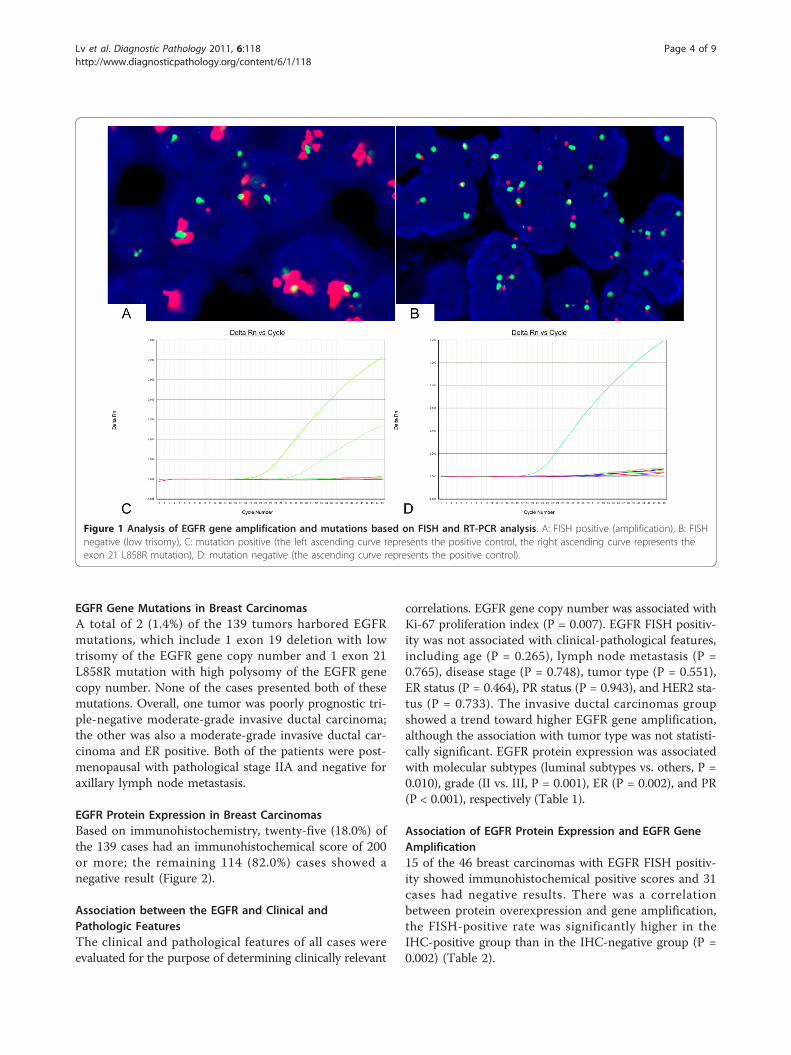
EGFR Gene Mutations in Breast CarcinomasA total of 2 (1.4%) of the 139 tumors harbored EGFRmutations, which include 1 exon 19 deletion with lowtrisomy of the EGFR gene copy number and 1 exon 21L858R mutation with high polysomy of the EGFR genecopy number. None of the cases presented both of thesemutations. Overall, one tumor was poorly prognostic tri-ple-negative moderate-grade invasive ductal carcinoma;the other was also a moderate-grade invasive ductal car-cinoma and ER positive. Both of the patients were post-menopausal with pathological stage IIA and negative foraxillary lymph node metastasis.
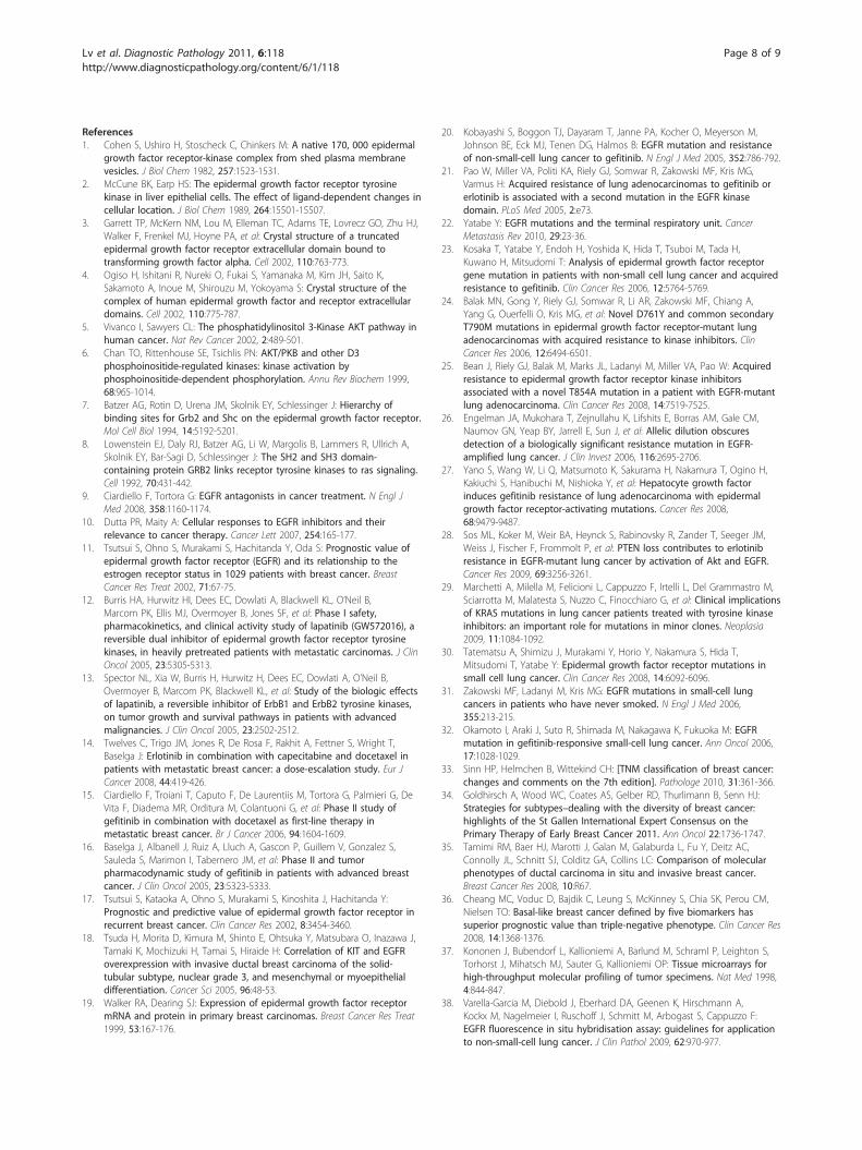
EGFR Protein Expression in Breast CarcinomasBased on immunohistochemistry, twenty-five (18.0%) ofthe 139 cases had an immunohistochemical score of 200or more; the remaining 114 (82.0%) cases showed anegative result (Figure 2).
Association between the EGFR and Clinical andPathologic FeaturesThe clinical and pathological features of all cases wereevaluated for the purpose of determining clinically relevant
correlations. EGFR gene copy number was associated withKi-67 proliferation index (P = 0.007). EGFR FISH positiv-ity was not associated with clinical-pathological features,including age (P = 0.265), lymph node metastasis (P =0.765), disease stage (P = 0.748), tumor type (P = 0.551),ER status (P = 0.464), PR status (P = 0.943), and HER2 sta-tus (P = 0.733). The invasive ductal carcinomas groupshowed a trend toward higher EGFR gene amplification,although the association with tumor type was not statisti-cally significant. EGFR protein expression was associatedwith molecular subtypes (luminal subtypes vs. others, P =0.010), grade (II vs. III, P = 0.001), ER (P = 0.002), and PR(P < 0.001), respectively (Table 1).
Association of EGFR Protein Expression and EGFR GeneAmplification15 of the 46 breast carcinomas with EGFR FISH positiv-ity showed immunohistochemical positive scores and 31cases had negative results. There was a correlationbetween protein overexpression and gene amplification,the FISH-positive rate was significantly higher in theIHC-positive group than in the IHC-negative group (P =0.002) (Table 2).
Figure 1 Analysis of EGFR gene amplification and mutations based on FISH and RT-PCR analysis. A: FISH positive (amplification), B: FISHnegative (low trisomy), C: mutation positive (the left ascending curve represents the positive control, the right ascending curve represents theexon 21 L858R mutation), D: mutation negative (the ascending curve represents the positive control).
Lv et al. Diagnostic Pathology 2011, 6:118http://www.diagnosticpathology.org/content/6/1/118
Page 4 of 9

DiscussionThe aim of this study was to evaluate the frequency ofEGFR gene amplification and mutations in 139 femalepatients with breast cancer. It is known that EGFR geneamplification indicates EGF-sensitive breast cancer. Inone study, EGFR gene amplification and/or high EGFRexpression were demonstrated as biological predictors ofpoor prognosis in breast carcinoma [43]. Due to theavailability and benefit of anti-EGFR therapies, includingboth monoclonal antibodies (MoAbs) and small mole-cule tyrosine kinase inhibitors (TKI), for the treatmentof various solid malignant tumors, such as non-smallcell lung cancer (NSCLC), squamous cell carcinoma ofthe head and neck (HNSCC), and colorectal cancer(CRC), the role of EGFR gene status has been investi-gated in a number of clinical studies. Several data setsregarding EGFR gene amplification in breast cancer areaccessible. In various trials, EGFR gene amplification inbreast carcinomas was different, ranging between 0.8-28percent. Khawla Al-Kuraya et al. [44] described EGFR
gene amplification in 0.8% of studied tumors, Jungsil Roet al. [43] reported positivity in 3 of the 21 evaluablecases, Rohit Bhargava et al. [45] found positive EGFRamplification in 11/175 (6%) of samples, Christian Ker-sting et al. [46] showed EGFR whole gene amplificationin 4.7% of investigated cases, and in the cohorts ofJudith A. Gilbert et al. [47] and Jorge S Reis-Filho et al.[48] 26% and 28% of the metastatic breast carcinomasdisplayed high EGFR copy number, respectively. It islikely that multiple techniques and scoring systems usedin the detection of EGFR amplification have led to theinconsistent outcomes of these different trials.In this current study we used FISH to detect EGFR
gene copy numbers in breast carcinomas. We identifiedEGFR gene amplification in 46 (33.1%) of the 139patients with breast cancer. This percentage is higherthan the range reported by the studies mentioned above.It seems that the Chinese origin of the specimens, possi-bly in addition to the use of various techniques andscoring criteria, may possibly have contributed to the
Figure 2 EGFR protein expression by immunohistochemistry. A: Negative EGFR expression; B: 1+ EGFR expression; C: 2+ EGFR expression; D:3+ EGFR expression.
Lv et al. Diagnostic Pathology 2011, 6:118http://www.diagnosticpathology.org/content/6/1/118
Page 5 of 9

Table 1 Summary of the relationship between EGFR copy number, expression, and the patients’ clinical-pathologicalcharacteristics
clinical-pathological characteristic
Copy number Expression
Positive, N/% Negative, N/% P value Positive, N/% Negative, N/% P value
Age(years)
≤35 5/10.9 4/4.3 0.265c 23/92.0 107/93.9 1.000c
>35 41/89.1 89/95.7 2/8.0 7/6.1
Lymph node metastasis
Positive 23/50 49/52.7 0.765a 16/64.0 56/49.1 0.178a
Negative 23/50 44/47.3 9/36.0 58/50.9
Stage
0 0/0 2/2.2 0.748a, $ 1/4.0 2/1.8 0.450a, $
I 10/21.7 18/19.4 6/24.0 21/18.4
II 25/54.3 53/57 11/44.0 67/58.8
III 10/21.7 20/21.5 7/28.0 22/20.2
IV 1/2.2 0/0 0/0 1/0.9
Tumor type
DCIS 0/0 3/3.2 0.551b, & 1/4.0 2/1.8 1.000c, &
LCIS 0/0 0/0 0/0 0/0
IDC 44/95.7 83/89.2 23/92.0 104/91.2
ILC 0/0 3/3.2 0/0 3/2.6
Other 2/4.3 4/4.3 1/4.0 5/4.4
ER
Positive 33/71.7 72/77.4 0.464a 13/52.0 92/80.7 0.002a
Negative 13/28.3 21/22.6 12/48.0 22/19.3
PR
Positive 27/58.7 54/58.1 0.943a 5/20.0 76/66.7 0.000a
Negative 19/41.3 39/41.9 20/80.0 38/33.3
HER2
Positive 9/19.6 16/17.2 0.733a 5/20.0 29/25.4 0.567a
Negative 37/80.4 77/82.8 20/80.0 85/74.6
Ki-67 (%)
< 14 7/15.2 35/37.6 0.007a 17/68.0 67/58.8 0.393a
> 16 39/84.8 58/62.4 8/32.0 47/41.2
Subtypes
LUMA 9/19.6 28/30.1 0.628a, ^ 4/16.0 33/28.9 0.010c, ^
LUMB (HER2-NEG) 17/37.0 32/34.4 7/28.0 42/36.8
LUMB (HER2-POS) 10/21.7 16/17.2 4/16.0 22/19.3
HER2 2/4.3 6/17.2 1/4.0 7/6.1
Basal-like 6/13.0 4/4.3 9/36.0 1/0.9
UC 2/4.3 7/7.5 0/0 9/7.9
Grade
I 0/0 0/0 0.921a, # 0/0 0/0 0.001a, #
II 31/67.4 52/55.9 10/40.0 73/64.0
III 12/26.1 21/22.6 13/52.0 20/17.5
UC 3/6.5 20/21.5 2/8.0 21/18.4aP values (two-sided) calculated using Pearson’s chi-square test.bP values (two-sided) calculated using Fisher’s exact test.cP values (two-sided) calculated using Continuity Correction of Pearson’s chi-square test.$Pearson’s chi-square test for stage 0-II and III- IV vs. EGFR status.&Fisher’s exact test for invasive ductal carcinoma and other types vs. EGFR status.
^Pearson’s chi-square test for luminal subtypes and other subtypes vs. EGFR status.#Grade II and III vs. EGFR status.
DCIS, ductal carcinoma in-situ; LCIS, lobular carcinoma in-situ; IDC, invasive ductal carcinoma; ILC, invasive lobular carcinoma; ER, estrogen receptor; PR,progesterone receptor; HER2, human epidermal growth factor receptor-2; Ki-67, Ki-67 proliferation index; LUMA, luminal A; LUMB (HER2-NEG), luminal B (HER2-negative); LUMB (HER2-POS), luminal B (HER2-positive); UC, unclassified.
Lv et al. Diagnostic Pathology 2011, 6:118http://www.diagnosticpathology.org/content/6/1/118
Page 6 of 9

difference in the results. In this study, 46 carcinomasshowed EGFR gene amplification, which include 42tumors (30.2%) presenting high polysomy and 4 tumors(2.9%) presenting amplification (based on the ratio ofEGFR gene copies to CEP7 gene copies in at least 100tumor cell nuclei). These data revealed that EGFR geneamplification is a frequent event in Chinese patientswith breast carcinomas. As observed from this study,EGFR positive immunostaining was consistent withEGFR gene amplification. It appears that positive EGFRprotein overexpression could predict gene amplificationin breast cancers.Activation of EGFR involves heterodimerization of
EGFR with HER2. Our results showed that there was nocorrelation between EGFR and HER2 protein expression(P = 0.567), which might indicated that activated EGFRcan form heterodimer not only with HER2, but alsowith other members of ErbB family.Currently, many trials have assayed for EGFR gene
amplification in order to identify patients that wouldbenefit from anti-EGFR therapy. Patients with EGFRgene amplification have been connected to poor prog-nosis in HNSCC [49,50] and NSCLC [51]. However,patients with EGFR mutations have demonstrated anincreased benefit as compared to patients having EGFRamplification [39]. EGFR gene mutations indicate sensi-tivity to gefitinib, and it was demonstrated that about85% of patients with NSCLC who obtained benefit fromgefitinib treatment were found to have mutations inexons 18 to 21 of the tyrosine kinase domain of theEGFR gene [51-54]. EGFR mutations in exon 19 (shortin-frame deletions) or 21 L858R (point mutation) affectthe adenosine triphosphate (ATP) pocket of the tyrosinekinase domain leading to the activation of 4-anilinoqui-nazoline compounds, which function to compete withATP [53]. Anti-EGFR therapy can consequently lead tothe downregulation of downstream signaling cascades,such as the PI3K/Akt, RAS/Erk, MAPK, and STATpathways, responsible for cell proliferation and survival,resulting in the inhibition of cell proliferation andinduction of cell apoptosis, respectively. The method forEGFR-mutations used in this study can detect the mostcommon mutations of exons 19 and 21, but there arestill other mutations of exons 19 and 21 that cannot bedetected. Additionally, mutations of exons 18 and 20,
which can harbor upto 15% of EGFR-mutations in lungcancer, cannot be analyzed in this way.It is reported in some studies that EGFR gene muta-
tions in the tyrosine kinase domain in patients withlung cancer are accompanied with a low increase inEGFR gene copy number [55,56]. However, in thisstudy, EGFR gene mutations could be identified in only2 out of 139 cases (1.4%) of the breast carcinoma sam-ples, confirming that EGFR gene mutations are rare inChinese patients. Further trials with large samples and/or different methods are highly recommended to be per-formed to validate the observations mentioned above.
ConclusionsWe observed that EGFR gene mutations were rare inbreast carcinomas, but EGFR gene amplification wasdetected in about one third of the cases in this popula-tion. In this study, rare mutations in the EGFR gene inpatients with breast cancer were detected, indicatingthat EGFR gene mutations are infrequent in this cohortof breast cancers. This suggested that EGFR mutationanalysis is not useful as a screening test for sensitivity toanti-EGFR therapy for breast cancers. Nevertheless,further studies will be required to investigate whetherEGFR gene copy number is a suitable screening test forEGFR targeted therapy.
AbbreviationsATP: adenosine triphosphate; CK5/6: cytokeratin 5/6; CRC: colorectal cancer;DAPI: 4’: 6’-diamidino-2-phenylindole; EGFR: epidermal growth factorreceptor; ER: estrogen receptor; FISH: fluorescence in-situ hybridization; HER2:human epidermal growth factor receptor-2; HNSCC: squamous cellcarcinoma of the head and neck; MBC: metastatic breast cancer; MoAbs:monoclonal antibodies; NSCLC: non-small cell lung cancer; PI3K:phosphatidyl inositol 3’-kinase; PR: progesterone receptor; Real Time PCR:Real-time quantitative Polymerase Chain Reaction; SSC: saline sodium citratebuffer; TGF-α: transforming growth factor-alpha; TKI: tyrosine kinaseinhibitors.
AcknowledgementsWe thank Tao Tang and Qiong Shao for their excellent technical assistancewith mutation analysis and FISH analysis.
Author details1Department of Breast Oncology, Sun Yat-Sen University Cancer Center,Guangzhou, Guangdong 510060, P. R. China. 2State Key Laboratory ofOncology in South China, Guangzhou, Guangdong 510060, P. R. China.3Department of Pathology, Sun Yat-Sen University Cancer Center,Guangzhou, Guangdong 510060, P. R. China.
Authors’ contributionsWDW conceived and designed the study, WDW and NL performed theexperiments, WDW and NL conducted the statistical analysis, and WDW andNL drafted the manuscript with substantial contributions from all authors. Allauthors read and approved the final manuscript.
Competing interestsThe authors declare that they have no competing interests.
Received: 14 July 2011 Accepted: 2 December 2011Published: 2 December 2011
Table 2 Correlation of EGFR gene amplification andprotein expression
Expression Amplification P value
Positive Negative
Positive 15/32.6 10/10.8 0.002$
Negative 31/67.4 83/89.2$ P values (two-sided) calculated using Pearson’s chi-square test.
Lv et al. Diagnostic Pathology 2011, 6:118http://www.diagnosticpathology.org/content/6/1/118
Page 7 of 9
References1. Cohen S, Ushiro H, Stoscheck C, Chinkers M: A native 170, 000 epidermal
growth factor receptor-kinase complex from shed plasma membranevesicles. J Biol Chem 1982, 257:1523-1531.
2. McCune BK, Earp HS: The epidermal growth factor receptor tyrosinekinase in liver epithelial cells. The effect of ligand-dependent changes incellular location. J Biol Chem 1989, 264:15501-15507.
3. Garrett TP, McKern NM, Lou M, Elleman TC, Adams TE, Lovrecz GO, Zhu HJ,Walker F, Frenkel MJ, Hoyne PA, et al: Crystal structure of a truncatedepidermal growth factor receptor extracellular domain bound totransforming growth factor alpha. Cell 2002, 110:763-773.
4. Ogiso H, Ishitani R, Nureki O, Fukai S, Yamanaka M, Kim JH, Saito K,Sakamoto A, Inoue M, Shirouzu M, Yokoyama S: Crystal structure of thecomplex of human epidermal growth factor and receptor extracellulardomains. Cell 2002, 110:775-787.
5. Vivanco I, Sawyers CL: The phosphatidylinositol 3-Kinase AKT pathway inhuman cancer. Nat Rev Cancer 2002, 2:489-501.
6. Chan TO, Rittenhouse SE, Tsichlis PN: AKT/PKB and other D3phosphoinositide-regulated kinases: kinase activation byphosphoinositide-dependent phosphorylation. Annu Rev Biochem 1999,68:965-1014.
7. Batzer AG, Rotin D, Urena JM, Skolnik EY, Schlessinger J: Hierarchy ofbinding sites for Grb2 and Shc on the epidermal growth factor receptor.Mol Cell Biol 1994, 14:5192-5201.
8. Lowenstein EJ, Daly RJ, Batzer AG, Li W, Margolis B, Lammers R, Ullrich A,Skolnik EY, Bar-Sagi D, Schlessinger J: The SH2 and SH3 domain-containing protein GRB2 links receptor tyrosine kinases to ras signaling.Cell 1992, 70:431-442.
9. Ciardiello F, Tortora G: EGFR antagonists in cancer treatment. N Engl JMed 2008, 358:1160-1174.
10. Dutta PR, Maity A: Cellular responses to EGFR inhibitors and theirrelevance to cancer therapy. Cancer Lett 2007, 254:165-177.
11. Tsutsui S, Ohno S, Murakami S, Hachitanda Y, Oda S: Prognostic value ofepidermal growth factor receptor (EGFR) and its relationship to theestrogen receptor status in 1029 patients with breast cancer. BreastCancer Res Treat 2002, 71:67-75.
12. Burris HA, Hurwitz HI, Dees EC, Dowlati A, Blackwell KL, O’Neil B,Marcom PK, Ellis MJ, Overmoyer B, Jones SF, et al: Phase I safety,pharmacokinetics, and clinical activity study of lapatinib (GW572016), areversible dual inhibitor of epidermal growth factor receptor tyrosinekinases, in heavily pretreated patients with metastatic carcinomas. J ClinOncol 2005, 23:5305-5313.
13. Spector NL, Xia W, Burris H, Hurwitz H, Dees EC, Dowlati A, O’Neil B,Overmoyer B, Marcom PK, Blackwell KL, et al: Study of the biologic effectsof lapatinib, a reversible inhibitor of ErbB1 and ErbB2 tyrosine kinases,on tumor growth and survival pathways in patients with advancedmalignancies. J Clin Oncol 2005, 23:2502-2512.
14. Twelves C, Trigo JM, Jones R, De Rosa F, Rakhit A, Fettner S, Wright T,Baselga J: Erlotinib in combination with capecitabine and docetaxel inpatients with metastatic breast cancer: a dose-escalation study. Eur JCancer 2008, 44:419-426.
15. Ciardiello F, Troiani T, Caputo F, De Laurentiis M, Tortora G, Palmieri G, DeVita F, Diadema MR, Orditura M, Colantuoni G, et al: Phase II study ofgefitinib in combination with docetaxel as first-line therapy inmetastatic breast cancer. Br J Cancer 2006, 94:1604-1609.
16. Baselga J, Albanell J, Ruiz A, Lluch A, Gascon P, Guillem V, Gonzalez S,Sauleda S, Marimon I, Tabernero JM, et al: Phase II and tumorpharmacodynamic study of gefitinib in patients with advanced breastcancer. J Clin Oncol 2005, 23:5323-5333.
17. Tsutsui S, Kataoka A, Ohno S, Murakami S, Kinoshita J, Hachitanda Y:Prognostic and predictive value of epidermal growth factor receptor inrecurrent breast cancer. Clin Cancer Res 2002, 8:3454-3460.
18. Tsuda H, Morita D, Kimura M, Shinto E, Ohtsuka Y, Matsubara O, Inazawa J,Tamaki K, Mochizuki H, Tamai S, Hiraide H: Correlation of KIT and EGFRoverexpression with invasive ductal breast carcinoma of the solid-tubular subtype, nuclear grade 3, and mesenchymal or myoepithelialdifferentiation. Cancer Sci 2005, 96:48-53.
19. Walker RA, Dearing SJ: Expression of epidermal growth factor receptormRNA and protein in primary breast carcinomas. Breast Cancer Res Treat1999, 53:167-176.
20. Kobayashi S, Boggon TJ, Dayaram T, Janne PA, Kocher O, Meyerson M,Johnson BE, Eck MJ, Tenen DG, Halmos B: EGFR mutation and resistanceof non-small-cell lung cancer to gefitinib. N Engl J Med 2005, 352:786-792.
21. Pao W, Miller VA, Politi KA, Riely GJ, Somwar R, Zakowski MF, Kris MG,Varmus H: Acquired resistance of lung adenocarcinomas to gefitinib orerlotinib is associated with a second mutation in the EGFR kinasedomain. PLoS Med 2005, 2:e73.
22. Yatabe Y: EGFR mutations and the terminal respiratory unit. CancerMetastasis Rev 2010, 29:23-36.
23. Kosaka T, Yatabe Y, Endoh H, Yoshida K, Hida T, Tsuboi M, Tada H,Kuwano H, Mitsudomi T: Analysis of epidermal growth factor receptorgene mutation in patients with non-small cell lung cancer and acquiredresistance to gefitinib. Clin Cancer Res 2006, 12:5764-5769.
24. Balak MN, Gong Y, Riely GJ, Somwar R, Li AR, Zakowski MF, Chiang A,Yang G, Ouerfelli O, Kris MG, et al: Novel D761Y and common secondaryT790M mutations in epidermal growth factor receptor-mutant lungadenocarcinomas with acquired resistance to kinase inhibitors. ClinCancer Res 2006, 12:6494-6501.
25. Bean J, Riely GJ, Balak M, Marks JL, Ladanyi M, Miller VA, Pao W: Acquiredresistance to epidermal growth factor receptor kinase inhibitorsassociated with a novel T854A mutation in a patient with EGFR-mutantlung adenocarcinoma. Clin Cancer Res 2008, 14:7519-7525.
26. Engelman JA, Mukohara T, Zejnullahu K, Lifshits E, Borras AM, Gale CM,Naumov GN, Yeap BY, Jarrell E, Sun J, et al: Allelic dilution obscuresdetection of a biologically significant resistance mutation in EGFR-amplified lung cancer. J Clin Invest 2006, 116:2695-2706.
27. Yano S, Wang W, Li Q, Matsumoto K, Sakurama H, Nakamura T, Ogino H,Kakiuchi S, Hanibuchi M, Nishioka Y, et al: Hepatocyte growth factorinduces gefitinib resistance of lung adenocarcinoma with epidermalgrowth factor receptor-activating mutations. Cancer Res 2008,68:9479-9487.
28. Sos ML, Koker M, Weir BA, Heynck S, Rabinovsky R, Zander T, Seeger JM,Weiss J, Fischer F, Frommolt P, et al: PTEN loss contributes to erlotinibresistance in EGFR-mutant lung cancer by activation of Akt and EGFR.Cancer Res 2009, 69:3256-3261.
29. Marchetti A, Milella M, Felicioni L, Cappuzzo F, Irtelli L, Del Grammastro M,Sciarrotta M, Malatesta S, Nuzzo C, Finocchiaro G, et al: Clinical implicationsof KRAS mutations in lung cancer patients treated with tyrosine kinaseinhibitors: an important role for mutations in minor clones. Neoplasia2009, 11:1084-1092.
30. Tatematsu A, Shimizu J, Murakami Y, Horio Y, Nakamura S, Hida T,Mitsudomi T, Yatabe Y: Epidermal growth factor receptor mutations insmall cell lung cancer. Clin Cancer Res 2008, 14:6092-6096.
31. Zakowski MF, Ladanyi M, Kris MG: EGFR mutations in small-cell lungcancers in patients who have never smoked. N Engl J Med 2006,355:213-215.
32. Okamoto I, Araki J, Suto R, Shimada M, Nakagawa K, Fukuoka M: EGFRmutation in gefitinib-responsive small-cell lung cancer. Ann Oncol 2006,17:1028-1029.
33. Sinn HP, Helmchen B, Wittekind CH: [TNM classification of breast cancer:changes and comments on the 7th edition]. Pathologe 2010, 31:361-366.
34. Goldhirsch A, Wood WC, Coates AS, Gelber RD, Thurlimann B, Senn HJ:Strategies for subtypes–dealing with the diversity of breast cancer:highlights of the St Gallen International Expert Consensus on thePrimary Therapy of Early Breast Cancer 2011. Ann Oncol 22:1736-1747.
35. Tamimi RM, Baer HJ, Marotti J, Galan M, Galaburda L, Fu Y, Deitz AC,Connolly JL, Schnitt SJ, Colditz GA, Collins LC: Comparison of molecularphenotypes of ductal carcinoma in situ and invasive breast cancer.Breast Cancer Res 2008, 10:R67.
36. Cheang MC, Voduc D, Bajdik C, Leung S, McKinney S, Chia SK, Perou CM,Nielsen TO: Basal-like breast cancer defined by five biomarkers hassuperior prognostic value than triple-negative phenotype. Clin Cancer Res2008, 14:1368-1376.
37. Kononen J, Bubendorf L, Kallioniemi A, Barlund M, Schraml P, Leighton S,Torhorst J, Mihatsch MJ, Sauter G, Kallioniemi OP: Tissue microarrays forhigh-throughput molecular profiling of tumor specimens. Nat Med 1998,4:844-847.
38. Varella-Garcia M, Diebold J, Eberhard DA, Geenen K, Hirschmann A,Kockx M, Nagelmeier I, Ruschoff J, Schmitt M, Arbogast S, Cappuzzo F:EGFR fluorescence in situ hybridisation assay: guidelines for applicationto non-small-cell lung cancer. J Clin Pathol 2009, 62:970-977.
Lv et al. Diagnostic Pathology 2011, 6:118http://www.diagnosticpathology.org/content/6/1/118
Page 8 of 9

39. Cappuzzo F, Hirsch FR, Rossi E, Bartolini S, Ceresoli GL, Bemis L, Haney J,Witta S, Danenberg K, Domenichini I, et al: Epidermal growth factorreceptor gene and protein and gefitinib sensitivity in non-small-cell lungcancer. J Natl Cancer Inst 2005, 97:643-655.
40. Hirsch FR, Varella-Garcia M, Bunn PA Jr, Di Maria MV, Veve R, Bremmes RM,Baron AE, Zeng C, Franklin WA: Epidermal growth factor receptor in non-small-cell lung carcinomas: correlation between gene copy number andprotein expression and impact on prognosis. J Clin Oncol 2003,21:3798-3807.
41. Toyama T, Yamashita H, Kondo N, Okuda K, Takahashi S, Sasaki H,Sugiura H, Iwase H, Fujii Y: Frequently increased epidermal growth factorreceptor (EGFR) copy numbers and decreased BRCA1 mRNA expressionin Japanese triple-negative breast cancers. BMC Cancer 2008, 8:309.
42. Endo K, Konishi A, Sasaki H, Takada M, Tanaka H, Okumura M, Kawahara M,Sugiura H, Kuwabara Y, Fukai I, et al: Epidermal growth factor receptorgene mutation in non-small cell lung cancer using highly sensitive andfast TaqMan PCR assay. Lung Cancer 2005, 50:375-384.
43. Ro J, North SM, Gallick GE, Hortobagyi GN, Gutterman JU, Blick M:Amplified and overexpressed epidermal growth factor receptor gene inuncultured primary human breast carcinoma. Cancer Res 1988,48:161-164.
44. Al-Kuraya K, Schraml P, Torhorst J, Tapia C, Zaharieva B, Novotny H,Spichtin H, Maurer R, Mirlacher M, Kochli O, et al: Prognostic relevance ofgene amplifications and coamplifications in breast cancer. Cancer Res2004, 64:8534-8540.
45. Bhargava R, Gerald WL, Li AR, Pan Q, Lal P, Ladanyi M, Chen B: EGFR geneamplification in breast cancer: correlation with epidermal growth factorreceptor mRNA and protein expression and HER-2 status and absenceof EGFR-activating mutations. Mod Pathol 2005, 18:1027-1033.
46. Kersting C, Tidow N, Schmidt H, Liedtke C, Neumann J, Boecker W, vanDiest PJ, Brandt B, Buerger H: Gene dosage PCR and fluorescence in situhybridization reveal low frequency of egfr amplifications despite proteinoverexpression in invasive breast carcinoma. Lab Invest 2004, 84:582-587.
47. Gilbert JA, Goetz MP, Reynolds CA, Ingle JN, Giordano KF, Suman VJ,Blair HE, Jenkins RB, Lingle WL, Reinholz MM, et al: Molecular analysis ofmetaplastic breast carcinoma: high EGFR copy number via aneusomy.Mol Cancer Ther 2008, 7:944-951.
48. Reis-Filho JS, Milanezi F, Carvalho S, Simpson PT, Steele D, Savage K,Lambros MB, Pereira EM, Nesland JM, Lakhani SR, Schmitt FC: Metaplasticbreast carcinomas exhibit EGFR, but not HER2, gene amplification andoverexpression: immunohistochemical and chromogenic in situhybridization analysis. Breast Cancer Res 2005, 7:R1028-1035.
49. Chung CH, Ely K, McGavran L, Varella-Garcia M, Parker J, Parker N, Jarrett C,Carter J, Murphy BA, Netterville J, et al: Increased epidermal growth factorreceptor gene copy number is associated with poor prognosis in headand neck squamous cell carcinomas. J Clin Oncol 2006, 24:4170-4176.
50. Temam S, Kawaguchi H, El-Naggar AK, Jelinek J, Tang H, Liu DD, Lang W,Issa JP, Lee JJ, Mao L: Epidermal growth factor receptor copy numberalterations correlate with poor clinical outcome in patients with headand neck squamous cancer. J Clin Oncol 2007, 25:2164-2170.
51. Sequist LV, Joshi VA, Janne PA, Muzikansky A, Fidias P, Meyerson M,Haber DA, Kucherlapati R, Johnson BE, Lynch TJ: Response to treatmentand survival of patients with non-small cell lung cancer undergoingsomatic EGFR mutation testing. Oncologist 2007, 12:90-98.
52. Lynch TJ, Bell DW, Sordella R, Gurubhagavatula S, Okimoto RA,Brannigan BW, Harris PL, Haserlat SM, Supko JG, Haluska FG, et al:Activating mutations in the epidermal growth factor receptorunderlying responsiveness of non-small-cell lung cancer to gefitinib. NEngl J Med 2004, 350:2129-2139.
53. Paez JG, Janne PA, Lee JC, Tracy S, Greulich H, Gabriel S, Herman P, Kaye FJ,Lindeman N, Boggon TJ, et al: EGFR mutations in lung cancer: correlationwith clinical response to gefitinib therapy. Science 2004, 304:1497-1500.
54. Pao W, Miller V, Zakowski M, Doherty J, Politi K, Sarkaria I, Singh B, Heelan R,Rusch V, Fulton L, et al: EGF receptor gene mutations are common inlung cancers from “never smokers” and are associated with sensitivity oftumors to gefitinib and erlotinib. Proc Natl Acad Sci USA 2004,101:13306-13311.
55. Amann J, Kalyankrishna S, Massion PP, Ohm JE, Girard L, Shigematsu H,Peyton M, Juroske D, Huang Y, Stuart Salmon J, et al: Aberrant epidermalgrowth factor receptor signaling and enhanced sensitivity to EGFRinhibitors in lung cancer. Cancer Res 2005, 65:226-235.
56. Kosaka T, Yatabe Y, Endoh H, Kuwano H, Takahashi T, Mitsudomi T:Mutations of the epidermal growth factor receptor gene in lung cancer:biological and clinical implications. Cancer Res 2004, 64:8919-8923.
doi:10.1186/1746-1596-6-118Cite this article as: Lv et al.: Epidermal growth factor receptor in breastcarcinoma: association between gene copy number and mutations.Diagnostic Pathology 2011 6:118.
Submit your next manuscript to BioMed Centraland take full advantage of:
• Convenient online submission
• Thorough peer review
• No space constraints or color figure charges
• Immediate publication on acceptance
• Inclusion in PubMed, CAS, Scopus and Google Scholar
• Research which is freely available for redistribution
Submit your manuscript at www.biomedcentral.com/submit
Lv et al. Diagnostic Pathology 2011, 6:118http://www.diagnosticpathology.org/content/6/1/118
Page 9 of 9