alix/aip1 antagonizes epidermal growth factor receptor
TRANSCRIPT
MOLECULAR AND CELLULAR BIOLOGY, Oct. 2004, p. 8981–8993 Vol. 24, No. 200270-7306/04/$08.00�0 DOI: 10.1128/MCB.24.20.8981–8993.2004Copyright © 2004, American Society for Microbiology. All Rights Reserved.
Alix/AIP1 Antagonizes Epidermal Growth Factor ReceptorDownregulation by the Cbl-SETA/CIN85 Complex
Mirko H. H. Schmidt,1,2 Daniela Hoeller,2 Jiuhong Yu,1 Frank B. Furnari,3,4
Webster K. Cavenee,3,4,5 Ivan Dikic,2 and Oliver Bogler1*William and Karen Davidson Laboratory of Brain Tumor Biology, Hermelin Brain Tumor Center, Department
of Neurosurgery, Henry Ford Hospital, Detroit, Michigan1; Institute of Biochemistry II, Goethe UniversityMedical School, Frankfurt am Main, Germany2; and San Diego Branch, Ludwig Institute for
Cancer Research,3 Department of Medicine and Cancer Center,4 and Centerfor Molecular Genetics,5 University of California at San Diego,
La Jolla, California
Received 14 March 2004/Returned for modification 5 April 2004/Accepted 22 July 2004
The assembly of the Cbl-SETA/CIN85-endophilin complex at the C terminus of the epidermal growth factorreceptor (EGFR) following ligand activation mediates its internalization and ubiquitination. We found that theSETA/CIN85-interacting protein Alix/AIP1, which also binds endophilins, modulates this complex. Alix wasfound to associate indirectly with EGFR, regardless of its activation state, and with �EGFR, which signals atlow intensity and does not bind Cbls or SETA/CIN85. In agreement with this, Alix interaction did not occur viaSETA/CIN85. However, SETA/CIN85 and Alix were capable of mutually promoting their interaction with theEGFR. Increasing the level of Alix weakened the interaction between SETA/CIN85 and Cbl and reduced thetyrosine phosphorylation of c-Cbl and the level of ubiquitination of EGFR, SETA/CIN85, and Cbls. Thisantagonism of the Cbl-SETA/CIN85 complex by Alix was reflected in its diminution of EGFR internalization.In agreement with this, small interfering RNA-mediated knockdown of Alix promoted EGFR internalizationand downregulation. It has been suggested that SETA/CIN85 promotes receptor internalization by recruitingendophilins. However, Alix was also capable of increasing the level of endophilin associated with EGFR,implying that this is not sufficient to promote receptor internalization. We propose that Alix inhibits EGFRinternalization by attenuating the interaction between Cbl and SETA/CIN85 and by inhibiting Cbl-mediatedubiquitination of the EGFR.
Receptor tyrosine kinase signaling plays a central role incellular growth control and is often deregulated in cancer.Escape from Cbl-mediated ubiquitination and downregulationis one common characteristic of receptor tyrosine kinases thathave undergone oncogenic deregulation (8, 35). Therefore,understanding how the interaction between the receptors andthe Cbl protein complex is regulated is important for the de-velopment of strategies aimed at impairing oncogenic signalingin transformed cells. Recent work by several laboratories hasdemonstrated that normal ligand activation of receptor ty-rosine kinases, which has long been recognized to lead to thebinding and phosphorylation of Cbl proteins via their PTBdomains, also results in the recruitment of the SETA/CIN85-endophilin complex by binding SETA/CIN85’s SH3 domains toa PXXXPR motif in the C termini of the Cbls (23, 39, 41, 42).Cbls recruit E2 ubiquitin conjugase in parallel, via their RINGfinger domains, and so cause the receptor to be ubiquitinated(11–13) and SETA/CIN85 to be monoubiquitinated in its Cterminus (15, 39, 41). The internalization and ubiquitination ofthe receptor can be mechanistically separated, and the inter-action with the SETA/CIN85-Cbl complex may be primarilyinvolved in internalization into clathrin-coated vesicles, while
the ubiquitination state may regulate subsequent sorting intorecycling or degradation pathways (20, 25, 39). While muta-tions in the receptor’s intracellular sequence can release itfrom this negative regulation (35), signal intensity is also im-portant. This is exemplified by the potent glioma-associatedoncogene product deleted-(2-7) epidermal growth factor re-ceptor (EGFR) (�EGFR or EGFRvIII; referred to here as�EGFR), which signals in a ligand-independent manner (10,19, 40, 47), albeit at a lower intensity and in the absence ofreceptor internalization (18). Interaction between wild-typeEGFR and the Cbl-SETA/CIN85 complex and internalizationare dependent on activation beyond a certain threshold, which�EGFR does not cross (38).
A central component of this complex, the adaptor moleculeSETA/CIN85/Ruk, offers multiple avenues of regulation ofreceptor internalization by virtue of its broad spectrum ofinteractions. It was independently identified as being expressedin association with malignant transformation of astrocytes(SETA was derived from SH3 domain encoding, expressed intumorigenic astrocytes [1]), as being a binding partner forc-Cbl (Cbl-interacting protein of 85 kDa or CIN85 [39, 42]), oras being a binding partner of p85� and a negative regulator ofphosphatidylinositol 3-kinase (regulator of ubiquitous kinaseor Ruk) (14). SETA/CIN85 proteins exist in several isoforms(2, 3, 14), the longest of which comprises three SH3 domains inthe N-terminal half and a C-terminal half with a proline-richregion and a coiled-coil domain involved in multimerization atthe terminus (2, 46). SETA/CIN85 constitutively associates
* Corresponding author. Mailing address: William and Karen Da-vidson Laboratory of Brain Tumor Biology, Hermelin Brain TumorCenter, Department of Neurosurgery, Henry Ford Hospital, 2799West Grand Blvd., Detroit, MI 48202. Phone: (313) 916-7293. Fax:(425) 732-8379. E-mail: [email protected].
8981
on February 20, 2018 by guest
http://mcb.asm
.org/D
ownloaded from
with endophilins, proteins thought to modify membrane phos-pholipids and to induce negative curvature and invagination ofthe plasma membrane during the early steps of endocytosis(13, 36, 39, 41). In addition to its binding to Cbls and endophi-lins, SETA/CIN85 interacts with other signaling molecules,regulators of the cytoskeleton, and modulators of apoptosis,including Crk-I, Crk-II, p130(Cas), Grb2, Sos1, and apoptosis-linked gene 2-interacting protein X/apoptosis-linked gene 2-in-teracting protein 1 (Alix/AIP1) (2, 6, 37, 46).
The recent observation (30, 45) that Alix/AIP1 (referred tohere as Alix to avoid confusion with other proteins that arenamed AIP1) itself interacts with endophilins (5) promptedthis investigation. Our data show that Alix also binds EGFRsbut does not discriminate between activation states and sobinds active and inactive EGFRs as well as �EGFR. WhileAlix binds EGFR indirectly, this does not occur via SETA/CIN85. However, Alix and SETA/CIN85 mutually strengthentheir interaction with active EGFR, suggesting that they caninteract with each other while bound to the receptor. Impor-tantly Alix appears to negatively regulate the interaction be-tween SETA/CIN85 and Cbls, as well as Cbl phosphorylationfollowing EGFR activation, and as a result Alix is capable ofattenuating EGFR, SETA/CIN85, and Cbl ubiquitination.Modulation of Alix levels also impacts EGFR internalization:overexpression of Alix antagonizes this process, while smallinterfering RNA (siRNA)-mediated knockdown stimulates it.We propose a model in which the Alix-SETA/CIN85 interac-tion antagonizes the activity of the Cbl-SETA/CIN85 complexin promoting EGFR downregulation, at the levels of bothSETA/CIN85-mediated internalization and Cbl-dependentubiquitination.
MATERIALS AND METHODS
Constructs and antibodies. Transfection experiments were performed usingthe following gene expression plasmid constructs. The coding sequences forfull-length SETA123cc and lacZ were cloned into pcDNA6 (Invitrogen) as pre-viously described (2, 6). Coding sequences for C-terminally Flag-tagged Alix(construct provided by Luciano D’Adamio) (44, 45) and sequence-altered ver-sions of Alix in pcDNA3, wild-type EGFR, and �EGFR (18) in 1726/zeo/Gretrovirus, a derivative of 1726/zeo (6) which carries a Gateway recombinationcassette in the unique EcoRI site, making it a Gateway destination vector (In-vitrogen), and wild-type EGFR as well as �EGFR and sequence-altered versions(see Fig. 3) were alternatively cloned into LRNL. Coding sequences for Cbl-bcarrying a C-terminal hemagglutinin (HA) tag and c-Cbl carrying an N-terminalHA tag in pCEFL (11, 22), FLAG-tagged ubiquitin in pcDNA3.1, and greenfluorescent protein (GFP) and HA-tagged endophilin A1 and Flag-taggedCIN85 in pcDNA3 were described previously (39).
For detection of proteins in Western blots we used the following antibodies.Polyclonal goat anti-EGFR (1005) and monoclonal mouse anti-HA (F-7) anti-bodies were purchased from Santa Cruz Biotechnology. Monoclonal anti-EGFRantibodies against the extracellular domain were Ab-1 (Oncogene Science) andAb-11 (Labvision Neomarkers). An anti-�EGFR monoclonal antibody (MAb;806) was used as previously described (21). Monoclonal anti-Flag (M2) waspurchased from Sigma, and a monoclonal mouse antiphosphotyrosine antibody(4G10) was purchased from Upstate Biotechnology. The monoclonal anti-Alixantibody was purchased from BD Pharmingen. Polyclonal anti-SETA/CIN85antibodies were made and used as previously described (6). Polyclonal rabbitanti-EGFR pY1045 was purchased from Cell Signaling Technology.
Cell lines, cell transfection, and EGF induction experiments. Human embry-onic kidney HEK293 cells and U87MG glioblastoma cells were cultured understandard conditions in Dulbecco’s modified Eagle medium supplemented withantibiotics and 10% fetal calf serum. CHO and CHO-EGFR cells were culturedin F12K medium (Kaighn’s modification) plus supplements. Cells were trans-fected with plasmids by a modified calcium phosphate procedure. For epidermalgrowth factor (EGF) induction experiments 5 million cells were transfected and
incubated at 37°C for 24 h. Cells were serum deprived for 18 to 20 h andsubsequently incubated with 50 or 100 ng of recombinant EGF/ml in serum-freemedium for 5 min. Recombinant human EGF, EGFR kinase inhibitor AG1478,and src kinase inhibitor PP2 were purchased from Oncogene Science.
siRNA silencing. Vector-based silencing of Alix was achieved by the use ofpTER-Alix, which was constructed as previously described (43). Briefly, comple-mentary oligonucleotides containing the Alix target sequence (boldface) de-scribed recently (28) (sense, 5�-GAT CCC GCC GCT GGT GAA GTT CAT CTTCAA GAG AGA TGA ACT TCA CCA GCG GCT TTT TGG AAA-3�; antisense,5�-AGC TTT TCC AAA AAG CCG CTG GTG AAG TTC ATC TCT CTT GAAGAT GAA CTT CAC CAG CGG CGG-3�) were annealed and cloned into BglIIand HindIII restriction sites of the pTER� vector (kindly provided by HansClevers). This construct was used to transfect 293T cells or HeLa cells withLipofectamine (Invitrogen). Forty-eight hours after transfection cells were col-lected and the level of Alix protein was determined by Western blotting (WB).
Optimal oligonucleotide-based silencing of Alix was determined by transfec-tion of different volumes (1 to 10 �l) of a 20 �M stock solution of an Alix siRNAduplex (QIAGEN; sequence as described above) into HeLa, HEK293, CHO-EGFR, NIH 3T3, and NIH SAA cells with Oligofectamine or LP2000 (Invitro-gen) according to the manufacturer’s guidelines. After 24, 48, and 72 h cells wereharvested and endogenous Alix levels were determined by immunoblotting usingan anti-Alix antibody kindly provided by Remy Sadoul (unpublished). Eventu-ally, HeLa and CHO-EGFR cells were chosen as model systems and transfectedwith 1 �l of 20 �M siRNA solution in combination with LP2000. After 72 h cellswere used for subsequent assays. With this combination a knockdown efficiencyof endogenous Alix of more than 90% could be achieved.
Mouse brain homogenates. Mouse brain was prepared from BL6 mice, trans-ferred to immunoprecipitation (IP) buffer (see below), and homogenized with anUltraTurrax homogenizer for two rounds, 1 min each, on ice. The insolublefraction was removed by centrifugation at 3,200 � g for 5 min at 4°C. Theresulting suspension was centrifuged at high speed (16,200 � g) five times at 4°C.Cloudy lower phases were pooled and termed the fat rich fraction, while the clearsupernatant is referred to as the low-fat fraction. Both were subjected to immu-noprecipitation studies.
Ubiquitination assays. To determine the degree of protein ubiquitination,HEK293 cells were cotransfected with EGFR, SETA/CIN85, Cbls, and Flag-tagged ubiquitin. The effect of Alix was measured by comparison with lacZ-transfected controls. Cells underwent EGF induction as described above andwere harvested, and subsequently EGFR, SETA/CIN85, and Cbls were immu-noprecipitated from the lysates. Precipitates were analyzed by immunoblottingwith an anti-Flag antibody.
Immunoprecipitation. Cells were washed two times with ice-cold phosphate-buffered saline (PBS) and were lysed on ice for 30 min in a modified radioim-munoprecipitation assay buffer (50 mM HEPES [pH 7.5], 150 mM NaCl, 1%IGEPAL [{octylphenoxy}polyethoxyethanol] CA-630 [Sigma], 0.5% deoxy-cholate, 0.1% sodium dodecyl sulfate [SDS], 5 mM EDTA, 1 mM EGTA, 1 mMdithiothreitol, 4 mM sodium azide, 1 mM phenylmethylsulfonyl fluoride, 5 mMbenzamidine, a protease inhibitor cocktail [2 �g of aprotinin and leupeptin/ml,10 �g of E-64 and a trypsin inhibitor/ml, 1 �g of pepstatin A/ml], and a phos-phatase inhibitor cocktail [2 mM sodium vanadate and sodium fluoride, 5 mMsodium molybdate, and 15 mM p-nitrophenylphosphate]). Additionally, to mea-sure ubiquitination, 50 �M MG132 was added to inhibit proteasomal degrada-tion. Following lysis, the cell suspension was sheared 10 times through an18G1[1/2] needle and 10 times through an IM1 needle and incubated on ice foranother 30 min. The cell solution was then cleared by centrifugation at 12,000 �g at 4°C. The supernatant was used for IP studies. Appropriate concentrations ofprimary antibody were added, and the solution was rotated at 4°C for at least anhour. Antibody-protein complexes were precipitated with 50 �l of protein A-agarose solution (Roche) by rotation at 4°C overnight. The agarose beads werecollected by centrifugation at 12,000 � g for 5 min at 4°C and were washed seventimes with precipitation buffer on ice. Finally, the sediment was boiled for 5 minat 95°C in 1� NuPAGE LDS sample buffer (Invitrogen) containing 10% �-mer-captoethanol and transferred to ice immediately. The solution was cleared ofagarose by centrifugation and stored at �80°C until further analysis by proteinelectrophoresis.
WB. Protein samples were analyzed by SDS-polyacrylamide gel electrophore-sis using an XCell SureLock minicell (Invitrogen) in combination with precast 4to 12% NuPAGE or 10% Bis-Tris gels (1 mm) at 200 V according to themanufacturer’s guidelines. Following electrophoresis, proteins were blotted to apolyvinylidene difluoride membrane and were incubated for at least 1 h inblocking buffer (5% bovine serum albumin [BSA] and 1% Tween 20 in Tris-buffered saline). Membranes were incubated overnight with appropriate dilu-tions of primary antibody in blocking buffer. The next day membranes were
8982 SCHMIDT ET AL. MOL. CELL. BIOL.
on February 20, 2018 by guest
http://mcb.asm
.org/D
ownloaded from
washed and incubated for 1 h with an appropriate alkaline phosphatase-conju-gated secondary antibody solution in blocking buffer (Sigma; dilutions: anti-mouse antibody, 1:3,000; anti-rabbit antibody, 1:5,000; anti-goat antibody,1:15,000). After additional washing steps, antibody complexes were visualized onfilm with Immun-Star AP substrate (Bio-Rad). In vitro confrontation assays wereperformed using the Promega TnT kit to generate [3H]leucine-labeled proteinsand carried out as described previously (6). Figure panels were made by digitallyphotographing films and assembling the relevant lanes in Photoshop. For eachrow in a panel a single exposure of the same film was used, so that evennoncontiguous lanes are from the same exposure of the same series of blots andthe same experiment. Lanes were rearranged for clarity of presentation. Molec-ular weight standards were run in each experiment, and the mobilities of thebands shown throughout the data are the same as those shown in Fig. 1. Mo-lecular weight labels have been omitted from the majority of the figures forclarity.
Receptor internalization and degradation assays. Receptor internalizationwas measured by two approaches. In the first, radiolabeled ligand was used todetect receptors. CHO cells were transfected with EGFR, Alix, and c-Cbl and,after 48 h, serum deprived for 30 min at 4°C in F12K medium–0.1% BSA–10 mMHEPES. Then, cells were incubated in medium containing 5 ng of EGF/ml and1 ng of 125I-EGF/ml for 1 h on ice. After a washing, internalization was initiatedat 37°C and stopped by transfer to wet ice. Cells were washed with PBS–0.1%BSA or stripped of surface EGF with PBS (pH 3.4)–0.1% BSA on ice for 5 min,washed, lysed, and analyzed in a gamma counter (1470 Wizard; Perkin-Elmer).The percentage of signal from stripped cells versus that from nonstripped cellswas calculated from triplicate data points and expressed as the percentage ofEGFR internalized. To measure the remaining plasma membrane-associatedEGFR or platelet-derived growth factor receptor (PDGFR), transfected CHOcells or siRNA-treated HeLa and CHO-EGFR cells were incubated with 50 ngof EGF or platelet-derived growth factor (PDGF)/ml at 37°C for various times.Cells underwent an acid wash with PBS–0.1% BSA (pH 3.4) to remove surfaceEGF and were incubated for 1.5 h with 1 ng of 125I-EGF or 125I-PDGF/ml at 4°C.Surface-bound 125I-EGF or 125I-PDGF was determined as described above andcompared to EGFR or PDGFR on the surfaces of nonstimulated cells.
In the second approach, receptor internalization was measured by flow cytom-etry. Forty-eight hours after transfection with pTER-Alix and pEGFP-C1 cellmonolayers of 293T or HeLa cells were incubated with 50 ng of EGF/ml at 37°Cto induce internalization of EGFR. At various time points cells were harvestedand blocked in 5% BSA-PBS for 30 min on ice. The amount of endogenoussurface resident EGFR was detected by incubation with an anti-EGFR antibody,conjugated with phycoerythrin (BD Pharmingen) for 1 h at 4°C. Cells werewashed with ice-cold PBS and analyzed with an Epics XL flow cytometer (Beck-man-Coulter). For each sample 10,000 cells were analyzed and GFP-expressingcells were gated for determining the amount of EGFR at the plasma membrane.Mean fluorescence intensity of each sample was calculated with Expo 32 ADCsoftware.
Receptor degradation was also measured by flow cytometry. Forty-eight hoursafter transfection with pTER-Alix and pEGFP-EGFR monolayers of 293T cellswere incubated with 50 ng of EGF/ml at 37°C to induce internalization anddegradation of EGFR. At various time points cells were harvested and analyzedwith an Epics XL flow cytometer (Beckman-Coulter). For each sample 10,000cells were analyzed and the amount of EGFR in the cell was determined bymeasuring the intensity of the GFP signal. Mean fluorescence intensity of eachsample was calculated with Expo 32 ADC software.
RESULTS
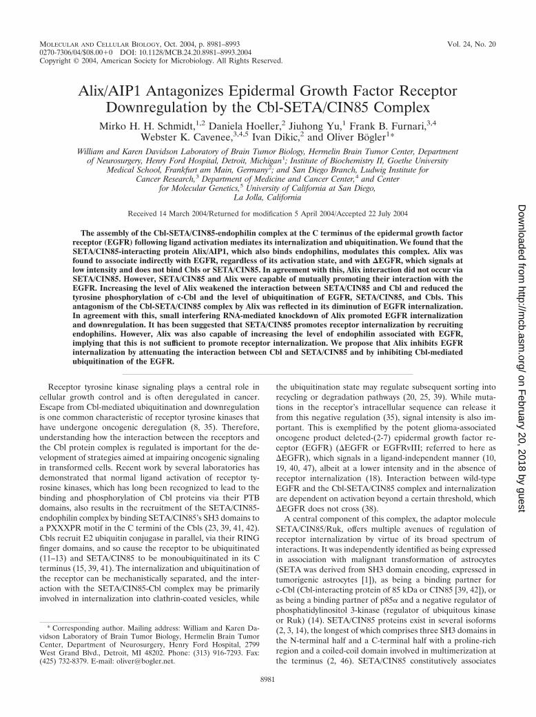
SETA/CIN85 interacts with ligand-activated EGFR and, to-gether with the Cbl proteins and endophilin, is involved in itsinternalization (39, 42). To test whether Alix, which associateswith SETA/CIN85’s SH3 domains via a proline-rich motif in itsC terminus (6, 37), also binds to EGFR, a Flag-tagged Alix wastransfected into U87 glioma cells that express wild-type EGFRand EGFR immunoprecipitates were analyzed (Fig. 1A). Alixwas detected in complexes of endogenous EGFR at robustlevels relative to its expression levels in the lysates from theseglioma cells, which are not very efficiently transfected (Fig.1A), demonstrating interaction between EGFR and Alix.
SETA/CIN85 and its associated proteins c-Cbl, Cbl-b, andendophilin A1 discriminate between the �EGFR mutant pro-
tein and EGFR in that they associate only with the latter (38).To test whether Alix also discriminates between these twoforms of the EGFR, we performed IP experiments with con-ditions specific for the wild type and �EGFR (Fig. 1B). EGFR-specific conditions were achieved by transfecting this receptoralone into HEK293 cells (which do not express endogenous�EGFR) and immunoprecipitating with an N-terminal EGFRantibody. To isolate �EGFR from cells that had been trans-fected with it, the MAb 806 was used, as it preferentiallyrecognizes this mutant form (21). However, as described pre-viously (38), in cells that also express endogenous EGFR, as isusually the case in gliomas, as well as the HEK293 cell modelused here, there is a degree of cross-reactivity with EGFR.Others have also reported this, particularly when EGFR ishighly expressed, and it is thought to be because MAb 806recognizes an activity-dependent conformation rather than aneo epitope of �EGFR (21, 27, 29). To reduce the level ofEGFR in �EGFR immunoprecipitates, lysates were pre-cleared by IP with an anti-EGFR antibody (Ab-1; OncogeneScience), followed by IP with MAb 806, which resulted inpredominant recovery of �EGFR (Fig. 1B, lane 1). Cotrans-fection of Flag-tagged Alix with wild-type EGFR or �EGFR inHEK293 cells and IP of the respective receptor showed thatAlix associated with both receptors, although lower levels ofAlix were associated with �EGFR (Fig. 1B). These data sug-gest that Alix does not interact with EGFR via Cbl and SETA/CIN85, as neither of these proteins binds to �EGFR (38).However, as the interaction of Alix with �EGFR is weakerthan that with wild-type EGFR, it is possible that SETA/CIN85and Cbls can positively contribute to Alix’s interaction withEGFR. Two experiments were then performed to test whetherAlix and EGFR interacted in situations where neither proteinwas overexpressed by transient transfection. First, CHO cellsstably expressing EGFR (CHO-EGFR) were subjected toEGFR IP, and it was found that endogenous Alix, as well astransfected Alix, could be recovered in the immunoprecipitates(Fig. 1C). The amount of endogenous Alix recovered was notaffected by stimulation with EGF (Fig. 1C). Second, endoge-nous EGFR was immunoprecipitated from either a fat rich ora low-fat fraction of mouse brain homogenates and endoge-nous Alix was detected in the recovered material (Fig. 1D).Direct confrontation of bacterially made Alix with in vitro-transcribed and -translated EGFR or �EGFR failed to showinteraction, while SETA/CIN85 and Alix bound in this situa-tion as previously described (6) (data not shown), suggestingthat the interaction between these proteins is not direct andrequires the presence of an unidentified cellular cofactor. Ad-ditional control experiments (unpublished) were also per-formed to establish that the recovery of Alix in EGFR IP isdependent on the presence of the EGFR, that the EGFRantibodies do not recognize Alix, that preimmune and second-ary antibodies are not capable of recovering Flag-tagged Alixin IPs, and that no Flag-tagged Alix is found in cells that arenot transfected with this construct.
The interaction between SETA/CIN85 and EGFR is depen-dent on the EGFR being in an activated state (38, 39). To testwhether the activation state modulated the interaction be-tween Alix and EGFR, HEK293 cells transfected with both ofthese constructs were serum starved and challenged with EGFfor 5 min to produce maximal EGFR activity. Although this
VOL. 24, 2004 Alix ANTAGONIZES EGFR DOWNREGULATION BY Cbl-SETA/CIN85 8983
on February 20, 2018 by guest
http://mcb.asm
.org/D
ownloaded from
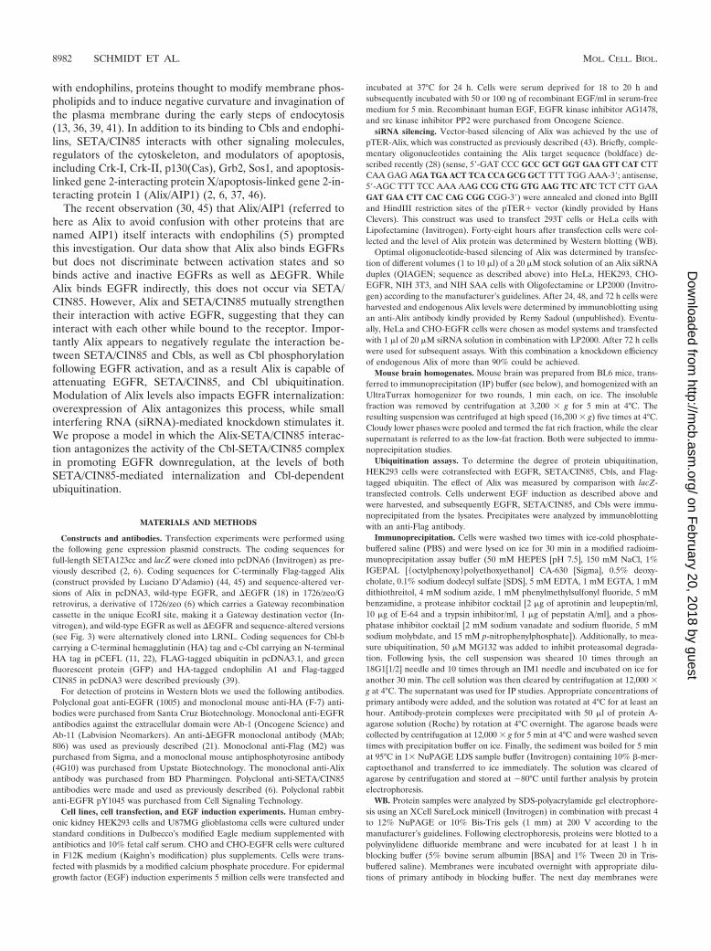
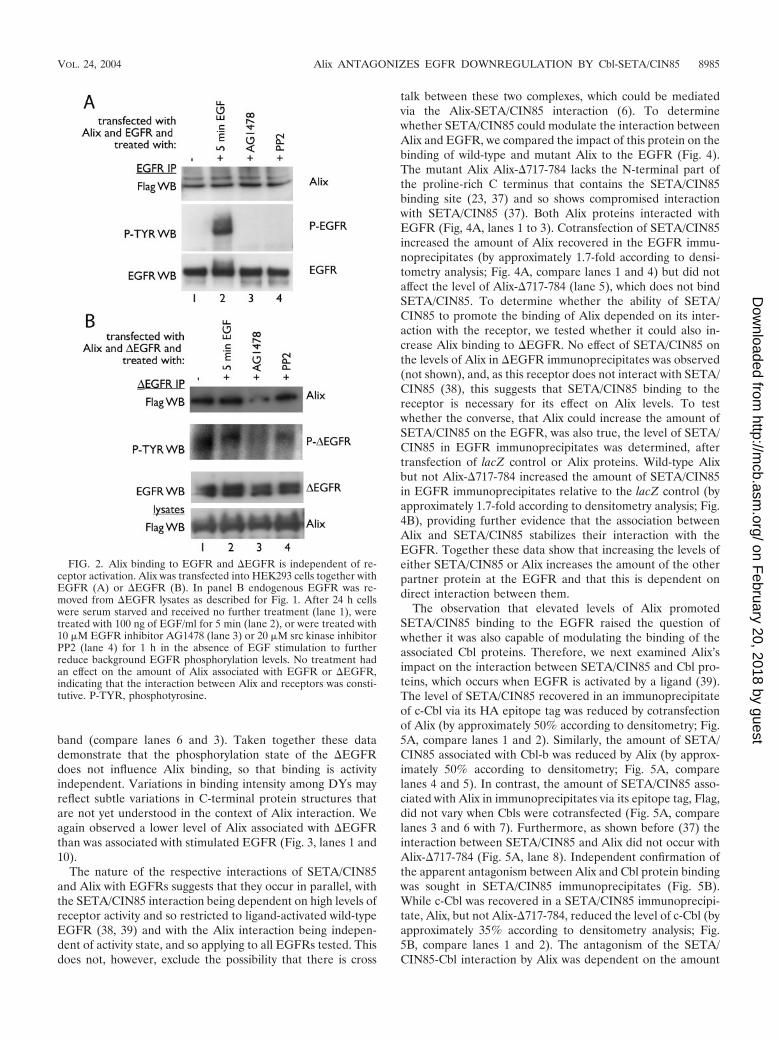
resulted in a dramatic increase in the tyrosine phosphorylationstate of the EGFR, it did not affect the amount of Alix that wasrecovered in the EGFR IP (Fig. 2A, lanes 1 and 2). Althoughno tyrosine-phosphorylated EGFR was detectable in the se-rum-starved cells (lane 1), cells were also treated with theEGFR inhibitor AG1478 or the src kinase inhibitor PP2 in theabsence of EGF, in an attempt to eliminate any residual phos-phorylation that might be present, but again this did not alterthe amount of Alix that was recovered in the EGFR complex(Fig. 2A, lanes 3 and 4). Therefore, the interaction betweenAlix and EGFR was not dependent on the activation or phos-phorylation state of the receptor. Similarly, the interactionbetween Alix and �EGFR was not affected by EGF stimula-tion, which has no effect on this ligand-independent mutantreceptor (Fig. 2B, lanes 1 and 2). It also did not appear to besignificantly affected by treatment with AG1478, which inhibitsthe �EGFR (17, 33). However, a slight reduction in Alix in the
AG1478-treated �EGFR complex and an incomplete attenu-ation of �EGFR phosphorylation were observed (Fig. 2B, lane3). The src kinase inhibitor PP2 had no effect.
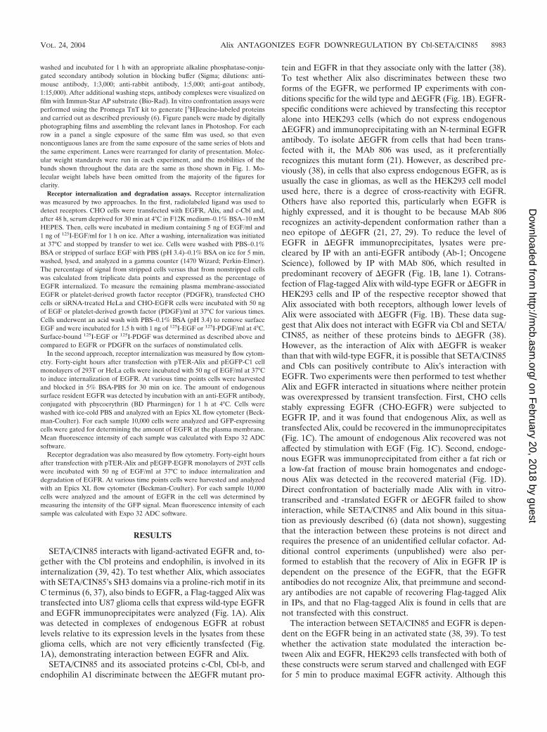
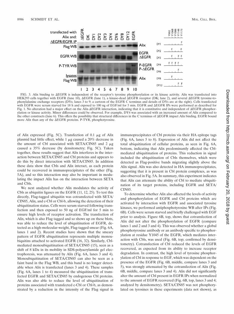
To evaluate the role of the phosphorylation state of theconstitutively active �EGFR in its interaction with Alix morethoroughly, various �EGFRs with tyrosine-to-phenylalaninesubstitutions (DY1, -2, -3, -4, -6, -8, and -12), as well as akinase-inactive mutant �EGFR (DK), were examined (Fig. 3).The amount of Alix recovered in the IPs of kinase-inactive�EGFR DK or the various DYs did not vary with phosphor-ylation status (Fig. 3 shows details of the alterations). Forexample, �EGFR DK recovered an amount of Alix similar tothat recovered by the active �EGFR, although it was notphosphorylated (Fig. 3, lanes 1 and 2). Interestingly, DY4,which showed very low levels of phosphorylation, bound rela-tively more Alix than DY1, which exhibited near-�EGFR lev-els of activity, compared to the intensity of the EGFR WB
FIG. 1. Alix binds to both EGFR and �EGFR. (A) Flag-tagged Alix was transfected into U87MG glioma cells. IP of endogenous EGFRrevealed receptor-associated Alix within the precipitate (lane 2). (B) Alix and �EGFR (lanes 1 and 3) or EGFR (lane 2) were transfected intoHEK293 cells, which were then serum starved for 18 h and treated with 100 ng of EGF/ml for 5 min before lysis. IPs were performed with anantibody against EGFR (Ab-1; Oncogene Science; lanes 2 and 3) or with EGFR preclearing of the �EGFR lysate with the same EGFR antibody,followed by IP with MAb 806 to specifically isolate the �EGFR (lane 1). Alix was recovered along with both receptors (lanes 1 to 3). However,less Alix was associated with �EGFR. (C) CHO-EGFR cells were either transfected with Flag-tagged Alix (lane 1) or not and subjected to EGFRIP followed by EGFR or Alix WB. Although a stronger Alix band is obtained in the EGFR IP when Alix is transfected, the endogenous Alix isalso recovered, and the levels of endogenous Alix are equivalent regardless of whether cells were serum starved for 18 h and either not treatedfurther (lane 2) or challenged with 50 ng of EGF/ml for 10 min (lane 3; ns, not starved). (D) Fat rich and low-fat fractions of mouse brainhomogenates were subjected to IP with the anti-EGFR antibody, and the recovered material was analyzed by Alix and EGFR WB. Alix wasrecovered in EGFR IPs from both fractions and was also observed in the lysates. EGFR was detected in the lysates at longer exposures (not shown).
8984 SCHMIDT ET AL. MOL. CELL. BIOL.
on February 20, 2018 by guest
http://mcb.asm
.org/D
ownloaded from
band (compare lanes 6 and 3). Taken together these datademonstrate that the phosphorylation state of the �EGFRdoes not influence Alix binding, so that binding is activityindependent. Variations in binding intensity among DYs mayreflect subtle variations in C-terminal protein structures thatare not yet understood in the context of Alix interaction. Weagain observed a lower level of Alix associated with �EGFRthan was associated with stimulated EGFR (Fig. 3, lanes 1 and10).
The nature of the respective interactions of SETA/CIN85and Alix with EGFRs suggests that they occur in parallel, withthe SETA/CIN85 interaction being dependent on high levels ofreceptor activity and so restricted to ligand-activated wild-typeEGFR (38, 39) and with the Alix interaction being indepen-dent of activity state, and so applying to all EGFRs tested. Thisdoes not, however, exclude the possibility that there is cross
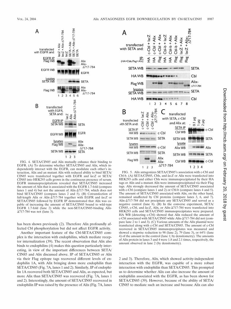
talk between these two complexes, which could be mediatedvia the Alix-SETA/CIN85 interaction (6). To determinewhether SETA/CIN85 could modulate the interaction betweenAlix and EGFR, we compared the impact of this protein on thebinding of wild-type and mutant Alix to the EGFR (Fig. 4).The mutant Alix Alix-�717-784 lacks the N-terminal part ofthe proline-rich C terminus that contains the SETA/CIN85binding site (23, 37) and so shows compromised interactionwith SETA/CIN85 (37). Both Alix proteins interacted withEGFR (Fig, 4A, lanes 1 to 3). Cotransfection of SETA/CIN85increased the amount of Alix recovered in the EGFR immu-noprecipitates (by approximately 1.7-fold according to densi-tometry analysis; Fig. 4A, compare lanes 1 and 4) but did notaffect the level of Alix-�717-784 (lane 5), which does not bindSETA/CIN85. To determine whether the ability of SETA/CIN85 to promote the binding of Alix depended on its inter-action with the receptor, we tested whether it could also in-crease Alix binding to �EGFR. No effect of SETA/CIN85 onthe levels of Alix in �EGFR immunoprecipitates was observed(not shown), and, as this receptor does not interact with SETA/CIN85 (38), this suggests that SETA/CIN85 binding to thereceptor is necessary for its effect on Alix levels. To testwhether the converse, that Alix could increase the amount ofSETA/CIN85 on the EGFR, was also true, the level of SETA/CIN85 in EGFR immunoprecipitates was determined, aftertransfection of lacZ control or Alix proteins. Wild-type Alixbut not Alix-�717-784 increased the amount of SETA/CIN85in EGFR immunoprecipitates relative to the lacZ control (byapproximately 1.7-fold according to densitometry analysis; Fig.4B), providing further evidence that the association betweenAlix and SETA/CIN85 stabilizes their interaction with theEGFR. Together these data show that increasing the levels ofeither SETA/CIN85 or Alix increases the amount of the otherpartner protein at the EGFR and that this is dependent ondirect interaction between them.
The observation that elevated levels of Alix promotedSETA/CIN85 binding to the EGFR raised the question ofwhether it was also capable of modulating the binding of theassociated Cbl proteins. Therefore, we next examined Alix’simpact on the interaction between SETA/CIN85 and Cbl pro-teins, which occurs when EGFR is activated by a ligand (39).The level of SETA/CIN85 recovered in an immunoprecipitateof c-Cbl via its HA epitope tag was reduced by cotransfectionof Alix (by approximately 50% according to densitometry; Fig.5A, compare lanes 1 and 2). Similarly, the amount of SETA/CIN85 associated with Cbl-b was reduced by Alix (by approx-imately 50% according to densitometry; Fig. 5A, comparelanes 4 and 5). In contrast, the amount of SETA/CIN85 asso-ciated with Alix in immunoprecipitates via its epitope tag, Flag,did not vary when Cbls were cotransfected (Fig. 5A, comparelanes 3 and 6 with 7). Furthermore, as shown before (37) theinteraction between SETA/CIN85 and Alix did not occur withAlix-�717-784 (Fig. 5A, lane 8). Independent confirmation ofthe apparent antagonism between Alix and Cbl protein bindingwas sought in SETA/CIN85 immunoprecipitates (Fig. 5B).While c-Cbl was recovered in a SETA/CIN85 immunoprecipi-tate, Alix, but not Alix-�717-784, reduced the level of c-Cbl (byapproximately 35% according to densitometry analysis; Fig.5B, compare lanes 1 and 2). The antagonism of the SETA/CIN85-Cbl interaction by Alix was dependent on the amount
FIG. 2. Alix binding to EGFR and �EGFR is independent of re-ceptor activation. Alix was transfected into HEK293 cells together withEGFR (A) or �EGFR (B). In panel B endogenous EGFR was re-moved from �EGFR lysates as described for Fig. 1. After 24 h cellswere serum starved and received no further treatment (lane 1), weretreated with 100 ng of EGF/ml for 5 min (lane 2), or were treated with10 �M EGFR inhibitor AG1478 (lane 3) or 20 �M src kinase inhibitorPP2 (lane 4) for 1 h in the absence of EGF stimulation to furtherreduce background EGFR phosphorylation levels. No treatment hadan effect on the amount of Alix associated with EGFR or �EGFR,indicating that the interaction between Alix and receptors was consti-tutive. P-TYR, phosphotyrosine.
VOL. 24, 2004 Alix ANTAGONIZES EGFR DOWNREGULATION BY Cbl-SETA/CIN85 8985
on February 20, 2018 by guest
http://mcb.asm
.org/D
ownloaded from
of Alix expressed (Fig. 5C). Transfection of 0.1 �g of Alixplasmid had little effect, while 1 �g caused a 20% decrease inthe amount of Cbl associated with SETA/CIN85 and 2 �gcaused a 35% decrease (by densitometry; Fig. 5C). Takentogether, these results suggest that Alix interferes in the inter-action between SETA/CIN85 and Cbl proteins and appears todo this by direct interaction with SETA/CIN85. In additionthese data show that Cbls and Alix interact, as each proteincould be recovered in immunoprecipitates of the other (Fig.5A), and so this interaction may also be important in modu-lating the impact Alix has on the interaction between SETAand Cbls.
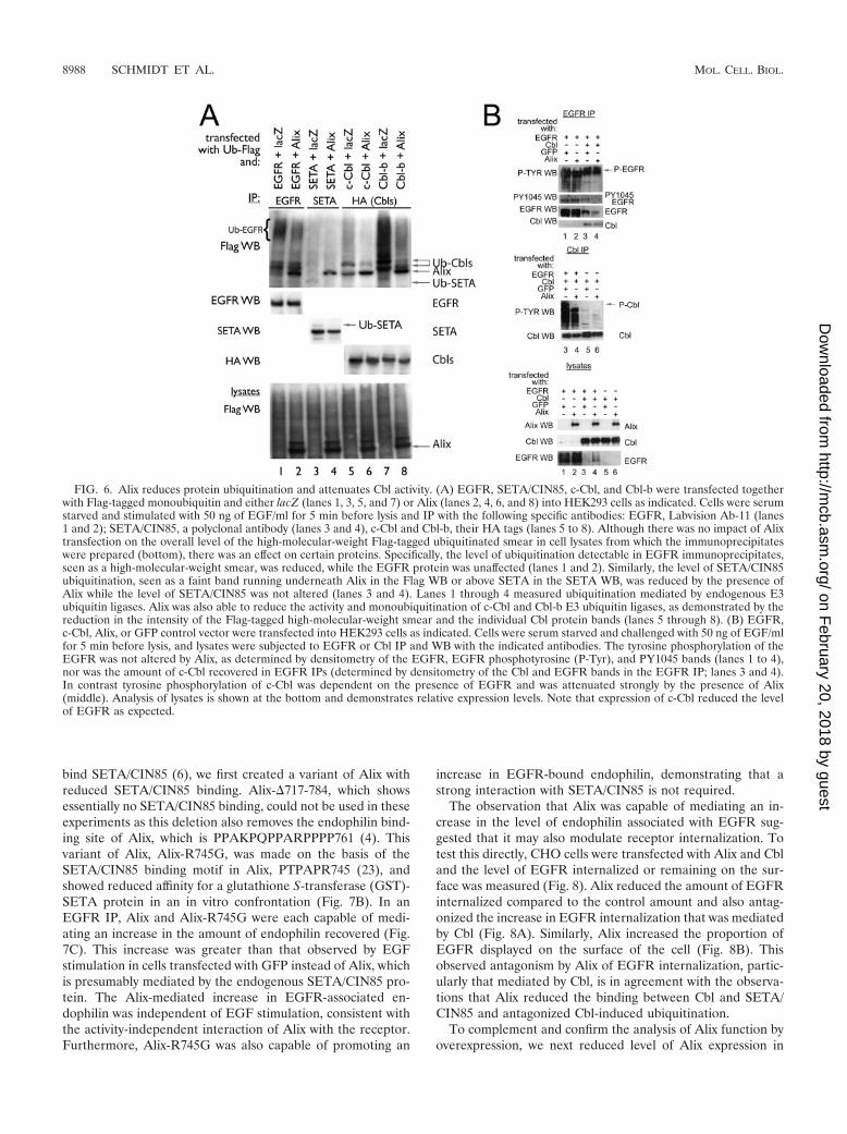
We next analyzed whether Alix modulates the activity ofCbls as ubiquitin ligases on the EGFR (11, 12, 25). To test thisdirectly, Flag-tagged ubiquitin was cotransfected with SETA/CIN85, Alix, and c-Cbl or Cbl-b, allowing the detection of theirubiquitination status. Cells were serum starved following trans-fection and then exposed to 50 ng of EGF/ml for 5 min toensure high levels of receptor activation. The transfection ofAlix, which is also Flag tagged and so shows up on these blots,was able to reduce the level of ubiquitination of EGFR, de-tected as a high-molecular-weight, Flag-tagged smear (Fig. 6A,lanes 1 and 2). Recent studies have shown that the smearypattern of EGFR ubiquitination represents multiple monou-biquitins attached to activated EGFR (16, 32). Similarly, Cbl-mediated monoubiquitination of SETA/CIN85 (15), seen as ashift of 8 kDa in its mobility in SDS-polyacrylamide gel elec-trophoresis, was attenuated by Alix (Fig. 6A, lanes 3 and 4).Monoubiquitination of SETA/CIN85 can also be seen as afaint band in the Flag WB, and this band is no longer detect-able when Alix is transfected (lanes 3 and 4). These samples(Fig. 6A, lanes 1 to 4) measured the ubiquitination of trans-fected EGFR and SETA/CIN85 by endogenous Cbl proteins.Alix was also able to reduce the level of ubiquitination ofproteins associated with transfected c-Cbl or Cbl-b, as demon-strated by a reduction in the intensity of the Flag signal in
immunoprecipitates of Cbl proteins via their HA epitope tags(Fig. 6A, lanes 5 to 8). Expression of Alix did not affect thetotal ubiquitination of cellular proteins, as seen in Fig. 6A,bottom, indicating that Alix predominantly affected the Cbl-mediated ubiquitination of proteins. This reduction in signalincluded the ubiquitination of Cbls themselves, which weredetected as Flag-positive bands migrating slightly above theAlix signal. Alix was also detected in HA immunoprecipitates,suggesting that it is present in Cbl protein complexes, as wasalso observed in Fig. 5A. In summary, this experiment indicatesthat Alix can attenuate the ability of Cbl to mediate ubiquiti-nation of its target proteins, including EGFR and SETA/CIN85.
To determine whether Alix also affected the levels of activityand phosphorylation of EGFR and Cbl proteins which areactivated by interaction with EGFR and associated tyrosinekinases, we performed antiphosphotyrosine WB after IPs (Fig.6B). Cells were serum starved and briefly challenged with EGFprior to analysis. Figure 6B, top, shows that cotransfection ofAlix did not alter the phosphorylation of EGFR (comparelanes 1 and 2 and 3 and 4). This was observed whether a globalphosphotyrosine antibody or an antibody specific to phosphor-ylation at residue Y1045 of the EGFR, which mediates inter-action with Cbls, was used (Fig. 6B, top; confirmed by densi-tometry). Cotransfection of Cbl reduced the levels of EGFRrecovered, as expected from its ability to increase receptordegradation. In contrast, the high level of tyrosine phosphor-ylation of Cbl in response to EGF, which was dependent on thepresence of the EGFR (Fig. 6B, middle, compare lanes 3 and5), was strongly attenuated by the cotransfection of Alix (Fig.6B, middle, compare lanes 3 and 4). Alix did not significantlyalter the amount of Cbl present in EGFR IPs when normalizedto the amount of EGFR recovered (Fig. 6B, top, lanes 3 and 4;analyzed by densitometry). SETA/CIN85 was not phosphory-lated on tyrosines in these experiments (data not shown), as
FIG. 3. Alix binding to �EGFR is independent of the receptor’s tyrosine phosphorylation or its kinase activity. Alix was transfected intoHEK293 cells together with EGFR (lane 10), �EGFR (lane 1), a kinase-dead �EGFR receptor (DK; lane 2), and several �EGFR tyrosine-to-phenylalanine exchange receptors (DYs; lanes 3 to 9; a cartoon of the EGFR C terminus and details of DYs are at the right). Cells transfectedwith EGFR were serum starved for 18 h and exposed to 100 ng of EGF/ml for 5 min. EGFR and �EGFR IPs were performed as described forFig. 1. No alteration had a major effect on the Alix-�EGFR interaction, indicating that it is constitutive and independent of �EGFR phosphor-ylation or kinase activity. Minor differences could be observed. For example, DY4 was associated with an increased amount of Alix compared tothe other constructs (lane 6). This offers the possibility that structural differences in the C terminus of �EGFR impact Alix binding. EGFR boundmore Alix than any of the �EGFR proteins. P-TYR, phosphotyrosine.
8986 SCHMIDT ET AL. MOL. CELL. BIOL.
on February 20, 2018 by guest
http://mcb.asm
.org/D
ownloaded from
has been shown previously (2). Therefore Alix profoundly af-fected Cbl phosphorylation but did not affect EGFR activity.
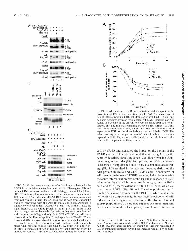
Another important feature of the Cbl-SETA/CIN85 com-plex is the interaction with endophilins, which mediate recep-tor internalization (39). The recent observation that Alix alsobinds to endophilins (4) makes this question particularly inter-esting, in view of the important differences between SETA/CIN85 and Alix discussed above. IP of SETA/CIN85 or Alixvia their Flag epitope tags recovered different levels of en-dophilin 1A, with Alix bringing down more endophilin thanSETA/CIN85 (Fig. 7A, lanes 1 and 2). Similarly, IP of endophi-lin 1A recovered both SETA/CIN85 and Alix, as expected, butmore Alix than SETA/CIN85 was recovered (Fig. 7A, lanes 1and 2). Interestingly, the amount of SETA/CIN85 recovered inendophilin IP was raised by the presence of Alix (Fig. 7A, lanes
2 and 3). Therefore, Alix, which showed activity-independentinteraction with the EGFR, was capable of a more robustinteraction with endophilin than SETA/CIN85. This promptedus to determine whether Alix can also increase the amount ofendophilin associated with the EGFR, as has been shown forSETA/CIN85 (39). However, because of the ability of SETA/CIN85 to mediate such an increase and because Alix can also
FIG. 4. SETA/CIN85 and Alix mutually enhance their binding toEGFR. (A) To determine whether SETA/CIN85 and Alix, which in-dependently interact with the EGFR, can modulate each other’s in-teraction, Alix and an mutant Alix with reduced ability to bind SETA/CIN85 were transfected together with EGFR and lacZ or SETA/CIN85 into HEK293 cells grown in the continuous presence of serum.EGFR immunoprecipitations revealed that SETA/CIN85 increasedthe amount of Alix that is associated with the EGFR 1.7-fold (comparelanes 1 and 4) but not the amount of Alix-�717-784, which does notbind SETA/CIN85 (compare lanes 2 and 5). (B) Cotransfection offull-length Alix or Alix-�717-784 together with EGFR and lacZ orSETA/CIN85 followed by EGFR IP demonstrated that Alix was ca-pable of increasing the amount of SETA/CIN85 bound to wild-typeEGFR 1.7-fold (lane 2) while the non-SETA/CIN85-binding Alix-�717-784 was not (lane 3).
FIG. 5. Alix antagonizes SETA/CIN85’s association with c-Cbl andCbl-b. (A) SETA/CIN85, Cbls, and lacZ or Alix were transfected intoHEK293 cells and either Cbls were immunoprecipitated by their HAtags or Alix and a mutant Alix were immunoprecipitated via their Flagtags. Alix strongly decreased the amount of SETA/CIN85 associatedwith c-Cbl (compare lanes 1 and 2) or Cbl-b (compare lanes 4 and 5).The amount of SETA/CIN85 associated with Alix, on the other hand,remained unaffected by Cbl proteins (compare lanes 3, 6, and 7).Alix-�717-784 did not precipitate any SETA/CIN85 and served as anegative control (lane 8). (B) In the converse experiment, SETA/CIN85, c-Cbl, and lacZ, Alix, or Alix-�717-784 were transfected intoHEK293 cells and SETA/CIN85 immunoprecipitates were prepared.HA WB (detecting c-Cbl) showed that Alix reduced the amount ofc-Cbl associated with SETA/CIN85 while Alix-�717-784 did not (com-pare lane 2 to 1 and 3). (C) Various amounts of the Alix plasmid weretransfected along with c-Cbl and SETA/CIN85. The amount of c-Cblrecovered in SETA/CIN85 immunoprecipitates was measured andshowed a stepwise reduction to 98 (lane 2), 79 (lane 3), or 64% (lane4) of the amount in the control (lane 1; by densitometry). The amountsof Alix protein in lanes 3 and 4 were 1.8 and 2.1 times, respectively, theamount observed in lane 2 (by densitometry).
VOL. 24, 2004 Alix ANTAGONIZES EGFR DOWNREGULATION BY Cbl-SETA/CIN85 8987
on February 20, 2018 by guest
http://mcb.asm
.org/D
ownloaded from
bind SETA/CIN85 (6), we first created a variant of Alix withreduced SETA/CIN85 binding. Alix-�717-784, which showsessentially no SETA/CIN85 binding, could not be used in theseexperiments as this deletion also removes the endophilin bind-ing site of Alix, which is PPAKPQPPARPPPP761 (4). Thisvariant of Alix, Alix-R745G, was made on the basis of theSETA/CIN85 binding motif in Alix, PTPAPR745 (23), andshowed reduced affinity for a glutathione S-transferase (GST)-SETA protein in an in vitro confrontation (Fig. 7B). In anEGFR IP, Alix and Alix-R745G were each capable of medi-ating an increase in the amount of endophilin recovered (Fig.7C). This increase was greater than that observed by EGFstimulation in cells transfected with GFP instead of Alix, whichis presumably mediated by the endogenous SETA/CIN85 pro-tein. The Alix-mediated increase in EGFR-associated en-dophilin was independent of EGF stimulation, consistent withthe activity-independent interaction of Alix with the receptor.Furthermore, Alix-R745G was also capable of promoting an
increase in EGFR-bound endophilin, demonstrating that astrong interaction with SETA/CIN85 is not required.
The observation that Alix was capable of mediating an in-crease in the level of endophilin associated with EGFR sug-gested that it may also modulate receptor internalization. Totest this directly, CHO cells were transfected with Alix and Cbland the level of EGFR internalized or remaining on the sur-face was measured (Fig. 8). Alix reduced the amount of EGFRinternalized compared to the control amount and also antag-onized the increase in EGFR internalization that was mediatedby Cbl (Fig. 8A). Similarly, Alix increased the proportion ofEGFR displayed on the surface of the cell (Fig. 8B). Thisobserved antagonism by Alix of EGFR internalization, partic-ularly that mediated by Cbl, is in agreement with the observa-tions that Alix reduced the binding between Cbl and SETA/CIN85 and antagonized Cbl-induced ubiquitination.
To complement and confirm the analysis of Alix function byoverexpression, we next reduced level of Alix expression in
FIG. 6. Alix reduces protein ubiquitination and attenuates Cbl activity. (A) EGFR, SETA/CIN85, c-Cbl, and Cbl-b were transfected togetherwith Flag-tagged monoubiquitin and either lacZ (lanes 1, 3, 5, and 7) or Alix (lanes 2, 4, 6, and 8) into HEK293 cells as indicated. Cells were serumstarved and stimulated with 50 ng of EGF/ml for 5 min before lysis and IP with the following specific antibodies: EGFR, Labvision Ab-11 (lanes1 and 2); SETA/CIN85, a polyclonal antibody (lanes 3 and 4), c-Cbl and Cbl-b, their HA tags (lanes 5 to 8). Although there was no impact of Alixtransfection on the overall level of the high-molecular-weight Flag-tagged ubiquitinated smear in cell lysates from which the immunoprecipitateswere prepared (bottom), there was an effect on certain proteins. Specifically, the level of ubiquitination detectable in EGFR immunoprecipitates,seen as a high-molecular-weight smear, was reduced, while the EGFR protein was unaffected (lanes 1 and 2). Similarly, the level of SETA/CIN85ubiquitination, seen as a faint band running underneath Alix in the Flag WB or above SETA in the SETA WB, was reduced by the presence ofAlix while the level of SETA/CIN85 was not altered (lanes 3 and 4). Lanes 1 through 4 measured ubiquitination mediated by endogenous E3ubiquitin ligases. Alix was also able to reduce the activity and monoubiquitination of c-Cbl and Cbl-b E3 ubiquitin ligases, as demonstrated by thereduction in the intensity of the Flag-tagged high-molecular-weight smear and the individual Cbl protein bands (lanes 5 through 8). (B) EGFR,c-Cbl, Alix, or GFP control vector were transfected into HEK293 cells as indicated. Cells were serum starved and challenged with 50 ng of EGF/mlfor 5 min before lysis, and lysates were subjected to EGFR or Cbl IP and WB with the indicated antibodies. The tyrosine phosphorylation of theEGFR was not altered by Alix, as determined by densitometry of the EGFR, EGFR phosphotyrosine (P-Tyr), and PY1045 bands (lanes 1 to 4),nor was the amount of c-Cbl recovered in EGFR IPs (determined by densitometry of the Cbl and EGFR bands in the EGFR IP; lanes 3 and 4).In contrast tyrosine phosphorylation of c-Cbl was dependent on the presence of EGFR and was attenuated strongly by the presence of Alix(middle). Analysis of lysates is shown at the bottom and demonstrates relative expression levels. Note that expression of c-Cbl reduced the levelof EGFR as expected.
8988 SCHMIDT ET AL. MOL. CELL. BIOL.
on February 20, 2018 by guest
http://mcb.asm
.org/D
ownloaded from
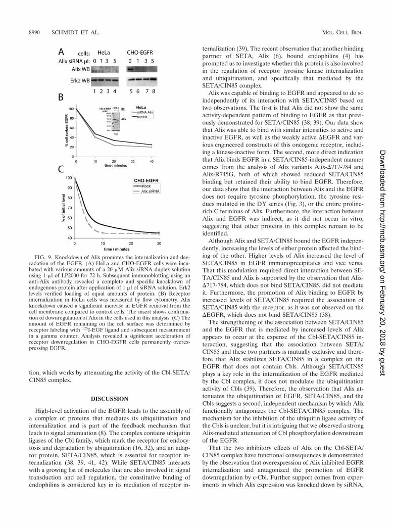
cells by siRNA and measured the impact on the biology of theEGFR (Fig. 9). These data showed that silencing Alix via therecently described target sequence (28), either by using trans-fected oligonucleotides (Fig. 9A; optimization of this approachis described in unpublished data) or by a vector-mediated strat-egy (Fig. 9B) resulted in the efficient downregulation of theAlix protein in HeLa and CHO-EGFR cells. Knockdown ofAlix resulted in increased EGFR downregulation by increasingthe acute internalization rate of the EGFR in response to EGFstimulation, by a small but measurable margin, both in HeLacells and to a greater extent in CHO-EGFR cells, which ex-press more EGFR (Fig. 9B and C and unpublished data).Similar data were obtained for the PDGFR, which also inter-acts with Alix (unpublished). Interestingly knockdown of Alixdid not result in a significant reduction in the absolute levels ofEGFR (unpublished). These data support our model that Alixis a negative regulator of receptor tyrosine kinase internaliza-
FIG. 7. Alix increases the amount of endophilin associated with theEGFR in an activity-independent manner. (A) Flag-tagged Alix andhuman CIN85 were cotransfected with HA-tagged endophilin A1 intoHEK293 cells, which were serum starved and stimulated for 5 min with100 ng of EGF/ml. Alix and SETA/CIN85 were immunoprecipitatedfrom cell lysates via their Flag epitopes, and in both cases endophilinwas also recovered, with the Alix IP containing more. Although aslightly lower level of SETA/CIN85 was expressed in the lysates, thesignal intensity of the CIN85 protein in the Flag IP was similar to thatof Alix, suggesting similar levels of protein, as they were both detectedwith the same anti-Flag antibody. Both SETA/CIN85 and Alix wererecovered in the HA-endophilin IP, and again less SETA/CIN85 wasrecovered. (B) In vitro confrontation of various radiolabeled Alix pro-teins made by in vitro transcription and translation with bacteriallymade GST proteins showed that GST-SETA binds Alix and Alix-784Stop (a truncation of Alix at position 784) efficiently but shows nobinding to Alix-�717-784 and low-efficiency binding to Alix-R745G
FIG. 8. Alix reduces EGFR internalization and antagonizes thepromotion of EGFR internalization by Cbl. (A) The percentage ofEGFR internalization in CHO cells transfected with EGFR, c-Cbl, andAlix was measured by using radiolabeled 125I-EGF. Expression of Alixresults in a decline in the amount of c-Cbl-mediated EGFR internal-ization. (B) The relative remaining EGFR on the surfaces of CHOcells, transfected with EGFR, c-Cbl, and Alix, was measured afterexposure to EGF for the times indicated to radiolabeled EGF. Thevalues are expressed as percentages of control cells that were notexposed to EGF. Expression of Alix inhibited the c-Cbl-induced de-cline in EGFR present at the cell surface.
that is equivalent to that observed for lacZ. Note that in this experi-ment Alix was relatively underloaded. (C) Transfection of Alix andAlix-R745G increased the level of endophilin that was recovered inEGFR immunoprecipitates beyond the increase mediated by stimula-tion with EGF.
VOL. 24, 2004 Alix ANTAGONIZES EGFR DOWNREGULATION BY Cbl-SETA/CIN85 8989
on February 20, 2018 by guest
http://mcb.asm
.org/D
ownloaded from
tion, which works by attenuating the activity of the Cbl-SETA/CIN85 complex.
DISCUSSION
High-level activation of the EGFR leads to the assembly ofa complex of proteins that mediates its ubiquitination andinternalization and is part of the feedback mechanism thatleads to signal attenuation (8). The complex contains ubiquitinligases of the Cbl family, which mark the receptor for endocy-tosis and degradation by ubiquitination (16, 32), and an adap-tor protein, SETA/CIN85, which is essential for receptor in-ternalization (38, 39, 41, 42). While SETA/CIN85 interactswith a growing list of molecules that are also involved in signaltransduction and cell regulation, the constitutive binding ofendophilins is considered key in its mediation of receptor in-
ternalization (39). The recent observation that another bindingpartner of SETA, Alix (6), bound endophilins (4) hasprompted us to investigate whether this protein is also involvedin the regulation of receptor tyrosine kinase internalizationand ubiquitination, and specifically that mediated by theSETA/CIN85 complex.
Alix was capable of binding to EGFR and appeared to do soindependently of its interaction with SETA/CIN85 based ontwo observations. The first is that Alix did not show the sameactivity-dependent pattern of binding to EGFR as that previ-ously demonstrated for SETA/CIN85 (38, 39). Our data showthat Alix was able to bind with similar intensities to active andinactive EGFR, as well as the weakly active �EGFR and var-ious engineered constructs of this oncogenic receptor, includ-ing a kinase-inactive form. The second, more direct indicationthat Alix binds EGFR in a SETA/CIN85-independent mannercomes from the analysis of Alix variants Alix-�717-784 andAlix-R745G, both of which showed reduced SETA/CIN85binding but retained their ability to bind EGFR. Therefore,our data show that the interaction between Alix and the EGFRdoes not require tyrosine phosphorylation, the tyrosine resi-dues mutated in the DY series (Fig. 3), or the entire proline-rich C terminus of Alix. Furthermore, the interaction betweenAlix and EGFR was indirect, as it did not occur in vitro,suggesting that other proteins in this complex remain to beidentified.
Although Alix and SETA/CIN85 bound the EGFR indepen-dently, increasing the levels of either protein affected the bind-ing of the other. Higher levels of Alix increased the level ofSETA/CIN85 in EGFR immunoprecipitates and vice versa.That this modulation required direct interaction between SE-TA/CIN85 and Alix is supported by the observation that Alix-�717-784, which does not bind SETA/CIN85, did not mediateit. Furthermore, the promotion of Alix binding to EGFR byincreased levels of SETA/CIN85 required the association ofSETA/CIN85 with the receptor, as it was not observed on the�EGFR, which does not bind SETA/CIN85 (38).
The strengthening of the association between SETA/CIN85and the EGFR that is mediated by increased levels of Alixappears to occur at the expense of the Cbl-SETA/CIN85 in-teraction, suggesting that the association between SETA/CIN85 and these two partners is mutually exclusive and there-fore that Alix stabilizes SETA/CIN85 in a complex on theEGFR that does not contain Cbls. Although SETA/CIN85plays a key role in the internalization of the EGFR mediatedby the Cbl complex, it does not modulate the ubiquitinationactivity of Cbls (39). Therefore, the observation that Alix at-tenuates the ubiquitination of EGFR, SETA/CIN85, and theCbls suggests a second, independent mechanism by which Alixfunctionally antagonizes the Cbl-SETA/CIN85 complex. Themechanism for the inhibition of the ubiquitin ligase activity ofthe Cbls is unclear, but it is intriguing that we observed a strongAlix-mediated attenuation of Cbl phosphorylation downstreamof the EGFR.
That the two inhibitory effects of Alix on the Cbl-SETA/CIN85 complex have functional consequences is demonstratedby the observation that overexpression of Alix inhibited EGFRinternalization and antagonized the promotion of EGFRdownregulation by c-Cbl. Further support comes from exper-iments in which Alix expression was knocked down by siRNA,
FIG. 9. Knockdown of Alix promotes the internalization and deg-radation of the EGFR. (A) HeLa and CHO-EGFR cells were incu-bated with various amounts of a 20 �M Alix siRNA duplex solutionusing 1 �l of LP2000 for 72 h. Subsequent immunoblotting using ananti-Alix antibody revealed a complete and specific knockdown ofendogenous protein after application of 1 �l of siRNA solution. Erk2levels verified loading of equal amounts of protein. (B) Receptorinternalization in HeLa cells was measured by flow cytometry. Alixknockdown caused a significant increase in EGFR removal from thecell membrane compared to control cells. The insert shows confirma-tion of downregulation of Alix in the cells used in this analysis. (C) Theamount of EGFR remaining on the cell surface was determined byreceptor labeling with 125I-EGF ligand and subsequent measurementin a gamma counter. Analysis revealed a significant acceleration ofreceptor downregulation in CHO-EGFR cells permanently overex-pressing EGFR.
8990 SCHMIDT ET AL. MOL. CELL. BIOL.
on February 20, 2018 by guest
http://mcb.asm
.org/D
ownloaded from
which resulted in a stimulation of EGFR internalization. Al-though the effect of Alix was relatively modest, it was consis-tently observed over different cell lines and with differentknockdown approaches and modes of measuring receptor in-ternalization. Interestingly although alterations of Alix levelchanged EGFR ubiquitination and internalization, they didnot profoundly affect EGFR degradation. This fits with amodel in which Alix modulates the process of EGFR internal-ization, rather than being involved in its direct execution.
Alix, like SETA/CIN85, binds endophilins constitutively (4)and mediated the binding of endophilins to the EGFR in ourexperiments. This recruitment of endophilins was more robustthan that mediated by stimulation of cells with EGF, whichpresumably relied on the activity of the endogenous Cbl-SETA/CIN85 complex. Alix-R745G, which shows reduced in-teraction with SETA/CIN85, was still capable of mediating thisincrease in endophilins at the EGFR, suggesting that Alix actsindependently of SETA. Alix was also able to mediate anincrease in endophilins at the inactive EGFR, consistent withAlix’s binding characteristics, and this was further evidencethat this mechanism of recruitment of endophilins to theEGFR is SETA/CIN85 independent. These data show thatendophilin recruitment, at least by Alix, does not mediatereceptor internalization, both because it occurs at inactive re-ceptors and because of direct evidence that Alix antagonizesreceptor internalization. It is possible that endophilin recruit-ment in the absence of Cbl-mediated ubiquitination is not
sufficient to trigger EGFR internalization and that the inhibi-tion of Cbl ubiquitin ligase activity is the more importantaspect of Alix function in this context. Alternately, the natureof the endophilin complex created by Alix could be differentfrom that created by SETA/CIN85 in terms of, for example, itsstoichiometry or structure, and so, although endophilins arepresent, they do not act to promote internalization. Regardlessof the mechanism, this explains the apparent contradiction thatthe endophilin binding protein Alix binds to the �EGFR,which is inefficiently internalized (7, 18, 34).
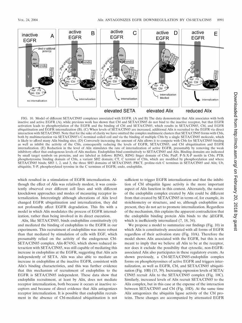
We propose a model to summarize our findings (Fig. 10) inwhich Alix is constitutively associated with all forms of EGFRregardless of their activation state (Fig. 10A). Therefore themodel shows Alix associated with the EGFR, but this is notmeant to imply that we believe all Alix to be at the receptor,nor does it exclude the possibility that cytosolic, non-EGFR-associated Alix also participates in these regulatory events. Asshown previously, a Cbl-SETA/CIN85-endophilin complexforms on phosphotyrosines of active EGFR and triggers inter-nalization, as well as EGFR, Cbl, and SETA/CIN85 ubiquiti-nation (Fig. 10B) (15, 39). Increasing expression levels of SETA/CIN85 recruit Alix to the SETA/CIN85 complex (Fig. 10C).Similarly, increased levels of Alix recruit SETA/CIN85 to theAlix complex, but in this case at the expense of the interactionbetween SETA/CIN85 and Cbl (Fig. 10D). At the same timeAlix antagonizes the ubiquitin ligase activity of the Cbl pro-teins. These changes are accompanied by attenuated EGFR
FIG. 10. Model of different SETA/CIN85 complexes associated with EGFR. (A and B) The data demonstrate that Alix associates with bothinactive and active EGFR (A), while previous work has shown that Cbl and SETA/CIN85 do not bind to the inactive receptor, but that EGFRactivation leads to phosphorylation of the EGFR and the binding of Cbl and SETA/CIN85, which results in SETA/CIN85, Cbl, and EGFRubiquitination and EGFR internalization (B). (C) When levels of SETA/CIN85 are increased, additional Alix is recruited to the EGFR via directinteraction with SETA/CIN85. Note that for the sake of clarity we have omitted the complex multimeric clusters that SETA/CIN85 forms with Cbls,both by multimerization via SETA/CIN85’s C-terminal coiled coil and via the binding of multiple Cbls by a single SETA/CIN85 molecule, whichis likely to afford many Alix binding sites. (D) Conversely increasing the amount of Alix allows it to compete with Cbls for SETA/CIN85 bindingas well as inhibit the activity of the Cbls, consequently reducing the levels of EGFR, SETA/CIN85, and Cbl ubiquitination and EGFRinternalization. (E) Reduction in the level of Alix stimulates the rate of internalization of active EGFR, presumably by removing the weakinhibitory effect that endogenous levels of Alix mediate. Endophilins bind constitutively to SETA/CIN85 and Alix. Binding domains are indicatedby small target symbols on proteins, and are labeled as follows: RING, RING finger domain of Cbls; PxxP, P-X-X-P motifs in Cbls; PTB,phosphotyrosine binding domain of Cbls, a variant SH2 domain; CT, C termini of Cbls, which are modified by phosphorylation and whereSETA/CIN85 binds; SH3 1, 2, and 3, the three SH3 domains of SETA/CIN85; PRCT, proline-rich C terminus in SETA/CIN85 and Alix; Ub,ubiquitin; Y-P, phosphorylated tyrosine in the C terminus of EGFR; endo, endophilin.
VOL. 24, 2004 Alix ANTAGONIZES EGFR DOWNREGULATION BY Cbl-SETA/CIN85 8991
on February 20, 2018 by guest
http://mcb.asm
.org/D
ownloaded from
internalization. The inhibition of the Cbl-SETA/CIN85 inter-action may well be sufficient to reduce the internalization ofthe EGFR (39). Last, reduction of Alix levels (Fig. 10E) pro-motes the internalization of the EGFR, suggesting that Alixexerts a constitutive inhibitory effect on these processes. Thiseffect is represented as weak in the model, both to distinguishit from the inhibitory impact seen when levels of Alix areelevated (Fig. 10C) and because EGFR internalization occursrobustly under normal levels of Alix. It is distinctly possiblethat Alix has similar impacts on the signaling of other receptortyrosine kinases, such as the PDGFR, with which we demon-strated its interaction and modulation of internalization.
A degree of controversy on the relative importance of ubiq-uitin signals in controlling internalization versus endocyticsorting of receptor tyrosine kinases remains. Therefore, whileoverexpression of Cbl enhances ubiquitination and downregu-lation of EGF, PDGF, and colony-stimulating factor 1 recep-tors (24, 26, 31), oncogenic mutant Cbls, which are impaired inthe ubiquitin ligase activity, block receptor degradation byshunting endocytosed receptors from the endosome to therecycling pathway and not by blocking receptor internalization(26). Furthermore, Cbl-mediated ubiquitination of EGFRs inmouse embryonic fibroblasts is required for endosomal recep-tor sorting and degradation but is dispensable for receptorinternalization (9). The ability of Alix to act both at the level ofthe SETA/CIN85 interaction and the level of ubiquitin ligaseactivity of Cbls suggests that it may attenuate EGFR internal-ization by the former and may modulate sorting by the latter.
ACKNOWLEDGMENTS
We thank Stan Lipkowitz (National Cancer Institute, National In-stitutes of Health, Bethesda, Md.) for the Cbl expression plasmids,Hans Clevers for the pTER vector, Luciano D’Adamio (Albert Ein-stein College of Medicine, Yeshiva University, Bronx, N.Y.) for AIP1/Alix constructs, and Remy Sadoul for the anti-Alix antibody. We grate-fully acknowledge the help of Susan Finniss, Laura Tahash, and FotiniNicolaou with technical aspects of the work.
This work was supported in part by CA-R01-84109 (O.B.) and CA-PO1-95616 (W.K.C. and F.B.F.) from the National Cancer Institute,the National Foundation for Cancer Research (W.K.C.), the DeutscheForschungsgemeinschaft DI 931/1-1 (I.D.), and Boehringer IngelheimFoundation (I.D.), as well as by the generosity of the Hermelin BrainTumor Center donors, with particular thanks to William and KarenDavidson (O.B.). M.H.H.S. is a fellow of the European MolecularBiology Organization (ALTF 881-2003).
REFERENCES
1. Bogler, O., F. B. Furnari, A. Kindler-Roehrborn, V. W. Sykes, R. Yung,H.-J. S. Huang, and W. K. Cavenee. 2000. SETA: a novel SH3 domain-containing adapter molecule associated with malignancy in astrocytes.Neuro-Oncology 2:6–15.
2. Borinstein, S. C., M. A. Hyatt, V. W. Sykes, R. E. Straub, S. Lipkowitz, J.Boulter, and O. Bogler. 2000. SETA is a multifunctional adapter protein withthree SH3 domains that binds Grb2, Cbl and the novel SB1 proteins. Cell.Signal. 12:769–779.
3. Buchman, V., C. Luke, E. Borthwick, I. Gout, and N. Ninkina. 2002. Orga-nization of the mouse Ruk locus and expression of isoforms in mouse tissues.Gene 295:13–17.
4. Chatellard-Causse, C., B. Blot, N. Cristina, S. Torch, M. Missotten, and R.Sadoul. 2002. Alix (ALG-2-interacting protein X), a protein involved inapoptosis, binds to endophilins and induces cytoplasmic vacuolization.J. Biol. Chem. 277:29108–29115.
5. Chatterjee, S., A. Matsumura, J. Schradermeier, and G. Y. Gillespie. 2000.Human malignant glioma therapy using anti-�v�3 integrin agents. J. Neu-rooncol. 46:135–144.
6. Chen, B., S. C. Borinstein, J. Gillis, V. W. Sykes, and O. Bogler. 2000. Theglioma associated protein SETA interacts with AIP1/Alix and ALG-2 andmodulates apoptosis in astrocytes. J. Biol. Chem. 275:19275–19281.
7. Chu, C. T., K. D. Everiss, C. J. Wikstrand, S. K. Batra, H. J. Kung, and D. D.Bigner. 1997. Receptor dimerization is not a factor in the signalling activityof a transforming variant epidermal growth factor receptor (EGFRvIII).Biochem. J. 324(Pt. 3):855–861.
8. Dikic, I., and S. Giordano. 2003. Negative receptor signalling. Curr. Opin.Cell Biol. 15:128–135.
9. Duan, L., Y. Miura, M. Dimri, B. Majumder, I. L. Dodge, A. L. Reddi, A.Ghosh, N. Fernandes, P. Zhou, K. Mullane-Robinson, N. Rao, S. Donoghue,R. A. Rogers, D. Bowtell, M. Naramura, H. Gu, V. Band, and H. Band. 2003.Cbl-mediated ubiquitinylation is required for lysosomal sorting of epidermalgrowth factor receptor but is dispensable for endocytosis. J. Biol. Chem.278:28950–28960.
10. Ekstrand, A. J., N. Sugawa, C. D. James, and V. P. Collins. 1992. Amplifiedand rearranged epidermal growth factor receptor genes in human glioblas-tomas reveal deletions of sequences encoding portions of the N- and/orC-terminal tails. Proc. Natl. Acad. Sci. USA 89:4309–4313.
11. Ettenberg, S. A., M. M. Keane, M. M. Nau, M. Frankel, L. M. Wang, J. H.Pierce, and S. Lipkowitz. 1999. cbl-b inhibits epidermal growth factor recep-tor signaling. Oncogene 18:1855–1866.
12. Ettenberg, S. A., Y. R. Rubinstein, P. Banerjee, M. M. Nau, M. M. Keane,and S. Lipkowitz. 1999. cbl-b inhibits EGF-receptor-induced apoptosis byenhancing ubiquitination and degradation of activated receptors. Mol. Cell.Biol. Res. Commun. 2:111–118.
13. Farsad, K., N. Ringstad, K. Takei, S. R. Floyd, K. Rose, and P. De Camilli.2001. Generation of high curvature membranes mediated by direct endophi-lin bilayer interactions. J. Cell Biol. 155:193–200.
14. Gout, I., G. Middleton, J. Adu, N. N. Ninkina, L. B. Drobot, V. Filonenko, G.Matsuka, A. M. Davies, M. Waterfield, and V. L. Buchman. 2000. Negativeregulation of PI 3-kinase by Ruk, a novel adaptor protein. EMBO J. 19:4015–4025.
15. Haglund, K., N. Shimokawa, I. Szymkiewicz, and I. Dikic. 2002. Cbl-directedmonoubiquitination of CIN85 is involved in regulation of ligand-induceddegradation of EGF receptors. Proc. Natl. Acad. Sci USA 99:12191–12196.
16. Haglund, K., S. Sigismund, S. Polo, I. Szymkiewicz, P. P. Di Fiore, and I.Dikic. 2003. Multiple monoubiquitination of RTKs is sufficient for theirendocytosis and degradation. Nat. Cell Biol. 5:461–466.
17. Han, Y., C. G. Caday, A. Nanda, W. K. Cavenee, and H. J. Huang. 1996.Tyrphostin AG 1478 preferentially inhibits human glioma cells expressingtruncated rather than wild-type epidermal growth factor receptors. CancerRes. 56:3859–3861.
18. Huang, H. S., M. Nagane, C. K. Klingbeil, H. Lin, R. Nishikawa, X. D. Ji,C. M. Huang, G. N. Gill, H. S. Wiley, and W. K. Cavenee. 1997. Theenhanced tumorigenic activity of a mutant epidermal growth factor receptorcommon in human cancers is mediated by threshold levels of constitutivetyrosine phosphorylation and unattenuated signaling. J. Biol. Chem. 272:2927–2935.
19. Humphrey, P. A., A. J. Wong, B. Vogelstein, M. R. Zalutsky, G. N. Fuller,G. E. Archer, H. S. Friedman, M. M. Kwatra, S. H. Bigner, and D. D. Bigner.1990. Anti-synthetic peptide antibody reacting at the fusion junction ofdeletion-mutant epidermal growth factor receptors in human glioblastoma.Proc. Natl. Acad. Sci. USA 87:4207–4211.
20. Jiang, X., F. Huang, A. Marusyk, and A. Sorkin. 2003. Grb2 regulatesinternalization of EGF receptors through clathrin-coated pits. Mol. Biol.Cell 14:858–870.
21. Johns, T. G., E. Stockert, G. Ritter, A. A. Jungbluth, H. J. Huang, W. K.Cavenee, F. E. Smyth, C. M. Hall, N. Watson, E. C. Nice, W. J. Gullick, L. J.Old, A. W. Burgess, and A. M. Scott. 2002. Novel monoclonal antibodyspecific for the de2–7 epidermal growth factor receptor (EGFR) that alsorecognizes the EGFR expressed in cells containing amplification of theEGFR gene. Int. J. Cancer 98:398–408.
22. Keane, M. M., S. A. Ettenberg, M. M. Nau, P. Banerjee, M. Cuello, J.Penninger, and S. Lipkowitz. 1999. cbl-3: a new mammalian cbl familyprotein. Oncogene 18:3365–3375.
23. Kowanetz, K., I. Szymkiewicz, K. Haglund, M. Kowanetz, K. Husnjak, J. D.Taylor, P. Soubeyran, U. Engstrom, J. Ladbury, and I. E. Dikic. 2003.Identification of a novel proline-arginine motif involved in CIN85-dependentclustering of Cbl and downregulation of EGF receptors. J. Biol. Chem.278:39735–39746.
24. Lee, P. S., Y. Wang, M. G. Dominguez, Y. G. Yeung, M. A. Murphy, D. D.Bowtell, and E. R. Stanley. 1999. The Cbl protooncoprotein stimulatesCSF-1 receptor multiubiquitination and endocytosis, and attenuates macro-phage proliferation. EMBO J. 18:3616–3628.
25. Levkowitz, G., H. Waterman, S. A. Ettenberg, M. Katz, A. Y. Tsygankov, I.Alroy, S. Lavi, K. Iwai, Y. Reiss, A. Ciechanover, S. Lipkowitz, and Y.Yarden. 1999. Ubiquitin ligase activity and tyrosine phosphorylation underliesuppression of growth factor signaling by c-Cbl/Sli-1. Mol. Cell 4:1029–1040.
26. Levkowitz, G., H. Waterman, E. Zamir, Z. Kam, S. Oved, W. Y. Langdon, L.Beguinot, B. Geiger, and Y. Yarden. 1998. c-Cbl/Sli-1 regulates endocyticsorting and ubiquitination of the epidermal growth factor receptor. GenesDev. 12:3663–3674.
27. Luwor, R. B., T. G. Johns, C. Murone, H. J. Huang, W. K. Cavenee, G. Ritter,L. J. Old, A. W. Burgess, and A. M. Scott. 2001. Monoclonal antibody 806
8992 SCHMIDT ET AL. MOL. CELL. BIOL.
on February 20, 2018 by guest
http://mcb.asm
.org/D
ownloaded from
inhibits the growth of tumor xenografts expressing either the de2–7 or am-plified epidermal growth factor receptor (EGFR) but not wild-type EGFR.Cancer Res. 61:5355–5361.
28. Matsuo, H., J. Chevallier, N. Mayran, I. Le Blanc, C. Ferguson, J. Faure,N. S. Blanc, S. Matile, J. Dubochet, R. Sadoul, R. G. Parton, F. Vilbois, andJ. Gruenberg. 2004. Role of LBPA and Alix in multivesicular liposomeformation and endosome organization. Science 303:531–534.
29. Mishima, K., T. G. Johns, R. B. Luwor, A. M. Scott, E. Stockert, A. A.Jungbluth, X. D. Ji, P. Suvarna, J. R. Voland, L. J. Old, H. J. Huang, andW. K. Cavenee. 2001. Growth suppression of intracranial xenografted glio-blastomas overexpressing mutant epidermal growth factor receptors by sys-temic administration of monoclonal antibody (mAb) 806, a novel monoclo-nal antibody directed to the receptor. Cancer Res. 61:5349–5354. (Erratum,61:7703–7705.)
30. Missotten, M., A. Nichols, K. Rieger, and R. Sadoul. 1999. Alix, a novelmouse protein undergoing calcium-dependent interaction with the apopto-sis-linked-gene 2 (ALG-2) protein. Cell Death Differ. 6:124–129.
31. Miyake, S., K. P. Mullane-Robinson, N. L. Lill, P. Douillard, and H. Band.1999. Cbl-mediated negative regulation of platelet-derived growth factorreceptor-dependent cell proliferation. A critical role for Cbl tyrosine kinase-binding domain. J. Biol. Chem. 274:16619–16628.
32. Mosesson, Y., K. Shtiegman, M. Katz, Y. Zwang, G. Vereb, J. Szollosi, andY. Yarden. 2003. Endocytosis of receptor tyrosine kinases is driven by mo-noubiquitylation, not polyubiquitylation. J. Biol. Chem. 278:21323–21326.
33. Nagane, M., Y. Narita, K. Mishima, A. Levitzki, A. W. Burgess, W. K.Cavenee, and H. J. Huang. 2001. Human glioblastoma xenografts overex-pressing a tumor-specific mutant epidermal growth factor receptor sensitizedto cisplatin by the AG1478 tyrosine kinase inhibitor. J. Neurosurg. 95:472–479.
34. Nishikawa, R., X. D. Ji, R. C. Harmon, C. S. Lazar, G. N. Gill, W. K.Cavenee, and H. J. Huang. 1994. A mutant epidermal growth factor receptorcommon in human glioma confers enhanced tumorigenicity. Proc. Natl.Acad. Sci. USA 91:7727–7731.
35. Peschard, P., and M. Park. 2003. Escape from Cbl-mediated downregula-tion. A recurrent theme for oncogenic deregulation of receptor tyrosinekinases. Cancer Cell 3:519–523.
36. Schmidt, A., M. Wolde, C. Thiele, W. Fest, H. Kratzin, A. V. Podtelejnikov,W. Witke, W. B. Huttner, and H. D. Soling. 1999. Endophilin I mediatessynaptic vesicle formation by transfer of arachidonate to lysophosphatidicacid. Nature 401:133–141.
37. Schmidt, M. H. H., B. Chen, L. M. Randazzo, and O. Bogler. 2003. SETA/CIN85/Ruk and its binding partner AIP1 associate with diverse cytoskeletalelements, including FAKs, and modulate cell adhesion. J. Cell Sci. 116:2845.
38. Schmidt, M. H. H., F. B. Furnari, W. K. Cavenee, and O. Bogler. 2003.Epidermal growth factor receptor signaling intensity determines intracellularprotein interactions, ubiquitination, and internalization. Proc. Natl. Acad.Sci. USA 100:6505–6510.
39. Soubeyran, P., K. Kowanetz, I. Szymkiewicz, W. Y. Langdon, and I. Dikic.2002. Cbl-CIN85-endophilin complex mediates ligand-induced downregula-tion of EGF receptors. Nature 416:183–187. (Erratum, 417:102.)
40. Sugawa, N., A. J. Ekstrand, C. D. James, and V. P. Collins. 1990. Identicalsplicing of aberrant epidermal growth factor receptor transcripts from am-plified rearranged genes in human glioblastomas. Proc. Natl. Acad. Sci. USA87:8602–8606.
41. Szymkiewicz, I., K. Kowanetz, P. Soubeyran, A. Dinarina, S. Lipkowitz, andI. Dikic. 2002. CIN85 participates in Cbl-b-mediated downregulation ofreceptor tyrosine kinases. J. Biol. Chem. 277:39666–39672.
42. Take, H., S. Watanabe, K. Takeda, Z. X. Yu, N. Iwata, and S. Kajigaya. 2000.Cloning and characterization of a novel adaptor protein, CIN85, that inter-acts with c-Cbl. Biochem. Biophys. Res. Commun. 268:321–328.
43. van de Wetering, M., I. Oving, V. Muncan, M. T. Pon Fong, H. Brantjes, D.van Leenen, F. C. Holstege, T. R. Brummelkamp, R. Agami, and H. Clevers.2003. Specific inhibition of gene expression using a stably integrated, induc-ible small-interfering-RNA vector. EMBO Rep. 4:609–615.
44. Vito, P., E. Lacana, and L. D’Adamio. 1996. Interfering with apoptosis:Ca2�-binding protein ALG-2 and Alzheimer’s disease gene ALG-3. Science271:521–525.
45. Vito, P., L. Pellegrini, C. Guiet, and L. D’Adamio. 1999. Cloning of AIP1, anovel protein that associates with the apoptosis-linked gene ALG-2 in aCa2�-dependent reaction. J. Biol. Chem. 274:1533–1540.
46. Watanabe, S., H. Take, K. Takeda, Z. X. Yu, N. Iwata, and S. Kajigaya. 2000.Characterization of the CIN85 adaptor protein and identification of compo-nents involved in CIN85 complexes. Biochem. Biophys. Res. Commun. 278:167–174.
47. Wong, A. J., J. M. Ruppert, S. H. Bigner, C. H. Grzeschik, P. A. Humphrey,D. S. Bigner, and B. Vogelstein. 1992. Structural alterations of the epidermalgrowth factor receptor gene in human gliomas. Proc. Natl. Acad. Sci. USA89:2965–2969.
VOL. 24, 2004 Alix ANTAGONIZES EGFR DOWNREGULATION BY Cbl-SETA/CIN85 8993
on February 20, 2018 by guest
http://mcb.asm
.org/D
ownloaded from