histamine antagonizes tnf signaling by stimulating … antagonizes tnf signaling 2 ... u.k) and...
TRANSCRIPT
Histamine antagonizes TNF signaling
1
Histamine antagonizes TNF signaling by stimulating TNF receptor shedding
from the cell surface and Golgi storage pool
Jun Wang1; Rafia S Al-Lamki1; Hui Zhang1; Nancy Kirkiles-Smith2; Mary Lou
Gaeta2; Sathia Thiru3; Jordan S Pober2 and John R Bradley1.
Department of Medicine1 and Pathology3, University of Cambridge School of Clinical
Medicine, Addenbrooke’s Hospital, Box 157, Hills Road, Cambridge CB2 2QQ UK
Interdepartmental Program in Vascular Biology and Transplantation2, The Boyer
Center for Molecular Medicine, Yale University School of Medicine, New Haven,
Connecticut
Corresponding author: John Bradley, Department of Medicine
University of Cambridge, Addenbrooke’s Hospital, Box 157, Level 5,
Hills Road, Cambridge CB2 2QQ UK. Fax: 0 11 44 1223-586506.
e-mail:[email protected]
Running title: Histamine antagonizes TNF signaling
Acknowledgement:
This work was supported by grants from the National Kidney Research Fund (United
Kingdom), Medical Research Council and the National Institutes of Health.
Copyright 2003 by The American Society for Biochemistry and Molecular Biology, Inc.
JBC Papers in Press. Published on March 19, 2003 as Manuscript M212662200 by guest on June 28, 2018
http://ww
w.jbc.org/
Dow
nloaded from
Histamine antagonizes TNF signaling
2
Summary
TNF activates pro-inflammatory functions of vascular endothelial cells (EC) through
binding to receptor type 1 (TNFR1) molecules expressed on the cell surface. The
majority of TNFR1 molecules are localized to the Golgi apparatus. Soluble forms of
TNFR1 (as well as of TNFR2) can be shed from the EC surface and inhibit TNF
actions. The relationships among cell surface, Golgi-associated and shed forms of
TNFR1 are unclear. Here we report that histamine causes transient loss of surface
TNFR1, TNFR1 shedding and mobilization of TNFR1 molecules from the Golgi in
cultured human EC. The Golgi pool of TNFR1 serves both to replenish cell surface
receptors and as a source of shed receptor. Histamine-induced shedding is blocked by
TAPI, an inhibitor of TNF-α converting enzyme (TACE), and through H1 receptor
via a MEK-1/p42,p44 MAP kinase pathway. Cultured EC with histamine-induced
surface receptor loss become transiently refractory to TNF. Histamine injection into
human skin engrafted on immunodeficient mice similarly causes shedding of TNFR1
and diminishes TNF-mediated induction of endothelial adhesion molecules. These
results both clarify relationships among TNFR1 populations and reveal a novel anti-
inflammatory activity of histamine.
Keywords: Endothelial cell /autacoid /TNF-α converting enzyme / sTNFR1 / Human
by guest on June 28, 2018http://w
ww
.jbc.org/D
ownloaded from
Histamine antagonizes TNF signaling
3
Introduction
The immunological and inflammatory capacities of vascular endothelial cells (EC)
are activated in response to binding of homotrimeric TNF with cell surface receptors
of 55 (TNFR1 or CD120a) or 75 (TNFR2 or CD 120b) kD(1). TNFR1 is the
predominant receptor involved in new EC gene expression, although TNFR2 may
increase the sensitivity of EC to TNF(2). New gene transcription results from
activation of parallel signaling pathways involving several protein kinases, notably
IκB kinase (IKK), various MAP kinases (including cJun N-terminal kinase, p42/44
MAP kinase and p38 MAP kinase) and protein kinase B (also known as Akt)(1). IKK
is central to the TNF activation response because this kinase uniquely phosphorylates
IκB proteins, such as IκBα, triggering their degradation and thereby releasing
sequestered transcription factor, NFκB(3). NFκB is essential for the transcription of
almost all of the pro-inflammatory gene products induced by TNF. IKK activation
through TNFR1 is initiated by recruitment of the adaptor protein TNF receptor-
associated death domain-containing protein (TRADD) to the cytoplasmic DD of the
ligand-occupied receptor molecule. Although the majority of TNFR1 molecules are
located within the Golgi apparatus, TRADD associates with surface expressed but not
Golgi-associated receptors(4;5). The significance of the Golgi pool of TNFR1
molecules is unclear. One hypothesis is that it may act as a reservoir to increase
surface receptor expression density, thereby sensitizing EC to the actions of TNF.
There is precedence for this idea in smooth muscle cells, in which the TNF receptor
family member Fas localizes predominantly to the Golgi, from where it can be
translocated to the cell surface, thereby sensitizing cells to Fas-ligand induced
killing(6).
by guest on June 28, 2018http://w
ww
.jbc.org/D
ownloaded from
Histamine antagonizes TNF signaling
4
Both types of TNF receptors can be released from the cell surface by the actions of
a metalloproteinase called TNF alpha converting enzyme (TACE)(7). The shed
extracellular domains of the receptors are soluble in water and are referred to as
sTNFR1 or sTNFR2(8). Receptor shedding, which can reduce the surface expression
of TNFR1 and TNFR2, may desensitize cells to TNF actions. Additionally, since
sTNFRs maintain their ability to bind ligand, they may serve as physiological
neutralizing agents for TNF(9;10), further dampening inflammatory responses. This
idea is supported by the observation that patients with structural mutations in TNFR1
that prevent shedding by TACE are hypersensitive to TNF(11). Thus a second
potential function of the Golgi pool of TNFR1 molecules is to serve as a reservoir for
sTNFR1, reducing EC responses.
TACE, which was initially identified as pro-inflammatory because of its role in
TNF secretion(12;13), may be either pro- or anti-inflammatory depending on whether
it acts on an effector (e.g. macrophage) or target (e.g. endothelial) cell, releasing
ligand or receptors, respectively. Pharmacological studies have suggested that TACE
activity in cells may be regulated by several mechanisms. For example, TNFR
shedding in many cell types can be initiated by phorbol esters, implicating a role for
PKC, the target of phorbol ester action(7;14;15). Shedding of amyloid precursor
protein from HEK293 cells, which is also mediated by TACE, is blocked by inhibitors
of MEK-1, the activator of p42 and p44 MAP kinases(16). In this case, PKC may lie
upstream of MEK-1. Salicylates, at concentrations that induce apoptosis, trigger
TNFR shedding from EC via a pathway blocked by an inhibitor of p38 MAP
kinase(17). It is unclear whether these differences are agonist-specific, cell-specific or
both.
by guest on June 28, 2018http://w
ww
.jbc.org/D
ownloaded from
Histamine antagonizes TNF signaling
5
Physiological activators of TACE in EC are unknown. Histamine is a vasoactive
autacoid, released by activated human mast cells or basophils, that produces a rapid
but transient EC response. Two well described effects of histamine are EC
contraction, resulting in loss of permselectivity and subsequent development of
edema, and EC synthesis of vasodilators, such as PGI2 and NO(18). Histamine-
mediated vascular leak and vasodilation underlie the classic “wheal and flare”
response of allergy. Histamine also stimulates regulated secretion of stored EC
proteins, such as von Willebrand factor and surface translocation of others such as P-
selectin(19). Previous studies have shown that TNF pretreatment potentiates some
histamine responses, such as vasodilator synthesis, but not others such as von
Willebrand factor secretion(20). Histamine acts through trimeric G protein coupled
receptors (H1, H2, H3 or H4) and may elicit calcium transients, protein kinase C
activation and MAP kinase activation(21). Histamine does not activate IKK in EC and
may actually inhibit activation of NFκB via calcium-dependent production of
NO(22).
In the present study, we have investigated the effect of histamine on TNFR1
expression in human EC. We find that this agent causes both mobilization of receptor
from the Golgi pool and shedding of receptor into the medium. This action appears to
utilize H1 type receptors and is mediated by a MEK-1/p42, p44 MAP kinase pathway.
These responses correlate with transiently diminished TNF-mediated endothelial
activation, identifying a new function for histamine and supporting the hypotheses
that the Golgi receptor pool is a reservoir for both cell surface and shed receptors.
by guest on June 28, 2018http://w
ww
.jbc.org/D
ownloaded from
Histamine antagonizes TNF signaling
6
Materials and Methods
Materials: Mouse monoclonal anti-human TNFR1, TNFR2, and control IgG,
Quantikine human TNFR1 and TNFR2 ELISA kits; human recombinant TNF-α and
human recombinant IL-1 were all purchased from R&D Systems Europe (Abingdon,
U.K). Goat anti-mouse FITC-conjugated antibody was from DAKO (Glostrup,
Denmark). Goat anti-human TACE antibody and rabbit anti-human IκB-α antibody
were from Santa Cruz (Wiltshire, U.K). Horse anti-goat and goat anti-rabbit
horseradish peroxidase-conjugated antibodies were from Vector Laboratories Ltd
(Peterborough, U.K) and Bio-Rad (Hertfordshire, U.K) respectively. TAPI, a specific
inhibitor of TACE, was purchased from Peptides International (Louisville, U.S.A).
Proteinase inhibitor cocktail was from Roche Diagnostics Ltd (East Sussex, U.K). The
ECL system was from Amersham Pharmacia Biotech UK Ltd (Bukinghamshire,
U.K). Bisindolylmaleimide, PD98059 and SB202810 were from Calbiochem
(Nottingham, U.K). Sulfo-NHS-biotin and NeutrAvidin were from Pierce (Chester,
U.K). Unless otherwise indicated, all reagents were from Sigma-Aldrich Company
Ltd (Dorset, U.K).
Cell Culture: Human umbilical vein EC (HUVEC) were isolated from human
umbilical cords and serially cultured in modified M199 culture medium, containing
20% v/v heat inactivated bovine fetal calf serum (FCS), 100 µg/ml heparin sodium
salt, 30 µg/ml endothelial cell growth supplement, 2 mM L-glutamine, 60 U/ml
penicillin and 0.5 µg/ml streptomycin at 37°C, in 5% CO2 on gelatin-coated tissue
culture plastic (Appleton Woods, U.K) as previously described(23). Cells were used
at passages 2-4. Such cultures are free of detectable leukocytes by immunostaining for
CD45.
by guest on June 28, 2018http://w
ww
.jbc.org/D
ownloaded from
Histamine antagonizes TNF signaling
7
Measurement of Cell Surface TNF Receptor Expression by Flow Cytometry:
HUVEC were seeded into 6-well tissue culture plate (1.5×10⁵ cells per well), and 24
hours later the confluent cells were treated with histamine 100 µM for 0.5 to 16 hours.
For experiments using Brefeldin A (10 µg/ml) or TAPI (25 µM), EC were pretreated
with either agent for half an hour before treatment with histamine. After each
treatment, cells were harvested using a non-enzymatic cell suspension solution
(EDTA in Hank’s balanced salt solution), washed twice with 1% FCS in PBS, and
then incubated with primary antibody on ice for 40 minutes. Cells were then washed
twice and incubated with secondary antibody for another 40 minutes on ice. EC were
then washed three times and resuspended in 500 µl 2% paraformaldehyde in PBS.
Fixed cells were analysed by flow cytometry using FACSCalibur machine (BD
Biosciences, Oxford, U.K). Data were analyzed using WinMDI 2.8 software.
Detection of Soluble Receptors by ELISA: HUVEC were seeded into 6-well tissue
culture plate as described above 24 hours before each experiment. Cells were then
washed in media containing 10% heat inactivated FCS and then treated with
histamine or PMA for one hour. In experiments using Brefeldin A or TAPI, the agents
were added half an hour before addition of histamine or PMA; other agents were
added 15 minutes before treatment with histamine or PMA. After treatment the media
from each well were collected, centrifuged at 1500 rpm (380 g) for 5 minutes and the
clarified supernatants were collected and stored at –20°C for 1 to 2 weeks until
analyzed. ELISA assays for sTNFR1 and sTNFR2 were performed following the
manufacturer’s instructions. Developed assay plates were read at wavelength 450 nm
and 540 nm with Titertek Multiscan plate reader and the results were calculated using
a standard curve generated each time an assay was performed.
by guest on June 28, 2018http://w
ww
.jbc.org/D
ownloaded from
Histamine antagonizes TNF signaling
8
Cell Surface Labeling and Sample Preparation for TACE: HUVECs grown to
confluence in T75 flasks (3×10⁶) were washed twice in ice-cold PBS (PH8.0). The
membrane impermeable biotinylaton reagent, NHS-SS-Biotin was added to a final
concentration 0.5 mg/ml in PBS and the cells were incubated at 4°C for 30 minutes.
The cells were then washed twice with ice-cold PBS and incubated with complete
media at 37°C for 15 minutes. Cells were then treated with 100 µM histamine or 0.1
µM PMA for 30 minutes. After treatment, the supernatants were removed and the
cells were then lysed using 25 mM Tris base, 135 mM NaCl, 2.6 mM KCl, 1%
Nonidet P-40, protein inhibitor cocktail, 1 mM PMSF and 25 µM TAPI for 30
minutes. Lysates were centrifuged at 10,000 rpm for 5 minutes, and the clarified
supernatant was transferred to tubes containing NeutrAvidin beads. After incubation
for 1 hour the beads were centrifuged down and washed. The supernatant (cytosolic
fraction) or beads (containing the biotinylated membrane proteins) were boiled in
sample buffer (125 mM Tris/HCl, 15% sucrose, 4% SDS, 10 mM EDTA, 0.1 mg/ml
bromophenol blue, 4% mercaptoethanol) for 3 minutes and analysed by immuno-
blotting as described below.
IκB-α Degradation Assay: HUVEC were grown to confluence in 6-well plates and
then treated with or without 100 µM histamine for various time points. The media
containing shed receptors was then removed and complete media with or without 50
uints/ml TNF or 1 ng/ml IL-1 was added for 15 minutes. (For the experiment of effect
of TAPI, 25 µM of TAPI was added half an hour before histamine treatment). Cells
were then washed with ice cold PBS twice and lysed in 25 mM Tris base, 135 mM
NaCl, 2.6 mM KCl, 1% Nonidet P-40, protein inhibitor cocktail and 1 mM PMSF for
30 minutes. Samples were centrifuged and the supernatants were collected and boiled
by guest on June 28, 2018http://w
ww
.jbc.org/D
ownloaded from
Histamine antagonizes TNF signaling
9
in sample buffer (75 mM Tris/HCl, 10% sucrose, 0.2 mg/ml bromophenol blue, 2%
SDS) for 3 minutes prior to analysis by immuno-blotting as described below. Protein
concentration was determined using BCA protein assay kits (Pierce, Chester, U.K).
Immuno-blotting: Proteins (25 µg) in sample buffer were separated by SDS
polyacrylamide gel electrophoresis and then transferred to nitrocellulose membrane
and immunoblotted (5). Polyclonal anti-TACE and anti-IκB-α antibodies were used at
a dilution of 1:500 and detected by enhanced chemiluminesence using ECL according
to the manufacturer’s instructions. Serial dilution of samples for immuno-blotting
confirmed that the density of bands was within the linear range of detection.
Confocal Immunofluorescence or Fluorescence Microscopy: HUVEC grown to
confluence on coverslips were treated with 0.75 µl/ml of Golgi Probe (Cambridge
Bioscience, Cambridge, UK) for 30 minutes, and then treated with or without 100 µM
histamine or 0.1 µM PMA for one hour at 37oC before fixation and staining. EC were
fixed by adding 1 ml of 2% paraformaldeyde in PBS to the 1 ml of complete growth
media in which the treatments were performed. This, and all subsequent steps were
performed at room temperature. After fixing for 2 minutes cells were washed three
times with PBS/1%BSA. Where indicated, fixed EC were permeabilized by
incubating in 0.1% Triton X-100 for 1 minute and then washed twice with
PBS/1%BSA. Cells were then incubated with mouse monoclonal anti-hTNFR1 in
PBS/1%BSA for 1 hour. After washing three times with PBS/1%BSA, EC were
incubated with secondary FITC-conjugated antibody for 45 minutes. EC were washed
twice with PBS/1%BSA and once in PBS, and coverslips were mounted in Citifluor
by guest on June 28, 2018http://w
ww
.jbc.org/D
ownloaded from
Histamine antagonizes TNF signaling
10
(Agar Scientific Ltd, Essex, UK) before viewing in a Leica TCS-NT Confocal
Microscope (Leica Microsystems Ltd, Milton Keynes, UK).
TNFR1 fusion constructs containing enhanced green fluorescent protein (gfp-
TNFR1) were introduced into HUVEC by transient transfection. In brief, HUVEC
were grown to 70% confluence on 100 mm diameter plastic culture plates were
transfected approximately 18 hours after passage with gfp-TNFR1(24) using a
modified DEAE-dextran protocol as previously described(25). Transfection
efficiencies typically were between 15% and 25%. 24 h after transfection cells were
plated onto fibronectin-coated glass-bottom culture plates (MatTek, Ashland, MA).
After 24 h replicate wells were either pretreated with or without 25 µM TAPI
(Peptides International), and then mock treated or exposed to 100 µM histamine for
the indicated times and imaged live using a Zeiss Confocal microscope running LSM
510 software.
Effects of histamine on human skin: The in vivo effects of histamine on human skin
were examined using immunodeficient (SCID/beige) C.B-17 mice stably engrafted
with two 1 cm2 split thickness grafts as previously described (49). Cadaveric human
skin was obtained from discarded specimens harvested by the skin bank at Yale
University School of Medicine and skin was engrafted under a protocol approved by
the Yale Animal Care and Use Committee and by the Yale Human Investigation
Committee.
To examine the effects of histamine on TNFR1 expression, grafts were injected
with 10 µl of histamine (Histatrol, composed of histamine base 0.1 mg/ml and
histamine phosphate 0.275 mg/ml, Center Laboratories, Port Washington NY) or 10
by guest on June 28, 2018http://w
ww
.jbc.org/D
ownloaded from
Histamine antagonizes TNF signaling
11
µl saline or untreated and harvested 30 min later. The tissue was then prepared for
immunoelectron microscopy (see below).
To examine the effects of TNF responses, one skin graft on each mouse was
injected with 10 µl of histamine, and the other graft was injected with physiological
saline, 30 minutes prior to TNF (R&D Sytems, Minneapolis, MN) administration.
Two mice at each dose (0, 3, 30, 100, 300, 1000 ng) of TNF were injected
subcutaneously in the scapular region, well separated from the graft site. Animals
were euthanised and skin grafts were harvested 6 hours after TNF injection.
Harvested grafts were snap frozen in liquid nitrogen and stored at –80 ºC until assay
for mRNA content (see below).
Electron microscopy of skin grafts: Human skin graft tissue was dissected in pieces
of less than 1 mm in thickness and fixed by immersion in 2% formaldehyde (J.T.
Baker, Philipsburg, NJ) in 0.1 M PIPES buffer, pH 7.6 for 1.5 hours at 4ºC. The tissue
was processed for freeze-substitution and low temperature embedding for immuno-
gold electron microscopy as previously described(26). In brief tissue was cryo-
protected in 30% propylene glycol for one hour at 4oC, and frozen in melting propane
cooled in liquid nitrogen, substituted against methanol containing 0.1% uranyl acetate
at -90oC for 24 hours at -70oC for 24 hours and at -50oC for 24 hours. The tissue was
then impregnated with Lowicryl HM 20 over a period of 3 days and the resin was
polymerised by ultraviolet irradiation at a temperature of -50oC. Ultrathin sections 70
nm in thickness were cut on a Leica Ultracut-S (Leica Vienna) ultramicrotome and
mounted on Formvar-coated grids.
by guest on June 28, 2018http://w
ww
.jbc.org/D
ownloaded from
Histamine antagonizes TNF signaling
12
Immunogold Labeling for Electron Microscopy: The grids were incubated, section
down, for half and hour at room temperature in blocking buffer containing 10% fetal
calf serum (FCS) in TBS to suppress non-specific antibody binding. Excess blocking
buffer was removed and they were incubated overnight, at room temperature, with
either, mouse anti-hTNFR-1 or mouse anti-hCytokeratin (MNF116, Dako, UK) at 1:5
dilution in blocking buffer. Omission of primary antibody and use of isotype-specific
primary antibody or non-immune serum were used as negative controls. After rinsing
extensively with TBS, the grids were incubated with goat anti-mouse conjugated with
either 1 nm-or 20nm-collidal gold particles (British Biocell International Ltd, Cardiff,
UK) diluted 1:100 in the blocking solution for one hour at room temperature.
Following thorough rinse in TBS, grids labeled with 1 nm-colloidal gold were
incubated with silver enhancement solution (British Biocell International Ltd, Cardiff
UK) for 4 minutes and washed in deionised water. All grids were then contrast stained
with uranyl acetate and lead citrate for 15 seconds each. They were then viewed in a
Philips TEM 410 electron microscope (Cambridge, UK) at an accelerating voltage of
80 kV. To quantify the labeling of membrane/extracellular versus intracellular
TNFR1 gold particles were counted in 10 fields containing on average 8 keratinocytes
at a magnification of × 3000 using a small screen attached to the microscope.
Counting was repeated using 3 different grids for each experiment.
Quantitative RT-PCR:
Total RNA was isolated from skin grafts as follows. Frozen skin was placed into 1
ml Trizol (Invitrogen, Carlsbad, CA) and homogenized using a polytron tissue grinder
until smooth. Samples were further processed according to the manufacturer’s
by guest on June 28, 2018http://w
ww
.jbc.org/D
ownloaded from
Histamine antagonizes TNF signaling
13
instructions, modified by centrifugation of the homogenate at 12,000 g at 4°C for 10
minutes to remove insoluble materials. Following Trizol extraction, RNA was further
purified using a Qiagen RNeasy (Valencia, CA) clean-up protocol with a DNase
digestion step.
First strand synthesis was performed using TaqMan Gold RT-PCR kit (Applied
Biosystems of Perkin Elmer [ABI-PE]; Foster City, CA) following the manufacturer’s
instructions. Random hexamers were used as primers to transcribe 700ng total RNA
per 35 µl reaction, and RT reactions were performed in a PTC-150 Minicycler (MJ
Research; Watertown, MA). Real-time quantitative RT-PCR was performed using the
TaqMan assay and PCR amplifications in BioRad iCycler IQ Multi-color Real-Time
Detection System (BioRad; Hercules, CA) as previously described (50). Briefly, a
solution of 2x TaqMan Universal PCR Master Mix (ABI-PE) containing primers and
probes were prepared and aliquoted into individual wells of iCycler iQ PCR Plates
(BioRad) and cDNA as added to give a final volume of 25 µl. Conditions for PCR
reactions included 2 minutes at 50oC, 10 minutes at 95oC and 50 cycles of
denaturation at 95oC for 15 seconds, and annealing/extension at 60oC for 1 min.
Threshold cycle (CT) during the exponential phase of amplification was determined
by real-time monitoring of fluorescent emission after cleavage of sequence-specific
probes by nuclease activity of taq polymerase. An increase in fluorescence is
proportional to the amount of PCR product, and the amplification cycle at which the
reporter dye fluorescence passes a selected baseline is the CT. Low CT values reflect
a high copy number and visa versa. CT values were exported to Excel for
calculations.
ICAM-1, ICAM-2, E-selectin and GAPDH RNA levels were quantified. ICAM-2
is not regulated by TNF and was used as an internal control gene to normalize values
by guest on June 28, 2018http://w
ww
.jbc.org/D
ownloaded from
Histamine antagonizes TNF signaling
14
for ICAM-1. E-selectin was normalized to GAPDH. Primers for ICAM-1 were
purchased from ABI-PE. Primers for E-selectin, ICAM-2, and GAPDH were designed
using Primer 3 software and synthesized by the Keck Foundation Bioresource
Laboratory at Yale University. Sequences were:
E-selectin forward: CATGGAGACCATGCAGTGTA,
E-selectin reverse: GGATTTGTCACAGCATCACA;
ICAM-2 forward: CTGACTGTGGCCCTCTTCAC,
ICAM-2 reverse: CACGTGTACCTCGAATACCTTCTC;
GAPDH forward: GAAGGTGAAGGTCGGAGTC,
GAPDH reverse: GAAGATGGTGATGGGATTTC.
Probes were purchased from ABI-PE with 6-carboxyfluescein as the emitter at the
5'end and 6-carboxytetramethylrhodamine as the quencher at the 3' end.
Statistics: The significance of differences between experimental values was assessed
by means of the paired Student’s t test.
by guest on June 28, 2018http://w
ww
.jbc.org/D
ownloaded from
Histamine antagonizes TNF signaling
15
Results
Effects of Histamine on Endothelial Cell Surface TNF Receptor Expression and
Shedding
As previously reported, cultured HUVEC express TNFR2, and to a lesser extent
TNFR1 on their cell surface(2). Treatment of confluent EC monolayers with
histamine (100 µM) for 30 minutes reduced cell surface expression levels of both
receptors as detected by FACS analysis. The level of TNFR1 on the cell surface had
recovered to basal level by one hour, while the recovery of TNFR2 was slower (Table
1).
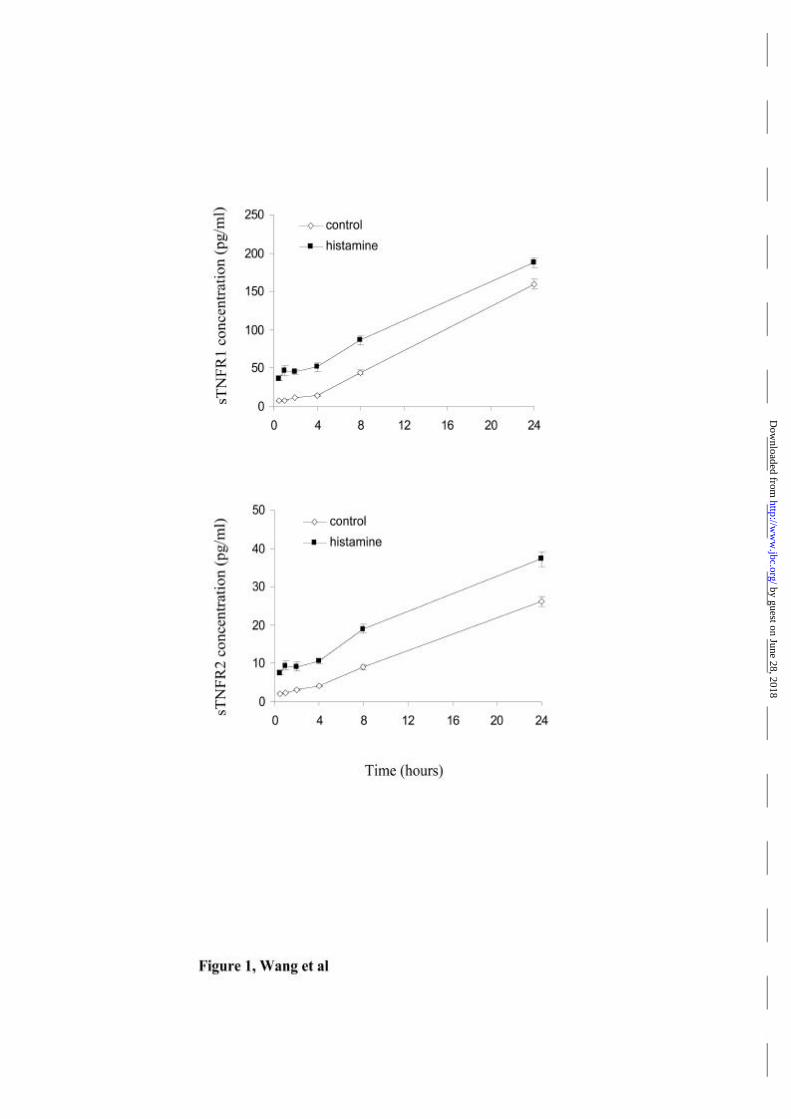
Concomitant with its effects of surface receptor expression, histamine treatment
induced an increase in soluble TNFR1 and TNFR2 shed into the culture media.
Receptor shedding was maximal during the first hour of histamine treatment (Fig 1).
Over this time period histamine induced shedding of TNFR1 was agonist
concentration dependent and inhibited by the histamine H1 receptor antagonist
diphenhydramine but not by the H2 antagonist cimetidine (Table 2). Similar results
were found for shedding of TNFR2, although the total amount shed was less.
Cumulatively, these data suggest that histamine-stimulated TNFR reduction on the
surface was caused by histamine-stimulated receptor shedding.
Role of TACE in Histamine Induced Shedding
TACE has been reported to cleave both TNF receptors from the cell surface, and it
was previously noted that receptor shedding induced by PMA could be inhibited by
the TACE inhibitor TAPI ( 7 and Table 3). To determine whether histamine induced
shedding involves TACE we pre-treated HUVEC with TAPI 25 µM for 30 minutes.
TAPI by itself increased the cell surface levels of both TNFR1 and TNFR2, consistent
by guest on June 28, 2018http://w
ww
.jbc.org/D
ownloaded from
Histamine antagonizes TNF signaling
16
with a basal rate of TACE-mediated shedding (Table 3). TAPI treatment completely
prevented the shedding caused by histamine. TAPI prevented the reduction of TNFR1
on the cell surface induced by histamine (Table 3), further supporting the link
between receptor loss and shedding. These results suggest that histamine increases
receptor loss through TACE-mediates shedding, although it is possible that other
TAPI-sensitive sheddase may be involved.
The apparent involvement of TACE in the histamine response prompted us to
examine TACE expression and localization in EC. By immuno-blotting, TACE
protein was found in both membrane and intracellular fractions of HUVEC (Fig 2).
TACE exists in two forms of molecular weights 120kD and 100kD, which correspond
to the pro-enzyme and the mature enzyme, respectively, as previously reported in
human mononuclear cell lines(27). Both forms of the enzyme were observed in
intracellular fractions whereas surface membrane-associated TACE was only detected
as the mature form. Although PMA decreases cell surface TACE in mononuclear
cells(28), the relative expression of the pro-enzyme and the mature form in different
endothelial cell fractions was not affected by either histamine or PMA treatment.
These data show that TACE is expressed in HUVEC but do not support a model in
which TACE activation by histamine is controlled by translocation to the membrane.
Effect of Brefeldin A on Cell Surface TNF Receptor Expression
The observation that endothelial cells express more TNFR2 than TNFR1 on their
surface(4), yet release higher concentrations of TNFR1 into the medium in response
to histamine, raises the possibility that intracellular TNFR1 molecules may contribute
to the shed receptor pool. We therefore directly examined whether intracellular
TNFR1 contributed to the amount of shed receptor. Brefeldin A is a fungal extract
by guest on June 28, 2018http://w
ww
.jbc.org/D
ownloaded from
Histamine antagonizes TNF signaling
17
that can disrupt cellular protein transportation from the Golgi apparatus to the plasmic
membrane(29). In one hour, Brefeldin A did not affect the amount of TNFR1
spontaneously shed into the media, but the cell surface level was reduced significantly
(Table 4). This indicated that mobilization of TNFR1 from an intracellular
compartment was required to maintain the constant cell surface level of the receptor.
Brefeldin A also reduced TNFR2 levels indicating that maintenance of this receptor
on a cell surface also depends on mobilization from an intracellular pool.
Furthermore, Brefeldin A reduced the amount of soluble receptors shed into the media
in response to histamine (Table 4). This suggests that histamine induced shedding also
involves mobilization of intracellular receptors. Brefeldin A also reduced receptor
shedding caused by PMA, sTNFR1 following treatment with PMA was (58.9±1.3
pg/ml), and was partially inhibited by pre-treatment with brefeldin A (15.6±2.0
pg/ml).
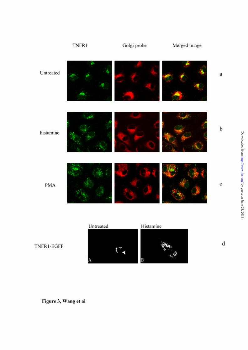
The Golgi pool constitutes the majority of TNFR1 molecules in EC. To determine
whether histamine mobilizes TNFR1 from the Golgi apparatus we examined the
distribution of TNFR1 in untreated and histamine-treated cells by confocal
immunofluorescence microscopy. As expected TNFR1 co-localizes with a Golgi
probe in untreated cells (Fig 3a). Treatment with histamine disperses TNFR1 from the
Golgi to give a punctate staining pattern throughout the cytoplasm (Fig 3b). PMA has
a similar but more pronounced effect (Fig 3c).
To extend the results seen in fixed and permeabilized cells, we generated HUVEC
transfected with EGFP-TNFR 1 fusion protein (gfp-TNFR1), and used these cells to
observe translocation of the fluorescent-labeled receptor in real time. As shown in Fig
3d Panel A, mock treated cells show fluorescence localized to the perinuclear region
consistent with receptor in the Golgi apparatus. Exposure of the cells to histamine
by guest on June 28, 2018http://w
ww
.jbc.org/D
ownloaded from
Histamine antagonizes TNF signaling
18
(Panel B) leads to a time dependent dispersal of the receptor from the perinuclear
region throughout the cytoplasm and eventually, to loss of fluorescence from the cell.
These results are consistent with observations in fixed and permeabilized cells, as
well as with the results of ELISA and FACS studies, and demonstrate that histamine
causes a redistribution of the receptor from the Golgi to the surface and into the
medium.
Signaling Pathway of Shedding caused by Histamine
To investigate the signaling pathway by which histamine activates TACE and
causes shedding of TNFRs, the effects of several pharmacological agents were tested.
Histamine activates nitric oxide synthase in EC and, via NO, can activate soluble
guanyl cyclase and protein kinase G. The NO synthase inhibitor L-NMMA (1 mM)
did not affect shedding caused by histamine or PMA. Histamine can activate protein
kinase C in EC. Bisindolylmaleimide(30), a protein kinase C inhibitor inhibited
shedding caused by PMA in a concentration dependent manner, but had no effect on
the shedding caused by histamine (Fig 4). Since the shedding of amyloid precursor
protein (APP), which is cleaved by TACE, involves MEK(16), we examined this
pathway as well. The specific MEK-1 inhibitor PD98059 (25 µM) significantly
inhibited shedding caused by histamine as well as PMA. In contrast, SB202810, the
inhibitor of the p38 mitogen activated protein kinase (p38MAPK) did not affect either
PMA or histamine induced shedding (Fig 4). These results are consistent with the
hypothesis that MEK/p42/44MAPK pathway is involved in TACE activation in EC,
but that the activation of this pathway by histamine is independent of PKC.
Effect of Histamine induced alterations in TNF Receptors on TNF responses
by guest on June 28, 2018http://w
ww
.jbc.org/D
ownloaded from
Histamine antagonizes TNF signaling
19
To investigate if the shedding of TNFR1 caused by histamine has any effect on
TNF responses in HUVEC, TNF-induced degradation of IκBα was analyzed by
immunoblotting. On its own histamine had no direct effect on the level of cellular
IκBα at any time point from 0.5 to 12 hours, whereas TNF, as previously reported(1),
induced rapid IκBα degradation (Fig 5a). Pretreatment with histamine for half an hour
prior to addition of TNF diminished the extent of TNF-induced IκBα degradation: the
effect of histamine pretreatment were lost at later time points. The time of maximal
effect corresponds to the time of the greatest reduction in the cell surface level of
TNFR1 (table 1). Furthermore, blocking the shedding of TNFR1 induced by
histamine with TAPI abolished the inhibitory effect of histamine on TNF-induced
IκBα degradation (Fig 5b). IL-1 also induces IκBα degradation in HUVEC, but IL-
1Rs are not subject to TACE-mediated shedding. To examine the specificity of the
histamine effect, histamine pre-treated cells were tested for IL-1 responsiveness. At
no time point did histamine pre-treatment show interference with IL-1 induced IκBα
degradation. These data cumulatively demonstrate that the effect of histamine on TNF
responses in HUVEC can be attributed to the shedding of cell surface receptors. It is
likely that cell surface receptor loss rather that neutralization of cytokine by sTNFR is
responsible for the effect because the medium containing shed receptors was replaced
before TNF treatment, and the effect of histamine was similar if TNF was added
without replacing the media (data not shown). The absence of neutralizing properties
in the medium can be explained by the concentrations of sTNF receptors that are
required to neutralize biological responses to TNF, which are approximately 5 ng/ml
for sTNFR1 and 500 ng/ml for sTNFR2(9). The concentration of sTNFRs in media
after histamine treatment typically reached only 30-40pg/ml.
by guest on June 28, 2018http://w
ww
.jbc.org/D
ownloaded from
Histamine antagonizes TNF signaling
20
Effect of histamine on TNF receptors and TNF responses in vivo
To determine whether the results observed with cultured HUVEC occur in vivo we
used a model involving transplantation of human skin grafts on to immunodeficient
(SCID beige) mice. First we injected replicate grafts with either saline or histamine
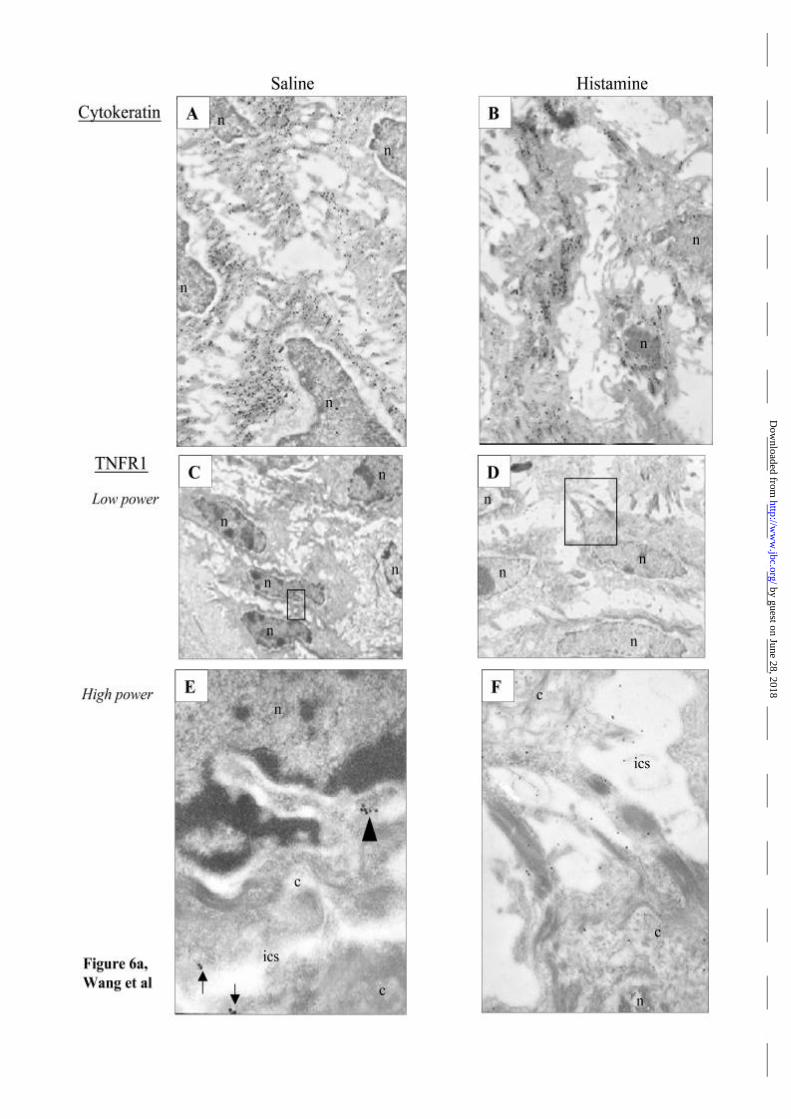
and examined the tissue 30 minutes later by immunelectron microscopy. TNFR1
molecules were most evident in the epidermis, associated with keratinocytes.
Compared with saline injected skin, histamine injection caused a marked
accumulation of human TNFR1 in intercellular space and near cell junctions of
keratinocytes in epidermis, while the distribution of cytokeratin was not altered (Fig
6a). Quantification of TNFR1 labeling by counting immuno-gold particles revealed
significantly more membrane/extracellular gold particles in histamine treated tissue
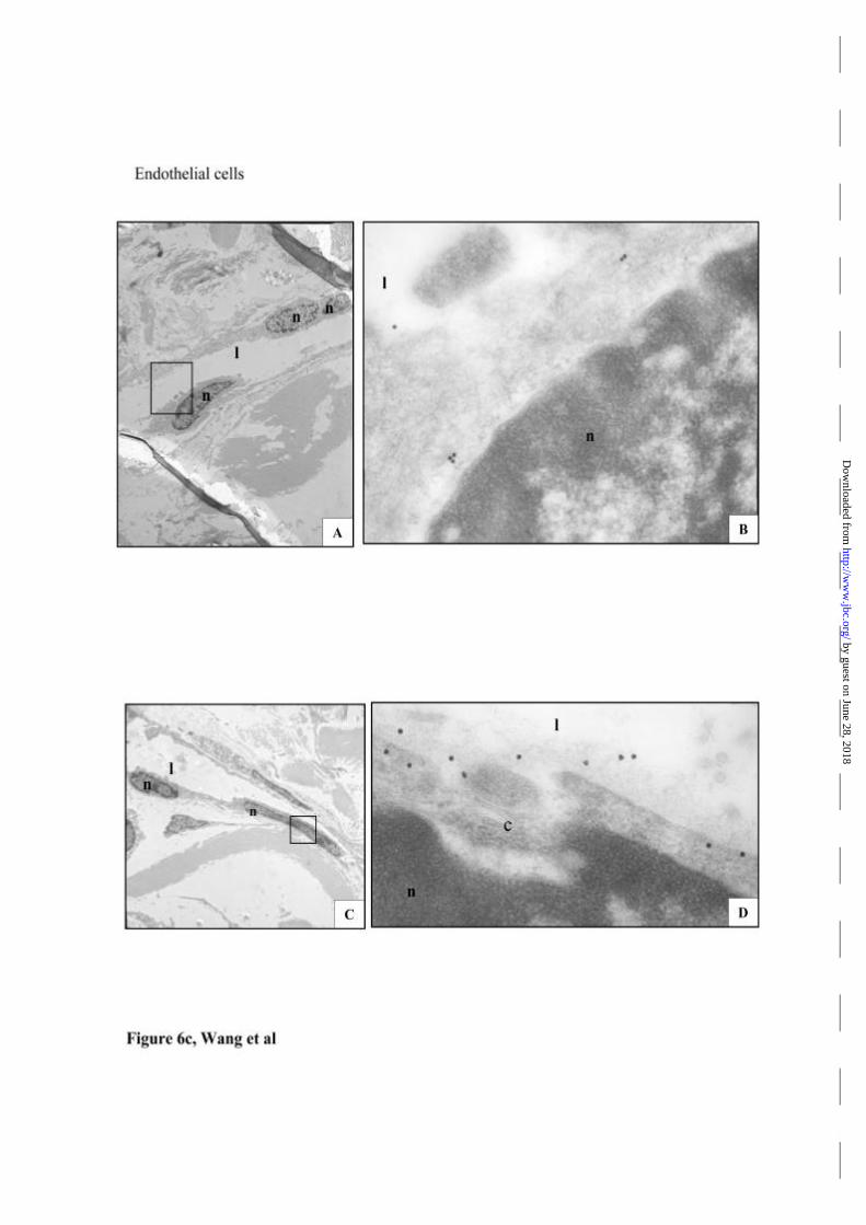
(Fig 6b). A similar redistribution of TNFR1 within EC lining dermal microvessels
was also noted (Fig 6c), but the lesser frequency of these structures did not permit
quantification.
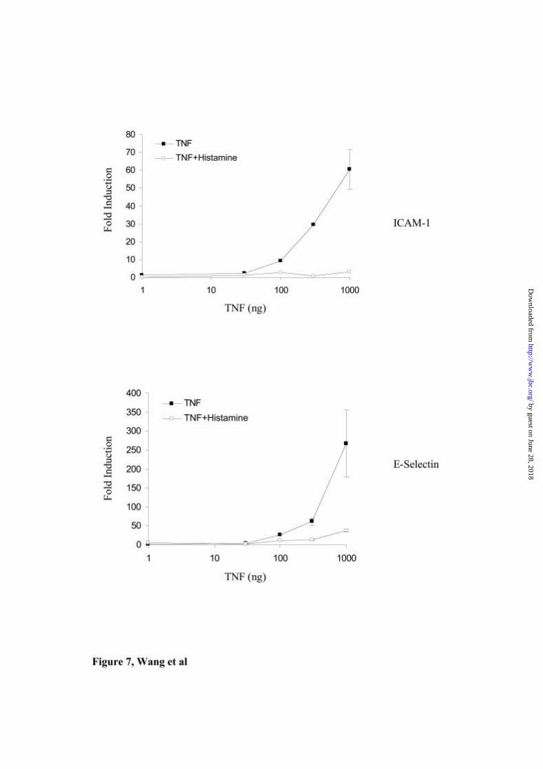
To test the effect of histamine pre-treatment on TNF responses in vivo we used
quantitative RT-PCR to evaluate ICAM-1 and E-selectin mRNA induction. E-selectin
is restricted in its expression to EC lining post-capillary venules. ICAM-1 is
expressed by EC throughout the microvasculature. Although keratinocytes can also
express ICAM-1, EC are the predominant cell types that respond to TNF. Injection of
TNF produced a dose-dependent increase in transcripts encoding both E-selectin and
ICAM-1. Pretreatment with histamine resulted in significant blunting of TNF
induction of both E-selectin and ICAM-1 mRNA (Fig 7).
by guest on June 28, 2018http://w
ww
.jbc.org/D
ownloaded from
Histamine antagonizes TNF signaling
21
Discussion
Histamine is a principal mediator of the immediate hypersensitivity reaction that
follows interaction of antigen with specific IgE molecules on the surface of mast cells
and / or basophils, and vascular endothelial cells are major targets for the biological
actions of histamine. Vascular responses occur within minutes of antigenic challenge,
and are often followed several hours later by a late phase reaction (LPR) characterized
by persistent edema and leukocyte infiltration. TNF is likely to be an important
mediator of the LPR. In skin organ culture TNF derived from resident cells in the
skin contributes to expression of E-selectin in elicited LPR(31), and in a murine
model of IgE dependent cutaneous LPR TNF contributes to mast cell dependent
recruitment of leukocytes(32). In addition mast-cell derived TNF is at least one of the
mediators involved in the recruitment of neutrophils during IgE-dependent gastric
inflammation in the mouse(33).
Mast cells contain preformed stores of biologically active TNF which can be
released into the extracellular space on degranulation(34;35). Mast cells thus provide
a source for the early release of both histamine and TNF at sites of evolving allergic
inflammation, and the biological actions of mast cell derived TNF are likely to be
important for the development of a LPR. Our results indicate that in this setting rapid
actions of histamine may limit subsequent TNF actions through effects on TNF
receptor shedding.
Histamine down-regulates both TNFR1 and TNFR2 on the cell surface of
endothelial cells by enhancing receptor shedding. TACE can cleave both TNF
receptors from the cell surface, and we have demonstrated that TACE is expressed at
high levels in EC. Histamine appears to cause shedding through activation of TACE,
as its effects could be completely blocked by the TACE inhibitor TAPI, but not other
by guest on June 28, 2018http://w
ww
.jbc.org/D
ownloaded from

Histamine antagonizes TNF signaling
22
metalloproteinase inhibitors, which is the characteristic profile for TACE(36).
Although TNFR2 is the predominant endothelial cell surface TNF receptor(4), higher
concentrations of TNFR1 were released into the media in response to histamine,
raising the possibility that TNFR1 was mobilised from the Golgi pool. This idea is
supported by the observation that Brefeldin A disrupts the Golgi, and reduces
histamine-induced receptor shedding, and also by direct observation of the
mobilization of transfected receptor in response to histamine. In EC shedding of
TNFR1 is also regulated by the expression of ARTS-1 (aminopeptidase regulator of
TNFR1 shedding), a protein that binds specifically to the extracellular domain of
TNFR1, and increases shedding of TNFR1 but not TNFR2(37). Histamine increases
shedding of both TNFR1 and TNFR2, indicating that its action is could not be fully
explained by a direct effect on ARTS-1, but expression of ARTS-1 in EC could
contribute to the increased shedding of TNFR1.
Shedding of TNFR1 was increased by both PMA and histamine, and PMA induced
shedding of TNFR1 could be inhibited by a PKC inhibitor, supporting the observation
that TACE can be activated by protein kinase C(15). However, our results
demonstrate that histamine acts through a PKC-independent pathway. It has been
reported that NGF induced β-APP shedding is regulated by a MEK-1/MAPK pathway
that can be activated by multiple first and second messengers in both a PKC-
dependent and independent manner(38;39). Shedding induced by histamine was
partially blocked by a selective MEK-1 inhibitor-PD98059, which suggested at least
part of the shedding induced by histamine was initiated through a MEK-1/MAPK
pathway. In contrast experiments using SB202810 suggest that p38MAPK was not
involved. An NO donor has been shown to be able to activate TACE(40). In our
system, shedding induced by either PMA or histamine, was not affected by the NOS
by guest on June 28, 2018http://w
ww
.jbc.org/D
ownloaded from
Histamine antagonizes TNF signaling
23
antagonist L-NMMA, although it is possible that other reactive oxygen species, which
can activate TACE(41), may be involved. Thus, there may be several enzymatic
cascades leading to activation of TACE and shedding of cell surface receptors, and
different signaling pathways may be activated by different stimuli(15;42).
Several observations suggest that histamine limits TNF responses through a direct
effect on TNF receptor shedding. The effect of histamine on both TNFR1 cell surface
expression and TNF induced IκBα degradation was transient, with both effects
occurring over the same time period. This is also consistent with the report that TNF
induces IκBα degradation predominantly through TNFR1(43). In addition the effect
of histamine on TNF induced IκBα degradation was lost if receptor shedding was
prevented by the TACE inhibitor TAPI. Finally, histamine had no effect on IL-1
induced IκBα degradation. The timing of exposure of cells to histamine in relation to
TNF is likely to be a key determinant of the effect on TNF responses, and may
explain why histamine does not inhibit TNF responses when administered
simultaneously with TNF(44;45).
Histamine exerts multiple regulatory effects during the development of an immune
inflammatory response(46). In cultured EC the effects of histamine on cell contraction
and release of vasodilators are accompanied by pro-inflammatory effects, which
include increased expression of P-selectin and IL-8, both of which are stored in
Weibel-Palade bodies(47) and can act in concert, in vitro, to promote the leukocyte
binding and transmigration. However, in vivo the principal response to histamine is
increased vascular permeability and vasodilatation without recruitment of leukocytes.
Our studies have shown a modest inhibitory effect of histamine on TNF responses,
but demonstrate a marked inhibitory effect of histamine on EC responses to TNF in
vivo. In cultured cells the effect of histamine on TNF responses appears to be through
by guest on June 28, 2018http://w
ww
.jbc.org/D
ownloaded from

Histamine antagonizes TNF signaling
24
loss of cell surface receptors rather than an inhibitory effect of shed soluble receptors.
However, neutralization of TNF by shed receptors may contribute to more dramatic
loss of EC responsiveness to TNF observed in vivo following histamine treatment of
human skin. Our ultrastructural studies suggest that keratinocytes may be a major
source of sTNFR in this context. Histamine caused a marked accumulation of
extracellular TNFR1 in human skin engrafted on to SCID mice, and diminished
upregulation of the endothelial cell specific gene E-selectin in response to TNF. The
predominant cell type, which displayed evidence of TNFR1 mobilization and
shedding in engrafted skin were keratinocytes, although mobilization of TNFR1 also
occurred in EC.
Soluble TNF receptors are emerging as important regulators of inflammatory
disease. Soluble TNF receptor fusion proteins suppress inflammation in experimental
models of inflammation, and a soluble TNFR2: Fc hybrid molecule has entered
clinical practice as an anti-inflammatory agent(11). In kidney, EC are the major cell
type expressing TNFR1(26), and our studies identify EC as a potentially important
source of sTNFR1. The role for soluble TNFR1 as a physiological inhibitor of
inflammatory responses is supported by the observation that patients with mutations
in the gene encoding TNFR1, which disrupt extracellular cysteines and impair
cleavage and shedding of the receptor develop a periodic-fever syndrome known as
TRAPS (TNF receptor-associated periodic syndrome). This syndrome is characterized
by attacks of fever, sterile peritonitis, arthralgia, myalgia, skin rash and / or
conjunctivitis(48).
In summary, the effect of histamine on mobilization of Golgi-associated TNFR1
and receptor shedding both clarifies the relationships among cell surface, Golgi-
associated and shed TNFR1 molecules and reveals a novel mechanism through which
by guest on June 28, 2018http://w
ww
.jbc.org/D
ownloaded from

Histamine antagonizes TNF signaling
25
histamine may limit the capacity of TNF to elicit an inflammatory response in an
evolving allergic reaction. They also point to EC as a major source of sTNFR1, and
that the Golgi pool of TNFR1 molecules may serve as an endogenous pool of anti-
inflammatory reagents.
by guest on June 28, 2018http://w
ww
.jbc.org/D
ownloaded from
Histamine antagonizes TNF signaling
26
Reference List
1. Madge, L. A. and Pober, J. S. (2001) Exp.Mol.Pathol. 70, 317-325
2. Slowik, M. R., De Luca, L. G., Fiers, W., and Pober, J. S. (1993) Am.J Pathol.
143, 1724-1730
3. Ledgerwood, E. C., Pober, J. S., and Bradley, J. R. (1999) Lab Invest 79, 1041-
1050
4. Bradley, J. R., Thiru, S., and Pober, J. S. (1995) Am.J Pathol. 146, 27-32
5. Jones, S. J., Ledgerwood, E. C., Prins, J. B., Galbraith, J., Johnson, D. R., Pober,
J. S., and Bradley, J. R. (1999) J Immunol 162, 1042-1048
6. Bennett, M., Macdonald, K., Chan, S. W., Luzio, J. P., Simari, R., and
Weissberg, P. (1998) Science 282, 290-293
7. Reddy, P., Slack, J. L., Davis, R., Cerretti, D. P., Kozlosky, C. J., Blanton, R.
A., Shows, D., Peschon, J. J., and Black, R. A. (2000) J Biol.Chem. 275, 14608-
14614
8. Hooper, N. M., Karran, E. H., and Turner, A. J. (1997) Biochem.J 321 ( Pt 2),
265-279
9. Van Zee, K. J., Kohno, T., Fischer, E., Rock, C. S., Moldawer, L. L., and
Lowry, S. F. (1992) Proc.Natl.Acad.Sci.U.S.A 89, 4845-4849
by guest on June 28, 2018http://w
ww
.jbc.org/D
ownloaded from
Histamine antagonizes TNF signaling
27
10. Hale, K. K., Smith, C. G., Baker, S. L., Vanderslice, R. W., Squires, C. H.,
Gleason, T. M., Tucker, K. K., Kohno, T., and Russell, D. A. (1995) Cytokine 7,
26-38
11. Galon, J., Aksentijevich, I., McDermott, M. F., O'Shea, J. J., and Kastner, D. L.
(2000) Curr.Opin.Immunol 12, 479-486
12. Black, R. A., Rauch, C. T., Kozlosky, C. J., Peschon, J. J., Slack, J. L., Wolfson,
M. F., Castner, B. J., Stocking, K. L., Reddy, P., Srinivasan, S., Nelson, N.,
Boiani, N., Schooley, K. A., Gerhart, M., Davis, R., Fitzner, J. N., Johnson, R.
S., Paxton, R. J., March, C. J., and Cerretti, D. P. (1997) Nature 385, 729-733
13. Moss, M. L., Jin, S. L., Milla, M. E., Bickett, D. M., Burkhart, W., Carter, H. L.,
Chen, W. J., Clay, W. C., Didsbury, J. R., Hassler, D., Hoffman, C. R., Kost, T.
A., Lambert, M. H., Leesnitzer, M. A., McCauley, P., McGeehan, G., Mitchell,
J., Moyer, M., Pahel, G., Rocque, W., Overton, L. K., Schoenen, F., Seaton, T.,
Su, J. L., Becherer, J. D., and . (1997) Nature 385, 733-736
14. Hwang, C., Gatanaga, M., Granger, G. A., and Gatanaga, T. (1993) J Immunol
151, 5631-5638
15. Merlos-Suarez, A. and Arribas, J. (1999) Biochem.Soc.Trans. 27, 243-246
16. Mills, J., Laurent, C. D., Lam, F., Beyreuther, K., Ida, N., Pelech, S. L., and
Reiner, P. B. (1997) J Neurosci. 17, 9415-9422
17. Madge, L. A., Sierra-Honigmann, M. R., and Pober, J. S. (1999) J Biol.Chem.
274, 13643-13649
18. Bolz, S. S. and Pohl, U. (1997) Cardiovasc.Res. 36, 437-444
by guest on June 28, 2018http://w
ww
.jbc.org/D
ownloaded from
Histamine antagonizes TNF signaling
28
19. Asako, H., Kurose, I., Wolf, R., DeFrees, S., Zheng, Z. L., Phillips, M. L.,
Paulson, J. C., and Granger, D. N. (1994) J Clin.Invest 93, 1508-1515
20. Zavoico, G. B., Ewenstein, B. M., Schafer, A. I., and Pober, J. S. (1989) J
Immunol 142, 3993-3999
21. Robinson, A. J. and Dickenson, J. M. (2001) Br.J Pharmacol. 133, 1378-1386
22. Spiecker, M., Darius, H., Kaboth, K., Hubner, F., and Liao, J. K. (1998) J
Leukoc.Biol. 63, 732-739
23. Bradley, J. R., Thiru, S., and Pober, J. S. (1995) Am.J Pathol. 147, 627-641
24. Gaeta, M. L., Johnson, D. R., Kluger, M. S., and Pober, J. S. (2000) Lab Invest
80, 1185-1194
25. Karmann, K., Min, W., Fanslow, W. C., and Pober, J. S. (1996) J Exp.Med. 184,
173-182
26. Al Lamki, R. S., Wang, J., Skepper, J. N., Thiru, S., Pober, J. S., and Bradley, J.
R. (2001) Lab Invest 81, 1503-1515
27. Schlondorff, J., Becherer, J. D., and Blobel, C. P. (2000) Biochem.J 347 Pt 1,
131-138
28. Doedens, J. R. and Black, R. A. (2000) J Biol.Chem. 275, 14598-14607
29. Pelham, H. R. (1991) Cell 67, 449-451
30. Krauss, S. and Brand, M. D. (2000) FASEB J 14, 2581-2588
31. Leung, D. Y., Pober, J. S., and Cotran, R. S. (1991) J Clin.Invest 87, 1805-1809
by guest on June 28, 2018http://w
ww
.jbc.org/D
ownloaded from
Histamine antagonizes TNF signaling
29
32. Wershil, B. K., Wang, Z. S., Gordon, J. R., and Galli, S. J. (1991) J Clin.Invest
87, 446-453
33. Furuta, G. T., Schmidt-Choudhury, A., Wang, M. Y., Wang, Z. S., Lu, L.,
Furlano, R. I., and Wershil, B. K. (1997) Gastroenterology 113, 1560-1569
34. Gordon, J. R. and Galli, S. J. (1990) Nature 346, 274-276
35. Walsh, L. J., Trinchieri, G., Waldorf, H. A., Whitaker, D., and Murphy, G. F.
(1991) Proc.Natl.Acad.Sci.U.S.A 88, 4220-4224
36. Dri, P., Gasparini, C., Menegazzi, R., Cramer, R., Alberi, L., Presani, G.,
Garbisa, S., and Patriarca, P. (2000) J Immunol 165, 2165-2172
37. Cui, X., Hawari, F., Alsaaty, S., Lawrence, M., Combs, C. A., Geng, W.,
Rouhani, F. N., Miskinis, D., and Levine, S. J. (2002) J.Clin.Invest. 110, 515-
526
38. Jolly-Tornetta, C. and Wolf, B. A. (2000) Biochemistry 39, 15282-15290
39. Buxbaum, J. D., Ruefli, A. A., Parker, C. A., Cypess, A. M., and Greengard, P.
(1994) Proc.Natl.Acad.Sci.U.S.A 91, 4489-4493
40. Zhang, Z., Kolls, J. K., Oliver, P., Good, D., Schwarzenberger, P. O., Joshi, M.
S., Ponthier, J. L., and Lancaster, J. R., Jr. (2000) J Biol.Chem. 275, 15839-
15844
41. Zhang, Z., Oliver, P., Lancaster, J. R., Jr., Schwarzenberger, P. O., Joshi, M. S.,
Cork, J., and Kolls, J. K. (2001) FASEB J 15, 303-305
42. Arribas, J. and Massague, J. (1995) J Cell Biol. 128, 433-441
by guest on June 28, 2018http://w
ww
.jbc.org/D
ownloaded from
Histamine antagonizes TNF signaling
30
43. McFarlane, S. M., Pashmi, G., Connell, M. C., Littlejohn, A. F., Tucker, S. J.,
Vandenabeele, P., and MacEwan, D. J. (2002) FEBS Lett. 515, 119-126
44. Miki, I., Kusano, A., Ohta, S., Hanai, N., Otoshi, M., Masaki, S., Sato, S., and
Ohmori, K. (1996) Cell Immunol 171, 285-288
45. Li, Y., Chi, L., Stechschulte, D. J., and Dileepan, K. N. (2001) Microvasc.Res.
61, 253-262
46. Schneider, E., Rolli-Derkinderen, M., Arock, M., and Dy, M. (2002) Trends
Immunol 23, 255-263
47. van Mourik, J. A., Romani, d. W., and Voorberg, J. (2002) Histochem.Cell Biol.
117, 113-122
48. Aksentijevich, I., Galon, J., Soares, M., Mansfield, E., Hull, K., Oh, H. H.,
Goldbach-Mansky, R., Dean, J., Athreya, B., Reginato, A. J., Henrickson, M.,
Pons-Estel, B., O'Shea, J. J., and Kastner, D. L. (2001) Am J Hum.Genet. 69,
301-314
49. Tellides, G., Kirkiles, N.C., Tereb, D.A., Schechner, J.S., Wilson, J.H., Lorber,
M.I. and J.S. Pober. Transplantation models in human/mouse chimeras. In
Organ Transplantation in Rats and Mice: Microsurgical Techniques and
Immunological Principals. W. Timmermann, H.J. Gassel, K. Ulrichs, R. Zhong,
and A, Thiede, eds. Springer-Verlag, Berlin, p. 615, (1998).
50. Heide CA., Stevens J., Livak KJ., and Williams PM. Real time quantitative PCR.
Genome Research 6:986-994, (1996).
by guest on June 28, 2018http://w
ww
.jbc.org/D
ownloaded from
Histamine antagonizes TNF signaling
31
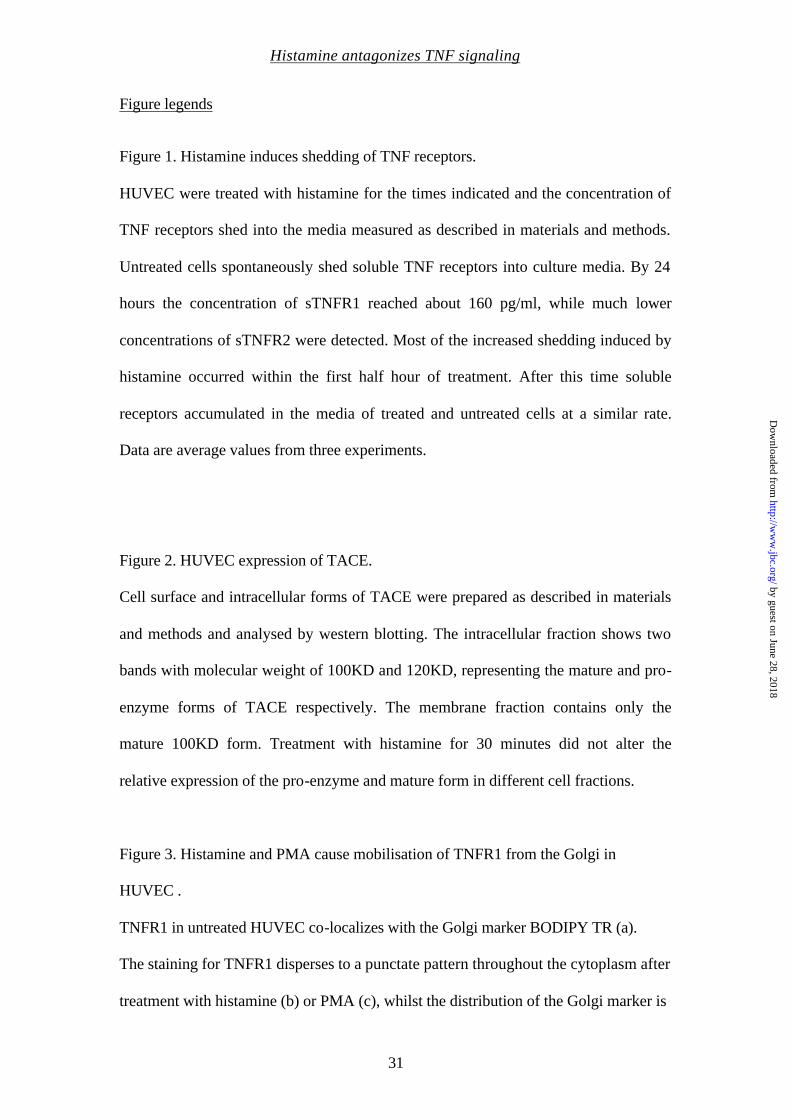
Figure legends
Figure 1. Histamine induces shedding of TNF receptors.
HUVEC were treated with histamine for the times indicated and the concentration of
TNF receptors shed into the media measured as described in materials and methods.
Untreated cells spontaneously shed soluble TNF receptors into culture media. By 24
hours the concentration of sTNFR1 reached about 160 pg/ml, while much lower
concentrations of sTNFR2 were detected. Most of the increased shedding induced by
histamine occurred within the first half hour of treatment. After this time soluble
receptors accumulated in the media of treated and untreated cells at a similar rate.
Data are average values from three experiments.
Figure 2. HUVEC expression of TACE.
Cell surface and intracellular forms of TACE were prepared as described in materials
and methods and analysed by western blotting. The intracellular fraction shows two
bands with molecular weight of 100KD and 120KD, representing the mature and pro-
enzyme forms of TACE respectively. The membrane fraction contains only the
mature 100KD form. Treatment with histamine for 30 minutes did not alter the
relative expression of the pro-enzyme and mature form in different cell fractions.
Figure 3. Histamine and PMA cause mobilisation of TNFR1 from the Golgi in
HUVEC .
TNFR1 in untreated HUVEC co-localizes with the Golgi marker BODIPY TR (a).
The staining for TNFR1 disperses to a punctate pattern throughout the cytoplasm after
treatment with histamine (b) or PMA (c), whilst the distribution of the Golgi marker is
by guest on June 28, 2018http://w
ww
.jbc.org/D
ownloaded from
Histamine antagonizes TNF signaling
32
unchanged. HUVEC transfected with gfp-TNFR1 show a Golgi pattern of
fluorescence (d, panel A). Histamine caused mobilisation of gfp-TNFR1 (d, panel B).
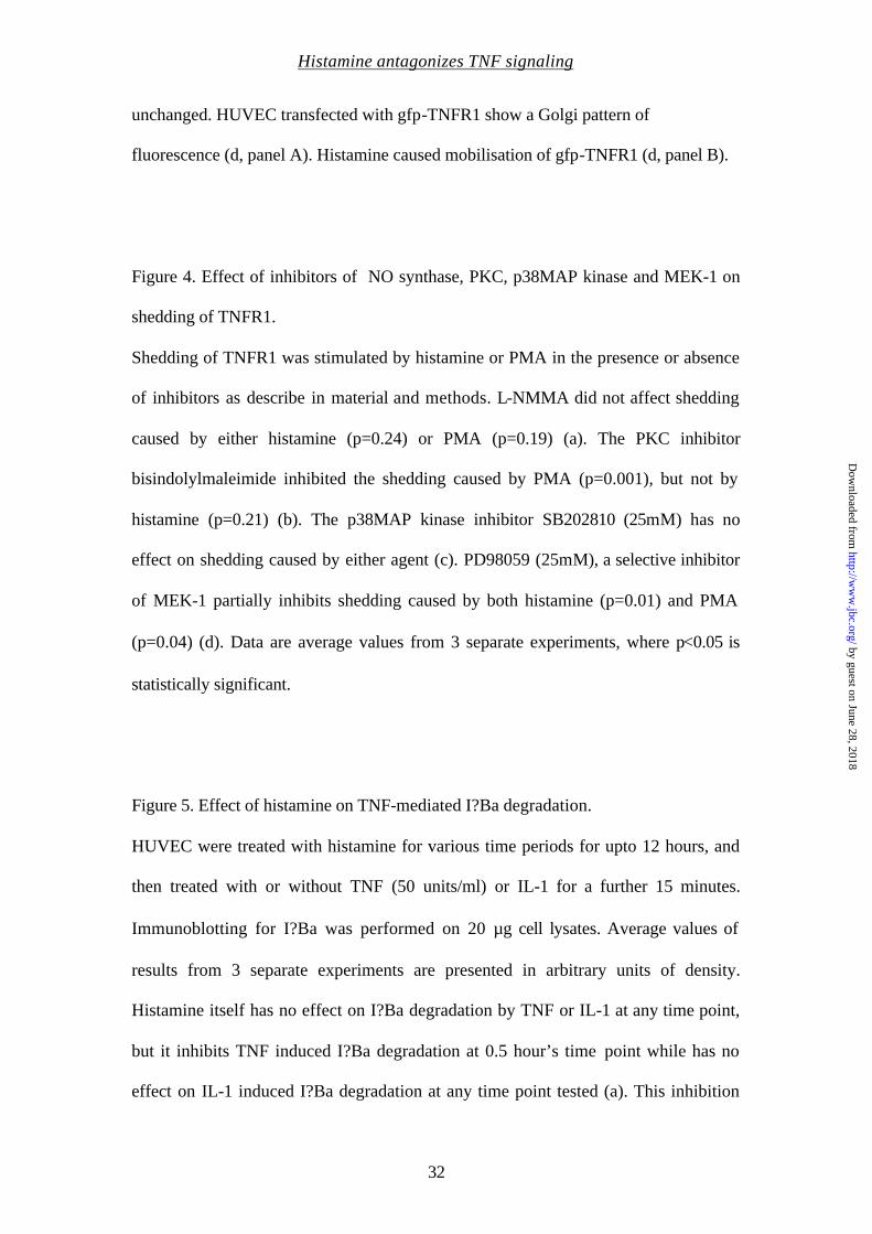
Figure 4. Effect of inhibitors of NO synthase, PKC, p38MAP kinase and MEK-1 on
shedding of TNFR1.
Shedding of TNFR1 was stimulated by histamine or PMA in the presence or absence
of inhibitors as describe in material and methods. L-NMMA did not affect shedding
caused by either histamine (p=0.24) or PMA (p=0.19) (a). The PKC inhibitor
bisindolylmaleimide inhibited the shedding caused by PMA (p=0.001), but not by
histamine (p=0.21) (b). The p38MAP kinase inhibitor SB202810 (25mM) has no
effect on shedding caused by either agent (c). PD98059 (25mM), a selective inhibitor
of MEK-1 partially inhibits shedding caused by both histamine (p=0.01) and PMA
(p=0.04) (d). Data are average values from 3 separate experiments, where p<0.05 is
statistically significant.
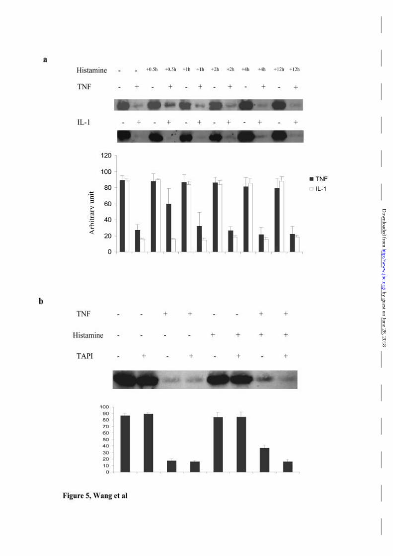
Figure 5. Effect of histamine on TNF-mediated I?Ba degradation.
HUVEC were treated with histamine for various time periods for upto 12 hours, and
then treated with or without TNF (50 units/ml) or IL-1 for a further 15 minutes.
Immunoblotting for I?Ba was performed on 20 µg cell lysates. Average values of
results from 3 separate experiments are presented in arbitrary units of density.
Histamine itself has no effect on I?Ba degradation by TNF or IL-1 at any time point,
but it inhibits TNF induced I?Ba degradation at 0.5 hour’s time point while has no
effect on IL-1 induced I?Ba degradation at any time point tested (a). This inhibition
by guest on June 28, 2018http://w
ww
.jbc.org/D
ownloaded from
Histamine antagonizes TNF signaling
33
of TNF’s effect by histamine was blocked by pre-treatment with TAPI (b); data are
average values from 2 experiments.
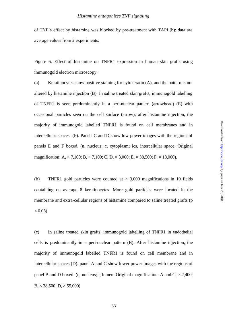
Figure 6. Effect of histamine on TNFR1 expression in human skin grafts using
immunogold electron microscopy.
(a) Keratinocytes show positive staining for cytokeratin (A), and the pattern is not
altered by histamine injection (B). In saline treated skin grafts, immunogold labelling
of TNFR1 is seen predominantly in a peri-nuclear pattern (arrowhead) (E) with
occasional particles seen on the cell surface (arrow); after histamine injection, the
majority of immunogold labelled TNFR1 is found on cell membranes and in
intercellular spaces (F). Panels C and D show low power images with the regions of
panels E and F boxed. (n, nucleus; c, cytoplasm; ics, intercellular space. Original
magnification: A, × 7,100; B, × 7,100; C, D, × 3,000; E, × 38,500; F, × 18,000).
(b) TNFR1 gold particles were counted at × 3,000 magnifications in 10 fields
containing on average 8 keratinocytes. More gold particles were located in the
membrane and extra-cellular regions of histamine compared to saline treated grafts (p
< 0.05).
(c) In saline treated skin grafts, immunogold labelling of TNFR1 in endothelial
cells is predominantly in a peri-nuclear pattern (B). After histamine injection, the
majority of immunogold labelled TNFR1 is found on cell membrane and in
intercellular spaces (D). panel A and C show lower power images with the regions of
panel B and D boxed. (n, nucleus; l, lumen. Original magnification: A and C, × 2,400;
B, × 38,500; D, × 55,000)
by guest on June 28, 2018http://w
ww
.jbc.org/D
ownloaded from
Histamine antagonizes TNF signaling
34
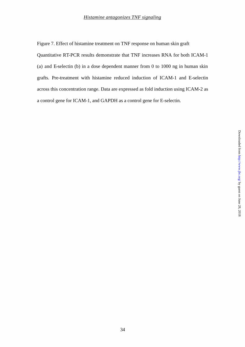
Figure 7. Effect of histamine treatment on TNF response on human skin graft
Quantitative RT-PCR results demonstrate that TNF increases RNA for both ICAM-1
(a) and E-selectin (b) in a dose dependent manner from 0 to 1000 ng in human skin
grafts. Pre-treatment with histamine reduced induction of ICAM-1 and E-selectin
across this concentration range. Data are expressed as fold induction using ICAM-2 as
a control gene for ICAM-1, and GAPDH as a control gene for E-selectin.
by guest on June 28, 2018http://w
ww
.jbc.org/D
ownloaded from
Histamine antagonizes TNF signaling
35
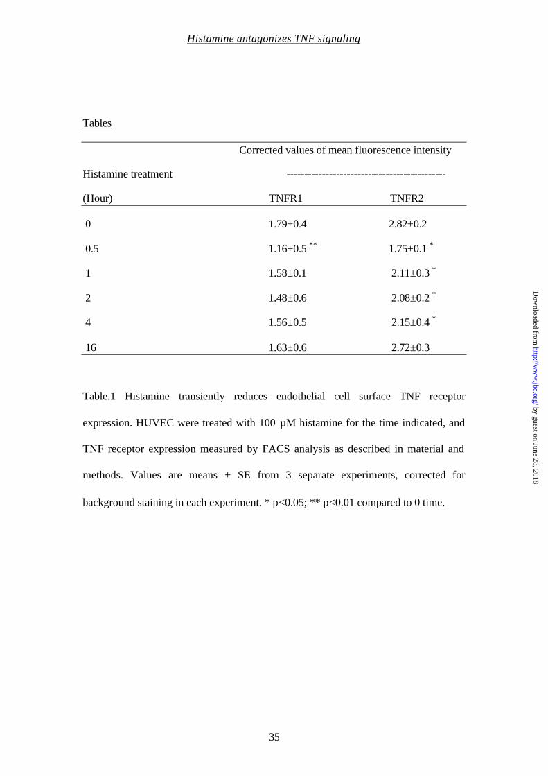
Tables
Corrected values of mean fluorescence intensity
Histamine treatment ---------------------------------------------
(Hour) TNFR1 TNFR2
0 1.79±0.4 2.82±0.2
0.5 1.16±0.5 ∗∗ 1.75±0.1 ∗
1 1.58±0.1 2.11±0.3 ∗
2 1.48±0.6 2.08±0.2 ∗
4 1.56±0.5 2.15±0.4 ∗
16 1.63±0.6 2.72±0.3
Table.1 Histamine transiently reduces endothelial cell surface TNF receptor
expression. HUVEC were treated with 100 µM histamine for the time indicated, and
TNF receptor expression measured by FACS analysis as described in material and
methods. Values are means ± SE from 3 separate experiments, corrected for
background staining in each experiment. * p<0.05; ** p<0.01 compared to 0 time.
by guest on June 28, 2018http://w
ww
.jbc.org/D
ownloaded from

Histamine antagonizes TNF signaling
36
Treatment sTNFR1 concentration (pg/ml) No treatment 7.04±2.0 Diphenhydramine 7.48±2.5 Cimetidine 7.77±1.6 Histamine 27.1±6.0 Histamine + Diphenhydramine 12.2±2.4 Histamine + Cimetidine 31.8±7.0 Table 2. Histamine induces shedding in a dose dependent manner through its H1
receptor. Levels of soluble TNFR1 in the culture media following treatment of
HUVEC for one hour with histamine across the concentration range from 0 to 1000
µM were: 8.5 pg/ml (0 µM), 8.0 pg/ml (1 µM), 23.3 pg/ml (10 µM), 32.4 pg/ml (100
µM), 35.1 pg/ml (1,000 µM). (Data are average values from two separate experiments
with similar results). Pre-treatment with the H1 antagonist diphenhydramine (100
µM) or H2 antagonist cimetidine (100 µM) was performed for 15 minutes prior to
treatment with histamine 100 µM for one hour. The H1 antagonist blocked the
histamine induced shedding, whilst the H2 antagonist had no effect. Data are
expressed as average values + SE from three experiments.
by guest on June 28, 2018http://w
ww
.jbc.org/D
ownloaded from
Histamine antagonizes TNF signaling
37
TNFR on the cell surface sTNFR1 in media
(arbitrary units) (pg/ml)
Treatment ------------------------------------- -----------------------
TNFR1 TNFR2
No treatment 1.13±0.4 1.42±0.3 8.9±1.5
TAPI 1.4±0.3 1.83 ±0.1 4.8±0.2
Histamine 0.56± 0.5∗ 0.83±0.1∗ 33.4±6.4
Histamine+TAPI 1.35±0.3 1.73± 0.5 6.9±0.91
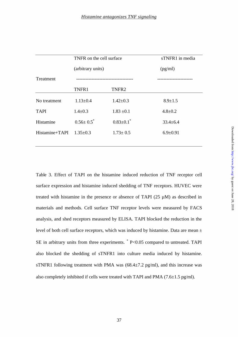
Table 3. Effect of TAPI on the histamine induced reduction of TNF receptor cell
surface expression and histamine induced shedding of TNF receptors. HUVEC were
treated with histamine in the presence or absence of TAPI (25 µM) as described in
materials and methods. Cell surface TNF receptor levels were measured by FACS
analysis, and shed receptors measured by ELISA. TAPI blocked the reduction in the
level of both cell surface receptors, which was induced by histamine. Data are mean ±
SE in arbitrary units from three experiments. ∗ P<0.05 compared to untreated. TAPI
also blocked the shedding of sTNFR1 into culture media induced by histamine.
sTNFR1 following treatment with PMA was (68.4±7.2 pg/ml), and this increase was
also completely inhibited if cells were treated with TAPI and PMA (7.6±1.5 pg/ml).
by guest on June 28, 2018http://w
ww
.jbc.org/D
ownloaded from
Histamine antagonizes TNF signaling
38
TNFR on the cell surface sTNFR1 in media
(arbitrary unit) (pg/ml)
Treatment -------------------------------------- ---------------------
TNFR1 TNFR2
No treatment 1.51±0.4 1.84±0.8 7.5±1.9
BrefeldinA 0.78±0.3∗ 1.01±0.5∗ 7.7±1.2
Histamine 0.78±0.3∗ 1.1±0.5∗ 30.2±0.7
Histamine+brefeldinA 0.68±0.5∗ 0.95±0.5∗ 13.7±3.3
Table 4. Effect of Brefeldin A on the histamine induced reduction of TNF receptor
cell surface expression, and histamine induced shedding of TNF receptors. HUVEC
were treated with histamine (100 µM) in the presence or absence of brefeldin A (10
µg/ml), cell surface TNF receptor level was measured by FACS analysis and shed
receptors measured by ELISA. Brefeldin A itself reduced cell surface TNF receptor
level without any effect on shedding. Brefeldin A appeared to potentiate the reduction
in the level of TNF cell surface receptors induced by histamine, and decreased the
shedding of sTNFR1 induced by histamine. Data are mean ± SE from three
experiments. ∗ P<0.05 compared to untreated.
by guest on June 28, 2018http://w
ww
.jbc.org/D
ownloaded from


Sathia Thiru, Jordan S. Pober and John R. BradleyJun Wang, Rafia S. Al-Lamki, Hui Zhang, Nancy Kirkiles-Smith, Mary Lou Gaeta,
the cell surface and Golgi storage poolHistamine antagonizes TNF signaling by stimulating TNF receptor shedding from
published online March 19, 2003J. Biol. Chem.
10.1074/jbc.M212662200Access the most updated version of this article at doi:
Alerts:
When a correction for this article is posted•
When this article is cited•
to choose from all of JBC's e-mail alertsClick here
by guest on June 28, 2018http://w
ww
.jbc.org/D
ownloaded from