Download - Impact of electrode micro - oatao
OATAO is an open access repository that collects the work of Toulouse researchers and makes it freely available over the web where possible
Any correspondence concerning this service should be sent
to the repository administrator: [email protected]
This is an author’s version published in: http://oatao.univ-toulouse.fr/20889
To cite this version:
Champigneux, Pierre and Delia-Dupuy, Marie-Line and Bergel, Alain Impact of electrode micro- and nano-scale topography on the formation and performance of microbial electrodes. (2018) Biosensors and Bioelectronics, 118. 231-246. ISSN 0956-5663
Official URL: https://doi.org/10.1016/j.bios.2018.06.059
Open Archive Toulouse Archive Ouverte
Impact of electrode micro- and nano-scale topography on the formation and
performance of microbial electrodes
Pierre Champigneux, Marie-Line Delia, Alain Bergel⁎
Laboratoire de Génie Chimique, CNRS, Université de Toulouse (INPT), 4 allée Emile Monso, 31432 Toulouse, France
A B S T R A C T
From a fundamental standpoint, microbial electrochemistry is unravelling a thrilling link between life and
materials. Technically, it may be the source of a large number of new processes such as microbial fuel cells for
powering remote sensors, autonomous sensors, microbial electrolysers and equipment for effluent treatment.
Microbial electron transfers are also involved in many natural processes such as biocorrosion. In these contexts, a
huge number of studies have dealt with the impact of electrode materials, coatings and surface functionalizations
but very few have focused on the effect of the surface topography, although it has often been pointed out as a key
parameter impacting the performance of electroactive biofilms.
The first part of the review gives an overview of the influence of electrode topography on abiotic electro-
chemical reactions. The second part recalls some basics of the effect of surface topography on bacterial adhesion
and biofilm formation, in a broad domain reaching beyond the context of electroactivity. On these well-estab-
lished bases, the effect of surface topography is reviewed and analysed in the field of electroactive biofilms.
General trends are extracted and fundamental questions are pointed out, which should be addressed to boost
future research endeavours. The objective is to provide basic guidelines useful to the widest possible range of
research communities so that they can exploit surface topography as a powerful lever to improve, or to mitigate
in the case of biocorrosion for instance, the performance of electrode/biofilm interfaces.
1. Introduction
A huge number of bacteria have revealed their capability to perform
extracellular electron transfer with electrodes (Logan and Regan, 2006;
Koch and Harnisch, 2016). Two main strains, Geobacter sulfurreducens
(Bond and Lovley, 2003; Lovley et al., 2011) and Shewanella oneidensis
(Ringeisen et al., 2006; Fredrickson et al., 2008) have been widely used
as model organisms for basic investigations, because of their early
discovery as electroactive strains and their high performance. In addi-
tion, G. sulfurreducens has the capability to achieve both anodic and
cathodic electron transfers (Bond and Lovley, 2003; Dumas et al.,
2008a; Soussan et al., 2013). However, multi-species microbial com-
munities are most often used as the inoculum when the objective is to
design microbial anodes to be implemented in microbial electro-
chemical processes. Environmental samples coming from marine or
lake sediments (Reimers et al., 2001; Girguis et al., 2010;
Zabihallahpoor et al., 2015; Grattieri and Minteer, 2018), wastewater,
sludge from treatment plants (Fornero et al., 2010; Kokko et al., n.d.),
and soils (Cercado Quezada et al., 2013; Doyle and Marsili, 2015) have
been particularly used as multi-species inocula and many others can be
implemented (Chabert et al., 2015).
The majority of studies have been devoted to microbial anodes
(Pham et al., 2009; Wagner et al., 2010; Kumar et al., 2013; Lu et al.,
2015). Microorganisms oxidize organic compounds to sustain their
metabolism and the resulting low energy electrons are released to the
electrode through the biofilm (Fig. 1). A smaller number of studies have
dealt with microbial cathodes, mainly for oxygen reduction (Bergel
et al., 2005) and, more recently, CO2 reduction (Rabaey et al., 2011;
ElMekawy et al., 2016; Bajracharya et al., 2017).
Microbial electrodes have been considered as the possible source of
a large number of new processes (Wang and Ren, 2013; Schröder et al.,
2015; Bajracharya et al., 2016). Some exaggeratedly enthusiastic fore-
casts about the future of microbial electrochemical technologies are
open to question (Blanchet et al., 2015; Oliot et al., 2016) but microbial
electrodes should, nevertheless, be at the core of many innovative ap-
plications, provided that suitable objectives are chosen. For instance,
microbial fuel cells may be appropriate energy production systems
when low power is sufficient (Shleev et al., 2015), such as for powering
remote sensors (Dewan et al., 2014) and designing autonomous sensors
(Di Lorenzo et al., 2009; Pasternak et al., 2017). Simplifying the
equipment to design low-cost devices that do not require attendance,
such as the electro-microbial snorkel (Erable et al., 2011; Matturro
et al., 2017), or focusing on specific environments, such as hypersaline
media (Rousseau et al., 2013; Carmona-Martinez et al., 2015; Grattieri
and Minteer, 2018) may also open up promising horizons. Furthermore,
microbial electrodes have led to fundamental discoveries on the elec-
trochemical link between living organisms and materials (Borole et al.,
2011; Shi et al., 2016; Kumar et al., 2017), which may be involved in
many natural processes, such as anaerobic digestion (Kato et al., 2012;
Liu et al., 2012) and microbially influenced corrosion (Beech and
Sunner, 2004; Mehanna et al., 2009a; Kip and van Veen, 2015).
Very many studies have focused on electrode materials and coatings
(Liu et al., 2007; Wei et al., 2011; Guo et al., 2013; Santoro et al., 2015;
Xie et al., 2015). Actually, the biofilm/electrode interface has been
widely investigated from the standpoint of surface chemistry (Lowy
et al., 2006; Rosenbaum et al., 2007; Scott et al., 2007; Kumar et al.,
2013; Du et al., 2014), with many attempts at surface functionalization
(Erable et al., 2009a; Picot et al., 2011; Lapinsonnière et al., 2013; Zhao
et al., 2013; Ding et al., 2015; Li et al., 2017). Surprisingly, the topo-
graphy of the biofilm/electrode interface has rarely been at the heart of
the studies, although it has often been pointed out as a key parameter
for the efficiency of microbial electrodes (Peng et al., 2010; Sun et al.,
2010; Fan et al., 2011; Pons et al., 2011a; Pocaznoi et al., 2012a). To
the best of our knowledge, the studies dealing with the impact of the
electrode topography on electroactive biofilms have never been re-
viewed so far. Such a review is the purpose of the present article, with
the objective of trying to extract general guidelines and suggesting, if
relevant, useful directions for future research.
The first part of the present article recalls the basics concerning the
effect of surface topography on bacterial adhesion and biofilm forma-
tion. It gives an overview of the overall knowledge that has been es-
tablished in a general field, out of the context of electroactivity. The
second part deals with the impact of electrode topography on abiotic
electrochemical reactions, i.e. in the absence of microbially-related
phenomena. On these two well-established bases, the studies that deal
with surface topography in the field of electroactive biofilms are then
reviewed and analysed.
One objective is to see whether general trends can be extracted and
whether or not they follow the knowledge previously established for
non-electroactive biofilms and in the field of abiotic electrochemistry.
Secondly, this cross-cutting analysis points to exciting questions that
should be addressed in order to encourage future research endeavours
in the most relevant directions. Among various suggestions, the main
conclusion that should be kept in mind is the necessity to take the
considerable effect of surface topography into better consideration
when analysing any experimental data related to electron transfer at
biofilm/electrode interfaces in the future.
We hope that this review will offer useful guidelines for exploiting
surface topography as a lever to improve the performance of electro-
active biofilms. Furthermore, even though the main current trend is
towards the design of electroactive biofilms for microbial electro-
chemical processes, it should not be forgotten that the same interfaces
can be at the heart of microbially influenced corrosion (Mehanna
et al., 2009a, 2009b). In this field, the same fundamental knowledge is
fully relevant to fight against corrosion by mitigating interfacial
electron transfers. In both cases, this review article has been written
with the objective of giving the widest possible range of research
communities some helpful information for further basic and techno-
logical thinking.
2. Basics of surface topography
2.1. Macro-, micro- and nano-roughness
Each surface, even the smoothest one, contains irregularities, which
can occur at macro-, micro- and nano-scale. This review focuses the
micro- and nano-scale surface topography, because this is the range of
sizes that can impact the mechanisms of bacterial adhesion, biofilm
formation and electron transfer reactions. The surface waviness, also
referred as macro-roughness, is not taken into account in this review
article. Actually, it may affect the global performance of microbial
electrochemical systems, by modifying the surface area that is available
for microbial colonization, but it is not thought to influence the me-
chanisms of electroactive biofilm formation and operation.
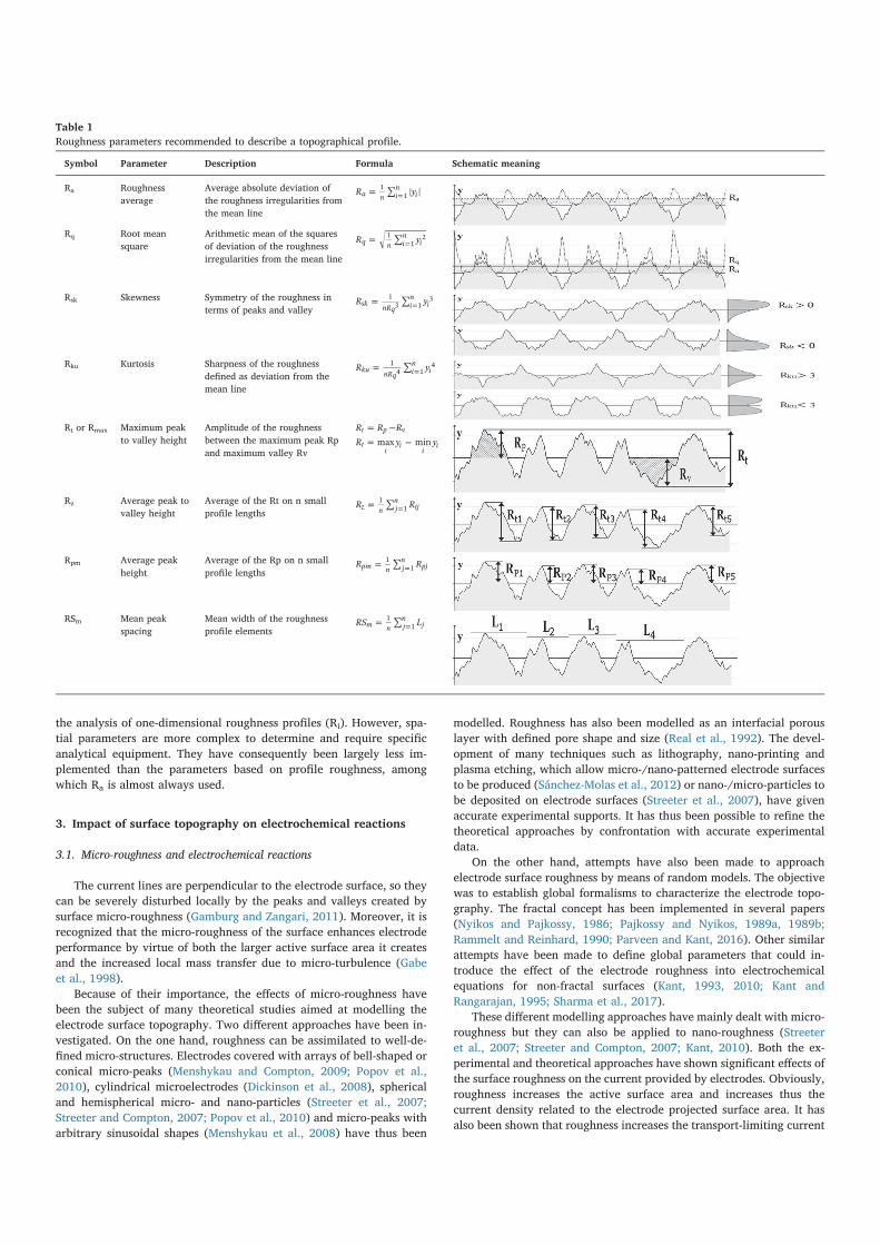
2.2. Characterization of surface topography: Ra is not perfect but is useful
Any surface is perturbed by asperities and valleys at micrometer and
nanometre scales. The surface roughness profile is commonly char-
acterized by the arithmetic mean roughness value (Ra) measured along
a line. Ra is defined as the average absolute deviation of the roughness
irregularities from the mean line (Table 1). For example, this means
that a surface with Ra of 1 µm presents peaks and valleys 1 µm above
and 1 µm below the mean line on average. Since it is one of the easiest
to measure, Ra has become a standard parameter. Nevertheless, it de-
picts the surface topography only vaguely (Donoso et al., 2007), since
various surface profiles can present the same Ra value. A large variety of
parameters have been described in the literature to better characterize
surface topography according to the field of study (Bharat Bhushan,
2000; Stout, 2000; Webb et al., 2012) (Table 1).
Several parameters have been based on the roughness profile
(Table 1), i.e. the roughness along a line:
– skewness (Rsk), whichmeasures the symmetry between peaks and valleys,
– kurtosis (Rku), which measures the sharpness of surface,
– root mean square surface roughness (Rq),
– maximum peak to valley height (Rt or Rmax),
– maximum peak height (Rp),
– maximum valley depth (Rv),
– average peak to valley height (Rz),
– average peak-to-mean height (Rpm),
– and mean width of the roughness elements (RSm).
Other parameters have been based on an analysis of the two-di-
mensional roughness, instead of the roughness profile along a line. The
so-called spatial parameters are: root mean square area roughness (Sq),
summit density (Sds), developed area ratio (Sdr), ten-point average
roughness (Sz), skewness (Ssk), texture aspect ratio (Str) and bearing
ratio (tp) as proposed by Crawford et al. (2012).
The (Si) parameters based on surface analysis result in better char-
acterization of the surface shape and organization than those based on
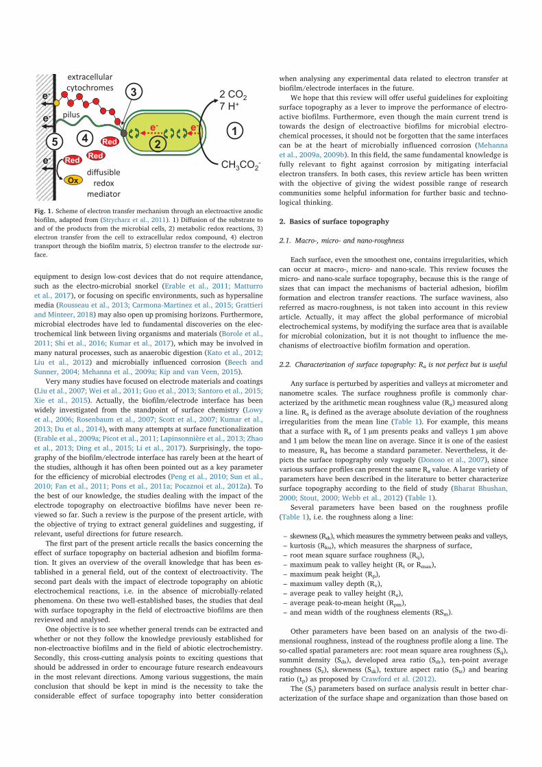
Fig. 1. Scheme of electron transfer mechanism through an electroactive anodic
biofilm, adapted from (Strycharz et al., 2011). 1) Diffusion of the substrate to
and of the products from the microbial cells, 2) metabolic redox reactions, 3)
electron transfer from the cell to extracellular redox compound, 4) electron
transport through the biofilm matrix, 5) electron transfer to the electrode sur-
face.
the analysis of one-dimensional roughness profiles (Ri). However, spa-
tial parameters are more complex to determine and require specific
analytical equipment. They have consequently been largely less im-
plemented than the parameters based on profile roughness, among
which Ra is almost always used.
3. Impact of surface topography on electrochemical reactions
3.1. Micro-roughness and electrochemical reactions
The current lines are perpendicular to the electrode surface, so they
can be severely disturbed locally by the peaks and valleys created by
surface micro-roughness (Gamburg and Zangari, 2011). Moreover, it is
recognized that the micro-roughness of the surface enhances electrode
performance by virtue of both the larger active surface area it creates
and the increased local mass transfer due to micro-turbulence (Gabe
et al., 1998).
Because of their importance, the effects of micro-roughness have
been the subject of many theoretical studies aimed at modelling the
electrode surface topography. Two different approaches have been in-
vestigated. On the one hand, roughness can be assimilated to well-de-
fined micro-structures. Electrodes covered with arrays of bell-shaped or
conical micro-peaks (Menshykau and Compton, 2009; Popov et al.,
2010), cylindrical microelectrodes (Dickinson et al., 2008), spherical
and hemispherical micro- and nano-particles (Streeter et al., 2007;
Streeter and Compton, 2007; Popov et al., 2010) and micro-peaks with
arbitrary sinusoidal shapes (Menshykau et al., 2008) have thus been
modelled. Roughness has also been modelled as an interfacial porous
layer with defined pore shape and size (Real et al., 1992). The devel-
opment of many techniques such as lithography, nano-printing and
plasma etching, which allow micro-/nano-patterned electrode surfaces
to be produced (Sánchez-Molas et al., 2012) or nano-/micro-particles to
be deposited on electrode surfaces (Streeter et al., 2007), have given
accurate experimental supports. It has thus been possible to refine the
theoretical approaches by confrontation with accurate experimental
data.
On the other hand, attempts have also been made to approach
electrode surface roughness by means of random models. The objective
was to establish global formalisms to characterize the electrode topo-
graphy. The fractal concept has been implemented in several papers
(Nyikos and Pajkossy, 1986; Pajkossy and Nyikos, 1989a, 1989b;
Rammelt and Reinhard, 1990; Parveen and Kant, 2016). Other similar
attempts have been made to define global parameters that could in-
troduce the effect of the electrode roughness into electrochemical
equations for non-fractal surfaces (Kant, 1993, 2010; Kant and
Rangarajan, 1995; Sharma et al., 2017).
These different modelling approaches have mainly dealt with micro-
roughness but they can also be applied to nano-roughness (Streeter
et al., 2007; Streeter and Compton, 2007; Kant, 2010). Both the ex-
perimental and theoretical approaches have shown significant effects of
the surface roughness on the current provided by electrodes. Obviously,
roughness increases the active surface area and increases thus the
current density related to the electrode projected surface area. It has
also been shown that roughness increases the transport-limiting current
Symbol Parameter Description Formula Schematic meaning
Ra Roughness
average
Average absolute deviation of
the roughness irregularities from
the mean line
= ∑ =R ya n in
i1
1
Rq Root mean
square
Arithmetic mean of the squares
of deviation of the roughness
irregularities from the mean line
= ∑ =R yq n in
i1
12
Rsk Skewness Symmetry of the roughness in
terms of peaks and valley= ∑ =R ysk
nRqin
i13 1
3
Rku Kurtosis Sharpness of the roughness
defined as deviation from the
mean line
= ∑ =R ykunRq
in
i14 1
4
Rt or Rmax Maximum peak
to valley height
Amplitude of the roughness
between the maximum peak Rp
and maximum valley Rv
= −R R Rt p v
= −R y ymax minti
ii
i
Rz Average peak to
valley height
Average of the Rt on n small
profile lengths= ∑ =R Rz n j
ntj
11
Rpm Average peak
height
Average of the Rp on n small
profile lengths= ∑ =R Rpm n j
npj
11
RSm Mean peak
spacing
Mean width of the roughness
profile elements= ∑ =RS Lm n j
nj
11
Table 1
Roughness parameters recommended to describe a topographical profile.
Waals and electrostatic double-layer forces, or short-range (< 3 nm)
forces, such as hydrogen bonding, ionic and dipole interactions, and
hydrophobic forces (Busscher and Weerkamp, 1987). In the case of
adsorption of solid particles, the DLVO theory has been complemented
with a surface roughness parameter (Czarnecki and Warszyński, 1987).
The application of DLVO theory to the adhesion of microbial cells
has been strongly debated. The wall of microbial cells cannot be con-
sidered as a well-defined, hard, non-permeable, uniform sheath, as the
conventional colloidal approaches assume, but should be approached
by soft, permeable heterogeneous interphases with a non-negligible
thickness (Gaboriaud et al., 2008; Duval and Gaboriaud, 2010; Hori and
Matsumoto, 2010). The complexity of the theoretical model is conse-
quently significantly increased.
The third step of the adhesion process, irreversible adhesion, in-
volves molecular reactions. Bacterial surface structures, such as ad-
hesins and transmembrane polymers, perform chemical bridging with
the conditioning film of the surface (Davey and O’toole, 2000; O’Toole
et al., 2000; Ubbink and Schaer-Zammaretti, 2007). The bonds must be
strong enough to overcome the repulsive and detachment forces, such
as shear forces induced by fluid flow. Increasing the contact area be-
tween bacterial cells and the electrode surface gives more opportunities
for linking and thus increases the adhesion strength. Once they are ir-
reversibly bonded with the surface, bacteria are able to change their
metabolism, switching from a free-swimming way of life to a complex
surface-attached community life (Flemming et al., 2016).
4.2. Basics of biofilm development and structural models
As the primo-adherent cells undergo irreversible adhesion, they
adopt a biofilm metabolism (Costerton et al., 1994; Costerton, 1995),
which is expressed by the growth and division of the cells to form
microbial aggregates. The growth of such cell clusters and their spatial
arrangement create the three-dimensional structure of the biofilm
(Klapper and Dockery, 2002). Biofilm growth and maturation involve
the establishment of an extracellular matrix that holds the three-di-
mensional structure of the biofilm. The extracellular matrix ensures the
structural integrity of the microbial community, protects it from en-
vironmental stresses, and enhances nutriment availability (Cogan and
Keener, 2004; Flemming et al., 2007; Flemming and Wingender, 2010).
The formation and growth of a biofilm has been described as a
developmental process (Doyle, 2001) evolving from primo-adherent
cells to structured microbial aggregates and extracellular matrix
(Characklis and Wilderer, 1989) to the final organised microbial biofilm
community. Understanding the mechanisms that govern how this
complex organization becomes established in connection with the en-
vironmental factors has been a great challenge for the microbiology
community in the past thirty years (Goller and Romeo, 2008). It has led
to detailed understanding of the causal relationships linking genotype
to phenotype within the biofilm communities (Monds and O’Toole,
2009), through genomic approaches linked with quorum sensing stu-
dies (Nealson et al., 1970; Eberhard, 1972; Irie and Parsek, 2008). In
this context, the chemical and topographical features of surfaces have
been shown to influence metabolic changes critical for biofilm forma-
tion (Shemesh et al., 2010). Nevertheless, there are still very few studies
that have addressed the link between the properties of a surface and the
response of bacterial communities growing on it.
4.3. Influence of surface topography on cell adhesion and biofilm formation
As detailed above (Section 4.1), cell adhesion mechanisms have
mainly been approached by theories developed in the domain of colloid
particles. In this context, surface topography has rarely been identified
as an essential parameter that may strongly influence cell adhesion.
Actually, DLVO studies are generally carried out by considering parti-
cles with a perfectly smooth surface and in the absence of shear stress.
Yet shear stresses due to solution flow are predominant in most aqueous
by acting on the local diffusion profile (Kant and Rangarajan, 1994;
Streeter and Compton, 2007). It also modified the conditions of ad-
sorption redox compounds on the electrode surface (Menshykau and
Compton, 2009) and impact the double layer capacity (Douglass Jr.
et al., 2008). The impact of surface roughness on abiotic electro-
chemistry has been judged so important that a recent article has stated
that: “…not accounting for roughness in data analysis may cause errors in
estimation of composition, diffusion coefficient, improper assignment of
electrode mechanism, and so forth” (Parveen and Kant, 2016).
To the best of our knowledge, this vast theoretical basis developed
for abiotic electrochemistry has not yet been exploited to investigate
the impact of roughness on electroactive biofilms, although it may di-
rectly affect the step 5, or even possibly step 4, of the electron transfer
chain (Fig. 1). Tapping into these studies would no doubt help the re-
search community to make significant advances in understanding and
improving microbial electrodes.
3.2. Nano-roughness and electrochemical reactions
Borisova and Ershler have reported pioneering work on the influ-
ence of nano-scale roughness on the double-layer capacitance (Borisova
and Ershler, 1950). Capacitance dispersion decreased as the surface
became flatter. Many studies then confirmed that capacitance disper-
sion was mainly of geometrical origin (De Levie, 1965; Scheider, 1975;
de Levie, 1989). Since this discovery, surface roughness has been
known to have a positive correlation with the electrical capacitance of
the surface (Albina et al., 2006). It has also been stated that electrical
conductance is reduced by deposited films, which decrease the nano-
roughness. This effect has been observed for thin films, tens of atoms
thick (Ke et al., 2009). Nano-roughness is also known to impact mole-
cular adsorption on electrode surfaces (Pfeifer et al., 1989). The con-
centration of adsorbed species tends to be smaller and the adsorption
process slower on a rough surface than on a smooth one, because of the
perturbation of the molecular arrangement caused by the roughness.
Adsorption can lead to great variations in the interfacial properties
(Bockris et al., 2008), especially in the electrode double-layer capaci-
tance (Douglass et al., 2008).
4. Impact of surface topography on cell adhesion and biofilm
development
4.1. Basics of cell adhesion
Settlement of a solid surface by a microbial biofilm is highly de-
pendent on the preliminary attachment of cells to the support. It is
agreed that biofilm formation takes place in eight successive steps
(Characklis and Marshall, 1990) and the term “cell adhesion” covers a
complex mechanism (Berkeley, 1980; Marshall, 1984), which com-
monly involves the first three steps.
The first step is the formation of a conditioning film on the surface.
This is composed of organic or inorganic matter. By modifying the
surface charge, potential, and possibly topography, it can promote or
lessen bacterial adhesion.
The second step involves a random or targeted contact between
planktonic cells and the surface. This step has been approached using
theories developed in colloidal science. There, physical forces are at
play: if the attractive forces are greater than the repulsive forces, bac-
teria reversibly adsorb to the surface. The forces to be considered in-
clude Van der Waals forces, steric interactions and electrostatic inter-
actions, which are strongly dependent on the compositions of the
conditioning film and the medium. Interfacial interactions have been
modelled using the DLVO theory applied to cells (Marshall et al., 1971),
which has been supplemented by an extended DLVO theory adding
hydrophobic/hydrophilic and osmotic interactions (Van Oss et al.,
1986; Van Oss, 1989; Hermansson, 1999). These forces act at the nano-
scale and are classified as long-range (< 150 nm), such as Van der
environments used to grow microbial cells. Neglecting the solution flow
has consequently been judged as a possible source of discrepancy be-
tween the colloidal-based theoretical approaches and the real world
(Perni et al., 2014).
When shear stress is taken into account, the surface topography
takes on great importance since it can create local variations of the
shear forces. It has been shown that, on altered surfaces, the initial
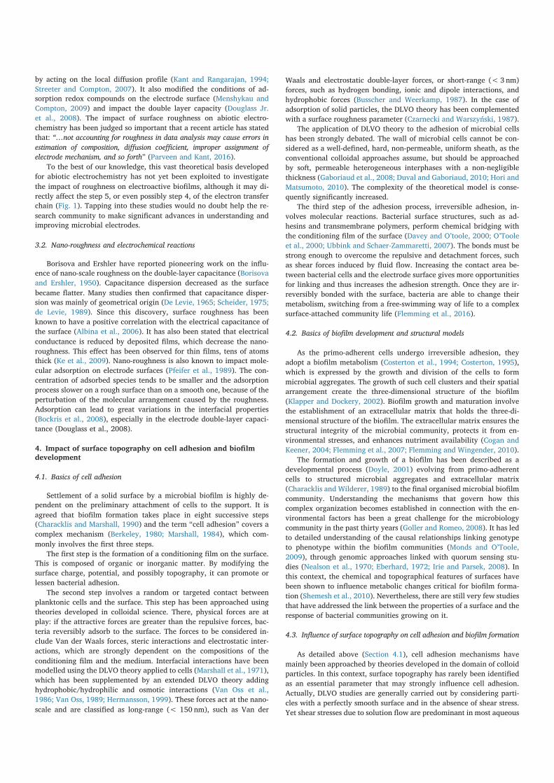
adhesion of bacteria is favoured by asperities and tends to occur in the
low shear force areas like scratches, holes or valleys (Characklis and
Marshall, 1990). These observations have been reported in many stu-
dies as illustrated in Fig. 1. They have been confirmed by implementing
well-controlled surface topographies obtained by patterning the surface
with micro-holes and micro-pillars (Díaz et al., 2007a, 2007b, 2010;
Lorenzetti et al., 2015; Hochbaum and Aizenberg, 2010; Helbig et al.,
2016) (Fig. 2). It now seems established that bacterial settlement on a
surface is enhanced by micro-roughness that creates low-shear-force
areas and thus provides the cells with calm spaces for adhesion.
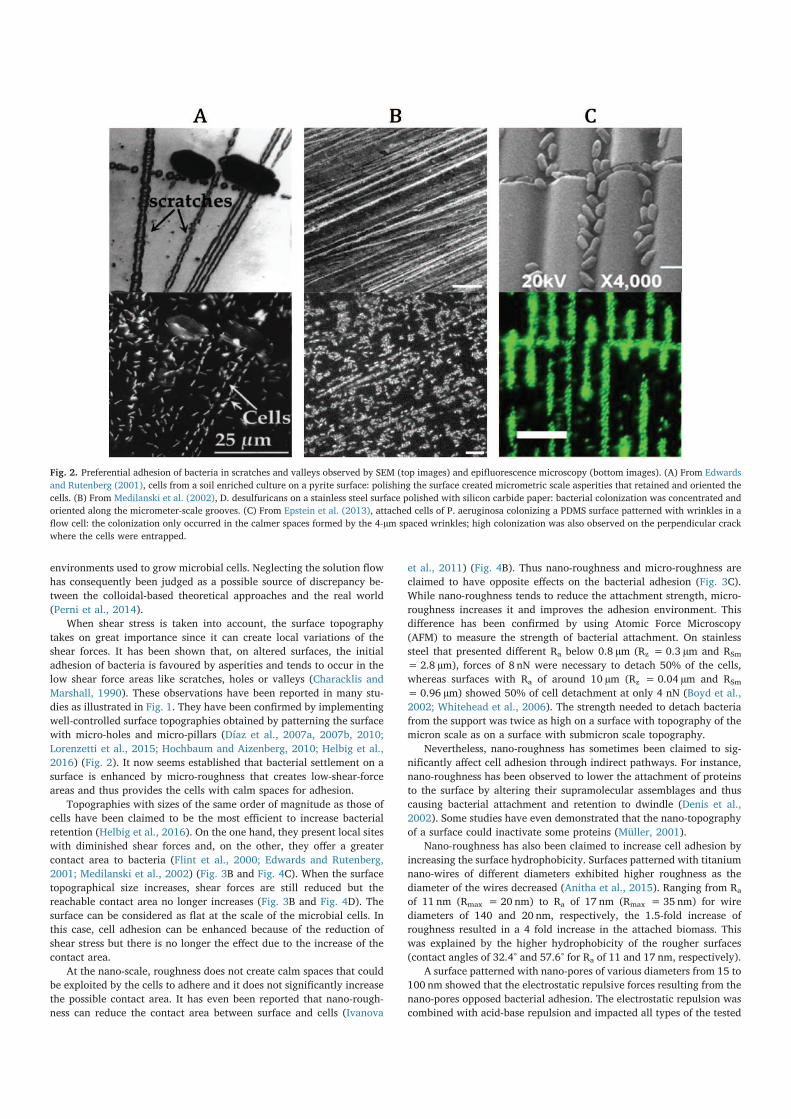
Topographies with sizes of the same order of magnitude as those of
cells have been claimed to be the most efficient to increase bacterial
retention (Helbig et al., 2016). On the one hand, they present local sites
with diminished shear forces and, on the other, they offer a greater
contact area to bacteria (Flint et al., 2000; Edwards and Rutenberg,
2001; Medilanski et al., 2002) (Fig. 3B and Fig. 4C). When the surface
topographical size increases, shear forces are still reduced but the
reachable contact area no longer increases (Fig. 3B and Fig. 4D). The
surface can be considered as flat at the scale of the microbial cells. In
this case, cell adhesion can be enhanced because of the reduction of
shear stress but there is no longer the effect due to the increase of the
contact area.
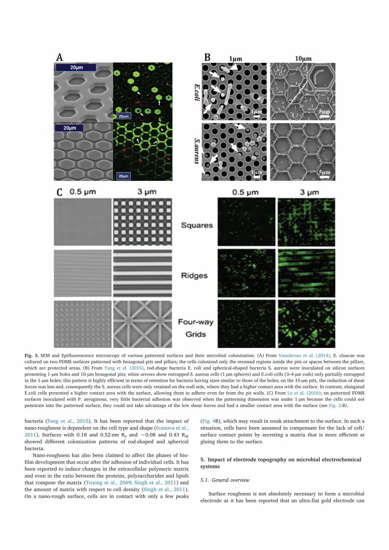
At the nano-scale, roughness does not create calm spaces that could
be exploited by the cells to adhere and it does not significantly increase
the possible contact area. It has even been reported that nano-rough-
ness can reduce the contact area between surface and cells (Ivanova
et al., 2011) (Fig. 4B). Thus nano-roughness and micro-roughness are
claimed to have opposite effects on the bacterial adhesion (Fig. 3C).
While nano-roughness tends to reduce the attachment strength, micro-
roughness increases it and improves the adhesion environment. This
difference has been confirmed by using Atomic Force Microscopy
(AFM) to measure the strength of bacterial attachment. On stainless
steel that presented different Ra below 0.8 µm (Rz =0.3 µm and RSm
=2.8 µm), forces of 8 nN were necessary to detach 50% of the cells,
whereas surfaces with Ra of around 10 µm (Rz =0.04 µm and RSm
=0.96 µm) showed 50% of cell detachment at only 4 nN (Boyd et al.,
2002; Whitehead et al., 2006). The strength needed to detach bacteria
from the support was twice as high on a surface with topography of the
micron scale as on a surface with submicron scale topography.
Nevertheless, nano-roughness has sometimes been claimed to sig-
nificantly affect cell adhesion through indirect pathways. For instance,
nano-roughness has been observed to lower the attachment of proteins
to the surface by altering their supramolecular assemblages and thus
causing bacterial attachment and retention to dwindle (Denis et al.,
2002). Some studies have even demonstrated that the nano-topography
of a surface could inactivate some proteins (Müller, 2001).
Nano-roughness has also been claimed to increase cell adhesion by
increasing the surface hydrophobicity. Surfaces patterned with titanium
nano-wires of different diameters exhibited higher roughness as the
diameter of the wires decreased (Anitha et al., 2015). Ranging from Ra
of 11 nm (Rmax =20 nm) to Ra of 17 nm (Rmax =35 nm) for wire
diameters of 140 and 20 nm, respectively, the 1.5-fold increase of
roughness resulted in a 4 fold increase in the attached biomass. This
was explained by the higher hydrophobicity of the rougher surfaces
(contact angles of 32.4° and 57.6° for Ra of 11 and 17 nm, respectively).
A surface patterned with nano-pores of various diameters from 15 to
100 nm showed that the electrostatic repulsive forces resulting from the
nano-pores opposed bacterial adhesion. The electrostatic repulsion was
combined with acid-base repulsion and impacted all types of the tested
Fig. 2. Preferential adhesion of bacteria in scratches and valleys observed by SEM (top images) and epifluorescence microscopy (bottom images). (A) From Edwards
and Rutenberg (2001), cells from a soil enriched culture on a pyrite surface: polishing the surface created micrometric scale asperities that retained and oriented the
cells. (B) From Medilanski et al. (2002), D. desulfuricans on a stainless steel surface polished with silicon carbide paper: bacterial colonization was concentrated and
oriented along the micrometer-scale grooves. (C) From Epstein et al. (2013), attached cells of P. aeruginosa colonizing a PDMS surface patterned with wrinkles in a
flow cell: the colonization only occurred in the calmer spaces formed by the 4-µm spaced wrinkles; high colonization was also observed on the perpendicular crack
where the cells were entrapped.
bacteria (Feng et al., 2015). It has been reported that the impact of
nano-roughness is dependent on the cell type and shape (Ivanova et al.,
2011). Surfaces with 0.18 and 0.52 nm Ra and −0.08 and 0.43 Rsk
showed different colonization patterns of rod-shaped and spherical
bacteria.
Nano-roughness has also been claimed to affect the phases of bio-
film development that occur after the adhesion of individual cells. It has
been reported to induce changes in the extracellular polymeric matrix
and even in the ratio between the proteins, polysaccharides and lipids
that compose the matrix (Truong et al., 2009; Singh et al., 2011) and
the amount of matrix with respect to cell density (Singh et al., 2011).
On a nano-rough surface, cells are in contact with only a few peaks
(Fig. 4B), which may result in weak attachment to the surface. In such a
situation, cells have been assumed to compensate for the lack of cell/
surface contact points by secreting a matrix that is more efficient at
gluing them to the surface.
5. Impact of electrode topography on microbial electrochemical
systems
5.1. General overview
Surface roughness is not absolutely necessary to form a microbial
electrode as it has been reported that an ultra-flat gold electrode can
Fig. 3. SEM and Epifluorescence microscopy of various patterned surfaces and their microbial colonization. (A) From Vasudevan et al. (2014), E. cloacae was
cultured on two PDMS surfaces patterned with hexagonal pits and pillars; the cells colonized only the recessed regions inside the pits or spaces between the pillars,
which are protected areas. (B) From Yang et al. (2015), rod-shape bacteria E. coli and spherical-shaped bacteria S. aureus were inoculated on silicon surfaces
presenting 1-µm holes and 10-µm hexagonal pits; white arrows show entrapped S. aureus cells (1 µm spheres) and E.coli cells (3–4 µm rods) only partially entrapped
in the 1-µm holes; this pattern is highly efficient in terms of retention for bacteria having sizes similar to those of the holes; on the 10 µm pits, the reduction of shear
forces was less and, consequently the S. aureus cells were only retained on the wall side, where they had a higher contact area with the surface. In contrast, elongated
E.coli cells presented a higher contact area with the surface, allowing them to adhere even far from the pit walls. (C) From Lu et al. (2016); on patterned PDMS
surfaces inoculated with P. aeruginosa, very little bacterial adhesion was observed when the patterning dimension was under 1 µm because the cells could not
penetrate into the patterned surface; they could not take advantage of the low shear forces and had a smaller contact area with the surface (see Fig. 3.B).
hold an electroactive biofilm that is as efficient as one developed on a
rough carbon cloth electrode (Richter et al., 2008). Nevertheless, it is
generally accepted as a rule of thumb that increasing the electrode
surface roughness increases the electrochemical performance of mi-
crobial electrodes (Dumas et al., 2008b; Erable et al., 2009b, 2010; In
Ho et al., 2011; Cercado-Quezada et al., 2011; Ye et al., 2012; Bombelli
et al., 2012; He et al., 2012; Kim et al., 2014; You et al., 2014; Guo
et al., 2014; Thung et al., 2016; Tao et al., 2016). Some studies com-
paring different electrode materials have even suggested that the sur-
face roughness may have a greater impact than the nature of the ma-
terial itself (Dumas et al., 2008b; Erable et al., 2009b). The better
performance observed with the rougher electrodes has most often been
attributed to enhancement of cell adhesion and biofilm development
(Erable et al., 2009b, 2010; In Ho et al., 2011; Ye et al., 2012; You et al.,
2014; W. Guo et al., 2014; Thung et al., 2016). The fact that biofilm
development is favoured by the surface roughness has even been
identified as a possible disadvantage for long-term operation because
biofilm overgrowth may finally result in a decrease in long-term per-
formance (Thung et al., 2016). In contrast, some studies have reported
that the rougher electrodes produce higher current, even though no
significant difference in biofilm development was observed (Dumas
et al., 2008b; Bombelli et al., 2012).
Numerous articles have noted the effect of surface roughness on the
current produced by microbial electrodes or on the power produced by
microbial fuel cells, although it was not the main purpose of the study.
In this context, several basic explanations have been suggested, some-
times only as speculative ideas that should be investigated further. The
increase of surface area available for cell adhesion has been evoked to
explain greater biofilm development (You et al., 2014). In contrast, the
importance of the larger available area has sometimes been denied by
pointing out the more significant effect of the difference in surface
energy linked to different roughnesses (Bombelli et al., 2012). The
lower charge-transfer resistance of rougher electrodes (Ye et al., 2012)
and a difference in local acidification of the biofilm (Kim et al., 2014)
(see Section 5.3) have also been evoked.
Actually, the main aim of most of these studies was to improve the
current produced by the electrode or the power supplied by the mi-
crobial fuel cell, so the accurate characterization of the effect of surface
topography was not an essential objective in this context. Drastic pro-
cedures were consequently used to modify the electrode surface, such
as plasma (He et al., 2012), electrochemical oxidation (Cercado-
Quezada et al., 2011) and various chemical surface modifications (Lai
et al., 2011; Bombelli et al., 2012; You et al., 2014; Guo et al., 2014),
and some studies compared electrodes of different materials. In such
conditions, the differences in surface roughness were only a con-
sequence of the modification of other physicochemical parameters that
may also play a key role in cell adhesion, biofilm formation and elec-
tron transfer rate, such as hydrophilicity (Zhou et al., 2017), surface
energy (Bombelli et al., 2012) and surface chemical composition
(Cercado-Quezada et al., 2011; Lai et al., 2011). In this context, the
effect of surface roughness could not be differentiated from the effect of
the other parameters that were changed.
From this preliminary overview, it can easily be concluded that
surface roughness most probably has an impact on microbial electrodes
and that this issue would deserve specifically dedicated studies. These
studies should be designed to minimize the number of parameters that
are modified when modifying the surface roughness. The detailed re-
view presented above is based on the articles that have tended towards
this objective.
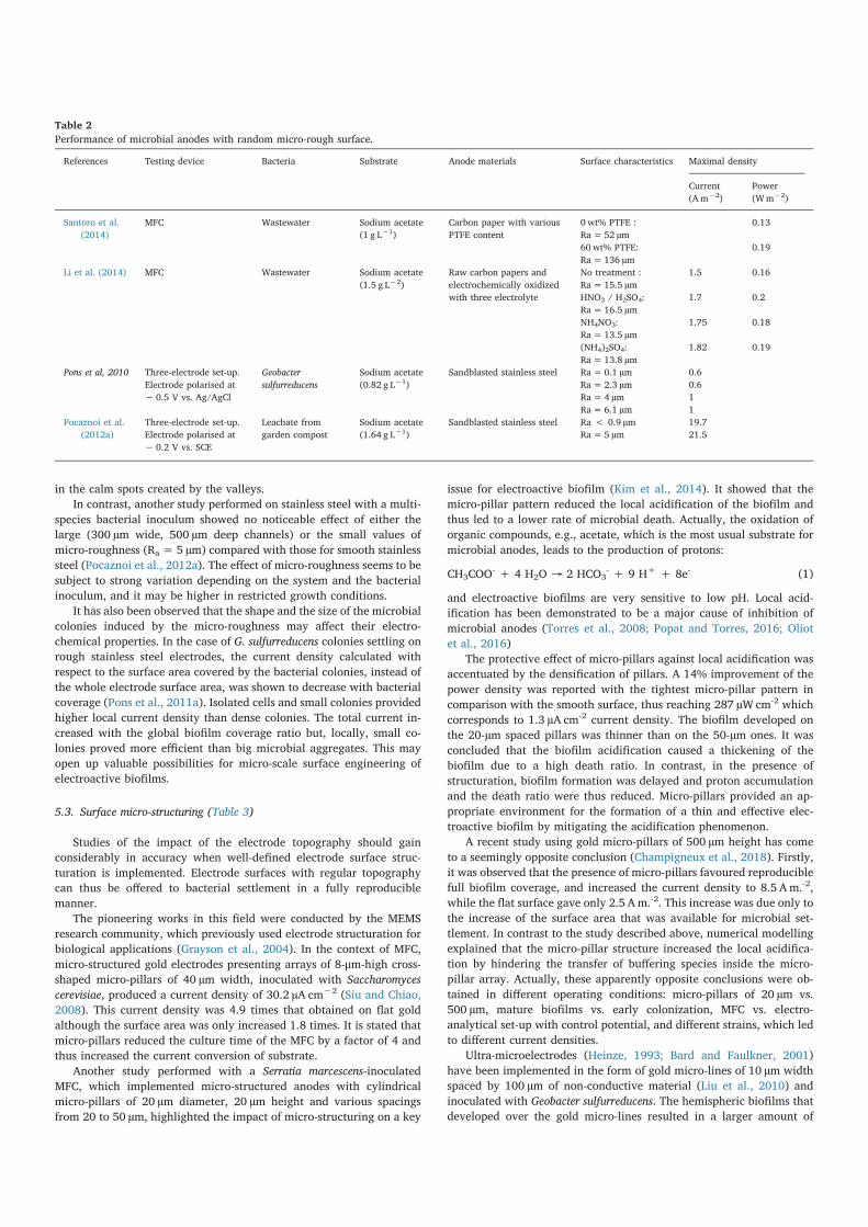
5.2. Random micro-roughness (Table 2)
The limit between nano-, micro- and macro-roughness is still un-
clear and may vary with the surface under study. Thus it is difficult to
clearly attribute an effect to each of the roughness scales but recent
works have been focusing on this issue (Santoro et al., 2014; Li et al.,
2014). The first study segregated roughness in two ranges: from 5 to
10 µm, where roughness was characterized by the high frequency
roughness RaH and skewness Rsk
H, and from 100 to 300 µm, char-
acterized by low frequency roughness RaL and skewness Rsk
L (Santoro
et al., 2014). This classification could be related to micro- and macro-
roughness. Various commercial carbon papers implemented with was-
tewater in a microbial fuel cell showed that the decrease of bacterial
attachment and current density was correlated with the decrease of
RskH, while no correlation could be established with the variation of
RskL. This means that the topographical variations measured at the scale
of 5–10 µm, close to the cell size, had an impact on the system while the
variations in the 100–300 scale were irrelevant in an analysis of the
microbial fuel cell performance. In order to better differentiate these
impacts, a segregation into four ranges was studied (20–100 nm,
0.9–5 µm, 6.5–50 µm and 163–450 µm) but it was not possible to cor-
relate the different parameters with the bacterial adhesion or with the
current produced. This study illustrated how complex surface char-
acterization can be (Li et al., 2014).
As recalled in Section 4.3, when approaching the size of a cell,
around a few mocrometers, roughness offers an obvious physical ben-
efit for bacterial adhesion by creating “calm areas” and increasing the
reachable contact area. The results presented above tend to confirm this
and the phenomenon, well-documented in the field of biofilms in
general, seems to also apply to electroactive biofilms. It has also been
observed on stainless steel electrodes inoculated with Geobacter sulfur-
reducens. Increasing the roughness from 0.1 to 2 µm produced almost no
effect on biofilm coverage and electrical output, while a shift from 2 to
4 µm multiplied the current density provided by the cathode by 1.6
(Pons et al., 2011b). The current rise was explained by the presence of
dense bacterial colonies on the roughest, 4 µm, surface, which formed
Fig. 4. Impact of contact surface and shear forces on bacterial adhesion depending on the surface roughness scale. On the flat and nano-rough surfaces (A, B), the cell
is directly impacted by the hydrodynamic flow and is subjected to high shear forces. On micro- and macro-rough surfaces (C, D), the hydrodynamic flow is reduced in
the grooves of the surface structure, which provides the cell with a calmer space to adhere. Regarding the contact area between the cell and the surface, the binding
surface is reduced in the nano-rough configuration (B) and enhanced when the roughness is of the same order of magnitude as the cell dimensions (C).
in the calm spots created by the valleys.
In contrast, another study performed on stainless steel with a multi-
species bacterial inoculum showed no noticeable effect of either the
large (300 µm wide, 500 µm deep channels) or the small values of
micro-roughness (Ra =5 µm) compared with those for smooth stainless
steel (Pocaznoi et al., 2012a). The effect of micro-roughness seems to be
subject to strong variation depending on the system and the bacterial
inoculum, and it may be higher in restricted growth conditions.
It has also been observed that the shape and the size of the microbial
colonies induced by the micro-roughness may affect their electro-
chemical properties. In the case of G. sulfurreducens colonies settling on
rough stainless steel electrodes, the current density calculated with
respect to the surface area covered by the bacterial colonies, instead of
the whole electrode surface area, was shown to decrease with bacterial
coverage (Pons et al., 2011a). Isolated cells and small colonies provided
higher local current density than dense colonies. The total current in-
creased with the global biofilm coverage ratio but, locally, small co-
lonies proved more efficient than big microbial aggregates. This may
open up valuable possibilities for micro-scale surface engineering of
electroactive biofilms.
5.3. Surface micro-structuring (Table 3)
Studies of the impact of the electrode topography should gain
considerably in accuracy when well-defined electrode surface struc-
turation is implemented. Electrode surfaces with regular topography
can thus be offered to bacterial settlement in a fully reproducible
manner.
The pioneering works in this field were conducted by the MEMS
research community, which previously used electrode structuration for
biological applications (Grayson et al., 2004). In the context of MFC,
micro-structured gold electrodes presenting arrays of 8-μm-high cross-
shaped micro-pillars of 40 µm width, inoculated with Saccharomyces
cerevisiae, produced a current density of 30.2 µA cm−2 (Siu and Chiao,
2008). This current density was 4.9 times that obtained on flat gold
although the surface area was only increased 1.8 times. It is stated that
micro-pillars reduced the culture time of the MFC by a factor of 4 and
thus increased the current conversion of substrate.
Another study performed with a Serratia marcescens-inoculated
MFC, which implemented micro-structured anodes with cylindrical
micro-pillars of 20 µm diameter, 20 µm height and various spacings
from 20 to 50 µm, highlighted the impact of micro-structuring on a key
issue for electroactive biofilm (Kim et al., 2014). It showed that the
micro-pillar pattern reduced the local acidification of the biofilm and
thus led to a lower rate of microbial death. Actually, the oxidation of
organic compounds, e.g., acetate, which is the most usual substrate for
microbial anodes, leads to the production of protons:
CH3COO- + 4 H2O → 2 HCO3
- + 9 H+ + 8e- (1)
and electroactive biofilms are very sensitive to low pH. Local acid-
ification has been demonstrated to be a major cause of inhibition of
microbial anodes (Torres et al., 2008; Popat and Torres, 2016; Oliot
et al., 2016)
The protective effect of micro-pillars against local acidification was
accentuated by the densification of pillars. A 14% improvement of the
power density was reported with the tightest micro-pillar pattern in
comparison with the smooth surface, thus reaching 287 µW cm-2 which
corresponds to 1.3 µA cm-2 current density. The biofilm developed on
the 20-µm spaced pillars was thinner than on the 50-µm ones. It was
concluded that the biofilm acidification caused a thickening of the
biofilm due to a high death ratio. In contrast, in the presence of
structuration, biofilm formation was delayed and proton accumulation
and the death ratio were thus reduced. Micro-pillars provided an ap-
propriate environment for the formation of a thin and effective elec-
troactive biofilm by mitigating the acidification phenomenon.
A recent study using gold micro-pillars of 500 µm height has come
to a seemingly opposite conclusion (Champigneux et al., 2018). Firstly,
it was observed that the presence of micro-pillars favoured reproducible
full biofilm coverage, and increased the current density to 8.5 Am.-2,
while the flat surface gave only 2.5 Am.-2. This increase was due only to
the increase of the surface area that was available for microbial set-
tlement. In contrast to the study described above, numerical modelling
explained that the micro-pillar structure increased the local acidifica-
tion by hindering the transfer of buffering species inside the micro-
pillar array. Actually, these apparently opposite conclusions were ob-
tained in different operating conditions: micro-pillars of 20 µm vs.
500 µm, mature biofilms vs. early colonization, MFC vs. electro-
analytical set-up with control potential, and different strains, which led
to different current densities.
Ultra-microelectrodes (Heinze, 1993; Bard and Faulkner, 2001)
have been implemented in the form of gold micro-lines of 10 µm width
spaced by 100 µm of non-conductive material (Liu et al., 2010) and
inoculated with Geobacter sulfurreducens. The hemispheric biofilms that
developed over the gold micro-lines resulted in a larger amount of
References Testing device Bacteria Substrate Anode materials Surface characteristics Maximal density
Current
(Am−2)
Power
(Wm−2)
Santoro et al.
(2014)
MFC Wastewater Sodium acetate
(1 g L−1)
Carbon paper with various
PTFE content
0 wt% PTFE :
Ra=52 µm
0.13
60 wt% PTFE:
Ra=136 µm
0.19
Li et al. (2014) MFC Wastewater Sodium acetate
(1.5 g L−2)
Raw carbon papers and
electrochemically oxidized
with three electrolyte
No treatment :
Ra=15.5 µm
1.5 0.16
HNO3 / H2SO4:
Ra=16.5 µm
1.7 0.2
NH4NO3:
Ra=13.5 µm
1.75 0.18
(NH4)2SO4:
Ra=13.8 µm
1.82 0.19
Pons et al, 2010 Three-electrode set-up.
Electrode polarised at
− 0.5 V vs. Ag/AgCl
Geobacter
sulfurreducens
Sodium acetate
(0.82 g L−1)
Sandblasted stainless steel Ra=0.1 µm 0.6
Ra=2.3 µm 0.6
Ra=4 µm 1
Ra=6.1 µm 1
Pocaznoi et al.
(2012a)
Three-electrode set-up.
Electrode polarised at
− 0.2 V vs. SCE
Leachate from
garden compost
Sodium acetate
(1.64 g L−1)
Sandblasted stainless steel Ra < 0.9 µm 19.7
Ra=5 µm 21.5
Table 2
Performance of microbial anodes with random micro-rough surface.
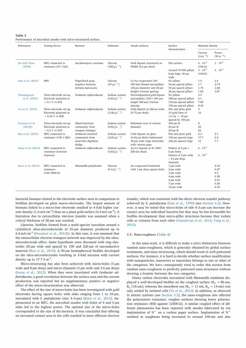
bacterial biomass related to the electrode surface area in comparison to
biofilms developed on plain macro-electrodes. The largest amount of
biomass linked to a micro-line electrode resulted in 4-fold higher cur-
rent density (1.6 mA cm-2) than on a plain gold surface (0.4 mA cm-2). A
limitation due to extracellular electron transfer was assumed when a
critical thickness of 20 µm was reached.
Likewise, biofilms formed from a multi-species inoculum around a
cylindrical ultra-microelectrode of 25 µm diameter produced up to
6.6 mA cm-2 (Pocaznoi et al., 2012b). In this case, it was assumed that
the extracellular electron transport network was improved by the ultra-
microelectrode effect. Same hypothesis were discussed with ring elec-
trodes 20 µm wide and spaced by 100 and 220 µm of unconductive
material (Ren et al., 2016). A 50 µm hemispherical biofilm developed
on the ultra-microelectrodes resulting in 3-fold increase with current
density up to 17.7 Am-2.
Micro-structuring has also been achieved with micro-holes (5 µm
wide and 8 µm deep) and micro-channels (5 µm wide and 3.6 µm deep)
(Inoue et al., 2012). When they were inoculated with Geobacter sul-
furreducens, a good correlation between the surface area and the current
production was reported but no supplementary positive or negative
effect of the micro-structuration was observed.
The effect of the size of micro-holes has been investigated with gold
electrodes having square holes with sides ranging from 1 to 10 µm,
inoculated with S. putrefaciens (size 3–5 µm) (Kano et al., 2012). Im-
plemented in an MFC, the microbial anodes with holes of 4 and 5 µm
sides led to the highest power. The optimal size of the micro-holes
corresponded to the size of the bacteria. It was concluded that offering
an increased contact area to the cells resulted in more efficient electron
transfer, which was consistent with the direct electron transfer pathway
achieved by S. putrefaciens (Kim et al., 1999) (see Section 4.3). How-
ever, it may be noted that micro-holes of side 4–5 µm can increase the
contact area for individual bacteria but they may be less favourable for
biofilm development than micro-pillar structures because they isolate
cells or colonies from each other (Vasudevan et al., 2014; Yang et al.,
2015).
5.4. Nano-roughness (Table 4)
At the nano-scale, it is difficult to make a strict distinction between
random nano-roughness, which is generally obtained by global surface
treatments, and nano-structuring, which should result in well-patterned
surfaces. For instance, it is hard to decide whether surface modification
with nanoparticles, nanowires or nanotubes belongs to one or other of
the categories. We have consequently tried to order this section from
random nano-roughness to perfectly patterned nano-structures without
drawing a frontier between the two categories.
Glassy carbon electrodes inoculated with Shewanella oneidensis dis-
played a well-developed biofilm on the roughest surface (Ra =86 nm,
Rq 116 nm), whereas the smoothest one (Ra =11 nm, Rq =14 nm) was
only settled by isolated cells (Ye et al., 2012). In addition, as observed
in abiotic systems (see Section 3.2), the nano-roughness also affected
the polarization resistance, rougher surfaces showing lower polariza-
tion resistance (850 against 12500Ω). A similar coupled effect of dif-
ferent phenomena has been reported with anodes fabricated by ion
implantation of N+ on a carbon paper surface. Implantation of N+
resulted in roughness being increased to around 100 nm and also
References Testing device Bacteria Substrate Anode surfaces Surface
characteristics
Maximal density
Current
(Am−2)
Power
(Wm−2)
Siu and Chiao.
(2008)
MFC connected to
resistance (47–1 kΩ)
Saccharomyces cerevisiae Glucose
(180 g L−1)
Gold deposit structured on
PDMS (0,2 µm thick)
Flat surface 6 . 10-3
(100Ω)
1 . 10-4
Around 70 000 pillars
8 µm high, 40 µm
wide
3 . 10-2
(100Ω)
4 . 10-3
Kim et al. (2014) MFC Flagellated gram
negative bacteria,
Serratia marcescens
Glucose
(25 g L−1)
Cr/Au evaporated (30/
300 nm) Round micropillars
(20 µm diameter and 20 µm
height) Various spacing
No pillars 1.5 2.5
50 µm spaced pillars 1.7 2.74
30 µm spaced pillars 1.75 2.68
20 µm spaced pillars 1.82 2.87
Champigneux
et al. (2018)
Three-electrode set-up.
Electrode polarised at
+ 0.1 V vs SCE
Geobacter sulfurreducens Sodium acetate
(0.82 g L−1)
Electrodeposited gold Square
micropillars (100×100 µm,
height 500 µm) Various
spacing
No pillars 2.5
200 µm spaced pillars 6.1
125 µm spaced pillars 7.85
100 µm spaced pillars 8.45
Liu et al. (2010) Three-electrode set-up.
Electrode polarised at
+ 0.24 V vs SHE
Geobacter sulfurreducens Sodium acetate
(2.46 g L−1)
Gold deposit on silicon wafer
(0.75 µm thick)
Flat and plain gold 4
10 gold lines of
1,5 cm × 10 µm
spaced by 100 µm
16
Pocaznoi et al.
(2012b)
Three-electrode set-up.
Electrode polarised at
− 0.2 V vs SCE
Mixed bacterial
community from
compost leekages
Sodium acetate
(0.82 g L−1)
Platinium wire of various
diameter
500 µm Ø 8
50 µm Ø 20
25 µm Ø 66
Ren et al. (2016) MFC connected to
resistance (148–1MΩ)
Geobacter-enriched
community from
anaerobic-digestion
sludge
Sodium acetate
(2.05 g L−1)
Gold deposit on glass
(0.42 µm thick) Imbricated
20 µm wide rings electrodes
with various gaps.
Flat and plain gold 5.2 2.1
100 µm spaced rings 17.7 6.9
220 µm spaced rings 15 7.7
Inoue et al. (2012) MFC connected to
resistance (lowered
stepwise)
Geobecter sulfurreducens Sodium acetate
(0.82 g L−1)
Au/Cr deposit on Si (400/
200 nm thick)
Pattern of 5 µm x
5 µm holes
6 . 10-3
Pattern of 5 µm wide
/ 3.6 µm deep
channels
2 . 10-3
Kano et al. (2012) MFC connected to
resistance
(100–3000Ω)
Shewanella putrefaciens Glucose
(0.4 g L−1)
Au evaporated Patterned
with 1 µm deep square holes
1 µm wide 0.18
2 µm wide 0.27
3 µm wide 0.3
4 µm wide 0.38
5 µm wide 0.41
7 µm wide 0.38
10 µm wide 0.35
Table 3
Performance of microbial anodes with micro-structured surface.
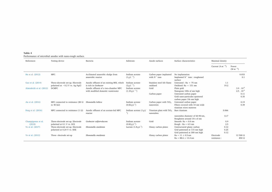
Table 4
Performance of microbial anodes with nano-rough surface.
References Testing device Bacteria Substrate Anode surfaces Surface characteristics Maximal density
Current (Am−2) Power
(Wm−2)
He et al. (2012) MFC Acclimated anaerobic sludge from
anaerobic reactor
Sodium acetate
(1 g L−1)
Carbon paper implanted
with N+ ions
No implantation 0.033
Implanted N+ ions : roughened
surface
0.1
Guo et al. (2014) Three-electrode set-up. Electrode
polarised at −0.2 V vs. Ag/AgCl
Anodic effluent of an existing BES, which
is rich in Geobacter
Sodium acetate
(2 g L−1)
Stainless steel felt flame
oxidized
Untreated : Ra = 75 nm 1.1
Oxidized: Ra = 151 nm 19.2
Alatraktchi et al. (2012) DCMFC Anodic effluent of a two-chamber MFC
with modified domestic wastewater
Sodium acetate
(1.33 g L−1)
Gold Plain gold 3.9 . 10-5
Nanograss 100s of nm high 2.5 . 10-3
Carbon paper Untreated carbon paper 0.11
Gold nano-particules sputtered
carbon paper 10s nm high
0.35
Jia et al. (2016) MFC connected to resistance (80 Ω
to 40 kΩ)
Shewanella loihica Sodium acetate
(0.82 g L-1)
Carbon paper with TiO2
nanowires
Untreated carbon paper 0.19
Fibers covered with 10 nm wide
titanium wires mattress
0.39
Feng et al. (2016) MFC connected to resistance (1 Ω) Anodic effluent of an acetate-fed MFC
reactor
Sodium acetate (1 g L-
1)
Titanium plate with TiO2
nanotubes
Bare titanium 0.066
nanotubes diameter of 60-90 nm,
Roughness around 10 s of nm
12.7
Champigneux et al.
(2018)
Three-electrode set-up. Electrode
polarised at 0.1 V vs. SCE
Geobacter sulfurreducens Sodium acetate
(0.82 g L-1)
Gold Smooth: Ra= 0.8 nm 0.9
Rough : Ra= 4.5 nm 2.5
Ye et al. (2017) Three-electrode set-up. Electrode
polarised at 0.24 V vs. SHE
Shewanella oneidensis Lactate (1.8 g L-1) Glassy carbon plates Unstructured glassy carbon 0.12
Grid patterned at 115 nm high 0.25
Grid patterned at 300 nm high 0.12
Ye et al. (2012) Three- electrode set-up Shewanella oneidensis Glassy carbon plates Ra= 11±0.5 nm Electrode
resistance :
12 500 Ω
Ra=88.6± 13.4 nm 850 Ω
gold surface (Champigneux et al., 2018). Inoculated with G. sulfurre-
ducens, the 0.8 and 4.5 nm-rough electrodes respectively produced 0.9
and 2.5 Am-2 on average. It was speculated that nano-roughness might
act by increasing the electron transfer rate. Another study implemented
nano-patterning to design well-controlled grids 3 µm wide and 115 or
300 nm high on glassy carbon surfaces. These electrodes were in-
oculated with S. oneidensis (Ye et al., 2017). The 115 nm high pattern
was 78% more effective in terms of cell attachment density than the
smooth surface and 40% more productive in current density. The
300 nm high pattern showed no significant improvement in terms of
current density compared to the smooth surface, even though the
bacterial attachment was a little greater. It was assumed that the
300 nm pattern created frontiers between the biofilm patches that
formed on different areas of the grid, and was thus detrimental to global
biofilm organization. In contrast, the 115 nm pattern was not too high
and did not hinder biofilm unity. The thermodynamic explanation
given was that the membrane deformation needed to overcome the
300 nm feature was too demanding in energy. It highlights an optimal
feature threshold of around 100 nm, which is consistent with cell en-
ergy limitations.
6. Open questions and thoughts
As a first obvious conclusion, according to the impressive influence
of the electrode surface topography on microbial electrochemical sys-
tems which have already been reported, it can be stated that surface
topographical engineering is a very promising avenue for improving the
efficiency of electroactive biofilms.
6.1. Take care when choosing the experimental set-up
Two different approaches can be followed to investigate microbial
electrochemical systems depending on the objective. The engineering
approach implements whole reactors and processes in order to shift the
technology towards actual application as fast as possible (Pocaznoi
et al., 2012c). In contrast, fundamental investigations try to single out a
given phenomenon in order to gain deeper fundamental understanding
of it. It is essential not to confuse the two approaches, which correspond
to different purposes and different experimental set-ups.
In the context of engineering, with the objective of improving the
performance of microbial electrodes, optimal surface topography can
be associated with porous electrodes, 3-dimensional structures and
surface functionalization, resulting in very complex electrode archi-
tecture. This can be illustrated by a glassy carbon electrode coated with
carbon nanotubes and then functionalized with polypyrrole and man-
ganese (Lu et al., 2013) or similarly, by carbon material doped with
nickel nano-particles, patterned in a micropillar array to form an
electrode and then coated with carbon nanofibers (Khare et al., 2016).
Such innovative anode designs can lead to valuable improvements in
performance but they are not ideally suited to analytical studies that
aim to characterize the effects of surface topography on electroactive
biofilms. Likewise, an MFC is not an appropriate electro-analytical
device (Rimboud et al., 2014), because many other rate-limiting steps
than the studied electrode can affect the global performance. As an il-
lustration of this general consideration, it has recently been reported
that the cathode roughness can increase MFC performance by im-
proving the electrical contact between the cathode and the current
collector (Santoro et al., 2015). In this case roughness had noting with
do with the bioelectrochemical processes. The design of MFCs should be
reserved for engineering purposes. A search for a fundamental ex-
planation of the impact of surface topography on electroactive biofilms
should be carried out in carefully designed electroanalytical set-ups
(Rimboud et al., 2014) with well-defined electrode surface topo-
graphies.
decreased the resistance and increased hydrophobicity (He et al.,
2012). It was consequently not possible to discriminate between the
contributions of the three phenomena to the 3-fold power density in-
crease obtained by implementing the electrodes in an MFC inoculated
with sludge.
At the nano-scale, did the roughest surface enhance the develop-
ment of electroactive biofilms by favouring bacterial adhesion and
biofilm formation or by decreasing the polarization resistance, i.e. im-
proving electron transfer? Bacteria use electron transfer to the electrode
to support their metabolic process. Facilitating electron transfer can
consequently lead to more intense biofilm development. Did nano-
roughness impact cell adhesion directly or did it favour the growth of
electroactive bacteria by accelerating electron transfer? A clear causal
chain can hardly be extracted yet.
The difficulty of drawing straightforward conclusions has been il-
lustrated by another study performed on stainless steel electrodes that
were either treated by flame oxidation or left untreated (Guo et al.,
2014). Flame oxidation not only increased the nano-scale roughness
from 75 to 151 nm but also altered the surface chemistry. The treatment
resulted in better biofilm coverage and higher current output from
enriched Geobacter psychrophilus and Geobacter sulfurreducens inocula
for the rougher oxidized electrode but, once again, the improvement
cannot be fully related to the increase in surface nano-roughness since
the flame oxidation also deteriorated the chromium-rich passive layer.
Actually, the nature of the passive layer can significantly affect electron
transfer rate with biofilms (Pons et al., 2011a). Similarly, the genera-
tion of current by Geobacter sulfurreducens has been shown to be in-
fluenced by the crystallographic nature and orientation of the electrode
surface (Maestro et al., 2014) and the crystallographic state can directly
influence the nano-roughness of the electrode surface.
The technique used to achieve the surface nano-topography may
also have a considerable impact on the performance of an electroactive
biofilm. Grass-like nano-roughness of 100 nm obtained by deep reactive
ion etching of a gold electrode has been compared with sputtering of
gold nanoparticles on carbon paper in an MFC inoculated with waste-
water (Alatraktchi et al., 2012). Nanoparticle sputtering led to con-
siderably higher power densities (346 vs. 2.5 mW/m2). It has generally
been observed that using nanoparticles sputtered on smooth electrodes
to increase their roughness leads to increased current output (Qiao
et al., 2007; Sharma et al., 2008; Sun et al., 2010; Fan et al., 2011; Quan
et al., 2015; Z. Lu et al., 2015; Xu et al., 2018) but, since nanoparticle
sputtering also impacts the surface chemistry and its electrical char-
acteristics, the current enhancement cannot be linked directly with the
nano-topography.
Surfaces modified with titanium oxide nanowires (Jia et al., 2016)
and titanium oxide nanotubes (Feng et al., 2016) have shown that a
titanium surface with a nanometre organization constitutes a suitable
electrode material for forming microbial anodes, in contrast to smooth
titanium, which has proved to be of no interest. After inoculation with
effluent from an existing acetate-fed MFC reactor, no biofilm developed
on smooth titanium, and consequently no current was produced, while
the titanium electrodes coated with nanotubes were completely covered
with biofilm and produced 12.7 A m-2. Titanium nanowires, on the
other hand, were believed to act as a substitute for bacterial pili by
promoting the bacterial adhesion and also favouring electron transfer.
Here again, the conclusions should be moderated by considering the
low conductivity of titanium oxide. In the field of electrochemical en-
gineering, pure titanium is not considered as appropriate anode mate-
rial because of the formation of a low-conductive oxide on its surface.
Surface modification by titanium nano-objects such as nanowires or
nanotubes may mainly act by modifying the surface conductivity.
Very recent studies have tended to single out the effect of nano-
topography by comparing surfaces of identical nature. Gold surfaces
with Ra of 0.8 and 4.5 nm have been compared. The 4.5-nm roughness
was obtained by electrolytic deposition of gold on the 0.8-nm smooth
roughness on electron transfer in abiotic conditions. This solid basic
knowledge should now be exploited for electroactive biofilms. For ex-
ample, surface nano-roughness has been shown to drastically change
the electrochemistry of an equine cytochrome c (Leopold and Bowden,
2002) by acting on the organization of the monolayer that self-assem-
bles on the electrode surface. This type of work, applied to the com-
ponent of the specific cytochromes c, quinone compounds and other
mediator types involved in biofilm extracellular electron transfer (Lidan
et al., 2014), could lead to a more accurate description of certain
transfer mechanisms.
Fascinating thoughts have been voiced on the possible impact of
nano-roughness from the standpoint of the metabolic changes it may
induce for adhering bacteria. A microbiological approach through
genomic analysis should be suitable to better understand bacterial re-
sponse to the surface topography. For example, effects of roughness on
extracellular matrix production and composition have already been
evoked (see Section 4.3) but no experimental confirmation has yet been
given. Genomics could be of great help in unravelling the bacterial
mechanism at play.
This issue should have great importance for electroactive biofilms
because the extracellular matrix plays a crucial role both in biofilm
structure and extracellular electron transfer. Using the electrode topo-
graphy to act on biofilm properties, such as thickness, structure and,
above all, conductivity, should be a great breakthrough for electro-
active biofilm design. In this direction, in the case of multispecies in-
ocula, it seems very attractive to investigate the possibility of orienting
the selection of the suitable electroactive species by means of the
electrode topography. For both pure cultures and multispecies inocula,
it has been shown that multiple pathways could be involved in extra-
cellular electron transfer (Zhu et al., 2012; Rimboud et al., 2016).
Trying to orient the synthesis of the most suitable extracellular med-
iator(s) through the nano-topography of the electrode would be a very
attractive way to optimize electroactive biofilms. This research direc-
tion seems very ambitious but it is supported by previous studies. These
studies did not deal with electroactive biofilms but suggested that the
production by bacterial cells of compounds involved in biofilm struc-
turing may be affected by the nano-topography of the support (see
Section 4.3).
7. Conclusion
Surface topography has an obvious, strong impact on the formation
and performance of electroactive biofilms. At the scale of around a
hundred micrometers and above, surface engineering can be used to
increase the electrode surface area. Optimal topographies are likely to
be found by improving mass transfer and mitigating local acidification
inside the micro-structure. At the micrometer scales, local shear stress
and cell-electrode contact angle seem to play the major roles. The nano-
scale opens up thrilling horizons, with some hope of acting on the in-
timate mechanisms of biofilm formation and extracellular electron
transfer.
Up to now, many studies have investigated surface topography in
parallel with other surface modifications, thus leading to a lack of
specific understanding. Conversely, it may be questioned whether the
improvements sometimes attributed to chemical or physicochemical
modifications of the surface were not, to some extent or even totally,
due to modification of the surface topography. The main conclusion of
this review is that it may be advisable to develop investigations of well-
controlled roughness by taking care to avoid the variation of other in-
terfacial parameters. Conversely, when the objective is to characterize a
chemical or physicochemical modification, it should be taken care to
work with the same surface topography throughout. This objective
seems fairly achievable at the micro-scale but more challenging at the
nanoscale.
6.2. Consensus on the impact of micro-roughness at cell size
Several reports tend to confirm that micro-roughness improves the
electroactive biofilm performance in two ways: by offering bacteria
sites with low shear forces, suitable for their attachment, and by in-
creasing the bacteria/electrode contact area for stronger attachment
and improved electron transfer. These two effects have already been
observed with bacterial adhesion and biofilm formation of non-elec-
troactive species (Figs. 1, 2). They are most marked when the average
roughness is of the same order of magnitude as the size of the cells, i.e.,
a few micrometers.
Above a few micrometers, random roughness does not seem to have
any great impact on electroactive biofilms, while ordered roughness,
which is achieved by surface micro-structuring, may increase the cur-
rent produced by increasing the surface area available for biofilm de-
velopment. For microbial anodes, the optimal order of magnitude for
micro-structures should be closely related with the diffusion limitation,
mainly to mitigate local acidification of the biofilm.
Actually, the impact of uncontrolled random surface topography on
electroactive biofilms is still unclear (see Section 5.2), certainly because
of the large variety of systems that have been considered in the context
of microbial electrochemical systems. Moreover, a large number of
these studies have been carried out in MFCs designed for purposes other
than characterizing the effect of surface topography. This is another
major reason of the lack of clear conclusion. The variation of roughness
has most often been the result of physical or chemical surface mod-
ifications, making it difficult to discriminate which parameter had the
main impact. Specific studies should now focus on roughness as the
single parameter tested, taking care to keep the other interfacial
properties unchanged. This direction has already been engaged with
surface micro-structured electrodes and should be intensified to grasp
the real effect of random roughness.
6.3. Interesting possibilities related to nano-roughness (see Section 5.4)
It has been claimed that bacterial cells could hardly grow over a
structure 300 nm high due to the deformation limitation. Sharp edges
above 100 nm or so may thus delay the formation of continuous bio-
films. This gives valuable guidelines on how to favour the development
of biofilms on an electrode: nano-structuration may be more efficient
when lower than 300 nm or presenting reduced sharpness (Rku lower
than 3) and wide peak to peak distance. Conversely, the same guide-
lines can also be considered if the objective is to hamper the develop-
ment of harmful biofilm, in the context of biocorrosion for instance.
It has been suggested that bacterial appendices could be mimicked
by decorating the electrode surface with metal nano-wires to provide
the cells with multiple “pilus-like” nanostructures to achieve extra-
cellular electron transfer. This particular patterning technique may
show great promise, but further studies need to be carried out, in
particular by avoiding the comparison with flat titanium, which is not
an appropriate material for anodes.
At the nano-scale it seems difficult to discriminate between the real
effect of the surface topography and that of other interfacial para-
meters, which are unavoidably changed when nano-topography is
modified. Are the characteristics and performance of electroactive
biofilms affected directly by the electrode nano-topography or by the
impact of the electrode nano-topography on surface chemistry, inter-
facial resistance, crystallographic state, etc.? (See Section 3.2.) Nano-
roughness corresponds to a scale at which it is difficult to avoid other
physicochemical properties being affected, so the causal chain can
hardly be unravelled. Basic conclusions should be carefully qualified.
6.4. Exciting horizons to be explored
Numerous electrochemical studies have approached, both experi-
mentally and theoretically (see Section 3), the impact of the electrode
None.
References
Alatraktchi, F.A., Zhang, Y., Noori, J.S., Angelidaki, I., 2012. Surface area expansion of
electrodes with grass-like nanostructures and gold nanoparticles to enhance elec-
tricity generation in microbial fuel cells. Bioresour. Technol. 123, 177–183. https://
doi.org/10.1016/j.biortech.2012.07.048.
Albina, A., Taberna, P.L., Cambronne, J.P., Simon, P., Flahaut, E., Lebey, T., 2006. Impact
of the surface roughness on the electrical capacitance. Microelectron. J. 37, 752–758.
https://doi.org/10.1016/j.mejo.2005.10.008.
Anitha, V.C., Lee, J.-H., Lee, J., Banerjee, A.N., Joo, S.W., Min, B.K., 2015. Biofilm for-
mation on a TiO 2 nanotube with controlled pore diameter and surface wettability.
Nanotechnology 26, 065102. https://doi.org/10.1088/0957-4484/26/6/065102.
Bajracharya, S., Sharma, M., Mohanakrishna, G., Dominguez Benneton, X., Strik,
D.P.B.T.B., Sarma, P.M., Pant, D., 2016. An overview on emerging bioelectrochemical
systems (BESs): technology for sustainable electricity, waste remediation, resource
recovery, chemical production and beyond. Renew. Energy, Spec. Issue.: New Horiz.
Biofuels Prod. Technol. 98, 153–170. https://doi.org/10.1016/j.renene.2016.03.002.
Bajracharya, S., Srikanth, S., Mohanakrishna, G., Zacharia, R., Strik, D.P., Pant, D., 2017.
Biotransformation of carbon dioxide in bioelectrochemical systems: state of the art
and future prospects. J. Power Sources 356, 256–273. https://doi.org/10.1016/j.
jpowsour.2017.04.024.
Bard, A.J., Faulkner, L.R., 2001. Electrochemical Methods: Fundamentals and
Applications, 2nd ed. John Wiley & Sons, New York.
Beech, W.B., Sunner, J., 2004. Biocorrosion: towards understanding interactions between
biofilms and metals. Curr. Opin. Biotechnol. 15, 181–186. https://doi.org/10.1016/j.
copbio.2004.05.001.
Bergel, A., Féron, D., Mollica, A., 2005. Catalysis of oxygen reduction in PEM fuel cell by
seawater biofilm. Electrochem. Commun. 7, 900–904. https://doi.org/10.1016/j.
elecom.2005.06.006.
Berkeley, R.C.W., 1980. Society of Chemical Industry (Great Britain)(Eds.). Microbial
adhesion to surfaces. Published for the Society of Chemical Industry, London by Ellis
Horwood ; New York : Distributors, North and South America and the rest of the
world, Halsted Press, Chichester [West Sussex].
Bharat Bhushan, 2000. Surface roughness analysis and measurement techniques. In:
Modern Tribology Handbook, Two Volume Set, Mechanics & Materials Science. CRC
Press, Boca Raton, FL.
Blanchet, E., Duquenne, F., Rafrafi, Y., Etcheverry, L., Erable, B., Bergel, A., 2015.
Importance of the hydrogen route in up-scaling electrosynthesis for microbial CO2
reduction. Energy Environ. Sci. 8, 3731–3744. https://doi.org/10.1039/
C5EE03088A.
Bockris, A.K.N.R., Gamboa-A, Maria E., John, O.’M., 2008. Modern Electrochemistry 2A:
Fundamentals of Electrodics, 2nd ed. Kluwer Academic/Plenum Publishers, New
York (2000. ed).
Bombelli, P., Zarrouati, M., Thorne, R.J., Schneider, K., Rowden, S.J.L., Ali, A., Yunus, K.,
Cameron, P.J., Fisher, A.C., Ian Wilson, D., Howe, C.J., McCormick, A.J., 2012.
Surface morphology and surface energy of anode materials influence power outputs
in a multi-channel mediatorless bio-photovoltaic (BPV) system. Phys. Chem. Chem.
Phys. PCCP 14, 12221–12229. https://doi.org/10.1039/c2cp42526b.
Bond, D.R., Lovley, D.R., 2003. Electricity production by geobacter sulfurreducens at-
tached to electrodes. Appl. Environ. Microbiol. 69, 1548–1555. https://doi.org/10.
1128/AEM.69.3.1548-1555.2003.
Borisova, T.I., Ershler, B.V., 1950. Determination of the zero voltage points of solid metals
from measurements of the capacity of the double layer. Zh. Fiz. Khimii 24, 337–344
(1950).
Borole, A.P., Reguera, G., Ringeisen, B., Wang, Z.-W., Feng, Y., Kim, B.H., 2011.
Electroactive biofilms: current status and future research needs. Energy Environ. Sci.
4, 4813–4834. https://doi.org/10.1039/C1EE02511B.
Boyd, R.D., Verran, J., Jones, M.V., Bhakoo, M., 2002. Use of the atomic force microscope
to determine the effect of substratum surface topography on bacterial adhesion.
Langmuir 18, 2343–2346. https://doi.org/10.1021/la011142p.
Busscher, H.J., Weerkamp, A.H., 1987. Specific and non-specific interactions in bacterial
adhesion to solid substrata. FEMS Microbiol. Lett. 46, 165–173. https://doi.org/10.
1016/0378-1097(87)90062-0.
Carmona-Martinez, A.A., Trably, E., Milferstedt, K., Lacroix, R., Etcheverry, L., Bernet, N.,
2015. Long-term continuous production of H-2 in a microbial electrolysis cell (MEC)
treating saline wastewater. Water Res. 81, 149–156. https://doi.org/10.1016/j.
watres.2015.05.041.
Cercado Quezada, B., Byrne, N., Bertrand, M., Pocaznoi, D., Rimboud, M., Achouak, W.,
Bergel, A., 2013. Garden compost inoculum leads to microbial bioanodes with po-
tential-independent characteristics. Bioresour. Technol. 134, 276–284. https://doi.
org/10.1016/j.biortech.2013.01.123.
Cercado-Quezada, B., Delia, M.-L., Bergel, A., 2011. Electrochemical micro-structuring of
graphite felt electrodes for accelerated formation of electroactive biofilms on mi-
crobial anodes. Electrochem. Commun. 13, 440–443. https://doi.org/10.1016/j.
elecom.2011.02.015.
Chabert, N., Amin Ali, O., Achouak, W., 2015. All ecosystems potentially host electro-
genic bacteria. Bioelectrochem., Spec. Issue Biol Fuel Cells 106, 88–96. https://doi.
org/10.1016/j.bioelechem.2015.07.004.
Champigneux, P., Renault-Sentenac, C., Bourrier, D., Rossi, C., Delia, M.-L., Bergel, A.,
2018. Effect of surface nano/micro-structuring on the early formation of microbial
anodes with Geobacter sulfurreducens: experimental and theoretical approaches.
Bioelectrochemistry 121, 191–200. https://doi.org/10.1016/j.bioelechem.2018.02.
005.
Characklis, W.G., Marshall, K.C., 1990. Biofilms. Wiley, New York.
Characklis, W.G., Wilderer, P.A., 1989. Structure and function of biofilms: report of the
Dahlem Workshop on Structure and Function of Biofilms, Berlin, 1988, November 27-
December 2. Wiley.
Cogan, N.G., Keener, J.P., 2004. The role of the biofilm matrix in structural development.
Math. Med. Biol. J. IMA 21, 147–166. https://doi.org/10.1093/imammb/21.2.147.
Costerton, J.W., 1995. Overview of microbial biofilms. J. Ind. Microbiol. 15, 137–140.
https://doi.org/10.1007/BF01569816.
Costerton, J.W., Lewandowski, Z., DeBeer, D., Caldwell, D., Korber, D., James, G., 1994.
Biofilms, the customized microniche. J. Bacteriol. 176, 2137–2142. https://doi.org/
10.1128/jb.176.8.2137-2142.1994.
Crawford, R.J., Webb, H.K., Truong, VK., Hasan, J., Ivanova, E.P., 2012. Surface topo-
graphical factors influencing bacterial attachment. Advances in Colloid and Interface
Science 179, 142–149. https://doi.org/10.1016/j.cis.2012.06.015.
Czarnecki, J., Warszyński, P., 1987. The evaluation of tangential forces due to surface
inhomogeneties in the particle deposition process. Colloids Surf. 22, 187–205.
https://doi.org/10.1016/0166-6622(87)80220-2.
Davey, M.E., O’toole, G.A., 2000. Microbial biofilms: from ecology to molecular genetics.
Microbiol. Mol. Biol. Rev. 64, 847–867. https://doi.org/10.1128/MMBR.64.4.847-
867.2000.
De Levie, R., 1965. The influence of surface roughness of solid electrodes on electro-
chemical measurements. Electrochim. Acta 10, 113–130. https://doi.org/10.1016/
0013-4686(65)87012-8.
Denis, F.A., Hanarp, P., Sutherland, D.S., Gold, J., Mustin, C., Rouxhet, P.G., Dufrêne,
Y.F., 2002. Protein adsorption on model surfaces with controlled nanotopography
and chemistry. Langmuir 18, 819–828. https://doi.org/10.1021/la011011o.
Dewan, A., Ay, S.U., Karim, M.N., Beyenal, H., 2014. Alternative power sources for re-
mote sensors: a review. J. Power Sources 245, 129–143. https://doi.org/10.1016/j.
jpowsour.2013.06.081.
Di Lorenzo, M., Curtis, T.P., Head, I.M., Scott, K., 2009. A single-chamber microbial fuel
cell as a biosensor for wastewaters. Water Res. 43, 3145–3154. https://doi.org/10.
1016/j.watres.2009.01.005.
Díaz, C., Cortizo, M.C., Schilardi, P.L., Saravia, S.G.G. de, Mele, M.A.F.L. de, 2007a.
Influence of the nano-micro structure of the surface on bacterial adhesion. Mater.
Res. 10, 11–14. https://doi.org/10.1590/S1516-14392007000100004.
Díaz, C., Schilardi, P.L., Salvarezza, R.C., Fernández Lorenzo de Mele, M., 2007b. Nano/
microscale order affects the early stages of biofilm formation on metal surfaces.
Langmuir 23, 11206–11210. https://doi.org/10.1021/la700650q.
Díaz, C., Salvarezza, R.C., Fernández Lorenzo de Mele, M.A., Schilardi, P.L., 2010.
Organization of Pseudomonas fluorescens on chemically different nano/micro-
structured surfaces. ACS Appl. Mater. Interfaces 2, 2530–2539. https://doi.org/10.
1021/am100313z.
Dickinson, E.J.F., Streeter, I., Compton, R.G., 2008. Theory of chronoamperometry at
cylindrical microelectrodes and their arrays. J. Phys. Chem. C 112, 11637–11644.
https://doi.org/10.1021/jp801867e.
Ding, C., Lv, M., Zhu, Y., Jiang, L., Liu, H., 2015. Wettability-regulated extracellular
electron transfer from the living organism of shewanella loihica PV-4. Angew. Chem.
Int. Ed. 54, 1446–1451. https://doi.org/10.1002/anie.201409163.
Donoso, M.G., Méndez-Vilas, A., Bruque, J.M., González-Martin, M.L., 2007. On the re-
lationship between common amplitude surface roughness parameters and surface
area: implications for the study of cell–material interactions. Int. Biodeterior.
Biodegrad., Proceedings of the 1st International Conference on Environmental,
Industrial and Applied Microbiology, 59, pp. 245–251 ⟨https://dx.doi.org/10.1016/
j.ibiod.2006.09.011⟩.
Douglass Jr, E.F., Driscoll, P.F., Liu, D., Burnham, N.A., Lambert, C.R., McGimpsey, W.G.,
2008. Effect of electrode roughness on the capacitive behavior of self-assembled
monolayers. Anal. Chem. 80, 7670–7677. https://doi.org/10.1021/ac800521z.
Doyle, L.E., Marsili, E., 2015. Methods for enrichment of novel electrochemically-active
microorganisms. Biores. Technol. Microb. Fuel Cells 195, 273–282. https://doi.org/
10.1016/j.biortech.2015.07.025.
Doyle, R.J., 2001. Microbial Growth in Biofilms: Developmental and molecular biological
aspects. Academic Press.
Du, J., Catania, C., Bazan, G.C., 2014. Modification of abiotic-biotic interfaces with small
molecules and nanomaterials for improved bioelectronics. Chem. Mater. 26,
686–697. https://doi.org/10.1021/cm401912j.
Dumas, C., Basseguy, R., Bergel, A., 2008a. Microbial electrocatalysis with Geobacter
sulfurreducens biofilm on stainless steel cathodes. Electrochim. Acta 53, 2494–2500.
https://doi.org/10.1016/j.electacta.2007.10.018.
Dumas, C., Basseguy, R., Bergel, A., 2008b. DSA to grow electrochemically active biofilms
of Geobacter sulfurreducens. Electrochim. Acta 53, 3200–3209. https://doi.org/10.
1016/j.electacta.2007.10.066.
Duval, J.F.L., Gaboriaud, F., 2010. Progress in electrohydrodynamics of soft microbial
particle interphases. Curr. Opin. Colloid Interface Sci. 15, 184–195. https://doi.org/
10.1016/j.cocis.2009.12.002.
Eberhard, A., 1972. Inhibition and activation of bacterial luciferase synthesis. J. Bacteriol.
Acknowledgements
This work was supported by funding from the French state, man-
aged by the Agence Nationale de la Recherche (ANR), within the fra-
mework of the Koropokkuru project (ANR-14-CE05-0004).
Declaration of interest
the role of metabolism in localized mineral dissolution. Chem. Geol. Exp. Theor.
Geomicrobiol. 180, 19–32. https://doi.org/10.1016/S0009-2541(01)00303-5.
ElMekawy, A., Hegab, H.M., Mohanakrishna, G., Elbaz, A.F., Bulut, M., Pant, D., 2016.
Technological advances in CO2 conversion electro-biorefinery: a step toward com-
mercialization. Bioresour. Technol. Waste Biorefinery – Advocating Circ. Econ. 215,
357–370. https://doi.org/10.1016/j.biortech.2016.03.023.
Epstein, A.K., Hong, D., Kim, P., Aizenberg, J., 2013. Biofilm attachment reduction on
bioinspired, dynamic, micro-wrinkling surfaces. New J. Phys. 15, 095018. https://
doi.org/10.1088/1367-2630/15/9/095018.
Erable, B., Duteanu, N., Kumar, S.M.S., Feng, Y., Ghangrekar, M.M., Scott, K., 2009a.
Nitric acid activation of graphite granules to increase the performance of the non-
catalyzed oxygen reduction reaction (ORR) for MFC applications. Electrochem.
Commun. 11, 1547–1549. https://doi.org/10.1016/j.elecom.2009.05.057.
Erable, B., Roncato, M.-A., Achouak, W., Bergel, A., 2009b. Sampling natural biofilms: a
new route to build efficient microbial anodes. Environ. Sci. Technol. 43, 3194–3199.
https://doi.org/10.1021/es803549v.
Erable, B., Vandecandelaere, I., Faimali, M., Délia, M.-L., Etcheverry, L., Vandamme, P.,
Bergel, A., 2010. Marine aerobic biofilm as biocathode catalyst. Bioelectrochemistry
78, 51–56. https://doi.org/10.1016/j.bioelechem.2009.06.006.
Erable, B., Etcheverry, L., Bergel, A., 2011. From microbial fuel cell (MFC) to microbial
electrochemical snorkel (MES): maximizing chemical oxygen demand (COD) removal
from wastewater. Biofouling 27, 319–326. https://doi.org/10.1080/08927014.2011.
564615.
Fan, Y., Xu, S., Schaller, R., Jiao, J., Chaplen, F., Liu, H., 2011. Nanoparticle decorated
anodes for enhanced current generation in microbial electrochemical cells. Biosens.
Bioelectron. 26, 1908–1912. https://doi.org/10.1016/j.bios.2010.05.006.
Feng, G., Cheng, Y., Wang, S.-Y., Borca-Tasciuc, D.A., Worobo, R.W., Moraru, C.I., 2015.
Bacterial attachment and biofilm formation on surfaces are reduced by small-dia-
meter nanoscale pores: how small is small enough? NPJ Biofilms Micro. 1. https://
doi.org/10.1038/npjbiofilms.2015.22.
Feng, H., Liang, Y., Guo, K., Chen, W., Shen, D., Huang, L., Zhou, Y., Wang, M., Long, Y.,
2016. TiO2 nanotube arrays modified titanium: a stable, scalable, and cost-effective
bioanode for microbial fuel cells. Environ. Sci. Technol. Lett. 3, 420–424. https://doi.
org/10.1021/acs.estlett.6b00410.
Flemming, H.-C., Wingender, J., 2010. The biofilm matrix. Nat. Rev. Microbiol. 8,
623–633. https://doi.org/10.1038/nrmicro2415.
Flemming, H.-C., Neu, T.R., Wozniak, D.J., 2007. The EPS matrix: the “house of biofilm
cells. J. Bacteriol. 189, 7945–7947. https://doi.org/10.1128/JB.00858-07.
Flemming, H.-C., Wingender, J., Szewzyk, U., Steinberg, P., Rice, S.A., Kjelleberg, S.,
2016. Biofilms: an emergent form of bacterial life. Nat. Rev. Microbiol. 14, 563–575.
https://doi.org/10.1038/nrmicro.2016.94.
Flint, S.H., Brooks, J.D., Bremer, P.J., 2000. Properties of the stainless steel substrate,
influencing the adhesion of thermo-resistant streptococci. J. Food Eng. 43, 235–242.
https://doi.org/10.1016/S0260-8774(99)00157-0.
Fornero, J.J., Rosenbaum, M., Angenent, L.T., 2010. Electric power generation from
municipal, food, and animal wastewaters using microbial fuel cells. Electroanalysis
22, 832–843. https://doi.org/10.1002/elan.200980011.
Fredrickson, J.K., Romine, M.F., Beliaev, A.S., Auchtung, J.M., Driscoll, M.E., Gardner,
T.S., Nealson, K.H., Osterman, A.L., Pinchuk, G., Reed, J.L., Rodionov, D.A.,
Rodrigues, J.L.M., Saffarini, D.A., Serres, M.H., Spormann, A.M., Zhulin, I.B., Tiedje,
J.M., 2008. Towards environmental systems biology of Shewanella. Nat. Rev.
Microbiol. 6, 592–603. https://doi.org/10.1038/nrmicro1947.
Gabe, D.R., Wilcox, G.D., Gonzalez-Garcia, J., Walsh, F.C., 1998. The rotating cylinder
electrode: its continued development and application. J. Appl. Electrochem. 28,
759–780. https://doi.org/10.1023/A:1003464415930.
Gaboriaud, F., Gee, M.L., Strugnell, R., Duval, J.F.L., 2008. Coupled electrostatic, hy-
drodynamic, and mechanical properties of bacterial interfaces in aqueous media.
Langmuir ACS J. Surf. Colloids 24, 10988–10995. https://doi.org/10.1021/
la800258n.
Gamburg, D.Y.D., Zangari, D.G., 2011. Current Distribution at Rough Electrodes. In:
Theory and Practice of Metal Electrodeposition. Springer, New York, pp. 169–187.
https://doi.org/10.1007/978-1-4419-9669-5_8.
Girguis, P.R., Nielsen, M.E., Figueroa, I., 2010. Harnessing energy from marine pro-
ductivity using bioelectrochemical systems. Curr. Opin. Biotechnol. Energy
Biotechnol. – Environ. Biotechnol. 21, 252–258. https://doi.org/10.1016/j.copbio.
2010.03.015.
Goller, C.C., Romeo, T., 2008. Environmental influences on biofilm development. Bact.
Biofilms 322, 37–66. https://doi.org/10.1007/978-3-540-75418-3_3.
Grattieri, M., Minteer, S.D., 2018. Microbial fuel cells in saline and hypersaline en-
vironments: advancements, challenges and future perspectives. Bioelectrochemistry
120, 127–137. https://doi.org/10.1016/j.bioelechem.2017.12.004.
Grayson, A.C.R., Shawgo, R.S., Johnson, A.M., Flynn, N.T., Li, Y., Cima, M.J., Langer, R.,
2004. A BioMEMS review: MEMS technology for physiologically integrated devices.
Proc. IEEE 92, 6–21. https://doi.org/10.1109/JPROC.2003.820534.
Guo, K., Freguia, S., Dennis, P.G., Chen, X., Donose, B.C., Keller, J., Gooding, J.J., Rabaey,
K., 2013. Effects of surface charge and hydrophobicity on anodic biofilm formation,
community composition, and current generation in bioelectrochemical systems.
Environ. Sci. Technol. 47, 7563–7570. https://doi.org/10.1021/es400901u.
Guo, K., Donose, B.C., Soeriyadi, A.H., Prévoteau, A., Patil, S.A., Freguia, S., Gooding,
J.J., Rabaey, K., 2014. Flame oxidation of stainless steel felt enhances anodic biofilm
formation and current output in bioelectrochemical systems. Environ. Sci. Technol.
48, 7151–7156. https://doi.org/10.1021/es500720g.
Guo, W., Cui, Y., Song, H., Sun, J., 2014. Layer-by-layer construction of graphene-based
microbial fuel cell for improved power generation and methyl orange removal.
Bioprocess Biosyst. Eng. 37, 1749–1758. https://doi.org/10.1007/s00449-014-
1148-y.
He, Y.-R., Xiao, X., Li, W.-W., Sheng, G.-P., Yan, F.-F., Yu, H.-Q., Yuan, H., Wu, L.-J., 2012.
Enhanced electricity production from microbial fuel cells with plasma-modified
carbon paper anode. Phys. Chem. Chem. Phys. PCCP 14, 9966–9971. https://doi.org/
10.1039/c2cp40873b.
Heinze, J., 1993. Ultramicroelectrodes in electrochemistry. Angew. Chem. -Int. Ed. 32,
1268–1288. https://doi.org/10.1002/anie.199312681.
Helbig, R., Günther, D., Friedrichs, J., Rößler, F., Lasagni, A., Werner, C., 2016. The
impact of structure dimensions on initial bacterial adhesion. Biomater. Sci. 4,
1074–1078. https://doi.org/10.1039/C6BM00078A.
Hermansson, M., 1999. The DLVO theory in microbial adhesion. Colloids Surf. B
Biointerfaces 14, 105–119. https://doi.org/10.1016/S0927-7765(99)00029-6.
Hochbaum, A.I., Aizenberg, J., 2010. Bacteria pattern spontaneously on periodic nanos-
tructure arrays. Nano Lett. 10, 3717–3721. https://doi.org/10.1021/nl102290k.
Hori, K., Matsumoto, S., 2010. Bacterial adhesion: from mechanism to control. Biochem.
Eng. J. Invit. Rev. Issue 2010 (48), 424–434. https://doi.org/10.1016/j.bej.2009.11.
014.
In Ho, P., Gnana Kumar, G., Kim, A.R., Kim, P., Suk Nahm, K., 2011. Microbial electricity
generation of diversified carbonaceous electrodes under variable mediators.
Bioelectrochemistry 80, 99–104. https://doi.org/10.1016/j.bioelechem.2010.06.
007.
Inoue, S., Parra, E.A., Higa, A., Jiang, Y., Wang, P., Buie, C.R., Coates, J.D., Lin, L., 2012.
Structural optimization of contact electrodes in microbial fuel cells for current den-
sity enhancements. Sens. Actuators Phys., Proceedings of the 24th IEEE International
Conference on Micro Electro Mechanical Systems 177, pp. 30–36 ⟨http://dx.doi.org/
10.1016/j.sna.2011.09.023⟩.
Irie, Y., Parsek, M.R., 2008. Quorum sensing and microbial biofilms. In: Bacterial
Biofilms, Current Topics in Microbiology and Immunology. Springer, Berlin,
Heidelberg, pp. 67–84. https://doi.org/10.1007/978-3-540-75418-3_4.
Ivanova, E.P., Truong, V.K., Webb, H.K., Baulin, V.A., Wang, J.Y., Mohammodi, N., Wang,
F., Fluke, C., Crawford, R.J., 2011. Differential attraction and repulsion of
Staphylococcus aureus and Pseudomonas aeruginosa on molecularly smooth titanium
films. Sci. Rep. 1. https://doi.org/10.1038/srep00165.
Jia, X., He, Z., Zhang, X., Tian, X., 2016. Carbon paper electrode modified with TiO2
nanowires enhancement bioelectricity generation in microbial fuel cell. Synth. Met.
215, 170–175. https://doi.org/10.1016/j.synthmet.2016.02.015.
Kano, T., Suito, E., Hishida, K., Miki, N., 2012. Effect of microscale surface geometry of
electrodes on performance of microbial fuel cells. Jpn. J. Appl. Phys. 51. https://doi.
org/10.1143/JJAP.51.06FK04.
Kant, R., 1993. Can current transients be affected by the morphology of the nonfractal
electrode? Phys. Rev. Lett. 70, 4094–4097. https://doi.org/10.1103/PhysRevLett.70.
4094.
Kant, R., 2010. General theory of arbitrary potential sweep methods on an arbitrary to-
pography electrode and its application to random surface roughness. J. Phys. Chem. C
114, 10894–10900. https://doi.org/10.1021/jp1022339.
Kant, R., Rangarajan, S.K., 1994. Effect of surface roughness on diffusion-limited charge
transfer. J. Electroanal. Chem. 368, 1–21. https://doi.org/10.1016/0022-0728(93)
03069-2.
Kant, R., Rangarajan, S.K., 1995. Diffusion to rough interfaces: finite charge transfer rates.
J. Electroanal. Chem. 396, 285–301. https://doi.org/10.1016/0022-0728(95)
03971-I.
Kato, S., Hashimoto, K., Watanabe, K., 2012. Methanogenesis facilitated by electric
syntrophy via (semi)conductive iron-oxide minerals. Environ. Microbiol. 14,
1646–1654. https://doi.org/10.1111/j.1462-2920.2011.02611.x.
Ke, Y., Zahid, F., Timoshevskii, V., Xia, K., Gall, D., Guo, H., 2009. Resistivity of thin Cu
films with surface roughness. Phys. Rev. B 79, 155406. https://doi.org/10.1103/
PhysRevB.79.155406.
Khare, P., Ramkumar, J., Verma, N., 2016. Carbon nanofiber-skinned three dimensional
Ni/carbon micropillars: high performance electrodes of a microbial fuel cell.
Electrochim. Acta 219, 88–98. https://doi.org/10.1016/j.electacta.2016.09.140.
Kim, B.H., Kim, H.J., Hyun, M.S., Park, D.H., 1999. Direct electrode reaction of Fe(III)-
reducing bacterium, Shewanella putrefaciens. J. Microbiol. Biotechnol. 9, 127–131.
Kim, J., Hwan, Ko, J., Lee, J., Jun Kim, M., Byun, D., 2014. Power enhancement of a μl-
scale microbial fuel cells by surface roughness. Appl. Phys. Lett. 104, 223702.
https://doi.org/10.1063/1.4880596.
Kip, N., van Veen, J.A., 2015. The dual role of microbes in corrosion. ISME J. 9, 542–551.
https://doi.org/10.1038/ismej.2014.169.
Klapper, I., Dockery, J., 2002. Finger formation in biofilm layers. SIAM J. Appl. Math. 62,
853–869. https://doi.org/10.1137/S0036139900371709.
Koch, C., Harnisch, F., 2016. Is there a specific ecological niche for electroactive micro-
organisms? ChemElectroChem 3, 1282–1295. https://doi.org/10.1002/celc.
201600079.
Kokko, M., Epple, S., Gescher, J., Kerzenmacher, S., 2018. Effects of wastewater con-
stituents and operational conditions on the composition and dynamics of anodic
microbial communities in bioelectrochemical systems. Bioresour. Technol. 258,
376–389. https://doi.org/10.1016/j.biortech.2018.01.090.
Kumar, A., Hsu, L.H.-H., Kavanagh, P., Barrière, F., Lens, P.N.L., Lapinsonnière, L.,
Lienhard V, J.H., Schröder, U., Jiang, X., Leech, D., 2017. The ins and outs of mi-
croorganism–electrode electron transfer reactions. Nat. Rev. Chem. 1, 0024. https://
doi.org/10.1038/s41570-017-0024.
Kumar, G.G., Sarathi, V.G.S., Nahm, K.S., 2013. Recent advances and challenges in the
anode architecture and their modifications for the applications of microbial fuel cells.
Biosens. Bioelectron. 43, 461–475. https://doi.org/10.1016/j.bios.2012.12.048.
de Levie, R., 1989. On the impedance of electrodes with rough interfaces. J. Electroanal.
Chem. Interfacial Electrochem. 261, 1–9. https://doi.org/10.1016/0022-0728(89)
109, 1101–1105.
Edwards, K.J., Rutenberg, A.D., 2001. Microbial response to surface microtopography:
Artyushkova, K., Atanassov, P., Schuler, A.J., 2014. Surface modification of microbial
fuel cells anodes: approaches to practical design. Electrochim. Acta 134, 116–126.
https://doi.org/10.1016/j.electacta.2014.04.136.
Li, S., Cheng, C., Thomas, A., 2017. Carbon-based microbial-fuel-cell electrodes: from
conductive supports to active catalysts. Adv. Mater. 29. https://doi.org/10.1002/
adma.201602547.
Lidan, L., Yong, X., Yicheng, W., Bilian, C., Feng, Z., 2014. Electron transfer mediators in
microbial electrochemical systems. Prog. Chem. 26, 1859–1866. https://doi.org/10.
7536/PC140740.
Liu, F., Rotaru, A.-E., Shrestha, P.M., Malvankar, N.S., Nevin, K.P., Lovley, D.R., 2012.
Promoting direct interspecies electron transfer with activated carbon. Energy
Environ. Sci. 5, 8982–8989. https://doi.org/10.1039/C2EE22459C.
Liu, J.l., Lowy, D.A., Baumann, R.G., Tender, L.M., 2007. Influence of anode pretreatment
on its microbial colonization. J. Appl. Microbiol. 102, 177–183. https://doi.org/10.
1111/j.1365-2672.2006.03051.x.
Liu, Y., Kim, H., Franklin, R., R. Bond, D., 2010. Gold line array electrodes increase
substrate affinity and current density of electricity-producing G. sulfurreducens
biofilms. Energy Environ. Sci. 3, 1782–1788. https://doi.org/10.1039/C0EE00242A.
Logan, B.E., Regan, J.M., 2006. Electricity-producing bacterial communities in microbial
fuel cells. Trends Microbiol. 14, 512–518. https://doi.org/10.1016/j.tim.2006.10.
003.
Lorenzetti, M., Dogša, I., Stošicki, T., Stopar, D., Kalin, M., Kobe, S., Novak, S., 2015. The
influence of surface modification on bacterial adhesion to titanium-based substrates.
ACS Appl. Mater. Interfaces 7, 1644–1651. https://doi.org/10.1021/am507148n.
Lovley, D.R., Ueki, T., Zhang, T., Malvankar, N.S., Shrestha, P.M., Flanagan, K.A.,
Aklujkar, M., Butler, J.E., Giloteaux, L., Rotaru, A.-E., Holmes, D.E., Franks, A.E.,
Orellana, R., Risso, C., Nevin, K.P., 2011. Geobacter: the microbe electric’s phy-
siology, ecology, and practical applications. Adv. Microb. Physiol. 59, 1–100. https://
doi.org/10.1016/B978-0-12-387661-4.00004-5.
Lowy, D.A., Tender, L.M., Zeikus, J.G., Park, D.H., Lovley, D.R., 2006. Harvesting energy
from the marine sediment–water interface II: kinetic activity of anode materials.
Biosens. Bioelectron. 21, 2058–2063. https://doi.org/10.1016/j.bios.2006.01.033.
Lu, M., Guo, L., Kharkwal, S., Wu, H., Ng, H.Y., Li, S.F.Y., 2013.
Manganese–polypyrrole–carbon nanotube, a new oxygen reduction catalyst for air-
cathode microbial fuel cells. J. Power Sources 221, 381–386. https://doi.org/10.
1016/j.jpowsour.2012.08.034.
Lu, M., Qian, Y., Huang, L., Xie, X., Huang, W., 2015. Improving the performance of
microbial fuel cells through anode manipulation. Chempluschem 80, 1216. https://
doi.org/10.1002/cplu.201500200.
Lu, N., Zhang, W., Weng, Y., Chen, X., Cheng, Y., Zhou, P., 2016. Fabrication of PDMS
surfaces with micro patterns and the effect of pattern sizes on bacteria adhesion. Food
Control 68, 344–351. https://doi.org/10.1016/j.foodcont.2016.04.014.
Lu, Z., Chang, D., Ma, J., Huang, G., Cai, L., Zhang, L., 2015. Behavior of metal ions in
bioelectrochemical systems: a review. J. Power Sources 275, 243–260. https://doi.
org/10.1016/j.jpowsour.2014.10.168.
Maestro, B., Ortiz, J.M., Schrott, G., Busalmen, J.P., Climent, V., Feliu, J.M., 2014.
Crystallographic orientation and electrode nature are key factors for electric current
generation by Geobacter sulfurreducens. Bioelectrochemistry 98, 11–19. https://doi.
org/10.1016/j.bioelechem.2014.02.001.
Marshall, K.C. (Ed.), 1984. Microbial Adhesion and Aggregation. Springer Berlin
Heidelberg, Berlin, Heidelberg. https://doi.org/10.1007/978-3-642-70137-5.
Marshall, K.C., Stout, R., Mitchell, R., 1971. Selective sorption of bacteria from seawater.
Can. J. Microbiol. 17, 1413–1416. https://doi.org/10.1139/m71-225.
Matturro, B., Cruz Viggi, C., Aulenta, F., Rossetti, S., 2017. Cable bacteria and the
bioelectrochemical snorkel: the natural and engineered facets playing a role in hy-
drocarbons degradation in marine sediments. Front. Microbiol. 8. https://doi.org/10.
3389/fmicb.2017.00952.
Medilanski, E., Kaufmann, K., Wick, L.Y., Wanner, O., Harms, H., 2002. Influence of the
surface topography of stainless steel on bacterial adhesion. Biofouling 18, 193–203.
https://doi.org/10.1080/08927010290011370.
Mehanna, M., Basseguy, R., Delia, M.-L., Bergel, A., 2009a. Role of direct microbial
electron transfer in corrosion of steels. Electrochem. Commun. 11, 568–571. https://
doi.org/10.1016/j.elecom.2008.12.019.
Mehanna, M., Basseguy, R., Delia, M.-L., Bergel, A., 2009b. Effect of Geobacter sulfur-
reducens on the microbial corrosion of mild steel, ferritic and austenitic stainless
steels. Corros. Sci. 51, 2596–2604. https://doi.org/10.1016/j.corsci.2009.06.041.
Menshykau, D., Compton, R.G., 2009. Influence of electrode roughness on stripping
voltammetry: mathematical modeling and numerical simulation. J. Phys. Chem. C
113, 15602–15620. https://doi.org/10.1021/jp904187t.
Menshykau, D., Streeter, I., Compton, R.G., 2008. Influence of electrode roughness on
cyclic voltammetry. J. Phys. Chem. C 112, 14428–14438. https://doi.org/10.1021/
jp8047423.
Monds, R.D., O’Toole, G.A., 2009. The developmental model of microbial biofilms: ten
years of a paradigm up for review. Trends Microbiol. 17, 73–87. https://doi.org/10.
1016/j.tim.2008.11.001.
Müller, B., 2001. Impact of nanometer-scale roughness on contact-angle hysteresis and
globulin adsorption. J. Vac. Sci. Technol. B Microelectron. Nanometer Struct. Process.
Meas. Phenom. 19, 1715–1720. https://doi.org/10.1116/1.1392402.
Nealson, K.H., Platt, T., Hastings, J.W., 1970. Cellular control of the synthesis and activity
of the bacterial luminescent system. J. Bacteriol. 104, 313–322.
Nyikos, L., Pajkossy, T., 1986. Diffusion to fractal surfaces. Electrochim. Acta 31,
1347–1350. https://doi.org/10.1016/0013-4686(86)80160-8.
O’Toole, G., Kaplan, H.B., Kolter, R., 2000. Biofilm formation as microbial development.
Annu. Rev. Microbiol. 54, 49–79. https://doi.org/10.1146/annurev.micro.54.1.49.
Oliot, M., Galier, S., Roux de Balmann, H., Bergel, A., 2016. Ion transport in microbial
fuel cells: key roles, theory and critical review. Appl. Energy 183, 1682–1704.
https://doi.org/10.1016/j.apenergy.2016.09.043.
Pajkossy, T., Nyikos, L., 1989a. Diffusion to fractal surfaces—II. Verification of theory.
Electrochim. Acta 34, 171–179. https://doi.org/10.1016/0013-4686(89)87082-3.
Pajkossy, T., Nyikos, L., 1989b. Diffusion to fractal surfaces—III. Linear sweep and cyclic
voltammograms. Electrochim. Acta 34, 181–186. https://doi.org/10.1016/0013-
4686(89)87083-5.
Parveen, Kant, R., 2016. Theory for cyclic staircase voltammetry of two step charge
transfer mechanism at rough electrodes. J. Phys. Chem. C 120, 4306–4321. https://
doi.org/10.1021/acs.jpcc.6b00810.
Pasternak, G., Greenman, J., Ieropoulos, I., 2017. Self-powered, autonomous Biological
Oxygen Demand biosensor for online water quality monitoring. Sens. Actuators B
Chem. 244, 815–822. https://doi.org/10.1016/j.snb.2017.01.019.
Peng, L., You, S.-J., Wang, J.-Y., 2010. Carbon nanotubes as electrode modifier promoting
direct electron transfer from Shewanella oneidensis. Biosens. Bioelectron. 25,
1248–1251. https://doi.org/10.1016/j.bios.2009.10.002.
Perni, S., Preedy, E.C., Prokopovich, P., 2014. Success and failure of colloidal approaches
in adhesion of microorganisms to surfaces. Adv. Colloid Interface Sci. Man. G.
Velarde 206, 265–274. https://doi.org/10.1016/j.cis.2013.11.008.
Pfeifer, P., Wu, Y.J., Cole, M.W., Krim, J., 1989. Multilayer adsorption on a fractally
rough surface. Phys. Rev. Lett. 62, 1997–2000. https://doi.org/10.1103/
PhysRevLett.62.1997.
Pham, T.H., Aelterman, P., Verstraete, W., 2009. Bioanode performance in bioelec-
trochemical systems: recent improvements and prospects. Trends Biotechnol. 27,
168–178. https://doi.org/10.1016/j.tibtech.2008.11.005.
Picot, M., Lapinsonnière, L., Rothballer, M., Barrière, F., 2011. Graphite anode surface
modification with controlled reduction of specific aryl diazonium salts for improved
microbial fuel cells power output. Biosens. Bioelectron. 28, 181–188. https://doi.org/
10.1016/j.bios.2011.07.017.
Pocaznoi, D., Calmet, A., Etcheverry, L., Erable, B., Bergel, A., 2012a. Stainless steel is a
promising electrode material for anodes of microbial fuel cells. Energy Environ. Sci.
5, 9645–9652. https://doi.org/10.1039/C2EE22429A.
Pocaznoi, D., Erable, B., Délia, M.-L., Bergel, A., 2012b. Ultra microelectrodes increase
the current density provided by electroactive biofilms by improving their electron
transport ability. Energy Environ. Sci. 5, 5287–5296. https://doi.org/10.1039/
C1EE01469B.
Pocaznoi, D., Erable, B., Etcheverry, L., Delia, M.-L., Bergel, A., 2012c. Towards an en-
gineering-oriented strategy for building microbial anodes for microbial fuel cells.
Phys. Chem. Chem. Phys. 14, 13332–13343. https://doi.org/10.1039/C2CP42571H.
Pons, L., Délia, M.-L., Basséguy, R., Bergel, A., 2011a. Effect of the semi-conductive
properties of the passive layer on the current provided by stainless steel microbial
cathodes. Electrochim. Acta 56, 2682–2688. https://doi.org/10.1016/j.electacta.
2010.12.039.
Pons, L., Délia, M.-L., Bergel, A., 2011b. Effect of surface roughness, biofilm coverage and
biofilm structure on the electrochemical efficiency of microbial cathodes. Bioresour.
Technol. 102, 2678–2683. https://doi.org/10.1016/j.biortech.2010.10.138.
Popat, S.C., Torres, C.I., 2016. Critical transport rates that limit the performance of mi-
crobial electrochemistry technologies. Bioresour. Technol. https://doi.org/10.1016/
j.biortech.2016.04.136.
Popov, K.I., Nikolić, N.D., Živković, P.M., Branković, G., 2010. The effect of the electrode
surface roughness at low level of coarseness on the polarization characteristics of
electrochemical processes. Electrochim. Acta 55, 1919–1925. https://doi.org/10.
1016/j.electacta.2009.10.085.
Qiao, Y., Li, C.M., Bao, S.-J., Bao, Q.-L., 2007. Carbon nanotube/polyaniline composite as
anode material for microbial fuel cells. J. Power Sources 170, 79–84. https://doi.org/
10.1016/j.jpowsour.2007.03.048.
Quan, X., Sun, B., Xu, H., 2015. Anode decoration with biogenic Pd nanoparticles im-
proved power generation in microbial fuel cells. Electrochim. Acta 182, 815–820.
https://doi.org/10.1016/j.electacta.2015.09.157.
Rabaey, K., Girguis, P., Nielsen, L.K., 2011. Metabolic and practical considerations on
microbial electrosynthesis. Curr. Opin. Biotechnol., Energy Biotechnol. – Environ.
Biotechnol. 22, 371–377. https://doi.org/10.1016/j.copbio.2011.01.010.
Rammelt, U., Reinhard, G., 1990. On the applicability of a constant phase element (CPE)
to the estimation of roughness of solid metal electrodes. Electrochim. Acta 35,
1045–1049. https://doi.org/10.1016/0013-4686(90)90040-7.
Real, S.G., Vilche, J.R., Arvia, A.J., 1992. The impedance response of electrochemically
roughened platinum electrodes. Surface modeling and roughness decay. J.
Electroanal. Chem. Int, J. Devoted Asp. Electrode Kinet. Interfacial Struct. Prop.
Electrolytes Colloid Biol. Electrochem. 341, 181–195. https://doi.org/10.1016/0022-
0728(92)80483-K.
Reimers, C.E., Tender, L.M., Fertig, S., Wang, W., 2001. Harvesting energy from the
marine sediment−water interface. Environ. Sci. Technol. 35, 192–195. https://doi.
org/10.1021/es001223s.
Ren, H., Rangaswami, S., Lee, H.-S., Chae, J., 2016. Enhanced current and power density
of micro-scale microbial fuel cells with ultramicroelectrode anodes. J. Micromech.
Microeng. 26, 095016. https://doi.org/10.1088/0960-1317/26/9/095016.
87121-9.
Lai, B., Tang, X., Li, H., Du, Z., Liu, X., Zhang, Q., 2011. Power production enhancement
with a polyaniline modified anode in microbial fuel cells. Biosens. Bioelectron. 28, 373–377. https://doi.org/10.1016/j.bios.2011.07.050.
Lapinsonnière, L., Picot, M., Poriel, C., Barrière, F., 2013. Phenylboronic acid modified
anodes promote faster biofilm adhesion and increase microbial fuel cell perfor-mances. Electroanalysis 25, 601–605. https://doi.org/10.1002/elan.201200351.
Leopold, M.C., Bowden, E.F., 2002. Influence of gold substrate topography on the vol-
tammetry of cytochrome c adsorbed on carboxylic acid terminated self-assembled
monolayers. Langmuir 18, 2239–2245. https://doi.org/10.1021/la011456c.
Li, B., Zhou, J., Zhou, X., Wang, X., Li, B., Santoro, C., Grattieri, M., Babanova, S.,
anodes for bioelectrochemical systems: basics, progress and perspectives. Phys.
Chem. Chem. Phys. PCCP 16, 16349–16366. https://doi.org/10.1039/c4cp01698j.
Rimboud, M., Bergel, A., Erable, B., 2016. Multiple electron transfer systems in oxygen
reducing biocathodes revealed by different conditions of aeration/agitation.
Bioelectrochemistry 110, 46–51. https://doi.org/10.1016/j.bioelechem.2016.03.
002.
Ringeisen, B.R., Henderson, E., Wu, P.K., Pietron, J., Ray, R., Little, B., Biffinger, J.C.,
Jones-Meehan, J.M., 2006. High power density from a miniature microbial fuel cell
using Shewanella oneidensis DSP10. Environ. Sci. Technol. 40, 2629–2634. https://
doi.org/10.1021/es052254w.
Rosenbaum, M., Zhao, F., Quaas, M., Wulff, H., Schröder, U., Scholz, F., 2007. Evaluation
of catalytic properties of tungsten carbide for the anode of microbial fuel cells. Appl.
Catal. B Environ. 74, 261–269. https://doi.org/10.1016/j.apcatb.2007.02.013.
Rousseau, R., Dominguez-Benetton, X., Délia, M.-L., Bergel, A., 2013. Microbial bioa-
nodes with high salinity tolerance for microbial fuel cells and microbial electrolysis
cells. Electrochem. Commun. 33, 1–4. https://doi.org/10.1016/j.elecom.2013.04.
002.
Sánchez-Molas, D., Esquivel, J.P., Sabaté, N., Muñoz, F.X., del Campo, F.J., 2012. High
aspect-ratio, fully conducting gold micropillar array electrodes: silicon micro-
machining and electrochemical characterization. J. Phys. Chem. C 116,
18831–18846. https://doi.org/10.1021/jp305339k.
Santoro, C., Guilizzoni, M., Correa Baena, J.P., Pasaogullari, U., Casalegno, A., Li, B.,
Babanova, S., Artyushkova, K., Atanassov, P., 2014. The effects of carbon electrode
surface properties on bacteria attachment and start up time of microbial fuel cells.
Carbon 67, 128–139. https://doi.org/10.1016/j.carbon.2013.09.071.
Santoro, C., Artyushkova, K., Gajda, I., Babanova, S., Serov, A., Atanassov, P., Greenman,
J., Colombo, A., Trasatti, S., Ieropoulos, I., Cristiani, P., 2015. Cathode materials for
ceramic based microbial fuel cells (MFCs). Int. J. Hydrog. Energy, Proceedings of the
2nd Euro-Mediterranean Hydrogen Technologies Conference (EmHyTeC2014), 9–12
December 2014, Taormina, Italy40, pp. 14706–14715 ⟨http://dx.doi.org/10.1016/j.
ijhydene.2015.07.054⟩.
Scheider, W., 1975. Theory of the frequency dispersion of electrode polarization.
Topology of networks with fractional power frequency dependence. J. Phys. Chem.
79, 127–136. https://doi.org/10.1021/j100569a008.
Schröder, U., Harnisch, F., Angenent, L.T., 2015. Microbial electrochemistry and tech-
nology: terminology and classification. Energy Environ. Sci. 8, 513–519. https://doi.
org/10.1039/C4EE03359K.
Scott, K., Rimbu, G.A., Katuri, K.P., Prasad, K.K., Head, I.M., 2007. Application of mod-
ified carbon anodes in microbial fuel cells. Process Saf. Environ. Prot. 85, 481–488.
https://doi.org/10.1205/psep07018.
Sharma, N., Goswami, N., Kant, R., 2017. Experimental corroboration of the theory of
chronoamperometry at high roughness electrode for reversible charge transfer. J.
Electroanal. Chem. 788, 83–90. https://doi.org/10.1016/j.jelechem.2017.01.058.
Sharma, T., Mohana Reddy, A.L., Chandra, T.S., Ramaprabhu, S., 2008. Development of
carbon nanotubes and nanofluids based microbial fuel cell. Int. J. Hydrog. Energy 33,
6749–6754. https://doi.org/10.1016/j.ijhydene.2008.05.112.
Shemesh, M., Tam, A., Aharoni, R., Steinberg, D., 2010. Genetic adaptation of
Streptococcus mutans during biofilm formation on different types of surfaces. BMC
Microbiol. 10, 51. https://doi.org/10.1186/1471-2180-10-51.
Shi, L., Dong, H., Reguera, G., Beyenal, H., Lu, A., Liu, J., Yu, H.-Q., Fredrickson, J.K.,
2016. Extracellular electron transfer mechanisms between microorganisms and mi-
nerals. Nat. Rev. Microbiol. 14, 651–662. https://doi.org/10.1038/nrmicro.2016.93.
Shleev, S., Bergel, A., Gorton, L., 2015. Biological fuel cells: divergence of opinion.
Bioelectrochem. Spec. Issue Biol. Fuel Cells 106, 1–2. https://doi.org/10.1016/j.
bioelechem.2015.07.006.
Singh, A.V., Vyas, V., Patil, R., Sharma, V., Scopelliti, P.E., Bongiorno, G., Podestà, A.,
Lenardi, C., Gade, W.N., Milani, P., 2011. Quantitative characterization of the in-
fluence of the nanoscale morphology of nanostructured surfaces on bacterial adhe-
sion and biofilm formation. PLoS One 6. https://doi.org/10.1371/journal.pone.
0025029.
Siu, C.P.B., Chiao, M., 2008. A microfabricated PDMS microbial fuel cell. J. Micro. Syst.
17, 1329–1341. https://doi.org/10.1109/JMEMS.2008.2006816.
Soussan, L., Erable, B., Delia, M.-L., Bergel, A., 2013. The open circuit potential of
Geobacter sulfurreducens bioanodes depends on the electrochemical adaptation of
the strain. Electrochem. Commun. 33, 35–38. https://doi.org/10.1016/j.elecom.
2013.04.013.
Stout, K., 2000. Development of Methods for the Characterisation of Roughness in Three
Dimensions. Penton Press, London (Rev. repr. ed.).
Streeter, I., Compton, R.G., 2007. Diffusion-limited currents to nanoparticles of various
shapes supported on an electrode; Spheres, hemispheres, and distorted spheres and
hemispheres. J. Phys. Chem. C 111, 18049–18054. https://doi.org/10.1021/
jp076593i.
Streeter, I., Baron, R., Compton, R.G., 2007. Voltammetry at nanoparticle and micro-
particle modified electrodes: theory and experiment. J. Phys. Chem. C 111,
17008–17014. https://doi.org/10.1021/jp076923z.
Strycharz, S.M., Malanoski, A.P., Snider, R.M., Yi, H., Lovley, D.R., Tender, L.M., 2011.
Application of cyclic voltammetry to investigate enhanced catalytic current genera-
tion by biofilm-modified anodes of Geobacter sulfurreducens strain DL1 vs. variant
strain KN400. Energy Environ. Sci. 4, 896–913. https://doi.org/10.1039/
C0EE00260G.
Sun, M., Zhang, F., Tong, Z.-H., Sheng, G.-P., Chen, Y.-Z., Zhao, Y., Chen, Y.-P., Zhou, S.-
Y., Liu, G., Tian, Y.-C., Yu, H.-Q., 2010. A gold-sputtered carbon paper as an anode for
improved electricity generation from a microbial fuel cell inoculated with Shewanella
oneidensis MR-1. Biosens. Bioelectron. 26, 338–343. https://doi.org/10.1016/j.bios.
2010.08.010.
Tao, Y., Liu, Q., Chen, J., Wang, B., Wang, Y., Liu, K., Li, M., Jiang, H., Lu, Z., Wang, D.,
2016. Hierarchically three-dimensional nanofiber based textile with high con-
ductivity and biocompatibility as a microbial fuel cell anode. Environ. Sci. Technol.
50, 7889–7895. https://doi.org/10.1021/acs.est.6b00648.
Thung, W.-E., Ong, S.-A., Ho, L.-N., Wong, Y.-S., Ridwan, F., Oon, Y.-L., Oon, Y.-S., Lehl,
H.K., 2016. Bioelectricity generation in batch-fed up-flow membrane-less microbial
fuel cell: effect of surface morphology of carbon materials as aqeuous biocathodes.
Water Air Soil Pollut. 227, 254. https://doi.org/10.1007/s11270-016-2961-5.
Torres, C.I., Marcus, A.K., Rittmann, B.E., 2008. Proton transport inside the biofilm limits
electrical current generation by anode-respiring bacteria. Biotechnol. Bioeng. 100,
872–881. https://doi.org/10.1002/bit.21821.
Truong, V.K., Rundell, S., Lapovok, R., Estrin, Y., Wang, J.Y., Berndt, C.C., Barnes, D.G.,
Fluke, C.J., Crawford, R.J., Ivanova, E.P., 2009. Effect of ultrafine-grained titanium
surfaces on adhesion of bacteria. Appl. Microbiol. Biotechnol. 83, 925–937. https://
doi.org/10.1007/s00253-009-1944-5.
Ubbink, J., Schaer-Zammaretti, P., 2007. Colloidal properties and specific interactions of
bacterial surfaces. Curr. Opin. Colloid Interface Sci. 12, 263–270. https://doi.org/10.
1016/j.cocis.2007.08.004.
Van Oss, C.J., 1989. Energetics of cell-cell and cell-biopolymer interactions. Cell Biophys.
14, 1–16. https://doi.org/10.1007/BF02797387.
Van Oss, C.J., Good, R.J., Chaudhury, M.K., 1986. The role of van der Waals forces and
hydrogen bonds in “hydrophobic interactions” between biopolymers and low energy
surfaces. J. Colloid Interface Sci. 111, 378–390. https://doi.org/10.1016/0021-
9797(86)90041-X.
Vasudevan, R., Kennedy, A.J., Merritt, M., Crocker, F.H., Baney, R.H., 2014. Microscale
patterned surfaces reduce bacterial fouling-microscopic and theoretical analysis.
Colloids Surf. B Biointerfaces 117, 225–232. https://doi.org/10.1016/j.colsurfb.
2014.02.037.
Wagner, R.C., Call, D.I., Logan, B.E., 2010. Optimal set anode potentials vary in bioe-
lectrochemical systems. Environ. Sci. Technol. 44, 6036–6041. https://doi.org/10.
1021/es101013e.
Wang, H., Ren, Z.J., 2013. A comprehensive review of microbial electrochemical systems
as a platform technology. Biotechnol. Adv. 31, 1796–1807. https://doi.org/10.1016/
j.biotechadv.2013.10.001.
Webb, H.K., Truong, V.K., Hasan, J., Fluke, C., Crawford, R.J., Ivanova, E.P., 2012.
Roughness parameters for standard description of surface nanoarchitecture. Scanning
34, 257–263. https://doi.org/10.1002/sca.21002.
Wei, J., Liang, P., Huang, X., 2011. Recent progress in electrodes for microbial fuel cells.
Bioresour. Technol. 102, 9335–9344. https://doi.org/10.1016/j.biortech.2011.07.
019.
Whitehead, K.A., Rogers, D., Colligon, J., Wright, C., Verran, J., 2006. Use of the atomic
force microscope to determine the effect of substratum surface topography on the
ease of bacterial removal. Colloids Surf. B Biointerfaces 51, 44–53. https://doi.org/
10.1016/j.colsurfb.2006.05.003.
Xie, X., Criddle, C., Cui, Y., 2015. Design and fabrication of bioelectrodes for microbial
bioelectrochemical systems. Energy Environ. Sci. 8, 3418–3441. https://doi.org/10.
1039/C5EE01862E.
Xu, H., Quan, X., Xiao, Z., Chen, L., 2018. Effect of anodes decoration with metal and
metal oxides nanoparticles on pharmaceutically active compounds removal and
power generation in microbial fuel cells. Chem. Eng. J. 335, 539–547. https://doi.
org/10.1016/j.cej.2017.10.159.
Yang, M., Ding, Y., Ge, X., Leng, Y., 2015. Control of bacterial adhesion and growth on
honeycomb-like patterned surfaces. Colloids Surf. B Biointerfaces 135, 549–555.
https://doi.org/10.1016/j.colsurfb.2015.08.010.
Ye, Z., Hou, J., Ellis, M.W., Behkam, B., 2012. Effect of anode surface roughness on power
generation in microbial fuel cells. ASME Proc. Energy 1409–1414. https://doi.org/
10.1115/IMECE2012-88643.
Ye, Z., Ellis, M.W., Nain, A.S., Behkam, B., 2017. Effect of electrode sub-micron surface
feature size on current generation of Shewanella oneidensis in microbial fuel cells. J.
Power Sources 347, 270–276. https://doi.org/10.1016/j.jpowsour.2017.02.032.
You, J., Santoro, C., Greenman, J., Melhuish, C., Cristiani, P., Li, B., Ieropoulos, I., 2014.
Micro-porous layer (MPL)-based anode for microbial fuel cells. Int. J. Hydrog. Energy
39, 21811–21818. https://doi.org/10.1016/j.ijhydene.2014.07.136.
Zabihallahpoor, A., Rahimnejad, M., Talebnia, F., 2015. Sediment microbial fuel cells as a
new source of renewable and sustainable energy: present status and future prospects.
RSC Adv. 5, 94171–94183. https://doi.org/10.1039/c5ra15279h.
Zhao, C., Wang, Y., Shi, F., Zhang, J., Zhu, J.-J., 2013. High biocurrent generation in
Shewanella-inoculated microbial fuel cells using ionic liquid functionalized graphene
nanosheets as an anode. Chem. Commun. Camb. Engl. 49, 6668–6670. https://doi.
org/10.1039/c3cc42068j.
Zhou, Y., Zhou, G., Yin, L., Guo, J., Wan, X., Shi, H., 2017. High-performance carbon
anode derived from sugarcane for packed microbial fuel cells. ChemElectroChem 4,
168–174. https://doi.org/10.1002/celc.201600510.
Zhu, X., Yates, M.D., Logan, B.E., 2012. Set potential regulation reveals additional oxi-
dation peaks of Geobacter sulfurreducens anodic biofilms. Electrochem. Commun.
22, 116–119. https://doi.org/10.1016/j.elecom.2012.06.013.
Richter, H., McCarthy, K., Nevin, K.P., Johnson, J.P., Rotello, V.M., Lovley, D.R., 2008.
Electricity generation by geobacter sulfurreducens attached to gold electrodes.
Langmuir 24, 4376–4379. https://doi.org/10.1021/la703469y.
Rimboud, M., Pocaznoi, D., Erable, B., Bergel, A., 2014. Electroanalysis of microbial


