effect of reproductive ageing on pregnant mouse uterus and
TRANSCRIPT

HAL Id: hal-01508153https://hal.archives-ouvertes.fr/hal-01508153
Submitted on 13 Apr 2017
HAL is a multi-disciplinary open accessarchive for the deposit and dissemination of sci-entific research documents, whether they are pub-lished or not. The documents may come fromteaching and research institutions in France orabroad, or from public or private research centers.
L’archive ouverte pluridisciplinaire HAL, estdestinée au dépôt et à la diffusion de documentsscientifiques de niveau recherche, publiés ou non,émanant des établissements d’enseignement et derecherche français ou étrangers, des laboratoirespublics ou privés.
Distributed under a Creative Commons Attribution - ShareAlike| 4.0 InternationalLicense
Effect of reproductive ageing on pregnant mouse uterusand cervix
Rima Patel, James D. Moffatt, Evangelia Mourmoura, Luc Demaison, Paul T.Seed, Lucilla Postson, Rachel M. Tribe
To cite this version:Rima Patel, James D. Moffatt, Evangelia Mourmoura, Luc Demaison, Paul T. Seed, et al.. Effect ofreproductive ageing on pregnant mouse uterus and cervix. The Journal of Physiology, Wiley, 2017,595 (6), pp.2065-2084. �10.1113/jp273350�. �hal-01508153�

This is an Accepted Article that has been peer-reviewed and approved for publication in the The Jour-
nal of Physiology, but has yet to undergo copy-editing and proof correction. Please cite this article as
an 'Accepted Article'; doi: 10.1113/JP273350.
This article is protected by copyright. All rights reserved.
Effect of reproductive ageing on pregnant mouse uterus and cervix.
Rima Patel1, James D. Moffatt
2, Evangelia Mourmoura
3, Luc Demaison
4, Paul T. Seed
1, Lu-
cilla Postson1 and Rachel M. Tribe
1*
1Division of Women's Health, King's College London, Women's Health Academic Centre,
King‟s Health Partners, St. Thomas' Hospital, London, United Kingdom 2Division of Biomedical Sciences, St George‟s University of London, London, United King-
dom
3Université Joseph Fourier, Laboratoire de Bioénergétique Fondamentale et Appliquée, BP
53, Grenoble F-38041, France.
4Unité de Nutrition Humaine, INRA, UMR 1019, Clermont Université, Université d'Au-
vergne, BP 10448, 63000 clermont-Ferrand, France.
Running title: Effect of reproductive ageing on pregnant uterus and cervix.
Keywords: maternal age, parturition, myometrium
Number of figures and tables: Figure 1-10, Table 1-2
*To whom correspondence should be addressed:
Dr Rachel M. Tribe, Division of Women's Health, 10th Floor, North Wing, St Thomas‟ Hos-
pital, London, SE1 7EH, United Kingdom; email: [email protected]
Table of Contents category: Reproductive physiology
Key points summary
- Older pregnant women have greater risk of operative delivery, still birth and post-
term induction.
- This suggests that maternal age can influence the timing of birth and processes of par-
turition.

2
This article is protected by copyright. All rights reserved.
- We have found that increasing maternal age in C57BL/6J mice is associated with pro-
longation of gestation and length of labour.
- Older pregnant mice also had delayed progesterone withdrawal and impaired myome-
trial function.
- Uterine ageing and labour dysfunction should be further investigated in older primi-
gravid women.
Abstract
Advanced maternal age (≥35 years) is associated with increased rates of operative delivery,
stillbirth, and post-term labour induction. The physiological causes remain uncertain, alt-
hough impaired myometrial function has been implicated. To investigate the hypothesis that
maternal age directly influences successful parturition, we assessed the timing of birth and
fetal outcome in pregnant C57BL/6J mice at 3 months (young), 5 (intermediate) months vs. 8
months (older) of age using infra-red video recording. Serum progesterone profiles, myome-
trium and cervix function, and mitochondrial electron transport chain complex enzymatic ac-
tivities were also examined. Older pregnant mice had longer mean gestation and labour dura-
tion (P < 0.001), as well as reduced litter size (P < 0.01) vs. 3 month old mice. Older mice
did not exhibit the same decline in serum progesterone concentrations as younger mice. Cer-
vical tissues from older mice were more distensible than younger mice (P < 0.05). Oxytocin
receptor and connexin-43 mRNA expression were reduced in myometrium from 8 month vs.
3 month old mice (P < 0.05, P < 0.01 respectively) in tandem with more frequent, but shorter
duration spontaneous myometrial contractions (P < 0.05) and an attenuated contractile re-
sponse to oxytocin. Myometrial mitochondrial copy number was reduced in older mice, but
there were no age-induced changes to the enzymatic activities of the mitochondrial electron
transport chain complexes. In conclusion, 8 month old mice provide a useful model of repro-
ductive ageing. This study has identified potential causes of labour dysfunction amenable to
investigation in older primigravid women.
Abbreviations
B2M, 2 microglobulin; CAP, contractile associated protein; Cx43, connexin-43; ELISA, en-
zyme-linked immunosorbent assay; ETC, electron transport chain; GAPDH, glyceraldehyde
3
This article is protected by copyright. All rights reserved.
3-phosphate dehydrogenase; MIT, mean integral tension; MMP2, matrix metalloproteinase-2;
MtDNA, mitochondrial DNA; Mt/N, mitochondrial/nuclear genome ratio; OT, oxytocin;
OTR, oxytocin receptor; PBS, phosphate-buffered saline; PGE2, prostaglandin E2; PTGS2,
prostaglandin-endoperoxide synthase 2; PSS, physiological saline solution; qPCR, quantita-
tive reverse transcription polymerase chain reaction; ROS, reactive oxygen species.
Introduction
Over recent decades, the average age of primigravid mothers in developed countries has pro-
gressively increased, with women (including multiparous women) over the age of 35 com-
prising a significant proportion of the pregnant population (Office for National Statistics for
England and Wales, 2013; Matthews & Hamilton, 2014). Such trends are accompanied by a
simultaneous rise in the incidence of pregnancy complications such as post-term induction,
failure to progress in labour, and postpartum haemorrhage (Ecker et al., 2001; Roos et al.,
2010; Yogev et al., 2010). Advanced maternal age is also associated with an increase in Cae-
sarean section and instrumental delivery rates (Smith et al., 2008; Ludford et al., 2012; Kara-
bulut et al., 2013; Herstad et al., 2015).
There is limited understanding of the links between maternal age and the processes of timing
of parturition. Spontaneous contraction of isolated human pregnant and non-pregnant myom-
etrium ex vivo is reported to decline with advancing maternal age, with myometrium from
pregnant women exhibiting increased multiphasic contractions (Smith et al., 2008; Ar-
rowsmith et al., 2012). Others have suggested that myometrial tissue responds less effective-
ly to uterotonic agents such as oxytocin or prostaglandins with increasing maternal age
(Greenberg et al., 2007; Arrowsmith et al., 2012), supported by the observation that women
of older age are more likely to require oxytocin-augmentation for induction of labour
(Adashek et al., 1993; Main et al., 2000).
Age may also influence cervical ripening. A retrospective, cohort study revealed that mater-
nal age above 30 years was an independent and significant predictor of cervical ripening fail-
ure in response to prostaglandin E2 (PGE2) (Melamed et al., 2010), suggesting abnormal
age-related timing of cervical ripening or impaired PGE2 responsiveness. Maternal age
greater than 35 years has also been implicated as an independent risk factor of „failure to pro-
4
This article is protected by copyright. All rights reserved.
gress in labour‟, during the first stage of labour, normally associated with cervical dilatation
and regular myometrial contractions (Sheiner et al., 2002).
Studies in rodents may give further insight into mechanism and improve upon experimental
control and tissue availability compared to the clinical setting, but have seldom been under-
taken in appropriately aged and nulliparous rodents. Holinka et al., (1978) suggested that
multiparous mice at the extreme limit of reproductive viability (11-12 months) have extended
gestations compared to younger multiparous mice (3-7 months), possibly due to changes in
the maternal progesterone status (Holinka et al., 1978). However, the impact of maternal age
in nulliparous mice at an age more relevant to older pregnant women, i.e. at the initial stages
of reproductive ageing (~ 8 months) was not studied. In another report, spontaneous myome-
trial contractility in vitro was reduced in tissues from older 24 week old Wistar rats compared
to 8 week old animals, but this was unrelated to expression of myometrium contraction asso-
ciated proteins, or plasma lipid or pregnancy-related hormone profiles. There was also no
evidence of a delay in timing of delivery (Elmes et al., 2015) suggesting their model does not
reflect adequately the situation in older pregnant women. A possible reason for this is that a
rat age of 24 weeks of age is more representative of an 18-20 year adult rather than a 35 year
old (Sengupta, 2013).
The aim of this study, therefore, was to develop a more appropriate mouse model of repro-
ductive ageing to address parallels with older (~ 35 years) nulliparous pregnant women and
explore the impact on gestational age at parturition as well as uterine and cervical function.
We hypothesised that the timing/initiation of labour would be prolonged in older dams as a
consequence of a delay in parturition signals affecting the physiological ripening of the cer-
vix and the activation of myometrium. We also explored the notion that ability of myometri-
um to contract at term would deteriorate in older mothers as a result of age-related mitochon-
drial dysfunction.
Materials and Methods
Ethical Approval
Institutional Animal Welfare Committee approval was not required as no regulated procedure
were used for this study. No animal was anaesthetised or underwent any surgical procedure.

5
This article is protected by copyright. All rights reserved.
All animals were treated and killed in accordance with the Animals (Scientific Procedures)
Act 1986 guidelines and experimental details conform to the animal ethics checklist (Grundy,
2015).
Animals
C57BL/6J mice (21.5-30.5 g; 3-8 months of age, Charles River Laboratories, UK) were
maintained under controlled conditions (25°C, 12-hour light/dark cycle), and received water
and standard chow diet ad libitum. Females (all ages) were mated with C57BL/6J males (3-4
months of age, Charles River Laboratories, UK). Conception was determined by the presence
of a vaginal plug (day 0 of gestation). A total of 510 mice were used for this study. This
number comprised non-pregnant 3 month old (n=60), 5 month old (n=50), 8 month old mice
(n=60), late pregnant 3 month old (n=90), 5 month old (n=40), 8 month old mice (n=110),
full term 3 month old (n=8), and 8 month old mice (n=8) as well as 84 pups born to the full
term mice.
Development of the mouse model of advanced maternal age
Three age groups of female mice were selected for experimentation; all were capable of re-
production (3, 5 and 8 months of age). This was informed by previous studies of the oestrous
cycle in ageing C57BL/6J mice (Nelson et al.,1982, Felicio et al.,1984) which reported that
peak cycling occurred at 3 to 5 months of age, but that by 9 months normal cycling was dis-
rupted and average cycle lengths became progressively longer, indicative of reproductive
ageing. Virgin C57BL/6J mice at 8 months of age were used to model human nulliparous
advanced maternal age. The 3 month old mice acted as controls, assuming this to be a period
of maximal reproductive potential. The 5 month old mice represented an intermediate age
group for some experiments. Where appropriate, experimental techniques were repeated with
age matched non-pregnant female mice.
Tissue collection
Mice were killed by cervical dislocation and exsanguination, in accordance with Schedule 1
of The Animals (Scientific Procedures) Act 1986. To control for changes to uterine smooth
muscle during the oestrous cycle, all non-pregnant tissues were collected in the oestrous stage
of the cycle, confirmed by the presence of large cornified epithelial cells with very few or no
visible nuclei in vaginal fluid collected by daily vaginal smearing. To control for changes to
6
This article is protected by copyright. All rights reserved.
uterine smooth muscle during the oestrous cycle, all non-pregnant tissues were collected in
the oestrous stage of the cycle, confirmed by the presence of large cornified epithelial cells
with very few or no visible nuclei in vaginal fluid collected by daily vaginal smearing.
Myometrium and cervixes from time mated late pregnant mice were collected on day 18 of
gestation and blood samples for progesterone analyses taken between day 15 and19 of gesta-
tion. Blood samples were immediately collected by cardiac puncture. Following centrifuga-
tion, (10 minutes, 13000 x g), serum was aspirated and stored at -80°C until analysis.
Uterine horns and cervices were dissected and placentas and fetuses removed. Fetuses were
killed by destruction of the brain and decapitation, in accordance with Schedule 1. Mouse
uterine horns were cut longitudinally down the midline, and the endometrium gently removed
with a cotton bud. Small, full thickness myometrium strips were dissected longitudinally with
fibre structure and mounted vertically in the organ bath.
Uterine and cervical tissues were either immediately snap frozen in liquid nitrogen for DNA
extraction and protein isolation, placed into RNAlater (Ambion, Warrington, UK) according
to manufacturer‟s recommendations for RNA extraction and stored at −80°C, or transferred
to ice-cold phosphate-buffered saline (PBS; Sigma-Aldrich, UK) for isometric tension re-
cording. Cervical biopsies for histological analysis were fixed by overnight immersion in
10% (v/v) formal saline solution (formaldehyde solution 10% v/v in 0.9% NaCl solution;
Fisher Scientific, UK), and then embedded in paraffin. Non-pregnant uterine and cervical
tissues were similarly collected and processed.
Serum progesterone assay
Serum progesterone concentrations from primiparous mice through late gestation (n=5-8)
were determined using a progesterone ELISA kit (Enzo Life Sciences, UK).
RNA isolation and cDNA synthesis
Myometrium (30 mg) was homogenised (TissueLyser; Qiagen, UK), and total RNA extracted
from the lysate (RNeasy mini kit; Qiagen, UK) according to the manufacturer‟s recommenda-
tions. RNA sample quality and concentrations were verified by gel electrophoresis and spec-
trophotometric analysis using a NanoDrop ND-1000 spectrophotometer (Labtech, Interna-
tional Ltd, UK). RNA (500 ng) was used to synthesise cDNA using 0.25 μg random hexanu-

7
This article is protected by copyright. All rights reserved.
cleotide primers (Promega, Southampton, UK) and 200 IU Superscript III (Invitrogen, Pais-
ley, UK).
DNA isolation
Total genomic DNA was extracted from 25 mg of myometrial tissue. Tissue was homoge-
nised in 160 µl of PBS (Sigma-Aldrich, UK). DNA was extracted from the lysate using the
ReliaPrep gDNA Purification kit (Promega, UK) according to manufacturer‟s recommenda-
tions. DNA was quantified as ng/µl and DNA integrity checked (NanoDrop ND-1000 spec-
trophotometer). Dilution was standardised to 50 ng/µl in a volume of 100 µl using
RNase/DNase free water (Qiagen, UK), and samples sonicated for 10 minutes (Pulsatron 55;
Kerry Ultrasonics Ltd., UK) to shear the DNA prior to Mt/N ratio determination by quantita-
tive real-time polymerase chain reaction (qPCR).
Quantitative real-time polymerase chain reaction
qPCR was performed in accordance with Bustin et al., (2008). SYBR green chemistry (2x
QuantiFAST SYBR green; Qiagen, UK) on a RotorGene 6000 (Qiagen, UK) using primers
listed in Table 1. Forward and reverse primers were optimised to be used at 300 nM working
concentrations, in a total reaction volume of 10 l. A pre-PCR cycle was run for 5 minute at
95°C followed by 42 cycles of 95°C for 10 s denaturation step, followed by 60°C for 30 s
combined annealing/extension step. Test samples (volume of 2 l cDNA) were run in dupli-
cate in parallel with cDNA standards of known gene copy number abundance (108-
101 copies). Melt-curve analysis was performed to confirm the presence of one single prod-
uct and non-template controls were run to assess genomic DNA contamination. Qunatifica-
tion cycle (also referred to as cycle threshold) values were used for analysis and abundance
data was obtained for test samples by the generation of a standard curve based on the quanti-
fied cDNA. Quantification data for the genes of interest were expressed relative to the most
stable reference genes from a panel of 3 [glyceraldehyde 3-phosphate dehydrogenase
(GAPDH), β-actin and β2 microglobulin (B2M)], using GeNorm software (Vandesompele et
al, 2002).
Infra-red video camera recording of mouse parturition
Primiparous late pregnant (day 17 of gestation onwards, 3 and 8 months of age, n=8 for each)
females were singly housed. Females were monitored continuously, using infrared video
8
This article is protected by copyright. All rights reserved.
cameras (600TVL Eyeball Dome; Qvis, USA) from the appearance of the first pup until the
completion of parturition. Minimal nesting bedding was provided (Enviro-dri; Shepherd
Speciality Papers, USA), to prevent the camera view being obscured. Mice were maintained
under controlled conditions (25°C, 12-hour light/dark cycle: 0700-1900 h), with access to
standard chow and water ad libitum. Cameras were connected to a Zeus Lite HDMI LX 4
Channel Full D1 Networked CCTV Recorder DVR 1TB Hard Drive (Qvis, USA), which rec-
orded continuously with tracked time and date. Litter size, live pup weights and any pup
mortality were recorded. Mice that failed to delivery after 21 days were culled and uteri dis-
sected to confirm pregnancy loss. The precise time of birth was determined from the video
recording by the appearance and complete delivery of the first pup, and gestation length cal-
culated (days). Total parturition duration (hours) was determined as the time between the
complete delivery of the first pup to the complete delivery of the final pup.
Ex vivo isometric tension measurements of mouse myometrium
Myometrial muscle strips (approximately 5 x 2 x 2 mm) were dissected from mouse uterine
horns from non-pregnant females in oestrous or late pregnant dams (gestation day 18). Strips
were mounted in a 10 ml organ bath chamber (Panlab 8 Chamber Organ Bath System;
ADInstruments, UK) and maintained in oxygenated physiological salt solution (PSS, pH 7.4;
mM: NaCl 119, KCl 4.7, MgSO4 1.17, NaHCO3 25, KH2PO4 1.18, EDTA 0.025, glucose 6
and CaCl2 2.5; 37°C, 95% air-5% CO2; BOC Gases, UK). A resting tension of 29 mN (x2
slack length) was applied before an equilibration period (45 minutes) to establish spontaneous
contractions. After 10 minutes of baseline measurement, spontaneous contractile activity
(minimum 1 hour), and response to the contractile agonist oxytocin were evaluated (10-12
-10-7
M oxytocin concentration response curve; Syntocinon; Alliance Pharmaceuticals, UK). Con-
tractile capacity of each myometrial tissue strip in response to a depolarising K+ solution
(PSS with 60 mM KCl substituted for NaCl) was measured following the recording of spon-
taneous contractility.
All data were recorded and analysed using LabChart 6 software (ADInstruments Limited,
UK). Contractile periods were assessed using mean integral tension (MIT; the sum of the
contraction integrals divided by the duration of the period assessed, and corrected for my-
ometrial active tissue wet weight, expressed as mN.s/g). Contraction force was calculated as
the mean amplitude of contractions over the assessment period. Contraction frequency was
expressed as contractions/second, and the duration of a contraction as the time (seconds) be-

9
This article is protected by copyright. All rights reserved.
tween start and end. As oxytocin dose response curves were not sigmoidal, EC50 and Emax
values could not be calculated; the slope of the linear response across the concentration range
was therefore analysed.
Ex vivo tensile strength measurements of mouse cervix
Cervical tensile strength was measured following an adaptation of a published method (Read
et al., 2007). Intact cervices from non-pregnant in oestrous or late pregnant (day 18) females
were dissected and all vaginal tissue removed (n=6-9). The preparation was mounted in a 10
ml organ bath chamber in oxygenated PSS (37°C). Cervices were initially held at a slack
length (at which a further 0.25 mm stretch led to an increase in passive tension), and equili-
brated for 15 minutes. The inner diameter (resting diameter of cervical os) was measured as
0 mm stretch and the os was then isometrically stretched incrementally (1 mm every two
minutes) until tension plateaued or, infrequently, when the tissue snapped/tore. The force
required to distend the cervix and the tension exerted by the stretched tissue were analysed,
force–strain curves generated and the slope of the linear component measured as a gauge of
tissue elasticity /stiffness.
Histological studies in mouse cervix
Whole intact cervical samples from non-pregnant females in oestrous or late pregnant (day
18) were fixed overnight (10% (v/v) formaldehyde solution in 0.9% NaCl solution; Fisher
Scientific, UK), followed by automated processing (TP1020 tissue processor; Leica Biosys-
tems, UK), and wax embedding (EG1150 heated paraffin embedding module; Leica Biosys-
tems, UK). Paraffin wax sections (5-10 µm longitudinally through cervical samples) were
mounted on SuperFrost Plus slides (VWR International Ltd, UK). Masson‟s Trichrome Kit
(Accustain® Trichrome stains; Sigma-Aldrich, UK) was used to identify structures and detect
collagen (blue stain), including a positive control section (Trichrome TISSUE-TROL, Sigma
Aldrich, UK). Images (x10 magnification of original) were taken using AZ100 multizoom
microscope. Collagen content (area of blue stain) in each cervical section was calculated (%
of total section area, binary units; NIS-Elements Version 4.0 software, Nikon Instruments
Europe, UK). Identities of the sections were unknown to the observer.
MMP2 (matrix metalloproteinase-2) expression was localised by immunostaining (Im-
munoCruz rabbit LSAB Staining System; Santa Cruz Biotechnology, Germany). Cervical
sections were deparaffinised, immersed in 10 mM Tris-EDTA buffer (10 mM Tris Base, 1
10
This article is protected by copyright. All rights reserved.
mM EDTA, 0.05% Tween 20, pH 8.0), and heated at highest power for 10 minutes in a mi-
crowave (antigen retrieval). Sections were incubated with MMP2 primary antibody (1:500;
Anti-MMP2 ab37150; Abcam, UK) overnight at room temperature in a humidified chamber,
and counterstained with hematoxylin (Gill‟s formulation number 2 Hematoxylin; Sigma-
Aldrich, UK). Images were taken at x10 magnification of original (AZ100 multizoom micro-
scope and NIS-Elements version 4.0 software; Nikon Instruments Europe, UK). Brown
staining was quantified using ImageScope (Aperio Technologies Ltd, UK). The percentage
of positive labelled cells per x10 magnification field was determined using the „positive pixel
count‟ function. Results were expressed as „positivity‟, accounting for the number of posi-
tive pixels and intensity of staining. Identities of the sections were unknown to the observer.
Mitochondrial DNA copy number in mouse myometrium
Genomic DNA was extracted from mouse myometrium from non-pregnant females in oes-
trous and late pregnant (day 18) mice as described; n=8. Real Time qPCR was performed for
the primers listed in Table 2 (Malik et al., 2016). Data were expressed as Mt/N ratio: the
qPCR derived copy number for the mouse mitochondrial genome relative to the mouse B2M
copy number (Malik et al., 2011; Malik et al., 2016).
Mitochondrial electron transport chain enzymatic activities in mouse myometrium
Myometrial samples (non-pregnant in oestrous and late pregnant gestation day 18, 50 mg,
n=8) were homogenised (4°C in 1:9 (w/v) 100 mM potassium phosphate (K2HPO4∙3∙H2O)
buffer; pH 7.4. and centrifuged (1,500 × g, 5 min, 4°C). Supernatants were stored at −80°C
until required. Enzymatic activities of mitochondrial respiratory chain complexes: NADH-
ubiquinone oxydo-reductase (complex I), succinate-ubiquinone oxydo-reductase (complex
II), ubiquinol cytochrome c reductase (complex III) and citrate synthase were assayed using a
spectrophotometer (Uvikon 941; Kontron Instruments, UK) according to previously pub-
lished methods (Mourmoura et al., 2011).
Protein content in homogenates was determined using the bicinchoninic acid (BCA) method
with a commercially available kit (Thermo Scientific, Rockford, IL). The activities of the
electron transport chain complexes were expressed in units/mg of homogenate protein con-
tent. To evaluate myometrial mitochondrial density, citrate synthase activity was adjusted
for protein content.

11
This article is protected by copyright. All rights reserved.
Statistical analysis
Student‟s t-test was used to assess data between two groups. One-way analysis of variance
(ANOVA) followed by all pairwise multiple comparison (Tukey‟s test) when comparing
three or more groups (GraphPad Prism version 5.0; GraphPad Software, USA); with compar-
isons presented are between pregnant and non-pregnant animals at the same age and between
animals at different ages with the same pregnant status.
Data were expressed as mean ± standard error of the mean (SEM) and P < 0.05 was consid-
ered significant; „n‟ refers to the number of animals or tissues used per test group. Linear re-
gression analyses were carried out using Stata version 11.2 (StataCorp, College Station,
USA), P < 0.05 was considered significant. Pup weight was calculated as an average per lit-
ter. For assessment of myometrium spontaneous contractile activity, when there was more
than one tissue strip per animal, the average value across all related strips was used. For as-
sessment of oxytocin augmented concentration-responses, the rate of change in mean integral
tension (slope) in response to increasing concentrations of oxytocin was calculated for each
myometrium tissue strip and the results compared generalised least squares regression clus-
tering on animal and with robust standard errors clustered by animal.
Power calculations were carried out based on published data. Animal numbers were calculat-
ed to give a minimum of 80% power to detect a minimum of 20% difference between groups
at the P < 0.05 level.
Results
Parturition in mice of advancing reproductive age
Eight mice per group were monitored in pregnancy and at delivery. Of the 8 month old mice,
only 6 delivered at term; 25% failed to maintain pregnancy (confirmed by uterine dissection
post day 21). All mice, regardless of age group, were confirmed pregnant on gestation day
15, but 63% of the older mice had some degree of intrauterine fetal loss and/or fetal resorp-
tion. All 3 month old mice delivered at term, with no fetal loss. Older 8 month old mice
demonstrated increased gestational length (20.1 ± 0.2 days, n=6, versus 19.1 ± 0.1 days for 3
month mice, n=8, P < 0.001) (Figure 1A). 8 month old pregnant mice also had longer labour
duration (3.7 ± 0.3 hours, n=6 versus 1.0 ± 0.2 for 3 month old mice, n=8, P < 0.001) (Figure

12
This article is protected by copyright. All rights reserved.
1B) and reduced litter size (4.8 ± 0.9 pups, n=6 litters, versus 7.5 ± 0.2 pups for 3 month old
mice; n=8 litters, P < 0.01; Figure 1C). Reproductive age had no effect on average pup
weight per litter; 8 month old mice (1.59 ± 0.07 g, 29 pups from n=6 litters, versus 1.54 ±
0.04 g, 60 pups from n=8 litters).
Linear regression analyses determined whether the prolongation of gestation and duration of
labour in older mice could be explained not only by an influence of maternal age but also
mechanistically because of the reduction in litter size. The impact of older age on gestation
length was 1.03 days (95% CI: 0.59 to 1.47; P < 0.001), which remained significant after ad-
justment for litter size. The effect of maternal age on gestation length was 0.64 days (95%
CI: 0.12 to 1.16; P < 0.05). Litter size had no significant influence on gestation length for
both 3 and 8 month old mice (3 months slope: 0.04 ± 0.19, r2: 0.007, P = 0.848; 8 months
slope: -0.16 ± 0.08, r2: 0.523, P = 0.104; Figure 1D). The impact of maternal age on labour
duration was 2.67 hours (95% CI: 1.93 to 3.41; P < 0.001); after adjustment for litter size, the
effect of maternal age on labour duration was 1.77 hours (95% CI: 1.14 to 2.39; P < 0.001).
Progesterone measurements in late gestation
The serum progesterone profile differed between 3 and 8 month old mice; in 3 month old
mice the progesterone concentration fell between gestation day 15 to day 18 (109.5 ± 22.55
ng/ml versus 30.5 ± 6.59; P < 0.01; Figure 2) whereas this decline was not observed in older
mice between gestation day 15 to day 19 (80.9 ± 12.13 to 58.4 ± 11.57 ng/ml; Figure 2).
Effect of reproductive ageing on cervical distensibility
Maximal cervical distension (increase in cervical os diameter) was similar in non-pregnant
mice aged 3 and 5 months, but at 8 months was increased 1.7 fold compared to both 3 month
old (10.6 ± 0.2 mm, n=8, versus 6.2 ± 0.2 mm, n=6, P < 0.001), and 5 month old non-
pregnant mice (6.3 ± 0.2 mm, n=8, P < 0.001; Figures 3A and 3B). In late pregnancy, cervi-
ces from 8 month old mice were more distensible compared to the 3 month old mice (19.3 ±
0.4 mm, n=6, versus 16.0 ± 0.5 mm, n=9, P < 0.001), and those of the 5 month old mice more
distensible than 3 month animals (18.0 ± 0.3 mm, n=6, P < 0.01; Figures 3A and 3B).
Maximum distension was greater in mice of all ages in late pregnancy compared to non-
pregnant mice; implying that cervical softening had occurred in all groups by day 18 of gesta-

13
This article is protected by copyright. All rights reserved.
tion. The maximal distension for late pregnant cervical tissues compared to maternal age
matched non-pregnant animals was 2.59, 2.88 and 1.82 fold higher for 3, 5 and 8 month old
mice respectively (n=6-9, P < 0.0001 for all; Figures 3A and 3B).
Cervical stiffness was determined as the slope of cervical distension stress-strain curves
shown in Figure 3A. There was no difference in cervical stiffness between non-pregnant 3
and 5 month old mice (7.1 ± 0.2 g/mm, n=6; 6.6 0.2 g/mm, n=8 respectively) but stiffness at
both ages was approximately twice that at 8 months (3.4 ± 0.2 g/mm, n=8, P < 0.001), P <
0.001 for both comparisons (Figure 3C). Slopes of the cervical distension stress-strain curves
for all ages of late pregnant mice were similar and all were lower than age matched non-
pregnant animals by a factor of 0.32, 0.27 and 0.49 for 3, 5 and 8 month old mice respective-
ly (n=6-9, P < 0.0001 for all; Figure 3C).
Determination of collagen in cervical tissues
Masson‟s trichrome blue staining (expressed as a percentage of the total cervical area) was
similar between the three age groups in the non-pregnant and late pregnant cervical tissues.
The transition from non-pregnant to late pregnancy in all age groups was associated with re-
duced cervical collagen content; a 28 % decline in 3 month old mice compared to age
matched non-pregnant animals (63.0 ± 4.2 to 34.6 ± 3.5 %, P < 0.001), a 29 % reduction in 5
month old mice (64.1 ± 3.4 to 34.9 ± 1.7 %, P < 0.001), and a 32 % reduction for 8 month old
mice (69.8 ± 2.4 to 37.8 ± 2.1 %, P < 0.001); n=6 for all groups (Figure 4C). This was evi-
denced by weaker and sparser blue staining as shown in Figures 4A and 4B.
Expression of MMP2 in cervical tissues
MMP2 staining was localised in the columnar and squamous epithelium as well as the stroma
in both non-pregnant and late pregnant cervical tissues for all three ages. MMP2 protein ex-
pression (mean ± SEM arbitrary units of positivity), was not significantly different between
the three age groups in non-pregnant or late pregnant cervical tissues. MMP2 expression was
0.086 ± 0.025, 0.074 ± 0.011 and 0.082 ± 0.010 arbitrary units in cervical tissues from non-
pregnant 3, 5 and 8 month old mice respectively. Expression was 0.040 ± 0.015, 0.055 ±
0.007 and 0.058 ± 0.015 arbitrary units in cervical tissues from late pregnant 3, 5 and 8
month old mice respectively (Figure 5, representative staining).
Contractile-associated protein gene expression analysis in late pregnant myometrium

14
This article is protected by copyright. All rights reserved.
PTGS2 (prostaglandin endoperoxide synthase 2) mRNA expression was similar in non-
pregnant and late pregnant myometrium (n=8 per group) and there were no age related differ-
ences in expression in myometrium from both groups. Cx43 (connexin 43) mRNA expres-
sion was reduced in late pregnant myometrium from 8 month (4.18 ± 0.03 log[Cx43 copy
number]) compared with 3 month old mice (4.40 ± 0.03; P < 0.01, n=8 for both groups, Fig-
ure 6A). There were no other age-related differences in expression across groups. Cx43 ex-
pression was low in non-pregnant myometrium from all age groups, compared with late
pregnancy, which was associated with increased expression in mice of all ages (fold change
6.16, 9.12 and 8.17 for 3, 5 and 8 month old mice respectively compared to non-pregnant tis-
sues; P < 0.001, n = 8 for all groups; Figure 6A).
Myometrial expression of OXTR (oxytocin receptor) was reduced in 8 month late pregnant
mice (2.67 ± 0.19 log[OXTR copy number]) compared to younger mice (3 month; 2.11 ±
0.12, P < 0.05, n=8 for both groups). There were no other age-induced differences in OXTR
expression levels across groups. Late pregnancy was associated with increased OXTR
mRNA expression in all ages of mice, compared with non-pregnant myometrium; OXTR
copy number increased 2.1 fold in 3 month old (P < 0.001, n=8 for both groups), 2.4 fold in 5
month old (P < 0.001, n=8 for both groups), and 1.8 fold in 8 month old (P < 0.01, n = 8 for
both groups) late pregnant myometrium (Figure 6B).
Spontaneous contractile activity of non-pregnant and late pregnant myometrium
MIT values for spontaneous myometrial contractions was similar between all age groups in
both non-pregnant and late pregnant cohorts (n=8 mice per group, average of 4 tissue strips
per animal) (Figure 7A). Spontaneous contractile activity was significantly higher in all late
pregnant mice compared to non-pregnant mice of all age groups (P < 0.001, P < 0.01, P <
0.05, n=8 mice per group, average of 4 strips per animal).
The contractile response (mean peak amplitude) induced by 40 mM K+ PSS), was similar in
myometrium from all non-pregnant age groups. In the late pregnant mouse, there numerical
age-associated reduction in the contraction amplitude was not significant (P = 0.073, Figure
7B). High K+-induced contraction
amplitudes were significantly increased in myometrium
from late pregnant mice compared to tissues from non-pregnant mice in all three age catego-
ries; P < 0.01, P < 0.05, n=8 mice for each group, average of 4 tissue strips per animal (Fig-
ure 7B).

15
This article is protected by copyright. All rights reserved.
The frequency and duration of spontaneous contractions in myometrium from non-pregnant
mice was similar regardless of age. In contrast, spontaneous contractions were more frequent
(1.7 times) in myometrium from late pregnant 8 month old mice (0.05 Hz, n=8 mice, average
of 4 tissue strips per animal) compared to 3 month old mice (0.03 Hz, P < 0.05, n=8 mice,
average of 4 tissue strips per animal) (Figures 7C, D and E).
Individual contraction duration in tissues from late pregnant mice aged 8 months was reduced
by 36 % and 41 % compared with 5 month old and 3 month old myometrium respectively (P
< 0.01 for both n = 8 mice for all groups, average of 4 tissue strips per animal). No statistical
difference was identified between mean contraction duration of myometrium from 3 versus 5
month old mice (Figure 7F).
Effect of oxytocin on spontaneous contractions of non-pregnant and late pregnant my-
ometrium
In non-pregnant mice, oxytocin (10-12
-10-7
M) did not augment contractile activity of sponta-
neously contracting myometrium in vitro. All statistical comparisons across all age groups
were also non-significant (n=8 mice per group, average of 4 tissue strips per animal).
Oxytocin (10-12
-10-7
M) augmented contractile activity (MIT) compared to baseline sponta-
neous contractile activity in late pregnant myometrium from mice in all age groups (Figure
8). Compared to 3 month old mice (slope 27.95, 95% CI: 23.41 to 32.49), the slope of the
dose response was lower in 5 month (mean difference: -7.29, 95% CI: -12.31 to -2.28, P <
0.01) and 8 month old mice (mean difference: -7.63, 95% CI: -13.92 to -1.35, P < 0.05).
Mitochondrial DNA (MtDNA) content in myometrium
MtDNA copy number (Mt/N ratio) was 26% lower in myometrium from non-pregnant 8
month (23.56 ± 1.59 copy number) compared to non-pregnant 3 month old mice (31.75 ±
2.07; P < 0.05) (Figure 9). There was an age related decline in MtDNA copy number in late
pregnant myometrium from 3 months (48.63 ± 1.67) to 5 months (38.61 ± 1.60; -25%, P <
0.01) from 3 months to 8 months (28.80 ± 1.72; -41%, P < 0.001) and from 5 months to 8
months (-21%) P < 0.01; Figure 9). MtDNA copy number was higher in late pregnant than
non-pregnant myometrium from 3 month (53%, P < 0.001) and 5 month old mice (34%, P <
0.01; Figure 9).
Mitochondrial electron transport chain enzyme activities in myometrium

16
This article is protected by copyright. All rights reserved.
Figure 10A demonstrates that citrate synthase activity in myometrial tissue protein was not
significantly affected by age in neither non-pregnant (3 months versus 8 months, P = 0.896,
n=8 for both groups); nor late pregnant mice (3 months versus 8 months, P = 0.952, n=8 for
both groups). Citrate synthase enzyme activity was numerically similar in late pregnant
mouse myometrium compared to the non-pregnant state for both age sets; the differences
were not significant (n=8 for all groups; Figure 10A).
In tissues from the same groups of animals, complex I (NADH dehydrogenase) activity in
myometrial protein was not modified by age in either non-pregnant (3 month old versus 8
month old, P = 0.465) nor late pregnant mice (3 month old versus 8 month old, p = 0.075)
(Figure 10B). Myometrial tissue NADH dehydrogenase activity was not significantly altered
by pregnancy in either age group compared to the non-pregnant state (Figure 10B).
Similarly, complex II (succinate dehydrogenase) activity was not significantly altered by age
in both non-pregnant (3 months versus 8 months, P = 0.205) and late pregnant mice (3
months versus 8 months, P = 0.654) (Figure 10C). However, succinate dehydrogenase activi-
ty was significantly increased by pregnancy in myometrium from both 3 and 8 month old
mice (p < 0.001, P < 0.01 respectively) (Figure 10C). Succinate dehydrogenase activity was
significantly higher in myometrium from pregnant 3 month old (64%) and 8 month old (43%)
mice compared to age-matched non-pregnant controls (P < 0.001 and 0.01 respectively).
Complex III (ubiquinol cytochrome c reductase) activity was also not significantly altered by
age in non-pregnant (3 months versus 8 months, P = 0.299) and late pregnant mice (3 months
versus 8 months, P = 0.399) (Figure 10D). For both age groups, the enzyme activity in the
myometrium was numerically higher in myometrial tissues from pregnant mice compared to
the non-pregnant state, but these did not reach significance (Figure 10D).
Discussion
This study confirmed the validity of using 8 month old pregnant C57BL/6J mice, a common
transgenic mouse strain, as a model of reproductive ageing in relation to parturition by
demonstrating that maternal age was associated not only with prolonged gestation, but also
greater duration of labour. Mechanistically, this was associated with an absence of progester-
one withdrawal at term in older mice and disrupted uterine priming activation, but the process
of cervical softening appeared to be unaffected. There was also a significant reduction in
17
This article is protected by copyright. All rights reserved.
MtDNA copy number in myometrium from older mice which could contribute to impaired
contractile activity via a reduction in overall ATP synthesis or altered intracellular Ca2+
regu-
lation
The impact of mouse maternal age on gestation length and labour duration
Accurate evaluation of timing of birth demonstrated that the older the age of mice at concep-
tion, the more likely they are to have prolonged pregnancies and longer labours compared to
younger mice. This supports previous data that report a marked increase in gestation length
seen with advancing maternal age across various species including mice and humans (Asdell,
1929; Terrill & Hazel, 1947; Brakel et al., 1952; Soderwall et al., 1960; Moore, 1963; Holin-
ka et al., 1978; Silk et al., 1993; Jolly et al., 2000; Roos et al., 2010). The only previous
study of aged C57BL/6J mice used proven breeders, rather than virgin mice, up to the age of
11 to 12 months and reported a gestation increase of 2.2 days (Holinka et al., 1978). Our data
shows that the length of gestation in primiparous C57BL/6J mice is prolonged from 8
months, an age that is more relevant to the human situation.
Labour duration was also significantly lengthened in older mice; previous animal studies
have not examined this in any detail, so as far as can be ascertained. Our data agrees, howev-
er, with observations on reproductive ageing in humans where length of labour and risk of
prolonged labour increases with advancing maternal age (Greenberg et al., 2007). There are
a number of possible explanations for the maternal age-related prolonged gestation length in
the mouse model, but the most likely is the observed absence of an effective progesterone
withdrawal, which is essential for the onset of parturition in rodents (Weiss, 2000). Young
animals showed a reduction in progesterone (72 %) the day before delivery; in contrast older
animals maintained progesterone concentrations the day before delivery (only 28 % reduc-
tion). Of interest is that despite the lack of progesterone withdrawal the older mice still went
into labour. This brings into question the role of progesterone withdrawal in labour onset,
although we cannot rule out that progesterone withdrawal occurs later and more quickly in
older mothers and was not detected by our daily measurements up to 19 days. The apparent
lack of progesterone withdrawal could also represent a disruption in the luteolysis pathway
that inhibits/delays progesterone decline in older mice. This was not assessed in our study,
but it has been reported that a significant decline in murine corpus luteum function during
pregnancy normally only occurs at 10 to 11 months of age in mice (Harman & Talbert, 1970).
One study of the effect of age on maternal progesterone concentrations in term parturient rats

18
This article is protected by copyright. All rights reserved.
aged 8 weeks and 24 weeks showed no difference, but this model was not one of advanced
reproductive age (Elmes et al., 2015).
We also identified a negative relationship between reproductive age at conception and litter
size at term. Older mice delivered smaller litters and had a greater incidence of stillbirth,
with a small number of older mice failing to deliver by 22 days of gestation, most likely due
to pup resorption. A reduction in litter size in older animals has been reported previously in
various species (Soderwall et al., 1960; Finn, 1963; Holinka et al., 1978, 1979; Lopes et al.,
2009; Monclus et al., 2011; Kong et al., 2012). Increased rates of resorption of conceptuses,
intrauterine fetal deaths as well as stillbirth rate have also been reported in ageing rodents
(Talbert, 1971; Holinka et al., 1979; Niggeschulze & Kast, 1994; Akiyama et al., 2006;
Lopes et al., 2009; Kong et al., 2012).
The reduction in litter size in older mothers in this study supports data from humans that indi-
cate that miscarriage increases with maternal age (Nybo Andersen et al., 2000; Nagaoka et
al., 2011; Khalil et al., 2013; Qiao et al., 2014). Stillbirth and fetal loss are also common risk
factors for mothers of advanced maternal age (Haavaldsen et al.,2010). . Possible causes for
fetal loss in older animals in this study could be a series of reproductive senescence changes
such as oocyte depletion and poor oocyte quality in aged ovaries resulting in the development
of fewer and/or non-viable fetuses (Liu et al., 2013). Decreased litter size can also be related
to an increase in the number of defective blastocysts (as a consequence of abnormal embry-
onic development patterns) available for implantation. (Day et al., 1991) or failure of the
aged uterus to maintain embryonic development (Talbert, 1971; Holinka et al., 1979; Kong et
al., 2012). Furthermore, as 8 month old mice tended to be slightly heavier, there may have
been some influence of increased adiposity, but this was not controlled for in this study. Un-
like, the study by Holinka et al., (1978) on older animals, we did not find a significant rela-
tionship between litter size and gestation length. However, as this was not the primary focus
of our study the numbers were small; it is possible that this relationship would become more
apparent if more animals had been studied.
The impact of mouse maternal age on cervical function

19
This article is protected by copyright. All rights reserved.
Having demonstrated that older mice have prolonged gestation and labour in parallel with
altered progesterone withdrawal, we determined how this impacted on reproductive tissues
and mechanisms underpinning the observed age-related phenotypic changes. Firstly, we hy-
pothesised that delayed cervical softening (which occurs prior to ripening) in older mice
could influence gestation length. Pregnant cervices were generally more distensible than
those of non-pregnant mice, data which complements the existing body of literature in ro-
dents (Harkness & Harkness, 1959; Read et al., 2007; Word et al., 2007; Vargis et al., 2012).
Cervices from older pregnant mice still displayed greater distensibility than the younger
pregnant mice, but intriguingly this did not result in shorter labours. It suggests perhaps, that
the acute cervical ripening phase may have more functional importance for labour progress
than cervical softening. We attempted to examine the contribution of collagen and MMP2
expression (Stygar et al., 2002; van Engelen et al., 2008; Choi et al., 2009; Akins et al.,
2011) to these changes in cervix biomechanics and softening using immunolocalisation tech-
niques. There were no apparent age related changes in collagen and MMP2 in non-pregnant
mouse tissues. This did not mirror the generalised age-associated changes reported for other
collagen rich tissues and organs such as skin and bone (Wang et al., 2002; Quan et al., 2010).
In pregnant compared to non-pregnant animals, there was a reduction in cervical tissue colla-
gen content, which could directly account for the pregnancy related change in cervical disten-
sion. There were no significant differences in collagen and MMP2 expression in cervices on
day 18 of gestation between younger and older animals. However, the period for cervical
softening is long (day 12-18) so differences may not be that apparent when labour is delayed.
Indeed, the role of MMPs has been attributed more to break down of extracellular matrix dur-
ing the ripening rather than the softening phase of parturition (Read et al., 2007). As ripening
usually occurs within 24 hours of parturition (Read et al., 2007) this may be a potential ex-
planation as to why no significant differences between MMP2 expression in non-pregnant
and late pregnant cervical tissues were detected. Cervical ripening also requires a drop in
progesterone levels (Mahendroo et al., 1999; Mahendroo, 2012) and since circulating proges-
terone remains relatively stable at the end of gestation in older 8 month old mice, this may be
an indication that the onset of cervical ripening was also perturbed.
Taken together, it is difficult to conclude that age-induced changes in collagen and MMP2
cervical properties and cervical distension have a major impact on the progress of labour in
this model. To definitively confirm this, further quantitative methods for comparing soluble

20
This article is protected by copyright. All rights reserved.
and interstitial collagen content should be performed, along with Western blot and zymogra-
phy for MMP2. Also the ~ 24 hour delay in parturition seen in 8 month old mice needs to be
taken into account by examining cervices on day 19 for this group.
The impact of mouse maternal age on uterine function
We also ascertained there was an inherent decrease in myometrial contractile function in vitro
which could explain the prolongation of pregnancy and labour in older mice. These data pro-
vide additional insight to previous literature where a negative correlation between contractile
activity and increasing maternal age has been reported for women (Smith et al., 2008) and in
rats where mature parturient females demonstrate reduced spontaneous myometrial contrac-
tile activity compared to adolescent rats (Elmes et al., 2015).
At the molecular level, we detected small changes in Cx43 and OXTR expression in the older
group of mice in parallel with the alterations in contractile frequency and duration in myome-
trium. This suppression may be due to the lack of progesterone withdrawal on day 18 in the
older mice, or reduced fetal occupancy (reduced litter size) limiting the influence of uterine
stretch on the induction of these CAPs (Ou et al., 1997; Ou et al., 1998; Lyall et al., 2002;
Terzidou et al., 2005). This contrasts with Elmes et al. (2015) who reported that maternal age
had no influence on Cx43 protein expression in pregnant rat myometrium, but as discussed
previously this study used 24 week old rats which represents full maturity rather than the
point of reproductive decline as used in our model (Elmes et al., 2015). The upregulation of
Cx43 and OXTR in late pregnancy compared to the non-pregnant state was similar to other
studies in rodents (Ou et al., 1997; Ou et al., 1998).
The functional impact of Cx43 and OXTR changes are difficult to directly elucidate as ex-
pression was still relatively high in older animals. However, assuming that the majority of
mRNA signal comes from smooth muscle rather than embedded blood vessels, this small de-
crease in mRNA could result in a reduction in gap junction number and oxytocin receptor in
myometrium and in part explain the reduction in oxytocin-augmented contractility in tissues
from older mice. These data are consistent with published findings that report reduced con-
tractile responses to oxytocin in myometrium from pregnant women aged 40 and above (Ar-
rowsmith et al., 2012).
In the present study, there was no specific age-induced decrease in spontaneous contractile
activity of non-pregnant mice and oxytocin did not augment contractile activity, most likely

21
This article is protected by copyright. All rights reserved.
because OXTR mRNA expression was low. In contrast, Arrowsmith and colleagues have re-
ported a striking reduction in spontaneous contractile force in non-pregnant human myome-
trium from older women (post 30 years old) when compared to myometrium from non-
pregnant 25-29 year olds (Arrowsmith et al., 2012).
Returning to molecular events, despite evidence to support the role of PTGS2 in human and
mouse pregnancy (Reese et al., 1999; Slater et al., 1999; Gross et al., 2000; Tsuboi et al.,
2000; Tsuboi et al., 2003), expression of PTGS2 was not induced in term myometrium or
regulated by age. This might be because we assessed myometrium on the day before delivery
for the younger mice and two days before the 8 month old group. Tsuboi et al., (2000) re-
ported an absence of PTGS2 mRNA upregulation in the myometrium of wild-type mice on
the day before parturition, but strong signals for PTGS2 mRNA on the day of parturition.
Since myometrial tissues were taken on day 18 of gestation, it would be beneficial to repeat
this study using myometrial tissues collected on day 19 of gestation in older primiparous
mice as well as on the day of parturition for both young and older mice.
The impact of mouse maternal age on uterine mitochondrial number and function
The unique set of experiments we undertook to assess mitochondrial number and activity in
myometrium from primiparous mice in early stages of reproductive ageing showed a clear
reduction in the Mt/N DNA copy number seen with age in myometrial tissues from pregnant
and non-pregnant animals. Interestingly, there was a pregnancy-associated enhancement in
the Mt/N in younger mice, but this was suppressed in older mice. In contrast, mean activities
of mitochondrial electron transport chain enzymes NADH dehydrogenase and ubiquinol cy-
tochrome c reductase (complexes I and III), as well as citrate synthase activity in mouse my-
ometrium were not influenced by age or pregnancy state. Succinate dehydrogenase (complex
II) activity was also unaffected by age, but enhanced in pregnancy.
These data differ from the bulk of published data for cardiac and skeletal muscles and to
some extent smooth muscle, where the process of ageing has often been associated with an
alteration in the enzymatic activities of all or some of the mitochondrial electron transport
chain complexes (Yen et al., 1989; Cooper et al., 1992; Torii et al., 1992; Sugiyama et al.,
1993; Takasawa et al., 1993; Ojaimi et al., 1999; Kwong & Sohal, 2000; Lin et al., 2000;
Short et al., 2005; Figueiredo et al., 2008; Muller et al., 2010; Padrao et al., 2012). Some

22
This article is protected by copyright. All rights reserved.
contradictory data do exist, with alterations in mitochondrial respiratory complex activities
not always apparent across all tissues in aged animals (Kwong and Sohal, 2000).
The reasons for minimal age-related changes in the present study may relate to the age of
mice used as the majority of published studies have reported alterations in mitochondrial
electron transport chain complex enzyme activities in muscles from animals far older than 8
months of age.
Irrespective of age, complex II was found to be significantly increased in pregnant myometri-
um. This is novel finding could be explained, in part, by increased cell number and/or hyper-
trophy in pregnant uterus (Douglas et al., 1988; Shynlova et al., 2006). However, we would
also expect to see an increase in activities of all other electron transport complexes, which
was not the case. Increased complex II activity is often associated with increased rates of
ROS production by reverse electron flux from complex II to complex I (Quinlan et al., 2012;
Dedkova et al., 2013). Increased ROS production leads to eventual reduction of oxidative
capacities and ATP synthesis rate, which may be why activities of complex I and III were not
increased during pregnancy. To definitively determine mitochondrial function in terms of
oxidative capacity and ATP production, NADH availability (NADH/NAD+ ratio) in the mito-
chondrial matrix should be measured.
The impact of pregnancy on one complex, cytochrome c-oxidase/complex IV activity, my-
ometrium has been studied by Geyer & Riebschläger (1974) and they showed an increase in
pregnant human and rat myometrium compared to non-pregnant myometrium (ages of human
subjects were not reported; pregnant rats were 40 days of age).
The generalised age-related reduction in Mt/N DNA copy number in non-pregnant and preg-
nant myometrium at 8 month of age suggests that this reduction precedes any change in mito-
chondrial complex activity. Reduced mitochondrial copy number has been previously report-
ed as a consequence of ageing in rodent and human skeletal muscle (Barazzoni et al., 2000;
Short et al., 2005; Peterson et al., 2012). According to the mitochondrial theory of ageing, a
decline in MtDNA is a direct result of mitochondrial ROS accumulation which in turn causes
progressive damage to MtDNA and other mitochondrial constituents. The limited increase in
mitochondrial copy number in pregnant older animals compared to non-pregnant animals is
likely to have physiological consequences in parturition and could help explain the reduction
in contractile potential in myometrium from older mice. A reduction in Mt/N could reflect a
23
This article is protected by copyright. All rights reserved.
concurrent reduction in capacity to generate sufficient cellular energy (ATP) to drive uterine
contractions but since several copies of MtDNA can be present in one mitochondria (Wiesner
et al., 1992) ATP production in mitochondria from older animals would have to be measured
to confirm this.
The role of mitochondria and ATP provision in the control of uterine contractions has previ-
ously been studied in mouse non-pregnant myometrium (Gravina et al., 2010). Pharmacolog-
ical inhibition of mitochondrial complex I and disruption of the mitochondrial membrane po-
tential caused a reduction in the force of myometrial contractions in vitro. The authors hy-
pothesised that these reductions were not linked to ATP generation but suggested that mito-
chondrial Ca2+
handling has an independent role in determining uterine contractility
(Chalmers & McCarron, 2008; Gravina et al., 2010; Gravina et al., 2011). This could be an
alternative explanation; given the reduction in MtDNA copy number, for our observations
that myometrium from older pregnant mice has altered contractility in vitro.
Summary
This study confirms that both gestation and parturition are prolonged in a primiparous mouse
model of maternal ageing, as well as exhibiting deterioration in reproductive capacity reflect-
ed by a decline in litter size. The mouse model mimics characteristics associated with ageing
in humans including delayed onset of parturition, prolonged duration of labour as well as
greater risk of fetal loss. It is a suitable model to further assess the mechanisms responsible
for the age-induced changes demonstrated in this study.
Our study demonstrates that maternal age influences expression of CAPsand alterations in
spontaneous and oxytocin augmented myometrial contractions in older mice. These data
suggest that myometrium from older mice on day 18 of gestation may not be fully “activated”
for parturition mediated either by the limited progesterone withdrawal in the older mice or
reduced uterine stretch due to litter size. Reduced Mt/N copy number in myometrial tissues
from older pregnant mice implies myometrium in older mice have fewer mitochondria which
will also potentially impact on myometrial contractile function in these animals.
Taken together, the data from this study highlight the physiological and cellular changes that
occur with reproductive ageing. The reproductive tissue that is predominantly affected by
ageing is the myometrium. This work highlights the need for additional research in this nas-
24
This article is protected by copyright. All rights reserved.
cent area in both animal models and human tissues in order to develop a more detailed under-
standing of the mechanistic impact of maternal ageing on labour. This is necessary to inform
management of labour in older women, a burgeoning clinical problem.
References
Adashek JA, Peaceman AM, Lopez-Zeno JA, Minogue JP & Socol ML (1993). Factors
contributing to the increased cesarean birth rate in older parturient women. Am J Ob-
stet Gynecol 169, 936-940.
Akins ML, Luby-Phelps K, Bank RA & Mahendroo M (2011). Cervical softening during
pregnancy: regulated changes in collagen cross-linking and composition of matricel-
lular proteins in the mouse. Biol Reprod 84, 1053-1062.
Akiyama T, Nagata M & Aoki F (2006). Inadequate histone deacetylation during oocyte
meiosis causes aneuploidy and embryo death in mice. Proc Natl Acad Sci U S A 103, 7339
7344.
Arrowsmith S, Robinson H, Noble K & Wray S (2012). What do we know about what
happens to myometrial function as women age? J Muscle Res Cell Motil 33, 209-217.
Asdell SA (1929). Variation in the duration of gestation in the goat. J Agric Sci 19, 382-396.
Barazzoni R, Short KR & Nair KS (2000). Effects of aging on mitochondrial DNA copy
number and cytochrome c oxidase gene expression in rat skeletal muscle, liver, and
heart. J Biol Chem 275, 3343-3347.
Brakel WJ, Rife DC & Salisbury SM (1952). Factors Associated with the Duration of Gesta-
tion in Dairy Cattle. J Dairy Sci 35, 179-194.
Bustin SA, Benes V, Garson JA, Hellemans J, Huggett J, Kubista M, Mueller R, Nolan T,
Pfaffl MW, Shipley GL Vandesompele J & Wittwer CT (2009). The MIQE guide-
lines: minimum information for publication of quantitative real-time PCR experi-
ments. Clin Chem 55, 611-622.
25
This article is protected by copyright. All rights reserved.
Chalmers S & McCarron JG (2008). The mitochondrial membrane potential and Ca2+ oscil-
lations in smooth muscle. J Cell Sci 121, 75-85.
Choi SJ, Jung KL, Oh SY, Kim JH & Roh CR (2009). Cervicovaginal matrix metalloprotein-
ase-9 and cervical ripening in human term parturition. Eur J Obstet Gynecol Reprod
Biol 142, 43-47.
Cooper JM, Mann VM & Schapira AH (1992). Analyses of mitochondrial respiratory chain
function and mitochondrial DNA deletion in human skeletal muscle: effect of ageing.
J Neurol Sci 113, 91-98.
Day JR, Lapolt PS & Lu JK (1991). Plasma patterns of prolactin, progesterone, and estradiol
during early pregnancy in aging rats: relation to embryonic development. Biol Reprod
44, 786-790.
Dedkova EN, Seidlmayer LK & Blatter LA (2013). Mitochondria-mediated cardioprotection
by trimetazidine in rabbit heart failure. J Mol Cell Cardiol 59, 41-54.
Douglas AJ, Clarke EW & Goldspink DF (1988). Influence of mechanical stretch on growth
and protein turnover of rat uterus. Am J Physiol 254, E543-548.
Ecker JL, Chen KT, Cohen AP, Riley LE & Lieberman ES (2001). Increased risk of cesarean
delivery with advancing maternal age: indications and associated factors in nullipa-
rous women. Am J Obstet Gynecol 185, 883-887.
Elmes M, Szyszka A, Pauliat C, Clifford B, Daniel Z, Cheng Z, Wathes C & McMullen S
(2015). Maternal age effects on myometrial expression of contractile proteins, uterine
gene expression, and contractile activity during labor in the rat. Physiol Rep 3, pii:
e12305. doi: 10.14814/phy2.12305.
Felicio LS, Nelson JF & Finch CE (1984). Longitudinal studies of estrous cyclicity in aging
C57BL/6J mice: II. Cessation of cyclicity and the duration of persistent vaginal corni-
fication. Biol Reprod 31, 446-453.
Figueiredo PA, Ferreira RM, Appell HJ & Duarte JA (2008). Age-induced morphological,
biochemical, and functional alterations in isolated mitochondria from murine skeletal
muscle. J Gerontol A Biol Sci Med Sci 63, 350-359.

26
This article is protected by copyright. All rights reserved.
Finn CA (1963). Reproductive capacity and litter size in mice: effect of age and environment.
J Reprod Fertil 6, 205-214.
Geyer H & Riebschläger M (1974). Effect of pregnancy on cytoplasmic and mitochondrial
enzymes in human and animal myometrium. Acta Endocrinol 77, 368-379.
Gravina FS, Jobling P, Kerr KP, de Oliveira RB, Parkington HC & van Helden DF (2011).
Oxytocin depolarizes mitochondria in isolated myometrial cells. Exp Physiol 96, 949-
956.
Gravina FS, Parkington HC, Kerr KP, de Oliveira RB, Jobling P, Coleman HA, Sandow SL,
Davies MM, Imtiaz MS & van Helden DF (2010). Role of mitochondria in contrac-
tion and pacemaking in the mouse uterus. Br J Clin Pharmacol 161, 1375-1390.
Greenberg MB, Cheng YW, Sullivan M, Norton ME, Hopkins LM & Caughey AB (2007).
Does length of labor vary by maternal age? Am J Obstet Gynecol 197, 428 e421-427.
Gross G, Imamura T, Vogt SK, Wozniak DF, Nelson DM, Sadovsky Y & Muglia LJ (2000).
Inhibition of cyclooxygenase-2 prevents inflammation-mediated preterm labor in the
mouse. Am J Physiol Regul Integr Comp Physiol 278, R1415-1423.
Grundy D (2015). Principles and standards for reporting animal experiments in The Journal
of Physiology and Experimental Physiology. Exp Physiol 100, 755-758.
Haavaldsen C, Sarfraz AA, Samuelsen SO & Eskild A (2010). The impact of maternal age on
fetal death: does length of gestation matter? Am J Obstet Gynecol 203, 554.e1-8.
Harkness ML & Harkness RD (1959). Changes in the physical properties of the uterine cer-
vix of the rat during pregnancy. Journal Physiol 148, 524-547.
Harman SM & Talbert GB (1970). The effect of maternal age on ovulation, corpora lutea of
pregnancy, and implantation failure in mice. J Reprod Fertil 23, 33-39.
Herstad L, Klungsoyr K, Skjaerven R, Tanbo T, Forsen L, Abyholm T & Vangen S (2015).
Maternal age and emergency operative deliveries at term: a population-based registry
study among low-risk primiparous women. BJOG 122, 1642-1651.

27
This article is protected by copyright. All rights reserved.
Holinka CF, Tseng YC & Finch CE (1978). Prolonged gestation, elevated preparturitional
plasma progesterone and reproductive aging in C57BL/6J mice. Biol Reprod 19, 807-
816.
Holinka CF, Tseng YC & Finch CE (1979). Reproductive aging in C57BL/6J mice: plasma
progesterone, viable embryos and resorption frequency throughout pregnancy. Biol
Reprod 20, 1201-1211.
Jolly M, Sebire N, Harris J, Robinson S & Regan L (2000). The risks associated with preg-
nancy in women aged 35 years or older. Hum Reprod 15, 2433-2437.
Karabulut A, Ozkan S, Bozkurt AI, Karahan T & Kayan S (2013). Perinatal outcomes and
risk factors in adolescent and advanced age pregnancies: comparison with normal re-
productive age women. J Obstet Gynaecol 33, 346-350.
Khalil A, Syngelaki A, Maiz N, Zinevich Y & Nicolaides KH (2013). Maternal age and ad-
verse pregnancy outcomes: a cohort study. Ultrasound Obstet Gynecol 42, 634-643.
Kong S, Zhang S, Chen Y, Wang W, Wang B, Chen Q, Duan E & Wang H (2012). Determi-
nants of uterine aging: lessons from rodent models. Sci China Life Sci 55, 687-693.
Kwong LK & Sohal RS (2000). Age-related changes in activities of mitochondrial electron
transport complexes in various tissues of the mouse. Arch Biochem Biophys 373, 16-
22.
Lin AT, Hsu TH, Yang C & Chang LS (2000). Effects of aging on mitochondrial enzyme ac-
tivity of rat urinary bladder. Urol Int 65, 144-147.
Liu M, Yin Y, Ye X, Zeng M, Zhao Q, Keefe DL & Liu L (2013). Resveratrol protects
against age-associated infertility in mice. Hum Reprod 28, 707-717.
Lopes FL, Fortier AL, Darricarrere N, Chan D, Arnold DR & Trasler JM (2009). Reproduc-
tive and epigenetic outcomes associated with aging mouse oocytes. Hum Mol Genet
18, 2032-2044.
Ludford I, Scheil W, Tucker G & Grivell R (2012). Pregnancy outcomes for nulliparous
women of advanced maternal age in South Australia, 1998-2008. Aust N Z J Obstet
Gynaecol 52, 235-241.

28
This article is protected by copyright. All rights reserved.
Lyall F, Lye S, Teoh T, Cousins F, Milligan G & Robson S (2002). Expression of Gsalpha,
connexin-43, connexin-26, and EP1, 3, and 4 receptors in myometrium of prelabor
singleton versus multiple gestations and the effects of mechanical stretch and steroids
on Gsalpha. J Soc Gynecol Investig 9, 299-307.
Mahendroo M (2012). Cervical remodeling in term and preterm birth: insights from an ani-
mal model. Reproduction 143, 429-438.
Mahendroo MS, Porter A, Russell DW & Word RA (1999). The parturition defect in steroid
5alpha-reductase type 1 knockout mice is due to impaired cervical ripening. Mol En-
docrinol 13, 981-992.
Main DM, Main EK & Moore DH, 2nd (2000). The relationship between maternal age and
uterine dysfunction: a continuous effect throughout reproductive life. Am J Obstet
Gynecol 182, 1312-1320.
Matthews TJ & Hamilton BE (2014). First births to older women continue to rise. NCHS Da-
ta Brief 152, 1-8.
Melamed N, Ben-Haroush A, Kremer S, Hod M & Yogev Y (2010). Failure of cervical ripen-
ing with prostaglandin-E2 can it be predicted? J Matern Fetal Neonatal Med 23, 536-
540.
Monclus R, Tiulim J & Blumstein DT (2011). Older mothers follow conservative strategies
under predator pressure: the adaptive role of maternal glucocorticoids in yellow-
bellied marmots. Horm Behav 60, 660-665.
Mourmoura E, Leguen M, Dubouchaud H, Couturier K, Vitiello D, Lafond JL, Richardson
M, Leverve X & Demaison L (2011). Middle age aggravates myocardial ischemia
through surprising upholding of complex II activity, oxidative stress, and reduced
coronary perfusion. Age (Dordr) 33, 321-336.
Moore HC (1963). Intra-uterine foetal death during prolonged pregnancy in rats receiving
progesterone: the effect of ovariectomy and oestrogens. BJOG 70, 151-153.
Muller WE, Eckert A, Kurz C, Eckert GP & Leuner K (2010). Mitochondrial dysfunction:
common final pathway in brain aging and Alzheimer's disease-therapeutic aspects.
Mol Neurobiol 41, 159-171.
29
This article is protected by copyright. All rights reserved.
Nagaoka SI, Hodges CA, Albertini DF & Hunt PA (2011). Oocyte-specific differences in
cell-cycle control create an innate susceptibility to meiotic errors. Curr Biol 21, 651-
657.
Nelson JF, Felicio LS, Randall PK, Sims C & Finch CE (1982). A longitudinal study of es-
trous cyclicity in aging C57BL/6J mice: I. Cycle frequency, length and vaginal cytol-
ogy. Biol Reprod 27, 327-339.
Niggeschulze A & Kast A (1994). Maternal age, reproduction and chromosomal aberrations
in Wistar derived rats. Lab Anim 28, 55-62.
Nybo Andersen AM, Wohlfahrt J, Christens P, Olsen J & Melbye M (2000). Maternal age
and fetal loss: population based register linkage study. BMJ 320, 1708-1712.
Ojaimi J, Masters CL, Opeskin K, McKelvie P & Byrne E (1999). Mitochondrial respiratory
chain activity in the human brain as a function of age. Mech Ageing Dev 111, 39-47.
Office for National Statistics for England and Wales (2013). Live Births in England and
Wales by Characteristics of Mother 1: 2013.
http://www.ons.gov.uk/peoplepopulationandcommunity/birthsdeathsandmarriages/liv
ebirths/bulletins/livebirthsinenglandandwalesbycharacteristicsofmother1/2014-10-16
Ou CW, Chen ZQ, Qi S & Lye SJ (1998). Increased expression of the rat myometrial oxyto-
cin receptor messenger ribonucleic acid during labor requires both mechanical and
hormonal signals. Biol Reprod 59, 1055-1061.
Ou CW, Orsino A & Lye SJ (1997). Expression of connexin-43 and connexin-26 in the rat
myometrium during pregnancy and labor is differentially regulated by mechanical and
hormonal signals. Endocrinol 138, 5398-5407.
Padrao AI, Ferreira R, Vitorino R, Alves RM, Figueiredo P, Duarte JA & Amado F (2012).
Effect of lifestyle on age-related mitochondrial protein oxidation in mice cardiac
muscle. Eur J Appl Physiol 112, 1467-1474.
Peterson CM, Johannsen DL & Ravussin E (2012). Skeletal muscle mitochondria and aging:
a review. J Aging Res 2012, 194821.
Qiao J, Wang ZB, Feng HL, Miao YL, Wang Q, Yu Y, Wei YC, Yan J, Wang WH, Shen W,
Sun SC, Schatten H & Sun QY (2014). The root of reduced fertility in aged women

30
This article is protected by copyright. All rights reserved.
and possible therapentic options: Current status and future perspects.
Mol Aspects Med 38, 54-85.
Quan T, Shao Y, He T, Voorhees JJ & Fisher GJ (2010). Reduced expression of connective
tissue growth factor (CTGF/CCN2) mediates collagen loss in chronologically aged
human skin. J Clin Investig Dermatol 130, 415-424.
Quinlan CL, Orr AL, Perevoshchikova IV, Treberg JR, Ackrell BA & Brand MD
(2012).Mitochondrial complex II can generate reactive oxygen species at high rates in
both the forward and reverse reactions. J Biol Chem 287, 27255-27264.
Read CP, Word RA, Ruscheinsky MA, Timmons BC & Mahendroo MS (2007). Cervical re-
modeling during pregnancy and parturition: molecular characterization of the soften-
ing phase in mice. Reproduction 134, 327-340.
Reese J, Brown N, Paria BC, Morrow J & Dey SK (1999). COX-2 compensation in the uterus
of COX-1 deficient mice during the pre-implantation period. Mol Cell Endocrinol
150, 23-31.
Roos N, Sahlin L, Ekman-Ordeberg G, Kieler H & Stephansson O (2010). Maternal risk fac-
tors for postterm pregnancy and cesarean delivery following labor induction. Acta
Obstet Gynecol Scand 89, 1003-1010.
Sengupta P (2013). The laboratory rat: relating its age with human's. Int J Prev Med 4, 624-
630.
Sheiner E, Levy A, Feinstein U, Hallak M & Mazor M (2002). Risk factors and outcome of
failure to progress during the first stage of labor: a population-based study. Acta Ob-
stet Gynecol Scand 81, 222-226.
Short KR, Bigelow ML, Kahl J, Singh R, Coenen-Schimke J, Raghavakaimal S & Nair KS
(2005). Decline in skeletal muscle mitochondrial function with aging in humans. Proc
Natl Acad Sci U S A 102, 5618-5623.
Shynlova O, Oldenhof A, Dorogin A, Xu Q, Mu J, Nashman N & Lye SJ (2006). Myometrial
apoptosis: activation of the caspase cascade in the pregnant rat myometrium at
midgestation. Biol of Reprod 74, 839-849.

31
This article is protected by copyright. All rights reserved.
Silk J, Short J, Roberts J & Kusnitz J (1993). Gestation length in rhesus macaques (Macaca
mulatta). Int J Primatol 14, 95-104.
Slater DM, Dennes WJ, Campa JS, Poston L & Bennett PR (1999). Expression of cyclo-
oxygenase types-1 and -2 in human myometrium throughout pregnancy. Mol Hum
Reprod 5, 880-884.
Smith GC, Cordeaux Y, White IR, Pasupathy D, Missfelder-Lobos H, Pell JP, Charnock-
Jones DS & Fleming M (2008). The effect of delaying childbirth on primary cesarean
section rates. PLoS Med 5, e144.
Soderwall AL, Kent HA, Jr., Turbyfill CL & Britenbaker AL. (1960). Variation in gestation
length and litter size of the golden hamster, Mesocricetus auratus. J Gerontol 15, 246-
248.
Stygar D, Wang H, Vladic YS, Ekman G, Eriksson H & Sahlin L (2002). Increased level of
matrix metalloproteinases 2 and 9 in the ripening process of the human cervix. Biol
Reprod 67, 889-894.
Sugiyama S, Takasawa M, Hayakawa M & Ozawa T (1993). Changes in skeletal muscle,
heart and liver mitochondrial electron transport activities in rats and dogs of various
ages. Biochem Mol Biol Int 30, 937-944.
Takasawa M, Hayakawa M, Sugiyama S, Hattori K, Ito T & Ozawa T (1993). Age-associated
damage in mitochondrial function in rat hearts. Exp Gerontol 28, 269-280.
Talbert GB (1971). Effect of maternal age on postimplantation reproductive failure in mice. J
Reprod Fertil 24, 449-452.
Terrill CE & Hazel LN (1947). Length of gestation in range sheep. Am J Vet Res 8, 66-72.
Terzidou V, Sooranna SR, Kim LU, Thornton S, Bennett PR & Johnson MR (2005). Me-
chanical stretch up-regulates the human oxytocin receptor in primary human uterine
myocytes. J Clin Endocrinol Metab 90, 237-246.
Torii K, Sugiyama S, Takagi K, Satake T & Ozawa T (1992). Age-related decrease in res-
piratory muscle mitochondrial function in rats. Am J Respir Cell Mol Biol 6, 88-92.

32
This article is protected by copyright. All rights reserved.
Tsuboi K, Iwane A, Nakazawa S, Sugimoto Y & Ichikawa A (2003). Role of prostaglandin
H2 synthase 2 in murine parturition: study on ovariectomy-induced parturition in
prostaglandin F receptor-deficient mice. Biol Reprod 69, 195-201.
Tsuboi K, Sugimoto Y, Iwane A, Yamamoto K, Yamamoto S & Ichikawa A (2000). Uterine
expression of prostaglandin H2 synthase in late pregnancy and during parturition in
prostaglandin F receptor-deficient mice. Endocrinol 141, 315-324.
Vandesompele J, De Preter K, Pattyn F, Poppe B, Van Roy N, De Paepe A & Speleman F
(2002). Accurate normalization of real-time quantitative RT-PCR data by geometric
averaging of multiple internal control genes. Genome Biol 3, RESEARCH0034.
van Engelen E, Breeveld-Dwarkasing VN, Taverne MA, Everts ME, van der Weijden GC &
Rutten VP (2008). MMP-2 expression precedes the final ripening process of the bo-
vine cervix. Mol Reprod Dev 75, 1669-1677.
Vargis E, Brown N, Williams K, Al-Hendy A, Paria BC, Reese J & Mahadevan-Jansen A
(2012). Detecting biochemical changes in the rodent cervix during pregnancy using
Raman spectroscopy. Ann Biomed Eng 40, 1814-1824.
Wang X, Shen X, Li X & Agrawal CM (2002). Age-related changes in the collagen network
and toughness of bone. Bone 31, 1-7.
Weiss G. (2000). Endocrinology of parturition. J Clin Endocrinol Metab 85, 4421-4425.
Wiesner RJ, Rüegg JC & Morano I (1992). Counting target molecules by exponential poly-
merase chain reaction: copy number of mitochondrial DNA in rat tissues. Biochem
Biophys Res Commun 183, 553-559.
Word RA, Li XH, Hnat M & Carrick K (2007). Dynamics of cervical remodeling during
pregnancy and parturition: mechanisms and current concepts. Semin Reprod Med 25,
69-79.
Yen TC, Chen YS, King KL, Yeh SH & Wei YH (1989). Liver mitochondrial respiratory
functions decline with age. Biochem Biophys Res Commun 165, 944-1003.
Yogev Y, Melamed N, Bardin R, Tenenbaum-Gavish K, Ben-Shitrit G & Ben-Haroush A
(2010). Pregnancy outcome at extremely advanced maternal age. A Journal Obstetrics
Gynecol 203, 558 e551-557.

33
This article is protected by copyright. All rights reserved.
Additional Information
Competing interests
The authors have no conflicts of interest to declare
Author contributions
RP, RMT, JDM, EM, LD and LP designed the experiments. RP performed most experiments
at King‟s College London, U.K. Immunohistochemistry was carried out by RP and JDM at
St George‟s University of London, U.K. Experiments to measure mitochondrial electron
transport chain enzymatic activities were performed by RP and EM at Université Joseph Fou-
rier, Grenoble, France. RP, EM and PTS analysed and interpreted data. RP, RMT, JDM,
EM, LD, PTS and LP wrote the manuscript and critically revised it before submission.
Funding
BBSRC PhD studentship and Tommy‟s charity (No. 1060508).
Acknowledgements
The authors would like to thank Dr Afshan Malik and Dr Anna Czajka (King‟s College Lon-
don) for providing primer sequences for Mt/N mitochondrial copy number experiments. We
are also thankful to the staff working at King‟s College London Biological Services for their
continued help with animal husbandry.
TABLES
Table 1. Primer sequences used in qPCR
Gene Primer Sequences
mouse GAPDH (glyceraldehyde-3-phosphate
dehydrogenase) (accession number: NM_001001303)
Forward primer: 5‟-TTGATGGCAACAATCTCCAC-3‟
Reverse primer: 5‟-CGTCCCGTAGACAAAATGGT-3‟
mouse B2M (beta-2 microglobulin) (accession number: NM_009735)
Forward primer: 5‟-TTCAGTATGTTCGGCTTCCC-3‟
Reverse primer: 5‟-TGGTGCTTGTCTCACTGACC-3‟
mouse - actin (accession number: NM_007393)
Forward primer: 5‟-ATGGAGGGGAATACAGCCC-3‟ Reverse primer: 5‟-TTCTTTGCAGCTCCTTCGTT-3‟

34
This article is protected by copyright. All rights reserved.
mouse OTR
(oxytocin receptor)
(accession number: NM_001081147.1)
Forward primer: 5‟-GTGCAGATGTGGAGCGTCT-3‟ Reverse primer: 5‟-GTTGAGGCTGGCCAAGAG-3‟
mouse Cx43
(connexin 43)
(accession number: X61576.1)
Forward primer: 5‟-GTGCCGGCTTCACTTTCA-3‟ Reverse primer: 5‟-GGAGTAGGCTTGGACCTTGTC-3‟
mouse PTGS2
(Prostaglandin-endoperoxide synthase 2)
(accession number: NM_011198.3)
Forward primer: 5‟-GGGAGTCTGGAACATTGTGAA-3‟ Reverse primer: 5‟-GTGCACATTGTAAGTAGGTGGACT-
3‟
Table 2. Primer sequences used to determine mitochondrial copy number
Gene Primer Sequences
mouse mitochondrion complete genome
(accession number: NC_005089.1) Forward primer: 5‟-CTAGAAACCCCGAAACCAAA-3‟
Reverse primer: 5‟-CCAGCTATCACCAAGCTCGT-3‟
mouse B2M
(accession number: NC_000068.8) Forward primer: 5‟-ATGGGAAGCCGAACATACTG-3‟ Reverse primer: 5‟-CAGTCTCAGTGGGGGTGAAT-3‟
FIGURE LEGENDS
Figure 1. Influence of maternal age on gestation length, labour duration and litter size.
(A) Scatter graph presenting individual gestation lengths in 3 month old (n=8, open circles)
and 8 month old (n=6, closed squares) pregnant mice. Gestation was longer in 8 month old
mice, *** P < 0.001. (B) Scatter graph presenting individual labour durations in 3 month old
(n=8, open circles) and 8 month old (n=6, closed squares) pregnant mice. Parturition was
longer in 8 month old mice, *** P < 0.001. Horizontal bars indicate means, and error bars
indicate ± SEM in both (A) and (B). (C) Bar graph illustrating litter size for 3 month old
(n=8, open bars) and 8 month old (n=6, closed dark grey bars) pregnant mice. Litter size was
reduced in 8 month old mice, ** P < 0.01. Data expressed as mean ± SEM. Data were ana-
lysed using Student‟s t-test for all (A), (B) and (C). (D) Scatter plot and linear regression
lines illustrating the relationship between gestation length and litter size in 3 month old (n=8,
open circles) and 8 month old (n=6, closed squares) pregnant mice. There was no significant
influence of litter size on gestation length (3 months slope: 0.04 ± 0.19, r2: 0.007, P = 0.848;
8 months slope: -0.16 ± 0.08, r2: 0.523, P = 0.104).
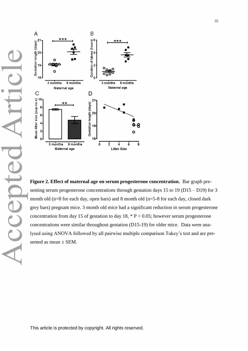
35
This article is protected by copyright. All rights reserved.
Figure 2. Effect of maternal age on serum progesterone concentration. Bar graph pre-
senting serum progesterone concentrations through gestation days 15 to 19 (D15 – D19) for 3
month old (n=8 for each day, open bars) and 8 month old (n=5-8 for each day, closed dark
grey bars) pregnant mice. 3 month old mice had a significant reduction in serum progesterone
concentration from day 15 of gestation to day 18, * P < 0.05; however serum progesterone
concentrations were similar throughout gestation (D15-19) for older mice. Data were ana-
lysed using ANOVA followed by all pairwise multiple comparison Tukey‟s test and are pre-
sented as mean ± SEM.

36
This article is protected by copyright. All rights reserved.
Figure 3. The impact of maternal age on cervical distension and stiffness in non-
pregnant and late pregnant tissues. (A) Tension generated by the cervix is plotted as a
function of incremental stretch of the cervical os diameter as stress-strain curves. Tissues
obtained from non-pregnant 3 month (n=6, open circles), 5 month (n=8, open triangles), and
8 month (n=8, open squares) old mice, as well as late pregnant 3 month (n=9, closed circles),
5 month (n=6, closed triangles), and 8 month (n=6, closed squares) old mice. Cervical stretch
(distension) means were different between non-pregnant compared to late pregnant mice of
all three age groups (*** P < 0.001). Cervices obtained from non-pregnant 3 month [NP(3),
n=6, open bar with diagonal lines], 5 month [NP(5), n=8, closed grey bar with diagonal
lines], and 8 month [NP(8), n=8, closed dark grey bar with diagonal lines] old mice, as well
as late pregnant 3 month [LP(3), n=9, open bar], 5 month [LP(5), n=6, closed grey bar], and 8
month [LP(8), n=6, closed dark grey bar] old mice for (B) and (C). (B) Transition from non-
pregnant to pregnant state, significantly increased maximal cervical distension in all three age
groups, *** P < 0.001. Advance in age from 3 to 8 months, and 5 to 8 months in non-
pregnant mice, and from 3 to 5 to 8 months in late pregnant mice also significantly increased
maximal cervical distension, ** P < 0.01 and *** P < 0.001. (C) Bar graph presenting cervi-
cal stiffness as determined by the slope of cervical distension stress-strain curves. Transition
from non-pregnant to pregnant state, significantly reduced cervical stiffness in 3, 5 and 8

37
This article is protected by copyright. All rights reserved.
month old mice, *** P < 0.001. Advance in age from 3 to 8 months and 5 to 8 months in
non-pregnant mice also significantly reduced cervical stiffness *** P < 0.001, however ad-
vance in age did not change cervical stiffness in tissues from late pregnant mice. Data were
analysed using ANOVA followed by all pairwise multiple comparison Tukey‟s test and are
presented as mean ± SEM for (A), (B) and (C).
Figure 4. Collagen content is lower in cervical tissues from late pregnant mice compared to non-pregnant mice. Repre-sentative images of Masson’s trichrome staining of collagen in cervical tissues from 8 month old non-pregnant (A) and late pregnant (B) mice. Collagen staining is noticeably weaker in the late pregnant cervix. Original magnification x10. (C) Bar graph presenting percentage of collagen content in cervical tissues obtained from non-pregnant 3 month [NP(3), n=6, open bar with diagonal lines], 5 month [NP(5), n=6, closed grey bar with diagonal lines] and 8 month [NP(8), n=6, closed dark grey bar with diagonal lines] old mice, as well as late pregnant 3 month [LP(3), n=6, open bar], 5 month [LP(5), n=6, closed grey bar], and 8 month [LP(8), n=6, closed dark grey bar] old mice. Transition from non-pregnant to pregnant state, signifi-

38
This article is protected by copyright. All rights reserved.
cantly reduced cervical collagen content in 3, 5 and 8 month old mice, *** P < 0.001. Data were analysed using ANOVA followed by all pairwise multiple comparison Tukey’s test and are presented as mean ± SEM.
Figure 5. Expression of MMP2 is not significantly altered by the effect of age in cervical tissues from both non-pregnant and late pregnant mice
Representative images of immunohistochemical staining of MMP2 in cervical tissues from 8 month old non-pregnant (A) and late pregnant (B) mice. (C) Bar graph presenting expression of MMP2 in cervical tissues obtained from non-pregnant 3 month [NP(3), n=6, open bar with diagonal lines], 5 month [NP(5), n=6, closed grey bar with diagonal lines] and 8 month [NP(8), n=6, closed dark grey bar with diagonal lines] old mice, as well as late pregnant 3 month [LP(3), n=6, open bar], 5 month [LP(5), n=6, closed grey bar], and 8 month [LP(8), n=6, closed dark grey bar] old mice. Comparisons across all groups did not reach significance. Data were analysed using ANOVA followed by all pairwise multiple comparison Tukey’s test and are presented as mean ± SEM arbitrary positivity score.

39
This article is protected by copyright. All rights reserved.
Figure 6. Effect of maternal age on mRNA expression of connexin-43 and oxytocin re-
ceptor in myometrium from non-pregnant and late pregnant mice. Myometrium was
obtained from non-pregnant 3 month old [NP(3), n=8, open bars with diagonal lines] late
pregnant 3 month old [LP(3), n=8, open bars], non-pregnant 5 month old [NP(5), n=8, closed
grey bars with diagonal lines], late pregnant 5 month old [LP(5), n=8, closed grey bars] non-
pregnant 8 month old [NP(8), n=8, closed dark grey bars with diagonal lines] and late preg-
nant 8 month old [LP(8), n=8, closed dark grey bars] mice for both (A) and (B). (A) Expres-

40
This article is protected by copyright. All rights reserved.
sion of connexin-43 was increased in late pregnant myometrium of all age groups compared
to non-pregnant, *** P < 0.001, and lower in late pregnant myometrium from 8 month old
mice compared to late pregnant 3 month old, ##
P < 0.01. (B) Expression of oxytocin receptor
was significantly increased in late pregnant myometrium of all age groups compared to non-
pregnant; *** P < 0.001, ** P < 0.01, and lower in late pregnant myometrium from 8 month
old mice compared to late pregnant 3 month old, # P < 0.05. Data were analysed using
ANOVA followed by all pairwise multiple comparison Tukey‟s test and are expressed as
log(mean of copy number) ± SEM normalised to reference genes B2M and GAPDH.
Figure 7. Effect of maternal age on spontaneous contractile activity in myometrium from non-pregnant and late preg-nant mice. Myometrium was obtained from non-pregnant 3 month old [NP(3), n=32 strips from n=8, open bars with diag-onal lines] late pregnant 3 month old [LP(3), n=32 strips from n=8, open bars], non-pregnant 5 month old [NP(5), n=32 strips from n=8, closed grey bar with diagonal lines], late pregnant 5 month old [LP(5), n=32 strips from n= 8, closed grey bar] non-pregnant 8 month old [NP(8), n=32 strips from n=8, closed dark grey bar with diagonal lines] and late pregnant 8 month old [LP(8), n=32 strips from n=8, closed dark grey bar] mice for (A), (B), (E) and (F). (A) Mean integral tension was significantly higher in late pregnant myometrium compared to non-pregnant across all age groups, *** P < 0.001, ** P < 0.01, * P < 0.05. (B) Force amplitude was significantly higher in late pregnant myometrium compared to non-pregnant across all age groups, ** P < 0.01,* P < 0.05. (C) Representative trace showing spontaneous myometrial contractions from late pregnant 3 month old mouse. (D) Representative trace showing spontaneous myometrial contractions from late preg-nant 8 month old mouse. (E) Spontaneous myometrial contraction frequency was significantly greater in late pregnant 8 month vs. late pregnant 3 month old mice, * P < 0.05. (F) Myometrial spontaneous contraction duration was shorter in late pregnant 8 month vs. late pregnant 5 month and 3 month old mice, ** P < 0.01. Data were analysed using ANOVA followed by all pairwise multiple comparison Tukey’s test and are presented as mean ± SEM for (A), (B), (E) and (F), all sig-nificant comparisons (P< 0.05) comparisons are indicated.

41
This article is protected by copyright. All rights reserved.
Figure 8. Effect of oxytocin (10-12
-10-7
M) on the contractile activity in myometrial tis-
sues taken from late pregnant mice at different ages. Line graph illustrating the effect of
oxytocin (10-12
-10-7
M) on contractile activity in myometrium from late pregnant 3 month old
[LP(3), n=32 strips from n=8, closed circles], late pregnant 5 month old [LP(5), n=32 strips
from n=8, closed triangles], and late pregnant 8 month old [LP(8), n=32 strips from n=8,
closed squares] mice. Data expressed as mean integral tension expressed as a percentage in-
crease of the baseline spontaneous activity ± SEM. Application of oxytocin at concentrations
10-12-
10-7
M were able to augment mean integral tension in myometrium from mice of all ag-
es; linear regression analysis.
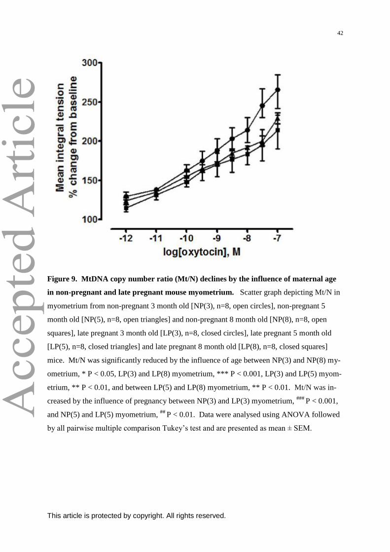
42
This article is protected by copyright. All rights reserved.
Figure 9. MtDNA copy number ratio (Mt/N) declines by the influence of maternal age
in non-pregnant and late pregnant mouse myometrium. Scatter graph depicting Mt/N in
myometrium from non-pregnant 3 month old [NP(3), n=8, open circles], non-pregnant 5
month old [NP(5), n=8, open triangles] and non-pregnant 8 month old [NP(8), n=8, open
squares], late pregnant 3 month old [LP(3), n=8, closed circles], late pregnant 5 month old
[LP(5), n=8, closed triangles] and late pregnant 8 month old [LP(8), n=8, closed squares]
mice. Mt/N was significantly reduced by the influence of age between NP(3) and NP(8) my-
ometrium, * P < 0.05, LP(3) and LP(8) myometrium, *** P < 0.001, LP(3) and LP(5) myom-
etrium, ** P < 0.01, and between LP(5) and LP(8) myometrium, ** P < 0.01. Mt/N was in-
creased by the influence of pregnancy between NP(3) and LP(3) myometrium, ###
P < 0.001,
and NP(5) and LP(5) myometrium, ##
P < 0.01. Data were analysed using ANOVA followed
by all pairwise multiple comparison Tukey‟s test and are presented as mean ± SEM.

43
This article is protected by copyright. All rights reserved.
Figure 10. Effect of maternal age on citrate synthase, complex I, complex II and com-
plex III enzymatic activities in myometrium from non-pregnant and late pregnant mice.
Myometrium was obtained from non-pregnant 3 month old [NP(3), n=8, open bar with diag-
onal lines], non-pregnant 8 month old [NP(8), n=8, closed dark grey bar with diagonal lines],
late pregnant 3 month old [LP(3), n=8, open bar], and late pregnant 8 month old [LP(8), n=8,
closed dark grey bar] mice for (A), (B), (C) and (D). (A) There were no differences in citrate
synthase enzyme activity. (B) There were no differences in complex I/NADH dehydrogen-
ase enzyme activity. (C) Transition from non-pregnant to pregnant state increased complex
II/succinate dehydrogenase activity in myometrium from both 3 and 8 month old mice, *** P
< 0.001, ** P < 0.01. (D) There were no differences in complex III/ubiquinol cytochrome c
reductase enzyme activity. Data were analysed using ANOVA followed by all pairwise mul-
tiple comparison Tukey‟s test and are presented as mean ± SEM.

44
This article is protected by copyright. All rights reserved.