xii national meeting of the mexican association
TRANSCRIPT
645Abstract section. XII National Meeting of the Mexican Association of Hepatology. , 2017; 16 (4): 645-693
July-August, Vol. 16 No. 4, 2017: 645-693
ABSTRACT SECTION
The Official Journal of the Mexican Association of Hepatology,the Latin-American Association for Study of the Liver and
the Canadian Association for the Study of the Liver
XII NATIONAL MEETING OF THEMEXICAN ASSOCIATION OF HEPATOLOGY
June 21-24, 2017. Merida, Yucatan, Mexico
I. MOLECULAR AND CELLULAR BIOLOGY
01ANTIOXIDANTS AND ANTIFIBROTICS PROPERTIES
OF N-ACETYLCYSTEINE IN THE EXPERIMENTALLIVER DAMAGE REVERTION
GALICIA-MORENO M,* MONROY-RAMÍREZ HC,* ARAUZ J, * MURIEL P**GALICIA-MORENO M,* MONROY-RAMÍREZ HC,* ARAUZ J, * MURIEL P**GALICIA-MORENO M,* MONROY-RAMÍREZ HC,* ARAUZ J, * MURIEL P**GALICIA-MORENO M,* MONROY-RAMÍREZ HC,* ARAUZ J, * MURIEL P**GALICIA-MORENO M,* MONROY-RAMÍREZ HC,* ARAUZ J, * MURIEL P***DEPARTAMENTO DE FARMACOLOGÍA, FACULTAD DE MEDICINA MEXICALI, U.A.B.C.,
MEXICALI, B.C. MÉXICO.**DEPARTAMENTO DE FARMACOLOGÍA, CINVESTAV-IPN, CIUDAD DE MÉXICO,
MÉXICO.
Background. N-acetylcysteine (NAC) is an antioxidant, pre-cursor of reduced glutathione and inhibitor of different profi-brotic cytokines involved in liver damage. The aim of this workwas to evaluate the effect of NAC to reverse liver cirrhosis in-duced with carbon tetrachloride (CCl4) in rats. Material andmethods. Four groups of rats (n = 8) were performed. Group1, control animals received the vehicle (oil). Group 2 was ad-ministered with intraperitoneal CCl4 (0.4 g/kg). Group 3 re-ceived CCl4 and then NAC (300 mg/kg, orally). Group 4received mineral oil (0.25 mL), and then NAC. CCl4 and oilwere administered by 2 months, three times per week and NACand CMC only by 1 month daily after treatment with CCl4.Alanine aminotransferase (ALT) was measured in plasma. Colla-gen, glycogen and MDA levels and reduced glutathione wereesteemed in liver samples; a histopathologycal analysis was per-formed. Results. ALT enzyme activity increased significantlyafter 2 months of CCl4-administration. Discontinuation ofCCl4 for 1 month resulted in a decrease levels of ALT enzymeactivity to normal values. MDA levels increased while GSH/GSSG ratio in liver decreased by the administration of CCl4 for2 months; discontinuation of CCl4 did not lead to normal val-ues of these parameters, again NAC prevented both effects. Liv-er glycogen content decreased by CCl4 intoxication, NACrestored normal glycogen levels. Fibrosis was quantified bymeasuring collagen levels, CCl4-intoxication during 2 monthsproduced an increased in liver collagen content. Discontinua-tion of CCl4 did not lead to fibrolysis, NAC administration re-sulted in a partial but significant reversion of CCl4-inducedfibrosis. Conclusions. our results strongly suggest that NACwas highly effective in reversing liver damage. This effect per-haps is related to the ability of NAC as scavenger as well as itsantifibrogenic capacity. We are currently studying the expres-
DOI: 10.5604/01.3001.0010.0318
sion of several cytokines related to liver damage as TGF-β,among others to be able to consider that NAC can be tested infibrotic or cirrhotic patients under control trials.This work was supported by Beca PRODEP- apoyo a la incor-poración de nuevos PTC UABC7PTC 570.
02PROTECTOR EFECT OF HGF IN INTRAHEPATIC
CHOLESTASISSALAS-SILVA S,* SIMONI-NIEVES A,* BELLO-MONROY O,*SALAS-SILVA S,* SIMONI-NIEVES A,* BELLO-MONROY O,*SALAS-SILVA S,* SIMONI-NIEVES A,* BELLO-MONROY O,*SALAS-SILVA S,* SIMONI-NIEVES A,* BELLO-MONROY O,*SALAS-SILVA S,* SIMONI-NIEVES A,* BELLO-MONROY O,*
BUCIO-ORTÍZ L,* ROMA M,** GÓMEZ-QUIROZ LE,* GUTIÉRREZ-RUÍZ MC*BUCIO-ORTÍZ L,* ROMA M,** GÓMEZ-QUIROZ LE,* GUTIÉRREZ-RUÍZ MC*BUCIO-ORTÍZ L,* ROMA M,** GÓMEZ-QUIROZ LE,* GUTIÉRREZ-RUÍZ MC*BUCIO-ORTÍZ L,* ROMA M,** GÓMEZ-QUIROZ LE,* GUTIÉRREZ-RUÍZ MC*BUCIO-ORTÍZ L,* ROMA M,** GÓMEZ-QUIROZ LE,* GUTIÉRREZ-RUÍZ MC**DEPARTAMENTO DE CIENCIAS DE LA SALUD, UNIVERSIDAD AUTÓNOMA
METROPOLITANA IZTAPALAPA. CIUDAD DE MÉXICO, MÉXICO.**INSTITUTO DE FISIOLOGÍA EXPERIMENTAL, FACULTAD DE CIENCIAS BIOQUÍMICAS
Y FARMACÉUTICAS, UNIVERSIDAD DE ROSARIO, ARGENTINA.
Introduction. Cholestasis is a common syndrome in a largenumber of hepatic diseases, such as drug-induced cholestasis whichis produced due to a primary lesion of the bile conducts eitherfunctional or obstructive, generating oxidative stress in the liver.Hepatocyte growth (HFG) factor, and its receptor, c-Met, repre-sents the first defense line against hepatotoxics factors because theyinduce the Nrf2 activation which, in turn, will activate its targetgenes to lead to an antioxidant and repair response. The aim of thisresearch is to characterize the anticholestatic molecular and cellularmechanism measured with the HGF in a (urine model of inflam-matory cholestasis induced by the α-naftil-isotiocianato (ANIT).Material and methods. CD1 Mice were used, treated with a-naftil isotiocianato (60 μg/kg) for 48 h intragastricaly. After 24 h ofANIT, HGF (10 μg/kg) were administrated intravenously. When48 h passed the sacrifice were done whereby biochemist tests wereundertaken such as AST, ALT, ALP and H-E staining. Similarlymouse hepatocytes were obtained for primary culture which weretreated with ANIT (20 μM) and HGF (50 ng) in order to carry outviability and functionality tests. Results. Animales treated withANIT present liver injury, (hepatomegalia) and cholecystitis,those effects were reverted when HGF was administrated in com-parison with the ones that were just damaged. In the ANITtreated cellular culture we could observe a decrease in the cellularviability as well as in its functionality, however when the culturewas pre-treated with HGF it was observed that the cellular viabil-ity was kept even though it was exposed to ANIT. Conclusion.Data shows that HGF treated animals improved significantly incomparison with the ANIT damaged, besides HGF provides cel-
646Abstract section. XII National Meeting of the Mexican Association of Hepatology. , 2017; 16 (4): 645-693
lular dead protection in hepatocytes primary culture. Based onthis we can considere HGF as a therapeutic intervention point incholestatic diseases.CONACYT: CB 252942
03HEPATIC EXPRESSSION OF INTERLEUKIN-6 (IL-6) INA MURINE MODEL OF HEPATIC FIBROSIS INDUCED
BY THIOACETAMIDEBAUTISTA-UBALDO MG,* ARÉVALO-SÁNCHEZ TA,*BAUTISTA-UBALDO MG,* ARÉVALO-SÁNCHEZ TA,*BAUTISTA-UBALDO MG,* ARÉVALO-SÁNCHEZ TA,*BAUTISTA-UBALDO MG,* ARÉVALO-SÁNCHEZ TA,*BAUTISTA-UBALDO MG,* ARÉVALO-SÁNCHEZ TA,*
RAMÍREZ-MENDOZA A,* KERSHENOBICH D,**RAMÍREZ-MENDOZA A,* KERSHENOBICH D,**RAMÍREZ-MENDOZA A,* KERSHENOBICH D,**RAMÍREZ-MENDOZA A,* KERSHENOBICH D,**RAMÍREZ-MENDOZA A,* KERSHENOBICH D,**GUTIÉRREZ-REYES G,* GUZMÁN C*GUTIÉRREZ-REYES G,* GUZMÁN C*GUTIÉRREZ-REYES G,* GUZMÁN C*GUTIÉRREZ-REYES G,* GUZMÁN C*GUTIÉRREZ-REYES G,* GUZMÁN C*
*LABORATORIO DE HÍGADO, PÁNCREAS Y MOTILIDAD, UNIDAD DE MEDICINAEXPERIMENTAL, FACULTAD DE MEDICINA, UNAM/HOSPITAL GENERAL DE MÉXICO.
MÉXICO.**INSTITUTO NACIONAL DE CIENCIAS MÉDICAS Y NUTRICIÓN “SALVADOR ZUBIRÁN”,
CIUDAD DE MÉXICO, MÉXICO.
Introduction and aim. Interleukin (IL)-6 is a highly pleio-tropic cytokine produced by a wide range of cell types. In hepat-ic pathologies, IL-6 has been shown to be related to processesincluding inflammation and regeneration. In the liver, IL-6 issynthesized by Kupffer cells, endothelial cells and hepatic stel-late cells, whereas in the regenerating liver is produced in theductular reactions by oval cells. Here we aimed to study the ex-pression of IL-6 in the liver of mice exposed to thioacetamide(TAA) with different degrees of fibrosis. Material and meth-ods. Female C57BL/6 mice weighing 22 ± 3 g and 16 weeksold were administered with increasing doses of TAA (50-400mg/kg) thrice a week for 4, 6, or 8 weeks. A control groups thatreceived the same number of doses of saline (vehicle) was in-cluded. Total proteins were isolated from the liver and IL-6 wasquantified by ELISA. Data is shown as Mean ± SD and wereanalyzed by one way ANOVA followed by the Tukey post hoctest. P < 0.05 was considered significant. Results. Increasingdegree of liver fibrosis was observed according to the number ofTAA doses. IL-6 expressed in the liver of TAA treated mice didnot show alterations after receiving 12 or 18 doses compared tocontrols. However after the 24 TAA doses, a significantly lowerIL-6 expression in comparison to controls as well as TAA12 andTAA18 was found (C = 91.8 ± 19.9, TAA12 = 104.4 ± 27.7,TAA18 = 89.7 ± 11.2, TAA24 = 31.8 ± 19.4 pg of protein/mgof tissue, n = 4-6 per group, p < 0.001). Conclusions. Al-though IL-6 is cytokine mainly associated to inflammation, itssynthesis is diminished in advanced fibrosis induced by TAAcompared to initial fibrosis or to the healthy liver. A loss of IL-6in the liver could be associated not only to a higher degree ofliver damage, but also to a decreased regenerative capacity.This work was funded by Conacyt (CB-221137).
04ASSESSMENT OF CONNECTIVE TISSUE GROWTH
FACTO (CTGF) IN A MURINE MODEL OF LIVERFIBROSIS INDUCED BY BILE DUCT LIGATION
ARÉVALO-SÁNCHEZ TA,* BAUTISTA-UBALDO MG,*ARÉVALO-SÁNCHEZ TA,* BAUTISTA-UBALDO MG,*ARÉVALO-SÁNCHEZ TA,* BAUTISTA-UBALDO MG,*ARÉVALO-SÁNCHEZ TA,* BAUTISTA-UBALDO MG,*ARÉVALO-SÁNCHEZ TA,* BAUTISTA-UBALDO MG,*RAMÍREZ-MENDOZA A,* GARCÍA-REBOLLAR JO,** DÍAZ-HERRERA G,**RAMÍREZ-MENDOZA A,* GARCÍA-REBOLLAR JO,** DÍAZ-HERRERA G,**RAMÍREZ-MENDOZA A,* GARCÍA-REBOLLAR JO,** DÍAZ-HERRERA G,**RAMÍREZ-MENDOZA A,* GARCÍA-REBOLLAR JO,** DÍAZ-HERRERA G,**RAMÍREZ-MENDOZA A,* GARCÍA-REBOLLAR JO,** DÍAZ-HERRERA G,**
KERSHENOBICH D,*** GUTIÉRREZ-REYES G,* GUZMÁN C*KERSHENOBICH D,*** GUTIÉRREZ-REYES G,* GUZMÁN C*KERSHENOBICH D,*** GUTIÉRREZ-REYES G,* GUZMÁN C*KERSHENOBICH D,*** GUTIÉRREZ-REYES G,* GUZMÁN C*KERSHENOBICH D,*** GUTIÉRREZ-REYES G,* GUZMÁN C**LABORATORIO DE HÍGADO, PÁNCREAS Y MOTILIDAD, UNIDAD DE MEDICINA
EXPERIMENTAL, FACULTAD DE MEDICINA, UNAM/HOSPITAL GENERAL DE MÉXICO.MÉXICO.
**INSTITUTO DE INVESTIGACIONES BIOMÉDICAS, UNIDAD DE MODELOS BIOLÓGICOS,UNAM. MÉXICO.
***INSTITUTO NACIONAL DE CIENCIAS MÉDICAS Y NUTRICIÓN “SALVADOR ZUBIRÁN”,CIUDAD DE MÉXICO, MÉXICO.
Background and aim. Connective Tissue Growth Factor(CTGF) is a matricellular protein involved in wound healingand scarring. In the liver it has not only been associated to fi-brosis, but also to oval cell differentiation and proliferation inthe ductular reactions during liver regeneration. Bile Duct Li-gation (BDL) is considered a murine model of biliary fibrosis inwhich ductular reactions are highly observed. We aimed to as-sess the CTGF protein in both liver tissue and serum of micesubjected to BDL. Material and methods. Male 16 weeks oldCD-1 mice weighing 25 ± 3 g were subjected to surgical BDL;a second group receiving surgical intervention with no BDL(SHAM) was included. Mice were kept for 7 or 25 days aftersurgery, liver and blood samples were collected. Total proteinwas isolated from liver samples and protein integrity was tested.CTGF was measured in protein extracts and serum by ELISA.Data is shown as Mean ± SD, and analyzed by one-way ANO-VA, followed by Tukey post-hoc test. P < 0.05 was consideredsignificant. N = 4-6. Results. After 7 days of BDL, mice ex-hibited mild fibrosis, whereas after 25 days severe fibrosis wasobserved, no histological alterations were observed in theSHAM group. CTGF protein contents in the liver were signifi-cantly increased in the group of 7 days (BDL7d) compared toSHAM (SHAM = 93.4 ± 29.36, BDL7d = 189.6 ± 80.96,BDL25d = 142.2 ± 52.10 pg of CTGF/mg of liver, p < 0.05).Serum levels of CTGF were increased after 25 days post-BDLcompared to SHAM and BDL7d (SHAM = 6833.2 ± 291.64,BDL7d = 8620.7 ± 711.37 y BDL25d = 17679.5 ± 2147.58pg/mL, p < 0.0001). Conclusions. Results here shown suggestan early increase in CTGF synthesis in BDL after 7 days post-surgery, whereas in the serum that increase is observable after25 days, which could be associated to an increased CTGF hepat-ic synthesis during early fibrosis with a higher secretion in theadvanced damage.This work was funded by Conacyt (CB-221137).
647Abstract section. XII National Meeting of the Mexican Association of Hepatology. , 2017; 16 (4): 645-693
II. CIRRHOSIS AND ITS COMPLICATIONS
01MALNUTRITION AS AN ADVERSE PROGNOSTIC
FACTOR RELATED TO INCREASE IN THEMORTALITY RISK AT TWO-YEAR FOLLOW-UP IN
PATIENTS WITH CIRRHOSIS. A PROSPECTIVESTUDY
HIGUERA-DE LA TIJERA F,* ROJAS-LOUREIRO G,*HIGUERA-DE LA TIJERA F,* ROJAS-LOUREIRO G,*HIGUERA-DE LA TIJERA F,* ROJAS-LOUREIRO G,*HIGUERA-DE LA TIJERA F,* ROJAS-LOUREIRO G,*HIGUERA-DE LA TIJERA F,* ROJAS-LOUREIRO G,*SERVÍN-CAAMAÑO AI,** CAMACHO-AGUILERA J,**SERVÍN-CAAMAÑO AI,** CAMACHO-AGUILERA J,**SERVÍN-CAAMAÑO AI,** CAMACHO-AGUILERA J,**SERVÍN-CAAMAÑO AI,** CAMACHO-AGUILERA J,**SERVÍN-CAAMAÑO AI,** CAMACHO-AGUILERA J,**
LÓPEZ LADRÓN-DE GUEVARA V,* MACÍAS-ÁNGELES Y,*LÓPEZ LADRÓN-DE GUEVARA V,* MACÍAS-ÁNGELES Y,*LÓPEZ LADRÓN-DE GUEVARA V,* MACÍAS-ÁNGELES Y,*LÓPEZ LADRÓN-DE GUEVARA V,* MACÍAS-ÁNGELES Y,*LÓPEZ LADRÓN-DE GUEVARA V,* MACÍAS-ÁNGELES Y,*PÉREZ-HERNÁNDEZ JL*PÉREZ-HERNÁNDEZ JL*PÉREZ-HERNÁNDEZ JL*PÉREZ-HERNÁNDEZ JL*PÉREZ-HERNÁNDEZ JL*
*GASTROENTEROLOGY DEPARTMENT/LIVER CLINIC. **INTERNAL MEDICINEDEPARTMENT. HOSPITAL GENERAL DE MÉXICO “DR. EDUARDO LICEAGA”, MEXICO
CITY, MEXICO.
Background. Malnutrition is highly prevalent in patients withcirrhosis. Previous studies have demonstrated its relationwith greater risk of complications in these patients. Aim. Toevaluate which prognostic factors are related to mortality in pa-tients with cirrhosis at two-year follow-up. Material andmethods. A cohort study which included patients with cirrho-sis by any etiology. The Global Subjective Assessment (GSA)was initially performed in all patients. Each visit, they receivednutritional counseling; nevertheless, a group did not have adher-ence to the recommendations, therefore did not have nutritionalimprovement. The exposed cohort was integrated for patientswith moderate to severe malnutrition (grades B and C) accord-ing to the GSA. The non-exposed were well-nourished patients(A of the GSA). For univariate analysis we used χ2 or exactFisher’s test, also were calculated odds ratio and 95% confidenceintervals. For the multivariate analysis we introduced variableswhich were statistical significant on the univariate analysis, weperformed Cox regression and Kaplan-Meier curves were con-structed. A P < 0.05 was considered significant. Results. A to-tal of 110 patients, 55.5% were women, mean age 54.5 ± 12.1year-old. Etiology: Alcohol 53.6%, viral 14.6%, non-alcoholicsteatohepatitis 21.8%, autoimmune 10%. Child-Pugh: 20% A,57.3% B, and 22.7% C. Nutritional status: 49 (44.5%) grade Aof the VGS, 58 (52.7%) grade B of the VGS, and 3 (2.7%) gradeC of the VGS. In the univariate analysis were negatively associ-ated with survival at two-year: decompensated cirrhosis orChild-Pugh B/C (65.5% vs. 92.3%; OR = 1.4; 95%CI: 1.2-1.7;P = 0.007), malnutrition (57.4% vs. 89.8%; OR = 1.6; 95%CI:1.2-2.0; P < 0.0001), presence of ascites (57.4% vs. 89.8%; OR= 1.6; 95%CI: 1.2-2.0; P < 0.0001), and history of hospitaliza-tion because of bacterial infection in the last two years (35.3%vs. 78.5%; OR = 2.2; 95%CI: 1.2-4.3; P = 0.001). Did not in-fluence: age, gender, etiology, variceal bleeding, hepatic enceph-alopathy. In the multivariate analysis malnutrition (HR = 3.4;IC al 95%:1.3 a 9.3; P = 0.01) and presence of ascites (HR =3.0; 95%CI: 1.1-8.5; P = 0.04) were factors that increase therisk of death of cirrhotic patients at two-year follow-up, inde-pendently of Child-Pugh stage. Conclusion. Malnutrition is arisk factor that increases the probability of death in cirrhotic pa-tients. Is important can count with a multidisciplinary team andhealth-care programs which takes in count the nutritional in-tervention in patients with cirrhosis.
02P300 POTENTIALS EVOKED BY AUDITORY
STIMULATION (A Y B) FOR DIAGNOSIS OF MINIMALHEPATIC ENCEPHALOPATHY
PÉREZ HERNÁNDEZ J,* HIGUERA DE LA TIJERA F,* PÉREZ SOTO F,*PÉREZ HERNÁNDEZ J,* HIGUERA DE LA TIJERA F,* PÉREZ SOTO F,*PÉREZ HERNÁNDEZ J,* HIGUERA DE LA TIJERA F,* PÉREZ SOTO F,*PÉREZ HERNÁNDEZ J,* HIGUERA DE LA TIJERA F,* PÉREZ SOTO F,*PÉREZ HERNÁNDEZ J,* HIGUERA DE LA TIJERA F,* PÉREZ SOTO F,*SERVIN CAAMAÑO A,* DURAN MEZA H,** JIMENEZ CORREA U,**SERVIN CAAMAÑO A,* DURAN MEZA H,** JIMENEZ CORREA U,**SERVIN CAAMAÑO A,* DURAN MEZA H,** JIMENEZ CORREA U,**SERVIN CAAMAÑO A,* DURAN MEZA H,** JIMENEZ CORREA U,**SERVIN CAAMAÑO A,* DURAN MEZA H,** JIMENEZ CORREA U,**
SANTANA VARGAS D**SANTANA VARGAS D**SANTANA VARGAS D**SANTANA VARGAS D**SANTANA VARGAS D***GASTROENTEROLOGY/LIVER CLINIC,
**SLEEP DISORDERS CLINIC, HOSPITAL GENERAL DE MÉXICO “DR. EDUARDOLICEAGA” CIUDAD DE MÉXICO. MÉXICO.
Background. Minimal hepatic encephalopathy (MHE) diag-nosis is complex. P300 potentials can be elicited by auditory orvisual stimulation. A two stimuli paradigm, standard and targetto obtain typical P3b and a three stimuli paradigm standard, dis-tractor and target to obtain P3a. Both types of P3 have not beenexplored in this setting. P3 potential are an objective tool, capa-ble of detecting minimal changes in cerebral function regardingprocessing of information. P3a/b are obtained from pairing stim-uli with ongoing electroencephalogram. Aim: To evaluate P300(A and B) for diagnosis of MHE, and to compare their latencyversus another diagnostic tools. Material and methods. Weincluded cirrhotic patients paired with a group of healthy-con-trols. We excluded patients with overt hepatic encephalopathy,taking medications as: antidepressants, anxiolytics, anti-ammo-nia; also infected patients, or with previous diagnosis of anyneurological disease. In one session neuropsychological batteryPHES, Critical Frequency Flicker (FCC), visual-P300 and audi-tory-P300a/b were obtained from each participant. P300b wasconsidered as gold standard. Results. 26 healthy-controls wereincluded, 17 women, age 39.69 ± 8.7; and 40 cirrhotic, 24 wom-en, age 56.10 ± 9.23. Child-Pugh A-B-C 24-14-02, the fre-quency of detection of MHE was: PHES 21 (52.50%) patients,CFF 29 (72.50%), P300v 30 (75%), P300a - 33 (82.5%) P300b27, (67.50%). Sensitivity and specificity were 51.7%, 45.5% forFCC-PHES; 60%, 70%, for visual-P300, 66.7%, and 76.9%, forP300a-B S= 72.7 E= 46.9; Positive and negative predictive val-ues were: 72.42%, 50.75% for P300a. Conclusions. Nowadaysthere is not an ideal tool for diagnosis of MHE. Both types ofP300 are new diagnostic tools available in this setting. Regardlessof the modality visual or auditory, P300 showed better sensitivityand specificity than CFF, and also better predictive values visual-P300 compared to other diagnostic tools.
03RETROSPECTIVE ANALYSIS OF THE EFFICACY AND
SAFETY OF TERLIPRESSIN PLUS VARICEALLIGATION VERSUS OCTREOTIDE PLUS VARICEALLIGATION IN PATIENTS WITH ACUTE VARICEAL
BLEEDINGCASTILLEJOS-GARCÍA KJ,* LÓPEZ-COLOMBO A,**CASTILLEJOS-GARCÍA KJ,* LÓPEZ-COLOMBO A,**CASTILLEJOS-GARCÍA KJ,* LÓPEZ-COLOMBO A,**CASTILLEJOS-GARCÍA KJ,* LÓPEZ-COLOMBO A,**CASTILLEJOS-GARCÍA KJ,* LÓPEZ-COLOMBO A,**
MENDOZA-TORRES MA***MENDOZA-TORRES MA***MENDOZA-TORRES MA***MENDOZA-TORRES MA***MENDOZA-TORRES MA****GASTROENTEROLOGÍA, HOSPITAL GENERAL DE ZONA NÚM. 2 TULANCINGO,
HIDALGO. MÉXICO.**DIRECCIÓN DE EDUCACIÓN E INVESTIGACIÓN EN SALUD, ***DEPARTAMENTO DE
GASTROENTEROLOGÍA UMAE HOSPITAL DE ESPECIALIDADES PUEBLA CMN MANUELÁVILA CAMACHO. MÉXICO.
Background. Acute variceal bleeding has high mortality (15-20%). There are just a few of trials about efficacy and safety ofvasoactive drugs plus endoscopic treatment. Objective: To
648Abstract section. XII National Meeting of the Mexican Association of Hepatology. , 2017; 16 (4): 645-693
analize the efficacy and safety of the terlipressin plus endo-scopic variceal ligation (TRL/VL) vs. octreotide plus endoscopicvariceal ligation (OCTR/VL). Material and methods. Trans-versal, comparative study including patients who had been di-agnosed with esophageal variceal bleeding between 2012 and2016 and whom were been treated with TRL/VL or OCTR/VL. Using χ2 and fisher exact probability test were contrastedvariables of efficacy as control of bleeding (CB), mortality at 6weeks (M-6W) and inpatient stay (IS) and variables of safety asperipheral ischemia and abdominal pain. Results. 45 patientswere been identified: 27 (60%) TRL/VL group and 18 (40%)OCTR/VL gruop. Main age 59.9 years, 66.6% men. Controlof bleeding was reached in 88.9% in both groups (p = 1.0).General M-6W was 11.1% and also for each group (p = 1.0).IS under 5 days was 81.5% in TRL/VL group and 94.4% inOCTR/VL (p = 0.38). In TRL/VL group were identified 7 cas-es of abdominal pain as an adversal effect and no one case inOCTR/VL group (p = 0.01). The prolongated inpatient stay(> 5 days) was observed in 7.7% of patients whom were sub-mitted to an early endoscopy (< 24 h) vs. 50% of those withan endoscopy after 24 h. Conclusions. The efficacy of TRL/VL y OCTR/VL was similar. Were observed more cases of ab-dominal pain in TRL/LV group. Patients submitted to an earlyendoscopy had a shorter IS.
04COMPARATION OF TWO PROPHYLACTIC
ANTIBIOTICS FOR SPONTANEOUS BACTERIALPERITONITIS IN PATIENTS WITH CIRRHOSIS AND
ASCITIS: RIFAXIMINE VS. CIPROFLOXACINO(preliminary report)
GARCÍA-GONZÁLEZ E, ACOSTA-GÓMEZ M, FRANQUEZ-FLORES B,GARCÍA-GONZÁLEZ E, ACOSTA-GÓMEZ M, FRANQUEZ-FLORES B,GARCÍA-GONZÁLEZ E, ACOSTA-GÓMEZ M, FRANQUEZ-FLORES B,GARCÍA-GONZÁLEZ E, ACOSTA-GÓMEZ M, FRANQUEZ-FLORES B,GARCÍA-GONZÁLEZ E, ACOSTA-GÓMEZ M, FRANQUEZ-FLORES B,GONZÁLEZ-HUEZO MGONZÁLEZ-HUEZO MGONZÁLEZ-HUEZO MGONZÁLEZ-HUEZO MGONZÁLEZ-HUEZO M
DEPARTAMENTO DE GASTROENTEROLOGÍA Y ENDOSCOPIA GASTROINTESTINAL.INSTITUTO DE SEGURIDAD SOCIAL DEL ESTADO DE MÉXICO Y MUNICIPIOS; CENTRO
MÉDICO ISSEMYM, METEPEC, ESTADO DE MÉXICO. MÉXICO.
Introduction. Systemic antibiotics as prophylaxis have de-creased risk for development spontaneous bacterial peritonitis(SBP) in cirrhotic patients (CP). It implies a risk developmentfor antimicrobial resistance. An intraluminal antibiotic could re-duce the number of SBP episodes without favoring the devel-opment of multiresistant strains. Objective: Compare rifaximinvs. ciprofloxacin, as prophylaxis in cirrhotic patients with risk
factors, determine number and characteristics of infections, gen-eral mortality and associated to sepsis. Material and methods.We perform a prospective, randomized and open study, with du-ration of one year. Started in September 2015 and ongoing. In-
(II.03) Table 1.
Total (n = 45) TRL/VL (n = 27) OCTR/VL (n = 18) p
• Comparison of vasoactives safety.
Adverse effectsAbdominal pain 7 (15.5) 7 (26.9) 0 (0) 0.01Peripheral ischemia 1 (2.2) 1 (3.7) 0 (0) 1
• Comparison of vasoactives efficacy
Control of bleeding, n (%) 40 (88.8) 24 (88.8) 16 (88.8) 1Impatient stay ≤ 5days 39 (86.7) 22 (81.5) 17 (94.4) 0.377Transfusion requeriment 31 (68.9) 19 (70) 12 (66.7) 0.792Use of rescue therapy 40 (88.8) 25 (92.6) 15 (83.3) 0.375Mortality at 6 weeks 5 (11.1) 3 (11.1) 2 (11.1) 1
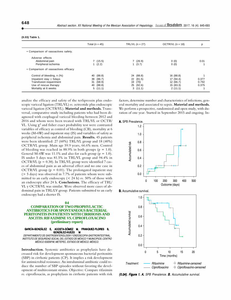
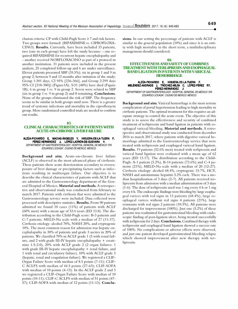
(II.04). Figure 1.(II.04). Figure 1.(II.04). Figure 1.(II.04). Figure 1.(II.04). Figure 1. A.A.A.A.A. SPB Prevalence. B. B. B. B. B. Accumulative survival.
Treatment: Rifaximine Rifaximine-censoredCiprofloxacino Ciprofloxacino-censored
0 100 200 300 400 500Outcome (days)
1.2
1.0
0.8
0.6
0.4
0.2
0.0
Accu
mul
ated
risk
A. A. A. A. A. SPB Prevalence.
B. B. B. B. B. Accumulative survival.
0 5 10 15 20Time (months)
1.0
0.8
0.6
0.4
0.2
0.0
Accu
mul
ated
surv
ival
649Abstract section. XII National Meeting of the Mexican Association of Hepatology. , 2017; 16 (4): 645-693
clusion criteria: CP with Child-Pugh Score ≥ 7 and risk factors.Two groups were formed: (RIFAXIMINE vs. CIPROFLOXA-CINO). Results. Currently, have been included 33 patients,two (one in each group) have left the study because: - one re-quired RIFAXIMINE for recurrent hepatic encephalopathy and- another received NORFLOXACINO as part of a protocol inanother institution. 31 patients were included in the presentanalysis, 25 completed follow-up and 6 are under surveillance.Eleven patients presented SBP (35.5%); six in group 1 and 9 ingroup 2; between 9 and 12 months after initiation of the study:Group 1:301 days, CI 95% [236-366], and Group 2:299 days95% CI [218-380]) (Figure1A). 5/31 (48%) have died (Figure1B), 6 in group 1 vs. 9 in group 2. Seven were related to SBP(six in group 1 vs. 9 in group 2) and 8 remaining. Conclusions.None of the groups eliminated the risk of SBP. The frequencyseems to be similar in both groups until now. There is a greatertrend of systemic infections and mortality in the ciprofloxacingroup. More randomized controlled trials are needed to confirmour results.
05CLINICAL CHARACTERISTICS OF PATIENTS WITH
ACUTE-ON-CHRONIC LIVER FAILURE
ALZÚA-PICHARDO E, MACIAS-ÁNGELES Y, HIGUERA-DE-LA-TIJERA F,ALZÚA-PICHARDO E, MACIAS-ÁNGELES Y, HIGUERA-DE-LA-TIJERA F,ALZÚA-PICHARDO E, MACIAS-ÁNGELES Y, HIGUERA-DE-LA-TIJERA F,ALZÚA-PICHARDO E, MACIAS-ÁNGELES Y, HIGUERA-DE-LA-TIJERA F,ALZÚA-PICHARDO E, MACIAS-ÁNGELES Y, HIGUERA-DE-LA-TIJERA F,PÉREZ-HERNÁNDEZ J, LÓPEZ-LADRÓN-DE-GUEVARA V.PÉREZ-HERNÁNDEZ J, LÓPEZ-LADRÓN-DE-GUEVARA V.PÉREZ-HERNÁNDEZ J, LÓPEZ-LADRÓN-DE-GUEVARA V.PÉREZ-HERNÁNDEZ J, LÓPEZ-LADRÓN-DE-GUEVARA V.PÉREZ-HERNÁNDEZ J, LÓPEZ-LADRÓN-DE-GUEVARA V.
DEPARTMENT OF GASTROENTEROLOGY, HOSPITAL GENERAL DE MÉXICO “DR.EDUARDO LICEAGA”, CIUDAD DE MÉXICO. MÉXICO.
Background and aim. Acute-on-chronic liver failure(ACLF) is observed in the most advanced phase of cirrhosis.These patients show acute deterioration secondary to overex-posure of liver damage or precipitating factors such as infec-tions resulting in multiorgan failure. Our objective is todescribe the clinical characteristics of patients with ACLF thatare admitted to the Gastroenterology department of the Gen-eral Hospital of Mexico. Material and methods. A retrospec-tive and observational study was conducted from february tomarch 2017. Patients with cirrhosis that were admitted to theGastroenterology service were included. Data collected wereprocessed with descriptive statistics. Results. From 90 patientsadmitted we found 10 cases (11%) of patients with ACLF(60% men) with a mean age of 53.6 years (ED 13.8). The dis-tribution according to the Child-Pugh score: B-3 patients andC-7 patients. MELD-Na scale with a median of 27 (11-37).Cirrhosis etiology: alcohol 70%, NASH 20%, and cryptogenic10%. The most common reason for admission was hepatic en-cephalopathy in 30% of patients and grade 3 ascites in 20% ofpatients. We classified 70% in ACLF grade 1 (5 with renal fail-ure, and 2 with grade III-IV hepatic encephalopathy + creati-nine 1.5-2.0), 20% with ACLF grade 2 (2 organ failures: 1with grade III-IV hepatic encephalopathy + renal failure, and1 with renal and circulatory failure), 10% with ACLF grade 3(hepatic, renal and coagulation failure). We registered a CLIF-Organ Failure Score with median of 8.5 points (7-11); CLIF-C ACLFS with median of 44.5 points (27-63); CLIF-SOFAwith median of 10 points (4-13). In the ACLF grade 2 and 3we registered a CLIF-Organ Failure Score with median of 10points (10-11); CLIF-C ACLFS with median of 51 points (47-57); CLIF-SOFA with median of 12 points (11-13). Conclu-
sions. In our setting the percentage of patients with ACLF issimilar to the general population (10%) and since it is an enti-ty with high mortality in the short term, a multidisciplinarymanagement should considered.
06EFFECTIVENESS AND SAFETY OF COMBINES
TREATMENT WITH TERLIPRESIN AND ESOPHAGEALBAND LIGATION IN PATIENTS WITH VARICEAL
HEMORRHAGEALZÚA-PICHARDO E, HIGUERA-DE-LA-TIJERA F,ALZÚA-PICHARDO E, HIGUERA-DE-LA-TIJERA F,ALZÚA-PICHARDO E, HIGUERA-DE-LA-TIJERA F,ALZÚA-PICHARDO E, HIGUERA-DE-LA-TIJERA F,ALZÚA-PICHARDO E, HIGUERA-DE-LA-TIJERA F,
MELÉNDEZ-ANDRADE J, TRÓCHEZ-MEJÍA C, LÓPEZ-PÉREZ RY,MELÉNDEZ-ANDRADE J, TRÓCHEZ-MEJÍA C, LÓPEZ-PÉREZ RY,MELÉNDEZ-ANDRADE J, TRÓCHEZ-MEJÍA C, LÓPEZ-PÉREZ RY,MELÉNDEZ-ANDRADE J, TRÓCHEZ-MEJÍA C, LÓPEZ-PÉREZ RY,MELÉNDEZ-ANDRADE J, TRÓCHEZ-MEJÍA C, LÓPEZ-PÉREZ RY,PÉREZ-HERNÁNDEZ JPÉREZ-HERNÁNDEZ JPÉREZ-HERNÁNDEZ JPÉREZ-HERNÁNDEZ JPÉREZ-HERNÁNDEZ J
DEPARTMENT OF GASTROENTEROLOGY, HOSPITAL GENERAL DE MÉXICO “DR.EDUARDO LICEAGA”, CIUDAD DE MÉXICO. MÉXICO.
Background and aim. Variceal hemorrhage is the most seriouscomplication of portal hypertension leading to high mortality incirrhotic patients. The optimal treatment for this requires an ad-equate strategy to control the acute event. The objective of thisstudy is to assess the effectiveness and security of combinedtreatment of terlipressin and band ligation in patients with es-ophageal variceal bleeding. Material and methods. A retro-spective and observational study was conducted from december2016 to march 2017, where patients with digestive variceal he-morrhage admitted to the Gastroenterology service that weretreated with terlipressin and esophageal variceal band ligation.Results. 19 patients (52.6% men) treated with terlipressin andvariceal band ligation were evaluated with a mean age of 62years (ED 13.17). The distribution according to the Child-Pugh: A-1 patient (5.2%), B-14 patients (73.6%) and C-4 pa-tients (21%), MELD-Na scale with a median of 20 (12-27).Cirrhosis etiology: alcohol 68.4%, cryptogenic 15.7%, HCV,NASH and autoimmune hepatitis 5.2% each. There was a me-dian hospitalization of 3 days (2-7). All patients received ter-lipressin from admission with a median administration of 3 days(1-6). The dose of terlipressin used was 1 mg every 4 h or 1 mgevery 6 h. The endoscopic findings were bleeding by: large esopha-geal varices with red signs in 13 patients (68.4%), large es-ophageal varices without red signs 4 patients (21%), largeremnants with red signs 2 patients (10.5%). All patients weredischarged for improvement (100%). Just one (5.2%) of thesepatients was readmitted for gastrointestinal bleeding with endo-scopic finding of post-ligation ulcer, being treated successfullywith terlipressin for 2 days. Conclusions. Combined therapy withterlipressin and esophageal band ligation showed a success rateof 100%. No complications or adverse effects were observed,and just one patient developed gastrointestinal bleeding relapsewhich showed improvement after new therapy with ter-lipressin.
650Abstract section. XII National Meeting of the Mexican Association of Hepatology. , 2017; 16 (4): 645-693
07HEPATIC ENCEPHALOPATHY OF MINIMUM
CHANGES AND COGNITIVE ALTERATIONS INELDERLY ADULTS WITH HEPATIC CIRRHOSIS
SANTANA VARGAS D,* HIGUERA DE LA TIJERA F,** PÉREZ SOTO F,**SANTANA VARGAS D,* HIGUERA DE LA TIJERA F,** PÉREZ SOTO F,**SANTANA VARGAS D,* HIGUERA DE LA TIJERA F,** PÉREZ SOTO F,**SANTANA VARGAS D,* HIGUERA DE LA TIJERA F,** PÉREZ SOTO F,**SANTANA VARGAS D,* HIGUERA DE LA TIJERA F,** PÉREZ SOTO F,**DURÁN MEZA H,* FORONDA C,* PÉREZ HERNÁNDEZ J**DURÁN MEZA H,* FORONDA C,* PÉREZ HERNÁNDEZ J**DURÁN MEZA H,* FORONDA C,* PÉREZ HERNÁNDEZ J**DURÁN MEZA H,* FORONDA C,* PÉREZ HERNÁNDEZ J**DURÁN MEZA H,* FORONDA C,* PÉREZ HERNÁNDEZ J**
*SLEEP DISORDERS CLINIC.**GASTROENTEROLOGY/LIVER CLINIC, HOSPITAL GENERAL DE MÉXICO “DR
EDUARDO LICEAGA” CIUDAD DE MÉXICO. MÉXICO.
Introduction. The most commonly used test to detect hepaticencephalopathy of minimal changes (MHCS) is the neuropsy-chological battery for hepatic encephalopathy (PHES). The waythis entity is distributed in older adults has not been exploredand could be accompanied by other types of mental alterationsthat add to aging, so that identifying the differences betweenolder and younger adults with cirrhosis and MChS may benefitthem in their Clinical approach. Objective. In elderly andyoung cirrhotic patients, compare the degree of cognitive im-pairment with and without a diagnosis of HCMV. Materialand methods. We included adults over 60 years of age andfrom 18 to 59 years old with liver cirrhosis without treatmentwith antidepressants or anxiolytics, without active infection, orwith known neurological diseases. The neuropsychological bat-tery was applied to detect MHCS and the neuropsychologicalNeuropsi test to detect cognitive impairment. A binary logisticregression was performed to determine the contribution of ageand detection of MHCS in cognitive impairment. Results.Seventy-one patients were included, 34 young (age 49.32 ±9.01, 19 women) and 37 adults older than 60 years (age 66.03 ±5.12, 21 women). EHCM was detected in 54.8% of older adultscompared to 45.2% in young adults. Cognitive impairment washigher in young adults 55.2% than in older adults 44.8%. Thebest predictor for cognitive impairment was to have a diagnosisof MHCS (OR = 2.78) than the age group. Conclusions. Inpatients with cirrhosis ECHM is more frequent in older adults,however, cognitive impairment is greater in young adults. It ismore than twice as likely to have cognitive impairment coupledwith MHCS in young adults than in older adults, which maybe associated with the severity of liver disease.
08COMPARISON BETWEEN CORPORAL
COMPOSITION EVALUATED BY CONVENTIONALBIOIMPEDANCE AND VECTORIAL ANALYSIS, WITH
HEPATIC STEATOSIS MEASURED BYELASTOGRAPHY AMONG CIRRHOTIC PATIENTS
GASCA PONCE-M, GARCÍA CEDILLO-F, ESPINOSA CUEVAS M,GASCA PONCE-M, GARCÍA CEDILLO-F, ESPINOSA CUEVAS M,GASCA PONCE-M, GARCÍA CEDILLO-F, ESPINOSA CUEVAS M,GASCA PONCE-M, GARCÍA CEDILLO-F, ESPINOSA CUEVAS M,GASCA PONCE-M, GARCÍA CEDILLO-F, ESPINOSA CUEVAS M,CASTRO NARRO-G, LÓPEZ MÉNDEZ-ECASTRO NARRO-G, LÓPEZ MÉNDEZ-ECASTRO NARRO-G, LÓPEZ MÉNDEZ-ECASTRO NARRO-G, LÓPEZ MÉNDEZ-ECASTRO NARRO-G, LÓPEZ MÉNDEZ-E
DEPARTAMENTO DE GASTROENTEROLOGÍA, INSTITUTO NACIONAL DE CIENCIASMÉDICAS Y NUTRICIÓN SALVADOR ZUBIRÁN, CIUDAD DE MÉXICO. MÉXICO.
Introduction. Nutritious evaluation is a problem among pa-tients with hepatic cirrhosis (HC) because of changes in thecorporal composition (fat mass, muscular depletion, and hidroe-lectrolitic). Obesity is an adverse factor associated to HC. Fewevidences exist on prevalence of overweight/obesity and its rela-tionship with the hepatic steatosis degree in cirrhotic patients.Aims: To evaluate adiposity excess (AE) by conventional bio-
impedance (CB) and vectorial analysis (RXc), related to hepaticsteatosis presence by means of elastography (FS). Material andmethods. Transversal research defined by AE the criterion ful-filled by conventional bioimpedance or vectorial analysis, relat-ing hepatic steatosis existence measured by FS, among cirrhoticpatients. Since November 2016 to February 2017, only com-pensated patients, included on Child-Pugh A and B scale, wererecruited. Results. 67 patients were evaluated, 41 women (61%)and 26 men. The AE prevalence by CB was 81% and 31% byRXc. In total, 29 patients (43%) presented hepatic steatosis byFS. From 41 evaluated women, 17 (41%) presented steatosisby FS, which 16 of them had AE by CB (94%) and only 5 byRXc (29%). From 26 men, instead, 12 (46%) had steatosisby FS, 10 of them presented AE (83%) by CB and 4 by RXc(33%) only. On the other hand, 20 women (48%) and 14 men(54%) were classified as cachectic ones by means of RXc. Con-clusions. This difference among methods to diagnose AE, con-firms the CB is based on healthy people showing customaryhydrated states, overestimating AE with 81% quite high preva-lence in our research. The RXc instead, evaluates the hydrationchanges in the state of hydration, obesity, cachexy and muscu-lature with prevalence AE of 31%. As a conclusion, using RXc,is the best way that reflects AE in cirrhotic patients, neverthe-less, there have been patients suffering by hepatic steatosismeasured by FS, with or without AE. More long term studiesand its continuity to know the AE impact in HC evolution.
09NONSELECTIVE βββββ-BLOCKERS EFFECT IN
DECOMPENSATED CIRRHOSIS AND ACUTE KIDNEYINJURY
GUERRERO-CABRERA JP, DOMÍNGUEZ-WAKIDA AJ,GUERRERO-CABRERA JP, DOMÍNGUEZ-WAKIDA AJ,GUERRERO-CABRERA JP, DOMÍNGUEZ-WAKIDA AJ,GUERRERO-CABRERA JP, DOMÍNGUEZ-WAKIDA AJ,GUERRERO-CABRERA JP, DOMÍNGUEZ-WAKIDA AJ,SÁNCHEZ-ÁVILA JF, LÓPEZ-MÉNDEZ EE.SÁNCHEZ-ÁVILA JF, LÓPEZ-MÉNDEZ EE.SÁNCHEZ-ÁVILA JF, LÓPEZ-MÉNDEZ EE.SÁNCHEZ-ÁVILA JF, LÓPEZ-MÉNDEZ EE.SÁNCHEZ-ÁVILA JF, LÓPEZ-MÉNDEZ EE.
GASTROENTEROLOGY DEPARTMENT, INSTITUTO NACIONAL DE CIENCIAS MÉDICAS YNUTRICIÓN “SALVADOR ZUBIRÁN”, MEXICO CITY. MEXICO.
Background and aim. Non-selective beta-blocker (NSBB) usein patients with decompensated cirrhosis (DC) is controversial. Ithas been suggested that the use of NSBB in DC predisposes pa-tients to acute kidney injury (AKI), negatively influencing the ev-olution and outcome. The aim was to determine the risk for acutekidney injury (AKI) related to NSBB use in decompensated cirrho-sis admitted patients and mortality outcome. Material and meth-ods. Transversal study. Patients with DC admitted at the InstitutoNacional de Ciencias Médicas y Nutrición “Salvador Zubirán”during the period 2015-2016 were enrolled. The presence of AKI atadmission/during hospital stay was evaluated and compared be-tween those taking NSBB (NSBB+) vs. not (NSBB-), mortalitywas also compared. Comparisons between variables were analyzedwith χ2, Cramer’s V and Student T. Results. One hundred andfive cirrhotic patients were enrolled (59 ± 15.2 years, women:64.8%, MELD: 19.0±7.3) hepatitis C (32.4%) and cryptogenic(22.9%). At admission, 67 (63.8%) patients were NSBB+ vs. 38(33.2%) NSBB-. NSBB+ group had lower mean arterial pressure(p = 0.041), there was no difference in heart rate (81 ± 14 vs. 86 ±19). AKI incidence was similar in both groups NSBB+ (67.16%)vs. the NSBB- (77.4%), most common AKI was type 1 (48.6%).There was no correlation between the NSBB+ and AKI develop-ment during hospitalization (p = 0.307). There was no significant
651Abstract section. XII National Meeting of the Mexican Association of Hepatology. , 2017; 16 (4): 645-693
difference in serum creatinine levels between the groups duringhospitalization and follow-up. Course of AKI was similar betweenthe groups. 11 patients NSBB+ died (16.4%) and 4 NSBB-(10.5%), without correlation between NSBB+ and death (p =0.407). Six months survival was not statistically different betweenNSBB+ vs. NSBB- (p = 0.141). Conclusion. We do not observedifferences between the two groups related mortality. There were11 deaths in NSBB+ and 4 in NSBB-, however, as we have agreater number of patients in NSBB+, no significant differencewas found. The results of our study suggest that the use of beta-blocker in DC admitted is safe, does not increase the frequency ofAKI or mortality. It is necessary to increase the number of patientsto confirm these findings.
10EVALUATION OF ANXIETY AND DEPRESSION IN
PATIENTS WITH LIVER CIRRHOSIS HOSPITALIZEDIN HOSPITAL GENERAL DE MÉXICO
TRÓCHEZ-MEJÍA C, RÁBAGO-ESCOTO R, FÚNEZ-MADRID V,TRÓCHEZ-MEJÍA C, RÁBAGO-ESCOTO R, FÚNEZ-MADRID V,TRÓCHEZ-MEJÍA C, RÁBAGO-ESCOTO R, FÚNEZ-MADRID V,TRÓCHEZ-MEJÍA C, RÁBAGO-ESCOTO R, FÚNEZ-MADRID V,TRÓCHEZ-MEJÍA C, RÁBAGO-ESCOTO R, FÚNEZ-MADRID V,PEÑA-HERRERA D, MAYORGA-MARÍN T, HIGUERA-DE LA TIJERA F,PEÑA-HERRERA D, MAYORGA-MARÍN T, HIGUERA-DE LA TIJERA F,PEÑA-HERRERA D, MAYORGA-MARÍN T, HIGUERA-DE LA TIJERA F,PEÑA-HERRERA D, MAYORGA-MARÍN T, HIGUERA-DE LA TIJERA F,PEÑA-HERRERA D, MAYORGA-MARÍN T, HIGUERA-DE LA TIJERA F,
PÉREZ-HERNÁNDEZ JPÉREZ-HERNÁNDEZ JPÉREZ-HERNÁNDEZ JPÉREZ-HERNÁNDEZ JPÉREZ-HERNÁNDEZ JSERVICIO DE GASTROENTEROLOGÍA, HOSPITAL GENERAL DE MÉXICO “DR. EDUARDO
LICEAGA”, CIUDAD DE MÉXICO. MÉXICO.
Background. Anxiety and depression in patients with cirrhosisaffects quality of life and mortality rate. The objective of thisstudy was to evaluate the presence and degree of anxiety anddepression in patients with liver cirrhosis in hospitalized patientsat Hospital General de México “Dr. Eduardo Liceaga”. Materi-al and methods. The Hamilton anxiety and depression scalewas used to identify cases among patients with no psychiatricdisease, and those with more than 8 points were considered pos-itive for depression and more than 7 points were diagnosed withanxiety in patients with hepatic cirrhosis from February to Aprilof 2017 hospitalized in the Service of Gastroenterology, exclud-ing those with hepatic encefalopathy manifest or another psy-chiatric disorder in the last 6 months. Results. 30 patients wereincluded, with a middle age 56 (range 32-71), 15 women and15 men, Child Pugh ABC 3-15-12, illiterates 4, elementary ed-ucation 10, high school education 14, college education 2. Eti-ology: alcohol 20, NASH 3, HCV 3, HBV 1, HAI 1,Cryptogenic 2; Diagnosed with anxiety 25 patients (83%), De-pression 25 patients (83%) (mild 4, moderate 9, severe 8 andvery severe 4) and 5 non-depressed. Conclusions. Patientshospitalized with cirrhosis have a high prevalence of anxiety anddepression, and even some with severe and very severe depres-sion, is an underdiagnosed and less treated complication, thissurely has an impact on the deterioration of their quality of life.Although the studied group was small, we can suggest psychiat-ric evaluations for all these patients.
11FREQUENCY OF UPPER GASTROINTESTINAL
BLEEDING RELATED TO PORTAL HIPERTENSIONDURING A SEMESTER IN A CENTER OF REFERENCE
TRÓCHEZ-MEJÍA C, RÁBAGO-ESCOTO R, MAYORGA-MARÍN T,TRÓCHEZ-MEJÍA C, RÁBAGO-ESCOTO R, MAYORGA-MARÍN T,TRÓCHEZ-MEJÍA C, RÁBAGO-ESCOTO R, MAYORGA-MARÍN T,TRÓCHEZ-MEJÍA C, RÁBAGO-ESCOTO R, MAYORGA-MARÍN T,TRÓCHEZ-MEJÍA C, RÁBAGO-ESCOTO R, MAYORGA-MARÍN T,MACÍAS-ANGELES Y, HIGUERA-DE LA TIJERA FMACÍAS-ANGELES Y, HIGUERA-DE LA TIJERA FMACÍAS-ANGELES Y, HIGUERA-DE LA TIJERA FMACÍAS-ANGELES Y, HIGUERA-DE LA TIJERA FMACÍAS-ANGELES Y, HIGUERA-DE LA TIJERA F
SERVICIO DE GASTROENTEROLOGÍA, HOSPITAL GENERAL DE MÉXICO “DR. EDUARDOLICEAGA”, CIUDAD DE MÉXICO. MÉXICO.
Background. Peptic ulcer has been the most common cause ofupper gastrointestinal bleeding (UGB). However, cirrhosis is thefourth leading cause of morbidity and mortality in Mexico to-day, suggesting that in recent years there has been an increase inUGB associated with portal hypertension. The main objectiveof the study was to know the most frequent causes that condi-tion UGB in a third level referral center. Material and meth-ods. Retrospective review of endoscopies performed fromJanuary to July 2016 in a Gastroenterology service due to uppergastrointestinal bleeding. Descriptive statistics were used. Re-sults. 2,662 endoscopies were performed in this period of which800 (30%) were indicated due to upper gastrointestinal bleeding.The mean age was 55.8 + 15.58 years, with 401 (50.13%)women. In order of frequency of endoscopic diagnoses were gas-troesophageal variceal hemorrhage 213 (26.63%), gastric ulcer117 (14.63%), erosive gastritis 107 (13.38%), duodenal ulcer 89(11.13%), unidentified lesion 82 (10.25%), tumors 75 (9.38%),erosive esophagitis 30 (3.75%), Angiodysplasia 16 (2%), Erosiveduodenitis 14 (1.75%), Antral gastric vascular ectasia 13(1.63%), Portal hypertensive gastropathy 12 (1.5%), Mallory-Weiss 9 (1.13%), Dieulafoy’s lesion 9 (1.13%), ectopic varices 8(1%), Cameron ulcer 5 (0.63%), post-band ligation ulcer 1(0.13%). Conclusions. The main causes of UGB due to portalhypertension were esophageal, gastric and ectopic varices, as wellas portal hypertensive gastropathy. However, causes not relatedto portal hypertension continue to predominate as the most fre-quent cause. UGB due to portal hypertension corresponds to30.89% of all causes of upper gastrointestinal bleeding in athird-level referral center.
12IMPACT OF INFECTIONS ON THE MORTALITY OF
PATIENTS WITH LIVER CIRRHOSIS
VÁZQUEZ-ANAYA G, DÍAZ-HERNÁNDEZ HA, RODRIGUEZ-PEREZ PA,VÁZQUEZ-ANAYA G, DÍAZ-HERNÁNDEZ HA, RODRIGUEZ-PEREZ PA,VÁZQUEZ-ANAYA G, DÍAZ-HERNÁNDEZ HA, RODRIGUEZ-PEREZ PA,VÁZQUEZ-ANAYA G, DÍAZ-HERNÁNDEZ HA, RODRIGUEZ-PEREZ PA,VÁZQUEZ-ANAYA G, DÍAZ-HERNÁNDEZ HA, RODRIGUEZ-PEREZ PA,VÁZQUEZ-DELGADILLO A, PINEDA-DEPAZ M, CASTRO-NARRO GEVÁZQUEZ-DELGADILLO A, PINEDA-DEPAZ M, CASTRO-NARRO GEVÁZQUEZ-DELGADILLO A, PINEDA-DEPAZ M, CASTRO-NARRO GEVÁZQUEZ-DELGADILLO A, PINEDA-DEPAZ M, CASTRO-NARRO GEVÁZQUEZ-DELGADILLO A, PINEDA-DEPAZ M, CASTRO-NARRO GE
DEPARTAMENTO DE GASTROENTEROLOGÍA, INSTITUTO NACIONAL DE CIENCIASMÉDICAS Y NUTRICIÓN “SALVADOR ZUBIRÁN”. MÉXICO.
Background. Infections are common in patients with hepaticcirrhosis (HC) and are associated with poor prognosis. Atpresent, in our population, there are no studies that analyze therisk of mortality of the different infections related to the severityscales in cirrhotic patients. Objectives. To evaluate the impactof different infections on the mortality of patients with HC.Materials and methods. A case-control study was performedin patients with HC between 18 and 70 years of age, hospital-ized from March 1st, 2014 to March 1st, 2016. Patients with ahistory of neoplasms (except hepatocellular carcinoma), immu-nosuppressive therapy, extra-hepatic autoimmune diseases, andHIV infection were excluded. We recorded general demographic
652Abstract section. XII National Meeting of the Mexican Association of Hepatology. , 2017; 16 (4): 645-693
variables, prognostic scales (Child-Pugh and MELD) and infec-tions occurred, according to the clinical guidelines of the Infec-tious Diseases Society of America besides to the primaryoutcome that was death. For the demographic analysis, num-bers and percentages were determined for the qualitative, medi-an and inter-quartile variables for the quantitative variables withabnormal distribution and for the comparison of populations χ2
and Kruskal-Wallis tests were used correspondingly. For themortality analysis, odds ratios and risk rates were calculated with95% confidence intervals by binary logistic regression and Coxregression in uni and multi-variant models correspondingly andfor the contrast of hypothesis χ2 test was used. Results. A totalof 100 patients with a mean age of 57 years with the predomi-nance of women (59%) were analyzed and the predominant eti-ology was hepatitis C virus infection (38%). Seventy-sevenpatients had an infection. A correlation was observed betweenthe prognostic scales and infection rates. Of the 77 infected pa-tients, 23 (28.8%) died and of the 23 non-infected 3 (13%) diedat the end of follow-up. In multivariate analysis, an increase inmortality was observed in patients with soft tissue infections(OR 5.22, CI 95% 1.19-22.87) and neuro-infections (OR 14.48CI 95%, 1.19-175.59). Conclusions. There was an increase inthe risk of mortality in patients hospitalized with CH who pre-sented soft tissue infections and neuro-infections.
13ASOCIATION BETWEEN DYSGEUSIAS,
QUALITY OF LIFE AND NUTRIENT CONSUMPTIONIN PATIENTS WITH CIRRHOSIS
JUÁREZ-HERNÁNDEZ E, LÓPEZ-SÁNCHEZ GN, CHÁVEZ-VELÁZQUEZ J,JUÁREZ-HERNÁNDEZ E, LÓPEZ-SÁNCHEZ GN, CHÁVEZ-VELÁZQUEZ J,JUÁREZ-HERNÁNDEZ E, LÓPEZ-SÁNCHEZ GN, CHÁVEZ-VELÁZQUEZ J,JUÁREZ-HERNÁNDEZ E, LÓPEZ-SÁNCHEZ GN, CHÁVEZ-VELÁZQUEZ J,JUÁREZ-HERNÁNDEZ E, LÓPEZ-SÁNCHEZ GN, CHÁVEZ-VELÁZQUEZ J,URIBE M, CHÁVEZ-TAPIA NCURIBE M, CHÁVEZ-TAPIA NCURIBE M, CHÁVEZ-TAPIA NCURIBE M, CHÁVEZ-TAPIA NCURIBE M, CHÁVEZ-TAPIA NC
TRANSLATIONAL RESEARCH UNIT, MEDICA SUR CLINIC & FOUNDATION, MEXICO CITY,MEXICO.
Background and aim. Nutrient consumption and quality oflife were affected in patients with cirrhosis. Is unknown theirassociation with presence of disgeusias. The aim of this studywas to determine the association between nutrient consump-tion and quality of life with presence of disgeusias in patientswith cirrhosis. Material and methods. We evaluated the pres-ence of dysgeusias for 5 basic tastes in 62 patients with cirrhosis.Dietary nutrient consumption was assessed with a validatedconsumption frequency in Mexican population; quality of lifewas evaluated by Spanish version of Liver Disease Quality of
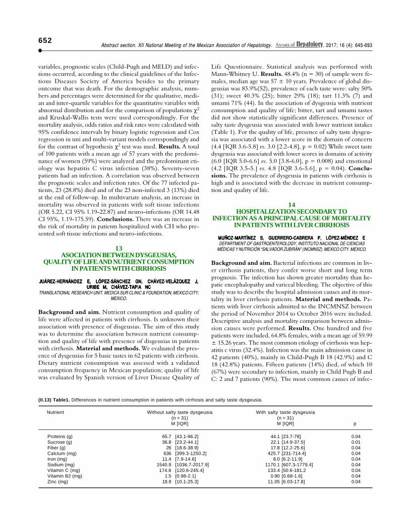
Life Questionnaire. Statistical analysis was performed withMann-Whitney U. Results. 48.4% (n = 30) of sample were fe-males, median age was 57 ± 10 years. Prevalence of global dis-geusias was 83.9%(52), prevalence of each taste were: salty 50%(31); sweet 40.3% (25); bitter 29% (18); tart 11.3% (7) andumami 71% (44). In the association of dysgeusia with nutrientconsumption and quality of life; bitter, tart and umami tastesdid not show statistically significant differences. Presence ofsalty taste dysgeusia was associated with lower nutrient intakes(Table 1). For the quality of life, presence of salty taste dysgeu-sia was associated with a lower score in the domain of concern(4.4 [IQR 3.6-5.8] vs. 3.0 [2.2-4.8], p = 0.02) While sweet tastedysgeusia was associated with lower scores in domains of activity(6.0 [IQR 5.0-6.6] vs. 5.0 [3.8-6.0], p = 0.008) and emotional(4.2 [IQR 3.5-5.] vs. 4.8 [IQR 3.6-5.6], p = 0.04). Conclu-sions. The prevalence of dysgeusia in patients with cirrhosis ishigh and is associated with the decrease in nutrient consump-tion and quality of life.
14HOSPITALIZATION SECONDARY TO
INFECTION AS A PRINCIPAL CAUSE OF MORTALITYIN PATIENTS WITH LIVER CIRRHOSIS
MUÑOZ-MARTÍNEZ S, GUERRERO-CABRERA P, LÓPEZ-MÉNDEZ EMUÑOZ-MARTÍNEZ S, GUERRERO-CABRERA P, LÓPEZ-MÉNDEZ EMUÑOZ-MARTÍNEZ S, GUERRERO-CABRERA P, LÓPEZ-MÉNDEZ EMUÑOZ-MARTÍNEZ S, GUERRERO-CABRERA P, LÓPEZ-MÉNDEZ EMUÑOZ-MARTÍNEZ S, GUERRERO-CABRERA P, LÓPEZ-MÉNDEZ EDEPARTMENT OF GASTROENTEROLOGY, INSTITUTO NACIONAL DE CIENCIAS
MÉDICAS Y NUTRICIÓN “SALVADOR ZUBIRÁN” (INCMNSZ), MEXICO CITY. MEXICO.
Background and aim. Bacterial infections are common in liv-er cirrhosis patients, they confer worse short and long termprognosis. The infection has shown greater mortality than he-patic encephalopathy and variceal bleeding. The objective of thisstudy was to describe the hospital admission causes and its mor-tality in liver cirrhosis patients. Material and methods. Pa-tients with liver cirrhosis admitted to the INCMNSZ betweenthe period of November 2014 to October 2016 were included.Descriptive analysis and mortality comparison between admis-sion causes were performed. Results. One hundred and fivepatients were included, 64.8% females, with a mean age of 59.99± 15.26 years. The most common etiology of cirrhosis was hep-atitis c virus (32.4%). Infection was the main admission cause in42 patients (40%), mainly in Child-Pugh B 18 (42.9%) and C18 (42.8%) patients. Fifteen patients (14%) died, of which 10(67%) were secondary to infection, mainly in Child Pugh B andC: 2 and 7 patients (90%). The most common causes of infec-
(II.13) Table1. Differences in nutrient consumption in patients with cirrhosis and salty taste dysgeusia.
Nutrient Without salty taste dysgeusia With salty taste dysgeusia(n = 31) (n = 31)M [IQR] M [IQR] p
Proteins (g) 65.7 [43.1-96.2] 44.1 [23.7-78] 0.04Sucrose (g) 36.8 [23.2-44.1] 22.1 [14.9-37.5] 0.01Fiber (g) 26 [18.6-38.9] 17.8 [12.2-25.6] 0.04Calcium (mg) 636 [399.3-1250.2] 425.7 [231-714.4] 0.04Iron (mg) 11.4 [7.9-14.6] 8.0 [6.2-11.9] 0.04Sodium (mg) 1540.9 [1036.7-2017.9] 1170.1 [607.3-1779.4] 0.04Vitamin C (mg) 174.6 [120.6-245.4] 133.4 [50.6-181.2 0.04Vitamin B2 (mg) 1.5 [0.98-2.1] 0.90 [0.68-1.6] 0.04Zinc (mg) 18.9 [10.1-25.3] 11.05 [6.03-17.8] 0.04

653Abstract section. XII National Meeting of the Mexican Association of Hepatology. , 2017; 16 (4): 645-693
tion were spontaneous bacterial peritonitis (n = 4) and pneu-monia (n = 3), p = 0.559. Ninety nine patients with a 6months follow-up were included in the survival analysis, thosewith hospital admission for infection died in the first 30 daysfollowing admission, with greater mortality among patientswith septic shock at admission, p = 0.020. There was no 6months mortality difference (p = 0.089), indicating that mor-tality is equal between infected and non-infected patients afterthe first month of hospitalization. Conclusions. Infection is themain reason of hospital admission in liver cirrhosis patients, theyshow high mortality in the first 30 days after admission. It’s im-portant to identify and treat infection in a timely and aggressivemanner in these patients. There is a necessity of more studies tostablish measures that can help to improve the prognosis.
15FREQUENCY OF ZINC DEFICIENCY
IN A PATIENT WITH CIRROSIS AND HEPATICENCEPHALOPATHY MANIFESTA IN EVALUATIONFOR ORTHOPEDIC HEPATIC TRANSPLANTATION
OF THE NATIONAL INSTITUTE OF MEDICALSCIENCE AND NUTRITION
VÁZQUEZ-DELGADILLO A, PINEDA-DEPAZ MR, VÁZQUEZ-ANAYA G,VÁZQUEZ-DELGADILLO A, PINEDA-DEPAZ MR, VÁZQUEZ-ANAYA G,VÁZQUEZ-DELGADILLO A, PINEDA-DEPAZ MR, VÁZQUEZ-ANAYA G,VÁZQUEZ-DELGADILLO A, PINEDA-DEPAZ MR, VÁZQUEZ-ANAYA G,VÁZQUEZ-DELGADILLO A, PINEDA-DEPAZ MR, VÁZQUEZ-ANAYA G,DÍAZ-HERNÁNDEZ HA, CASTRO-NARRO GEDÍAZ-HERNÁNDEZ HA, CASTRO-NARRO GEDÍAZ-HERNÁNDEZ HA, CASTRO-NARRO GEDÍAZ-HERNÁNDEZ HA, CASTRO-NARRO GEDÍAZ-HERNÁNDEZ HA, CASTRO-NARRO GE
DEPARTMENT OF GASTROENTEROLOGY, INSTITUTO NACIONAL DE CIENCIASMÉDICAS Y NUTRICIÓN “SALVADOR ZUBIRÁN”, MEXICO CITY, MEXICO.
Introduction. Several studies have suggested the relationshipbetween the pathophysiology of hepatic encephalopathy (EH),hepatic cirrhosis (HC) and decreased serum levels of zinc, as wellas zinc supplementation as a therapeutic option. It is, therefore,important to know the frequency of zinc deficiency in patientswith HC and manifest EH. Objectives. To determine the fre-quency of zinc deficiency in patients with HC and EH who arebeing evaluated for orthotopic liver transplantation (THO) atthe Instituto Nacional de Ciencias Médicas y Nutrición Salva-dor Zubirán (INCMNSZ). Material and methods. We in-cluded 45 randomly selected patients treated as an outpatientbasis at the INCMNSZ THO clinic over a period of one year(January to December, 2016), with the diagnosis of EH mani-fest according to the West Haven criteria. An observational, de-scriptive, transversal and comparative study was performed.Demographic, biochemical and clinical variables were obtained.For statistical analysis, SPSS v21 was used, taking as p < 0.05.Results. The mean age was 58 (47-63) males (53%). The pre-dominant etiology was HCV (30%), autoimmune etiology(20%), cryptogenic etiology and NASH (15%). The most fre-quent clinical stage of HD was II (69%), the most frequentChild Pugh-Turcotte stage was C (53%), MELD media 14 (12-18), ammonium 59 mg/dL (33-83). The frequency of zinc defi-ciency in the studied population of 80% and mean values was44 mg/dL (35.5-56.5), in patients with portosystemic short-cir-cuit zinc deficiency was 100%, Child Pugh-Turcotte C 95.5%(p 0.001), in EH III 93% (p 0.14). Conclusion. Patients withcirrhosis and grade II or III hepatic encephalopathy have highfrequency of zinc deficiency. It is higher in patients with porto-systemic shunting, decompensated patients and grade III en-fephalopathy. Diagnose and treat zinc deficiency couldcontribute to the clinical management of these patients.
16CAUSAL MICROORGANISMS OF BACTERIAL
PERITONITIS IN PATIENTS WITH CHRONIC HEPATICINSUFFICIENCY COMPLICATED WITH BACTERIAL
PERITONITIS
MELÉNDEZ-ANDRADE J, RÁBAGO-ESCOTO R,MELÉNDEZ-ANDRADE J, RÁBAGO-ESCOTO R,MELÉNDEZ-ANDRADE J, RÁBAGO-ESCOTO R,MELÉNDEZ-ANDRADE J, RÁBAGO-ESCOTO R,MELÉNDEZ-ANDRADE J, RÁBAGO-ESCOTO R,HIGUERA-DE LA TIJERA F, PÉREZ-HERNÁNDEZ JHIGUERA-DE LA TIJERA F, PÉREZ-HERNÁNDEZ JHIGUERA-DE LA TIJERA F, PÉREZ-HERNÁNDEZ JHIGUERA-DE LA TIJERA F, PÉREZ-HERNÁNDEZ JHIGUERA-DE LA TIJERA F, PÉREZ-HERNÁNDEZ J
SERVICIO DE GASTROENTEROLOGÍA, HOSPITAL GENERAL DE MÉXICO “DR. EDUARDOLICEAGA”, CIUDAD DE MÉXICO. MÉXICO.
Background. Spontaneous bacterial peritonitis (SBP) is themost frequent infection in cirrhotic patients; in Mexico, thereare no statistics regarding the bacterial etiological agents thatcause this infection. Objectives. To describe the most frequentmicroorganisms reported in cultures of peritoneal fluid of pa-tients with cirrhosis and SBP. Material and methods. A de-scriptive, observational, study that included patients withdiagnostic of cirrhosis and SBP defined by the American Associ-ation for the Study of liver diseases, as the presence of asciticfluid polymorphonuclear leukocyte counts greater than or equalto ≥ 250 cells/mm3, without a source of intra-abdominal infec-tion, treated at Hospital from January to December 2016. Re-sults. In the hospital records database of 2016, 16 patients withcirrhosis and SBP, 11 male (69%), 5 female (31%), Mean age 50years with standard deviation of ± 18 years of which 5 patients(31%) corresponded to their first episode of SBP and 11 patients(69%) with previous history of SBP. 8 ascitic cultures were neg-ative (50%) and 8 were positive (50%). The microorganismsidentified were 50% Escherichia coli, Staphylococcus aureus 37%,Staphylococcus epidermidis 13%, hepatorenal syndrome waspresent in 6%. Conclusions. The proportion of patients withrecurrent SPB events is very high; studies that evaluate antimi-crobial resistance in Mexican populations are required.
17SECONDARY VARICEAL BLEEDING (VB)
PROPHYLAXIS WITH PROPRANOLOL ADJUSTEDTO PONDERAL DOSE IN MEXICAN POPULATION
HERRERO-MACEDA MR, JUÁREZ-BARRIENTOS TE, GARCÍA-RUÍZ E,HERRERO-MACEDA MR, JUÁREZ-BARRIENTOS TE, GARCÍA-RUÍZ E,HERRERO-MACEDA MR, JUÁREZ-BARRIENTOS TE, GARCÍA-RUÍZ E,HERRERO-MACEDA MR, JUÁREZ-BARRIENTOS TE, GARCÍA-RUÍZ E,HERRERO-MACEDA MR, JUÁREZ-BARRIENTOS TE, GARCÍA-RUÍZ E,MEJÍA-LOZA SMEJÍA-LOZA SMEJÍA-LOZA SMEJÍA-LOZA SMEJÍA-LOZA S
HOSPITAL JUÁREZ DE MÉXICO, MEXICO CITY, MEXICO.
Background and aim. VB continues to be the complicationthat is most associated with mortality in 6 weeks in patientswith cirrhosis (10-20%), so the impact of early therapy, coupled withetiological treatment, with emphasis on prevention of recurrentVB is the main action to prolong survival and improve thequality of life of these patients. Less than 25% of HJM pa-tients tolerate the recommended dose of VB secondary prophy-laxis with variceal ligation plus non-selective beta-blockersNSBB (propranolol) according to current Baveno VI guidelines.Objective. To adjust the dose of NSBB (propranolol) at weightdose to patients with intolerability. As secondary objective thepresence of rebleeding was evaluated in the following 3 months.Material and methods. Ambispective case-control study inwhich the outpatient from January 1, 2016 to March 31, 2017 >18 years, with a history of variceal hemorrhage, who were treatedwith variceal ligation, were reviewed (0.3-1 mg/kg/d), assessing thetolerability of the treatment as well as the desired beta blockade. In-
654Abstract section. XII National Meeting of the Mexican Association of Hepatology. , 2017; 16 (4): 645-693
tolerability is defined as refractory headache, hypotension (TAM <60 mmHg) or FC < 50 lpm and vasomotor syncope. HCV was ex-cluded. Descriptive IBM-SPSSv2.1 statistical analysis and relativerisks. Results. A total of 378 files with cirrhosis diagnosis were re-viewed: 174 were discarded by endoscopic report of small varices orabsence of varicose oesophageal veins. 204 patients reported largevarices and 99 (48%) had documented variceal hemorrhage, all ofwhich were treated with variceal ligation. 45 Child-Pugh C de-compensated for which NSBB was not offered, with a total of 19patients with adjusted dose: 42% (8) female. Average age 53 years.Median FC61x’ (50-90) TAM70mmHg (60-80). Average dose0.54 mgkgd (0.3-1). It has been documented resorting to 14% ofpatients with full doses of NSBB. OR1.1 Conclusions. The ad-justment to the dose of BBNS to the Mexican population, whichpresumably has a lower weight, could be safe without increasingthe risk of rebleeding, according to the population studied in theHJM. Although studies involving a larger population with hetero-geneous characteristics are required.
18HEMORRHAGIC PANCREATITIS AND INTESTINAL
ISCHEMIA SECONDARY TO THROMBOSIS PORTO-MESENTERIC VEIN IN PATIENT WITH CIRRHOSIS
MONTES DE OCA SALINAS F,* HIGUERA DE LA TIJERA M,*MONTES DE OCA SALINAS F,* HIGUERA DE LA TIJERA M,*MONTES DE OCA SALINAS F,* HIGUERA DE LA TIJERA M,*MONTES DE OCA SALINAS F,* HIGUERA DE LA TIJERA M,*MONTES DE OCA SALINAS F,* HIGUERA DE LA TIJERA M,*ARISTI URISTA G,** MOTOLA KUBA M*ARISTI URISTA G,** MOTOLA KUBA M*ARISTI URISTA G,** MOTOLA KUBA M*ARISTI URISTA G,** MOTOLA KUBA M*ARISTI URISTA G,** MOTOLA KUBA M*
*UNIDAD DE GASTROENTEROLOGIA, **UNIDAD DE PATOLOGÍA, HOSPITAL GENERALDE MÉXICO, CIUDAD DE MÉXICO, MÉXICO.
Background. Portal thrombosis associated with hepatic cirrho-sis appears silently, presenting manifestations such as hemor-rhage, abdominal pain, ascites or deterioration in liver function.Objective. Report the case of hemorrhagic thrombus compli-cations in a patient with liver cirrhosis. Case report. Female 37years old with liver cirrhosis of unknown etiology diagnosed in2014, history of ascites decompensation and presence of esopha-geal varices with variceal ligation. We entered our unit for gen-eralized abdominal pain of 72 h of evolution, with no evidenceof digestive hemorrhage and hemoglobin of 2.9 g/dL. Endosco-py with remaining large varices without red signs, post-ligationchanges and severe portal hypertensive gastropathy. During herhospital stay persisted with abdominal pain, bacterioascitis sec-ondary to streptococcus of the Viridans group was identifiedand empirical coverage with ertapenem was obtained with mor-bid evolution. She presented neurological deterioration and re-fractory shock to vasopressors, resulting in the death of thepatient. An autopsy was performed which revealed coagulativenecrosis of the liver, portal vein vascular occlusion, superior me-senteric, multiple subacute splenic infarctions, acute ischemicenteritis and acute hemorrhagic pancreatitis. Discussion. Thecase corresponds to a patient with decompensated chronic liver
disease, persistent abdominal pain who developed multiorganfailure with thrombosis of venous portal splenic vein, superiormesenteric and pancreatic hemorrhage with a fatal outcome.Coagulation abnormalities in patients with cirrhosis are general-ly manifested with hemorrhage, despite the presence of throm-botic-hemorrhage complications at different levels in awell-recognized phenomenon. The most common site ofthrombosis is the portal vein and mesenteric veins, ranging from8-16%. Therefore, thrombotic complications should always beconsidered in patients with hepatopathy with acute abdominalpain. This work has not been fully or partially sponsored by anygovernmental or commercial system.
19ACUTE ON CHRONIC LIVER FAILURE (ACLF), ONE
YEAR EXPERIENCE IN THE DEPARTMENT OFGASTROENTEROLOGY OF HOSPITAL JUÁREZ DE
MÉXICO
AGUILAR-MORENO RU, FERNÁNDEZ-MARTÍNEZ NC,AGUILAR-MORENO RU, FERNÁNDEZ-MARTÍNEZ NC,AGUILAR-MORENO RU, FERNÁNDEZ-MARTÍNEZ NC,AGUILAR-MORENO RU, FERNÁNDEZ-MARTÍNEZ NC,AGUILAR-MORENO RU, FERNÁNDEZ-MARTÍNEZ NC,VELASCO-SANTIAGO YM, RIVERA-FLORES AG, BARRAZA-ORTIZ DB,VELASCO-SANTIAGO YM, RIVERA-FLORES AG, BARRAZA-ORTIZ DB,VELASCO-SANTIAGO YM, RIVERA-FLORES AG, BARRAZA-ORTIZ DB,VELASCO-SANTIAGO YM, RIVERA-FLORES AG, BARRAZA-ORTIZ DB,VELASCO-SANTIAGO YM, RIVERA-FLORES AG, BARRAZA-ORTIZ DB,
CANO-CONTRERAS AD, DURÁN-ROSAS C, LLORENTE-RAMÓN A,CANO-CONTRERAS AD, DURÁN-ROSAS C, LLORENTE-RAMÓN A,CANO-CONTRERAS AD, DURÁN-ROSAS C, LLORENTE-RAMÓN A,CANO-CONTRERAS AD, DURÁN-ROSAS C, LLORENTE-RAMÓN A,CANO-CONTRERAS AD, DURÁN-ROSAS C, LLORENTE-RAMÓN A,OVIEDO-MAGLIONE MA, RAMOS-AGUILAR GA, MEJÍA-LOZA SMI,OVIEDO-MAGLIONE MA, RAMOS-AGUILAR GA, MEJÍA-LOZA SMI,OVIEDO-MAGLIONE MA, RAMOS-AGUILAR GA, MEJÍA-LOZA SMI,OVIEDO-MAGLIONE MA, RAMOS-AGUILAR GA, MEJÍA-LOZA SMI,OVIEDO-MAGLIONE MA, RAMOS-AGUILAR GA, MEJÍA-LOZA SMI,
ZAMARRIPA-DORSEY FZAMARRIPA-DORSEY FZAMARRIPA-DORSEY FZAMARRIPA-DORSEY FZAMARRIPA-DORSEY FDEPARTMENT OF GASTROENTEROLOGY, HOSPITAL JUÁREZ DE MÉXICO, MEXICO
CITY. MEXICO.
Introduction and objectives. Acute on chronic liver failure(ACLF) is a common patology in our setting. There are currentlyno studies in Mexico on the epidemiological panorama of this dis-ease. The objective of this work is to present the frequency andbehavior of the ACLF. Material and methods. Type of study:Descriptive, transversal, observational. Patients with hepatic cir-rhosis admitted to hospital for acute decompensation or ACLF, inthe Department of Gastroenterology from March 1, 2016 to Feb-ruary 28, 2017. Variables: Age, sex, precipitating factors, patientswith acute decompensation, ACLF, ACLF grade and mortality.Results. A total of 211 patients, aged between 27 to 88 years (80women and 131 men) with diagnosis of hepatic cirrhosis withacute decompensation or ACLF, 164 presented acute decompen-sation (77.73%) and 47 ACLF (22.27%). Of the patients withACLF it was observed that 46.81% presented ACLF grade I,42.55% ACLF grade II, and 10.64% ACLF grade III. The mainprecipitating factor in patients with acute decompensation wasgastrointestinal bleeding in 67.68% while in patients with ACLFwere infections in 63.83%. The most frequent organic failure wasthe renal failure, with 65.96% of patients with ACLF, the generalmortality rate was 18.01%, of the deceased patients 39.47% corre-sponded to acute decompensation and 60.53% to ACLF. Con-clusions. The Acute on Chronic Liver failure is a frequent inpatients with hepatic cirrosis, approximately 2 out of 10 hospital-ized cirrhotic patients will present ACLF (22.27%), approachingthe frequency of 32.4% that was observed in the CANONICstudy. The main precipitating factor in patients with ACLF wereinfections, observing that the most common organic failure inACLF was the renal, and this is of great relevance for the treat-ment approach in patients with hepatic cirrhosis, 6 out of 10 pa-tients who died had ACLF, So it is of importance to identifypatients with ACLF from their admission to treat the precipitat-ing factor and manage the organic failures that will impact on theprognosis of the patient.
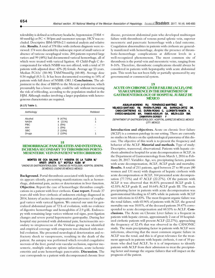
(II.17) Table 1.
Aethiology Rebleed
Alcohol 9 (47%) 1NASH 4 (21%) 1PBC 1 (5%) -AIH 1 (5%) -SBC 2 (10%) -Drugs 2 (10%) 1
655Abstract section. XII National Meeting of the Mexican Association of Hepatology. , 2017; 16 (4): 645-693
20LONG TERM CLINICAL AND REAL-WORLD
EXPERIENCE WITH A SLOW RELEASEFORMULATION OF PIRFENIDONE IN PATIENTS
WITH ADVANCED HEPATIC FIBROSIS. PROMETEOSTUDY
POO JL,* MUÑOZ L,POO JL,* MUÑOZ L,POO JL,* MUÑOZ L,POO JL,* MUÑOZ L,POO JL,* MUÑOZ L,††††† TORRE A, TORRE A, TORRE A, TORRE A, TORRE A,‡‡‡‡‡ CRUZ M, CRUZ M, CRUZ M, CRUZ M, CRUZ M,§§§§§ RIVERA JF, RIVERA JF, RIVERA JF, RIVERA JF, RIVERA JF,||||||||| | PATIÑO A, PATIÑO A, PATIÑO A, PATIÑO A, PATIÑO A,¶¶¶¶¶
BOSQUES F,** VELÁZQUEZ A,BOSQUES F,** VELÁZQUEZ A,BOSQUES F,** VELÁZQUEZ A,BOSQUES F,** VELÁZQUEZ A,BOSQUES F,** VELÁZQUEZ A,†††††††††† HERNÁNDEZ L,* AGUILAR JR HERNÁNDEZ L,* AGUILAR JR HERNÁNDEZ L,* AGUILAR JR HERNÁNDEZ L,* AGUILAR JR HERNÁNDEZ L,* AGUILAR JR‡‡‡‡‡‡‡‡‡‡
*SAN JERONIMO CLINIC, MEXICO CITY, MEXICO.†UNIVERSITY HOSPITAL, MONTERREY, MEXICO.
‡INSTITUTO NACIONAL DE CIENCIAS MÉDICAS Y NUTRICIÓN “SALVADOR ZUBIRÁN”,MEXICO CITY, MEXICO.
§HGZ NO. 4, IMSS CELAYA, MEXICO.||SPANISH HOSPITAL, MEXICO CITY, MEXICO.
¶ SURGICAL MEDICAL CENTER, MEXICO.** CELAYA, ZAMBRANO HELLION HOSPITAL, ITESM, MONTERREY, MEXICO.
††INTERNAL MEDICINE CLINIC, PUEBLA.‡‡LIVER CLINIC, MEXICO CITY. MEXICO.
Introduction. Pirfenidone (PF), a drug with potent anti-in-flammatory, anti-oxidant and antifibrotic effects has grantedmarketing authorization by EMA, FDA and Mexican COFE-PRIS, for the treatment of idiopathic pulmonary fibrosis. How-ever, few studies have focused on its clinical utilization inpatients with hepatic fibrosis of diverse etiology. Aim. To de-scribe the clinical and real-world experience with a slow releasePF formulation (KitosCell LP®) in patients with liver fibrosis inMexico. Material and methods. A total of 139 patients usingKitosCell LP® (600 mg bid) were identified from 5 centers; 94subjects received the formulation for less than one year and 45for at least 1 year or more; mean results are reported at M0 andM12. Efficacy was assessed using METAVIR scoring charts, bynon-invasive tests (APRI, Fibrotest) or by elastography (Fibros-can or ARFI); variations greater than or equal to 0.5 units inAPRI, 0.10 units or 5 kPa or 1 point in METAVIR, were con-sidered as significant. Baseline fibrosis, according to Fibrotestwas F4 in 77.8%, F3-F4 in 4.4%, and F3 in 17.8%. Results.44.4% were women, 60 ± 9 years old; 31.1% with metabolicdamage, 26.6% viral, 24.4% alcohol, 15.5% autoimmune and2.2% others. Mean biochemical values were: Hb (g/dL): 14.0 ±2.1, 13.6 ± 1.5; Platelets (x 103): 97 ± 6, 97 ± 8; TB (mg/dL):2.0 ± 0.8, 1.3 ± 0.6; Albumin (g/dL): 3.5 ± 0.6, 3.5 ± 0.5; PT(INR): 1.3 ± 0.4, 1.2 ± 0.3; ALT (IU/L): 53 ± 4.48 ± 3; AST(IU/L): 69 ± 7, 53 ± 3. In relation to fibrosis we detected: Interms of safety, 8 patients (11%) reported transient burning ornausea, 7% photosensitivity. In 4/45 patient’s progression tomortality (8.8%) was observed due to variceal hemorrhage (2),ascites (1) or sepsis (1). Conclusion. Administration of KitoscellLP at 12 months in patients with hepatic fibrosis was well toler-ated and associated with encouraging efficacy results.This work has been partially supported by Cellpharma. (Accessto study drug in some centers).
2120 YEARS OF TREATMENT WITH PENTOXIFYLLINEIN PATIENTS WITH HEPATIC CIRRHOSIS CAUSED
BY VIRUS HEPATITIS C
JIMÉNEZ-LUEVANO MA, RODRÍGUEZ-VILLA P, RAMÍREZ-FLORES S,JIMÉNEZ-LUEVANO MA, RODRÍGUEZ-VILLA P, RAMÍREZ-FLORES S,JIMÉNEZ-LUEVANO MA, RODRÍGUEZ-VILLA P, RAMÍREZ-FLORES S,JIMÉNEZ-LUEVANO MA, RODRÍGUEZ-VILLA P, RAMÍREZ-FLORES S,JIMÉNEZ-LUEVANO MA, RODRÍGUEZ-VILLA P, RAMÍREZ-FLORES S,JIMÉNEZ-PARTIDA MA, CERVANTES-RODRÍGUEZ G,JIMÉNEZ-PARTIDA MA, CERVANTES-RODRÍGUEZ G,JIMÉNEZ-PARTIDA MA, CERVANTES-RODRÍGUEZ G,JIMÉNEZ-PARTIDA MA, CERVANTES-RODRÍGUEZ G,JIMÉNEZ-PARTIDA MA, CERVANTES-RODRÍGUEZ G,
FRANCO-TOPETE R, BRAVO-CUELLAR AFRANCO-TOPETE R, BRAVO-CUELLAR AFRANCO-TOPETE R, BRAVO-CUELLAR AFRANCO-TOPETE R, BRAVO-CUELLAR AFRANCO-TOPETE R, BRAVO-CUELLAR ACLÍNICA DE HÍGADO, HOSPITAL REGIONAL “VALENTÍN GÓMEZ FARÍAS” (ISSSTE),
ZAPOPAN, JALISCO, MÉXICO.
Introduction. Cirrhosis results from chronic liver disease, andit is characterized by advanced fibrosis, scarring, and formationof regenerative nodules leading to architectural distortion. InMexico, it is the fourth leading cause of death; alcohol con-sumption remains the main etiology, followed by viral hepatitis,in which hepatitis C virus (HCV) infection is the most com-mon. Approximately 180 million people worldwide have chron-ic HCV infection, with a prevalence of 1.4% in Mexico; and ithas an average age of presentation at 42 years. Although thenatural history is highly variable, it is estimated that between20-30% of HCV infected Mexicans will progressed to cirrhosisin the next 3 decades. The median survival of patients withcompensated cirrhosis is much longer than in patients with de-compensation and is about 9 years; however, they will eventu-ally developed decompensated cirrhosis and death, unless a livertransplant is performed. Objective. To evaluate the response of159 cirrhotic patients by virus hepatitis C with Pentoxifline af-ter 20 years of treatment. Material and methods. A retrospec-tive, analytical study with a sample of 159 patients diagnosedwith hepatitis C virus cirrhosis treated with pentoxifylline dur-ing 7 and 20 years. Results. It was observed that 26 (16%) ofthe 159 patients with virus C cirrhosis treated with pentoxifyl-line had a 20-year survival without progress of Child A or Bstage; 34 of them died during the course of treatment due totheir comorbidities and in 99 of the patients the follow-up waslost, having a record of survival up to 7 years after treatment be-gan. It was obtained a OR of 2.5. Conclusion. Pentoxifyllinehas shown to have a beneficial effect in these patients, whichmotivates the use of this drug in patients with cirrhosis to im-prove survival up to 20 years.
22CHANGES IN MAGNETIC RESONANCE
SPECTROSCOPY IN PATIENTS WITH CIRROSIS ANDINFECTION
ASTUDILLO-GARCÍA F, MÁRQUEZ-GUILLÉN E, MACÍAS RODRÍGUEZ RU,ASTUDILLO-GARCÍA F, MÁRQUEZ-GUILLÉN E, MACÍAS RODRÍGUEZ RU,ASTUDILLO-GARCÍA F, MÁRQUEZ-GUILLÉN E, MACÍAS RODRÍGUEZ RU,ASTUDILLO-GARCÍA F, MÁRQUEZ-GUILLÉN E, MACÍAS RODRÍGUEZ RU,ASTUDILLO-GARCÍA F, MÁRQUEZ-GUILLÉN E, MACÍAS RODRÍGUEZ RU,ROMERO-SÁNCHEZ GT, TORRE-DELGADILLO AROMERO-SÁNCHEZ GT, TORRE-DELGADILLO AROMERO-SÁNCHEZ GT, TORRE-DELGADILLO AROMERO-SÁNCHEZ GT, TORRE-DELGADILLO AROMERO-SÁNCHEZ GT, TORRE-DELGADILLO A
DEPARTAMENT OF GASTROENTEROLOGY, INSTITUTO NACIONAL DE CIENCIASMÉDICAS Y NUTRICIÓN “SALVADOR ZUBIRÁN”. MEXICO CITY. MEXICO.
Background. Hepatic encephalopathy (HE) is a cirrhosis compli-cation, with infections being one of the major triggers. Protonmagnetic resonance spectroscopy (1H-MRS) is a technique that al-lows in vivo quantification of different metabolites concentrationsin specific tissues. Using 1H-MRS in patients with HE, changes innormal concentration of metabolites have been demonstrated, suchas an increase in glutamine and a decrease in myo-inositol, second-ary to increased cerebral ammonia and osmotic stress derived fromit. Objective: To assess changes in cerebral metabolites of infected
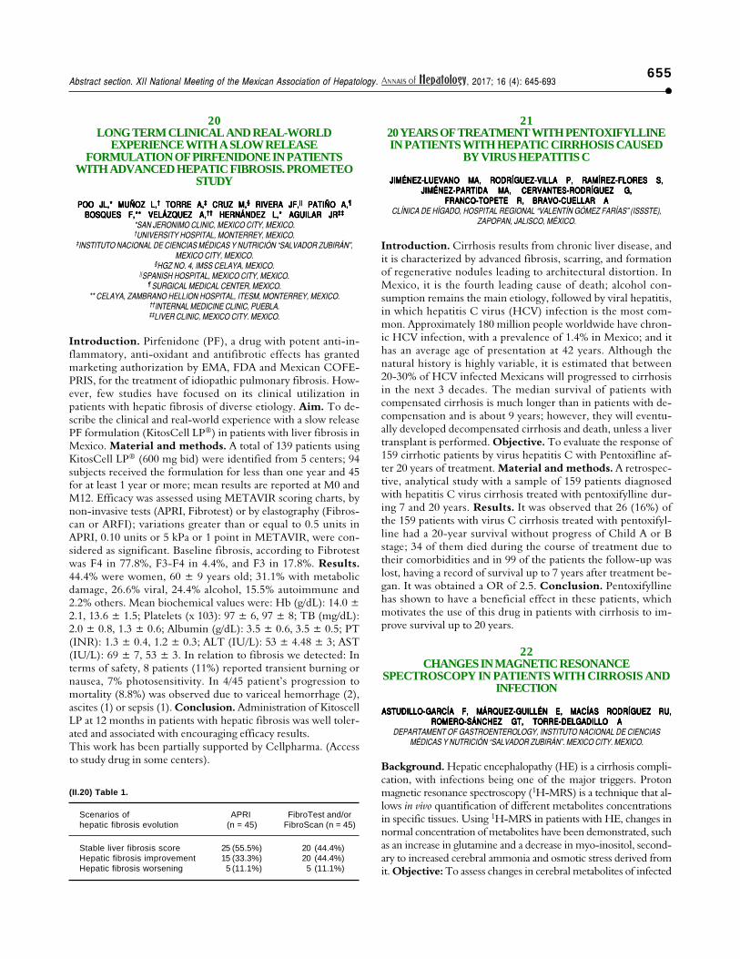
(II.20) Table 1.
Scenarios of APRI FibroTest and/orhepatic fibrosis evolution (n = 45) FibroScan (n = 45)
Stable liver fibrosis score 25 (55.5%) 20 (44.4%)Hepatic fibrosis improvement 15 (33.3%) 20 (44.4%)Hepatic fibrosis worsening 5 (11.1%) 5 (11.1%)
656Abstract section. XII National Meeting of the Mexican Association of Hepatology. , 2017; 16 (4): 645-693
cirrhotic patients by 1H-MRS evaluation. Materials and meth-ods. Case-control study. Participants were divided into threegroups: non-infected compensated cirrhotics (group 1, n = 10), in-fected cirrhotics (with or without encephalopathy, group 2, n =10) and healthy individuals (group 3, n = 10). Participantes wereevaluated by using 1H-MRS (Simmens® equipment, Tarquin®
software) with metabolites measurement in territory of basal gan-glia and white matter of temporal region. U Mann Whitney testwas used to evaluate differences between two groups, Student’s Tfor related independent samples, χ2 for qualitative variables, Pear-son’s correlation coefficient to establish a relationship between twovariables, and linear regression analysis was performed to evaluatethe relationship between variables. A value of p < 0.05 was consid-ered as statiscally significant. Results. The main infections ingroup 2 were spontaneous bacterial peritonitis in 40% and urinarytract infection in 30% of the patients. In the analysis of metabolitesby 1H-MRS, a significant decrease of myo-inositol was observed inpatients in group 1 compared to group 3 and in group 2 vs. group 3(p < 0.001), this mainly in the temporal region. Additionally, a sig-nificant difference of N-Acetylaspartate was observed betweenhealthy and cirrhotic patients infected (p < 0.05). The decrease inthe myo-inositol peak correlates with the Child-Pugh classifica-tion. Conclusion. Acute infection in patients with cirrhosischanges the concentration of brain metabolites (mainly N-Acety-laspartate and myo-inositol) and may be a factor related to the de-velopment of hepatic encephalopathy.
23ESTIMATION OF HEPATIC FIBROSIS BY SHEARWAVE ELASTOGRAPHY IN MEXICAN PATIENTS
WITH CHRONIC LIVER DISEASE. PANDORA STUDY
POO JL,* HERNÁNDEZ L,* ZAPATA C,** MUÑOZ L**POO JL,* HERNÁNDEZ L,* ZAPATA C,** MUÑOZ L**POO JL,* HERNÁNDEZ L,* ZAPATA C,** MUÑOZ L**POO JL,* HERNÁNDEZ L,* ZAPATA C,** MUÑOZ L**POO JL,* HERNÁNDEZ L,* ZAPATA C,** MUÑOZ L***SAN JERONIMO CLINIC, MEXICO CITY. MEXICO.
**UNIVERSITY HOSPITAL, MONTERREY, NUEVO LEÓN. MEXICO.
Introduction. Elastography has emerged as the noninvasivemethod of reference for estimating liver fibrosis by using elasticshear waves emitted manually or automatically by the vibratorattached to the ultrasound transducer probe. Methods includetransient elastography (Fibroscan) and the most recent ARFI-based ultrasound devices, allowing evaluation of patients withobesity and ascites. Additionally, Shear Wave Elastography(SWE) has the advantages of providing anatomic B-mode USimages and elastographic color maps according to the degree ofstiffness in specific regions of interest. However, there are fewMexican studies describing its experience. Objective. To de-scribe the prevalence, type and grade of hepatic fibrosis in agroup of patients from two Mexican centers using a SWE equip-ment. Material and methods. A total of 184 patients (site 1 =104, site 2 = 80) were studied, with a mean age of 56 ± 13years; 57% were women, with suspected chronic liver disease (in42%) or cirrhosis (in 58%). Causes of liver damage includedNASH in 35.8%, HCV 32.8%, autoimmune 21.2%, alcoholic9.5% and other 1%. Patients were evaluated with an Aixplorerultrafast US system (Supersonic Imagine) equipped with anSXC 6-1 convex transducer. Measurements (at least 5) wereperformed in fasting conditions, in apnea, at the level of seg-ments 6 and 7, with results expressed in kilo Pascals and m/sec.Optimal reliability criteria was met with values lower 0.3, calcu-
lated by the interquartile range/median value (IQR/M). Hepaticrigidity was estimated according to the semi-quantitativeMETAVIR scoring chart. Those patients with F4 scores werefurther sub classified by using the 7 grades-METAVIR extendedsystem to evaluate risk of esophageal varices, variceal bleeding,sepsis, cancer or increase mortality. Results. The study was reli-able in 179 subjects (97%), including 6 patients with ascites andvarious degrees of obesity, with BMI ranging between 16.2 and40. The findings demonstrated a wide range of fibrosis from F0in 10.1%, F1 in 19.6%, F2 in 17.9%, F3 in 11.2, F4 in 41.3%.Among subjects with F4 (n = 73) scores, 30 were classified asF4.1 (41%), 35 in F4.2 (48%) and 9 in F4.3 (12%). Conclu-sion. Liver stiffness evaluation with an Aixplorer equipment(SW-Elastography) proved to be a simple, high applicability andreliable method for the estimation of liver fibrosis in patientswith chronic liver disease.
24RELATION BETWEEN DETACHMENT TO MEDICAL
CHECKUP AND COMPLICATIONS IN PATIENTSWITH CIRRHOSIS OF THE CENTER FOR RESEARCH
ON LIVER DISEASES AND GASTROENTEROLOGY OFPACHUCA, HIDALGO
TÉLLEZ-JAÉN S,* CONTRERAS-OMAÑA R**TÉLLEZ-JAÉN S,* CONTRERAS-OMAÑA R**TÉLLEZ-JAÉN S,* CONTRERAS-OMAÑA R**TÉLLEZ-JAÉN S,* CONTRERAS-OMAÑA R**TÉLLEZ-JAÉN S,* CONTRERAS-OMAÑA R***MEDICINE SCHOOL OF UAEH, PACHUCA, HIDALGO, MEXICO.
**CENTER FOR RESEARCH ON LIVER DISEASES AND GASTROENTEROLOGY,PACHUCA, HIDALGO, MEXICO.
Background. Patients with decompensated liver cirrhosispresent as main complications, hepatic insufficiency, jaundice,ascites, variceal hemorrhage, or hepatic encephalopathy. InMexico, there aren’t evidence of the relationship between medi-cal checkup, and complications that can exist in patients withcirrhosis, in the private practice. Aim. Determine the relation-ship between detachment from the medical checkup and com-plications of patients with cirrhosis in the Center for Researchon Liver Diseases and Gastroenterology (CIEHG) Pachuca,Hidalgo. Material and methods. Observational, retrospective,descriptive and transversal study in which reviewed and collect-ed data from patients with cirrhosis, attended by the CIEHG,period 2014-2016, getting 65 records of patients with this pa-thology, who maintained a control in this center in this period.Results. Of the 65 patients with cirrhosis, 60% (n = 39) wom-en, and 40% (n = 26) men, with a mean of 61.6 years, a medi-an of 61, and a mode of 60. We were found, as etiologies:NASH 49.23% = 32), Alcohol 20% (n = 13), chronic infectionof HCV 13.85% (n = 9), primary biliary cholangitis 9.23% (n= 6), autoimmune hepatitis 6.15% (n = 4) and primary scleros-ing cholangitis 1.54% (n = 1). Of these patients, 21.54% (n =14) presented Child Pugh A, 64.61% (n = 42) Child Pugh B,and 13.85% (n = 9) Child Pugh C. Within the attachment tofollow up, 36.92% (n=24) maintain it adequately, while63.08% (n = 41) present detachment; of which 7.32% (n = 3)stopped following of the first medical checkup, 46.34% (n =19), 46.34% (n = 19), after a lapse between consultation ofone month, and 46.34% (n = 19) after a lapse of more thanthree months and reentry to this center with decompensatedcirrhosis presenting 42.1% (n = 8) Hepatic Encephalopathy,21.06% (n = 4) Variceal hemorrhage, 10.53% (n = 2) Sponta-
657Abstract section. XII National Meeting of the Mexican Association of Hepatology. , 2017; 16 (4): 645-693
neous Bacterial Peritonitis, and other causes of decompensation26.31% (n = 5). Conclusions. This study show that, of thepatients with cirrhosis, 36.92% maintained a good follow-up,and 46.34% of the patients with detachment with a period ofmore than three months, come back with decompensation data,the main cause of which was hepatic encephalopathy, followedOf Variceal Hemorrhage, and other causes such as Ascites andDehydration. Given the above, more studies are required to val-idate outcome and to propose attachment strategies.
25FREQUENCY OF HEPATOPULMONARY SYNDROMEIN MEXICAN PATIENTS WITH HEPATIC CIRRHOSIS
SUDER-CASTRO LS, ÁLVAREZ LÓPEZ F, VELARDE RUIZ VELASCO JA,SUDER-CASTRO LS, ÁLVAREZ LÓPEZ F, VELARDE RUIZ VELASCO JA,SUDER-CASTRO LS, ÁLVAREZ LÓPEZ F, VELARDE RUIZ VELASCO JA,SUDER-CASTRO LS, ÁLVAREZ LÓPEZ F, VELARDE RUIZ VELASCO JA,SUDER-CASTRO LS, ÁLVAREZ LÓPEZ F, VELARDE RUIZ VELASCO JA,HERNÁNDEZ DEL RÍO JE, ALDANA LEDESMA JMHERNÁNDEZ DEL RÍO JE, ALDANA LEDESMA JMHERNÁNDEZ DEL RÍO JE, ALDANA LEDESMA JMHERNÁNDEZ DEL RÍO JE, ALDANA LEDESMA JMHERNÁNDEZ DEL RÍO JE, ALDANA LEDESMA JM
HOSPITAL CIVIL “FRAY ANTONIO ALCALDE”. GUADALAJARA, JALISCO. MEXICO.
Background. The reported prevalence of hepatopulmonarysyndrome (SHP) in patients with hepatic cirrhosis (CH) is het-erogeneous. Objective. Determine frequency, severity and fre-quent clinical characteristics of SHP in Mexican patients withHC, and utility of pulse-oximetry for SHP diagnosis. Materialand methods. We carried out an analytical cross-sectionalstudy at the Gastroenterology Service of Hospital Civil “FrayAntonio Alcalde”, May 2015 to June 2016. Including patients ≥18 years of age, with CH of any etiology, hospitalized for anycause. Excluded patients with respiratory disease due to othercauses, those with hemoglobin < 7 g/dL, patients with chest X-ray with pulmonary parenchyma or pleural involvement at in-clusion. Oxygen saturation (SatO2) measured with digital pulseoximetry in sedestation and supine decubitus; Arterial blood gasanalysis performed if had SatO2 ≤ 94%, and patients with ahigh arterial alveolus gradient (PA-at O2 ≥ 15 mmHg or ≥ 20mmHg if age > 64 years) were submitted to contrast-enhancedtransthoracic echocardiography (ECCT). Patients with left atrialopacification in ≥ 4 heart beats after the initial appearance ofright atrial contrast, in absence of intra-cardiac communicationswere considered with SHP, which was classified as mild withPaO2 ≥ 80 mmHg, moderate PaO2 ≥ 60 and < 80 mmHg, se-vere PaO2 ≥ 50 and < 60 mmHg and very severe with PaO2 <50 or < 300 mmHg breathing 100% oxygen. Results. 63 pa-tients were included, men 88%, mean age 51.1 ± 11.7 years.CH was alcoholic 69.5%, hepatitis C virus (HCV) 11%, alcohol/HCV 6.8%, non-alcoholic fatty liver disease 8% and idiopathic4% other 2%. Child Pugh (CP) 9.4 ± 1.6 points: 2 (3%) A, 29(45%) B, 33 (52%) in C. Mean MELD 18.2 ± 7.6. I9 (30%) pa-tients SatO2 ≤ 94% was identified, of which 16 patients had el-evated PA-to O2, 13 (20.3%) patients had positive ECCT forSHP. SHP patients, 50% reported dyspnoea, 19% were platyp-nea, 19% digital clubbing, and 12% showed ordeoxy. The sensi-tivity of SatO2 to diagnose SHP was 100% and specificity was88.2%, positive predictive value was 68.4% negative predictivevalue was 100%. A Pearson correlation was performed to deter-mine if SHP severity associated with CH severity by CP, Rho of0.23 (p = 0.43) MELD of 0.15 (p = 0.62). Conclusions. Fre-quency of SHP in Mexican patients with CH was 20.3%, simi-lar to that reported in the literature, 55% was moderate SHP.The most common symptom was dyspnea. SatO2 is very sensi-
tive but less specific to detect SHP in CH and because of itshigh PPV, it is possible to safely rule out severe SHP by restrict-ing the use of more expensive and invasive tests for patientswith significant hypoxemia. There was no association betweenCH severity and SHP severity.
26VALIDATION OF THE CLIF-C-ACLF SCORE TO
PREDICT 28-DAY MORTALITY IN HOSPITALIZEDMEXICAN CIRRHOTIC PATIENTS
MAYORGA-MARÍN T, PÉREZ-HERNÁNDEZ JL, HIGUERA-DE LA TIJERA FMAYORGA-MARÍN T, PÉREZ-HERNÁNDEZ JL, HIGUERA-DE LA TIJERA FMAYORGA-MARÍN T, PÉREZ-HERNÁNDEZ JL, HIGUERA-DE LA TIJERA FMAYORGA-MARÍN T, PÉREZ-HERNÁNDEZ JL, HIGUERA-DE LA TIJERA FMAYORGA-MARÍN T, PÉREZ-HERNÁNDEZ JL, HIGUERA-DE LA TIJERA FGASTROENTEROLOGY DEPARTMENT. HOSPITAL GENERAL DE MÉXICO “DR.
EDUARDO LICEAGA”, MEXICO CITY, MEXICO.
Background. Fifteen percent of the patients with cirrhosis de-velop complications each year. The severity of the complicationsis strongly related to the hepatic reservoir function and the liv-er’s structural lesion. Decompensated cirrhosis can lead to re-duction of the patient’s life expectancy, organ failure andshort-term mortality. The CLIF-C-ACLF Acute Decompensa-tion score (CLIF-C ACLF) is a score that predicts the 30 and90-day mortality. There is not enough national data on the ac-curacy of this prognostic score. Objective. To validate the ac-curacy of CLIF-C-ACLF to predict 28-day mortality inMexican patients with cirrhosis and acute-on-chronic liver fail-ure (ACLF). Material and methods. Observational analyticprospective study that evaluated 75 consecutive patients withliver disease between October 2016 and March 2017. At admis-sion, the CLIF-C ACLF score was assessed. Results. The meanage was 54.6 ± 12.51 year-old, 57.3% females. The cause of cir-rhosis was alcoholic liver disease in 49.3% and nonalcoholic fat-ty liver disease in 12% of the patients. The major complicationswhere hepatic encephalopathy (42%) and ascites (24%). Six pa-tients (8%) did not met the criteria for ACLF, 27 (36%) had ACLFgrade I, 28 (37.3%) had ACLF grade II and 14 (18.7%) ACLF gradeIII. The 28-days mortality was 46.4% for the ACLF patients incomparison to 16.7% who did not meet ACLF criteria. The areaunder the curve to predict 28-day mortality was 0.663 ± 0.062.Conclusion. The CLIF-C-ACLF score demonstrated validityto predict 28-day mortality in our population.
27PREDICTIVE FACTORS OF BLEEDING RELATED TOPOST-BANDING ULCER FOLLOWING ENDOSCOPICVARICEAL LIGATION IN CIRRHOTIC PATIENTS: A
CASE-CONTROL STUDY
MAYORGA-MARÍN T, SOTO-HERNÁNDEZ K, RÁBAGO-ESCOTO R,MAYORGA-MARÍN T, SOTO-HERNÁNDEZ K, RÁBAGO-ESCOTO R,MAYORGA-MARÍN T, SOTO-HERNÁNDEZ K, RÁBAGO-ESCOTO R,MAYORGA-MARÍN T, SOTO-HERNÁNDEZ K, RÁBAGO-ESCOTO R,MAYORGA-MARÍN T, SOTO-HERNÁNDEZ K, RÁBAGO-ESCOTO R,TROCHEZ-MEJÍA C, FOSADO-GAYOSSO M, PÉREZ-HERNÁNDEZ JL,TROCHEZ-MEJÍA C, FOSADO-GAYOSSO M, PÉREZ-HERNÁNDEZ JL,TROCHEZ-MEJÍA C, FOSADO-GAYOSSO M, PÉREZ-HERNÁNDEZ JL,TROCHEZ-MEJÍA C, FOSADO-GAYOSSO M, PÉREZ-HERNÁNDEZ JL,TROCHEZ-MEJÍA C, FOSADO-GAYOSSO M, PÉREZ-HERNÁNDEZ JL,
HIGUERA-DE LA TIJERA FHIGUERA-DE LA TIJERA FHIGUERA-DE LA TIJERA FHIGUERA-DE LA TIJERA FHIGUERA-DE LA TIJERA FGASTROENTEROLOGY DEPARTMENT. HOSPITAL GENERAL DE MÉXICO “DR.
EDUARDO LICEAGA”, MEXICO CITY, MEXICO.
Introduction. Variceal bleeding accounts for 10-30% of casesof upper gastrointestinal bleeding and is the most frequentcomplication of portal hypertension. The treatment of choiceis endoscopic ligation. Although rare, post-banding ulcer(PBU) is a complication. Objective. To determine the predic-tive factors of PBU bleeding in cirrhotic patients. Materialand methods. Case studies (cirrhotic with esophageal varices
658Abstract section. XII National Meeting of the Mexican Association of Hepatology. , 2017; 16 (4): 645-693
that developed PBU) and controls (without PBU). Results.From January 2012 to January 2017, 5475 endoscopic proce-dures were recorded; 117 patients with esophageal varices werediagnosed for cirrhosis and portal hypertension; 61 were men(52.1%); mean age 58.4 ±11.5 year-old; cause of cirrhosis: 56(47.9%) alcohol, 18 (15.4%) viral, 43 (36.8%) others. Accord-ing to Child-Pugh: 33 (28.2%) A, 46 (39.3%) B, 38 (32.5%)C. Indication of endoscopy: 21 (17.9%) primary prophylaxis,68 (58.1%) secondary prophylaxis, 28 (23.9%) active bleeding.We found 39 cases (33.3%) of PBU. In the univariate analysis,the following were associated with an increased risk of PBU:The decompensated cirrhosis (Child B/C) (38/84 (45.2%) vs. 1/33 (3.0%), OR = 17.9, CI 95% 3.3-97.4, p < 0.0001), thepresence of endoscopic signs with poor prognosis (35/44(77.3%) vs. 4/73 (5.5%), OR = 14.5, 95% CI 5.5-38.1; p <0.0001). Prophylaxis with beta-blockers behaved as a protectivefactor (27/101 (26.7%) vs. 12/16 (75%), OR = 0.1, 95% CI0.04-0.4, p < 0.0001). Other factors did not influence. Con-clusions. The use of beta-blockers is a protective factor toprevent the development of PBU; on the other hand, the de-compensation of cirrhosis is associated with an increased risk ofPBU, but in our patients the presence of endoscopic signswith poor prognosis was the worst factor associated with PBUdevelopment.
28FREQUENCY OF DREAM DISORDERS IN PATIENTSWITH CIRROSIS ACCORDING TO THE DEGREE OF
HEPATIC INSUFFICIENCYRIVAS MARTÍNEZ H, CASTILLO BARRADAS M.RIVAS MARTÍNEZ H, CASTILLO BARRADAS M.RIVAS MARTÍNEZ H, CASTILLO BARRADAS M.RIVAS MARTÍNEZ H, CASTILLO BARRADAS M.RIVAS MARTÍNEZ H, CASTILLO BARRADAS M.
DEPARTAMENTO DE GASTROENTEROLOGÍA, HOSPITAL DE ESPECIALIDADES “DR.ANTONIO FRAGA MOURET”, CENTRO MÉDICO NACIONAL LA RAZA, IMSS. CIUDAD DE
MÉXICO. MÉXICO.
Introduction. In patients with hepatic cirrhosis exists alterationin the regulation and synthesis of melatonin, which participatesin the regulation of the circadian sleep rhythm. There are threegroups of sleep diseases: dysomnias, parasomnias and sleep disor-ders associated with medical or psychiatric processes.One way toevaluate sleep disorders is to apply questionnaires such as theone in Monterrey, which contains 30 questions that measure in-somnia, excessive daytime sleepiness, symptoms of obstructivesleep apnea, somnambulism, restless legs syndrome and sleep pa-ralysis. Objective. To determine the frequency of sleep disor-ders in patients with cirrhosis and according to the hepaticimpairment degree. Material and methods. An analyticalcross-sectional study was carried out. Subjects with advanced,non-advanced liver cirrhosis and without liver disease were in-cluded. All patients with manifest and minimal hepatic enceph-alopathy were excluded. The Monterrey questionnaire was usedto identify sleep disorders. Results. 105 subjects were studied;35 had no liver disease, 35 had non-advanced liver failure (ChildPugh A) and 35 had advanced liver disease, of which 19 hadChild Pugh B and 16 Child Pugh C. In all analyzed subjects itwas found that 51.4% (n = 54) had at least one sleep disorder.In the advanced stages of liver disease (Child Pugh B and C) ahigher frequency of sleep disorders was observed (p = 0.03 andp = 0.01, respectively). Insomnia and excessive daytime sleepi-ness were the most prevalent disorders. Association with insom-
nia in patients with Child Pugh C (OR 3.29 CI 95% 1.11-9.79p = 0.03), and excessive daytime sleepiness (OR 4.25 CI 95%1.29-14.02 p = 0.02), was found. Conclusions. Sleep disordersare more frequent in patients with an advanced degree of hepat-ic impairment compared to patients without advanced grade andwithout hepatic insufficiency. Insomnia is the major sleep disor-der in most individuals, with a higher prevalence in those withmore advanced hepatic impairment.
III. DRUG-INDUCED LIVER INJURY
01MAIN RISK FACTORS RELATED TO THE
DEVELOPMENT OF ACUTE LIVER FAILURE INPATIENTS DIAGNOSED WITH DRUG INDUCED LIVER
INJURY (DILI), IN A TERTIARY CARE CENTER
HIGUERA-DE LA TIJERA F,* SERVÍN-CAAMAÑO AI,**HIGUERA-DE LA TIJERA F,* SERVÍN-CAAMAÑO AI,**HIGUERA-DE LA TIJERA F,* SERVÍN-CAAMAÑO AI,**HIGUERA-DE LA TIJERA F,* SERVÍN-CAAMAÑO AI,**HIGUERA-DE LA TIJERA F,* SERVÍN-CAAMAÑO AI,**CÓRDOBA-REYES M,* CAMACHO-AGUILERA J,** CRUZ-ESTRADA A,**CÓRDOBA-REYES M,* CAMACHO-AGUILERA J,** CRUZ-ESTRADA A,**CÓRDOBA-REYES M,* CAMACHO-AGUILERA J,** CRUZ-ESTRADA A,**CÓRDOBA-REYES M,* CAMACHO-AGUILERA J,** CRUZ-ESTRADA A,**CÓRDOBA-REYES M,* CAMACHO-AGUILERA J,** CRUZ-ESTRADA A,**
ALEXANDERSON-ROSAS G,** PÉREZ-HERNÁNDEZ JL*ALEXANDERSON-ROSAS G,** PÉREZ-HERNÁNDEZ JL*ALEXANDERSON-ROSAS G,** PÉREZ-HERNÁNDEZ JL*ALEXANDERSON-ROSAS G,** PÉREZ-HERNÁNDEZ JL*ALEXANDERSON-ROSAS G,** PÉREZ-HERNÁNDEZ JL**GASTROENTEROLOGY DEPARTMENT/LIVER CLINIC, **INTERNAL MEDICINE
DEPARTMENT. HOSPITAL GENERAL DE MÉXICO “DR. EDUARDO LICEAGA”, MEXICOCITY, MEXICO.
Background. Idiosyncratic drug induced liver injury (DILI)can lead to liver failure or death. DILI is a diagnosis of exclusion.On the basis of R-value it can be classified into hepatocellular,cholestasis, or mixed types. The hallmark of treatment is with-drawal of the causal agent. No definitive therapy is available.Aim. To describe the main characteristics of patients diagnosedwith DILI, and to identify risk factors for develop acute liverfailure (ALF) and death. Material and methods. We collecteddata retrospectively from medical records of 75 patients diag-nosed with DILI, treated between January 2006 and January2017 at a Tertiary care Center. Univariate and multivariate re-gression models were performed to identify risk factors associat-ed with ALF and death. Results. The following drugs wereidentified as causal agents of DILI: Herbal 31 cases (39.7%);quinolones 9 (11.5%); ceftriaxone, amoxicillin/clavulanate, ke-toconazole 6 (7.7%) each one; statins 4 (5.1%); antituberculosisdrugs, carbamazepine 3 (3.8%) each one; diclofenac, oral contra-ceptives 2 (2.6%) each one; isotretinoin, piracetam, valproic acid,dapsone, tamoxifen, nimesulide 1 (1.3%) each one. The meanage was 38.9 ± 10.9 year-old; 59 (75.6%) were female; accord-ing to R-value 37 (47.4%) had hepatocellular injury, 26(33.3%)mixed, 15 (19.2%) cholestasis; 33 (42.3%) were obese; ALF oc-curred in 35 (44.9%) cases, and 11 (14.1%) died. Risk factors fordevelop ALF were obesity (60.6% vs. 33.3%, P = 0.02; OR 3.1,95%CI = 1.2-7.8); hepatocellular injury (64.9% vs. 26.8%, P =0.001; OR 5.0, 95%CI = 1.9-13.2); and herbal (67.7% vs.29.8%, P = 0.001; OR 5.0, 95%CI = 1.9-13.2). Gender was notassociated in this cohort with higher ALF development rate. Be-cause of a small sample size we did not obtain statistical signifi-cance in the multivariate analysis. Risk factors for death weredevelopment of ALF (31.4% vs. 0%, P < 0.0004; OR = 40.8,95%CI = 2.3-723.4); obesity (30.3% vs. 2.2%, P < 0.0006; OR13.3, 95%CI = 2.2-78.8); hepatocellular injury (29.7% vs. 0%;P = 0.0001; OR 36.0, 95%CI = 2.0-637.2); herbal (32.3% vs.2.1%, P = 0.0001; OR 21.9, 95%CI = 2.6-182.4). Gender was
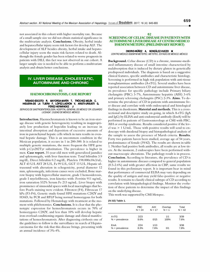
659Abstract section. XII National Meeting of the Mexican Association of Hepatology. , 2017; 16 (4): 645-693
not associated in this cohort with higher mortality rate. Becauseof a small sample size we did not obtain statistical significance inthe multivariate analysis. Conclusions. Obesity, herbal intakeand hepatocellular injury seem risk factors for develop ALF. Thedevelopment of ALF besides obesity, herbal intake and hepato-cellular injury seem the main risk factors related to death. Al-though the female gender has been related to worse prognosis inpatients with DILI, this fact was not observed in our cohort. Alarger sample size is needed to be able to perform a multivariateanalysis and obtain better conclusions.
IV. LIVER DISEASE, CHOLESTATIC,AUTOIMNUME AND CHRONIC
01HAEMOCHROMATOSIS, CASE REPORT
RÁBAGO-ESCOTO R, MAYORGA-MARÍN T, TRÓCHEZ-MEJÍA C,RÁBAGO-ESCOTO R, MAYORGA-MARÍN T, TRÓCHEZ-MEJÍA C,RÁBAGO-ESCOTO R, MAYORGA-MARÍN T, TRÓCHEZ-MEJÍA C,RÁBAGO-ESCOTO R, MAYORGA-MARÍN T, TRÓCHEZ-MEJÍA C,RÁBAGO-ESCOTO R, MAYORGA-MARÍN T, TRÓCHEZ-MEJÍA C,HIGUERA-DE LA TIJERA F, LÓPEZ-LADRÓN V, ARISTI-URSUSTI G,HIGUERA-DE LA TIJERA F, LÓPEZ-LADRÓN V, ARISTI-URSUSTI G,HIGUERA-DE LA TIJERA F, LÓPEZ-LADRÓN V, ARISTI-URSUSTI G,HIGUERA-DE LA TIJERA F, LÓPEZ-LADRÓN V, ARISTI-URSUSTI G,HIGUERA-DE LA TIJERA F, LÓPEZ-LADRÓN V, ARISTI-URSUSTI G,
PÉREZ-HERNÁNDEZ JPÉREZ-HERNÁNDEZ JPÉREZ-HERNÁNDEZ JPÉREZ-HERNÁNDEZ JPÉREZ-HERNÁNDEZ JSERVICIO DE GASTROENTEROLOGÍA, HOSPITAL GENERAL DE MÉXICO “DR. EDUARDO
LICEAGA”, CIUDAD DE MÉXICO. MÉXICO.
Introduction. Haemochromatosis is known to be an iron-stor-age disease with genetic heterogeneity resulting in inappropri-ately low production of hepcidin leading to an increase inintestinal absorption and deposition of excessive amounts ofiron in parenchymal hepatic cells which in turn results in even-tual hepatic damage. This is an uncommon condition in theMexican population, it occurs in 1 in 200-300. It is related tomultiple genetic mutations, the more frequent the HFE genewith p.Cys282Tyr substitution. The prevalence is higher inmen. Case report. 31-year-old men with generalized jaundiceand splenomegaly, with liver function tests: Total bilirubin 3.9mg/dL, Direct bilirubin 0.2 mg/dL, Platelets 198.000x10e3/uL,ALT 43 U/L AST 28 U/L, Fa 99 U/L, GGT 13 U/L, Hepatic ul-trasound with alteration in echogenicity, portal diameter 12mm, splenomegaly, infectious causes were excluded, Bone mar-row biopsy with hypercellular marrow, grade I hemosiderosis,grade I myelofibrosis, iron kinetics with: Ferritin 911 ng/mL,iron saturation 532% Serum Fe 213 ng/mL. Liver biopsy withprominence of sinusoidal spaces with local macrophages that be-fore Pearls staining were evident. Fibrotest (F4), Fibroscan 17kPa (F3-F4), Genetic study found HFE mutations C282Y andH36D, by PCR and RFLP being normal homozygous for bothmutations. Followed by Hematology with treatment at the mo-ment with phlebotomies. Conclusions. It is clear that the phe-notypic expression for hemochromatosis occurs in 70% ofhomozygotes C282Y, and less than 10% will develop hepaticiron overload conditioning organic damage and clinical manifes-tations of hemochromatosis. After diagnosing cirrhosis one ofthe guidelines to follow is the surveillance in search of Hepato-carcinoma for the risk that this disease brings, presenting withan annual incidence of 3%-4%.
02SCREENING OF CELIAC DISEASE IN PATIENTS WITHAUTOIMMUNE LIVER DISEASE AT CENTRO MEDICO
ISSEMYM METEPEC (PRELIMINARY REPORT)
OSORIO-NÚÑEZ A, GONZÁLEZ-HUEZO MOSORIO-NÚÑEZ A, GONZÁLEZ-HUEZO MOSORIO-NÚÑEZ A, GONZÁLEZ-HUEZO MOSORIO-NÚÑEZ A, GONZÁLEZ-HUEZO MOSORIO-NÚÑEZ A, GONZÁLEZ-HUEZO MCENTRO MÉDICO ISSEMYM METEPEC. METEPEC, ESTADO DE MÉXICO. MÉXICO.
Background. Celiac disease (CD) is a chronic, immune-medi-ated inflammatory disease of small intestine characterized bymalabsorption that is induced by dietary gluten in geneticallypredisposed individuals. The diagnosis is based on a suggestiveclinical features, specific antibodies and characteristic histology.Screening is performed in high risk population with anti-tissuetransglutaminase antibodies (A-tTG). Several studies have beenreported association between CD and autoimmune liver disease,its prevalence for specific pathology include Primary biliarycholangitis (PBC) 3-7%, Autoimmune hepatitis (AIH) 3-6%and primary sclerosing cholangitis (PSC) 2-3%. Aims. To de-termine the prevalence of CD in patients with autoimmune liv-er disease and correlate with with endoscopical and histologicalfindings in duodenum. Material and methods. This is a cross-sectional and descriptive study on going, in which A-tTG (IgAand IgG) by ELISA and anti-endomysial antibody (EmA) will beperformed in patients of Gastroenterology at CMI with PBC,AIH or overlap syndrome. Results considered positive if the lev-el was > 1 U/mL. Those with positive results will send to en-doscopy with duodenal biopsy and histopathological analysis ofthe sample to assess the presence of Marsh criteria. Results.Forty two patients haven been studied, average age of 54 years,predominance of female (39/42). The results are shown in table1. Neither had positive both antibodies, all results are at low tit-ers. At the moment, 2 endoscopies have been performed with-out macroscopic alterations. The pathology result is in process.Conclusion. According to literature, the prevalence of CD ishigher in autoimmune diseases compared to general population(0.5-2.6%) and with greater affection in CBP, same results wefound in this preliminary report. It is important bear in mindthat performance of commercial ELISA may vary depending onthe quality of antigen and may yield false-positive or negativeresults. It remains to classify clinical subtype of CD according tocorrelation with hitopathological findings. Monitor the evolu-tion of these patients to determine the impact of this findingson the underlying disease.This work was supported by CMI Metepec.
(IV.02) Table 1.
PBC AIH Overlap TotalN = 24 N = 17 N = 1 N = 42
A-tTG 2/23 1/1 3/39% = prevalence 8.6% 0/15 100% 7.6%
EmA 2/24 1/13 3/38% = prevalence 8.3% 7.6% 0/1 7.8%
660Abstract section. XII National Meeting of the Mexican Association of Hepatology. , 2017; 16 (4): 645-693
03MOLECULAR DIAGNOSIS OF INDIRECT
HYPERBILIRUBINEMIA (CRIGLER NAJJARSYNDROME) A CASE REPORT
RODRÍGUEZ PÉREZ PA, GARRIDO SÁNCHEZ GA, LÓPEZ MONTALVO S,RODRÍGUEZ PÉREZ PA, GARRIDO SÁNCHEZ GA, LÓPEZ MONTALVO S,RODRÍGUEZ PÉREZ PA, GARRIDO SÁNCHEZ GA, LÓPEZ MONTALVO S,RODRÍGUEZ PÉREZ PA, GARRIDO SÁNCHEZ GA, LÓPEZ MONTALVO S,RODRÍGUEZ PÉREZ PA, GARRIDO SÁNCHEZ GA, LÓPEZ MONTALVO S,AGUILAR RAMÍREZ JR. MARTÍNEZ LÓPEZ H, CERDA REYES EAGUILAR RAMÍREZ JR. MARTÍNEZ LÓPEZ H, CERDA REYES EAGUILAR RAMÍREZ JR. MARTÍNEZ LÓPEZ H, CERDA REYES EAGUILAR RAMÍREZ JR. MARTÍNEZ LÓPEZ H, CERDA REYES EAGUILAR RAMÍREZ JR. MARTÍNEZ LÓPEZ H, CERDA REYES E
HOSPITAL CENTRAL MILITAR, CIUDAD DE MÉXICO. MÉXICO.
Introduction. Mutations of the UGT1A1 gene, which en-codes the enzyme UDP glucoroniltransferasa originates disor-ders of bilirubin conjugation; Gilbert’s syndrome clinicallyinsignificant and with BT ≥ 4 mg/dL. Crigler-Najjar syndrometype I, which is usually fatal in childhood and type II most sur-vive to age adult treatment with phenobarbital (enzyme inducerthat favors the conjugation of bilirrubinas) the definitive diag-nosis is with genetic analysis. Objective. Presentation of a caseand review of the literature. Material and methods. Reportedthe case of a male 35 years that income to the Hospital in Mayof 2014 with fever, asthenia, in physical examination withjaundice of sclera or mucous +++, abdomen with hepatomeg-aly of 5 cm below the costal edge, no peritoneal irritation, no as-cites, no hepatic encephalopathy, following laboratory studies:BT = 54 01 mg/dL, BD = 30 mg/dL AST = 74 u/l, ALT = 155u/l, FA = 152 u/l, INR = 1 26, leukocytes = 10.34 platelets =157, 000 hb = 13. 7 g/dL ultrasound of liver: with hepat-osplenomegaly without bile duct dilatation. By indirect hyper-bilirubinemia is on board to exclude hemolysis with: DHL =312 u/direct coombs = negative, EGO = without hemo-globunuria, without esferositosis peripheral blood smear. Ham’stest, osmotic fragility, negative hemoglobin electrophoresis. In-fectious causes were discarded: AgsVHB and AG not reactive,negative TORCH profile, with Microarrays for herpes and Cy-tomegalovirus undetected. ANAS, antimitochondrial MIT3,antiLKM1, antiLKM and anti smooth muscle negative IgG80.9, IgG 80.9, Alpha-1antitripsina: quantification and muta-tion undetected. Percutaneous liver biopsy with spaces pre-served trabecular architecture portal with mild chronicinflammatory infiltrate, mild and focal intracanicular Cholesta-sis, without necrosis, without fibrosis, negative perls staining.CT of liver triple phase: hepatosplenomegaly. Results. Is ship-ping the gene UGT1A1 AT repetition: homozygous for poly-morphism TA7. Began treatment with phenobarbital with goodbiochemical response: BT = 9 3 mg/dL, AST = 27 u/l, ALT =39 u/l FA = 90 u/l INR = 1 11 leukocytes = 8, 21 platelets =217, 000. Conclusion. Crigler-Najjar must suspect it in adultswith unconjugated hyperbilirubinemia and the rest of normalliver tests, the absence of hemolysis is a key part for the diagno-sis and confirmed with the detection of mutations of the geneUGT1A1.
04INTESTINAL DYSBIOSIS AND INCREASED
EXPRESSION OF MUC-2 IS ASSOCIATED TOBACTERIAL TRANSLOCATION IN EXPERIMENTAL
CHOLESTASIS
VEGA-MAGAÑA N,* JAVE-SUÁREZ LF, ** DELGADO-RIZO V,***VEGA-MAGAÑA N,* JAVE-SUÁREZ LF, ** DELGADO-RIZO V,***VEGA-MAGAÑA N,* JAVE-SUÁREZ LF, ** DELGADO-RIZO V,***VEGA-MAGAÑA N,* JAVE-SUÁREZ LF, ** DELGADO-RIZO V,***VEGA-MAGAÑA N,* JAVE-SUÁREZ LF, ** DELGADO-RIZO V,***DEL TORO-ARREOLA S, * AGUILAR-LEMARROY A,**DEL TORO-ARREOLA S, * AGUILAR-LEMARROY A,**DEL TORO-ARREOLA S, * AGUILAR-LEMARROY A,**DEL TORO-ARREOLA S, * AGUILAR-LEMARROY A,**DEL TORO-ARREOLA S, * AGUILAR-LEMARROY A,**GARCÍA BENAVIDES L,**** BASTIDAS-RAMÍREZ BE,*GARCÍA BENAVIDES L,**** BASTIDAS-RAMÍREZ BE,*GARCÍA BENAVIDES L,**** BASTIDAS-RAMÍREZ BE,*GARCÍA BENAVIDES L,**** BASTIDAS-RAMÍREZ BE,*GARCÍA BENAVIDES L,**** BASTIDAS-RAMÍREZ BE,*
FAFUTIS-MORRIS M,*** ESCARRÁ SENMARTÍ M*,FAFUTIS-MORRIS M,*** ESCARRÁ SENMARTÍ M*,FAFUTIS-MORRIS M,*** ESCARRÁ SENMARTÍ M*,FAFUTIS-MORRIS M,*** ESCARRÁ SENMARTÍ M*,FAFUTIS-MORRIS M,*** ESCARRÁ SENMARTÍ M*,GUTIÉRREZ- FRANCO J*, BUENO-TOPETE MR*GUTIÉRREZ- FRANCO J*, BUENO-TOPETE MR*GUTIÉRREZ- FRANCO J*, BUENO-TOPETE MR*GUTIÉRREZ- FRANCO J*, BUENO-TOPETE MR*GUTIÉRREZ- FRANCO J*, BUENO-TOPETE MR*
* INSTITUTO DE ENFERMEDADES CRÓNICO DEGENERATIVAS, CUCS U DE G.**CENTRO DE INVESTIGACIÓN BIOMÉDICA DE OCCIDENTE (CIBO)-IMSS.
*** LABORATORIO DE INMUNOLOGÍA, CUCS U DE G. MÉXICO. **** INSTITUTO DETERAPÉUTICA EXPERIMENTAL Y CLÍNICA, CUCS U DE G. MÉXICO.
Introduction and aim. Bacterial infections in cirrhotic pa-tients are an important cause of death. Bacterial translocation(BT) plays a crucial role in this process. Recently, our groupdemonstrated that BT was present at 8 days after bile duct liga-tion (BDL) in rats. However, in the model of BDL there is lim-ited information concerning the mechanisms related to improveBT; for instance, intestinal and mesenteric lymph nodes (MLN)microbiota diversity and Mucin-2 expression, which is a majorstructural protein of intestinal mucus layer. The aim of thisstudy was to evaluate parameters associated to the developmentof BT in BDL model. Material and methods. Male Wistar ratswere sacrificed at 8 and 30 days after BDL. BT was detected bymicrobiological cultures of mesenteric lymph nodes (MLN).Bacterial load was assessed through estimating faecal Escherichia/Shigella by qPCR. Diversity of microbiota in faeces and MLNwas determined by 16S rDNA pyrosequencing GS-Junior 454.The colonic mucus layer was analyzed by alcian blue stainingand mucin-2 by immunohistochemistry. Results. BT was de-tected as early as 8 days after BDL. Intestinal bacterial overloadof Escherichia/Shigella was significantly increased 2 and 4 loga-rithms at 8 and 30 days post-BDL respectively, in comparisonto control group. BDL induced changes in intestinal microbiotacomposition. Firmicutes percentage was importantly decreased,whereas Bacteroidetes and Proteobacteria were increased concomi-tantly with liver damage. These Gram-negative bacteria havethe ability to adhere and degrade intestinal mucus oligosaccha-rides and have been related to chronic inflammatory processes.In addition, we observed a dramatic decrease of beneficial bacte-ria, such as Lactobacillus, Enterococcus, and Ruminococcus. Bacte-rial composition in mesenteric lymph nodes at 8 days after BDLwas dominated by Proteobacteria (77%), followed by Firmicutes(22%). This correlation was reversed in control rats. Mucus lay-er was surprisingly increased during liver damage. Conclusions.BT in BDL model is associated to intestinal and MLN dysbiosis,bacterial overgrowth and overexpression of Mucin-2.
661Abstract section. XII National Meeting of the Mexican Association of Hepatology. , 2017; 16 (4): 645-693
05EXPRESSION OF IL-22 AND IL-22BP IN ANEXPERIMENTAL MODEL OF CHOLESTASIS
SAAVEDRA-GONZÁLEZ A,* VEGA-MAGAÑA N,* DEL TORO-ARREOLA S,*SAAVEDRA-GONZÁLEZ A,* VEGA-MAGAÑA N,* DEL TORO-ARREOLA S,*SAAVEDRA-GONZÁLEZ A,* VEGA-MAGAÑA N,* DEL TORO-ARREOLA S,*SAAVEDRA-GONZÁLEZ A,* VEGA-MAGAÑA N,* DEL TORO-ARREOLA S,*SAAVEDRA-GONZÁLEZ A,* VEGA-MAGAÑA N,* DEL TORO-ARREOLA S,*PEREIRA-SUÁREZ AL,** FAFUTIS-MORRIS M,**PEREIRA-SUÁREZ AL,** FAFUTIS-MORRIS M,**PEREIRA-SUÁREZ AL,** FAFUTIS-MORRIS M,**PEREIRA-SUÁREZ AL,** FAFUTIS-MORRIS M,**PEREIRA-SUÁREZ AL,** FAFUTIS-MORRIS M,**
GUTIÉRREZ FRANCO J,* ESCARRÁ SENMARTÍ M,* BUENO-TOPETEGUTIÉRREZ FRANCO J,* ESCARRÁ SENMARTÍ M,* BUENO-TOPETEGUTIÉRREZ FRANCO J,* ESCARRÁ SENMARTÍ M,* BUENO-TOPETEGUTIÉRREZ FRANCO J,* ESCARRÁ SENMARTÍ M,* BUENO-TOPETEGUTIÉRREZ FRANCO J,* ESCARRÁ SENMARTÍ M,* BUENO-TOPETEMR*MR*MR*MR*MR*
*INSTITUTO DE ENFERMEDADES CRÓNICO DEGENERATIVAS, CUCS U DE G. MÉXICO.**LABORATORIO DE INMUNOLOGÍA, CUCS U DE G. MÉXICO.
Introduction and aim. The immune response under liverdamage in the bile duct ligation (BDL) model has been associat-ed to the activation of Kupffer cells and hepatic stellate cells, aswell as to over-production of pro-inflammatory cytokines, suchas IL-1β and TNF-α. Nonetheless, IL-22, which is consideredan important cytokine in the control of inflammatory and re-generative processes, has not been explored yet in the BDLmodel, and even less is known about the expression of its recep-tors. Recently, to increase in the systemic IL-22 levels have beenrelated as a prognostic factor in patients with advanced liver cir-rhosis. The aim of this study was to evaluate the expression pat-ters of IL-22 and its receptors IL-22BP (soluble receptor) andIL-22R1 (membrane subunit receptor) during the progression ofcholestasis fibrosis. Material and methods. Hepatic fibrosiswas induced by BDL at 8 and 30 days of evolution. IL-22 andSTAT-3p levels in liver homogenates were analyzed by Westernblotting and gene expression of IL-22BP and IL-22R1 by qRT-PCR. IL-22 in serum was assessed by Western blotting.Results. IL-22 in liver homogenates was significantly in-creased at 8 days post-BDL compared to control group (p <0.01). Hepatic expression of STAT-3p tends to increase in paral-lel to liver damage, with significant differences only at 30 dayspost-BDL (p < 0.05) in comparison to control group. Systemiclevels of IL-22 showed a dramatic increase at 8 (p < 0.05) and30 (p < 0.01) days after BDL. In liver, lL-22BP and IL-22R1gene expression was increased throughout the damage process.Conclusions. The increase of hepatic IL-22 in early state of fi-brosis may indicate a protector effect against hepatocellularnecrosis and inflammation. However, increased IL-22BP geneexpression suggests the possibility of neutralization of the IL-22effector functions, favoring liver damage. Interestingly, systemiclevels of IL-22 were increased in parallel to liver damage, there-fore may be considered a progression marker of cholestasic fibrosis.
V. VIRAL HEPATITIS
01POLYMORPHISMS IN IFNL4 (TT/ΔΔΔΔΔG) AND IL10 (-1082
A/G) GENES INCREASE LEVELS OF TOTALCHOLESTEROL AND LDL-C IN PATIENTS WITH
HEPATITIS C VIRUS INFECTION
GONZÁLEZ-ALDACO K, ROMAN S, FIERRO NA, TORRES-REYES LA,GONZÁLEZ-ALDACO K, ROMAN S, FIERRO NA, TORRES-REYES LA,GONZÁLEZ-ALDACO K, ROMAN S, FIERRO NA, TORRES-REYES LA,GONZÁLEZ-ALDACO K, ROMAN S, FIERRO NA, TORRES-REYES LA,GONZÁLEZ-ALDACO K, ROMAN S, FIERRO NA, TORRES-REYES LA,PANDURO APANDURO APANDURO APANDURO APANDURO A
DEPARTMENT OF MOLECULAR BIOLOGY IN MEDICINE, CIVIL HOSPITAL OFGUADALAJARA, “FRAY ANTONIO ALCALDE”, AND HEALTH SCIENCES CENTER,
UNIVERSITY OF GUADALAJARA, GUADALAJARA, JALISCO, MEXICO.
Background. Hepatitis C virus (HCV) alters liver lipid metab-
olism. High levels of total cholesterol (TC) and low-density li-poprotein cholesterol (LDL-c) have been associated with sus-tained virological response (SVR). IFNL4 and IL10 cytokinesare expressed during HCV infection. Some genetic variants ofimmune mediators may modulate cholesterol levels. Variants inIFNL4 (TT/ΔG) and IL10 (-1082 A/G) genes have been associ-ated with spontaneous clearance (SC). However, it is unclear ifthey are related to lipid alterations that may favor SC. Aim. Toanalyze if IFNL4 (TT/ΔG) and IL10 (-1082 A/G) gene variantsare associated with serum lipid levels in treatment-naïve HCVpatients. Material and methods. In a cross-sectional study,299 treatment-naïve patients were included. Of these, 206 pa-tients had chronic infection (positive viral load (VL), and 93 pa-tients were SC (undetectable VL for at least one year).Biochemical tests were accessed by an dry chemistry assay. VLwas determined by COBAS® TaqMan 48 HCV test and geno-types were identified by TaqMan Real-Time PCR. Comparativestatistics, Odds Ratio Test and linear regression analysis werecalculated by SPSS software, v21. Results. Among the chroni-cally-infected patients, the IFNL4-TT/TT genotype was associ-ated with higher levels of TC and LDL-c than the ΔG/TT+ΔG/ΔG genotypes (176.6 ± 48.7 mg/dL vs. 160.3 ± 45.6mg/dL, p = 0.042; and 107.7 ± 43.7 mg/dL vs. 84.3 ± 39.5 mg/dL, p = 0.01, respectively). A 6.4% (β = 23.39, p = 0.02) varia-tion in LDL-c levels was attributable to the IFNL4-TT/TTgenotype whereas the IL10-1082-AA genotype was associatedwith higher levels of TC and LDL-c among SC patients thanthe AG+GG genotypes (193.6 ± 44.6 mg/dL vs. 173.2 ± 36.0mg/dL, p = 0.041; and 118.3 ± 36.8 vs. 101.7 ± 25.2 mg/dL, p= 0.045, respectively). Also, the AA genotype was responsiblefor a 6.6% (β = 0.004, p = 0.045) increase in LDL-c and 6.1%(β = 0.003, p = 0.041) increase in TC. Conclusions. IFNL4-TT/TT and IL10-AA genotypes differentially modulate TC andLDL-c in chronic HCV-infected and SC patients. This studysuggests an interaction between the immune response and cho-lesterol levels that could be involved in SVR. Therefore, modu-lation of cholesterol levels by diet and genetic factors appears toinfluence the outcome of HCV infection.Funding: This work was subsidized by Prodep-University ofGuadalajara (UDG-CA-478) and PRO-SNI to Arturo Panduro.
02HIV/HCV CO-INFECTION, ASSOCIATED RISK
FACTORS AND HCV GENOTYPES IN PATIENTSFROM WEST MEXICO
TRUJILLO-TRUJILLO ME, PANDURO A, TORRES-VALADEZ R,TRUJILLO-TRUJILLO ME, PANDURO A, TORRES-VALADEZ R,TRUJILLO-TRUJILLO ME, PANDURO A, TORRES-VALADEZ R,TRUJILLO-TRUJILLO ME, PANDURO A, TORRES-VALADEZ R,TRUJILLO-TRUJILLO ME, PANDURO A, TORRES-VALADEZ R,SEPÚLVEDA-VILLEGAS M, GONZÁLEZ-ALDACO K,SEPÚLVEDA-VILLEGAS M, GONZÁLEZ-ALDACO K,SEPÚLVEDA-VILLEGAS M, GONZÁLEZ-ALDACO K,SEPÚLVEDA-VILLEGAS M, GONZÁLEZ-ALDACO K,SEPÚLVEDA-VILLEGAS M, GONZÁLEZ-ALDACO K,
JOSE-ABREGO A, ROMAN S.JOSE-ABREGO A, ROMAN S.JOSE-ABREGO A, ROMAN S.JOSE-ABREGO A, ROMAN S.JOSE-ABREGO A, ROMAN S.DEPARTMENT OF MOLECULAR BIOLOGY IN MEDICINE, HOSPITAL CIVIL DE
GUADALAJARA “FRAY ANTONIO ALCALDE” AND HEALTH SCIENCES CENTER,UNIVERSITY OF GUADALAJARA, GUADALAJARA JALISCO, MÉXICO.
Background. Patients with immunodeficiency virus (HIV)have a good response to antiretroviral therapy (ART). However,liver disease constitutes the main co-morbidity that leads to fataloutcomes among HIV patients. In HIV/HCV co-infected pa-tients, HIV accelerates hepatitis C virus (HCV) replication de-creasing the occurrence of spontaneous clearance and sustainedantiviral response. In Mexico, there are scarce studies aboutHIV/HCV co-infection and it is unclear which risk factors pre-
662Abstract section. XII National Meeting of the Mexican Association of Hepatology. , 2017; 16 (4): 645-693
dispose for co-infection as well as the circulating HCV geno-types among HIV-infected patients. Aim. To determine the fre-quency of HIV/HCV co-infection, associated risk factors and HCVgenotype distribution among HIV patients from West Mexico. Ma-terial and methods. A cross-sectional study was conducted in 611patients. Of these, 287 were positive to anti-HIV and 324 were posi-tive to anti-HCV. Serological antibodies were detected by third-gen-eration ELISA and HCV genotypes by LIPA assay. Anova,t-Student, χ2, and logistic regression analysis were performed withSPSS, v. 21. Results. The frequency of HIV/HCV co-infection was37%. HIV-monoinfected patients were mainly men (89%), and thehighest number of cases were between the ages of 19-29 years. Con-versely, HCV-infected patients were older women (50-59 years),whereas the majority of HIV/HCV co-infections were found in the30-40 year age group. Surgeries (78%) and transfusions (58%) werethe main risk factors for HCV mono-infection. However, promiscu-ity (78%), surgeries (71%), transfusions (43%), tattoos (64%) and in-travenous (IV) drug use (34%) were the main risk factors amongHIV/HCV co-infected patients. By logistic regression test, the inde-pendent predictors associated with risk to co-infection were transfu-sions (OR= 2.4, 95% CI 1.3-4.4, p = 0.004), tattoos (OR = 2.0,95% CI 1.1-3.7, p = 0.015) and IV drugs use (OR = 6.1, 95% CI2.5-14.9, p = 6 x 10⁻⁵). The distribution of HCV genotypes in co-infected patients was 1a (70%), followed by 3a (14%), 1b (11%), 2,2b and 4a with 1.5%. Finally, the frequency of HCV genotypesamong HCV-monoinfected patients were mainly 1a (52%), 1b(13%) and 3a (12%). Conclusions. The frequency of HIV/HCV co-infected patients was 37%. The independent predictors associatedwith HIV/HCV coinfection were transfusions, IV drug use and tat-toos. The main HCV genotypes in HIV patients were 1a, 1b and 3a.This work was subsidized by Promep-University of adalajara (UDG-CA-478) to Arturo Panduro and PRO-SNI to Sonia Roman.
03DESCRIPTION OF SOCIO-DEMOGRAPHIC
CHARACTERISTICS AND SEXUAL BEHAVIOR OFHEPATITIS C AND HIV CO-INFECTED PATIENTS IN
MEXICO
RAMÍREZ-GONZÁLEZ LE,* PIÑEIRUA-MENÉNDEZ A,**RAMÍREZ-GONZÁLEZ LE,* PIÑEIRUA-MENÉNDEZ A,**RAMÍREZ-GONZÁLEZ LE,* PIÑEIRUA-MENÉNDEZ A,**RAMÍREZ-GONZÁLEZ LE,* PIÑEIRUA-MENÉNDEZ A,**RAMÍREZ-GONZÁLEZ LE,* PIÑEIRUA-MENÉNDEZ A,**SÁNCHEZ-ÁVILA JF,* HIRATA HERNÁNDEZ AH,***SÁNCHEZ-ÁVILA JF,* HIRATA HERNÁNDEZ AH,***SÁNCHEZ-ÁVILA JF,* HIRATA HERNÁNDEZ AH,***SÁNCHEZ-ÁVILA JF,* HIRATA HERNÁNDEZ AH,***SÁNCHEZ-ÁVILA JF,* HIRATA HERNÁNDEZ AH,***
PÉREZ-CARRIZOSA A,* CAMIRO-ZUÑIGA A,* SIMENTAL-ALDABA E,*PÉREZ-CARRIZOSA A,* CAMIRO-ZUÑIGA A,* SIMENTAL-ALDABA E,*PÉREZ-CARRIZOSA A,* CAMIRO-ZUÑIGA A,* SIMENTAL-ALDABA E,*PÉREZ-CARRIZOSA A,* CAMIRO-ZUÑIGA A,* SIMENTAL-ALDABA E,*PÉREZ-CARRIZOSA A,* CAMIRO-ZUÑIGA A,* SIMENTAL-ALDABA E,*SIERRA-MADERO JG*SIERRA-MADERO JG*SIERRA-MADERO JG*SIERRA-MADERO JG*SIERRA-MADERO JG*
* DEPARTMENT OF INFECTIOUS DISEASES INSTITUTO NACIONAL DE CIENCIASMÉDICAS Y NUTRICIÓN “SALVADOR ZUBIRÁN” (INCMNSZ), MEXICO CITY, MEXICO.
** CLÍNICA ESPECIALIZADA CONDESA IZTAPALAPA (CECI), MEXICO CITY, MEXICO.*** DEPARTMENT OF MENTAL HEALTH, CLÍNICA ESPECIALIZADA CONDESA (CEC),
MEXICO CITY, MEXICO.
Background. HCV/HIV co-infected patients is a heterogenousgroup, with significant regional variations. Numerous studieshave shown elevated risk of HCV in intravenous drug users(IVDU), and has been recognized as the main risk factor associ-ated with HCV transmission. However in central regions ofMexico the prevalence of IV drug use is less than 1%. The roleof different forms of transmission in Latin America is still scarce.Objectives. To describe socio-demographic characteristics aswell as sexual behavior of patients diagnosed with HCV/HIVco-infection. Material and methods. Participants were re-cruited between May 2016 and March 2017, every participantswas assessed with a 119 item questionnaire, covering, socio-de-
mographic, risk of parenteral exposition, sexual behavior withfocus on history of sexually transmitted diseases, condom useand high-risk sexual behavior. ASSIST Questionnaire Version 3was used as a screening of substance abuse. Data was analyzedwith STATA 13 using descriptive statistics. Results. 71 partici-pants were recruited, 63 male (88.8%), with a mean of age of37.5 years (ICR 33-44), 76% were born in Mexico City. Themost frequent genotype was 1a, 62.8%. From the group of eval-uated men 83% (59/63) were men who have sex with men.From the total of patients 2.9% (2/71) recognized the past con-sumption of injected drugs, cocaine consumption was reportedin 19.8% (14/71). History of transfusion before 1992 was found in23.9% (17/71), 32.3% (23/71) have a previos tattoo, 43.4% ofthem recognized that tattoo was crafted in unhealthy situations.The 76% (54/71) of participants had participated in high risksexual practices wth out condom. Limitations: Associationscould not be established until further studies. The developmentof this project will continue for a further pair matched case con-trol study with HCV uninfected participants. Conclusions.High risk sexual behavior might have a important role in HCVtransmission in this population, we found a low rate of IVDU.The 40.8% of the participants have high risk of HCV infectionsby parenteral routes.
04DEGREE OF OBESITY IN PATIENTS WITH CHRONIC
INFECTION BY HEPATITIS C VIRUS AND ITSRELATION WITH STEATOSIS BY STEATOTEST
GARCÍA SOTO ML, GÓMEZ GARCÍA T,GARCÍA SOTO ML, GÓMEZ GARCÍA T,GARCÍA SOTO ML, GÓMEZ GARCÍA T,GARCÍA SOTO ML, GÓMEZ GARCÍA T,GARCÍA SOTO ML, GÓMEZ GARCÍA T,DEL REAL CALZADA C, RIZO ROBLES TDEL REAL CALZADA C, RIZO ROBLES TDEL REAL CALZADA C, RIZO ROBLES TDEL REAL CALZADA C, RIZO ROBLES TDEL REAL CALZADA C, RIZO ROBLES T
DEPARTMENT OF GASTROENTEROLOGY; HOSPITAL OF SPECIALTIES IMSS CMN LARAZA, “ANTONIO FRAGA MOURET”. CITY OF MEXICO, MEXICO.
Introduction. Hepatic steatosis is reported by biopsy in 40-80%of patients with chronic hepatitis C virus infection (HCV), act-ing as a co-factor to fibrosis.The biopsy represents a diagnosticobstacle due to the cost and associated complications, this is whythat non-invasive studies like steatotest have taken relevance,showing significant sensitivity compared with classic markers.Aim of the research. Determining the number of patientswith HCV, with overweight and obesity who present hepaticsteatosis by Steatotest. Material and methods. This is an ob-servational study, we studied 78 patients with Steatotest, ofwhich 47 had HCV, and established its relation with body massindex(BMI). Inclusion criteria: patients with chronic infectionof HCV subscribed to the hepatitis clinic. Statistical analysisused was square chi, using SPSS 20. Results. Of the 47 patientswith chronic HCV infection, 37 presented steatosis by Stea-totest (4 normal weight, 19 overweight, 11 obesity grade 1 and3 obesity grade 2). Discussion: the most frequent causes of he-patic steatosis in people infected with genotypes other than 3are the increase of BMI and visceral obesity, factors associated toresistance to insulin. This study establishes the relation of stea-tosis between the report of steatotest and the degree of obesity inpatients with HCV chronic infection. The most of patients withsevere steatosis presented overweigth than obesity. The impor-tance resides in establishing non-invasive measures in patientswith metabolic risk factors that correlate to the degree of steato-sis, it is possible that BMI can not be the best way to value it,
663Abstract section. XII National Meeting of the Mexican Association of Hepatology. , 2017; 16 (4): 645-693
and we must consider other parameters related to insulin resist-ance, like the abdominal perimeter. Conclusions. Patients withHCV infection have comorbilities that cause steatosis, perpetu-ating the damage, despite the virus eradication. There is no rela-tion between the degree of obesity and the degree of steatosis bySteatotest in patients with chronic infection by HCV. Conflictof interests: this work has been subsidized partially by medifar-ma group (fibromax tests).
05SEROPREVALENCE OF HEPATITIS C VIRUS IN
BLOOD DONORS FROM THE GENERAL HOSPITALOF MEXICO “DR. EDUARDO LICEAGA”
BONILLA-RAMOS A,* HIGUERA-DE LA TIJERA F,* RÁBAGO-ESCOTO R,*BONILLA-RAMOS A,* HIGUERA-DE LA TIJERA F,* RÁBAGO-ESCOTO R,*BONILLA-RAMOS A,* HIGUERA-DE LA TIJERA F,* RÁBAGO-ESCOTO R,*BONILLA-RAMOS A,* HIGUERA-DE LA TIJERA F,* RÁBAGO-ESCOTO R,*BONILLA-RAMOS A,* HIGUERA-DE LA TIJERA F,* RÁBAGO-ESCOTO R,*BÉJAR-RAMÍREZ Y,** PÉREZ-HERNÁNDEZ J*BÉJAR-RAMÍREZ Y,** PÉREZ-HERNÁNDEZ J*BÉJAR-RAMÍREZ Y,** PÉREZ-HERNÁNDEZ J*BÉJAR-RAMÍREZ Y,** PÉREZ-HERNÁNDEZ J*BÉJAR-RAMÍREZ Y,** PÉREZ-HERNÁNDEZ J*
*GASTROENTEROLOGY SERVICE, ** BLOOD BANK GENERAL HOSPITAL OF MEXICO“DR. EDUARDO LICEAGA”, MEXICO CITY. MEXICO.
Background. Chronic Hepatitis C virus (HCV) is a globalhealth problem. It is known that the disease is widespread allover the world; However, in Mexico there are few studiesshowing the prevalence of this disease. In our country is esti-mated a prevalence around 0.4 - 1.4%. Objectives. To de-termine the seroprevalence of HCV in blood donors of theGeneral Hospital of Mexico “Dr. Eduardo Liceaga” in a peri-od of 3 years, from 2014 to 2016. Material and methods.Retrospective, observational study, carried out in donor files,that came to the Blood Bank of the General Hospital ofMexico, from January 1, 2014 to December 31, 2016, andwhich met the criteria of NOM-003- SSA2-1993 “For thedisposal of human blood and its components for therapeuticpurposes”. We included accepted donors who presented anti-HCV positive with ELISA technique and with positive con-firmatory test by PCR. Results. 69,999 blood donors, 50,040(71.4%) men and 19,959 (28.6%) women were evaluated; Ofthese 103 (0.14%) donors had anti-HCV positive PCR con-firmed, yielding a prevalence of 0.0014. All risk factors forHCV were denied by donors. Conclusions. HCV seropreva-lence in blood donors at the General Hospital of Mexico “Dr.Eduardo Liceaga” is inferior to the one reported at nationaland international level; Probably these low indices reflect thepositive impact of the preventive measures, improvement inthe detection strategies of risky donors which are classified asunfit donors.This work was not sponsored.
06SEROPREVALENCE OF HEPATITIS B VIRUS IN
BLOOD DONORS FROM THE GENERAL HOSPITALOF MEXICO “DR. EDUARDO LICEAGA”
BONILLA-RAMOS A,* HIGUERA-DE LA TIJERA F,* RÁBAGO-ESCOTO R,*BONILLA-RAMOS A,* HIGUERA-DE LA TIJERA F,* RÁBAGO-ESCOTO R,*BONILLA-RAMOS A,* HIGUERA-DE LA TIJERA F,* RÁBAGO-ESCOTO R,*BONILLA-RAMOS A,* HIGUERA-DE LA TIJERA F,* RÁBAGO-ESCOTO R,*BONILLA-RAMOS A,* HIGUERA-DE LA TIJERA F,* RÁBAGO-ESCOTO R,*BÉJAR-RAMÍREZ Y,** PÉREZ-HERNÁNDEZ J*BÉJAR-RAMÍREZ Y,** PÉREZ-HERNÁNDEZ J*BÉJAR-RAMÍREZ Y,** PÉREZ-HERNÁNDEZ J*BÉJAR-RAMÍREZ Y,** PÉREZ-HERNÁNDEZ J*BÉJAR-RAMÍREZ Y,** PÉREZ-HERNÁNDEZ J*
*GASTROENTEROLOGY SERVICE, ** BLOOD BANK GENERAL HOSPITAL OF MEXICO“DR. EDUARDO LICEAGA”, MEXICO CITY. MEXICO.
Background. National epidemiological research on transmitteddiseases by blood transfusion is important, because throughblood banks it is possible to know the distribution and seroprev-alence of these diseases. In Mexico, the seroprevalence of hepati-tis B virus (HBV) has fluctuated between 0.16 and 0.4%.Objectives. To determine the seroprevalence of HBV in blooddonors of the General Hospital of Mexico “Dr. Eduardo Licea-ga” for three years, in the period from 2014 to 2016. Materialand methods. Retrospective, observational study of donorswho attended the Blood Bank of the General Hospital of Mexi-co, from January 1, 2014 to December 31, 2016, and who metthe criteria of NOM-003-SSA2-1993 “For the disposal of hu-man blood and its components for therapeutic purposes.” Ac-cepted donors who were positive for HBsAg were included.Results. A total of 69,999 blood donors, 50,040 (71.4%) men,and 19,959 (28.6%) women, were evaluated; of these 12 (0.01%)donors had positive HBsAg that yields a prevalence of 0.00017.All denied risk factors, only one donor reported on 5 sexual part-ners. Conclusions. HBV seroprevalence in blood donors at theGeneral Hospital of Mexico “Dr. Eduardo Liceaga” is lower thanthat reported at national and international level. Studies in blooddonors involve the selection of patients, excluding those with riskfactors; the actual prevalence may be higher. It is necessary to car-ry out a greater number of epidemiological studies, especially inthe open population and with risk factors.This work was not sponsored.
07PROTECTIVE EFFECT OF MORINGA OLEIFERA ON
HBV GENOTYPE C AND H TRANSIENTLYTRANSFECTED Huh7 CELLS
SÁNCHEZ-OROZCO LV, FEUSTEL S, AYÓN-PÉREZ MF,SÁNCHEZ-OROZCO LV, FEUSTEL S, AYÓN-PÉREZ MF,SÁNCHEZ-OROZCO LV, FEUSTEL S, AYÓN-PÉREZ MF,SÁNCHEZ-OROZCO LV, FEUSTEL S, AYÓN-PÉREZ MF,SÁNCHEZ-OROZCO LV, FEUSTEL S, AYÓN-PÉREZ MF,SANDOVAL-RODRIGUEZ A, RODRÍGUEZ ECHEVARRÍA R,SANDOVAL-RODRIGUEZ A, RODRÍGUEZ ECHEVARRÍA R,SANDOVAL-RODRIGUEZ A, RODRÍGUEZ ECHEVARRÍA R,SANDOVAL-RODRIGUEZ A, RODRÍGUEZ ECHEVARRÍA R,SANDOVAL-RODRIGUEZ A, RODRÍGUEZ ECHEVARRÍA R,
ARMENDARIZ-BORUNDA JARMENDARIZ-BORUNDA JARMENDARIZ-BORUNDA JARMENDARIZ-BORUNDA JARMENDARIZ-BORUNDA JINSTITUTO DE BIOLOGÍA MOLECULAR EN MEDICINA, DEPARTAMENTO DE BIOLOGÍA
MOLECULAR Y GENÓMICA, CENTRO UNIVERSITARIO DE CIENCIAS DE LA SALUD,UNIVERSIDAD DE GUADALAJARA, JALISCO, MÉXICO.
Background. Chronic hepatitis B infection treatment implicatesa long lasting treatment with antiviral drugs. M. oleifera extractscontain compounds with antiviral, antioxidant and antifibroticproperties. Objective. To evaluate the effect of M. oleifera inHuh7 cells expressing either HBV genotypes C or H for the anti-viral, antifibrotic and antioxidative responses. Material andmethods. The M. oleifera leaves were obtained from the southcoast of Jalisco, Mexico and were validated by the Instituto Na-cional de Investigaciones Forestales, Agrícolas y Pecuarias (INI-FAP). Huh7 cells were treated with a water-soluble extract of M.oleifera (leaves) at doses of 0, 30, 45 or 60 μg/mL. TGF-β1,CTGF, CAT, IFN-β1 and pgRNA expression was measured byreal time. Secretion of HBsAg and levels of nuclear activated Nrf2
(V.04) Table 1.
BMI Steatotest Steatotest Steatotest Totalmild moderade severe
Obesity grade II 2 1 0 3Obesity grade I 4 1 6 11Overweight 6 6 7 19Normal weight 4 0 0 4
664Abstract section. XII National Meeting of the Mexican Association of Hepatology. , 2017; 16 (4): 645-693
were determined by ELISA and Western blot respectively. Re-sults. Concentrations of 30, 45, 60 and 120 μg/mL of M. oleiferadecreased the number of viable cells in an average of 2.8%. Thus,for further experiments the three lowest concentrations of M.oleifera (30, 45 and 60 μg/mL) were used to test our proof of con-cept. CTGF, TGF-β1 and pgRNA expression decreased with M.oleifera treatment independently of the HBV genotype. M. oleiferatreatment reduced IFN-β1expression for both genotypes. HBsAgsecretion in the supernatant of transfected Huh7 cells with bothHBV genotypes was decreased independently of the dose of M.oleifera. Transfection with both HBV genotypes strongly de-creased CAT and Nrf2 expression, which are retrieved with M.oleifera treatment. Conclusions. M. oleifera treatment reducedpgRNA expression, fibrosis markers and HBsAg secretion inHBV genotypes C and H. Furthermore, an important antioxi-dant response was detected with the recovery of CAT expressionand the nuclear activated Nrf2 protein.
08HEPATITIS E INFECTION: ACUTE PANCREATITIS AS
INITIAL EXTRAHEPATIC MANIFESTATION ANDPROGRESSION TO CHRONICITY WITH HEPATIC
MANIFESTATION SIMILAR TO AUTOIMMUNEHEPATITIS
LÓPEZ-PÉREZ RY,* MAYORGA-MARÍN TA,* MOTOLA-KUBA M,*LÓPEZ-PÉREZ RY,* MAYORGA-MARÍN TA,* MOTOLA-KUBA M,*LÓPEZ-PÉREZ RY,* MAYORGA-MARÍN TA,* MOTOLA-KUBA M,*LÓPEZ-PÉREZ RY,* MAYORGA-MARÍN TA,* MOTOLA-KUBA M,*LÓPEZ-PÉREZ RY,* MAYORGA-MARÍN TA,* MOTOLA-KUBA M,*GARDUÑO-JANETH J,** ARISTI- URISTA G,**GARDUÑO-JANETH J,** ARISTI- URISTA G,**GARDUÑO-JANETH J,** ARISTI- URISTA G,**GARDUÑO-JANETH J,** ARISTI- URISTA G,**GARDUÑO-JANETH J,** ARISTI- URISTA G,**
HIGUERA-DE LA TIJERA F*HIGUERA-DE LA TIJERA F*HIGUERA-DE LA TIJERA F*HIGUERA-DE LA TIJERA F*HIGUERA-DE LA TIJERA F**GASTROENTEROLOGY DEPARTMENT, **PATHOLOGY DEPARTMENT HOSPITAL
GENERAL DE MÉXICO “DR. EDUARDO LICEAGA” AND SCHOOL OF MEDICINE UNAM.MEXICO.
Introduction. In Mexico, hepatitis E virus (HEV) is endemic,the most frequent genotypes are 2 and 3, and the prevalence ofIgG-HEV antibodies has been reported up to 26.5% in cirrhoticpatients. Several extrahepatic manifestations related to HEVhave been described; like 50 cases of acute pancreatitis, only re-lated to genotype 1. The relationship between HEV and au-toimmune hepatitis is controversial. Aim. To describe a rarecase of HEV infection with acute pancreatitis as extrahepaticmanifestation and clinical data similar to autoimmune hepatitis.Case presentation. A 45-year-old female with no relevant his-tory, presented with pain located in epigastrium and irradiatedto left hipocondrium, transfective, intense 10/10, constant, ac-companied by vomiting of partially digested food, graduallyadding jaundice, coluria, without acolia, without pruritus . Onexamination normal vital signs, generalized jaundice, hyperalge-sia in the left hipocondrium, Orlowski and Preioni points werepositive. No clinical stigmas of hepatic decompensation. Labs:Platelets 106,000 x 10e 3/uL Urea 16.3 mg/dL, Creatinine 0.7mg/dL Total Bilirubin 7.57 mg/dL Total protein 7.9 g/dL ALT587 U/L AST 1026 U/L FA 224 U/L GGT U/L 120 DHL 310U/L Amylase 263 U/L Lipase 206 U/L INR 1.5 TORCH nega-tive, ANA Negative IgG 4090 mg/dL Ig M 366 mg/dL IgG12920 mg/dL IgG2 233 mg/20.50 mg/dL IgG 4: 56.6 mg/dL,Anti-LKM-1, Anti mitochondrial and Anti Muscle NegativeHsAg, Anti HAV IgM, and Anti HCV IgM. Hepatitis E IgG0.2 IU IgM 2.18 IU (Positive). Liver biopsy with inflammatoryinfiltrate in portal triads and hepatic lobules, necrosis of the lim-iting plaque, interface hepatitis, mild cholestasis. Activity III/IVand grade II/III fibrosis. Discussion. The evolution of HEV in-
fection is generally acute with extrahepatic viral replication andthe formation of immune complexes that detonate autoimmu-nity, associating this as a trigger for autoimmune hepatitis.Chronic evolution and acute pancreatitis as an extrahepaticmanifestation are uncommon. Conclusion. Chronic HEV in-fection is uncommon, it induces systemic manifestations by im-mune complexes and should be suspected in endemic areas.
09STEVIOSIDE, STEVIA AND PIRFERIDONE REDUCESHEPATITIS C VIRAL REPLICATION AND IMPROVESTHE ANTIFIBROTIC AND ANTIOXIDANT RESPONSE
IN JFH1 CELLS
SÁNCHEZ-OROZCO LV, FABIOLA AYÓN-PÉREZ M, FEUSTEL S,SÁNCHEZ-OROZCO LV, FABIOLA AYÓN-PÉREZ M, FEUSTEL S,SÁNCHEZ-OROZCO LV, FABIOLA AYÓN-PÉREZ M, FEUSTEL S,SÁNCHEZ-OROZCO LV, FABIOLA AYÓN-PÉREZ M, FEUSTEL S,SÁNCHEZ-OROZCO LV, FABIOLA AYÓN-PÉREZ M, FEUSTEL S,GARCÍA BAÑUELOS J, SANDOVAL-RODRÍGUEZ AS,GARCÍA BAÑUELOS J, SANDOVAL-RODRÍGUEZ AS,GARCÍA BAÑUELOS J, SANDOVAL-RODRÍGUEZ AS,GARCÍA BAÑUELOS J, SANDOVAL-RODRÍGUEZ AS,GARCÍA BAÑUELOS J, SANDOVAL-RODRÍGUEZ AS,
ARMENDÁRIS-BORUNDA JARMENDÁRIS-BORUNDA JARMENDÁRIS-BORUNDA JARMENDÁRIS-BORUNDA JARMENDÁRIS-BORUNDA JINSTITUTO DE BIOLOGÍA MOLECULAR EN MEDICINA, CUCS, U DE G. MEXICO.
Background. HCV treatment, have been improved considera-bly with the introduction of directly acting antiviral agents(DAA), achieving a sustained virological response in over 90%of treated patients, but the treatment is costly. Stevioside (STV)a dipertene glycoside from stevia (ST) has an antioxidant, anti-inflammatory and antiviral responses and Pirferidone (PFD) isan antifrotic agent. Objective. To evaluate the effect of STV,ST or PFD alone and combined with IFN-β1a in JFH1 cells forthe antiviral, antifibrotic, and antioxidant responses. Materialand methods. JFH1 cells were treated with: a) ST a 50 μg/mL,72 h, b) STV 200 μg/mL, 48 h; c) PFD 100 μM, 12 h; d) IFN-β1a 100 IU/mL, 48 h and IFN-β1a combined with ST, STV orPFD. Viral replication and gene TGFβ-1, CTGF, SOD, CAT,NOS2 e IFN-β1a expression were determined by rtPCR. Re-sults. Viral replication decreased with ST or PFD treatmentalso, with combined IFN-β1a plus ST, STV or PFD (p < 0.05).TGFβ1 expression decrease with PFD, STV or ST treatment (p< 0.05). CAT expression increase with ST or IFN-β1a treat-ment (p < 0.05). SOD expression was increased with PFD (p <0.05) and NOS2 expression was increased with IFN-β1a, aloneor in combination with PFD, STV or ST (p < 0.05). STV plusIFN-β1a decrease NOS2 expression compared with IFN-β1aalone (p < 0.05). IFN-β1a expression increases with STV treat-ment (p < 0.05). Conclusions. Hepatitis C viral replicationwas diminished with PFD or ST treatment and fibrosis markerswere reduced with ST, STV or PFD treatments. ST and PFD fa-vor the antioxidant response (CAT and SOD respectively) andIFN-β1a expression increased with STV treatment.
10SECRETION OF CXCL-9 AND CXCL-10 IN CHRONIC
HEPATITIS C
GARCÍA-LÓPEZ B,* FLORES-VASCONCELOS I,* MEDINA-ÁVILA Z,*GARCÍA-LÓPEZ B,* FLORES-VASCONCELOS I,* MEDINA-ÁVILA Z,*GARCÍA-LÓPEZ B,* FLORES-VASCONCELOS I,* MEDINA-ÁVILA Z,*GARCÍA-LÓPEZ B,* FLORES-VASCONCELOS I,* MEDINA-ÁVILA Z,*GARCÍA-LÓPEZ B,* FLORES-VASCONCELOS I,* MEDINA-ÁVILA Z,*ROSIQUE-ORAMAS D,* VERDEJA-PADILLA P,* RIZO-TÉLLEZ S,*ROSIQUE-ORAMAS D,* VERDEJA-PADILLA P,* RIZO-TÉLLEZ S,*ROSIQUE-ORAMAS D,* VERDEJA-PADILLA P,* RIZO-TÉLLEZ S,*ROSIQUE-ORAMAS D,* VERDEJA-PADILLA P,* RIZO-TÉLLEZ S,*ROSIQUE-ORAMAS D,* VERDEJA-PADILLA P,* RIZO-TÉLLEZ S,*
PÉREZ-HERNÁNDEZ JL,** HIGUERA F,** KERSHENOBICH D,*PÉREZ-HERNÁNDEZ JL,** HIGUERA F,** KERSHENOBICH D,*PÉREZ-HERNÁNDEZ JL,** HIGUERA F,** KERSHENOBICH D,*PÉREZ-HERNÁNDEZ JL,** HIGUERA F,** KERSHENOBICH D,*PÉREZ-HERNÁNDEZ JL,** HIGUERA F,** KERSHENOBICH D,*GUTIÉRREZ-REYES G*GUTIÉRREZ-REYES G*GUTIÉRREZ-REYES G*GUTIÉRREZ-REYES G*GUTIÉRREZ-REYES G*
* LABORATORY (HIPAM) EXPERIMENTAL MEDICINE UNIT, UNAM.** LIVER CLINIC, GASTROENTEROLOGY DEPARTMENT, GENERAL HOSPITAL OF
MEXICO. MEXICO.
Introduction. Hepatic fibrosis contributes to the global burdenof mortality and disease. The etiologic factors that induce fibro-
665Abstract section. XII National Meeting of the Mexican Association of Hepatology. , 2017; 16 (4): 645-693
sis are chronic hepatitis C (CHC) among others. It has beenfound an association of chemokines CXCL-9 and CXCL-10 toviral immune response and the progression to liver fibrosis, sotheir study is necessary to identify a biomarker during the viralinfection or to fibrosis progression. Objective. EvaluateCXCL-9 and CXCL-10 concentrations in blood serum of CHCpatients. Material and methods. A transversal and observa-tional study was performed, including 21 patients with CHCwithout another hepathopathy and 40 control subjects. CXCL-9 concentrations in both groups were quantified using ELISAand CXCL-10 quantification was done by Luminex (Biorad).Statistical analysis was done with SPSS version 15 using UMann Whitney probe. Results. CXCL-9 concentrations (pg/mL) for patients were 1025 ± 962, while for control group re-sulted 747 ± 745 (p = 0.162). CXCL-10 concentrations (pg/mL) for patients were higher than for control group, 88.9 ± 13and 22.6 ± 13 respectively (p < 0.001). Conclusion. CXCL-9did not show differences between patients and control subjects.On the other hand, CXCL-10 in our study, an increase of thischemokine is demonstrated in patients with HCC, reason whywe consider that it can be a potential biomarker in chronic Hep-atitis C.CONACyT: SALUD-2016-1-272579.
11HEPATITIS E: A MISSED OUT DISEASE IN AN
ENDEMIC COUNTRY. CASE REPORT
ALONZO-GARCÍA CJ, GARCÍA-JIMÉNEZ E, GUZMÁN-LEPE A,ALONZO-GARCÍA CJ, GARCÍA-JIMÉNEZ E, GUZMÁN-LEPE A,ALONZO-GARCÍA CJ, GARCÍA-JIMÉNEZ E, GUZMÁN-LEPE A,ALONZO-GARCÍA CJ, GARCÍA-JIMÉNEZ E, GUZMÁN-LEPE A,ALONZO-GARCÍA CJ, GARCÍA-JIMÉNEZ E, GUZMÁN-LEPE A,ÁLVAREZ-LOPEZ F, VELARDE-RUIZ VELASCO JAÁLVAREZ-LOPEZ F, VELARDE-RUIZ VELASCO JAÁLVAREZ-LOPEZ F, VELARDE-RUIZ VELASCO JAÁLVAREZ-LOPEZ F, VELARDE-RUIZ VELASCO JAÁLVAREZ-LOPEZ F, VELARDE-RUIZ VELASCO JA
DEPARTAMENTO DE GASTROENTEROLOGÍA, HOSPITAL CIVIL “FRAY ANTONIOALCALDE”, GUADALAJARA, JALISCO, MEXICO.
Introduction. Since the discovery of a new species of non A,non B hepatitis in 1978 in India, Hepatitis E Virus (HEV) hasspread in endemic and hyperendemic regions, especially in de-veloping countries, where consumption of infected animals,contaminated water and person to person spreading exist. Thisrepresents a challenge for health systems. Our country is classi-fied as a hyperendemic region, and due to the heterogeneity ofthe virus and its variable clinical picture, it should raise a sys-tematic approach into searching for a missed out disease in ourpopulation. Objective. We present a case of HEV infection.Case report. A 63 year old man, ironmongery clerk, with un-remarkable history of diseases, no alcohol nor drug abuse, no
tattoos, no transfusions and no herb consumption. Chief com-plaint was a 2 week history of malaise and nausea and a 1 weekhistory of jaundice and dark urine without pruritus. Physicalexam confirms jaundice in skin, mucous membranes and scler-ae; no visible telangiectasia or rubi nevi on thorax; pulmonaryexam was unremarkable; abdomen with moderate fatty tissue,no visible collateral circulation, symmetric, adequate peristalticsounds, no tension, with no palpable organs. Liver measure bypercussion 10 cm in right mid-clavicular line, no tenderness topalpation. Liver chemistry reports AST 522 IU/L, ALT 605 IU/L, GGT 548 IU/L, FA 264 IU/L, TB 46.8 mg/dL, DB 18.8 mg/dL, Albumin 3.8 mg/dL, LDH 181 IU/L. Normal CBC. Pro-thrombin time 22.1 seconds, INR 1.83. Abdominal ultrasounddenotes normal structures, including biliary ducts. Hepatitis A(HAV), hepatitis B (HBV), and hepatitis C (HCV) serology wasnegative; autoimmune tests (ANA, ASMA) were negative, IgG940 mg/dL. In this context, with an hepatocellular damage pat-tern with transaminases more than 10 times the upper normallimit, we considered an acute event that might be secondary toan uninvestigated viral process, therefore running serology forHEV with positive IgM and negative IgG.
12NON-INVASIVE ASSESSMENT OF LIVER
FIBROSIS IN HIV/HCV AND HIV/HBV COINFECTEDPATIENTS BY TRANSIENT ELASTOGRAPHY
(FIBROSCAN) AND SERUM FIBROSIS MARKERS(FIBROTEST, APRI AND FIB4)
AVEYTIA-CAMACHO A,* FAGUNDO-SIERRA R,** SIXTOS-ALONSO M,*AVEYTIA-CAMACHO A,* FAGUNDO-SIERRA R,** SIXTOS-ALONSO M,*AVEYTIA-CAMACHO A,* FAGUNDO-SIERRA R,** SIXTOS-ALONSO M,*AVEYTIA-CAMACHO A,* FAGUNDO-SIERRA R,** SIXTOS-ALONSO M,*AVEYTIA-CAMACHO A,* FAGUNDO-SIERRA R,** SIXTOS-ALONSO M,*RAMÍREZ-GONZÁLEZ L,*** TELIO-AGUIRRE A,** CAMIRO-ZÚÑIGA A,***RAMÍREZ-GONZÁLEZ L,*** TELIO-AGUIRRE A,** CAMIRO-ZÚÑIGA A,***RAMÍREZ-GONZÁLEZ L,*** TELIO-AGUIRRE A,** CAMIRO-ZÚÑIGA A,***RAMÍREZ-GONZÁLEZ L,*** TELIO-AGUIRRE A,** CAMIRO-ZÚÑIGA A,***RAMÍREZ-GONZÁLEZ L,*** TELIO-AGUIRRE A,** CAMIRO-ZÚÑIGA A,***
SIMENTAL-ALDABA E,*** PIÑEIRÚA-MENÉNDEZ A,****SIMENTAL-ALDABA E,*** PIÑEIRÚA-MENÉNDEZ A,****SIMENTAL-ALDABA E,*** PIÑEIRÚA-MENÉNDEZ A,****SIMENTAL-ALDABA E,*** PIÑEIRÚA-MENÉNDEZ A,****SIMENTAL-ALDABA E,*** PIÑEIRÚA-MENÉNDEZ A,****RODRÍGUEZ-DÍAZ R,** ÁVALOS-MARTÍNEZ R,* CEDILLO-MARTÍNEZ J,*RODRÍGUEZ-DÍAZ R,** ÁVALOS-MARTÍNEZ R,* CEDILLO-MARTÍNEZ J,*RODRÍGUEZ-DÍAZ R,** ÁVALOS-MARTÍNEZ R,* CEDILLO-MARTÍNEZ J,*RODRÍGUEZ-DÍAZ R,** ÁVALOS-MARTÍNEZ R,* CEDILLO-MARTÍNEZ J,*RODRÍGUEZ-DÍAZ R,** ÁVALOS-MARTÍNEZ R,* CEDILLO-MARTÍNEZ J,*
CANTÚ-LLANOS E,* SIERRA-MADERO J,*** SÁNCHEZ-ÁVILA JF*CANTÚ-LLANOS E,* SIERRA-MADERO J,*** SÁNCHEZ-ÁVILA JF*CANTÚ-LLANOS E,* SIERRA-MADERO J,*** SÁNCHEZ-ÁVILA JF*CANTÚ-LLANOS E,* SIERRA-MADERO J,*** SÁNCHEZ-ÁVILA JF*CANTÚ-LLANOS E,* SIERRA-MADERO J,*** SÁNCHEZ-ÁVILA JF** DEPARTMENT OF GATROENTEROLOGY, **CLINICAL LABORATORY, ***DEPARTMENT
OF INFECTOLOGY, INSTITUTO NACIONAL DE CIENCIAS MÉDICAS Y NUTRICIÓN“SALVADOR ZUBIRÁN” (INCMNSZ), MEXICO CITY. MEXICO.
****CLÍNICA CONDESA-ITZTAPALAPA, MEXICO CITY. MEXICO.
Background. Approximately 10 to 50% of human immunode-ficiency virus (HIV) patients are coinfected with hepatitis B(HBV) or hepatitis C virus (HCV). Coinfected subjects havehigher risk to develop significant fibrosis. In our country, infor-mation related to these populations is lacking. Aim. To assessliver fibrosis by transient elastography and serological biomarkersin coinfected patients, and the correlation in liver fibrosis stag-
(V.12). Table 1. Concordance between non-invasive fibrosis methods in 56 HIV/HBV and HIV/HCV coinfected patients.
Fibrosis % Agreement κ p
Mild fibrosis (F0, F1) Ftest and Fscan 58.93% 0.2088 0.027Ftest and APRI ≤ 0.5 67.86% 0.3571 0.0037Ftest and FIB4 ≤ 1.5 62.50% 0.2668 0.0153
Fscan and APRI ≤ 0.5 55.36% 0.1071 0.1712Fscan and FIB4 ≤ 1.5 75% 0.396 0.0011
Significant fibrosis (F ≥ 2) Ftest and Fscan 57.14% 0.1765 0.0466Ftest and APRI ≥ 1.5/FIB4 ≥ 3.25 51.79% 0.0935 0.0487Fscan and APRI ≥ 1.5/FIB4 ≥ 3.25 80.36% 0.1979 0.0248
Advanced fibrosis (F ≥ 3) Ftest and Fscan 83.93% 0.3793 0.0019
666Abstract section. XII National Meeting of the Mexican Association of Hepatology. , 2017; 16 (4): 645-693
ing obtained with each non-invasive test. Material and meth-ods. Patients from Viral Hepatitis and Infectology clinics wereinvited to participate. After informed consent, liver fibrosis wasmeasured by fibroscan and fibrometer. The calculation of APRIand FIB-4 was according to the established algorithms. Descrip-tive statistics were performed with demographic and biochemi-cal variables as well as a concordance analysis betweennon-invasive fibrosis tests with STATA version 11.0 statisticalpackage. Results. At present 56 patients have been studied,89.3% male, with an average age of 44 (25-72) and a meanBMI of 24.2 kg/m2 (20-32.86). The average years since the HIVdiagnosis was 12.9 (2-34), 13.5 (8-28) for HBV and 6.4 (0-34)for HCV. The mean AST, ALT, and platelet count values were:52.1 (14-255), 68.7 (13-240) IU/mL, and 221.5 (112-414)platelets/mm3, respectively. Fibroscan results indicated that43 patients (76.79%) had no fibrosis or early-stage fibrosis (F0-F1) and the remaining 23.21% had significant fibrosis (F ≥ 2),7.14% of whom had cirrhosis (F4). Using Fibrotest 26 patients(46.42%) had no fibrosis or early-stage fibrosis and 30 patients(53.58%) had significant fibrosis, 8.93% of whom presented cir-rhosis. The concordance between Fibroscan and Fibrotest evalu-ated by the Kappa index obtained an agreement of 48.21% (κ0.1283, p = 0.0442). Concordance between the differentnon-invasive fibrosis evaluated methods are shown in table 1.Conclusions. In the analyzed cohort the majority of coinfect-ed patients were in early stages of fibrosis. Concordance be-tween noninvasive tests was better at advanced stages of fibrosiscompared to early stages. These results must be confirmed in alarger group of subjects.
13TREATMENT OF HEPATITIS C CHRONIC IN MEXICAN
PATIENTS WITH GENOTYPE 2
MALÉ-VELÁZQUEZ R,* ALFARO-RIVERA CHG,**MALÉ-VELÁZQUEZ R,* ALFARO-RIVERA CHG,**MALÉ-VELÁZQUEZ R,* ALFARO-RIVERA CHG,**MALÉ-VELÁZQUEZ R,* ALFARO-RIVERA CHG,**MALÉ-VELÁZQUEZ R,* ALFARO-RIVERA CHG,**BETANCOURT-SÁNCHEZ F,* TORRES-MADE L*BETANCOURT-SÁNCHEZ F,* TORRES-MADE L*BETANCOURT-SÁNCHEZ F,* TORRES-MADE L*BETANCOURT-SÁNCHEZ F,* TORRES-MADE L*BETANCOURT-SÁNCHEZ F,* TORRES-MADE L*
*INSTITUTO DE SALUD DIGESTIVA Y HEPÁTICA, GUADALAJARA, JALISCO, MÉXICO.**HOSPITAL ÁNGELES DEL CARMEN, GUADALAJARA, JALISCO, MÉXICO.
Introduction. It has been recently described as an election re-gime in patients with genotype 2 the combination of sofosbu-vir/velpatasvir and sofosbuvir + daclatasvir. In recent studies, ithas been proposed that the response with sofosbuvir + ribavir-ine is similar to the regimes previously described. Objective. Toevaluate the sustained virological response (SVR) in mexicanpatients with genotype 2 treated with the regimes: sofosbuvir +ribavirine and sofosbuvir + daclatasvir. Materials and meth-od. 15 patients were included with genotype 2 that receivedtreatment based on direct acting antivirals from 2014 to 2016.All were evaluated with Fibroscan before treatment. The pa-tients received sofosbuvir + ribavirine for 12 weeks in case osF0-2 and sofosbuvir + ribavirine for 16 weeks if F3-F4. 1 trans-planted patient received sofosbuvir + daclatasvir for history ofribavirine intolerance. 3 patients that had failed received re-treatment base don sofosbuvir + daclatasvir. Results. The av-erage age was 59 years. 7 (46%) were men and 8 women (54%).Six (40%) had advanced fibrosis (F3-4) and 9 mild fibrosis (F1-2). Of the 15 that received sofosbuvir + ribavirine, 5 presentedtreatment failure. 4 of this failed at the end and 1 presented viralresurgence at 6 weeks from start. The SVR with sofosbuvir +
ribavirine was of 73%. The regime sofosbuvir + daclatasvir wasgiven to four patients, one treatment naive and three as a re-treatment. The SVR with sofosbuvir + daclatasvir was of 100%.Two patients with treatment failure have not received othertreatment. There wasn’t statistical difference between in theprevalence of advanced fibrosis, viral load, nor history of previ-ous treatments between the patients that did respond and thosethat did not to the regime sofosbuvir + ribavirina. Conclusion.The SVR with the sofosbuvir + ribavirine regime in Mexicanpatients with chronic genotype 2 Hepatitis C infection is sub-optimal, with treatment failure of 27%. The SVR with sofosbu-vir + daclatasvir was of 100%, including patients with previousfailure to sofosbuvir + ribavirine. This results support the use ofsofosbuvir + a NS5A inhibitor as initial treatment in patientswith genotype 2, independently of the fibrosis severity and his-tory of previous treatments.
14REGRESSION OF CIRRHOSIS AND HEPATICFIBROSIS IN PATIENTS WITH HEPATITIS CCHRONIC INFECTION IN SUSTAINED VIRALRESPONSE EVALUATED WITH TRANSIENT
ELASTOGRAPHY
BETANCOURT-SÁNCHEZ F,* ALFARO-RIVERA CH G,**BETANCOURT-SÁNCHEZ F,* ALFARO-RIVERA CH G,**BETANCOURT-SÁNCHEZ F,* ALFARO-RIVERA CH G,**BETANCOURT-SÁNCHEZ F,* ALFARO-RIVERA CH G,**BETANCOURT-SÁNCHEZ F,* ALFARO-RIVERA CH G,**CORONA-MELÉNDEZ JC,** MEDINA-MEDRANO MA,**CORONA-MELÉNDEZ JC,** MEDINA-MEDRANO MA,**CORONA-MELÉNDEZ JC,** MEDINA-MEDRANO MA,**CORONA-MELÉNDEZ JC,** MEDINA-MEDRANO MA,**CORONA-MELÉNDEZ JC,** MEDINA-MEDRANO MA,**
BADIAL-LÓPEZ JP,** MALÉ-VELÁZQUEZ R*BADIAL-LÓPEZ JP,** MALÉ-VELÁZQUEZ R*BADIAL-LÓPEZ JP,** MALÉ-VELÁZQUEZ R*BADIAL-LÓPEZ JP,** MALÉ-VELÁZQUEZ R*BADIAL-LÓPEZ JP,** MALÉ-VELÁZQUEZ R**INSTITUTO DE SALUD DIGESTIVA Y HEPÁTICA, GUADALAJARA, JALISCO, MÉXICO.** MEDICINA INTERNA, HOSPITAL ÁNGELES DEL CARMEN, GUADALAJARA, JALISCO,
MÉXICO.
Introduction. Chronic Hepatitis C infection is a public healthproblema. The advanced fibrosis has been associated with longterm complications such as the presence of cirrhosis and hepato-carcinoma. The transient elastography is a method that notonly identifies the presence of advanced fibrosis; but also its evo-lution through time. Materials and methods. We did a retro-spective studie of the cohort of patients in the Hepatic andDigestive Health Institute in Guadalajara, Jalisco from 2014 to2016, with chronic hepatitis C infection, in sustained viral re-sponse and had a transient elastography before and after treat-ment. Patients with hepatic transplant were excluded. Results.29 patients were included. 16 (56%) were women and 13 (44%)men. Genotype 1A (41.7%) and genotype 1B with 33.3% werethe more frequent. 10 patients had cirrhosis (F4) of which 4 pa-tients (40%) achieved a regression of the severity of fibrosis fromF4 to F2. All the patients with this regression had measurementsof < 22 kPa; whereas patients that did not achieve a regressionhad measurements of > 22 kPa, but one. Independently of theseverity of fibrosis at the start of treatment, all the patients had aregression in the severity of hepatic rigidity. Conclusions. Inmexican patients with chronic hepatitis c infection trated withdirect acting antivirals and sustained viral response, a regressionfrom F4 was demonstrated in 40%. All of which had < 22kPameasured in the initial elastography. All the patients had a re-gression in the severity of rigidity.
667Abstract section. XII National Meeting of the Mexican Association of Hepatology. , 2017; 16 (4): 645-693
15POSTCHOLECYSTECTOMY SYNDROME (PCS) THE
PANORAMA IN GASTROENTEROLOGY
LARGACHA-BARREIRO ML, ZAMARRIPA-DORSEY F,LARGACHA-BARREIRO ML, ZAMARRIPA-DORSEY F,LARGACHA-BARREIRO ML, ZAMARRIPA-DORSEY F,LARGACHA-BARREIRO ML, ZAMARRIPA-DORSEY F,LARGACHA-BARREIRO ML, ZAMARRIPA-DORSEY F,CHÁVEZ-GARCÍA MA, CERNA-CARDONA J, JUÁREZ-BARRIENTOS TE,CHÁVEZ-GARCÍA MA, CERNA-CARDONA J, JUÁREZ-BARRIENTOS TE,CHÁVEZ-GARCÍA MA, CERNA-CARDONA J, JUÁREZ-BARRIENTOS TE,CHÁVEZ-GARCÍA MA, CERNA-CARDONA J, JUÁREZ-BARRIENTOS TE,CHÁVEZ-GARCÍA MA, CERNA-CARDONA J, JUÁREZ-BARRIENTOS TE,
HERRERO-MACEDA MR, RIVERA-FLORES AG, JUÁREZ-VALDÉS EIHERRERO-MACEDA MR, RIVERA-FLORES AG, JUÁREZ-VALDÉS EIHERRERO-MACEDA MR, RIVERA-FLORES AG, JUÁREZ-VALDÉS EIHERRERO-MACEDA MR, RIVERA-FLORES AG, JUÁREZ-VALDÉS EIHERRERO-MACEDA MR, RIVERA-FLORES AG, JUÁREZ-VALDÉS EIDEPARTMENT OF GASTROENTEROLOGY; HOSPITAL JUÁREZ DE MÉXICO, MEXICO
CITY. MEXICO.
Background. Between 15% and 20% of patients with cholecys-tectomy have a variety of gastrointestinal symptoms such as fattyfood intolerance, nausea and vomiting, heartburn, flatulence, in-digestion, diarrhea, mild occasional abdominal pain, and severepain. The (PCS), has accumulated a large volume of medical in-formation; The variation of the individual and the sensitivity tothe pain, as well as the psychosocial disorders, influence its devel-opment. Objective. Determine the percentage of cholecystecto-my patients referred to the Gastroenterology service for gastricsymptoms at the Hospital Juárez de México. Material andmethods. A retrospective, longitudinal and descriptive study wascarried out, in which the patients who attended the External Gas-troenterology Consultation post cholecystectomized service wereincluded during the period from January 2014 to December 2016;Which persistently refer to symptoms after surgery, regardless ofgender, age, ethnicity, or social status. Results. 211 records werereviewed, the average age was 50 years with predominance of thefemale gender. The presentation time of onset of the symptomswas variable: 34.59% presented at one week postoperative, and at5 years at 14.69%. The predominant manifestations were residualcholedocholithiasis (65.40%), choledochal and sphincteric stenosis(3.31%), irritable bowel syndrome (4.26%), diarrhea (5.21%), dys-pepsia (5.21%), right upper quadrant pain (9.47%), reactive gastri-tis (7.10%). The total number of prophylactic surgeries was 185;12 patients presented technical difficulties and required T-tubeplacement at discharge. Conclusions. In the Hospital Juárez themain manifestation of PCS was residual choledocholithiasis, fol-lowed by pain in the right hypochondrium. Of these patients,143 were women, and 73 patients presented symptoms at the firstweek post surgery. Patients with this condition are still present asa diagnosis and therapeutic challenge for physicians, so it is con-sidered necessary to carry out extension studies as determined bythe literature.
16EVALUATION OF GROWTH FACTOR BINDING
PROTEIN (IGFBP) INSULIN IN SERUM OF PATIENTSWITH CHRONIC HEPATITIS C
ROSIQUE-ORAMAS D,* MEDINA-ÁVILA Z,* GUZMAN C,*ROSIQUE-ORAMAS D,* MEDINA-ÁVILA Z,* GUZMAN C,*ROSIQUE-ORAMAS D,* MEDINA-ÁVILA Z,* GUZMAN C,*ROSIQUE-ORAMAS D,* MEDINA-ÁVILA Z,* GUZMAN C,*ROSIQUE-ORAMAS D,* MEDINA-ÁVILA Z,* GUZMAN C,*PÉREZ-HERNÁNDEZ JL,** HIGUERA-DE LA TIJERA F,**PÉREZ-HERNÁNDEZ JL,** HIGUERA-DE LA TIJERA F,**PÉREZ-HERNÁNDEZ JL,** HIGUERA-DE LA TIJERA F,**PÉREZ-HERNÁNDEZ JL,** HIGUERA-DE LA TIJERA F,**PÉREZ-HERNÁNDEZ JL,** HIGUERA-DE LA TIJERA F,**
SÁNCHEZ-ÁVILA F,*** KERSHENOBICH D,*** GUTIÉRREZ-REYES G*SÁNCHEZ-ÁVILA F,*** KERSHENOBICH D,*** GUTIÉRREZ-REYES G*SÁNCHEZ-ÁVILA F,*** KERSHENOBICH D,*** GUTIÉRREZ-REYES G*SÁNCHEZ-ÁVILA F,*** KERSHENOBICH D,*** GUTIÉRREZ-REYES G*SÁNCHEZ-ÁVILA F,*** KERSHENOBICH D,*** GUTIÉRREZ-REYES G**LABORATORIO HÍGADO, PÁNCREAS Y MOTILIDAD (HIPAM), DPTO. MEDICINA
EXPERIMENTAL, UNAM. MÉXICO.**CLÍNICA DE HÍGADO, HOSPITAL GENERAL DE MÉXICO. MÉXICO.
***INSTITUTO NACIONAL DE CIENCIAS MÉDICAS Y NUTRICIÓN “SALVADOR ZUBIRÁN”.MÉXICO.
Introduction. In Mexico one of the main causes of liver cir-rhosis is Chronic Hepatitis C (cHC). Fibrosis and cirrhosis arecharacterized by an excess of extracellular matrix that is attribut-able to the hepatic stellate cells (HSC). IGF-1 is produced by the
HSC and is modulated by the INSULIN growth factor bindingproteins (IGFBPs) The liver is the main source of IGF andIGFBPs which are in circulation. Objective. To evaluate theconcentration of IGFBPs (1-7) in patients with HCc and non-infected subjects. Material and methods. Blood samples wereobtained from 108 patients with Chronic Hepatitis C (cHC)and 165 participants without CHC were included as a controlgroup, and informed consent was obtained from all participants.Levels of IGFBPs (1-7) were quantified by arrangement in mul-tiple suspension (Luminex, Biorad) technology. For statisticalanalysis we used Man Whitney U. Results. We included 108patients with HCc, which compared with 165 CT. The averageage was 51 ± 10 for HCc and 37 ± 9 CT. Conclusion. Ourstudy shows that the IGFBP-3 is the most abundant circulatingprotein followed by IGFBP-5. There was a significant increaseof IGFBP-2 and IGFBP-4 and IGFBP-7 in patients with HCc.This indicates that these proteins are related to liver disease andpossibly by modulating the production of liver fibrosis.This work has been partially supported by CONACYT:SALUD-2016-1-272579 and PAPIIT-UNAM TA200515.
17MULTICENTRIC STUDY TO EVALUATE THEPREVALENCE OF Q80K POLYMORPHISM
ASSOCIATED TO THE SECOND GENERATION DAA,SIMEPREVIR, IN MEXICAN PATIENTS WITH
CHRONIC HEPATITIS C GENOTYPE 1A
SÁNCHEZ-ÁVILA JF,* MUÑOZ-ESPINOZA LE,** FAGUNDO-SIERRA R,*SÁNCHEZ-ÁVILA JF,* MUÑOZ-ESPINOZA LE,** FAGUNDO-SIERRA R,*SÁNCHEZ-ÁVILA JF,* MUÑOZ-ESPINOZA LE,** FAGUNDO-SIERRA R,*SÁNCHEZ-ÁVILA JF,* MUÑOZ-ESPINOZA LE,** FAGUNDO-SIERRA R,*SÁNCHEZ-ÁVILA JF,* MUÑOZ-ESPINOZA LE,** FAGUNDO-SIERRA R,*PÉREZ-RIOS AM,*** CEDILLO-MARTÍNEZ J,* AVEYTIA-CAMACHO A,*PÉREZ-RIOS AM,*** CEDILLO-MARTÍNEZ J,* AVEYTIA-CAMACHO A,*PÉREZ-RIOS AM,*** CEDILLO-MARTÍNEZ J,* AVEYTIA-CAMACHO A,*PÉREZ-RIOS AM,*** CEDILLO-MARTÍNEZ J,* AVEYTIA-CAMACHO A,*PÉREZ-RIOS AM,*** CEDILLO-MARTÍNEZ J,* AVEYTIA-CAMACHO A,*
SIXTOS-ALONSO S,* CASTRO-NARRO GE,* GARCÍA-JUÁREZ I,*SIXTOS-ALONSO S,* CASTRO-NARRO GE,* GARCÍA-JUÁREZ I,*SIXTOS-ALONSO S,* CASTRO-NARRO GE,* GARCÍA-JUÁREZ I,*SIXTOS-ALONSO S,* CASTRO-NARRO GE,* GARCÍA-JUÁREZ I,*SIXTOS-ALONSO S,* CASTRO-NARRO GE,* GARCÍA-JUÁREZ I,*TELIO-AGUIRRE A,* MOCTEZUMA-VELÁZQUEZ C,*TELIO-AGUIRRE A,* MOCTEZUMA-VELÁZQUEZ C,*TELIO-AGUIRRE A,* MOCTEZUMA-VELÁZQUEZ C,*TELIO-AGUIRRE A,* MOCTEZUMA-VELÁZQUEZ C,*TELIO-AGUIRRE A,* MOCTEZUMA-VELÁZQUEZ C,*
DECANINI-TREVIÑO A,* CANTÚ-LLANOS E,* RAMÍREZ-POLO A,*DECANINI-TREVIÑO A,* CANTÚ-LLANOS E,* RAMÍREZ-POLO A,*DECANINI-TREVIÑO A,* CANTÚ-LLANOS E,* RAMÍREZ-POLO A,*DECANINI-TREVIÑO A,* CANTÚ-LLANOS E,* RAMÍREZ-POLO A,*DECANINI-TREVIÑO A,* CANTÚ-LLANOS E,* RAMÍREZ-POLO A,*CORDERO P,** ÁVALOS-MARTÍNEZ R, KERSHENOBICH D*CORDERO P,** ÁVALOS-MARTÍNEZ R, KERSHENOBICH D*CORDERO P,** ÁVALOS-MARTÍNEZ R, KERSHENOBICH D*CORDERO P,** ÁVALOS-MARTÍNEZ R, KERSHENOBICH D*CORDERO P,** ÁVALOS-MARTÍNEZ R, KERSHENOBICH D*
* INSTITUTO NACIONAL DE CIENCIAS MÉDICAS Y NUTRICIÓN SALVADOR ZUBIRÁN,CIUDAD DE MÉXICO. MÉXICO.
** UNIDAD DE HÍGADO, HOSPITAL UNIVERSITARIO “DR. ELEUTERIO GONZÁLEZ” UANL,MONTERREY, N.L. MÉXICO.
*** CENTRO DE INVESTIGACIÓN FARMACOLÓGICA ESPECIALIZADA, GUADALAJARA,JALISCO, MÉXICO.
Background. Therapy of Hepatitis C infection has been one ofthe major fields of investigation in recent years, achieving sus-tained virological response in most of those patients infectedby genotype 1 (GT1). One of the emergent issues related withthe usage of the new direct antiviral agents (DAA) has been theidentification of resistant associated variants (RAVs). The preva-
(V.16) Table 1. Results and demographic data of IGFBPs quantification instudy subjects.
HCc (108) CT (165) pGenus n (%)
Man 22 (29) 138 (89) < 0.001Woman 86 (71) 27 (11)
IGFBP-1 (ng/mL) 0.58 ± 0.08 0.73 ±0.14 0.439IGFBP-2 (ng/mL) 17.29 ± 4.52 3.96 ±0.35 0.001IGFBP-3 (ng/mL) 765 ± 43 875 ±39 0.067IGFBP-4 (ng/mL) 63 ± 16 21 ±1.9 0.006IGFBP-5 (ng/mL) 289 ± 29 239 ±21 0.160IGFBP-6 (ng/mL) 122 ± 56 122 ±42 0.943IGFBP-7 (ng/mL) 58 ± 5 33 ±3 < 0.001
Results in mean ± SD.
668Abstract section. XII National Meeting of the Mexican Association of Hepatology. , 2017; 16 (4): 645-693
lence of the Q80K variant conferring resistance to simeprevir hasbeen observed in 9-48% of untreated HCV GT1a infected pa-tients. Information regarding the prevalence of the differentRAVs in Mexico is lacking. Aim. To determine the prevalenceof the NS3/NS4 Q80K variant associated to resistance tosimeprevir in chronic hepatitis C, GT1a infected Mexican pa-tients. Material and methods. We included consecutive adultHCV, GT1a infected patients, naïve to DAA agents and quanti-fiable serum HCV-RNA > 3 log IU/mL, evaluated in threeMexican Reference Centers: INCMNSZ, México City; LiverUnit, Hospital Universitario, UANL, Monterrey, Nuevo Leónand Centro de Investigación Farmacologica Especializada(CIFE) at Guadalajara, Jalisco. In all cases informed consent wasobtained. Plasma samples were analyzed by reverse transcriptionand one step amplification RT-PCR of NS3 catalytic region byHeminested- PCR (amplification of a 543 pb fragment). Se-quentiaton by Sanger’ method with capilar lecture and analysisto determine the aa for the Q80K polymorphism at Labco NousBarcelona, Spain. Demographics variables and the prevalence ofQ80K polymorphism were analyzed, with SPSS v20 program.Results. We evaluated 504 patients and 184 HCV GT1a wereincluded (65.5% female), age: 52.58 ± 12.97 years. SerumHCV-RNA: 1,085,713 ± 1,619,083 IU/mL. HCV-RNA didnot amplify in 31 samples. Q80K polymorphism was positive in97/153 patients (63.39%). Geographical prevalence was: 31.32%(10/32) in Northwestern, 55.81% (24/43) in center-occidentand 79.48% (62/78) in the center of Mexico. Other NS3 muta-tions detected included: N174S in 22 (14.37%); S122S/G in 10(6.53%) and F43C in 4 (2.6%) patients. Conclusions. In thismulticentric study the prevalence of Q80K polymorphism inMexican patients with chronic hepatitis C, genotype 1a wasunexpectedly high (63.39%). In Mexico, when a simeprevircontaining treatment is considered, testing Q80K RAVs shouldbe performed.This study was partially supported by Janssen México, protocolTMC435-HPC3023.
18ASSOCIATION OF LDLR/APOB HAPLOGROUPSWITH UNDETECTABLE VIRAL LOAD AND HIGH
CHOLESTEROL AMONG HCV-INFECTED PATIENTSFROM WEST MEXICO
TORRES-VALADEZ R, ROMAN-S, OJEDA-GRANADOS C,TORRES-VALADEZ R, ROMAN-S, OJEDA-GRANADOS C,TORRES-VALADEZ R, ROMAN-S, OJEDA-GRANADOS C,TORRES-VALADEZ R, ROMAN-S, OJEDA-GRANADOS C,TORRES-VALADEZ R, ROMAN-S, OJEDA-GRANADOS C,GONZÁLEZ-ALDACO K, TRUJILLO-TRUJILLO ME, PANDURO AGONZÁLEZ-ALDACO K, TRUJILLO-TRUJILLO ME, PANDURO AGONZÁLEZ-ALDACO K, TRUJILLO-TRUJILLO ME, PANDURO AGONZÁLEZ-ALDACO K, TRUJILLO-TRUJILLO ME, PANDURO AGONZÁLEZ-ALDACO K, TRUJILLO-TRUJILLO ME, PANDURO ADEPARTMENT OF MOLECULAR BIOLOGY IN MEDICINE, CIVIL HOSPITAL OF
GUADALAJARA, “FRAY ANTONIO ALCALDE”, AND HEALTH SCIENCES CENTER,UNIVERSITY OF GUADALAJARA, GUADALAJARA, JALISCO, MEXICO.
Background. The entry of HCV into hepatocyte is mediatedby endocytosis, where key molecules such as APOB and LDLRplay a significant role. Chronic HCV infection is characterizedby low cholesterol levels. The SNPs -516C/T of APOB andC*52T of LDLR have been associated with hypercholestero-lemia. Therefore, the presence of these polymorphisms in pa-tients with HCV could modify the natural course of disease.Aim. To analyze the association of LDLR (C*52T) and APOB(-516C/T) haplogroups with the viral load and cholesterol levelsin HCV-infected patients. Material and methods. In thiscross-sectional study, 311 anti-HCV positive subjects were in-
cluded. Patients were classified in Detectable Viral Load (VL) (n= 214) who had two positive VL, and Undetectable VL (n =97) after two negative VL in the last 12 months. Biochemicaltests were determined by dry chemistry assay. VL was accessedby COBAS® TaqMan 48 HCV test. APOB and LDLR geno-types were identified by TaqMan Real-Time PCR. ANOVA, t-Student, χ2 and Odds Ratio tests were used. Statistical analyzeswere performed in SPSS (20.v). A p-value < 0.05 was consid-ered significant. Results. Total cholesterol (T-Chol), LDL-cand hypercholesterolemia were higher in Undetectable VL thanDetectable VL patients (182.6 ± 39.7 vs. 150.1 ± 44.6, p =2X10⁻⁷; 109.7 ± 33.2 vs. 86.3 ± 38.6, p = 5X10⁻⁵; and 30% vs.9.1%, p = 2X10⁻⁵ respectively). The LDLR CT genotype fre-quency was higher in patients with Undetectable VL, and wasassociated as protective factor in these patients (51.8% vs.38.7%, OR = 0.10, 95%CI 0.013-0.788, p = 0.029). In patientswith Undetectable VL and carriers of APOB TT genotype, T-Chol and hypercholesterolemia were higher compared to carriersof CC genotype (212.9 ± 32.0 vs. 174.8 ± 39.2, p = 0.012, and60.0% vs. 26.1%, p = 0.010, respectively). The protective C/Thaplogroup of LDLR/APOB genes was associated with Unde-tectable VL (OR = 0.474, 95%CI 0.244-0.921, p = 0.028), andwith high T-Chol and hypercholesterolemia compared with theother haplogroups (207.6 ± 40.3 vs. 175.0 ± 40.8 & 177.7 ±35.9, and 52.2% vs. 25% and 25% respectively). Conclusions.Lipid profile was different in both groups. The C/T haplogroupwas associated as independent factor for Undetectable VL andhigh cholesterol levels. The presence of these SNPs could inter-fere with the entry of HCV and thus protect the host.This work was subsidized by Prodep-University of Guadalajara(UDG-CA-478) and PRO-SNI to Arturo Panduro.
VI. PEDIATRIC HEPATOLOGY
01COPPER LIVER DISEASE: EXPERIENCE IN CHILDREN
OGAZ-CAMPOS A, CARO-SAMANIEGO J, LEDESMA-RAMÍREZ S,OGAZ-CAMPOS A, CARO-SAMANIEGO J, LEDESMA-RAMÍREZ S,OGAZ-CAMPOS A, CARO-SAMANIEGO J, LEDESMA-RAMÍREZ S,OGAZ-CAMPOS A, CARO-SAMANIEGO J, LEDESMA-RAMÍREZ S,OGAZ-CAMPOS A, CARO-SAMANIEGO J, LEDESMA-RAMÍREZ S,FLORES-CALDERÓN JFLORES-CALDERÓN JFLORES-CALDERÓN JFLORES-CALDERÓN JFLORES-CALDERÓN J
DEPARTAMENTO DE GASTROENTEROLOGÍA Y NUTRICIÓN PEDIÁTRICA. UMAEHOSPITAL DE PEDIATRÍA CENTRO MÉDICO NACIONAL SIGLO XXI. INSTITUTO
MEXICANO DEL SEGURO SOCIAL. CIUDAD DE MÉXICO. MÉXICO.
Background. Wilson’s disease (WD) and idiopathic coppertoxicosis (ICT) are entities characterized by intrahepatic copperaccumulation. Both conditions can condition chronic terminalliver disease and their lethality in the medium term can be re-versed with appropriate treatment. Objective. Describe thepresentation of WD and ICT. Case serie. Case 1. 15-year-oldfemale, secondary amenorrhea 8 months of evolution, acute he-patic failure, serum copper 56 mg/100 mL, ceruloplasmin 1.1mg/dL, urinary copper 104 mg/L. Dies and at autopsy hepatictissue with copper levels 250.57 mg/g. Case 2. 6 –year-old fe-male, antecedent of bacterial meningitis and secondary epilepsy,treated with DFH and valproic acid. Two years later withtransaminasemia, study protocol ceruloplasmin 1.6 mg/dL, uri-nary copper 200 mg/L, liver biopsy showed cirrhosis. Case 3. 9-year-old female, depressive disorder, plus transaminasemia,ceruloplasmin 1.2 mg/dL and urinary copper 152 mg/dL, liver
669Abstract section. XII National Meeting of the Mexican Association of Hepatology. , 2017; 16 (4): 645-693
biopsy with diffuse steatosis. Case 4. 2-year-old female, brotherdied due to unknown liver disease, jaundice evolving to hepaticinsufficiency in 2 months and dying, ceruloplasmin 31 mg/dL,urinary copper 218 mcg/L and serum 100 mcg/L, hepatic biopsydiagnosis of ICT. Discussion. Copper accumulation in hepatictissue characterizes both entities, EW is associated with neu-ropsychiatric symptoms and low levels of ceruloplasmin, TICcan occur in children younger than 2 years, has no extrahepaticdamage and ceruloplasmin is normal. The combination of clini-cal findings, specific tests and genetic study are the basis of thediagnosis. The treatment chelates copper as well as prevent itsabsorption at the intestinal level. Conclusion. The EW andICT are rare entities in our country. Various clinical manifesta-tions with established differences that may differentiate onefrom the other. A high degree of suspicion is required for diag-nosis. The treatment will prevent progression of liver damage byinstalling it in a timely manner
02FREQUENCY OF HEPATIC FIBROSIS AND LIVER
STEATOSIS IN OBESE AND OVERWEIGHT CHILDRENBY NON-INVASIVE METHODS
CÓRDOBA MORALES N,* FLORES CALDERÓN J,*CÓRDOBA MORALES N,* FLORES CALDERÓN J,*CÓRDOBA MORALES N,* FLORES CALDERÓN J,*CÓRDOBA MORALES N,* FLORES CALDERÓN J,*CÓRDOBA MORALES N,* FLORES CALDERÓN J,*GARRIDO MAGAÑA E,** MORÁN VILLOTA S**GARRIDO MAGAÑA E,** MORÁN VILLOTA S**GARRIDO MAGAÑA E,** MORÁN VILLOTA S**GARRIDO MAGAÑA E,** MORÁN VILLOTA S**GARRIDO MAGAÑA E,** MORÁN VILLOTA S**
*DEPARTAMENTO DE GASTROENTEROLOGÍA Y NUTRICIÓN PEDIÁTRICA,**DEPARTAMENTO DE ENDOCRINOLOGÍA Y NUTRICIÓN PEDIÁTRICA,
***LABORATORIO DE INVESTIGACIÓN EN GASTROENTEROLOGÍA Y HEPATOLOGÍA.UMAE HOSPITAL DE PEDIATRÍA CMN SXXI. MEXICO CITY MEXICO.
Background. Non-alcoholic fatty liver disease (NAFLD) is acomplication of obesity, with a histopathological spectrumranging from simple steatosis, steatohepatitis, advanced fibrosisto cirrosis. To determine the presence of cirrhosis, noninvasivemarkers of fibrosis have now proved to be useful in adults how-ever there are few studies in children. Aim. To know the fre-quency of hepatic fibrosis and steatosis by transient elastography(fibroscan) and fibromax score in obese and overweight chil-dren. Material and methods. A descriptive, prospective studyincluding overweight and obese children was done. Data col-lected in all cases were: demographic data and liver functiontests results. Fibrosis was determined by Fibroscan > 7 kPa andthe presence of steatosis with a value > 233 dB/m; the severityof fibrosis was determined by Fibromax: mild > 0.28 to 0.48,moderate 0.49-0.58 and severe > 0.59. Analysis was done bySpearman test and simple frequencies. Results. We included 30children, 18 males, with an average age of 12 years. They hadobesity 86%; elevation of transaminases 40%, dyslipidemia 63%and hyperglycemia 10%. Fibromax detected mild fibrosis in13.2%, none patient had significant fibrosis. FibroScan detectedsteatosis in 86.6%, this method no fibrosis was detected. By theSpearman test, no relationship was observed between the pres-ence of fibrosis obtained by Fibroscan and Fibromax; however,with both methods there was a significant correlation to detect-ed the presence of steatosis. Conclusions. Fibroscan detectedfatty liver in 86% of the cases. Fibrosis was detected by fibromaxin few children and in none with fibroscan. The absence of fi-brosis in 87% of the cases by both methods in this group reflectsmild hepatic disease, not meritorious of liver biopsy.This work was sponsored by Scienty Med and Cellpharma.
03NEW PEDIATRIC METABOLIC INDEX
CORRELATES WITH VISCERAL FAT ADIPOSITY,PREPERITONEAL FAT THICKNESS,
TRANSAMINASES, FATTY LIVER ANDINSULIN RESISTANCE
GARCÉS MJ,* QUISPE R,** HERRERA A,* GARIBAY N,***GARCÉS MJ,* QUISPE R,** HERRERA A,* GARIBAY N,***GARCÉS MJ,* QUISPE R,** HERRERA A,* GARIBAY N,***GARCÉS MJ,* QUISPE R,** HERRERA A,* GARIBAY N,***GARCÉS MJ,* QUISPE R,** HERRERA A,* GARIBAY N,***LÓPEZ-ALVARENGA JC,**** HERNÁNDEZ-RUIZ J*LÓPEZ-ALVARENGA JC,**** HERNÁNDEZ-RUIZ J*LÓPEZ-ALVARENGA JC,**** HERNÁNDEZ-RUIZ J*LÓPEZ-ALVARENGA JC,**** HERNÁNDEZ-RUIZ J*LÓPEZ-ALVARENGA JC,**** HERNÁNDEZ-RUIZ J*
*HIPAM, UME, UNAM, **UNIDAD DE INVESTIGACIÓN Y DESARROLLO TECNOLÓGICO,UNAM, ***CLÍNICA DE OBESIDAD INFANTIL, HOSPITAL GENERAL DE MEXICO “DR.
EDUARDO LICEAGA”. MÉXICO.****SOUTH TEXAS DIABETES AND OBESITY INSTITUTE, UNIVERSITY OF TEXAS RIO
GRANDE VALLEY.
Introduction. We recently created the Pediatric Metabolic In-dex (PMI) to obtain a score for metabolic risk. This new indexhas not been evaluated against variables of hepatic and cardi-ometabolic damage to assess its usefulness. Objectives. To as-sess the correlation of PMI with hepatic enzymes, fatty liver,preperitoneal fat thickness and insulin resistance in children.Material and methods. We evaluated 85 children (5-12 years)for serum AST, ALT and GGT; intima media thickness (IMT),flow mediated dilatation (FMD), fatty liver (FL) and preperito-neal fat thickness (PP) by ultrasound; visceral fat adiposity(VFA) and body fat by bio-impedance. Also, HOMA-IR, Mat-suda-ISI and QUICKI indexes were calculated. Pearson andSpearman correlations were used and cut points were calculatedby ROC curve. p < 0.05. Results. Children analyzed were dis-tributed in normal (35), overweight (23) and obesity (32)groups. Obesity had higher diastolic AT, glucose at 120 min,basal insulin, TG, uric acid, AST, ALT and LDL, respect to oth-er groups. Strong correlation was found between PMI and BodyFat (r = 0.735, p = 0.001), VFA (r = 0.616, p = 0.001) and PP(r = 0.571, p = 0.001). Moderate correlation was found be-tween PMI and ALT (r = 0.475, p = 0.001) HOMA-IR (r =0.404, p = 0.001), Matsuda (r = -0.470, p = 0.001), QUICKI(r = –0.406, p = 0.001), IMT (r = 0.571, p = 0.001) and FMD(r = –0.381, p = 0.001), and low correlation with AST (r =0.278, p = 0.001) and FL (r = 0.273, p = 0.011). A cut of pointof 2 was considered at risk. Conclusions. PMI has correlationwith adiposity, cardiovascular and metabolic distress, and hepaticdamage. It could be a helpful tool for identifying children at riskfor cardiometabolic diseases and assessment during treatment.
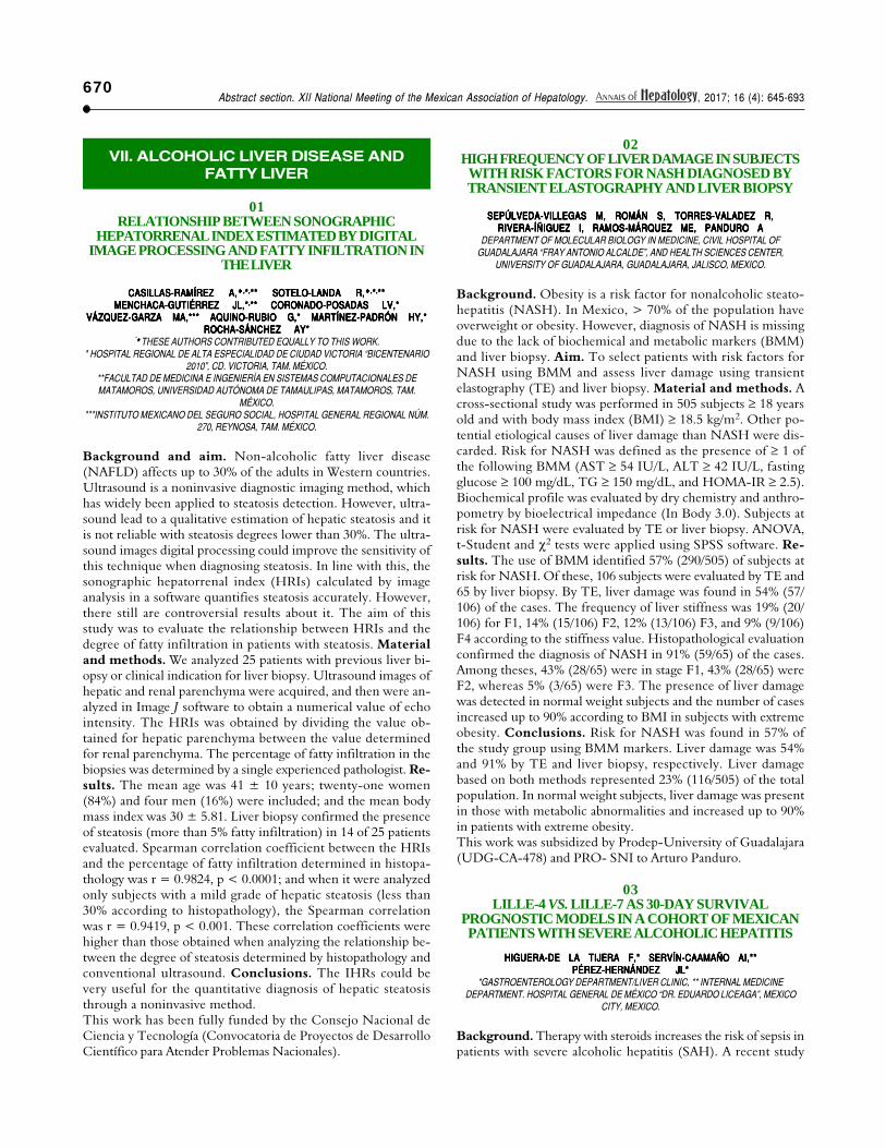
670Abstract section. XII National Meeting of the Mexican Association of Hepatology. , 2017; 16 (4): 645-693
VII. ALCOHOLIC LIVER DISEASE ANDFATTY LIVER
01RELATIONSHIP BETWEEN SONOGRAPHIC
HEPATORRENAL INDEX ESTIMATED BY DIGITALIMAGE PROCESSING AND FATTY INFILTRATION IN
THE LIVER
CASILLAS-RAMÍREZ A,CASILLAS-RAMÍREZ A,CASILLAS-RAMÍREZ A,CASILLAS-RAMÍREZ A,CASILLAS-RAMÍREZ A,♦♦♦♦♦ ,,,,,*****,,,,,** SOTELO-LANDA R,** SOTELO-LANDA R,** SOTELO-LANDA R,** SOTELO-LANDA R,** SOTELO-LANDA R,♦♦♦♦♦,,,,,*****,,,,,**********MENCHACA-GUTIÉRREZ JL,*MENCHACA-GUTIÉRREZ JL,*MENCHACA-GUTIÉRREZ JL,*MENCHACA-GUTIÉRREZ JL,*MENCHACA-GUTIÉRREZ JL,*,,,,,** CORONADO-POSADAS LV,*** CORONADO-POSADAS LV,*** CORONADO-POSADAS LV,*** CORONADO-POSADAS LV,*** CORONADO-POSADAS LV,*
VÁZQUEZ-GARZA MA,*** AQUINO-RUBIO G,* MARTÍNEZ-PADRÓN HY,*VÁZQUEZ-GARZA MA,*** AQUINO-RUBIO G,* MARTÍNEZ-PADRÓN HY,*VÁZQUEZ-GARZA MA,*** AQUINO-RUBIO G,* MARTÍNEZ-PADRÓN HY,*VÁZQUEZ-GARZA MA,*** AQUINO-RUBIO G,* MARTÍNEZ-PADRÓN HY,*VÁZQUEZ-GARZA MA,*** AQUINO-RUBIO G,* MARTÍNEZ-PADRÓN HY,*ROCHA-SÁNCHEZ AY*ROCHA-SÁNCHEZ AY*ROCHA-SÁNCHEZ AY*ROCHA-SÁNCHEZ AY*ROCHA-SÁNCHEZ AY*
¨♦THESE AUTHORS CONTRIBUTED EQUALLY TO THIS WORK.* HOSPITAL REGIONAL DE ALTA ESPECIALIDAD DE CIUDAD VICTORIA “BICENTENARIO
2010”, CD. VICTORIA, TAM. MÉXICO.**FACULTAD DE MEDICINA E INGENIERÍA EN SISTEMAS COMPUTACIONALES DEMATAMOROS, UNIVERSIDAD AUTÓNOMA DE TAMAULIPAS, MATAMOROS, TAM.
MÉXICO.***INSTITUTO MEXICANO DEL SEGURO SOCIAL, HOSPITAL GENERAL REGIONAL NÚM.
270, REYNOSA, TAM. MÉXICO.
Background and aim. Non-alcoholic fatty liver disease(NAFLD) affects up to 30% of the adults in Western countries.Ultrasound is a noninvasive diagnostic imaging method, whichhas widely been applied to steatosis detection. However, ultra-sound lead to a qualitative estimation of hepatic steatosis and itis not reliable with steatosis degrees lower than 30%. The ultra-sound images digital processing could improve the sensitivity ofthis technique when diagnosing steatosis. In line with this, thesonographic hepatorrenal index (HRIs) calculated by imageanalysis in a software quantifies steatosis accurately. However,there still are controversial results about it. The aim of thisstudy was to evaluate the relationship between HRIs and thedegree of fatty infiltration in patients with steatosis. Materialand methods. We analyzed 25 patients with previous liver bi-opsy or clinical indication for liver biopsy. Ultrasound images ofhepatic and renal parenchyma were acquired, and then were an-alyzed in Image J software to obtain a numerical value of echointensity. The HRIs was obtained by dividing the value ob-tained for hepatic parenchyma between the value determinedfor renal parenchyma. The percentage of fatty infiltration in thebiopsies was determined by a single experienced pathologist. Re-sults. The mean age was 41 ± 10 years; twenty-one women(84%) and four men (16%) were included; and the mean bodymass index was 30 ± 5.81. Liver biopsy confirmed the presenceof steatosis (more than 5% fatty infiltration) in 14 of 25 patientsevaluated. Spearman correlation coefficient between the HRIsand the percentage of fatty infiltration determined in histopa-thology was r = 0.9824, p < 0.0001; and when it were analyzedonly subjects with a mild grade of hepatic steatosis (less than30% according to histopathology), the Spearman correlationwas r = 0.9419, p < 0.001. These correlation coefficients werehigher than those obtained when analyzing the relationship be-tween the degree of steatosis determined by histopathology andconventional ultrasound. Conclusions. The IHRs could bevery useful for the quantitative diagnosis of hepatic steatosisthrough a noninvasive method.This work has been fully funded by the Consejo Nacional deCiencia y Tecnología (Convocatoria de Proyectos de DesarrolloCientífico para Atender Problemas Nacionales).
02HIGH FREQUENCY OF LIVER DAMAGE IN SUBJECTS
WITH RISK FACTORS FOR NASH DIAGNOSED BYTRANSIENT ELASTOGRAPHY AND LIVER BIOPSY
SEPÚLVEDA-VILLEGAS M, ROMÁN S, TORRES-VALADEZ R,SEPÚLVEDA-VILLEGAS M, ROMÁN S, TORRES-VALADEZ R,SEPÚLVEDA-VILLEGAS M, ROMÁN S, TORRES-VALADEZ R,SEPÚLVEDA-VILLEGAS M, ROMÁN S, TORRES-VALADEZ R,SEPÚLVEDA-VILLEGAS M, ROMÁN S, TORRES-VALADEZ R,RIVERA-ÍÑIGUEZ I, RAMOS-MÁRQUEZ ME, PANDURO ARIVERA-ÍÑIGUEZ I, RAMOS-MÁRQUEZ ME, PANDURO ARIVERA-ÍÑIGUEZ I, RAMOS-MÁRQUEZ ME, PANDURO ARIVERA-ÍÑIGUEZ I, RAMOS-MÁRQUEZ ME, PANDURO ARIVERA-ÍÑIGUEZ I, RAMOS-MÁRQUEZ ME, PANDURO A
DEPARTMENT OF MOLECULAR BIOLOGY IN MEDICINE, CIVIL HOSPITAL OFGUADALAJARA “FRAY ANTONIO ALCALDE”, AND HEALTH SCIENCES CENTER,
UNIVERSITY OF GUADALAJARA, GUADALAJARA, JALISCO, MEXICO.
Background. Obesity is a risk factor for nonalcoholic steato-hepatitis (NASH). In Mexico, > 70% of the population haveoverweight or obesity. However, diagnosis of NASH is missingdue to the lack of biochemical and metabolic markers (BMM)and liver biopsy. Aim. To select patients with risk factors forNASH using BMM and assess liver damage using transientelastography (TE) and liver biopsy. Material and methods. Across-sectional study was performed in 505 subjects ≥ 18 yearsold and with body mass index (BMI) ≥ 18.5 kg/m2. Other po-tential etiological causes of liver damage than NASH were dis-carded. Risk for NASH was defined as the presence of ≥ 1 ofthe following BMM (AST ≥ 54 IU/L, ALT ≥ 42 IU/L, fastingglucose ≥ 100 mg/dL, TG ≥ 150 mg/dL, and HOMA-IR ≥ 2.5).Biochemical profile was evaluated by dry chemistry and anthro-pometry by bioelectrical impedance (In Body 3.0). Subjects atrisk for NASH were evaluated by TE or liver biopsy. ANOVA,t-Student and χ2 tests were applied using SPSS software. Re-sults. The use of BMM identified 57% (290/505) of subjects atrisk for NASH. Of these, 106 subjects were evaluated by TE and65 by liver biopsy. By TE, liver damage was found in 54% (57/106) of the cases. The frequency of liver stiffness was 19% (20/106) for F1, 14% (15/106) F2, 12% (13/106) F3, and 9% (9/106)F4 according to the stiffness value. Histopathological evaluationconfirmed the diagnosis of NASH in 91% (59/65) of the cases.Among theses, 43% (28/65) were in stage F1, 43% (28/65) wereF2, whereas 5% (3/65) were F3. The presence of liver damagewas detected in normal weight subjects and the number of casesincreased up to 90% according to BMI in subjects with extremeobesity. Conclusions. Risk for NASH was found in 57% ofthe study group using BMM markers. Liver damage was 54%and 91% by TE and liver biopsy, respectively. Liver damagebased on both methods represented 23% (116/505) of the totalpopulation. In normal weight subjects, liver damage was presentin those with metabolic abnormalities and increased up to 90%in patients with extreme obesity.This work was subsidized by Prodep-University of Guadalajara(UDG-CA-478) and PRO- SNI to Arturo Panduro.
03LILLE-4 VS. LILLE-7 AS 30-DAY SURVIVAL
PROGNOSTIC MODELS IN A COHORT OF MEXICANPATIENTS WITH SEVERE ALCOHOLIC HEPATITIS
HIGUERA-DE LA TIJERA F,* SERVÍN-CAAMAÑO AI,**HIGUERA-DE LA TIJERA F,* SERVÍN-CAAMAÑO AI,**HIGUERA-DE LA TIJERA F,* SERVÍN-CAAMAÑO AI,**HIGUERA-DE LA TIJERA F,* SERVÍN-CAAMAÑO AI,**HIGUERA-DE LA TIJERA F,* SERVÍN-CAAMAÑO AI,**PÉREZ-HERNÁNDEZ JL*PÉREZ-HERNÁNDEZ JL*PÉREZ-HERNÁNDEZ JL*PÉREZ-HERNÁNDEZ JL*PÉREZ-HERNÁNDEZ JL*
*GASTROENTEROLOGY DEPARTMENT/LIVER CLINIC, ** INTERNAL MEDICINEDEPARTMENT. HOSPITAL GENERAL DE MÉXICO “DR. EDUARDO LICEAGA”, MEXICO
CITY, MEXICO.
Background. Therapy with steroids increases the risk of sepsis inpatients with severe alcoholic hepatitis (SAH). A recent study
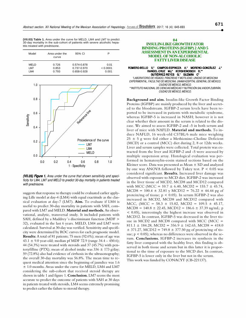
671Abstract section. XII National Meeting of the Mexican Association of Hepatology. , 2017; 16 (4): 645-693
suggests that response to therapy could be evaluated earlier apply-ing Lille model at day-4 (LM4) with equal exactitude as the clas-sical evaluation at day-7 (LM7). Aim. To evaluate if LM4 isuseful to predict 30-day mortality in patients with SAH, com-pared with LM7 and MELD. Material and methods. An obser-vational, analytic, transversal study. It included patients withSAH, defined by a Maddrey´s discriminant function (MDF >32), evaluated in the last 6 years. MELD, LM4 and LM7 werecalculated. Survival at 30-day was verified. Sensitivity and specifi-city were determined by ROC curves for each prognostic model.Results: A total of 81 patients; 75 men (92.6%); mean of age was43.1 ± 9.0 year-old; median pf MDF 72.9 (range 34.4 – 450.6);44 (54.3%) were treated with steroids and 37 (45.7%) with pen-toxyfilline (PTX); mean of alcohol intake was 336 ± 173 g/day;59 (72.8%) also had evidence of cirrhosis in the ultrasonography;the overall 30-day mortality was 56.8%. The mean time to re-quest medical attention since the beginning of jaundice was 1.6± 1.0 months. Areas under the curve for MELD, LM4 and LM7considering the sub-cohort that received steroid therapy areshown in table 1 and figure 1. Conclusion. LM7 seems the mostaccurate to predict the outcome of patients with SAH at 30 daysin patients treated with steroids, LM4 seems extremely promisingto predict earlier the failure to steroid therapy.
04INSULIN-LIKE GROWTH FATOR
BINDING PROTEINS (IGFBP) 2 AND 5ASSESSMENT IN AN EXPERIMENTAL
MODEL OF NON-ALCOHOLICFATTY LIVER DISEASE
ROMERO-BELLO II,* CAMPOS-ESPINOZA A,* MORENO-GONZÁLEZ J,*ROMERO-BELLO II,* CAMPOS-ESPINOZA A,* MORENO-GONZÁLEZ J,*ROMERO-BELLO II,* CAMPOS-ESPINOZA A,* MORENO-GONZÁLEZ J,*ROMERO-BELLO II,* CAMPOS-ESPINOZA A,* MORENO-GONZÁLEZ J,*ROMERO-BELLO II,* CAMPOS-ESPINOZA A,* MORENO-GONZÁLEZ J,*RANGEL-CRUZ MJ,* KERSHENOBICH D,**RANGEL-CRUZ MJ,* KERSHENOBICH D,**RANGEL-CRUZ MJ,* KERSHENOBICH D,**RANGEL-CRUZ MJ,* KERSHENOBICH D,**RANGEL-CRUZ MJ,* KERSHENOBICH D,**
GUTIÉRREZ-REYES G,* GUZMÁN C*GUTIÉRREZ-REYES G,* GUZMÁN C*GUTIÉRREZ-REYES G,* GUZMÁN C*GUTIÉRREZ-REYES G,* GUZMÁN C*GUTIÉRREZ-REYES G,* GUZMÁN C**LABORATORIO DE HÍGADO, PÁNCREAS Y MOTILIDAD, UNIDAD DE MEDICINA
EXPERIMENTAL, FACULTAD DE MEDICINA, UNAM/HOSPITAL GENERAL DE MÉXICO,CIUDAD DE MÉXICO, MÉXICO.
**INSTITUTO NACIONAL DE CIENCIAS MÉDICAS Y NUTRICIÓN SALVADOR ZUBIRÁN,CIUDAD DE MÉXICO, MÉXICO.
Background and aim. Insulin-like Growth Factor BindingProteins (IGFBP) are mainly produced by the liver and secret-ed to the bloodstream. IGFBP-2 serum levels have been re-ported to be increased in patients with metabolic syndrome,whereas IGFBP-5 is increased in NASH; however it is notclear whether their amount in the serum is related to the dis-ease. We aimed to assess IGFBP-2 and -5 in both serum andliver of mice with NAFLD. Material and methods. To in-duce NAFLD, 16 week-old C57BL/6 male mice weighing25 ± 5 g were fed either a Methionine-Choline Deficient(MCD) or a control (MCC) diet during 2, 8 or 12dx weeks.Liver and serum samples were collected. Total protein was ex-tracted from the liver and IGFBP-2 and -5 were assessed bymultiple suspension array. Histological evaluation was per-formed in hematoxylin-eosin stained sections based on theKleiner score. Data was presented as Mean ± SD and analyzedby one way ANOVA followed by Tukey test. P < 0.05 wasconsidered significant. Results. Increased liver damage wasobserved with exposure to MCD diet. IGFBP-2 was increasedin the liver tissue of MCD2, MCD8 and MCD12 comparedwith MCC (MCC = 10.7 ± 6.40, MCD2 = 155.7 ± 45.74,MCD8 = 100.4 ± 32.81 y MCD12 = 76.22 ± 44.44 pg ofprotein/mg of tissue; p < 0.05). In serum IGFBP-2 was alsoincreased in MCD2, MCD8 and MCD12 compared withMCC, (MCC = 58.3 ± 15.02, MCD2 = 109.5 ± 45.17,MCD8 = 140.8 ± 22.45, MCD12 = 186.6 ± 37.39 ng/mL; p< 0.05), interestingly the highest increase was observed inMCD12. In contrast, IGFBP-5 was decreased in the liver tis-sue in MCD2 and MCD8 compared with MCC (MCC =811.1 ± 184.28, MCD2 = 356.9 ± 142.65, MCD8 = 418.0± 371.27, MCD12 = 749.8 ± 377.50 pg of protein/mg of tis-sue; p < 0.05); whereas no differences were observed in the se-rum. Conclusions. IGFBP-2 increases its synthesis in thefatty liver compared with the healthy liver, this finding is ob-served in both tissue and serum but in this latter it is propor-tional to the time of exposure to the MCD diet. In contrast,IGFBP-5 is lower only in the liver but not in the serum.This work was funded by CONACYT (CB-221137).
(VII.03) Table 1. Area under the curve for MELD, LM4 and LM7 to predict30-day mortality in the sub-cohort of patients with severe alcoholic hepa-titis treated with prednisone.
Model Area under the 95% CI Pcurve
MELD 0.726 0.574-0.878 0.01LM7 0.853 0.737-0.970 < 0.0001LM4 0.793 0.658-0.928 0.001
Sens
itivity
1.0
0.8
0.6
0.4
0.2
0.0
0.0 0.2 0.4 0.6 0.8 1.01 - Specificity
(VII.03) Figure 1. (VII.03) Figure 1. (VII.03) Figure 1. (VII.03) Figure 1. (VII.03) Figure 1. Area under the curve that shown sensitivity and speci-ficity for LM4, LM7 and MELD to predict 30-day mortality in patients treatedwith prednisone.
LM7LM4MELD
Procedence of the curve
Referen
ce lin
e
672Abstract section. XII National Meeting of the Mexican Association of Hepatology. , 2017; 16 (4): 645-693
05SEVERE MALNUTRITION IS A PROGNOSTIC
FACTOR RELATED TO EARLY MORTALITY INMEXICAN PATIENTS WITH SEVERE ALCOHOLIC
HEPATITIS
HIGUERA-DE LA TIJERA F,* SERVÍN-CAAMAÑO AI,** LÓPEZ-LADRÓNHIGUERA-DE LA TIJERA F,* SERVÍN-CAAMAÑO AI,** LÓPEZ-LADRÓNHIGUERA-DE LA TIJERA F,* SERVÍN-CAAMAÑO AI,** LÓPEZ-LADRÓNHIGUERA-DE LA TIJERA F,* SERVÍN-CAAMAÑO AI,** LÓPEZ-LADRÓNHIGUERA-DE LA TIJERA F,* SERVÍN-CAAMAÑO AI,** LÓPEZ-LADRÓNDE GUEVARA V,* MACÍAS-ÁNGELES Y,* PÉREZ-HERNÁNDEZ JL*DE GUEVARA V,* MACÍAS-ÁNGELES Y,* PÉREZ-HERNÁNDEZ JL*DE GUEVARA V,* MACÍAS-ÁNGELES Y,* PÉREZ-HERNÁNDEZ JL*DE GUEVARA V,* MACÍAS-ÁNGELES Y,* PÉREZ-HERNÁNDEZ JL*DE GUEVARA V,* MACÍAS-ÁNGELES Y,* PÉREZ-HERNÁNDEZ JL**GASTROENTEROLOGY DEPARTMENT/LIVER CLINIC, ** INTERNAL MEDICINE
DEPARTMENT. HOSPITAL GENERAL DE MÉXICO “DR. EDUARDO LICEAGA”, MEXICOCITY, MEXICO.
Background. Severe alcoholic hepatitis (SAH) has a very highmortality rate in Mexican population, becoming as high as 50 to81% in those classified as ABIC B or C. Aim. To know whichclinical factors are related to early mortality (30 days) in Mexi-can patients with SAH. Material and methods. A historic co-hort study which included patients with SAH defined by aMaddrey‘s discriminant function > 32, evaluated in a five-yearperiod (2010-2015). Quantitative variables were expressed inmean and standard deviation and were compared using t Stu-dent’s test; univariate analysis for qualitative variables was per-formed using χ2 or exact Fisher’s test, odds ratio and 95%confidence intervals were also calculated; the multivariate anal-ysis was performed through Cox regression. Results. 76 pa-tients were included; 72 (94.7%) were men; mean age was 43 ±9.1 year-old, 58 (76.3%) had changes of cirrhosis in the ultra-sonography. According with the subjective global assessment(SGA) 38 (50%) had severe malnutrition, 22 (28.9%) were atrisk of malnutrition, 16 (21.1%) were well-nourished. Overall30-day mortality in this cohort was 46 patients (60.5%). In theunivariate analysis, the following were associated with 30.daymortality: Concomitant cirrhosis (OR = 2.3; 95%CI: 1.5-3.4; P< 0.0001), development of encephalopathy (OR = 5.6; 95%CI:2.5-12.5; P < 0.0001), development of renal failure (OR = 6.2;95%CI:2.5-15.6; P < 0.0001), variceal bleeding (OR = 4.4;95%CI:1.7-11.3; P < 0.0001), infection (OR = 2.7;95%CI:1.3-5.8; P = 0.004), according with the SGA: at risk ofmalnutrition (OR = 2.0; 95%CI:1.2-3.3; P = 0.01), and severemalnutrition (OR = 5.8; 95%CI: 2.0-16.4; P < 0.0001); thera-py with prednisone or pentoxyfilline did not influence on sur-vival, and was not difference between therapy groups. Whenthese variables were adjusted in a multivariate model, only se-vere malnutrition was related to 30-day mortality: OR = 6.4;95%CI: 1.9-22.1; P = 0.003. Conclusions. Nutritional statusis a key factor associated with early mortality (30 days) in Mexi-can patients with SAH.
06HISTOPATHOLOGIC DIFFERENCES IN AFLD-NAFLDIN A MURINE MODEL OF CHRONIC INTAKE OF HIGH-
FAT DIET, ETHANOL AND THEIR INTERACTION
RAMÍREZ-MENDOZA A,* LEÓN-MANCILLA B,* MEDINA-ÁVILA Z,*RAMÍREZ-MENDOZA A,* LEÓN-MANCILLA B,* MEDINA-ÁVILA Z,*RAMÍREZ-MENDOZA A,* LEÓN-MANCILLA B,* MEDINA-ÁVILA Z,*RAMÍREZ-MENDOZA A,* LEÓN-MANCILLA B,* MEDINA-ÁVILA Z,*RAMÍREZ-MENDOZA A,* LEÓN-MANCILLA B,* MEDINA-ÁVILA Z,*ARÉVALO-SÁNCHEZ T,* CAMPOS-ESPINOSA A,* GARCÍA-LÓPEZ B,*ARÉVALO-SÁNCHEZ T,* CAMPOS-ESPINOSA A,* GARCÍA-LÓPEZ B,*ARÉVALO-SÁNCHEZ T,* CAMPOS-ESPINOSA A,* GARCÍA-LÓPEZ B,*ARÉVALO-SÁNCHEZ T,* CAMPOS-ESPINOSA A,* GARCÍA-LÓPEZ B,*ARÉVALO-SÁNCHEZ T,* CAMPOS-ESPINOSA A,* GARCÍA-LÓPEZ B,*
RANGEL-CRUZ MJ,* ROMERO-BELLO II,* ROSIQUE-ORAMAS D,*RANGEL-CRUZ MJ,* ROMERO-BELLO II,* ROSIQUE-ORAMAS D,*RANGEL-CRUZ MJ,* ROMERO-BELLO II,* ROSIQUE-ORAMAS D,*RANGEL-CRUZ MJ,* ROMERO-BELLO II,* ROSIQUE-ORAMAS D,*RANGEL-CRUZ MJ,* ROMERO-BELLO II,* ROSIQUE-ORAMAS D,*VERDEJA P,* PÉREZ-TORRES A,** SÁNCHEZ J,* GUZMÁN C,*VERDEJA P,* PÉREZ-TORRES A,** SÁNCHEZ J,* GUZMÁN C,*VERDEJA P,* PÉREZ-TORRES A,** SÁNCHEZ J,* GUZMÁN C,*VERDEJA P,* PÉREZ-TORRES A,** SÁNCHEZ J,* GUZMÁN C,*VERDEJA P,* PÉREZ-TORRES A,** SÁNCHEZ J,* GUZMÁN C,*
GUTIÉRREZ-REYES G*GUTIÉRREZ-REYES G*GUTIÉRREZ-REYES G*GUTIÉRREZ-REYES G*GUTIÉRREZ-REYES G** LABORATORIO DE HÍGADO, PÁNCREAS Y MOTILIDAD, UNIDAD DE MEDICINA
EXPERIMENTAL, FACULTAD DE MEDICINA, UNAM/HOSPITAL GENERAL DE MÉXICO.**FSIPYM DEPTO. BIOLOGÍA CELULAR Y TISULAR, FACULTAD DE MEDICINA, UNAM.
MÉXICO.
Background and aim. Excessive alcohol intake is a risk factorfor the development of alcoholic fatty liver disease (AFLD). Inthe last decade, prevalence of obesity has increased diseases in-cluding non-alcoholic fatty liver disease (NAFLD). Bothpathologies are extended in our population and are risk factors todeveloping liver fibrosis. These diseases have been independent-ly studied, but evidence of the coincidence of them in the samesubject is lacking. Previously, we have shown the mid-term in-teraction of both etiologies. We aimed to identify the morpho-logical alterations in the liver due to High-Fat diet and ethanolintake in mice at long-term. Material and methods. 6 groupsof C57BL/6 mice, aged 10 weeks were assigned to the fol-lowing treatments: (1) Chow Diet/Water (DC/A), (2) ChowDiet/Ethanol (DC/OH), (3) High-Fat Diet/Water (DHF/A), (4)High-Fat Diet/Ethanol (DHF/OH), (5) 1stHigh-Fat Diet/Wa-ter-2nd Chow Diet/Ethanol (DHF/A-DC/OH) and (6) 1st
Chow Diet/Ethanol-2nd High-Fat Diet/Water (DC/OH-DHF/A). Mice in groups 1-4 received their treatments for 6 months.Groups 5 and 6 received the 1st treatment for 4 months fol-lowed by the 2nd for 6 months. Histological assessment wasperformed by using H&E and Masson’s tricrhome staining. Re-sults. Histological changes were observed in the liver accordingto the experimental group. DC/OH showed slight architecturalchanges due to sinusoidal distension and no steatosis comparedwith DC/A. DHF/A showed microvesicular diffused steatosismainly in zones 2 and 3 in the hepatic acinus with inflammatoryinfiltrate. DHF/OH group exhibited diffused micro-macrovesic-ular steatosis in zones 2 and 3. DHF/A-DC/OH showed micro-vesicular focal steatosis in zone 2 whereas DC/OH-DHF/Ashowed micro and macrovesicular diffused steatosis in the wholeacinus. Conclusion. Steatosis and other architectural changesin the liver depend on the interaction of the diet and the drink-ing. Structural changes in specific locations of the acinus wereobserved according to the group. Our data suggest that, at leastin mice, a chronic alcohol intake period followed by anobesogenic diet induces a severer steatosis when compared to in-dependent etiologies.This work was partially funded by Conacyt (CB-221137).
673Abstract section. XII National Meeting of the Mexican Association of Hepatology. , 2017; 16 (4): 645-693
07PREVALENCE OF NON-ALCOHOLIC FATTY LIVER
DISEASE DIAGNOSED BY BIOPSY IN A PERIODO OF5 YEARS
MACÍAS ÁNGELES Y, HIGUERA-DE LA TIJERA,MACÍAS ÁNGELES Y, HIGUERA-DE LA TIJERA,MACÍAS ÁNGELES Y, HIGUERA-DE LA TIJERA,MACÍAS ÁNGELES Y, HIGUERA-DE LA TIJERA,MACÍAS ÁNGELES Y, HIGUERA-DE LA TIJERA,LÓPEZ-LADRÓN DE GUEVARA V, PÉREZ HERNÁNDEZ JLÓPEZ-LADRÓN DE GUEVARA V, PÉREZ HERNÁNDEZ JLÓPEZ-LADRÓN DE GUEVARA V, PÉREZ HERNÁNDEZ JLÓPEZ-LADRÓN DE GUEVARA V, PÉREZ HERNÁNDEZ JLÓPEZ-LADRÓN DE GUEVARA V, PÉREZ HERNÁNDEZ J
SERVICIO DE GASTROENTEROLOGÍA. HOSPITAL GENERAL DE MÉXICO. “DR.EDUARDO LICEAGA”, CIUDAD DE MÉXICO. MÉXICO.
Background. Non alcoholic fatty liver disease (NAFDL), isthe number one cause of impaired liver function test. México70% of adults are overweight and obese, but few studies haveevaluated the prevalence (NAFDL). Aim. Establish the preva-lence NAFDL in a period of 5 years of patients who underwenthepatic biopsy, describe the histological findings and characteris-tic biochemistry compare between simple steatosis and steato-hepatitis/fibrosis. Material and methods. Analyticalobservational study. The Hospital General de México biopsyrecord was review, in the period from January 2012 to march2016 the diagnosis was intentionally sough NAFDL and steato-hepatitis. Registering the histopathological characteristic, thedemographic, clinical and biochemical data were collected clini-cal files. Comparing the characteristics between group 1 (simplesteatosis = group of low risk of disease progression and groupinflammation/fibrosis group at high risk of disease progression.Result. In the studied period 292 hepatic biopsies were pe-formed, of which 49 reported NAFDL. La prevalence was16.7%, mean age 42.4 ± 14.8 years, woman the occupied 77.6%(n = 38), IMC 23.4 ± 5.2 kg/m2, the diabetic 9.7% and pre-di-abetic 32.3%.The repoted characteristic of the biopsies were61.2% (n = 30); presented some degree of steatosis: 43.3% (n =13) mixed steatosis, 36.6% (n = 11) macrovesicular steatosis, st-eatosis only 13.3%, microvesicular steatosis 6.6% (n = 2). Weidentified nineteen patients (38.7%) whit steatohepatitis, in-flammatory infiltrate n = 8 (42%), fibrosis n = 5 (26.3%) andhepatitis 31.5%. When comparing between groups 1 and 2 therewas only significant difference in fasting serum glucose (87.3 ±11.8 vs. 109.2 ± 31.4 mg/dL respectively, (p = 0.03). Conclu-sion. Is a pathology affecting women of productive age, the al-teration of serum glucose was in greater patients with factorsconsidered to be at risk of progression of disease (inflammation/fibrosis).This work did not receive any sponsorship.
08PREVALENCE OF FIBROSIS AND STEATOSIS
DIAGNOSED WITH TRANSIENT ELASTOGRAPHY(FIBROSCAN) IN PATIENTS OF HOSPITAL GENERAL
DE MEXICO O.D. “EDUARDO LICEAGA”
MACIAS ANGELES Y, HIGUERA-DE LA TIJERA,MACIAS ANGELES Y, HIGUERA-DE LA TIJERA,MACIAS ANGELES Y, HIGUERA-DE LA TIJERA,MACIAS ANGELES Y, HIGUERA-DE LA TIJERA,MACIAS ANGELES Y, HIGUERA-DE LA TIJERA,LÓPEZ-LADRÓN DE GUEVARA V, PÉREZ HERNÁNDEZ JLÓPEZ-LADRÓN DE GUEVARA V, PÉREZ HERNÁNDEZ JLÓPEZ-LADRÓN DE GUEVARA V, PÉREZ HERNÁNDEZ JLÓPEZ-LADRÓN DE GUEVARA V, PÉREZ HERNÁNDEZ JLÓPEZ-LADRÓN DE GUEVARA V, PÉREZ HERNÁNDEZ J
SERVICIO DE GASTROENTEROLOGÍA/CLÍNICA DE HÍGADO. HOSPITAL GENERAL DEMÉXICO. “DR. EDUARDO LICEAGA”, CIUDAD DE MÉXICO. MÉXICO.
Background. Transiet elastography (TE) measures liver stiff-ness in patients suffering from different chronic liver diseases.This novel parameter “controlled atenuation parameter” (CAP)is efficient in detecting low grade steatosis. Aim. To determinethe prevalence of hepatic fibrosis and steatosis in the group of
(VII.10) Table 1. Alcohol consumption, cell populations, and cytokine con-centrations in the different groups.
Control Alcoholism Cirrhosisn = 294 n = 27 n = 37
Alcohol intake (g/day) 2 ± 1a 185 ±45b 322 ±297c,d
Years of consumption NA 22 ±14 26 ±9Neutrophil(%) 34 ± 15a 54 ±14b 61 ±12c
Lymphocyte (%) 57 ± 19a 34 ±13b 27 ±10c
Monocyte (%) 6 ± 4a 9 ±2b 9 ±4c
IL-6 (pg/mL) 0.3 (0-16)a 0.7 (0.2-3)b 4.3 (0.4-3181)c,d
IL-8/CXCL-8 (pg/mL) 1 (0.07-21)a 8 (0.7-106)b 18 (1-455)c
IL-10 (pg/mL) 0.4 (0-349)a 0.1 (0-118) 1.2 (0-177)c
TNF-α (pg/mL) 0.1 (0-5)a 0.4 (0-1) 1 (0-30)c,d
Data expressed in mean ± SD; median (min-max). Significant differencesbetween: a: all groups. b: control and alcoholism. C: control and cirrhosis.d: alcoholism and cirrhosis. NA: not apply.
patients who submitted the study of TE in the period from Jan-uary to March 2017. Material and methods. Observationalanalytic study, the result of TE performed of Hospital Generalde México in the 3-month period. The following date were col-lected; age, sex, weight, heigth, body mass index (BMI), withdiabetes mellitus (DM), dyslipidemia and sending reason. TheET with the fibroscan interpretation guide was determined inkilopascals kPa. The level of hepatic fibrosis F1-F4 and steatosisby CAP dB/m with cohort point 233 dB/m. Results. Therewere 80 TE, only studies 57 that were complete. Predominating61.4% (n = 35) women, average age 48 ± 24, BMI 25.6, DM40% (n = 14) and dyslipidemia 54% (n = 19). The main indi-cations for the realization of this study were; obese patients, st-eatosis by usg and VHC. Getting 19 patients without fibrosis, n= 16 with some degree of fibrosis F1-F4. F1: (n = 3), F2 (n =4), F3 (n = 5), the characteristic that predominated; obesity,overweight and dyslipidemia, F4 (n = 4), the 4 patients present-ed some degree of dyslipidemia but only 2 with overweight. Thedegree of steatosis: n = 19 mild steatosis (S1), n = 3 moderate(S2), severe (S3) n = 4, predominating obese, DM y dyslipi-demia and 6 normal weight. The men 38% (n = 22), averageage 50 ± 25, BMI 25, the 40% with DM (n = 9), 50% dyslipi-demia (n = 11). Fourteen patients with fibrosis (F1-F4): F1 (n= 1), F2(n = 2), F3 (n = 1). F4 (n = 10) three obesity patients,six BMI < 25 kg/m, seven of them with DM, the degree of st-eatosis S1-S3, prevail BMI < 25 kg/m y no diabetic. In patientssubmitted to this study were found 52% have a degree of fibro-sis and 64% degree of steatosis. Conclusions. Patients withmetabolic syndrome have fibrosis whithout being liverworts.
09FIBROSIS AND STEATOSIS IN PATIENTS WITHMORBID OBESITY EVALUATED BY TRANSIENT
ELASTOGRAPHY
PÉREZ HERNÁNDEZ J, LÓPEZ LADRÓN DE GUEVARA V,PÉREZ HERNÁNDEZ J, LÓPEZ LADRÓN DE GUEVARA V,PÉREZ HERNÁNDEZ J, LÓPEZ LADRÓN DE GUEVARA V,PÉREZ HERNÁNDEZ J, LÓPEZ LADRÓN DE GUEVARA V,PÉREZ HERNÁNDEZ J, LÓPEZ LADRÓN DE GUEVARA V,MACÍAS ÁNGELES Y, PÉREZ SOTO F, HIGUERA DE LA TIJERA F.MACÍAS ÁNGELES Y, PÉREZ SOTO F, HIGUERA DE LA TIJERA F.MACÍAS ÁNGELES Y, PÉREZ SOTO F, HIGUERA DE LA TIJERA F.MACÍAS ÁNGELES Y, PÉREZ SOTO F, HIGUERA DE LA TIJERA F.MACÍAS ÁNGELES Y, PÉREZ SOTO F, HIGUERA DE LA TIJERA F.
LIVER CLINIC FROM GASTROENTEROLOGY DEPARTMENT, HOSPITAL GENERAL DEMÉXICO “DR. EDUARDO LICEAGA”, MEXICO CITY, MEXICO.
Introduction. Obesity are closely related to the development ofliver steatosis and fibrosis; transient elastography (TE) is a usefulnon-invasive method to evaluate these conditions, and it is val-
674Abstract section. XII National Meeting of the Mexican Association of Hepatology. , 2017; 16 (4): 645-693
id if the interquartile range (IQR) is < 30 and with a successrate > 60% (valid measures between all taken measures). Inmore than 25% of the morbid obese patients, the TE cannot beperformed because is not easy to find a suitable window. Aim.To describe the findings on TE in patients with morbid obesity.Material and methods. A case-series study that included pa-tients with body mass index (BMI) > 40 who had a successfulTE. Results. A total of 20 patients, 15(75%) females, age 42.5± 10.6 year-old, weight 133.2 ± 31.4 kg, height 162 ±8.88cm, BMI 50.52 ± 8.56 kg/m2, Kpa 17.90 ± 7.2, IQR 5.2 ±5.2, grade of fibrosis 2.2 ± 1.4, CAP 371 ± 37.8 IQR 21.0512.2, percent of success 75%, range 60 a 100%. The grade of st-eatosis was III in a100% of patients, 5(25%) patients had not fi-brosis, 15(75%) had advanced fibrosis. Conclusions: All patientshad an IQR less than 20 and a percent of success greater than60%, which validate the study. In Patients with morbid obesitythe frequency of severe steatosis and advanced fibrosis is high.
10CHRONIC AND ACTIVE ALCOHOL CONSUMPTION
MODIFIED THE IMMUNOLOGICAL RESPONSE
MEDINA-ÁVILA Z,* RIZO-TÉLLEZ S,* ROSIQUE ORAMAS D,*MEDINA-ÁVILA Z,* RIZO-TÉLLEZ S,* ROSIQUE ORAMAS D,*MEDINA-ÁVILA Z,* RIZO-TÉLLEZ S,* ROSIQUE ORAMAS D,*MEDINA-ÁVILA Z,* RIZO-TÉLLEZ S,* ROSIQUE ORAMAS D,*MEDINA-ÁVILA Z,* RIZO-TÉLLEZ S,* ROSIQUE ORAMAS D,*PÉREZ-HERNÁNDEZ JL,** BEJAR Y,** HIGUERA F,**PÉREZ-HERNÁNDEZ JL,** BEJAR Y,** HIGUERA F,**PÉREZ-HERNÁNDEZ JL,** BEJAR Y,** HIGUERA F,**PÉREZ-HERNÁNDEZ JL,** BEJAR Y,** HIGUERA F,**PÉREZ-HERNÁNDEZ JL,** BEJAR Y,** HIGUERA F,**
CORDERO-PÉREZ P,*** KERSHENOBICH D,* GUTIÉRREZ REYES G*CORDERO-PÉREZ P,*** KERSHENOBICH D,* GUTIÉRREZ REYES G*CORDERO-PÉREZ P,*** KERSHENOBICH D,* GUTIÉRREZ REYES G*CORDERO-PÉREZ P,*** KERSHENOBICH D,* GUTIÉRREZ REYES G*CORDERO-PÉREZ P,*** KERSHENOBICH D,* GUTIÉRREZ REYES G** LABORATORY HIPAM, EXPERIMENTAL MEDICINE UNIT, UNIVERSIDAD NACIONAL
AUTÓNOMA DE MÉXICO. MÉXICO.** BLOOD BANK, LIVER CLINIC, GENERAL HOSPITAL OF MÉXICO. MÉXICO.
*** LIVER UNIT AND MOLECULAR MEDICINE. UNIVERSITY HOSPITAL. UNIVERSIDADAUTÓNOMA DE NUEVO LEÓN, MONTERREY, NUEVO LEÓN. MÉXICO.
Background. It is known that chronic alcohol intake alters in-nate and adaptive immune response in humans; however, theseeffects have not been evaluated in regard to active consump-tion. Aim. To evaluate the immune response in subjects withchronic and active alcohol consumption. Material and meth-ods. Transversal study which includes: group 1: control sub-jects; group 2: alcoholics without clinical or biochemical data ofhepatic lesion; group 3: subjects with alcoholic liver cirrhosis.Neutrophil, lymphocyte and monocyte percentage was obtainedthrough Complete Blood Count; and serum cytokine concen-trations with Luminex technology. Data were analyzed usingANOVA, orthogonal analysis and U-Mann Whitney, statisti-cally significant differences were considered from p < 0.05. Re-sults. We included 358 subjects; the mean age by group was: 35± 10, 40 ± 13, 43 ± 7 years, respectively (p < 0.001). Conclu-sions. In subjects, who presented active alcoholism without he-patic damage, innate immune cells were increased as well aspro-inflammatory cytokine concentrations and the adaptive re-sponse decreased; when the subjects presented alcoholic cirrhosisand chronic alcohol consumption is active, other cytokines wereincreased as TNF-α, IL-6 (considered in this study as hepato-protector) and IL-10 as anti-inflammatory, that could be offset-ting the damage generated.This work has been partially supported by CONACyT:SALUD-2016-1-272579 and PAPIIT-UNAM TA200515.
11COMPARATIVE STUDY OF LIPID PERCENTAGE INTHE LIVER OF TWO EXPERIMENTAL MODELS OF
NON-ALCOHOLIC FATTY LIVER DISEASE:METHIONINE-CHOLINE DEFICIENT (MCD) DIET VS.
HIGH-FAT DIET (HFD)
CAMPOS-ESPINOSA A,* RAMÍREZ-MENDOZA A,* ROMERO-BELLO II,*CAMPOS-ESPINOSA A,* RAMÍREZ-MENDOZA A,* ROMERO-BELLO II,*CAMPOS-ESPINOSA A,* RAMÍREZ-MENDOZA A,* ROMERO-BELLO II,*CAMPOS-ESPINOSA A,* RAMÍREZ-MENDOZA A,* ROMERO-BELLO II,*CAMPOS-ESPINOSA A,* RAMÍREZ-MENDOZA A,* ROMERO-BELLO II,*ARÉVALO-SÁNCHEZ AT,* OLMOS-VALDEZ K,* LEÓN-MANCILLA B,*ARÉVALO-SÁNCHEZ AT,* OLMOS-VALDEZ K,* LEÓN-MANCILLA B,*ARÉVALO-SÁNCHEZ AT,* OLMOS-VALDEZ K,* LEÓN-MANCILLA B,*ARÉVALO-SÁNCHEZ AT,* OLMOS-VALDEZ K,* LEÓN-MANCILLA B,*ARÉVALO-SÁNCHEZ AT,* OLMOS-VALDEZ K,* LEÓN-MANCILLA B,*
MEDINA-ÁVILA Z,* GARCÍA-LÓPEZ B,* RANGEL-CRUZ MJ,*MEDINA-ÁVILA Z,* GARCÍA-LÓPEZ B,* RANGEL-CRUZ MJ,*MEDINA-ÁVILA Z,* GARCÍA-LÓPEZ B,* RANGEL-CRUZ MJ,*MEDINA-ÁVILA Z,* GARCÍA-LÓPEZ B,* RANGEL-CRUZ MJ,*MEDINA-ÁVILA Z,* GARCÍA-LÓPEZ B,* RANGEL-CRUZ MJ,*ROSIQUE-ORAMAS D,* VERDEJA P,* PÉREZ- TORRES A,**ROSIQUE-ORAMAS D,* VERDEJA P,* PÉREZ- TORRES A,**ROSIQUE-ORAMAS D,* VERDEJA P,* PÉREZ- TORRES A,**ROSIQUE-ORAMAS D,* VERDEJA P,* PÉREZ- TORRES A,**ROSIQUE-ORAMAS D,* VERDEJA P,* PÉREZ- TORRES A,**
SÁNCHEZ J,* GUTIÉRREZ-REYES G,* GUZMÁN C*SÁNCHEZ J,* GUTIÉRREZ-REYES G,* GUZMÁN C*SÁNCHEZ J,* GUTIÉRREZ-REYES G,* GUZMÁN C*SÁNCHEZ J,* GUTIÉRREZ-REYES G,* GUZMÁN C*SÁNCHEZ J,* GUTIÉRREZ-REYES G,* GUZMÁN C**LABORATORIO DE HÍGADO, PÁNCREAS Y MOTILIDAD, UNIDAD DE MEDICINA
EXPERIMENTAL, FACULTAD DE MEDICINA, UNAM/HOSPITAL GENERAL DE MÉXICO.CIUDAD DE MÉXICO, MÉXICO.
**FSIPYM DEPTO. BIOLOGÍA CELULAR Y TISULAR, FACULTAD DE MEDICINA, UNAM.MÉXICO.
Introduction and aim. Experimental models of non-alcoholicfatty liver disease induced by diet exhibit differences in the timeof exposure that is needed to achieve different degrees of thedisease as well as the susceptibility of the strains used. Methio-nine-Choline Deficient (MCD) Diet is a model of NAFLD andis not associated to insulin resistance or metabolic alterations.High-Fat Diet (HFD) model induces obesity, insulin resistanceand NAFLD. We aimed to compare lipid accumulation in theliver among these two models of NAFLD, MCD vs HFD. Ma-terial and methods. Male C57BL76 mice weighing 25 ± 5gand 14 ± 2 weeks old, were randomly assigned to the followinggroups: MCD group was subdivided in control (MCC) and de-ficient (MCD) and fed the corresponding diet for 2, 8 or 12weeks; HFD group was divided in control (CD) and HFD and fedfor 4 or 6 months. Frozen sections were obtained andstained Oil-Red O. Morphometric analysis was performed toassess the percentage of fat. Data=Median(rank), one-wayANOVA followed by Dunn’s test. p ≤ 0.0001. Results. MCDmodel showed an increased %lipid in the liver of MCD treatedcompared with MCC; MCD12 %lipid was significantly higherthan the observed in MCC and MCD2 (MCC = 0.19 ± 0.42,MCD2 = 6.09 ± 6.00, MCD = 8.00 ± 5.76, MCD12 = 9.65± 5.19 %). In the obesity group, an increased %lipid was ob-served in HFD6m compared with CD (CD = 1.1 ± 0.40;HFD4m = 3.3 ± 3.02; HFD6m = 11.5 ± 9.28 %). Conclu-sions. Both models of NAFLD show progressive increase in st-eatosis with the time of exposure to the diet. In the MCDmodel, hepatic steatosis is observed at short-term compared withHDF. Although both models show a similar %lipid at the timeanalyzed, other studies have shown a higher degree of liverdamage en MCD (e.g. NASH is observed in MCD12). Choiceof the appropriate model should consider not only the amountof fat in the liver, but also the stage of NAFLD and the pres-ence or absence of other metabolic complications.This work was funded by Conacyt (CB-221137).
675Abstract section. XII National Meeting of the Mexican Association of Hepatology. , 2017; 16 (4): 645-693
12EVALUATION OF OXIDATIVE STRESS THROUGHTHE LEVELS OF GSH AND GSH/GSSG RATIO IN
PEDIATRIC PATIENTS WITH NAFLD
VERDEJA-PADILLA PA,* MEJÍA L,** MEDINA-ÁVILA Z,* CRUZ-BAUTISTA R,**VERDEJA-PADILLA PA,* MEJÍA L,** MEDINA-ÁVILA Z,* CRUZ-BAUTISTA R,**VERDEJA-PADILLA PA,* MEJÍA L,** MEDINA-ÁVILA Z,* CRUZ-BAUTISTA R,**VERDEJA-PADILLA PA,* MEJÍA L,** MEDINA-ÁVILA Z,* CRUZ-BAUTISTA R,**VERDEJA-PADILLA PA,* MEJÍA L,** MEDINA-ÁVILA Z,* CRUZ-BAUTISTA R,**SUÁREZ L,** ROSIQUE-ORAMAS D,* RIZO-TÉLLEZ S,*SUÁREZ L,** ROSIQUE-ORAMAS D,* RIZO-TÉLLEZ S,*SUÁREZ L,** ROSIQUE-ORAMAS D,* RIZO-TÉLLEZ S,*SUÁREZ L,** ROSIQUE-ORAMAS D,* RIZO-TÉLLEZ S,*SUÁREZ L,** ROSIQUE-ORAMAS D,* RIZO-TÉLLEZ S,*
GALICIA-MORENO M,* CORDERO P,*** MUÑOZ L,***GALICIA-MORENO M,* CORDERO P,*** MUÑOZ L,***GALICIA-MORENO M,* CORDERO P,*** MUÑOZ L,***GALICIA-MORENO M,* CORDERO P,*** MUÑOZ L,***GALICIA-MORENO M,* CORDERO P,*** MUÑOZ L,***GUTIÉRREZ-REYES G*GUTIÉRREZ-REYES G*GUTIÉRREZ-REYES G*GUTIÉRREZ-REYES G*GUTIÉRREZ-REYES G*
*HIPAM LABORATORY, UNIT OF EXPERIMENTAL MEDICINE, FACULTY OF MEDICINE,UNAM. GENERAL HOSPITAL OF MEXICO. MEXICO.
**PEDIATRIC HOSPITAL OF IZTAPALAPA, SEDESA. GDF. *** AUTONOMOUSUNIVERSITY OF NUEVO LEÓN. MEXICO.
Introduction. The prevalence of non-alcoholic fatty liver dis-ease (NAFLD) has recently increased in pediatric patients by itsassociation with metabolic syndrome and obesity. Glutathioneis a molecule with antioxidant capacity that inactivates perox-ides. More than 90% of Glutathione is found in reduced form(GSH) and the rest it’s in oxidized form (GSSG). GSH/GSSGratio is useful to determine the presence of Oxidative Stress(OS). However, the concentrations of GSH and GSSG, as wellas the role in the pathophysiology of NAFLD in pediatric pa-tient, are currently unknown. Objetives. Determine the con-centrations of GSH and GSSG, as well as the GSH/GSSG ratioin pediatric patients with obesity and NAFLD. Material andmethods. A cross-sectional study was performed. Patients ofpediatric age with body mass index over 95th percentile accord-ing to World Health Organization criteria, and without previ-ous liver or metabolic disease, were included. The diagnosis ofNAFLD was performed by Hepatic Ultrasonography. GSH andGSSG levels were determined in blood (Calbiochem, USA) andGSH/GSSG ratio was calculated. Finally, we compare the differ-ences between NAFLD and non-NAFLD groups. We deter-mined statistically significant differences at 95% confidence usingnon-parametric test of median differences. Results. A total of116 patients were included. The mean age was 10.59 ± 3.17 years,with male predominance (62.5%). The diagnosis of NALFD wasmade in 89 patients (76.72%). The results of GSH and GSH/GSSG ratio are expressed in median and interquartile range. Pa-tients with NAFLD compared to non-NAFLD patients had de-creased levels of GSH (378.5 ± 349.5-443.2 μmol vs. 490.8 ±409.8-771.3 μmol, p = 0.0001) and the GSH/GSSG ratio (0.17 ±-0.67-1.41 vs. 1.44 ± -0.36-8.93, p = 0.048). Conclusions. Lowlevels of GSH indicate decreased antioxidant capacity. For otherhand, decrement of GSH/GSSG ratio suggest increased OS inpediatric patients with obesity and NAFLD.Financial support by: UNAM-PAPIIT TA 200515.
13SELECTION OF TREATMENTS IN NON-ALCOHOLIC
FATTY LIVER IN CLINICAL PRACTICE THROUGHTHE EVALUATION OF A LITERATURE REVIEW
LARA ESPARZA M, CASTILLO BARRERA J,LARA ESPARZA M, CASTILLO BARRERA J,LARA ESPARZA M, CASTILLO BARRERA J,LARA ESPARZA M, CASTILLO BARRERA J,LARA ESPARZA M, CASTILLO BARRERA J,GALICIA HERNÁNDEZ G, PAREDES MARTÍNEZ DE ARREDONDO H,GALICIA HERNÁNDEZ G, PAREDES MARTÍNEZ DE ARREDONDO H,GALICIA HERNÁNDEZ G, PAREDES MARTÍNEZ DE ARREDONDO H,GALICIA HERNÁNDEZ G, PAREDES MARTÍNEZ DE ARREDONDO H,GALICIA HERNÁNDEZ G, PAREDES MARTÍNEZ DE ARREDONDO H,
TORRE DELGADILLO A, Y MARTÍNEZ VÁZQUEZ S.TORRE DELGADILLO A, Y MARTÍNEZ VÁZQUEZ S.TORRE DELGADILLO A, Y MARTÍNEZ VÁZQUEZ S.TORRE DELGADILLO A, Y MARTÍNEZ VÁZQUEZ S.TORRE DELGADILLO A, Y MARTÍNEZ VÁZQUEZ S.DEPARTMENT OF GASTROENTEROLOGY. INSTITUTO NACIONAL DE CIENCIAS
MÉDICAS Y NUTRICIÓN “SALVADOR ZUBIRÁN”. CIUDAD DE MÉXICO, MÉXICO.
Background and objective. Non-alcoholic fatty liver diseasetreatment guidelines highlight four recommendations with
greater strength of evidence including lifestyle interventions,vitamin E supplementation, omega-3 fatty acid, and statins.The aim of this study was to contrast the recommendationswith the selection of treatment carried out in clinical practicein a third level hospital through a review of the literature bygastroenterologist. Material and methods. Based on themethodology of a systematic review of the literature, the re-search was made in PubMed, Scielo, Cochrane and Elsevierwith the following keywords: “NASH in obesity patients”,“NAFLD in obesity patients”, “Nutrition therapy in NASHpatients “And” Supplementation in NASH patients” collectingfree complete, randomized clinical trial articles published be-tween 2009 and 2015. The first selection was made by one in-dependent reviewer considering the selection criteria, thesecond choice was made by two reviewers considering the arti-cles had the keywords in the title. Using the CONSORTguidelines, the reviewers selected the sample of articles concen-trated according to the best qualifications by a third reviewer,with the current monthly costs of each treatment. Results.There were found 660 items located in the electronic bases,642 were excluded, which not meet the selection criteria, leav-ing 18 articles. Of the second selection they were 8 articlesthat met the CONSORT criteria for the quality of clinical tri-als. The reviewers rated ezetimibe as the best treatment andwith less punctuation they rate changes for lifestyle. The costsfor the treatment of ezetimibe sum up a total of 70 USD permonth and for the change in lifestyle 108 USD per month.Conclusion. In clinical practice, according to the review ofliterature, the pharmacological use as ezetimibe/statins wasconsidered the best treatment for non-alcoholic fatty liver dis-ease although the effect is low, the monthly price is apparentlylower than changes in lifestyle which are more effective in thetreatment of the disease.
14DETERMINATION OF IGFBP-3 IN SUBJECTS WITH
DIFFERENT BODY MASS INDEX AS A NONALCOHOLIC GREASE LIVER MARKER
PÉREZ HERNÁNDEZ J,* HIGUERA DE LA TIJERA F,* PÉREZ SOTO F,*PÉREZ HERNÁNDEZ J,* HIGUERA DE LA TIJERA F,* PÉREZ SOTO F,*PÉREZ HERNÁNDEZ J,* HIGUERA DE LA TIJERA F,* PÉREZ SOTO F,*PÉREZ HERNÁNDEZ J,* HIGUERA DE LA TIJERA F,* PÉREZ SOTO F,*PÉREZ HERNÁNDEZ J,* HIGUERA DE LA TIJERA F,* PÉREZ SOTO F,*ROSIQUE ORAMAS D,ROSIQUE ORAMAS D,ROSIQUE ORAMAS D,ROSIQUE ORAMAS D,ROSIQUE ORAMAS D,††††† MEDINA AVILA Z, MEDINA AVILA Z, MEDINA AVILA Z, MEDINA AVILA Z, MEDINA AVILA Z,††††† GUZMÁN C, GUZMÁN C, GUZMÁN C, GUZMÁN C, GUZMÁN C,††††† BEJAR Y, BEJAR Y, BEJAR Y, BEJAR Y, BEJAR Y,‡‡‡‡‡
CORDERO PÉREZ P,CORDERO PÉREZ P,CORDERO PÉREZ P,CORDERO PÉREZ P,CORDERO PÉREZ P,§§§§§ MUÑOZ ESPINOSA L, MUÑOZ ESPINOSA L, MUÑOZ ESPINOSA L, MUÑOZ ESPINOSA L, MUÑOZ ESPINOSA L,§§§§§ DAVID KERSHENOBICH, DAVID KERSHENOBICH, DAVID KERSHENOBICH, DAVID KERSHENOBICH, DAVID KERSHENOBICH,||||||||||
GUTIERREZ-REYES GGUTIERREZ-REYES GGUTIERREZ-REYES GGUTIERREZ-REYES GGUTIERREZ-REYES G†1†1†1†1†1
* SERVICIO DE GASTROENTEROLOGÍA/CLÍNICA DE HÍGADO HOSPITAL GENERAL DEMÉXICO “DR EDUARDO LICEAGA”, MÉXICO. † LABORATORIO HÍGADO PÁNCREAS Y
MOTILIDAD. UNIVERSIDAD NACIONAL AUTÓNOMA DE MÉXICO, MÉXICO. ‡ BANCO DESANGRE HOSPITAL GENERAL DE MÉXICO. MÉXICO. § HOSPITAL UNIVERSITARIO.UNIVERSIDAD AUTÓNOMA DE NUEVO LEÓN, MÉXICO. || INSTITUTO NACIONAL DECIENCIAS MÉDICAS Y NUTRICIÓN “DR. SALVADOR ZUBIRÁN” CIUDAD DE MÉXICO.
MÉXICO.
Background. Overweight and obesity are a growing epidemicthat is associated with insulin resistance, an increase in the risk ofnon-alcoholic fatty liver, this is a common condition that affects25% of the people in the world. The visceral fatty tissue secretescytokines that generate a systemic inflammatory state. IGF-1 isproduced by hepatic stellate cells and is modulated by insulin-likegrowth factor binding proteins (IGFBPs). One of the independ-ent functions of IGFBP-3 is to act as an anti-inflammatory mol-ecule. In human adipocytes, IGFPB-3 inhibits the induction ofTNF-α and NFκB activation independently of IGF-1. Objec-
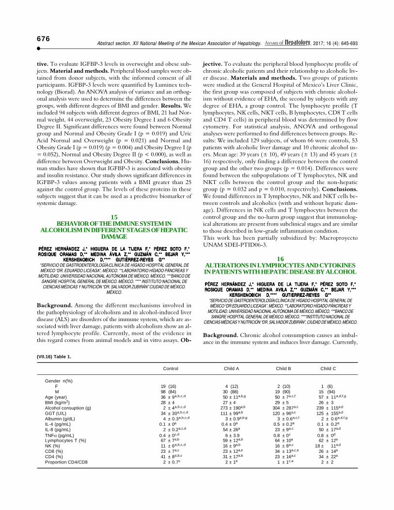
676Abstract section. XII National Meeting of the Mexican Association of Hepatology. , 2017; 16 (4): 645-693
tive. To evaluate IGFBP-3 levels in overweight and obese sub-jects. Material and methods. Peripheral blood samples were ob-tained from donor subjects, with the informed consent of allparticipants. IGFBP-3 levels were quantified by Luminex tech-nology (Biorad). An ANOVA analysis of variance and an orthog-onal analysis were used to determine the differences between thegroups, with different degrees of BMI and gender. Results. Weincluded 94 subjects with different degrees of BMI, 21 had Nor-mal weight, 44 overweight, 23 Obesity Degree I and 6 ObesityDegree II. Significant differences were found between Normalgroup and Normal and Obesity Grade I (p = 0.019) and UricAcid Normal and Overweight (p = 0.021) and Normal andObesity Grade I (p = 0.019) (p = 0.004) and Obesity Degree I (p= 0.052), Normal and Obesity Degree II (p < 0.000), as well asdifference between Overweight and Obesity. Conclusions. Hu-man studies have shown that IGFBP-3 is associated with obesityand insulin resistance. Our study shows significant differences inIGFBP-3 values among patients with a BMI greater than 25against the control group. The levels of these proteins in thesesubjects suggest that it can be used as a predictive biomarker ofsystemic damage.
15BEHAVIOR OF THE IMMUNE SYSTEM IN
ALCOHOLISM IN DIFFERENT STAGES OF HEPATICDAMAGE
PÉREZ HERNÁNDEZ J,* HIGUERA DE LA TIJERA F,* PÉREZ SOTO F,*PÉREZ HERNÁNDEZ J,* HIGUERA DE LA TIJERA F,* PÉREZ SOTO F,*PÉREZ HERNÁNDEZ J,* HIGUERA DE LA TIJERA F,* PÉREZ SOTO F,*PÉREZ HERNÁNDEZ J,* HIGUERA DE LA TIJERA F,* PÉREZ SOTO F,*PÉREZ HERNÁNDEZ J,* HIGUERA DE LA TIJERA F,* PÉREZ SOTO F,*ROSIQUE ORAMAS D,** MEDINA ÁVILA Z,** GUZMÁN C,** BEJAR Y,***ROSIQUE ORAMAS D,** MEDINA ÁVILA Z,** GUZMÁN C,** BEJAR Y,***ROSIQUE ORAMAS D,** MEDINA ÁVILA Z,** GUZMÁN C,** BEJAR Y,***ROSIQUE ORAMAS D,** MEDINA ÁVILA Z,** GUZMÁN C,** BEJAR Y,***ROSIQUE ORAMAS D,** MEDINA ÁVILA Z,** GUZMÁN C,** BEJAR Y,***
KERSHENOBICH D,**** GUTIÉRREZ-REYES G**KERSHENOBICH D,**** GUTIÉRREZ-REYES G**KERSHENOBICH D,**** GUTIÉRREZ-REYES G**KERSHENOBICH D,**** GUTIÉRREZ-REYES G**KERSHENOBICH D,**** GUTIÉRREZ-REYES G***SERVICIO DE GASTROENTEROLOGÍA/CLÍNICA DE HÍGADO HOSPITAL GENERAL DEMÉXICO “DR. EDUARDO LICEAGA”, MÉXICO. **LABORATORIO HÍGADO PÁNCREAS Y
MOTILIDAD. UNIVERSIDAD NACIONAL AUTÓNOMA DE MÉXICO, MÉXICO. ***BANCO DESANGRE HOSPITAL GENERAL DE MÉXICO. MÉXICO. **** INSTITUTO NACIONAL DECIENCIAS MÉDICAS Y NUTRICIÓN “DR. SALVADOR ZUBIRÁN” CIUDAD DE MÉXICO.
MÉXICO.
Background. Among the different mechanisms involved inthe pathophysiology of alcoholism and in alcohol-induced liverdisease (ALS) are disorders of the immune system, which are as-sociated with liver damage, patients with alcoholism show an al-tered lymphocyte profile. Currently, most of the evidence inthis regard comes from animal models and in vitro assays. Ob-
jective. To evaluate the peripheral blood lymphocyte profile ofchronic alcoholic patients and their relationship to alcoholic liv-er disease. Materials and methods. Two groups of patientswere studied at the General Hospital of Mexico’s Liver Clinic,the first group was composed of subjects with chronic alcohol-ism without evidence of EHA, the second by subjects with anydegree of EHA, a group control. The lymphocyte profile (Tlymphocytes, NK cells, NKT cells, B lymphocytes, CD8 T cellsand CD4 T cells) in peripheral blood was determined by flowcytometry. For statistical analysis, ANOVA and orthogonalanalyses were performed to find differences between groups. Re-sults: We included 129 subjects, of whom 66 were controls, 53patients with alcoholic liver damage and 10 chronic alcohol us-ers. Mean age: 39 years (± 10), 49 years (± 13) and 45 years (±16) respectively, only finding a difference between the controlgroup and the other two groups (p = 0.014). Differences werefound between the subpopulations of T lymphocytes, NK andNKT cells between the control group and the non-hepaticgroup (p = 0.032 and p = 0.010, respectively). Conclusions.We found differences in T lymphocytes, NK and NKT cells be-tween controls and alcoholics (with and without hepatic dam-age). Differences in NK cells and T lymphocytes between thecontrol group and the no-harm group suggest that immunolog-ical alterations are present from subclinical stages and are similarto those described in low-grade inflammation condition.This work has been partially subsidized by: MacroproyectoUNAM SDEI-PTID06-3.
16ALTERATIONS IN LYMPHOCYTES AND CYTOKINESIN PATIENTS WITH HEPATIC DISEASE BY ALCOHOL
PÉREZ HERNÁNDEZ J,* HIGUERA DE LA TIJERA F,* PÉREZ SOTO F,*PÉREZ HERNÁNDEZ J,* HIGUERA DE LA TIJERA F,* PÉREZ SOTO F,*PÉREZ HERNÁNDEZ J,* HIGUERA DE LA TIJERA F,* PÉREZ SOTO F,*PÉREZ HERNÁNDEZ J,* HIGUERA DE LA TIJERA F,* PÉREZ SOTO F,*PÉREZ HERNÁNDEZ J,* HIGUERA DE LA TIJERA F,* PÉREZ SOTO F,*ROSIQUE ORAMAS D,** MEDINA AVILA Z,** GUZMÁN C,** BEJAR Y,***ROSIQUE ORAMAS D,** MEDINA AVILA Z,** GUZMÁN C,** BEJAR Y,***ROSIQUE ORAMAS D,** MEDINA AVILA Z,** GUZMÁN C,** BEJAR Y,***ROSIQUE ORAMAS D,** MEDINA AVILA Z,** GUZMÁN C,** BEJAR Y,***ROSIQUE ORAMAS D,** MEDINA AVILA Z,** GUZMÁN C,** BEJAR Y,***
KERSHENOBICH D,**** GUTIERREZ-REYES G**KERSHENOBICH D,**** GUTIERREZ-REYES G**KERSHENOBICH D,**** GUTIERREZ-REYES G**KERSHENOBICH D,**** GUTIERREZ-REYES G**KERSHENOBICH D,**** GUTIERREZ-REYES G***SERVICIO DE GASTROENTEROLOGÍA/CLÍNICA DE HÍGADO HOSPITAL GENERAL DEMÉXICO “DR EDUARDO LICEAGA”. MÉXICO. **LABORATORIO HÍGADO PÁNCREAS Y
MOTILIDAD. UNIVERSIDAD NACIONAL AUTÓNOMA DE MÉXICO. MÉXICO. ***BANCO DESANGRE HOSPITAL GENERAL DE MÉXICO. MÉXICO. ****INSTITUTO NACIONAL DE
CIENCIAS MÉDICAS Y NUTRICIÓN “DR. SALVADOR ZUBIRÁN”, CIUDAD DE MÉXICO. MÉXICO.
Background. Chronic alcohol consumption causes an imbal-ance in the immune system and induces liver damage. Currently,
(VII.16) Table 1.
Control Child A Child B Child C
Gender n(%)F 19 (16) 4 (12) 2 (10) 1 (6)M 98 (84) 30 (88) 19 (90) 15 (94)
Age (year) 36 ± 9a,b,c,d 50 ± 11a,b,g 50 ± 7a,c,f 57 ± 11a,d,f,g
BMI (kg/m2) 28 ± 4 27 ± 4 29 ± 5 26 ± 3Alcohol consuption (g) 2 ± 4a,b,c,d 273 ± 190a,b 304 ± 287a,c 239 ± 115a,d
GGT (U/L) 34 ± 30a,b,c,d 111 ± 99a,b 120 ± 96a,c 125 ± 155a,d
Albumin (g/dL) 4 ± 0.3a,b,c,d 3 ± 0.9a,b,g 3 ± 0.6a,c,f 2 ± 0.6a,d,f,g
IL-4 (pg/mL) 0.1 ± 0a 0.4 ± 0a 0.5 ± 0.2a 0.1 ± 0.2a
IL-8 (pg/mL) 2 ± 0.2a,c,d 54 ± 26a 23 ± 9a,c 50 ± 17a,d
TNFα (pg/mL) 0.4 ± 0c,d 6 ± 3.9 0.8 ± 0c 0.8 ± 0d
Lymphocytes T (%) 67 ± 7a,b 59 ± 12a,b 64 ± 10a 62 ± 12a
NK (%) 11 ± 6a,b,c,d 16 ± 9a,b 16 ± 8a,c 18 ± 11a,d
CD8 (%) 23 ± 7a,c 23 ± 12a,e 34 ± 13a,c,e 26 ± 14a
CD4 (%) 41 ± 8a,b,c 31 ± 17a,b 23 ± 16a,c 34 ± 22a
Proportion CD4/CD8 2 ± 0.7c 2 ± 1e 1 ± 1c,e 2 ± 2
677Abstract section. XII National Meeting of the Mexican Association of Hepatology. , 2017; 16 (4): 645-693
the most important evidence comes from animal models and invitro assays. Objective. To evaluate the lymphocyte profile andcytokines in peripheral blood of subjects with Alcoholic LiverDisease (EHA). Material and methods. Participants were di-vided into two groups. Group 1 (control): Subjects with ethanolconsumption <10 g/day and AUDIT < 8. Group 2, subjectswith EHA who were classified according to the Child-Pughscale. The lymphocyte profile (T cells, B, NK, NKT) was deter-mined in peripheral blood by flow cytometry and serum cy-tokines by Luminex technology (Bio-Rad). Data were analyzedusing ANOVA and orthogonal analyzes. Results. We included188 subjects, 71 diagnosed with EHA and 117 healthy subjects.Conclusions. These results demonstrate that in EHA the per-centage of cytotoxic cells and pro-inflammatory cytokines is in-creased, whereas CD4 cells decrease so that in EHA the immuneresponse is altered and is persistent even in more severe stages.
17CORRELATION OF FIBROSCAN WITH NAFLD
SCORE AND FIB4 IN PATIENTS WITH NON-ALCOHOLIC FATTY LIVER DISEASE AND INSULIN
RESISTANCE
VELASCO-SANTIAGO YM,* FERNÁNDEZ-MARTÍNEZ NC,*VELASCO-SANTIAGO YM,* FERNÁNDEZ-MARTÍNEZ NC,*VELASCO-SANTIAGO YM,* FERNÁNDEZ-MARTÍNEZ NC,*VELASCO-SANTIAGO YM,* FERNÁNDEZ-MARTÍNEZ NC,*VELASCO-SANTIAGO YM,* FERNÁNDEZ-MARTÍNEZ NC,*AGUILAR-MORENO RU,* RIVERA-FLORES AG,* BARRAZA-ORTIZ DB,*AGUILAR-MORENO RU,* RIVERA-FLORES AG,* BARRAZA-ORTIZ DB,*AGUILAR-MORENO RU,* RIVERA-FLORES AG,* BARRAZA-ORTIZ DB,*AGUILAR-MORENO RU,* RIVERA-FLORES AG,* BARRAZA-ORTIZ DB,*AGUILAR-MORENO RU,* RIVERA-FLORES AG,* BARRAZA-ORTIZ DB,*
CANO-CONTRERAS AD,* DURÁN-ROSAS C,* LLORENTE-RAMÓN A,*CANO-CONTRERAS AD,* DURÁN-ROSAS C,* LLORENTE-RAMÓN A,*CANO-CONTRERAS AD,* DURÁN-ROSAS C,* LLORENTE-RAMÓN A,*CANO-CONTRERAS AD,* DURÁN-ROSAS C,* LLORENTE-RAMÓN A,*CANO-CONTRERAS AD,* DURÁN-ROSAS C,* LLORENTE-RAMÓN A,*OVIEDO-MAGLIONE MA,* RAMOS-AGUILAR GA,* MEJÍA-LOZA SMI,*OVIEDO-MAGLIONE MA,* RAMOS-AGUILAR GA,* MEJÍA-LOZA SMI,*OVIEDO-MAGLIONE MA,* RAMOS-AGUILAR GA,* MEJÍA-LOZA SMI,*OVIEDO-MAGLIONE MA,* RAMOS-AGUILAR GA,* MEJÍA-LOZA SMI,*OVIEDO-MAGLIONE MA,* RAMOS-AGUILAR GA,* MEJÍA-LOZA SMI,*
PÉREZ-LÓPEZ N,* ZAMARRIPA-DORSEY F,* TORRES-LÓPEZ E**PÉREZ-LÓPEZ N,* ZAMARRIPA-DORSEY F,* TORRES-LÓPEZ E**PÉREZ-LÓPEZ N,* ZAMARRIPA-DORSEY F,* TORRES-LÓPEZ E**PÉREZ-LÓPEZ N,* ZAMARRIPA-DORSEY F,* TORRES-LÓPEZ E**PÉREZ-LÓPEZ N,* ZAMARRIPA-DORSEY F,* TORRES-LÓPEZ E***GASTROENTEROLOGY DEPARTMENT, HOSPITAL JUÁREZ DE MÉXICO, MEXICO CITY,
MEXICO.**GENERAL SURGERY DEPARTMENT, HOSPITAL ÁNGELES LINDAVISTA, MEXICO CITY,
MÉXICO.
Background and aim. The prevalence of non-alcoholic fatty liv-er disease (NAFLD) is higher in patients with diabetes mellitus, be-ing > 70%. The NALFD has a clear correlation with the presenceof insulin resistance, which can be determine with the measure-ment of glycosylated hemoglobin (HbA1C) from values > 5.7%(ADA 2016). Fibrosis that accompanies this pathology can lead tocirrhosis and hepatocellular carcinoma, so an early diagnosis is es-sential. This study aims to determine the correlation of FibroScanwith the scales in patients with NAFLD and insulin resistance inthe Gastroenterology Department of the Hospital Juárez de Méxi-co. Material and methods. An observational, cross-sectionalstudy of patients with NAFLD who assessed their age, gender,HbA1C levels, body mass index (BMI), NAFLD score and FIB4determination which were correlated with FibroScan. The stages offibrosis were defined by KPa. Results. We included 40 patients, 32women and 8 men (80 and 20% respectively), mean age of 49.85years. They were divided into 2 groups, patients with 5.7% (17 =42.5%) and HbA1C < 5.7% (17 = 42.5%). Of the total number ofpatients, 7 (17.5%) showed advanced fibrosis with FibroScan (F3-F4). A positive correlation was found between FIB4 and Kpa in pa-tients with insulin resistance (r = 0.5575), lower than that foundwith the NAFLD score (r = 0.2434). Conclusions. The FIB4scale showed a higher correlation with FibroScan in patients withNAFLD and insulin resistance, it is required to expand the samplesize to support the result.
18RELATIVE CHANGES IN BIOCHEMICAL AND
ANTHROPOMETRIC INDICATORS IN PATIENTSWITH NON ALCOHOLIC FATTY LIVER DISEASEACCORDING TO NUTRITIONAL INTERVENTION.
TWO YEAR FOLLOW-UP REPORT
PÉREZ-RODRÍGUEZ DL, LARA-ESPARZA MP, DÁVILA-BONILLA S,PÉREZ-RODRÍGUEZ DL, LARA-ESPARZA MP, DÁVILA-BONILLA S,PÉREZ-RODRÍGUEZ DL, LARA-ESPARZA MP, DÁVILA-BONILLA S,PÉREZ-RODRÍGUEZ DL, LARA-ESPARZA MP, DÁVILA-BONILLA S,PÉREZ-RODRÍGUEZ DL, LARA-ESPARZA MP, DÁVILA-BONILLA S,GUTIÉRREZ-MARTÍNEZ G, MARTÍNEZ-VÁZQUEZ SEGUTIÉRREZ-MARTÍNEZ G, MARTÍNEZ-VÁZQUEZ SEGUTIÉRREZ-MARTÍNEZ G, MARTÍNEZ-VÁZQUEZ SEGUTIÉRREZ-MARTÍNEZ G, MARTÍNEZ-VÁZQUEZ SEGUTIÉRREZ-MARTÍNEZ G, MARTÍNEZ-VÁZQUEZ SE
DEPARTMENT OF GASTROENTEROLOGY. INSTITUTO NACIONAL DE CIENCIASMÉDICAS Y NUTRICIÓN “SALVADOR ZUBIRÁN”. MEXICO CITY, MEXICO.
Introduction. The clinical practice guidelines for the treatmentto the fatty liver disease point out that a weight loss of 3% re-duces steatosis level; the loss weight must be gradual, accompa-nied by exercise. Several studies indicate that initial changes intreatment, influence transaminases. Objective. Describe thechanges produced by hypocaloric diet and exercise in biochemi-cal indicators and anthropometry in patients with the fatty liverdisease through 2 years of follow up. Material and methods.In 173 patients belonging to Non alcoholic fatty liver diseasecohort, received hypocaloric diet using adjusted weight and pre-scription of exercise (1,200-1,800 kcals/day; 50% carbohydrates,30% lipids and 20% protein and more than 180 min/week ofaerobic exercise); the changes of two years in body compositionand biochemical indicators disease related, were analyzed withWilcoxon and Friedman test using the SPSS v20 statistic soft-ware. Results. 69.3% of patients remained in this cohort dur-ing the first 18 months and 47.9% at 24 months. The medianof the weight loss was of 2.4 kg (p = 0.003), arm circumferenceloss was of 0.05 cm (p = 0.005), waist circumference loss was of0.22 cm (p = 0.003) and Body Mass Index (BMI) loss was of0.1 kg/m2 (p = 0.027). The distance between nutritional period-ic reviews was 6-7 months; from the first to the second check-up, the differences were in the body weight and armcircumference (P = 0.019 and 0.004 respectively) from the sec-ond to the third, change in BMI (p = 0.026) and between thethird and fourth, in glucose (p = 0.027), glycated haemoglobin(p = 0.028) and ALT (p = 0.007) were see. Conclusion. Theweight loss in this cohort was lower than the guidelines pointout, so the changes in the both, anthropometric and biochemi-cal indicators were small and inconstant. It is suggested radicalsdietetic strategies, structured/supervised exercise and a minortime between periodic reviews.
19IMMUNOTYPICAL PROFILE ALTERATIONS IN
PERIPHERAL POLYMORPHONUCLEAR CELLS OFBINGE DRINKERS
PÉREZ-GARCÍA A,*PÉREZ-GARCÍA A,*PÉREZ-GARCÍA A,*PÉREZ-GARCÍA A,*PÉREZ-GARCÍA A,*,†,†,†,†,† ARROYO-VALERIO A, ARROYO-VALERIO A, ARROYO-VALERIO A, ARROYO-VALERIO A, ARROYO-VALERIO A,‡‡‡‡‡ BUSTOS-ESQUIVEL M, BUSTOS-ESQUIVEL M, BUSTOS-ESQUIVEL M, BUSTOS-ESQUIVEL M, BUSTOS-ESQUIVEL M,‡‡‡‡‡
ZALDÍVAR FUJIGAKI JL,* GASTELUM-STROZZI A,ZALDÍVAR FUJIGAKI JL,* GASTELUM-STROZZI A,ZALDÍVAR FUJIGAKI JL,* GASTELUM-STROZZI A,ZALDÍVAR FUJIGAKI JL,* GASTELUM-STROZZI A,ZALDÍVAR FUJIGAKI JL,* GASTELUM-STROZZI A,§§§§§
PADILLA-CASTAÑEDA MA,PADILLA-CASTAÑEDA MA,PADILLA-CASTAÑEDA MA,PADILLA-CASTAÑEDA MA,PADILLA-CASTAÑEDA MA,§§§§§ REDING-BERNAL A, REDING-BERNAL A, REDING-BERNAL A, REDING-BERNAL A, REDING-BERNAL A,‡‡‡‡‡ KERSENOBICH D, KERSENOBICH D, KERSENOBICH D, KERSENOBICH D, KERSENOBICH D,||||||||| |
HERNÁNDEZ-RUIZ J*HERNÁNDEZ-RUIZ J*HERNÁNDEZ-RUIZ J*HERNÁNDEZ-RUIZ J*HERNÁNDEZ-RUIZ J*,‡,‡,‡,‡,‡
*LABORATORIO DE HÍGADO, PÁNCREAS Y MOTILIDAD. UNIDAD DE MEDICINAEXPERIMENTAL. UNAM, MÉXICO. †SERVICIO DE CIRUGÍA EXPERIMENTAL, ‡DIRECCIÓN
DE INVESTIGACIÓN, §UNIDAD DE INVESTIGACIÓN Y DESARROLLO TECNOLÓGICOCCADET UNAM, HOSPITAL GENERAL DE MÉXICO “DR. EDUARDO LICEAGA”. MÉXICO.||INSTITUTO NACIONAL DE CIENCIAS MÉDICAS Y NUTRICIÓN “SALVADOR ZUBIRÁN”.
MÉXICO.
Background. High alcohol intake on weekends or “bingedrinking”, could cause early liver damage. Alcohol-induced liver
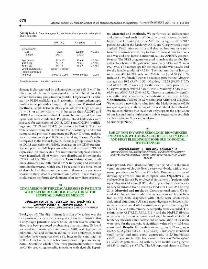
678Abstract section. XII National Meeting of the Mexican Association of Hepatology. , 2017; 16 (4): 645-693
damage is characterized by polymorphonuclear cell (PMN) in-filtration, which can be represented in the peripheral blood byaltered trafficking and activation profiles. Objective. To evalu-ate the PMN trafficking and activation immunophenotypicprofiles in people with a binge drinking pattern. Material andmethods. People between 20-29 years old with binge drinking(n = 18) or at low risk (n = 16) based on their AUDIT andHEPCA scores were studied. Hematic biometry and liver en-zyme tests were conducted. Peripheral blood leukocytes wereanalyzed for expression of CCR5, CCR4 and CXCR4 (traffick-ing), and CD69 and CD127 (activation) by FACS. The datawere analyzed using the T-test and Mann-Whitney’s U-test forcontrasts and principal component and Fuzzy C means analysesfor clustering, with p < 0.05 considered significant. Results.Compared to the low-risk group, the binge group showed high-er CCR5 expression on PMNs, decreases in the CD69 percent-age and positive PMNs per microliter, and decreased CXCR4expression on monocytes. Six immunophenotypical clusterswere identified, all of which were distributed following theCCR5 and CXCR4 main vectors. Conclusion. Young adultbinge drinkers have differential PMN trafficking and activationimmunophenotypes, which could be related to the initial onsetof alcoholic liver disease and a systemic inflammatory state in re-sponse to their alcohol consumption pattern. These findingscould lead to the future development of an early diagnostic tool.
20COMPARISON OF THREE SCALES SEES IN PATIENTS
WITH SEVERE ALCOHOLIC HEPATITIS AT THEHOSPITAL JUÁREZ DE MÉXICO
JUÁREZ-BARRIENTOS TE, MEJÍA-LOZA SMI, GARCÍA-RUÍZ E,JUÁREZ-BARRIENTOS TE, MEJÍA-LOZA SMI, GARCÍA-RUÍZ E,JUÁREZ-BARRIENTOS TE, MEJÍA-LOZA SMI, GARCÍA-RUÍZ E,JUÁREZ-BARRIENTOS TE, MEJÍA-LOZA SMI, GARCÍA-RUÍZ E,JUÁREZ-BARRIENTOS TE, MEJÍA-LOZA SMI, GARCÍA-RUÍZ E,ZAMARRIPA-DORSEY F, HERRERO-MACEDA RZAMARRIPA-DORSEY F, HERRERO-MACEDA RZAMARRIPA-DORSEY F, HERRERO-MACEDA RZAMARRIPA-DORSEY F, HERRERO-MACEDA RZAMARRIPA-DORSEY F, HERRERO-MACEDA R
GASTROENTEROLOGÍA, HOSPITAL JUÁREZ DE MÉXICO, MEXICO CITY, MEXICO.
Background. The discriminant function of Maddrey was thefirst prognostic scale to be developed and has the limitation thatit only staged patients in severe (> 32) or non-severe. Other in-dices have been proposed by observing that creatinine, INR andage are determinants of survival, so the ABIC scale (age, serumbilirubin, INR and serum creatinine) is later performed, whichincludes three categories (low risk, Intermediate and high) aswell as Glasgow, which is becoming more important today.Aim. Determine which of the three prognostic scales is mostuseful for predicting mortality in patients with alcoholic hepati-
tis. Material and methods. We performed an ambispectiveand observational analysis of 106 patients with severe alcoholichepatitis at Hospital Juárez de México during the 2013-2017period, to whom the Maddrey, ABIC and Glasgow scales wereapplied. Descriptive statistics and data exploration were per-formed to corroborate if they followed a normal distribution, aone-way and one-factor Bonferroni post-hoc ANOVA was per-formed. The SPSS program was used to analyze the results. Re-sults. We obtained 106 patients, 8 women (7.54%) and 98 men(92.45%). The average age for the male gender was 42.73% andfor the female gender of 49.75%. The total number of live pa-tients was 46 (44.89% male and 25% female) and 60 (55.10%male and 75% female). For the deceased patients the Glasgowaverage was 10.2 (9.87-10.52), Maddrey 102.78 (88.86-116.7)and ABIC 9.26 (8.47-9.76). In the case of living patients theGlasgow average was 9.17 (8.71-9.64), Maddrey 57.16 (49.1-65.8) and ABIC 7.93 (7.46-8.37). There is a statistically signifi-cant difference between the methods studied with a p < 0.05.Conclusions. Two scales were correlated ABIC and Glasgow.We obtained a new cohort value from the Maddrey index (65.8)to express gravity, so the utility of this scale should be evaluated.We must emphasize that these data only support the populationof our hospital and a multicenter study is suggested to establisha cohort value in Mexican population.Sponsorship: None.
21USE OF NON-INVASIVE SEROLOGIC BIOMARKERSIN PATIENTS WITH NON-ALCOHOLIC FATTY LIVER
AND FIRST BLEEDING EPISODE DUE TO PORTALHYPERTENSION
MENDOZA-RAMÍREZ J, ANZÁSTIGA DELGADILLO DMENDOZA-RAMÍREZ J, ANZÁSTIGA DELGADILLO DMENDOZA-RAMÍREZ J, ANZÁSTIGA DELGADILLO DMENDOZA-RAMÍREZ J, ANZÁSTIGA DELGADILLO DMENDOZA-RAMÍREZ J, ANZÁSTIGA DELGADILLO DGASTROENTEROLOGY AND GASTROINTESTINAL ENDOSCOPY DEPARTMENT.
HOSPITAL GENERAL REGIONAL (HGR) 251. IMSS. METEPEC, STATE OF MEXICO.MEXICO.
Background. Non-alcoholic fatty liver (NAFL) is the mostcommon cause of chronic liver disease worldwide, with an esti-mated prevalence in Mexico of 10-14%. Patients are at risk ofdeveloping cirrhosis and its complications. Objectives. Toevaluate liver fibrosis by serological biomarkers of patients withupper digestive bleeding (UDB) due to portal hypertension sec-ondary to chronic liver disease by NAFL in HGR 251 during2016. Material and methods. Cross-sectional study. We in-cluded adults, admitted to the emergency room and hospitaliza-tion during 2016, diagnosed with UDB, with laboratories,abdominal ultrasound (US) and upper digestive endoscopy. Pa-tients with current alcohol consumption, positive serology forHCV, HBV and autoimmune hepatopathy were excluded. Therelationship AST/ALT, APRI, FIB-4 and the NAFLD fibrosisscore were used as non-invasive serological biomarkers. Centraltendency measures and coefficient of correlation of Pearsonwere used for the analysis. A significant value of p < 0.05 wasconsidered. Results. Of the 44 patients analyzed, 23 were men(52%), 59.2 years old (+ 11.49 years). Endoscopy identifiedsmall varices and mild portal gastropathy in 23 individuals(53%), respectively. The body mass index was 24.84 kg/m2
(+ 2.32), 28 patients (63%) with diabetes mellitus and glucoseof 139.11 mg/dL (+ 87.67). The US reported chronic diffuse
(VII.21) Table 1. Data demographic, biochemical and protein carbonyls ofstudy subjects.
cHC (30) CT (166) p
Gender n (%)Male 7(23) 139(90) < 0.001Female 24(77) 27(11)
Age (years) 51 ± 10 37 ±9 < 0.001AST (U/I) 84 ± 7 30 ±1 < 0.001ALT (U/I) 90 ± 6 28 ±2 < 0.001Viral Load (UI/mL) 12x105 ± 20x105 — —Protein carbonyls(mg/mL) 0.092 ± 0.066 0.046 ±0.082 0.004
Results in mean ± standard deviation.
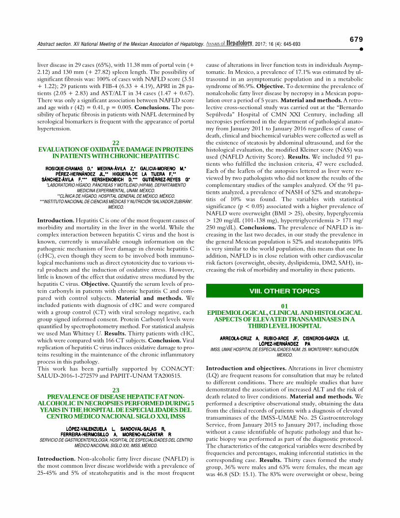
679Abstract section. XII National Meeting of the Mexican Association of Hepatology. , 2017; 16 (4): 645-693
liver disease in 29 cases (65%), with 11.38 mm of portal vein (+2.12) and 130 mm (+ 27.82) spleen length. The possibility ofsignificant fibrosis was: 100% of cases with NAFLD score (3.51+ 1.22); 29 patients with FIB-4 (6.33 + 4.19), APRI in 28 pa-tients (2.05 + 2.83) and AST/ALT in 34 cases (1.47 + 0.67).There was only a significant association between NAFLD scoreand age with r (42) = 0.41, p = 0.005. Conclusions. The pos-sibility of hepatic fibrosis in patients with NAFL determined byserological biomarkers is frequent with the appearance of portalhypertension.
22EVALUATION OF OXIDATIVE DAMAGE IN PROTEINS
IN PATIENTS WITH CHRONIC HEPATITIS C
ROSIQUE-ORAMAS D,* MEDINA-ÁVILA Z,* GALICIA-MORENO M,*ROSIQUE-ORAMAS D,* MEDINA-ÁVILA Z,* GALICIA-MORENO M,*ROSIQUE-ORAMAS D,* MEDINA-ÁVILA Z,* GALICIA-MORENO M,*ROSIQUE-ORAMAS D,* MEDINA-ÁVILA Z,* GALICIA-MORENO M,*ROSIQUE-ORAMAS D,* MEDINA-ÁVILA Z,* GALICIA-MORENO M,*PÉREZ-HERNÁNDEZ JL,** HIGUERA-DE LA TIJERA F,**PÉREZ-HERNÁNDEZ JL,** HIGUERA-DE LA TIJERA F,**PÉREZ-HERNÁNDEZ JL,** HIGUERA-DE LA TIJERA F,**PÉREZ-HERNÁNDEZ JL,** HIGUERA-DE LA TIJERA F,**PÉREZ-HERNÁNDEZ JL,** HIGUERA-DE LA TIJERA F,**
SÁNCHEZ-ÁVILA F,*** KERSHENOBICH D,*** GUTIÉRREZ-REYES G*SÁNCHEZ-ÁVILA F,*** KERSHENOBICH D,*** GUTIÉRREZ-REYES G*SÁNCHEZ-ÁVILA F,*** KERSHENOBICH D,*** GUTIÉRREZ-REYES G*SÁNCHEZ-ÁVILA F,*** KERSHENOBICH D,*** GUTIÉRREZ-REYES G*SÁNCHEZ-ÁVILA F,*** KERSHENOBICH D,*** GUTIÉRREZ-REYES G**LABORATORIO HÍGADO, PÁNCREAS Y MOTILIDAD (HIPAM), DEPARTAMENTO
MEDICINA EXPERIMENTAL, UNAM. MÉXICO.**CLÍNICA DE HÍGADO, HOSPITAL GENERAL DE MÉXICO. MÉXICO.
***INSTITUTO NACIONAL DE CIENCIAS MÉDICAS Y NUTRICIÓN “SALVADOR ZUBIRÁN”.MÉXICO.
Introduction. Hepatitis C is one of the most frequent causes ofmorbidity and mortality in the liver in the world. While thecomplex interaction between hepatitis C virus and the host isknown, currently is unavailable enough information on thepathogenic mechanism of liver damage in chronic hepatitis C(cHC), even though they seem to be involved both immuno-logical mechanisms such as direct cytotoxicity due to various vi-ral products and the induction of oxidative stress. However,little is known of the effect that oxidative stress mediated by thehepatitis C virus. Objective. Quantify the serum levels of pro-tein carbonyls in patients with chronic hepatitis C and com-pared with control subjects. Material and methods. Weincluded patients with diagnosis of cHC and were comparedwith a group control (CT) with viral serology negative, eachgroup signed informed consent. Protein Carbonyl levels werequantified by spectrophotometry method. For statistical analysiswe used Man Whitney U. Results. Thirty patients with cHC,which were compared with 166 CT subjects. Conclusion. Viralreplication of hepatitis C virus induces oxidative damage to pro-teins resulting in the maintenance of the chronic inflammatoryprocess in this pathology.This work has been partially supported by CONACYT:SALUD-2016-1-272579 and PAPIIT-UNAM TA200515.
23PREVALENCE OF DISEASE HEPATIC FAT NON-
ALCOHOLIC IN NECROPSIES PERFORMED DURING 5YEARS IN THE HOSPITAL DE ESPECIALIDADES DEL
CENTRO MÉDICO NACIONAL SIGLO XXI, IMSS
LÓPEZ-VALENZUELA L, SANDOVAL-SALAS R,LÓPEZ-VALENZUELA L, SANDOVAL-SALAS R,LÓPEZ-VALENZUELA L, SANDOVAL-SALAS R,LÓPEZ-VALENZUELA L, SANDOVAL-SALAS R,LÓPEZ-VALENZUELA L, SANDOVAL-SALAS R,FERREIRA-HERMOSILLO A, MORENO-ALCÁNTAR RFERREIRA-HERMOSILLO A, MORENO-ALCÁNTAR RFERREIRA-HERMOSILLO A, MORENO-ALCÁNTAR RFERREIRA-HERMOSILLO A, MORENO-ALCÁNTAR RFERREIRA-HERMOSILLO A, MORENO-ALCÁNTAR R
SERVICIO DE GASTROENTEROLOGÍA, HOSPITAL DE ESPECIALIDADES DEL CENTROMÉDICO NACIONAL SIGLO XXI, IMSS. MÉXICO.
Introduction. Non-alcoholic fatty liver disease (NAFLD) isthe most common liver disease worldwide with a prevalence of25-45% and 5% of steatohepatitis and is the most frequent
cause of alterations in liver function tests in individuals Asymp-tomatic. In Mexico, a prevalence of 17.1% was estimated by ul-trasound in an asymptomatic population and in a metabolicsyndrome of 86.9%. Objective. To determine the prevalence ofnonalcoholic fatty liver disease by necropsy in a Mexican popu-lation over a period of 5 years. Material and methods. A retro-lective cross-sectional study was carried out at the “BernardoSepúlveda” Hospital of CMN XXI Century, including allnecropsies performed in the department of pathological anato-my from January 2011 to January 2016 regardless of cause ofdeath, clinical and biochemical variables were collected as well asthe existence of steatosis by abdominal ultrasound, and for thehistological evaluation, the modified Kleiner score (NAS) wasused (NAFLD Activity Score). Results. We included 91 pa-tients who fulfilled the inclusion criteria, 47 were excluded.Each of the leaflets of the autopsies lettered as liver were re-viewed by two pathologists who did not know the results of thecomplementary studies of the samples analyzed. Of the 91 pa-tients analyzed, a prevalence of NASH of 52% and steatohepa-titis of 10% was found. The variables with statisticalsignificance (p < 0.05) associated with a higher prevalence ofNAFLD were overweight (BMI > 25), obesity, hyperglycemia> 120 mg/dL (101-138 mg), hypertriglyceridemia > 171 mg/250 mg/dL). Conclusions. The prevalence of NAFLD is in-creasing in the last two decades, in our study the prevalence inthe general Mexican population is 52% and steatohepatitis 10%is very similar to the world population, this means that one Inaddition, NAFLD is in close relation with other cardiovascularrisk factors (overweight, obesity, dyslipidemia, DM2, SAH), in-creasing the risk of morbidity and mortality in these patients.
VIII. OTHER TOPICS
01EPIDEMIOLOGICAL, CLINICAL AND HISTOLOGICAL
ASPECTS OF ELEVATED TRANSAMINASES IN ATHIRD LEVEL HOSPITAL
ARREOLA-CRUZ A, RUBIO-ARCE JF, CISNEROS-GARZA LE,ARREOLA-CRUZ A, RUBIO-ARCE JF, CISNEROS-GARZA LE,ARREOLA-CRUZ A, RUBIO-ARCE JF, CISNEROS-GARZA LE,ARREOLA-CRUZ A, RUBIO-ARCE JF, CISNEROS-GARZA LE,ARREOLA-CRUZ A, RUBIO-ARCE JF, CISNEROS-GARZA LE,LÓPEZ-HERNÁNDEZ PALÓPEZ-HERNÁNDEZ PALÓPEZ-HERNÁNDEZ PALÓPEZ-HERNÁNDEZ PALÓPEZ-HERNÁNDEZ PA
IMSS, UMAE HOSPITAL DE ESPECIALIDADES NÚM. 25. MONTERREY, NUEVO LEÓN,MEXICO.
Introduction and objectives. Alterations in liver chemistry(LQ) are frequent reasons for consultation that may be relatedto different conditions. There are multiple studies that havedemonstrated the association of increased ALT and the risk ofdeath related to liver conditions. Material and methods. Weperformed a descriptive observational study, obtaining the datafrom the clinical records of patients with a diagnosis of elevatedtransaminases of the IMSS-UMAE No. 25 GastroenterologyService, from January 2015 to January 2017, including thosewithout a cause identifiable of hepatic pathology and that he-patic biopsy was performed as part of the diagnostic protocol.The characteristics of the categorical variables were described byfrequencies and percentages, making inferential statistics in thecorresponding case. Results. Thirty cases formed the studygroup, 36% were males and 63% were females, the mean agewas 46.8 (SD: 15.1). The 83% were overweight or obese, being
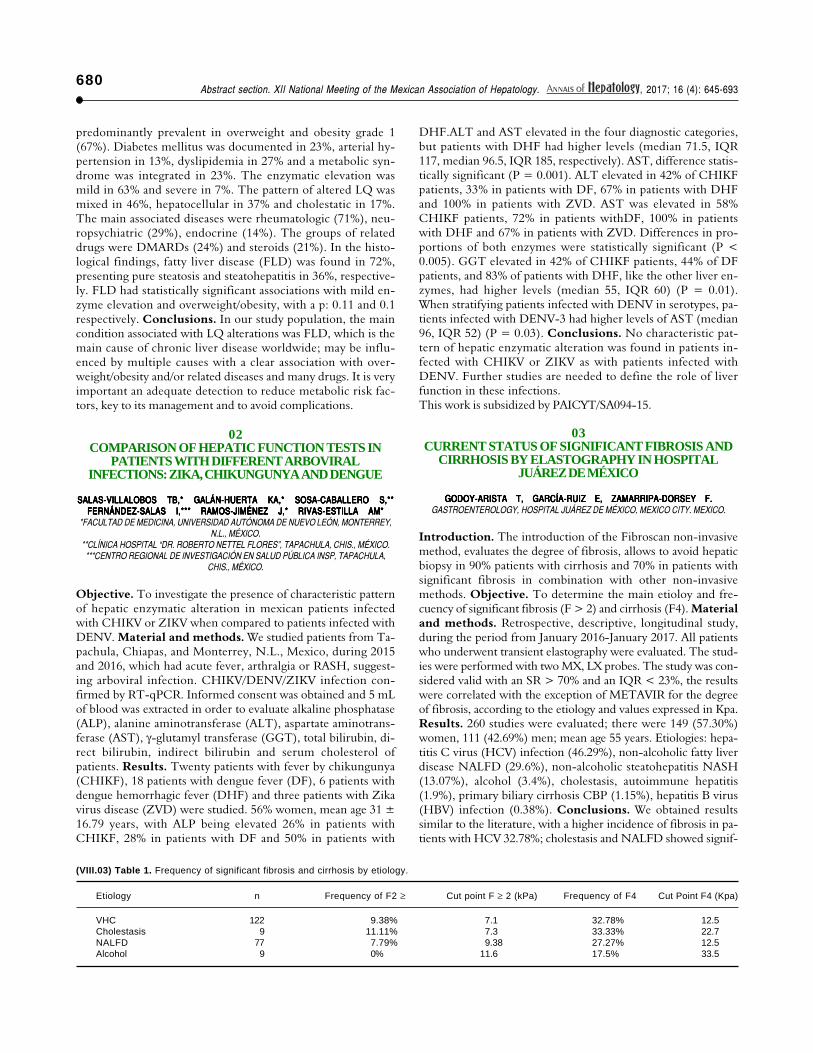
680Abstract section. XII National Meeting of the Mexican Association of Hepatology. , 2017; 16 (4): 645-693
predominantly prevalent in overweight and obesity grade 1(67%). Diabetes mellitus was documented in 23%, arterial hy-pertension in 13%, dyslipidemia in 27% and a metabolic syn-drome was integrated in 23%. The enzymatic elevation wasmild in 63% and severe in 7%. The pattern of altered LQ wasmixed in 46%, hepatocellular in 37% and cholestatic in 17%.The main associated diseases were rheumatologic (71%), neu-ropsychiatric (29%), endocrine (14%). The groups of relateddrugs were DMARDs (24%) and steroids (21%). In the histo-logical findings, fatty liver disease (FLD) was found in 72%,presenting pure steatosis and steatohepatitis in 36%, respective-ly. FLD had statistically significant associations with mild en-zyme elevation and overweight/obesity, with a p: 0.11 and 0.1respectively. Conclusions. In our study population, the maincondition associated with LQ alterations was FLD, which is themain cause of chronic liver disease worldwide; may be influ-enced by multiple causes with a clear association with over-weight/obesity and/or related diseases and many drugs. It is veryimportant an adequate detection to reduce metabolic risk fac-tors, key to its management and to avoid complications.
02COMPARISON OF HEPATIC FUNCTION TESTS IN
PATIENTS WITH DIFFERENT ARBOVIRALINFECTIONS: ZIKA, CHIKUNGUNYA AND DENGUE
SALAS-VILLALOBOS TB,* GALÁN-HUERTA KA,* SOSA-CABALLERO S,**SALAS-VILLALOBOS TB,* GALÁN-HUERTA KA,* SOSA-CABALLERO S,**SALAS-VILLALOBOS TB,* GALÁN-HUERTA KA,* SOSA-CABALLERO S,**SALAS-VILLALOBOS TB,* GALÁN-HUERTA KA,* SOSA-CABALLERO S,**SALAS-VILLALOBOS TB,* GALÁN-HUERTA KA,* SOSA-CABALLERO S,**FERNÁNDEZ-SALAS I,*** RAMOS-JIMÉNEZ J,* RIVAS-ESTILLA AM*FERNÁNDEZ-SALAS I,*** RAMOS-JIMÉNEZ J,* RIVAS-ESTILLA AM*FERNÁNDEZ-SALAS I,*** RAMOS-JIMÉNEZ J,* RIVAS-ESTILLA AM*FERNÁNDEZ-SALAS I,*** RAMOS-JIMÉNEZ J,* RIVAS-ESTILLA AM*FERNÁNDEZ-SALAS I,*** RAMOS-JIMÉNEZ J,* RIVAS-ESTILLA AM*
*FACULTAD DE MEDICINA, UNIVERSIDAD AUTÓNOMA DE NUEVO LEÓN, MONTERREY,N.L., MÉXICO.
**CLÍNICA HOSPITAL “DR. ROBERTO NETTEL FLORES”, TAPACHULA, CHIS., MÉXICO.***CENTRO REGIONAL DE INVESTIGACIÓN EN SALUD PÚBLICA INSP, TAPACHULA,
CHIS., MÉXICO.
Objective. To investigate the presence of characteristic patternof hepatic enzymatic alteration in mexican patients infectedwith CHIKV or ZIKV when compared to patients infected withDENV. Material and methods. We studied patients from Ta-pachula, Chiapas, and Monterrey, N.L., Mexico, during 2015and 2016, which had acute fever, arthralgia or RASH, suggest-ing arboviral infection. CHIKV/DENV/ZIKV infection con-firmed by RT-qPCR. Informed consent was obtained and 5 mLof blood was extracted in order to evaluate alkaline phosphatase(ALP), alanine aminotransferase (ALT), aspartate aminotrans-ferase (AST), γ-glutamyl transferase (GGT), total bilirubin, di-rect bilirubin, indirect bilirubin and serum cholesterol ofpatients. Results. Twenty patients with fever by chikungunya(CHIKF), 18 patients with dengue fever (DF), 6 patients withdengue hemorrhagic fever (DHF) and three patients with Zikavirus disease (ZVD) were studied. 56% women, mean age 31 ±16.79 years, with ALP being elevated 26% in patients withCHIKF, 28% in patients with DF and 50% in patients with
(VIII.03) Table 1. Frequency of significant fibrosis and cirrhosis by etiology.
Etiology n Frequency of F2 ≥ Cut point F ≥ 2 (kPa) Frequency of F4 Cut Point F4 (Kpa)
VHC 122 9.38% 7.1 32.78% 12.5Cholestasis 9 11.11% 7.3 33.33% 22.7NALFD 77 7.79% 9.38 27.27% 12.5Alcohol 9 0% 11.6 17.5% 33.5
DHF.ALT and AST elevated in the four diagnostic categories,but patients with DHF had higher levels (median 71.5, IQR117, median 96.5, IQR 185, respectively). AST, difference statis-tically significant (P = 0.001). ALT elevated in 42% of CHIKFpatients, 33% in patients with DF, 67% in patients with DHFand 100% in patients with ZVD. AST was elevated in 58%CHIKF patients, 72% in patients withDF, 100% in patientswith DHF and 67% in patients with ZVD. Differences in pro-portions of both enzymes were statistically significant (P <0.005). GGT elevated in 42% of CHIKF patients, 44% of DFpatients, and 83% of patients with DHF, like the other liver en-zymes, had higher levels (median 55, IQR 60) (P = 0.01).When stratifying patients infected with DENV in serotypes, pa-tients infected with DENV-3 had higher levels of AST (median96, IQR 52) (P = 0.03). Conclusions. No characteristic pat-tern of hepatic enzymatic alteration was found in patients in-fected with CHIKV or ZIKV as with patients infected withDENV. Further studies are needed to define the role of liverfunction in these infections.This work is subsidized by PAICYT/SA094-15.
03CURRENT STATUS OF SIGNIFICANT FIBROSIS AND
CIRRHOSIS BY ELASTOGRAPHY IN HOSPITALJUÁREZ DE MÉXICO
GODOY-ARISTA T, GARCÍA-RUIZ E, ZAMARRIPA-DORSEY F.GODOY-ARISTA T, GARCÍA-RUIZ E, ZAMARRIPA-DORSEY F.GODOY-ARISTA T, GARCÍA-RUIZ E, ZAMARRIPA-DORSEY F.GODOY-ARISTA T, GARCÍA-RUIZ E, ZAMARRIPA-DORSEY F.GODOY-ARISTA T, GARCÍA-RUIZ E, ZAMARRIPA-DORSEY F.GASTROENTEROLOGY, HOSPITAL JUÁREZ DE MÉXICO, MEXICO CITY. MEXICO.
Introduction. The introduction of the Fibroscan non-invasivemethod, evaluates the degree of fibrosis, allows to avoid hepaticbiopsy in 90% patients with cirrhosis and 70% in patients withsignificant fibrosis in combination with other non-invasivemethods. Objective. To determine the main etioloy and fre-cuency of significant fibrosis (F > 2) and cirrhosis (F4). Materialand methods. Retrospective, descriptive, longitudinal study,during the period from January 2016-January 2017. All patientswho underwent transient elastography were evaluated. The stud-ies were performed with two MX, LX probes. The study was con-sidered valid with an SR > 70% and an IQR < 23%, the resultswere correlated with the exception of METAVIR for the degreeof fibrosis, according to the etiology and values expressed in Kpa.Results. 260 studies were evaluated; there were 149 (57.30%)women, 111 (42.69%) men; mean age 55 years. Etiologies: hepa-titis C virus (HCV) infection (46.29%), non-alcoholic fatty liverdisease NALFD (29.6%), non-alcoholic steatohepatitis NASH(13.07%), alcohol (3.4%), cholestasis, autoimmune hepatitis(1.9%), primary biliary cirrhosis CBP (1.15%), hepatitis B virus(HBV) infection (0.38%). Conclusions. We obtained resultssimilar to the literature, with a higher incidence of fibrosis in pa-tients with HCV 32.78%; cholestasis and NALFD showed signif-
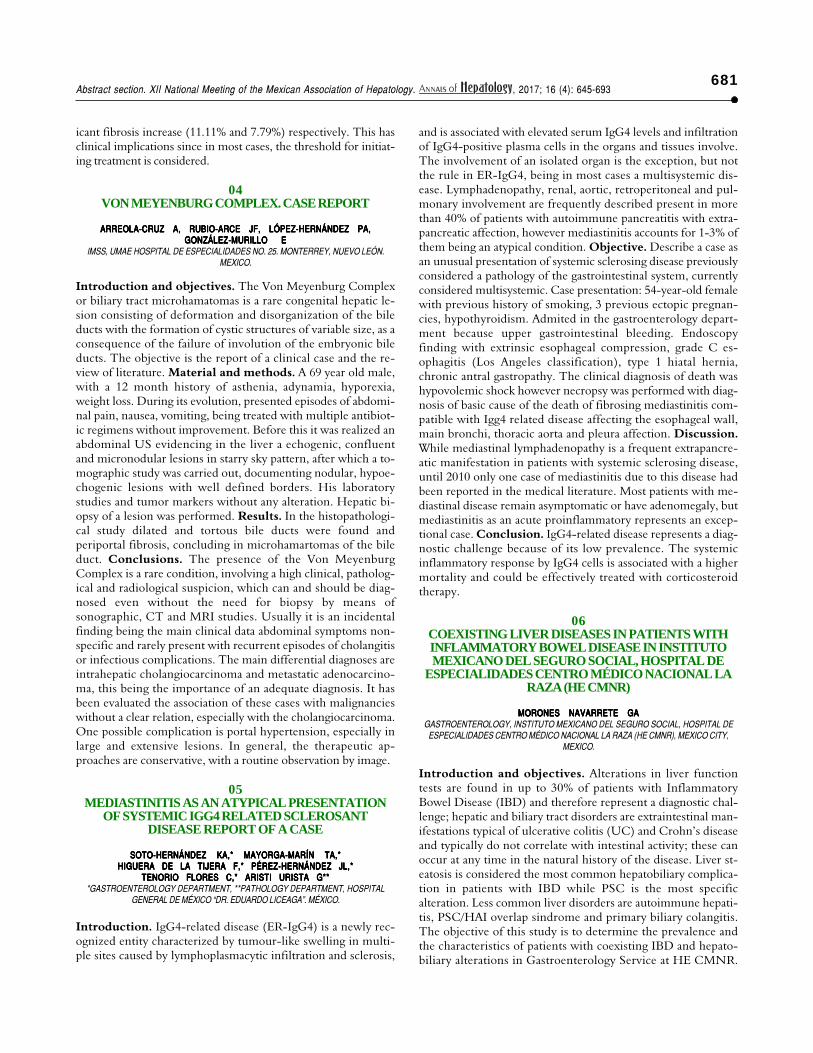
681Abstract section. XII National Meeting of the Mexican Association of Hepatology. , 2017; 16 (4): 645-693
icant fibrosis increase (11.11% and 7.79%) respectively. This hasclinical implications since in most cases, the threshold for initiat-ing treatment is considered.
04VON MEYENBURG COMPLEX. CASE REPORT
ARREOLA-CRUZ A, RUBIO-ARCE JF, LÓPEZ-HERNÁNDEZ PA,ARREOLA-CRUZ A, RUBIO-ARCE JF, LÓPEZ-HERNÁNDEZ PA,ARREOLA-CRUZ A, RUBIO-ARCE JF, LÓPEZ-HERNÁNDEZ PA,ARREOLA-CRUZ A, RUBIO-ARCE JF, LÓPEZ-HERNÁNDEZ PA,ARREOLA-CRUZ A, RUBIO-ARCE JF, LÓPEZ-HERNÁNDEZ PA,GONZÁLEZ-MURILLO EGONZÁLEZ-MURILLO EGONZÁLEZ-MURILLO EGONZÁLEZ-MURILLO EGONZÁLEZ-MURILLO E
IMSS, UMAE HOSPITAL DE ESPECIALIDADES NO. 25. MONTERREY, NUEVO LEÓN.MEXICO.
Introduction and objectives. The Von Meyenburg Complexor biliary tract microhamatomas is a rare congenital hepatic le-sion consisting of deformation and disorganization of the bileducts with the formation of cystic structures of variable size, as aconsequence of the failure of involution of the embryonic bileducts. The objective is the report of a clinical case and the re-view of literature. Material and methods. A 69 year old male,with a 12 month history of asthenia, adynamia, hyporexia,weight loss. During its evolution, presented episodes of abdomi-nal pain, nausea, vomiting, being treated with multiple antibiot-ic regimens without improvement. Before this it was realized anabdominal US evidencing in the liver a echogenic, confluentand micronodular lesions in starry sky pattern, after which a to-mographic study was carried out, documenting nodular, hypoe-chogenic lesions with well defined borders. His laboratorystudies and tumor markers without any alteration. Hepatic bi-opsy of a lesion was performed. Results. In the histopathologi-cal study dilated and tortous bile ducts were found andperiportal fibrosis, concluding in microhamartomas of the bileduct. Conclusions. The presence of the Von MeyenburgComplex is a rare condition, involving a high clinical, patholog-ical and radiological suspicion, which can and should be diag-nosed even without the need for biopsy by means ofsonographic, CT and MRI studies. Usually it is an incidentalfinding being the main clinical data abdominal symptoms non-specific and rarely present with recurrent episodes of cholangitisor infectious complications. The main differential diagnoses areintrahepatic cholangiocarcinoma and metastatic adenocarcino-ma, this being the importance of an adequate diagnosis. It hasbeen evaluated the association of these cases with malignancieswithout a clear relation, especially with the cholangiocarcinoma.One possible complication is portal hypertension, especially inlarge and extensive lesions. In general, the therapeutic ap-proaches are conservative, with a routine observation by image.
05MEDIASTINITIS AS AN ATYPICAL PRESENTATION
OF SYSTEMIC IGG4 RELATED SCLEROSANTDISEASE REPORT OF A CASE
SOTO-HERNÁNDEZ KA,* MAYORGA-MARÍN TA,*SOTO-HERNÁNDEZ KA,* MAYORGA-MARÍN TA,*SOTO-HERNÁNDEZ KA,* MAYORGA-MARÍN TA,*SOTO-HERNÁNDEZ KA,* MAYORGA-MARÍN TA,*SOTO-HERNÁNDEZ KA,* MAYORGA-MARÍN TA,*HIGUERA DE LA TIJERA F,* PÉREZ-HERNÁNDEZ JL,*HIGUERA DE LA TIJERA F,* PÉREZ-HERNÁNDEZ JL,*HIGUERA DE LA TIJERA F,* PÉREZ-HERNÁNDEZ JL,*HIGUERA DE LA TIJERA F,* PÉREZ-HERNÁNDEZ JL,*HIGUERA DE LA TIJERA F,* PÉREZ-HERNÁNDEZ JL,*
TENORIO FLORES C,* ARISTI URISTA G**TENORIO FLORES C,* ARISTI URISTA G**TENORIO FLORES C,* ARISTI URISTA G**TENORIO FLORES C,* ARISTI URISTA G**TENORIO FLORES C,* ARISTI URISTA G***GASTROENTEROLOGY DEPARTMENT, **PATHOLOGY DEPARTMENT, HOSPITAL
GENERAL DE MÉXICO “DR. EDUARDO LICEAGA”. MÉXICO.
Introduction. IgG4-related disease (ER-IgG4) is a newly rec-ognized entity characterized by tumour-like swelling in multi-ple sites caused by lymphoplasmacytic infiltration and sclerosis,
and is associated with elevated serum IgG4 levels and infiltrationof IgG4-positive plasma cells in the organs and tissues involve.The involvement of an isolated organ is the exception, but notthe rule in ER-IgG4, being in most cases a multisystemic dis-ease. Lymphadenopathy, renal, aortic, retroperitoneal and pul-monary involvement are frequently described present in morethan 40% of patients with autoimmune pancreatitis with extra-pancreatic affection, however mediastinitis accounts for 1-3% ofthem being an atypical condition. Objective. Describe a case asan unusual presentation of systemic sclerosing disease previouslyconsidered a pathology of the gastrointestinal system, currentlyconsidered multisystemic. Case presentation: 54-year-old femalewith previous history of smoking, 3 previous ectopic pregnan-cies, hypothyroidism. Admited in the gastroenterology depart-ment because upper gastrointestinal bleeding. Endoscopyfinding with extrinsic esophageal compression, grade C es-ophagitis (Los Angeles classification), type 1 hiatal hernia,chronic antral gastropathy. The clinical diagnosis of death washypovolemic shock however necropsy was performed with diag-nosis of basic cause of the death of fibrosing mediastinitis com-patible with Igg4 related disease affecting the esophageal wall,main bronchi, thoracic aorta and pleura affection. Discussion.While mediastinal lymphadenopathy is a frequent extrapancre-atic manifestation in patients with systemic sclerosing disease,until 2010 only one case of mediastinitis due to this disease hadbeen reported in the medical literature. Most patients with me-diastinal disease remain asymptomatic or have adenomegaly, butmediastinitis as an acute proinflammatory represents an excep-tional case. Conclusion. IgG4-related disease represents a diag-nostic challenge because of its low prevalence. The systemicinflammatory response by IgG4 cells is associated with a highermortality and could be effectively treated with corticosteroidtherapy.
06COEXISTING LIVER DISEASES IN PATIENTS WITHINFLAMMATORY BOWEL DISEASE IN INSTITUTOMEXICANO DEL SEGURO SOCIAL, HOSPITAL DE
ESPECIALIDADES CENTRO MÉDICO NACIONAL LARAZA (HE CMNR)
MORONES NAVARRETE GAMORONES NAVARRETE GAMORONES NAVARRETE GAMORONES NAVARRETE GAMORONES NAVARRETE GAGASTROENTEROLOGY, INSTITUTO MEXICANO DEL SEGURO SOCIAL, HOSPITAL DE
ESPECIALIDADES CENTRO MÉDICO NACIONAL LA RAZA (HE CMNR), MEXICO CITY,MEXICO.
Introduction and objectives. Alterations in liver functiontests are found in up to 30% of patients with InflammatoryBowel Disease (IBD) and therefore represent a diagnostic chal-lenge; hepatic and biliary tract disorders are extraintestinal man-ifestations typical of ulcerative colitis (UC) and Crohn’s diseaseand typically do not correlate with intestinal activity; these canoccur at any time in the natural history of the disease. Liver st-eatosis is considered the most common hepatobiliary complica-tion in patients with IBD while PSC is the most specificalteration. Less common liver disorders are autoimmune hepati-tis, PSC/HAI overlap sindrome and primary biliary colangitis.The objective of this study is to determine the prevalence andthe characteristics of patients with coexisting IBD and hepato-biliary alterations in Gastroenterology Service at HE CMNR.
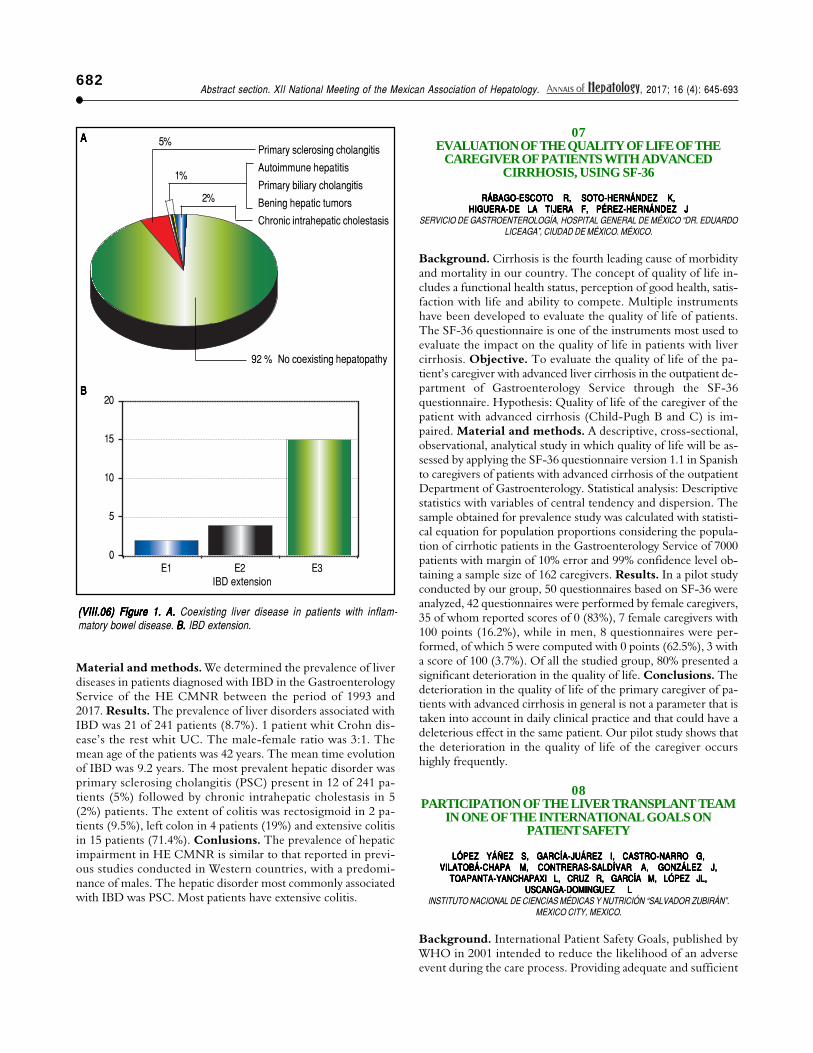
682Abstract section. XII National Meeting of the Mexican Association of Hepatology. , 2017; 16 (4): 645-693
(VIII.06) Figure 1. A.(VIII.06) Figure 1. A.(VIII.06) Figure 1. A.(VIII.06) Figure 1. A.(VIII.06) Figure 1. A. Coexisting liver disease in patients with inflam-matory bowel disease. B.B.B.B.B. IBD extension.
AAAAA
BBBBB20
15
10
5
0E1 E2 E3
IBD extension
Material and methods. We determined the prevalence of liverdiseases in patients diagnosed with IBD in the GastroenterologyService of the HE CMNR between the period of 1993 and2017. Results. The prevalence of liver disorders associated withIBD was 21 of 241 patients (8.7%). 1 patient whit Crohn dis-ease’s the rest whit UC. The male-female ratio was 3:1. Themean age of the patients was 42 years. The mean time evolutionof IBD was 9.2 years. The most prevalent hepatic disorder wasprimary sclerosing cholangitis (PSC) present in 12 of 241 pa-tients (5%) followed by chronic intrahepatic cholestasis in 5(2%) patients. The extent of colitis was rectosigmoid in 2 pa-tients (9.5%), left colon in 4 patients (19%) and extensive colitisin 15 patients (71.4%). Conlusions. The prevalence of hepaticimpairment in HE CMNR is similar to that reported in previ-ous studies conducted in Western countries, with a predomi-nance of males. The hepatic disorder most commonly associatedwith IBD was PSC. Most patients have extensive colitis.
07EVALUATION OF THE QUALITY OF LIFE OF THE
CAREGIVER OF PATIENTS WITH ADVANCEDCIRRHOSIS, USING SF-36
RÁBAGO-ESCOTO R, SOTO-HERNÁNDEZ K,RÁBAGO-ESCOTO R, SOTO-HERNÁNDEZ K,RÁBAGO-ESCOTO R, SOTO-HERNÁNDEZ K,RÁBAGO-ESCOTO R, SOTO-HERNÁNDEZ K,RÁBAGO-ESCOTO R, SOTO-HERNÁNDEZ K,HIGUERA-DE LA TIJERA F, PÉREZ-HERNÁNDEZ JHIGUERA-DE LA TIJERA F, PÉREZ-HERNÁNDEZ JHIGUERA-DE LA TIJERA F, PÉREZ-HERNÁNDEZ JHIGUERA-DE LA TIJERA F, PÉREZ-HERNÁNDEZ JHIGUERA-DE LA TIJERA F, PÉREZ-HERNÁNDEZ J
SERVICIO DE GASTROENTEROLOGÍA, HOSPITAL GENERAL DE MÉXICO “DR. EDUARDOLICEAGA”, CIUDAD DE MÉXICO. MÉXICO.
Background. Cirrhosis is the fourth leading cause of morbidityand mortality in our country. The concept of quality of life in-cludes a functional health status, perception of good health, satis-faction with life and ability to compete. Multiple instrumentshave been developed to evaluate the quality of life of patients.The SF-36 questionnaire is one of the instruments most used toevaluate the impact on the quality of life in patients with livercirrhosis. Objective. To evaluate the quality of life of the pa-tient’s caregiver with advanced liver cirrhosis in the outpatient de-partment of Gastroenterology Service through the SF-36questionnaire. Hypothesis: Quality of life of the caregiver of thepatient with advanced cirrhosis (Child-Pugh B and C) is im-paired. Material and methods. A descriptive, cross-sectional,observational, analytical study in which quality of life will be as-sessed by applying the SF-36 questionnaire version 1.1 in Spanishto caregivers of patients with advanced cirrhosis of the outpatientDepartment of Gastroenterology. Statistical analysis: Descriptivestatistics with variables of central tendency and dispersion. Thesample obtained for prevalence study was calculated with statisti-cal equation for population proportions considering the popula-tion of cirrhotic patients in the Gastroenterology Service of 7000patients with margin of 10% error and 99% confidence level ob-taining a sample size of 162 caregivers. Results. In a pilot studyconducted by our group, 50 questionnaires based on SF-36 wereanalyzed, 42 questionnaires were performed by female caregivers,35 of whom reported scores of 0 (83%), 7 female caregivers with100 points (16.2%), while in men, 8 questionnaires were per-formed, of which 5 were computed with 0 points (62.5%), 3 witha score of 100 (3.7%). Of all the studied group, 80% presented asignificant deterioration in the quality of life. Conclusions. Thedeterioration in the quality of life of the primary caregiver of pa-tients with advanced cirrhosis in general is not a parameter that istaken into account in daily clinical practice and that could have adeleterious effect in the same patient. Our pilot study shows thatthe deterioration in the quality of life of the caregiver occurshighly frequently.
08PARTICIPATION OF THE LIVER TRANSPLANT TEAM
IN ONE OF THE INTERNATIONAL GOALS ONPATIENT SAFETY
LÓPEZ YÁÑEZ S, GARCÍA-JUÁREZ I, CASTRO-NARRO G,LÓPEZ YÁÑEZ S, GARCÍA-JUÁREZ I, CASTRO-NARRO G,LÓPEZ YÁÑEZ S, GARCÍA-JUÁREZ I, CASTRO-NARRO G,LÓPEZ YÁÑEZ S, GARCÍA-JUÁREZ I, CASTRO-NARRO G,LÓPEZ YÁÑEZ S, GARCÍA-JUÁREZ I, CASTRO-NARRO G,VILATOBÁ-CHAPA M, CONTRERAS-SALDÍVAR A, GONZÁLEZ J,VILATOBÁ-CHAPA M, CONTRERAS-SALDÍVAR A, GONZÁLEZ J,VILATOBÁ-CHAPA M, CONTRERAS-SALDÍVAR A, GONZÁLEZ J,VILATOBÁ-CHAPA M, CONTRERAS-SALDÍVAR A, GONZÁLEZ J,VILATOBÁ-CHAPA M, CONTRERAS-SALDÍVAR A, GONZÁLEZ J,
TOAPANTA-TOAPANTA-TOAPANTA-TOAPANTA-TOAPANTA-YANCHAPAXI L, CRUZ R, GARCÍA M, LÓPEZ JL,YANCHAPAXI L, CRUZ R, GARCÍA M, LÓPEZ JL,YANCHAPAXI L, CRUZ R, GARCÍA M, LÓPEZ JL,YANCHAPAXI L, CRUZ R, GARCÍA M, LÓPEZ JL,YANCHAPAXI L, CRUZ R, GARCÍA M, LÓPEZ JL,USCANGA-DOMINGUEZ LUSCANGA-DOMINGUEZ LUSCANGA-DOMINGUEZ LUSCANGA-DOMINGUEZ LUSCANGA-DOMINGUEZ L
INSTITUTO NACIONAL DE CIENCIAS MÉDICAS Y NUTRICIÓN “SALVADOR ZUBIRÁN”.MEXICO CITY, MEXICO.
Background. International Patient Safety Goals, published byWHO in 2001 intended to reduce the likelihood of an adverseevent during the care process. Providing adequate and sufficient
5%
1%
2%
Primary sclerosing cholangitis
Autoimmune hepatitis
Primary biliary cholangitis
Bening hepatic tumors
Chronic intrahepatic cholestasis
92 % No coexisting hepatopathy
683Abstract section. XII National Meeting of the Mexican Association of Hepatology. , 2017; 16 (4): 645-693
information to patients prevents interpretation and content lim-ited errors. Health education groups are a learning strategywhere skills such as self-care and decision-making are favoredand they can impact in quality of life and in the social environ-ment of a patient. Objective. To strengthen the information toliver transplant (LT) candidates and their families throughhealth education groups. Material and methods. LT candi-dates and their families were included from August 2015 to Sep-tember 2016. A social group work method and a traditionalteaching with variants (controlled participation) were per-formed. For this purpose, health education courses were de-signed with a focus on evaluation and selection of LTcandidates, surgical, psychiatric and anesthetic procedures aswell as nutritional education, organ donation promotion andsocio-familial alterations derived from the disease and LT. Re-sults. Twelve group sessions involving 59 families were execut-ed. A total of 221 participants with mean of 3 family per patientwere observed. All participants answer an initial survey aboutthe initial knowledge in the involved areas. Among all, 64% ofparticipants were women, with an age of 70 ± 14 years. At leastsome basic education (middle or junior high) was coursed by91% of the attendants. At the end of the sessions, 81% of theparticipants were able to identify the patient’s own characteris-tics in relation to LT (donor and recipient), associated care need,surgical risks, importance of mental health and family participa-tion as well as the need of a post-biopsychosocial follow-upTHO. At the moment, a 200% increase in attendance was ob-served. Conclusion. The participation of patients and theirfamilies in health education groups allowed them to acquiresupplemental information about LT process, reduced the anxie-ty associated to list wait-time and provided comprehension ofbehavioral changes that patients can have. It also promoted anactive participation of the family, self-management, and com-munication with members of the health team.
09CASE REPORT: ACALCULOUS CHOLECYSTITISASSOCIATED WITH HEPATITIS A VIRUS (HAV)
OVIEDO MAGLIONE MA, MEJÍA S, GARCÍA E, ZAMARRIPA FOVIEDO MAGLIONE MA, MEJÍA S, GARCÍA E, ZAMARRIPA FOVIEDO MAGLIONE MA, MEJÍA S, GARCÍA E, ZAMARRIPA FOVIEDO MAGLIONE MA, MEJÍA S, GARCÍA E, ZAMARRIPA FOVIEDO MAGLIONE MA, MEJÍA S, GARCÍA E, ZAMARRIPA FHOSPITAL JUÁREZ DE MÉXICO, MEXICO CITY, MEXICO.
Introduction. Acalculous cholecystitis secondary to viral hepa-titis A, is a rare presentation which can influence the presence ofacute abdomen. Objective. Report uncommon case of acuteacalculous cholecystitis secondary to infection by HAV. Case.female, 18 years, that 1 week prior to admission with asthenia,adynamia, hiporexia; including abdominal pain in right upperquadrant, EVA 6-10, lancinating pain, irradiated to right lumbarregion; concomitant nausea, vomiting, 38-degrees fever, jaun-diced tinge. Physical exam: BP: 120/70 HR: 128, RR: 24, drymucous membranes, abdomen: decreased peristalsis, painful tothe superficial and deep palpation of predominance in right up-per quadrant, Murphy sign (+), with data of peritoneal irrita-tion. With the following analytical alterations: AST: 1630, ALT:2083, TB: 8.2, DB: 7.9, GGT: 441, ALP: 262, LDH: 597, viralPanel: IgG hep. To (-) IgM hep. A (+), US of abdomen: Vesic-ular inflammatory process of heterogeneous content. CT of Ab-domen: images in relation to acalculous cholecystitis. Treatment
was Conservative and symptomatic with satisfactory evolutionbeing discharged in 6th day. Studies of control to the 4th week:AST: 48, ALT: 61, TB: 0.7, DB: 0.5, GGT: 40, ALP: 90, LDH:55. Discussion. The theory of cellular tropism described inhepatitis E virus infection is being observed more frequently inpatients with HAV, affecting the vesicular wall and ducts, pro-ducing a severe inflammatory reaction, peri-cholangitis andacalculous cholecystitis. Conditioning greater severity and mor-bidity and mortality. The treatment so far is controversial andthe therapeutic decision depends on the evolution of the pa-tient. Conclusion. Acute acalculous cholecystitis is a rare com-plication of hepatitis A however should be suspected in allpatients with clinical presentation of acute cholecystitis becausethat can precipitate a fulminant hepatic failure.
10CASE REPORT: HEPATIC ABSCESUS BY
BURKHOLDERIA PSEUDOMALLEI IN HOSPITALJUÁREZ DE MÉXICO
DURÁN ROSAS C, MEJÍA S, GARCÍA E, ZAMARRIPA FDURÁN ROSAS C, MEJÍA S, GARCÍA E, ZAMARRIPA FDURÁN ROSAS C, MEJÍA S, GARCÍA E, ZAMARRIPA FDURÁN ROSAS C, MEJÍA S, GARCÍA E, ZAMARRIPA FDURÁN ROSAS C, MEJÍA S, GARCÍA E, ZAMARRIPA FHOSPITAL JUÁREZ DE MÉXICO, MEXICO CITY, MEXICO.
Introduction. In Mexico the main etiological agents of the liverabscesses piogenes son E. coli and Klebsiella, a difference of Asiancountries that caused by a single pathogen K. pneumoniae. Howev-er, there are rare agents such as Burkholderia pseudomiallei that canlead to multiple hepatic abscesses. Taiwan has been considered en-demic since 2001. Objective. To present an uncommon etiologicagent of hepatic abscess in Mexico. Case. A 24-year-old femalefrom Taiwan residing 6 months ago in Mexico City, she beginswith pain in the right hypochondrium, fever of evening origin andlater with no predominance of schedule. EF: TA 100/60 mmHg,FC 100x, T 38.8°, generalized pallor, conjunctival jaundice, painfulhepatomegaly 5 cm below the costal border, the following paraclin-ics are requested: AST 69 IU/L, ALT 71 UI/L, DHL 566. Abdomi-nal CT: multiple hepatic abscesses, initiating empiric treatmentwith Ceftriaxone and Metronidazole without clinical improve-ment; hemoculture negative, performing puncture guided by TACobtaining positive culture for B. pseudomallei, based on an antibio-gram management with Ceftazidime 2 g started every 8 h for 14days. Discussion. Meloidosis is a systemic infection caused by B.pseudomallei and is endemic to Taiwan, has a mortality rate of about37% in acute septicemia, antimicrobial therapy should be given atleast 5 months otherwise there is a risk of relapse of approximatelythe 10%. The culture of liver abscess is the gold standard for diag-nosis. The treatment of choice son: tetracyclines, trimethoprim,ceftazidime and carbapenemics. Conclusion. In Mexico there areno reports of hepatic abscesses by B. pseudomallei, we should suspectin all (a) original patient or antecedent of recent trip to Taiwan, tosuspect B. pseudomallei as an etiological agent.
684Abstract section. XII National Meeting of the Mexican Association of Hepatology. , 2017; 16 (4): 645-693
11MANAGEMENT OF GIST TUMOR WITH CELECOXIB
AND PENTOXIFYLLINE: A CASE REPORT
JIMÉNEZ-LUEVANO MA, RODRÍGUEZ-VILLA P, RAMÍREZ-FLORES S,JIMÉNEZ-LUEVANO MA, RODRÍGUEZ-VILLA P, RAMÍREZ-FLORES S,JIMÉNEZ-LUEVANO MA, RODRÍGUEZ-VILLA P, RAMÍREZ-FLORES S,JIMÉNEZ-LUEVANO MA, RODRÍGUEZ-VILLA P, RAMÍREZ-FLORES S,JIMÉNEZ-LUEVANO MA, RODRÍGUEZ-VILLA P, RAMÍREZ-FLORES S,JIMÉNEZ PARTIDA MA, CERVANTES-RODRÍGUEZ G,JIMÉNEZ PARTIDA MA, CERVANTES-RODRÍGUEZ G,JIMÉNEZ PARTIDA MA, CERVANTES-RODRÍGUEZ G,JIMÉNEZ PARTIDA MA, CERVANTES-RODRÍGUEZ G,JIMÉNEZ PARTIDA MA, CERVANTES-RODRÍGUEZ G,
FRANCO-TOPETE R, BRAVO-CUELLAR AFRANCO-TOPETE R, BRAVO-CUELLAR AFRANCO-TOPETE R, BRAVO-CUELLAR AFRANCO-TOPETE R, BRAVO-CUELLAR AFRANCO-TOPETE R, BRAVO-CUELLAR ACLÍNICA DE HÍGADO, HOSPITAL REGIONAL “VALENTÍN GÓMEZ FARÍAS” (ISSSTE),
ZAPOPAN, JALISCO, MEXICO.
Introduction. Gastrointestinal Stromal Tumors (GIST) aremesenchymal neoplasms of spindle cells derived from Cajal cells;characterized by expressing in 95% of cases KIT protein. Repre-sent less than 1% of digestive tumors, with an incidence of 10-20 per million inhabitants and predominance in male sex,between 40 and 60 years. It affects the small intestine by 20 to30% the final diagnosis is histological and immunohistochemi-cal. Surgery remains the gold standard treatment, but it also ex-ists drugs that improve survival such as imatinib; however, dueto its side effects and high resistance rates, it was used a therapybased on celecoxib and pentoxifylline because of their anti-in-flammatory, antiangiogenic and pro-apoptotic properties. Obje-tive. To assess the response of GIST tumor to the treatmentwith celecoxib and pentoxifylline. Clinical case. A 74 years oldfemale patient with no chronic degenerative diseases was admit-ted in the Gastroenterology Service of Hospital Regional“Valentín Gómez Farías” (ISSSTE), Zapopan, Jalisco, for ab-dominal pain and jaundice; BT 6.0 mg/dL, BD 5.0 mg/dL, GGT400 mg/dL, AST 15 mg/dL, ALT 80 mg/dL and FA 300 mg/dL.A liver and biliary tree ultrasound reported choledochal dilata-tion of 13 mm. Colangioresonance showed tumor in the secondportion of the duodenum of 3.5 x 2.5 cm causing distal com-pression of the common bile duct. Enteroscopy showed a tumorin the second and third portions of duodenum, obstructing 40%of the intestinal lumen. Biopsies were taken and sent to his-topathological study reporting compatible tissue with GIST.Due to the patient’s condition and surgical risk, it was iniciatedtreatment with Pentoxifylline 400 mg every 12 h and celecoxib100 mg every 12 h. After 5 months of treatment, she is asymp-tomatic, and with normal liver function tests. Conclusion.Management of duodenal GIST tumors continues to be a chal-lenge in patients who are not candidates for surgery. In this case,as in previous research, it was shown that an alternative therapywith Pentoxifylline and Celecoxib improves clinical and labora-tory status of the patient; however, more studies are required toassess their real effectiveness.
12IMPACT OF THE COSTS OF HOSPITALIZATION IN
PATIENTS WITH HEPATIC CIRRHOSIS AT THEHOSPITAL JUÁREZ DE MEXICO
FERNÁNDEZ-MARTÍNEZ NC,* AGUILAR-MORENO RU,*FERNÁNDEZ-MARTÍNEZ NC,* AGUILAR-MORENO RU,*FERNÁNDEZ-MARTÍNEZ NC,* AGUILAR-MORENO RU,*FERNÁNDEZ-MARTÍNEZ NC,* AGUILAR-MORENO RU,*FERNÁNDEZ-MARTÍNEZ NC,* AGUILAR-MORENO RU,*VELASCO-SANTIAGO YM,* RIVERA-FLORES AG,*VELASCO-SANTIAGO YM,* RIVERA-FLORES AG,*VELASCO-SANTIAGO YM,* RIVERA-FLORES AG,*VELASCO-SANTIAGO YM,* RIVERA-FLORES AG,*VELASCO-SANTIAGO YM,* RIVERA-FLORES AG,*
RAMOS-AGUILAR GA,* BARRAZA-ORTIZ DA,* LLORENTE-RAMÓN A,*RAMOS-AGUILAR GA,* BARRAZA-ORTIZ DA,* LLORENTE-RAMÓN A,*RAMOS-AGUILAR GA,* BARRAZA-ORTIZ DA,* LLORENTE-RAMÓN A,*RAMOS-AGUILAR GA,* BARRAZA-ORTIZ DA,* LLORENTE-RAMÓN A,*RAMOS-AGUILAR GA,* BARRAZA-ORTIZ DA,* LLORENTE-RAMÓN A,*DURÁN-ROSAS C,* OVIEDO-MAGLIONE MA,* MEJÍA-LOZA S MI,*DURÁN-ROSAS C,* OVIEDO-MAGLIONE MA,* MEJÍA-LOZA S MI,*DURÁN-ROSAS C,* OVIEDO-MAGLIONE MA,* MEJÍA-LOZA S MI,*DURÁN-ROSAS C,* OVIEDO-MAGLIONE MA,* MEJÍA-LOZA S MI,*DURÁN-ROSAS C,* OVIEDO-MAGLIONE MA,* MEJÍA-LOZA S MI,*
GARCÍA-RUIZ E,* JUÁREZ-VALDÉS E,* PÉREZ-LÓPEZ N,*GARCÍA-RUIZ E,* JUÁREZ-VALDÉS E,* PÉREZ-LÓPEZ N,*GARCÍA-RUIZ E,* JUÁREZ-VALDÉS E,* PÉREZ-LÓPEZ N,*GARCÍA-RUIZ E,* JUÁREZ-VALDÉS E,* PÉREZ-LÓPEZ N,*GARCÍA-RUIZ E,* JUÁREZ-VALDÉS E,* PÉREZ-LÓPEZ N,*ZAMARRIPA-DORSEY F*ZAMARRIPA-DORSEY F*ZAMARRIPA-DORSEY F*ZAMARRIPA-DORSEY F*ZAMARRIPA-DORSEY F*
*GASTROENTEROLOGY SERVICE; HOSPITAL JUÁREZ DE MÉXICO, MEXICO CITY.MEXICO.
Introduction. Liver cirrhosis is one of the main causes of mor-bidity and mortality in our country, particularly affecting peo-ple of productive age and therefore considered a public healthproblem. Our objective is to present the economic impact con-sidered an intrahospital management of hepatic cirrhosis. Mate-rial and methods. This is a descriptive, cross-sectional study of211 patients with liver cirrhosis hospitalized in one year (March2016 to February 2017) at Hospital Juárez de México. Age, sex,cause of liver cirrhosis, reason for admission, days of hospitalstay, hospitalization costs were studied. Results. Of 211 pa-tients, 62.6% were men and the mean age was 58 years. Themajor cause of cirrhosis was the following: Alcohol (51.18%),NASH (20.85%), HCV (12.80%), autoimmune (2.84%), medi-cation (1.42%), HBV (0.48%). The mortality rate was 1.8 pa-tients/year, with 38 deaths in total. The median number of daysof in-hospital stay was 3, the total annual cost was 2,448,205.6,the average cost per day was 3,859.06, and of which the medi-cations accounted for 81.6%, while medical care representedonly 2% due to the low socioeconomic level granted. Conclu-sions. The main cause of liver cirrhosis is secondary to alcoholconsumption predominating in males, the highest proportion ofpatients is of productive age, economically impacting our healthsystem, the cost of hospitalization increases proportionally tothe days of (e.g., terlipressin, endoscopy, transfusion expenses,antibiotic prophylaxis), infectious processes (e.g., EBP, antibiotictherapy), as well as the use of high-cost medications, as well asother resources derived from the main complications, includinggastrointestinal bleeding). Encephalopathy (antiammoniummeasures). More than 70% of patients with cirrhosis (alcohol,fatty liver) with modifiable risk factors could be prevented,which would lead to a decrease in the economic impact of ourhospital.
13HEPATIC FIBROSIS IN HEALTH PROFESSIONALS,ACCORDING TO A NON-INVASIVE BIOLOGICAL
TEST. CONCIENCIA PREVALENCE STUDY
POO JL,* AGUILAR JR,* PEÑA P,** HERNÁNDEZ L,* MUÑOZ L***POO JL,* AGUILAR JR,* PEÑA P,** HERNÁNDEZ L,* MUÑOZ L***POO JL,* AGUILAR JR,* PEÑA P,** HERNÁNDEZ L,* MUÑOZ L***POO JL,* AGUILAR JR,* PEÑA P,** HERNÁNDEZ L,* MUÑOZ L***POO JL,* AGUILAR JR,* PEÑA P,** HERNÁNDEZ L,* MUÑOZ L****MEXICAN GROUP FOR THE STUDY OF LIVER DISEASES, MEXICO CITY. MEXICO.
**CELLPHARMA LABORATORIES, MEXICO CITY, MEXICO.***MONTERREY UNIVERSITY HOSPITAL, NL. MEXICO.
Introduction. Despite knowing the impact of fibrosis on thenatural history of chronic liver diseases, there is few informa-tion about its prevalence in health professionals in Mexico.Objective. To describe the prevalence and degree of hepaticfibrosis in health professionals based on a commercially availa-
685Abstract section. XII National Meeting of the Mexican Association of Hepatology. , 2017; 16 (4): 645-693
ble noninvasive marker. Material and methods. A total of209 specialized health professionals: gastroenterologist (97),hepatologist (63) or Internist (49) were studied with the com-mercially available test, called Fibromax®, under 10 h fastingconditions. Clinical, somatometric, and biochemical data werecollected. Results. 44% were women, 49 ± 13 years old. Ac-cording to BMI 32% were normal, 41% overweight, and 27 %with obesity. Waist circumference was elevated (> 102 cm inmen and > 88 cm in women) in 35 and 33%, respectively;37% had chronic alcohol intake and 39% had health associatedconditions. Fibromax® test results in table 1. Additionally,26.2% presented some degree of necroinflammatory activity,21.3% with varying degrees of steatosis and 33.3% with stea-tohepatitis. Conclusions. A quarter of the total health profes-sional sample evaluated depicted some degree of liver fibrosis;hepatologist subgroup were the least affected. One-third pre-sented different degrees of potential metabolic damage, sug-gesting the need for additional confirmatory studies toimplement appropriate corrective measures.This work has been sponsored by Cellpharma (Fibromax®test).
14ALAGILLE SYNDROME AND LIVER
TRANSPLANTATION
MARTÍNEZ-SALDÍVAR B,* VALDÉS-ÁNGELES V,*MARTÍNEZ-SALDÍVAR B,* VALDÉS-ÁNGELES V,*MARTÍNEZ-SALDÍVAR B,* VALDÉS-ÁNGELES V,*MARTÍNEZ-SALDÍVAR B,* VALDÉS-ÁNGELES V,*MARTÍNEZ-SALDÍVAR B,* VALDÉS-ÁNGELES V,*SANTOS-CABALLERO M,SANTOS-CABALLERO M,SANTOS-CABALLERO M,SANTOS-CABALLERO M,SANTOS-CABALLERO M,††††† SANSÓN-NAVARRO G, SANSÓN-NAVARRO G, SANSÓN-NAVARRO G, SANSÓN-NAVARRO G, SANSÓN-NAVARRO G,‡‡‡‡‡ DÍAZ-PINEDA M, DÍAZ-PINEDA M, DÍAZ-PINEDA M, DÍAZ-PINEDA M, DÍAZ-PINEDA M,‡‡‡‡‡
FLORES-ARROYO A,FLORES-ARROYO A,FLORES-ARROYO A,FLORES-ARROYO A,FLORES-ARROYO A,§§§§§ HERNÁNDEZ-AGUILAR C, HERNÁNDEZ-AGUILAR C, HERNÁNDEZ-AGUILAR C, HERNÁNDEZ-AGUILAR C, HERNÁNDEZ-AGUILAR C,||||||||| |
HERNÁNDEZ-DOMÍNGUEZ MHERNÁNDEZ-DOMÍNGUEZ MHERNÁNDEZ-DOMÍNGUEZ MHERNÁNDEZ-DOMÍNGUEZ MHERNÁNDEZ-DOMÍNGUEZ M¶¶¶¶¶
*LIVER TRANSPLANT UNIT HG CMN LA RAZA. †CHIEF TRANSPLANT UNIT HG CMN LARAZA. ‡STUDENTS UNAM MEDICINE SCHOOL. §LIVER UNIT INSTITUTO DE PENSIONES
DEL ESTADO DE JALISCO.||HOSPITAL CENTRAL SUR PEMEX PICACHO. ¶CHIEF OFTRANSPLANT DIVISION HGC CMN LA RAZA. MEXICO.
Introduction. Alagille syndrome is an infrequent autosomaldominant disorder, with great penetrance and variable expressivi-ty. It usually manifests before 2 years of age and is characterizedby intrahepatic biliary hypoplasia associated with three to five ma-jor features: chronic cholestasis, cardiac disease, skeletal abnormal-ities, ocular anomalies and characteristic facial phenotype. Livertransplantation is indicated in children with cirrhosis and hepaticinsufficiency. Objective. To describe the characteristics of pa-tients with Alagille syndrome referred for evaluation to the livertransplantation in the Transplant Unit, Hospital General CMNLa Raza, IMSS. Material and methods. A retrospective and de-
scriptive study of a series of patients with Alagille syndrome re-ferred to the liver transplant unit of CMN La Raza Hospital Gen-eral during the period 2014-2016. Results. We have studied 3patients with Alagille syndrome. The mean age at diagnosis was at24 months and the mean age at which they were sent for evalua-tion for transplantation was at 7 years. Male was seen in 100%,cholestasis was seen in 100%, liver biopsy specimens showed pau-city of the interlobular ducts and characteristic facies in 100%,cardiac anomalies (pulmonary stenosis) in 66.6%, pruritus refrac-tory to treatment in 66.6%, xanthomas in 33%, skeletal altera-tions in 66.6%, and growth retardation in 66.6%. One patientwas in the stage of decompensated cirrhosis (Child C PELD of 30points) and underwent liver transplantation in March 2015. An-other patient is on the waiting list for liver transplant due to re-fractory pruritus and multiple xanthomas. The third patient is infollow-up and comprehensive treatment, without criteria for livertransplantation. Conclusion. The prognosis of the patient withlong-term Alagille syndrome depends on the severity of the liverdisease and associated malformations, requiring liver transplanta-tion from 21 to 31%.
15ACUTE HEPATIC FAILURE OF SECONDARY
LEPTOSPIROSIS: REPORT CASE OF AUTOPSIAFLORES-TORRES A,* MAYORGA- MARÍN T,*FLORES-TORRES A,* MAYORGA- MARÍN T,*FLORES-TORRES A,* MAYORGA- MARÍN T,*FLORES-TORRES A,* MAYORGA- MARÍN T,*FLORES-TORRES A,* MAYORGA- MARÍN T,*
HIGUERA-DE LA TIJERA F,* MURILLO-ELOISA JR,** GÓMEZ-APO E**HIGUERA-DE LA TIJERA F,* MURILLO-ELOISA JR,** GÓMEZ-APO E**HIGUERA-DE LA TIJERA F,* MURILLO-ELOISA JR,** GÓMEZ-APO E**HIGUERA-DE LA TIJERA F,* MURILLO-ELOISA JR,** GÓMEZ-APO E**HIGUERA-DE LA TIJERA F,* MURILLO-ELOISA JR,** GÓMEZ-APO E***GASTROENTEROLOGY DEPARTMENT, ** PATHOLOGY DEPARTMENT, HOSPITAL
GENERAL DE MÉXICO “DR. EDUARDO LICEAGA”, MEXICO CITY, MEXICO.
Background. It is a zoonosis of worldwide distribution, withavailable reports the incidence varies approximately 0.1-1 per100 thousand inhabitants in tropical humid climates. 10%presents the severe form called icteric hemorrhagic or Weil’s dis-ease, with a mortality of 40-50%. Report of case. Male of 23years old from Hidalgo, nurse, with sudden onset 9 days prior toadmission with headache, myalgia, arthralgia, nausea, vomitingassociated with quantified fever of 38.8°C after chills, 3 days af-ter generalized jaundice, coluria. He went to the hospital pre-senting tachycardia, tachypnea, diaphoresis, generalized jaundiceof mucosa and teguments, hemithorax with discrete disseminat-ed rales, altered heart sounds, distended abdomen, hepatomegaly3 cm below costal margin, painful palpation, ecchymosis anddisseminated petechiae. 24 h post admission hypotension withvasopressor requirement, epistaxis and neurological deteriorationwith Glasgow of 13 points. Laboratory: leukopenia, procalciton-in 1.56, creatinine 1.6 mg/dL, urea 46 mg/dL, normal electro-lytes, total bilirubin 35.9 direct standard, AST 1,228, ALT 1,516,alkaline phosphatase 183, GGT 50, TP 10.8%, INR 6.9, chestX-ray bilateral hilar congestion hepatic ultrasound withoutthrombosis, viral panel and TORCH negative cultures negative.Immunologic negative, PCR for leptospirosis negative. Terribleevolution and death. Pathological findings Microscopic: lungalveolar spaces with blood material. Liver extensive necrosis andstaining of warthin-starry photomicrograph of one of the ves-sels of the hepatic parenchyma, with evidence of filiform struc-tures in the vascular wall corresponding to leptospira sp. Renaltubules with extensive necrosis some with bile cylinders, peri-pancreatic adipose tissue with extensive necrosis. Discussion.Leptospirosis disease is a zoonosis that affects a large part of
(VIII.13) Table 1. Fibromax® test results.
Liver Gastroenterologist Hepatologist Internist Totalfibrosis populationscore (%)
F0 74 (76.2%) 55 (87%) 27 (55%) 156 (74.6%)F0-F1 11 (11.3%) 3 (5%) 5 (10%) 19 (9.1%)F1 1 (1%) 0 (0%) 7 (14%) 8 (3.8%)F1-F2 7 (7%) 4 (6%) 6 (12%) 17 (8.1%)F2 2 (2%) 1 (2%) 2 (4%) 5 (2.4%)F3 0 (0%) 0 (0%) 2 (4%) 2 (0.95%)F3-F4 2 (2%) 0 (0%) 0 (0%) 2 (0.95%)F4 0 (0%) 0 (0%) 0 (0%) 0 (0%)
686Abstract section. XII National Meeting of the Mexican Association of Hepatology. , 2017; 16 (4): 645-693
Mexico’s tropical zones and does not have accurate records. PCRwas performed to detect DNA from leptospira with a negativereport which could be associated with the late phase of the dis-ease. Therefore serology and molecular studies increase the sen-sitivity and diagnostic specificity since the progression to latephase is associated with an increase in mortality. Conclusion. Itis interesting that the case is that we must take into account somany risk factors and false negative studies depending on thetime of evolution of the disease.This work has been fully subsidized by the GastroenterologyService, Hospital General de México.
16COST OF INTRAHOSPITAL STAY OF
THE MAIN CAUSES OF REIGNATION OFPATIENTS WITH CIRRHOSIS DESCOMPENSED IN
THE HGZ MF NO. 1 IMSS
CONTRERAS-OMAÑA R,* TÉLLEZ-JAÉN S**CONTRERAS-OMAÑA R,* TÉLLEZ-JAÉN S**CONTRERAS-OMAÑA R,* TÉLLEZ-JAÉN S**CONTRERAS-OMAÑA R,* TÉLLEZ-JAÉN S**CONTRERAS-OMAÑA R,* TÉLLEZ-JAÉN S***CENTER FOR RESEARCH ON LIVER DISEASES AND GASTROENTEROLOGY,
** MEDICINE SCHOOL OF UAEH, PACHUCA, HIDALGO, MÉXICO.
Background. Patients with hepatic cirrhosis are a group vul-nerable to several comorbidities, which decompensate their gen-eral condition, requiring on occasion, in-hospital stay for themanagement of each of these. Aim. Determinate the cost gen-erated by the main causes of hospital readmission, in decompen-sated cirrhotic patients in Hospital General de Zona No.1 IMSS,Pachuca, Hidalgo. Material and methods. Observational,transversal, descriptive and retrospective study, performed inHGZMF No.1 IMSS Pachuca, Hidalgo, from January 2012 toAugust 2014, in which the main causes of hospital readmission,in decompensated cirrhotic patients were evaluated, and Subse-quently used the table of unit costs for care to non-entitledIMSS 2017, published in Diario Oficial de la Federación (DOF).Results. There were 77 cirrhotic patients who returned to thisunit 20 of them with Child-Pugh A (25.97%), 33 in Child-Pugh B (42.85%) and 24 in Child-Pugh C (31.16%), determin-ing as the main causes of hospital readmission, to hepaticencephalopathy (EH) with 33 patients (42.86%), followed byvariceal hemorrhage with 17 (22.07%), infections, ascites andjaundice with 9 each (11.69% each); within the causes of hos-pitalization average, for (HE) and jaundice 7 days, while forvariceal hemorrhage, infections and ascites is 5 days; subse-quently, according to the table of unit costs for care to non-en-titled IMSS 2017, it was determined that the in-hospital stay ofpatients with cirrhosis during the period studied would be$3,893,299.00; divided into: hepatic encephalopathy$1,859,913.00, variceal hemorrhage $783,547.00, ascites $371,295.00, jaundice $ 507,249.00, infections $ 371,295.00,considering that per day/patient and, the condition is on averagefor HE of $56,361.00, variceal hemorrhage $46,091.00, ascites$41,255.00, jaundice $56,361.00 , and infections $41,255.00.Conclusions. In the absence of prior evidence of the cost ofhospitalization for the main causes of reentry of patients withcirrhosis, this study shows the importance of performing thistype of study in other units, and determine adequately the Im-pact of Hepatic Cirrhosis on the Health System.
17ASSOCIATION OF PRURITE SEVERITY WITH
PERINATAL COMPLICATIONS IN PATIENTS WITHINTRAHEPACTIC CHOLESTASIS OF PREGNANCY
GÁLVEZ CASTRO M, CASTILLO BARRADAS MGÁLVEZ CASTRO M, CASTILLO BARRADAS MGÁLVEZ CASTRO M, CASTILLO BARRADAS MGÁLVEZ CASTRO M, CASTILLO BARRADAS MGÁLVEZ CASTRO M, CASTILLO BARRADAS MDEPARTAMENTO DE GASTROENTEROLOGÍA, HOSPITAL DE ESPECIALIDADES “DR.
ANTONIO FRAGA MOURET”, CENTRO MÉDICO NACIONAL LA RAZA, CIUDAD DEMÉXICO, MEXICO.
Introduction. Intrahepatic Cholestasis of Pregnancy (IHCP)occurs in the second or third trimester of pregnancy and it ischaracterized by the presence of intense pruritus and increasesin serum total bile acids and hepatic transaminases. It is associat-ed with an increased risk of preterm labor, prematurity, fetal hy-poxia, meconium amniotic fluid, and intrauterine fetal death. Ithas been proposed to use questionnaires that measure pruritusintensity in patients with IHCP in order to find an associationof the pruritus intensity with the perinatal outcome. Objec-tive. To determine the association of pruritus severity with peri-natal complications in patients with IHCP. Material andmethods. Pregnant women were included during their secondand third trimester, with a presumptive diagnosis of IHCP. Thediagnosis of IHCP was suspected by discrete liver biochemicalalterations and the presence of pruritus. At the time of diagnosistwo methods of measuring pruritus severity, the 5D question-naire and a visual analog 10 cm line scale were used by patientscrossing the line scale at the corresponding pruritus severitypoint. Results. 45 patients were included; (N = 14) had a pre-term labor threat, 11.1% (n = 5) fetal hypoxia, 24.4% (n = 11)showed meconium amniotic fluid and only 1 fetal death (2.2%).100% of evaluated patients showed some degree of pruritus attheir examination time. Combining the two scales to determinepruritus intensity, mild pruritus was found in 6.7% (n = 3),moderate in 55.6% (n = 25) and severe in 37.8% (n = 17). Thehigher the intensity of pruritus the greater the possibility ofperinatal complications such as the threat of preterm labor (P =0.003). Conclusions. The present study indicated that a greaterseverity of pruritus is associated with preterm delivery. There-fore the scales used may help to identify a risk of a poor neonatalprognosis group that requires more vigilance and could be usedas a diagnostic tool in those institutions where there is notavailable the bile acids test.
687Abstract section. XII National Meeting of the Mexican Association of Hepatology. , 2017; 16 (4): 645-693
IX. TRANSPLANTATION/LIVER SURGERY
01APPLICATION OF ULTRASOUND TO
IMPROVE THE PROTECTIVE EFFECTS OFDRUGS AGAINST ISCHEMIA-REPERFUSION
INJURY IN HEPATIC SURGERY
CASILLAS-RAMÍREZ A,CASILLAS-RAMÍREZ A,CASILLAS-RAMÍREZ A,CASILLAS-RAMÍREZ A,CASILLAS-RAMÍREZ A,♦♦♦♦♦,,,,,*****,†,†,†,†,† SÁNCHEZ-CASTRO J, SÁNCHEZ-CASTRO J, SÁNCHEZ-CASTRO J, SÁNCHEZ-CASTRO J, SÁNCHEZ-CASTRO J,♦♦♦♦♦,*,†,*,†,*,†,*,†,*,†
AQUINO-RUBIO G,*AQUINO-RUBIO G,*AQUINO-RUBIO G,*AQUINO-RUBIO G,*AQUINO-RUBIO G,* BARRÓN-VARGAS C.A,BARRÓN-VARGAS C.A,BARRÓN-VARGAS C.A,BARRÓN-VARGAS C.A,BARRÓN-VARGAS C.A,‡‡‡‡‡ ROCHA-SÁNCHEZ AY,* ROCHA-SÁNCHEZ AY,* ROCHA-SÁNCHEZ AY,* ROCHA-SÁNCHEZ AY,* ROCHA-SÁNCHEZ AY,*MARTÍNEZ-PADRÓN HY,*MARTÍNEZ-PADRÓN HY,*MARTÍNEZ-PADRÓN HY,*MARTÍNEZ-PADRÓN HY,*MARTÍNEZ-PADRÓN HY,* JIMÉNEZ-CASTRO MB, JIMÉNEZ-CASTRO MB, JIMÉNEZ-CASTRO MB, JIMÉNEZ-CASTRO MB, JIMÉNEZ-CASTRO MB,§§§§§ PERALTA C PERALTA C PERALTA C PERALTA C PERALTA C§,||§,||§,||§,||§,||
♦THESE AUTHORS CONTRIBUTED EQUALLY TO THIS WORK.* HOSPITAL REGIONAL DE ALTA ESPECIALIDAD DE CIUDAD VICTORIA “BICENTENARIO
2010”, CD. VICTORIA, TAM. MÉXICO, †FACULTAD DE MEDICINA E INGENIERÍA ENSISTEMAS COMPUTACIONALES DE MATAMOROS, UNIVERSIDAD AUTÓNOMA DE
TAMAULIPAS, MATAMOROS, TAM. MÉXICO. ‡HOSPITAL VETERINARIO DE PEQUEÑASESPECIES, FACULTAD DE MEDICINA VETERINARIA Y ZOOTECNIA, UNIVERSIDAD
AUTÓNOMA DE TAMAULIPAS, CD. VICTORIA, TAM. MÉXICO.§TRANSPLANTS BIOMEDICALS, S.L., ||INSTITUTO DE INVESTIGACIONES BIOMÉDICAS
AUGUSTO PI Y SUÑER, BARCELONA, ESPAÑA.
Background and aim. In clinical practice, hepatic resection isusually performed under vascular occlusion. Ischemia-reper-fusion (I/R) injury, inherent to vascular occlusion, adversely af-fects liver regeneration and is a determining factor ofpostoperative morbidity after resection. In recent years, the useof ultrasound (US) as a drug delivery technique has received theattention of many research groups. In several cell types and tis-sues, it has been reported that US facilitate drug delivery andtherefore improve the therapeutic effect. To date, the utility ofUS to enhance the effects of drugs on hepatic I/R injury has notbeen evaluated. In the present work we investigate whether theapplication of continuous or pulsed US improves the beneficialeffect of melatonin on I/R injury associated with hepatic resec-tion under vascular occlusion. Material and methods. An ex-perimental model of 70% hepatic resection under vascularocclusion for 60 min (RH-IR) in Wistar rats was performed, andthe following experimental groups were studied: RH-IR; RH-IRwith melatonin administration; RH-IR with application of con-tinuous or pulsed US; RH-IR with the administration of mela-tonin combined with the application of continuous or pulsedUS. Results. Administration of melatonin reduced hepatic in-jury. In experimental groups with application of US alone (ei-ther in continuous or pulsed mode), transaminase levels werealso reduced. The administration of melatonin combined withthe application of continuous or pulse US did not modify theparameters of hepatic injury, when compared with the experi-mental group treated with only melatonin. Conclusions. Theapplication of continuous or pulsed US did not improve thebeneficial effect of melatonin on hepatic I/R injury. However,the application of continuous or pulsed US by itself reduced I/Rinjury in hepatic resection under vascular occlusion, and theprotective effect was similar to that afforded by melatonin. Thisindicates a non-invasive therapeutic strategy against hepatic I/Rthat had not been previously described.This work has been fully funded by the Fondo Sectorial de In-vestigación en Salud y Seguridad Social from Consejo Nacionalde Ciencia y Tecnología.
02EVOLUTION OF THE GLOMERULAR FILTRATION
RATE DURING THE FIRST 12 MONTHS AFTER LIVERTRANSPLANTATION IN MEXICAN PATIENTS
TOAPANTA-YANCHAPAXI LA,* CHIQUETE E,** CALLE-RODAS D,*TOAPANTA-YANCHAPAXI LA,* CHIQUETE E,** CALLE-RODAS D,*TOAPANTA-YANCHAPAXI LA,* CHIQUETE E,** CALLE-RODAS D,*TOAPANTA-YANCHAPAXI LA,* CHIQUETE E,** CALLE-RODAS D,*TOAPANTA-YANCHAPAXI LA,* CHIQUETE E,** CALLE-RODAS D,*PÉREZ-ESCOBAR J,* RAMÍREZ POLO A,* PÁEZ ZAYAS V,* MUÑOZ S,*PÉREZ-ESCOBAR J,* RAMÍREZ POLO A,* PÁEZ ZAYAS V,* MUÑOZ S,*PÉREZ-ESCOBAR J,* RAMÍREZ POLO A,* PÁEZ ZAYAS V,* MUÑOZ S,*PÉREZ-ESCOBAR J,* RAMÍREZ POLO A,* PÁEZ ZAYAS V,* MUÑOZ S,*PÉREZ-ESCOBAR J,* RAMÍREZ POLO A,* PÁEZ ZAYAS V,* MUÑOZ S,*
VILATOBÀ CHAPA M,*** CONTRERAS SALDIVAR A,***VILATOBÀ CHAPA M,*** CONTRERAS SALDIVAR A,***VILATOBÀ CHAPA M,*** CONTRERAS SALDIVAR A,***VILATOBÀ CHAPA M,*** CONTRERAS SALDIVAR A,***VILATOBÀ CHAPA M,*** CONTRERAS SALDIVAR A,***GARCÍA-JUÁREZ I*GARCÍA-JUÁREZ I*GARCÍA-JUÁREZ I*GARCÍA-JUÁREZ I*GARCÍA-JUÁREZ I*
* DEPARTMENT OF GASTROENTEROLOGY AND LIVER TRANSPLANTATION. **DEPARTMENT OF NEUROLOGY AND PSIQUIATRY. *** DEPARTMENT OF SURGERY
AND TRANSPLANTATION, NATIONAL INSTITUTE OF SCIENCE AND NUTRITIONSALVADOR ZUBIRÁN. MEXICO CITY. MEXICO.
Introduction and aims. Renal dysfunction is an importantproblem after liver transplantation (LT). Early changes inglomerular filtration rate (GFR) have been associated with a lat-er development of chronic kidney disease (CKD). Thus, know-ing these changes and their conditional factors can be beneficialfor this population. We aimed to evaluate changes in GFR, dur-ing evaluation for LT and at 12-month follow-up, as well as theassociated factors with these changes. Materials and methods.This retrospective cohort study included adult patients who re-ceived LT between 2012 and 2016. All patients had a completeevaluation protocol and had clinical consultations at 1-, 3-, 6-and 12-month follow-up. Clinical, demographic and listingdata were reviewed. GFR was determined by three differentequations (Cockcroft-Gault (CG), CG-adjusted to body surfacearea and CKD epidemiology collaboration (CKD-EPI). Results.Among 100 patients initially evaluated, 85 met inclusion criteria(mean age 46.61 ± 13.68 years, 54% females). The main indica-tion for LT were Hepatitis C virus (n = 28). At baseline, 16.5%of patients had hypertension, 14.1% had diabetes. At LT, 48.1%of patients were in a Child-Pugh C stage and a MELD of 21.46± 4.52. Patients had a mean basal creatinine (Crea) of 0.90 ±0.93 mg/dL and mean basal GFR of 111.17 ± 46.96 mL/min byCG, of 110.16 ± 44.37 by CG-BSA and 97.87 ± 28.33 byCKD-EPI. At 12-month follow-up, Crea increased to 1.0 ±0.30 mg/dL and GFR decreased to 85.90 ± 26.45 mL/min(26.7% decrease), to 85.44 ± 27.01 (22.44% decrease) and to82.29 ± 23.76 (15.91% decrease); respectively. Basal predictor ofany CKD grade (GFR < 90 mL/min) were age ≥ 40 years (OR:8.468, 95% CI: 2.764-25.943) and serum creatinine ≥ 1.0 mg/dL (OR: 5.263, 95% CI: 1.003-27.604). The binary logistic re-gression models were adjusted by gender, hypertension, diabetes,indication for LT and quantity of iodine contrast media receivedduring the first year after LT. Conclusions. Deterioration ofrenal function occurs in the majority of patients during the firstyear after LT. The most important predictor of CKD is age anddecreased liver function. Our data also suggest that with 3 dif-ferent GFR estimation models the renal function changes canbe detected early and comparably.
688Abstract section. XII National Meeting of the Mexican Association of Hepatology. , 2017; 16 (4): 645-693
03IMPACT OF NASH IN THE ETIOLOGY OF
HEPATOCELLULAR CARCINOMA INNORTHEASTERN MEXICO
MUÑOZ LE,* APARICIO JE,* ZAPATA HA,* CORDERO P,* ELIZONDO G,*MUÑOZ LE,* APARICIO JE,* ZAPATA HA,* CORDERO P,* ELIZONDO G,*MUÑOZ LE,* APARICIO JE,* ZAPATA HA,* CORDERO P,* ELIZONDO G,*MUÑOZ LE,* APARICIO JE,* ZAPATA HA,* CORDERO P,* ELIZONDO G,*MUÑOZ LE,* APARICIO JE,* ZAPATA HA,* CORDERO P,* ELIZONDO G,*ALARCÓN G,* HERNÁNDEZ MA,* PÉREZ E,* ESCOBEDO MM,*ALARCÓN G,* HERNÁNDEZ MA,* PÉREZ E,* ESCOBEDO MM,*ALARCÓN G,* HERNÁNDEZ MA,* PÉREZ E,* ESCOBEDO MM,*ALARCÓN G,* HERNÁNDEZ MA,* PÉREZ E,* ESCOBEDO MM,*ALARCÓN G,* HERNÁNDEZ MA,* PÉREZ E,* ESCOBEDO MM,*
HERNÁNDEZ D**HERNÁNDEZ D**HERNÁNDEZ D**HERNÁNDEZ D**HERNÁNDEZ D***LIVER TRANSPLANT GROUP OF THE UNIVERSITY HOSPITAL, **UNIVERSITY CENTERAGAINST CANCER. UNIVERSITY HOSPITAL “DR. JOSÉ E. GONZÁLEZ”, UNIVERSIDAD
AUTÓNOMA DE NUEVO LEÓN, MONTERREY, NL, MEXICO.
Introduction. Hepatocellular carcinoma (HCC) is the 4thmost common cause of death in the state of NL. The incidenceis in 7th place. Main risk factors: liver cirrhosis, hepatitis B andC. However, the metabolic syndrome and non-alcoholic steato-hepatitis (NASH) are increasing considerably. Material andmethods. We studied 51 patients: 27 (53%) of the Liver Unit(LU) and 24 (47%) of the University Center against Cancer(UCAC) of the University Hospital; 32 males (63%), age 65 ±10y. NASH 22 (43%), OH 20 (39%), HCV 7 (14%). Alpha-fe-toprotein (AFP) in 42/51 (82%) diagnosis was made by biopsyin 27 (47%), CT scan 46 (90%), and elevation of AFP 31 (61%).Cirrhosis (100%), BMI 24 ± 15 kg/m2. We applied the func-tional classifications of Child-Pugh and MELD and the CHCclassifications of Okuda, Barcelona (BCLC), and Milan Results:Single lesion (54%). Only 10% a liver transplant (LT) (n = 5)in both centers. Mean AFP 3547 ± 199,431 ng/mL (0.79 to1,294,000 ng/mL). Ten pat. (19%) metastases. Ten (19%)chemoembolization 1 to 3 times, the rest of them are in theprocess of receiving it. Attended more than once at LU 18 pa-tients, and 21 to the UCAC, in spite of that for economic rea-sons very few get appropriate treatment. Conclusions. Themost common etiology of HCC was NASH at both centers. Pa-tients from UCAC presented a more advanced stage by the clas-sifications of Okuda, BCLC and Milan, the majority presented asingle lesion (54%). Only a small percentage received the benefitof LT (10%) in both centers.
04ASSOCIATION BETWEEN CMV SEROLOGY IN THE
PRE-TRANSPLANT PERIOD AND THE INCIDENCE OFPOST-TRANSPLANT CMV INFECTION.
PINEDA DE PAZ MR, AGUILAR RJ, LEÓN-PÉREZ S, GARCÍA-GARCÍA C,PINEDA DE PAZ MR, AGUILAR RJ, LEÓN-PÉREZ S, GARCÍA-GARCÍA C,PINEDA DE PAZ MR, AGUILAR RJ, LEÓN-PÉREZ S, GARCÍA-GARCÍA C,PINEDA DE PAZ MR, AGUILAR RJ, LEÓN-PÉREZ S, GARCÍA-GARCÍA C,PINEDA DE PAZ MR, AGUILAR RJ, LEÓN-PÉREZ S, GARCÍA-GARCÍA C,LÓPEZ MÉNDEZ I, LEAL VILLALPANDO P, CONTRERAS SALDÍVAR A,LÓPEZ MÉNDEZ I, LEAL VILLALPANDO P, CONTRERAS SALDÍVAR A,LÓPEZ MÉNDEZ I, LEAL VILLALPANDO P, CONTRERAS SALDÍVAR A,LÓPEZ MÉNDEZ I, LEAL VILLALPANDO P, CONTRERAS SALDÍVAR A,LÓPEZ MÉNDEZ I, LEAL VILLALPANDO P, CONTRERAS SALDÍVAR A,
VILATOBA CHAPA M, CASTRO NARRO GEVILATOBA CHAPA M, CASTRO NARRO GEVILATOBA CHAPA M, CASTRO NARRO GEVILATOBA CHAPA M, CASTRO NARRO GEVILATOBA CHAPA M, CASTRO NARRO GEINSTITUTO NACIONAL DE CIENCIAS MÉDICAS Y NUTRICIÓN “SALVADOR ZUBIRÁN”,
MEXICO CITY. MEXICO.
Background. Cytomegalovirus (CMV) infection is a commoncomplication after liver transplantation (LT). The most importantclinical predictors for the development of CMV infection after LTare the serostatus of the donor (D) and the recipient (R). That iswhy screening is performed with anti-CMV IgG. The highestrisk is in CMV seronegative IgG receptors and/or seropositive(D+ / R-) grafts.In contrast, double negative donor-receptor (D-/ R-) combinations have less risk. Objectives. To evaluate the as-sociation between serology for CMV in the pre-transplant periodand the incidence of post-transplant CMV infection. Materialand methods. Descriptive and retrolective study. We includedpost-transplant patients treated at the National Institute of Nu-trition between 2010 and 2015. Intermediate risk was defined as:(D+ / R+) and as high risk: (D + / R-). All transplanted patientswere followed with pp65 / PCR antigenemia every week for 3months; and all patients at high risk received prophylaxis withValganciclovir for 3 months. Demographic, anthropometric andlaboratory data were measured. The analysis was done using thestatistical package: SPSS v21. The p was significant if < 0.05. Re-sults. We evaluated 188 patients who underwent liver transplan-tation; 164 (87.2%) had intermediate risk (D+ / R+) and 24(12.8%) had a high risk for CMV infection (D+ / R-). The over-all prevalence of CMV infection after transplantation was 16%(30 patients: 16 men and 14 women); and the average age waslower than the non-infected (45 vs 51 years old p 0.02); 83.3% (n= 25) were intermediate risk, and 20.8% (n = 5) were high riskfor transplantation, (OR 0.72 95% CI 0.29-1.78 p 0.32). Two pa-tients (8%) died in the intermediate risk group who had post-transplant CMV infection (0R 1.21 CI 95% 1.02-1.44, p NS).No high-risk patient died. Conclusion. Cytomegalovirus infec-tion is 16% in patients after liver transplantation. High risk pa-tients had a lower association with CMV infection thanintermediate risk patients. This can be explained by prophylaxiswith valganciclovir in the high risk; it should be investigatedwhether patients with intermediate risk with other predisposingfactors could receive prophylaxis in selected cases.Sponsorship: None.
(IX.03) Table 1.
CUCC UdeH
AFP elevated 15 63% 16 59%ALT elevated 11 46% 12 44%AST elevated 20 83% 17 63%Etiology EHNA 46% EHNA 48%Child-Pugh A 46% A 52%MELD 6-15 63% 6-15 70%Okuda II 63% I 56%BCLC B 50% A 52%Milan (surpassed) 17 89% 17 71%
689Abstract section. XII National Meeting of the Mexican Association of Hepatology. , 2017; 16 (4): 645-693
05CURRENT OVERVIEW OF THE CIRRHOSIS BY
HEPATITIS C AND LIVER TRANSPLANTATION INTHE TRANSPLANT UNIT, HOSPITAL GENERAL CMN
LA RAZA, IMSS
MARTÍNEZ-SALDÍVAR B,* VALDÉS-ÁNGELES V,* SANTOS-CABALLERO M,MARTÍNEZ-SALDÍVAR B,* VALDÉS-ÁNGELES V,* SANTOS-CABALLERO M,MARTÍNEZ-SALDÍVAR B,* VALDÉS-ÁNGELES V,* SANTOS-CABALLERO M,MARTÍNEZ-SALDÍVAR B,* VALDÉS-ÁNGELES V,* SANTOS-CABALLERO M,MARTÍNEZ-SALDÍVAR B,* VALDÉS-ÁNGELES V,* SANTOS-CABALLERO M,†††††
SANSON-NAVARRO G,SANSON-NAVARRO G,SANSON-NAVARRO G,SANSON-NAVARRO G,SANSON-NAVARRO G,‡‡‡‡‡ DÍAZ-PINEDA M, DÍAZ-PINEDA M, DÍAZ-PINEDA M, DÍAZ-PINEDA M, DÍAZ-PINEDA M,‡‡‡‡‡ FLORES-ARROYO A, FLORES-ARROYO A, FLORES-ARROYO A, FLORES-ARROYO A, FLORES-ARROYO A,§§§§§
HERNÁNDEZ-AGUILAR C,HERNÁNDEZ-AGUILAR C,HERNÁNDEZ-AGUILAR C,HERNÁNDEZ-AGUILAR C,HERNÁNDEZ-AGUILAR C,||||||||| | HERNÁNDEZ-DOMÍNGUEZ M HERNÁNDEZ-DOMÍNGUEZ M HERNÁNDEZ-DOMÍNGUEZ M HERNÁNDEZ-DOMÍNGUEZ M HERNÁNDEZ-DOMÍNGUEZ M¶¶¶¶¶
*LIVER TRANSPLANT UNIT HG, CMN LA RAZA, †CHIEF TRANSPLANT UNIT HG,CMN LA RAZA.
‡STUDENTS UNAM MEDICINE SCHOOL, §LIVER UNIT, INSTITUTO DE PENSIONES DELESTADO DE JALISCO. ||HOSPITAL CENTRAL SUR PEMEX PICACHO.
¶CHIEF OF TRANSPLANT DIVISION HGC CMN LA RAZA.
Introduction. The chronic infection for the virus of the hepa-titis C (HCV) is the main indication of liver transplant (LT) inadults. Although the recurrence of the infection for the HCVafter the LT is almost universal, the advances in the antiviralagents of direct action (DAA) have revolutionized the manage-ment of the infection for the HCV later to LT. Objective. Todetermine clinical and epidemiological characteristics of patientswith HCV cirrhosis submitted to evaluation for LT. Materialand methods. Cross-sectional, observational, descriptive studyof the epidemiological and clinical characteristics of patientswith HCV cirrhosis submitted for evaluation for LT in Trans-plant Unit of HG CMN La Raza IMSS during the period fromJanuary 2014 to May 2017. Results. We evaluated 55 patientswith HCV cirrhosis referred for LT. Most females (56.36%) av-erage age 49 years. Regarded to the genotype, 1b was the mostreported (61.81%). Received antiviral treatment with pegylatedinterferon and ribavirin 65.45% and only 5.55% had a sustainedviral response. Two patients (3.63%) received treatment withinterferon-free regimens, obtaining adequate response at theend of treatment (100%) and only one patient presented spon-taneous clearance of the virus after liver transplantation. At themoment, 43% of these patients have been transplanted, 14.54%are on the waiting list, 12.72% are in the evaluation for LT and29% are carriers of compensated cirrhosis. Conclusion. Cirrho-sis secondary to HCV is the main indication of LT in theTransplant Unit of HG CMN La Raza. Most patients have re-ceived antiviral treatment with pegylated interferon and ribavi-rin without achieving adequate response and universalrecurrence of the virus after transplantation. It is necessary toknow the clinical and epidemiological characteristics of these pa-tients to propose treatment measures before or after liver trans-plantation.
X. HEPATIC TUMORS
01RESECTION OF FOCAL NODULAR HYPERPLASIA IN
A PATIENT WITH HEPATITIS C VIRUS
GUZMÁN-LEPE A,* GARCÍA-ZERMEÑO KR,* ALONZO-GARCÍA CJ,*GUZMÁN-LEPE A,* GARCÍA-ZERMEÑO KR,* ALONZO-GARCÍA CJ,*GUZMÁN-LEPE A,* GARCÍA-ZERMEÑO KR,* ALONZO-GARCÍA CJ,*GUZMÁN-LEPE A,* GARCÍA-ZERMEÑO KR,* ALONZO-GARCÍA CJ,*GUZMÁN-LEPE A,* GARCÍA-ZERMEÑO KR,* ALONZO-GARCÍA CJ,*VELARDE-RUIZ VELASCO JA,* SALADO-RENTERÍA JH,**VELARDE-RUIZ VELASCO JA,* SALADO-RENTERÍA JH,**VELARDE-RUIZ VELASCO JA,* SALADO-RENTERÍA JH,**VELARDE-RUIZ VELASCO JA,* SALADO-RENTERÍA JH,**VELARDE-RUIZ VELASCO JA,* SALADO-RENTERÍA JH,**
DELGADO-BORUNDA JA,** HARO-GARCÍA MA**DELGADO-BORUNDA JA,** HARO-GARCÍA MA**DELGADO-BORUNDA JA,** HARO-GARCÍA MA**DELGADO-BORUNDA JA,** HARO-GARCÍA MA**DELGADO-BORUNDA JA,** HARO-GARCÍA MA***DEPARTAMENTO DE GASTROENTEROLOGÍA, **DEPARTAMENTO DE CIRUGÍAGENERAL, HOSPITAL CIVIL FRAY ANTONIO ALCALDE, GUADALAJARA, JALISCO,
MÉXICO.
Background. Focal Nodular Hyperplasia (FHN) is the secondmost common benign hepatic lesion. Has been reported in 0.6-3% of general population. Predominant in women 8:1 (30-50yo). In 2/3 of the cases, Nodular regenerative hyperplasia is soli-tary. Regarded as a vascular malformation, conformed by cells ofthe Normal parenchyma. Aim. Case report of surgical resectionof FNH with co-infection of Hepatitis C virus. Case report.Female 55 yo, previously healthy without history of bloodtransfusions or drug consumption, two cesarean sections morethan 30 years ago. Reason of consultation: abdominal pain, pal-pable mass and postprandial fullness. Decreasing her intake andweigh loss of 3 kg during the last month. Diffuse abdominalpain, oppressive. Physical examination with abdominal asymme-try because of epigastric tumor, well delimited, soft, with a 4x4cm of diameter, follow the respiratory movements and painfulwit deep palpation. Murphy sign negative, no hepatomegaly,without signs of peritoneal irritation, without free abdominalfluid or collateral venous network was detected. Laboratoryanalysis with slightly increase of liver enzymes (ALT 59 IU/I,AST 58 IU/I, GGT 137 IU/I and FA 227 IU/I). Normal levels ofbilirubin, hemoglobin, platelets, albumin and clotting times.Anti-HCV was positive 13.2 (genotype 1b, viral load 341,693.00IU/mL) Ag surface HBV was negative. Alfa Fetoprotein 18.6ng/mL. CT Scan with contrast showing a lesion in left hepaticlobe, isodense mass with enhancement in portal phase and acentral scar. Liver biopsy was performed with a histopathologicalreport of regenerative nodular hyperplasia. Because of compres-sion symptoms was surgical removed, macroscopic examinationshowed a yellow and soft nodular mass, 4 cm of diameter; his-topathological report conclusive in FNH with minor steatosis.Conclusions. FNH consist of masses with capsule, well-de-limited wall and fibrous central scar. If the diagnosis is accurate,resection is not recommended. However, in some cases is rec-ommended (painful, organs compression, or rule out hepatocar-cinoma).
690Abstract section. XII National Meeting of the Mexican Association of Hepatology. , 2017; 16 (4): 645-693
02GROWTH DIFFERENTIATION FACTOR 11 INDUCESCELL CYCLE ARREST DECRESIG MALIGNANCY INHUMAN HEPATOCELLULAR CARCINOMA CELLS
GGGGGERARDO-RAMÍREZ M,* JIMÉNEZ-SALAZAR J,** LAZZARINI-LECHUGA R,**ERARDO-RAMÍREZ M,* JIMÉNEZ-SALAZAR J,** LAZZARINI-LECHUGA R,**ERARDO-RAMÍREZ M,* JIMÉNEZ-SALAZAR J,** LAZZARINI-LECHUGA R,**ERARDO-RAMÍREZ M,* JIMÉNEZ-SALAZAR J,** LAZZARINI-LECHUGA R,**ERARDO-RAMÍREZ M,* JIMÉNEZ-SALAZAR J,** LAZZARINI-LECHUGA R,**HERNÁNDEZ-RIZO S,* SIMONI-NIEVES A,* FERNÁNDEZ-CHECA JC,***HERNÁNDEZ-RIZO S,* SIMONI-NIEVES A,* FERNÁNDEZ-CHECA JC,***HERNÁNDEZ-RIZO S,* SIMONI-NIEVES A,* FERNÁNDEZ-CHECA JC,***HERNÁNDEZ-RIZO S,* SIMONI-NIEVES A,* FERNÁNDEZ-CHECA JC,***HERNÁNDEZ-RIZO S,* SIMONI-NIEVES A,* FERNÁNDEZ-CHECA JC,***
GARCÍA-RUIZ C,*** MARQUARDT JU,**** GUTIÉRREZ-RUIZ M C,*GARCÍA-RUIZ C,*** MARQUARDT JU,**** GUTIÉRREZ-RUIZ M C,*GARCÍA-RUIZ C,*** MARQUARDT JU,**** GUTIÉRREZ-RUIZ M C,*GARCÍA-RUIZ C,*** MARQUARDT JU,**** GUTIÉRREZ-RUIZ M C,*GARCÍA-RUIZ C,*** MARQUARDT JU,**** GUTIÉRREZ-RUIZ M C,*PÉREZ-AGUILAR BE,* GÓMEZ-QUIROZ LE*PÉREZ-AGUILAR BE,* GÓMEZ-QUIROZ LE*PÉREZ-AGUILAR BE,* GÓMEZ-QUIROZ LE*PÉREZ-AGUILAR BE,* GÓMEZ-QUIROZ LE*PÉREZ-AGUILAR BE,* GÓMEZ-QUIROZ LE*
*DEPARTAMENTO DE CIENCIAS DE LA SALUD, **DEPARTAMENTO DE BIOLOGÍA DE LAREPRODUCCIÓN, UNIVERSIDAD AUTÓNOMA METROPOLITANA, MEXICO.
***DEPARTMENT OF CELL DEATH AND PROLIFERATION, INSTITUTOINVESTIGACIONES BIOMÉDICAS DE BARCELONA, CSIC, BARCELONA, SPAIN.
****DEPARTMENT OF MEDICINE I, JOHANNES GUTENBERG UNIVERSITY, MAINZ,GERMANY.
Background and aim. Growth differentiation factor 11(GDF11) has been recently characterized as an important regu-lator of proliferation and differentiation in cells that retainstemness features but its role in cancer cells is not characterizedyet. Here, we focused to elucidate the regulatory effects inducedby GDF11 in hepatocellular carcinoma (HCC). Material andmethods. Human HCC cell line Huh7 and mouse Hepa1-6cell line were cultured with and without the presence of 50 ng/mL rhGDF11 for 3 days. Proliferation, spheroid, colony forma-tion as well as wound-healing assays and invasion assays wereperformed. Main cell cycle-, mesenchymal-epithelial-relatedproteins were addressed by Western blot and immunofluores-cence. To assess long-term effects of GDF11, proliferative prop-erties were also assessed after initial exposure of the cells for 3days followed by culturing in the absence rhGFD11 for subse-quent 5 days (reprograming). Results. GFD11 significantly re-duced proliferation, colony and, spheroid formation in Huh7and Hepa1-6 cells. Consistently, down-regulation of cyclin D1,cdk2 and cdk4 and concomitant upregulation of p27 was recog-nized after 24 h of treatment. Interestingly, viability of the cellsremained unchanged. Additionally, GDF11 significantly re-duced invasive properties of Huh7 in chick embryo chorioallan-toic membrane. These effects were potentially induced byinduction of E-cadherin and occludin expression as well as Snailand N-cadherin repression in a time dependent manner. Fur-thermore, reprograming experiments showed that cells exposedto GDF11 were incapable of sustaining their colony formingand sphere forming capacity indicating that the effect ofGDF11 on self-renewal capacity is not transient. Conclusion.GDF11 induces an antitumorigenic response and diminishes in-vasive properties of hepatoma cells.CONACYT: CB-252942, and Fronteras de la Ciencia.
03CHARACTERIZATION OF HEPATOCELLULAR
CARCINOMA CASES PRESENTING IN A TERTIARYHEALTCARE FACILITY
FLOREZ MEDELLÍN D, VILLEGAS GARCÍA F, DEL REAL CALZADA C,FLOREZ MEDELLÍN D, VILLEGAS GARCÍA F, DEL REAL CALZADA C,FLOREZ MEDELLÍN D, VILLEGAS GARCÍA F, DEL REAL CALZADA C,FLOREZ MEDELLÍN D, VILLEGAS GARCÍA F, DEL REAL CALZADA C,FLOREZ MEDELLÍN D, VILLEGAS GARCÍA F, DEL REAL CALZADA C,MARTÍNEZ SILVA F, VARGAS ÁNGELES R, BERNARDINO DEL RÍO M,MARTÍNEZ SILVA F, VARGAS ÁNGELES R, BERNARDINO DEL RÍO M,MARTÍNEZ SILVA F, VARGAS ÁNGELES R, BERNARDINO DEL RÍO M,MARTÍNEZ SILVA F, VARGAS ÁNGELES R, BERNARDINO DEL RÍO M,MARTÍNEZ SILVA F, VARGAS ÁNGELES R, BERNARDINO DEL RÍO M,
CASTILLO BARRADAS M, RIZO ROBLES MCASTILLO BARRADAS M, RIZO ROBLES MCASTILLO BARRADAS M, RIZO ROBLES MCASTILLO BARRADAS M, RIZO ROBLES MCASTILLO BARRADAS M, RIZO ROBLES MDEPARTMENT OF GASTROENTEROLOGY, INSTITUTO MEXICANO DEL SEGURO
SOCIAL, HOSPITAL DE ESPECIALIDADES “DR. ANTONIO FRAGA MOURET”, CENTROMÉDICO NACIONAL LA RAZA (HECMNR), MEXICO CITY, MEXICO.
Background. Hepatocellular carcinoma (HCC) is the sixthmost frequent cancer. It represents 90% of primary liver tumors
and is the third cause of cancer related death worldwide. Mostof them are a complication of chronic liver disease. The inci-dence has increased due to older age. It has a male predominanceand its risk factors are cirrhosis, viral chronic hepatitis, alcoholconsumption, steatohepatitis and exposure to aflatoxins. In cir-rhotic patients, the risk increases with increased hepatopathy se-verity and portal hypertension (PH). Since 2001, non-invasivediagnostic procedures have been accepted in cirrhotic patients.Objectives. Characterization of patients with HCC. Materialand methods. Descriptive and retrospective study. We ana-lyzed files of patients with Hepatocellular carcinoma at theHECMNR, from January 2012 to March 2017. Patient’s age,gender, age at diagnosis, presence of cirrhosis, cirrhosis etiology,Child-Turcotte-Pugh score (CTP), PH and treatment weretaken. Results. Overall, 31 patients were evaluated (54.8%women), 90.32% had cirrhosis due to HCV infection. Mean ageat diagnosis of HCC was 63.5 years. They were classified per theBCLC-staging system. Six (19.3%), 9 (29.03%), 9 and 7(22.58%) patients were in stage A, B, C and D respectively. InBCLC-A, 90% were cirrhotic, 60% had CTP-B, and 66.6% ofthem had grade 1-2 PH. With BCLC-D, 90% were older than60-year-old and CTP-C, in 71.4% of the patients who hadgrade 4 or 5 of PH. Liver biopsy was performed in 12 patients,100% with a well-differentiated pattern. In two patients tumorresection was performed, 6 were treated with sorafenib and 2 re-ceived chemoembolization, in one of them was added sorafenib,which reported the highest survival (36 months). Conclusion.An incidence of 35 cases of HCC were documented at 5 years,90% were cirrhotic, the most common etiology was HCV infec-tion. NAFLD was present in 90% of the non-cirrhotic patients.Patients with BCLC-D had the most advanced age and stages ofliver disease at the time of diagnosis. This study demonstratessome important features of patients with HCC. This knowledgewill be useful for the development of surveillance programsand management of HCC.
04HYDATID CYST AS HEPATIC INCIDENTALOMA
GARCÍA-ZERMEÑO KR, VELARDE-RUIZ VELASCO JA, GUZMÁN-LEPE A,GARCÍA-ZERMEÑO KR, VELARDE-RUIZ VELASCO JA, GUZMÁN-LEPE A,GARCÍA-ZERMEÑO KR, VELARDE-RUIZ VELASCO JA, GUZMÁN-LEPE A,GARCÍA-ZERMEÑO KR, VELARDE-RUIZ VELASCO JA, GUZMÁN-LEPE A,GARCÍA-ZERMEÑO KR, VELARDE-RUIZ VELASCO JA, GUZMÁN-LEPE A,OROZCO-MONTUFAR JA, ZARAGOZA-SHERMAN CF,OROZCO-MONTUFAR JA, ZARAGOZA-SHERMAN CF,OROZCO-MONTUFAR JA, ZARAGOZA-SHERMAN CF,OROZCO-MONTUFAR JA, ZARAGOZA-SHERMAN CF,OROZCO-MONTUFAR JA, ZARAGOZA-SHERMAN CF,
ÁLVAREZ-LÓPEZ F,ÁLVAREZ-LÓPEZ F,ÁLVAREZ-LÓPEZ F,ÁLVAREZ-LÓPEZ F,ÁLVAREZ-LÓPEZ F,SERVICE OF GASTROENTEROLOGY, CIVIL HOSPITAL “FRAY ANTONIO ALCALDE»,
GUADALAJARA, JALISCO, MEXICO.
Background. The hydatid cyst or echinococcosis is an endemicdisease caused by the larval form of Echinococcus granulosus. It candevelop in any organ of the human body, but more frequently inthe liver (60-70%). Objective. Through this entity can be symp-tomatic or identified as an incidental finding when undergoingstudies not related to that disease, its medical knowledge is of vitalimportance and should be part of the differential diagnosis of he-patic incidentalomas. Results. A 65-year-old woman with a per-sonal history of diabetes mellitus. She was admitted at hospital inthe service of neurosurgery because a low back pain and hy-poesthesia in bilateral T12-L1 dermatome, secondary to L4-L5disc extrusion. During the study of low back pain, in abdominalradiography a radiopaque oval image was identified in the topog-raphy of the right hypochondrium. Therefore, they asked forevaluation by the Gastroenterology Service. The hepatic ultra-
691Abstract section. XII National Meeting of the Mexican Association of Hepatology. , 2017; 16 (4): 645-693
sound demonstrates a hypoechoic image with a defined hypere-choic wall. A double-contrast abdominal computed tomographyscan showed a hepatic lesion in segment VII and VIII, hypodensethat does not reinforce with contrast medium, with regular bor-ders and hyperdenses with central calcification. Conclusion. Thehydatid cyst does not have a pathognomonic pattern of signs orsymptoms, and in the presence of a cystic hepatic mass we musthave a high index of suspected diagnosis.
05MANAGEMENT OF HEPATOCARCINOMA WITH
CELECOXIB AND PENTOXIFILINE: 3 CASES REPORT
JIMÉNEZ-LUEVANO MA, RAMÍREZ-FLORES S, RODRÍGUEZ-VILLA P,JIMÉNEZ-LUEVANO MA, RAMÍREZ-FLORES S, RODRÍGUEZ-VILLA P,JIMÉNEZ-LUEVANO MA, RAMÍREZ-FLORES S, RODRÍGUEZ-VILLA P,JIMÉNEZ-LUEVANO MA, RAMÍREZ-FLORES S, RODRÍGUEZ-VILLA P,JIMÉNEZ-LUEVANO MA, RAMÍREZ-FLORES S, RODRÍGUEZ-VILLA P,JIMÉNEZ-PARTIDA MA, CERVANTES-RODRÍGUEZ G,JIMÉNEZ-PARTIDA MA, CERVANTES-RODRÍGUEZ G,JIMÉNEZ-PARTIDA MA, CERVANTES-RODRÍGUEZ G,JIMÉNEZ-PARTIDA MA, CERVANTES-RODRÍGUEZ G,JIMÉNEZ-PARTIDA MA, CERVANTES-RODRÍGUEZ G,
FRANCO-TOPETE R, BRAVO-CUELLAR AFRANCO-TOPETE R, BRAVO-CUELLAR AFRANCO-TOPETE R, BRAVO-CUELLAR AFRANCO-TOPETE R, BRAVO-CUELLAR AFRANCO-TOPETE R, BRAVO-CUELLAR ACLÍNICA DE HÍGADO, HOSPITAL REGIONAL VALENTÍN GÓMEZ FARÍAS (ISSSTE),
ZAPOPAN, JALISCO, MEXICO.
Introduction. Hepatocarcinoma is the third cause of cancerin the world, in Mexico it is the fifth cause of cancer deathand its survival is limited, even with treatment. In this study,3 patients with hepatocarcinoma were treated with Celecoxiband Pentoxifylline; because has been found COX-2 overex-pression in liver tumors among others, involved in angiogen-esis, inhibition of endothelial growth factors, TNF andtranscription factors, which are inhibited by both drugs partic-ularly celecoxib. Objective. To assess the response to treat-ment with pentoxifylline and celecoxib in patients withhepatocarcinoma. Case report. 60-year-old male patient withalcohol cirrhosis presents a tumor that affects the whole of theright lobe. Laboratory tests comparing baseline and at 18months were TGO of 192-18.2 U/L, TGP of 115-19.4 U/L,total proteins of 5.6-3.8 g/dL, Albumin of 1.8-3.6 g/dL, bi-lirubin of 3.6-1.1 mg/dL, ALF of 240-2.97 μg/L. A 86 yearsold female with cirrhosis due to C virus, with a tumor in theright lobe 32 x 23 x 23 mm at diagnosis and 30 x 26 x 32 mmat 18 months, with laboratory studies of TGO of 70-68 U/L,TGP Of 54.5-46 U/L, total proteins of 6.1-6.6 g/dL, Albuminof 3.3-3.2 g/dL, bilirubin of 1.14-0.8 mg/dL, ALF of 47.71-22.13 μg/L. A 65-year-old male with alcohol cirrhosis, with atumor of 39 mm x 39 mm at diagnosis and 48 mm x 46 mm at24 months, presented laboratory studies of TGO of 42.2-31U/L, TGP of 16.3-12 U/L, total proteins of 7.0-7.5 g/dL, Al-bumin of 3.3-3.98 g/dL, bilirubin of 1.85-2.58 mg/dL, ALF of4.09-2.87 μg/L. Conclusion. The treatment with Celecoxiband Pentoxifylline synergized the response in these patientspreserving a Child A stage and reducing or maintaining thesize of the tumor, presenting too clinical improvement after 2years of treatment. This promising combination invites multi-center and randomized studies to assess its real effectiveness.
06NODULAR REGENERATIVE HYPERPLASIA OF THELIVER A DIAGNOSTIC TO CONSIDER IN PATIENTS
WITH CIRROSIS AND LIVER TUMOR:PRESENTATION OF TWO CASES
MARTÍNEZ-SALDÍVAR B,* VALDÉS-ÁNGELES V,* SANTOS-CABALLERO M,MARTÍNEZ-SALDÍVAR B,* VALDÉS-ÁNGELES V,* SANTOS-CABALLERO M,MARTÍNEZ-SALDÍVAR B,* VALDÉS-ÁNGELES V,* SANTOS-CABALLERO M,MARTÍNEZ-SALDÍVAR B,* VALDÉS-ÁNGELES V,* SANTOS-CABALLERO M,MARTÍNEZ-SALDÍVAR B,* VALDÉS-ÁNGELES V,* SANTOS-CABALLERO M,†††††
SANSON-NAVARRO G,SANSON-NAVARRO G,SANSON-NAVARRO G,SANSON-NAVARRO G,SANSON-NAVARRO G,‡‡‡‡‡ DÍAZ-PINEDA M, DÍAZ-PINEDA M, DÍAZ-PINEDA M, DÍAZ-PINEDA M, DÍAZ-PINEDA M,‡‡‡‡‡ FLORES-ARROYO A, FLORES-ARROYO A, FLORES-ARROYO A, FLORES-ARROYO A, FLORES-ARROYO A,§§§§§
HERNÁNDEZ-AGUILAR C,HERNÁNDEZ-AGUILAR C,HERNÁNDEZ-AGUILAR C,HERNÁNDEZ-AGUILAR C,HERNÁNDEZ-AGUILAR C,||||||||| | HERNÁNDEZ-DOMÍNGUEZ M HERNÁNDEZ-DOMÍNGUEZ M HERNÁNDEZ-DOMÍNGUEZ M HERNÁNDEZ-DOMÍNGUEZ M HERNÁNDEZ-DOMÍNGUEZ M¶¶¶¶¶
*LIVER TRANSPLANT UNIT HG CMN LA RAZA. †CHIEF TRANSPLANT UNIT HG CMN LARAZA. MEXICO.
‡STUDENTS UNAM MEDICINE SCHOOL. §LIVER UNIT INSTITUTO DE PENSIONES DELESTADO DE JALISCO. MEXICO.
||HOSPITAL CENTRAL SUR PEMEX PICACHO. MEXICO.¶CHIEF OF TRANSPLANT DIVISION HGC CMN LA RAZA. MEXICO.
Introduction. Nodular regenerative hyperplasia of the liver(NRHL) is a rare benign liver lesion, characterized by diffusehepatic micronodular transformation in groups without fibroussepta between the nodules. The disease is diagnosed in 0.52% ofliver biopsies and in 0.72% of autopsies. Clinically, it is usuallyasymptomatic. Its pathogenesis is unknown. We present 2 casesof patients referred for evaluation to the liver transplantation inthe Transplant Unit Hospital General CMN La Raza IMSSwith diagnosis of cryptogenic liver cirrhosis and hepatocarcino-ma during the period march 2016 to February 2017. Both pa-tients in Child B functional class and MELD between 15 and17 points. The diagnosis of hepatocarcinoma was made by im-aging findings (three-phase abdomen CT scan), alpha-fetopro-tein levels were normal, both were chemoembolized in order toreduce the size of the tumor prior to ht and both patients wereprioritized and submitted to LT, one in april 2016 and anotherin january 2017. The histopathological report of the explantedlivers was nodular regenerative hyperplasia without histologicalalterations in relation to hepatocarcinoma. Conclusion. Inthese patients the clinical manifestations and the radiologicalimages were not specific enough to allow an accurate diagnosis.HNRH is a rare diagnosis that should be considered in all pa-tients with cirrhotic liver tumor as it may mimic hepatocarcino-ma. The liver biopsy is mandatory for diagnosis.
07PRURITUS AS A PARANEOPLASTIC
MANIFESTATION OF HEPATOCELLULARCARCINOMA IN NON-CIRRHOTIC LIVER:
A CASE REPORT
MARTÍNEZ-VÁZQUEZ KB, FLORES-PADILLA LG, ANDA-GARAY JC,MARTÍNEZ-VÁZQUEZ KB, FLORES-PADILLA LG, ANDA-GARAY JC,MARTÍNEZ-VÁZQUEZ KB, FLORES-PADILLA LG, ANDA-GARAY JC,MARTÍNEZ-VÁZQUEZ KB, FLORES-PADILLA LG, ANDA-GARAY JC,MARTÍNEZ-VÁZQUEZ KB, FLORES-PADILLA LG, ANDA-GARAY JC,GARCÍA-FLORES ORGARCÍA-FLORES ORGARCÍA-FLORES ORGARCÍA-FLORES ORGARCÍA-FLORES OR
SERVICIO DE MEDICINA INTERNA, HOSPITAL DE ESPECIALIDADES “DR. BERNARDOSEPÚLVEDA”, CENTRO MÉDICO NACIONAL SIGLO XXI, CIUDAD DE MÉXICO, MÉXICO.
Introduction. Hepatocellular carcinoma (HCC) is the sixthmost common cancer. Most (> 80%) are due to liver cirrhosisassociated with HBV, HCV, alcohol, NAFLD and hemochro-matosis. The CHC can develop in cirrhotic patients, the report-ed prevalence varies from 2% to 20%, and the diagnosis can bedelayed, so that CHC that arises in the non-cirrhotic surgeonpresents more advanced disease with poor prognosis and lesstherapeutic options. The association with paraneoplastic syn-dromes is uncertain, mainly with hypoglycemia, pemphigus,hypercalcemia, hypercholesterolemia and erythrocytosis. Pruri-
692Abstract section. XII National Meeting of the Mexican Association of Hepatology. , 2017; 16 (4): 645-693
tus is related to malignancy, particularly with lymphoprolifera-tive diseases; in solid tumors has been reported anecdotally, itsappearance is early and precedes the clinical evidence of cancer,it is not a product of invasion or neoplastic compression and itremits with tumor resection. Objective. To describe a case ofpruritus as a paraneoplastic manifestation of CHC in a womanwith non-cirrhotic liver. Clinical Case. A 63-year-old female,a history of type 2 diabetes mellitus, pruritus of 2 years of evolu-tion without response to treatment. Sent to Internal Medicinefor the diagnostic approach of systemic pruritus. Paraclinicalstudies were performed to identify hepatic lesions in USG,three-phase CT of the liver was observed, with multiple lesionssuggestive of CHC plus 1476 ng/mL alpha-fetoprotein. Panen-doscopy, colonoscopy, normal tomography and chest tomogra-phy, liver biopsy-guided biopsy with histological findings ofwell differentiated hepatocellular carcinoma with pseudoglan-dular pattern, immunohistochemistry with antibodies againstHEPAR I positive, CD34 positive, Ki67 positive, CK8 and non-valuable AFP. Conclusions. The pathophysiological mecha-nisms of paraneoplastic pruritus are poorly understood. Inpruritus caused by hepatobiliary tumors, cholestasis is often thecause, however in our patient bilirubin levels were normal, aswell as the synthetic function of the liver, indicating that pruri-tus was not due to accumulation of toxins pruritogenic by he-patic dysfunction. There are few reports of pruritus as an initialmanifestation of CHC in patients with non-cirrhotic liver, thepathophysiological mechanism and adequate treatment are un-known. Paraneoplastic manifestations in HCC are associatedwith poor prognosis.
08INCIDENCE AND EPIDEMIOLOGY OF
HEPATOCECELLULAR CARCINOMA A IN A CENTEROF THIRD LEVEL OF ATTENTION IN MEXICO: ITS
BEHAVIOR IN 5 YEARS
FLORES RENTERIA F, REYES ZERMEÑO M, RAMOS GÓMEZ V,FLORES RENTERIA F, REYES ZERMEÑO M, RAMOS GÓMEZ V,FLORES RENTERIA F, REYES ZERMEÑO M, RAMOS GÓMEZ V,FLORES RENTERIA F, REYES ZERMEÑO M, RAMOS GÓMEZ V,FLORES RENTERIA F, REYES ZERMEÑO M, RAMOS GÓMEZ V,PEDROZA TERÁN L, RAMOS RAUDRY EPEDROZA TERÁN L, RAMOS RAUDRY EPEDROZA TERÁN L, RAMOS RAUDRY EPEDROZA TERÁN L, RAMOS RAUDRY EPEDROZA TERÁN L, RAMOS RAUDRY E
GASTROENTEROLOGY SERVICE OF THE NATIONAL MEDICAL CENTER NOVEMBER 20,ISSSTE, MEXICO CITY. MEXICO.
Introduction and objective. Liver cancer is the fifth most com-mon cancer in the world, and the second cancer-related cause ofdeath, GLOBOCAN 2012. An epidemiological analysis will allowus to create strategies for prevention, diagnosis and timely treat-ment. There is a variation in the incidence of hepatocellular carci-noma worldwide, in Mexico it represents 1.4% of malignancies,associated with alcoholism and chronic HCV infection. The objec-tive of this study is to know the incidence and epidemiology ofhepatocellular carcinoma in a third level hospital. Material andmethods. A descriptive study of 148 patients diagnosed with he-patic tumor collected by electronic file was carried out at the Na-tional Medical Center November 20, ISSSTE, from January 2011to December 2015. Hepatocellular carcinoma patient selection wasdetermined by liver biopsy and/or image study (USG, dynamic CT,NMR). Patients with incomplete information in their file were ex-cluded. Descriptive statistics were used with the SPSS 24 softwareto calculate the incidence and demographic characteristics of thepopulation. Results. A total of 70 patients with a diagnosis ofhepatocellular carcinoma were included, reaching an incidence of0.27% during the period of time studied. It predominated in thefemale sex 55.7% and advanced age 72%. The mean age was 66years SD ± 10.7, with a minimum age of 21 years and a maximumof 86 years. Most of the patients had a history of cirrhosis 70%, ofwhich the main etiology was chronic HCV infection 40% (28 pa-tients), followed by alcoholic etiology 10% (7 patients); finding >2/3 of the BCLC intermediate stage population, who were treatedwith TACE. Conclusions. The hepatocellular carcinoma is a tu-mor of high incidence in cirrhotic patients and infected by theHCV, which concurs with the reported in the international litera-ture. Unlike other studies in our population, the female genderpredominates. It is advisable to include high-risk patients in screen-ing to make possible an improvement in the timely diagnosis andprognosis.This work has been fully subsidized by the author of the study.