why are olfactory ensheathing cell tumors so rare?
TRANSCRIPT
Murtaza et al. Cancer Cell Int (2019) 19:260 https://doi.org/10.1186/s12935-019-0989-5
REVIEW
Why are olfactory ensheathing cell tumors so rare?Mariyam Murtaza1,2,3, Anu Chacko1,2,3, Ali Delbaz1,2,3, Ronak Reshamwala1,2,3, Andrew Rayfield1,2,3, Brent McMonagle4, James A. St John1,2,3† and Jenny A. K. Ekberg1,2,3*†
Abstract
The glial cells of the primary olfactory nervous system, olfactory ensheathing cells (OECs), are unusual in that they rarely form tumors. Only 11 cases, all of which were benign, have been reported to date. In fact, the existence of OEC tumors has been debated as the tumors closely resemble schwannomas (Schwann cell tumors), and there is no definite method for distinguishing the two tumor types. OEC transplantation is a promising therapeutic approach for nervous system injuries, and the fact that OECs are not prone to tumorigenesis is therefore vital. However, why OECs are so resistant to neoplastic transformation remains unknown. The primary olfactory nervous system is a highly dynamic region which continuously undergoes regeneration and neurogenesis throughout life. OECs have key roles in this process, providing structural and neurotrophic support as well as phagocytosing the axonal debris resulting from turnover of neurons. The olfactory mucosa and underlying tissue is also frequently exposed to infectious agents, and OECs have key innate immune roles preventing microbes from invading the central nervous system. It is possible that the unique biological functions of OECs, as well as the dynamic nature of the primary olfactory nervous system, relate to the low incidence of OEC tumors. Here, we summarize the known case reports of OEC tumors, discuss the difficulties of correctly diagnosing them, and examine the possible reasons for their rare incidence. Understanding why OECs rarely form tumors may open avenues for new strategies to combat tumorigenesis in other regions of the nervous system.
Keywords: Glioma, Olfactory nervous system, Schwannoma, Olfactory bulb, Schwann cell, Anterior cranial fossa
© The Author(s) 2019. This article is distributed under the terms of the Creative Commons Attribution 4.0 International License (http://creat iveco mmons .org/licen ses/by/4.0/), which permits unrestricted use, distribution, and reproduction in any medium, provided you give appropriate credit to the original author(s) and the source, provide a link to the Creative Commons license, and indicate if changes were made. The Creative Commons Public Domain Dedication waiver (http://creativecommons.org/publicdomain/zero/1.0/) applies to the data made available in this article, unless otherwise stated.
Types of glial cells and tumors arising from glial cellsTumors consisting of glial cells can occur within the central and peripheral nervous system (CNS and PNS, respectively), with those occurring within the CNS referred to as glioma while those within the PNS are referred to as peripheral nerve sheath tumors. Within the CNS, gliomas are the most common intracranial tumors observed in adults and account for 80% of all malignant brain tumors [1, 2]. The main types of CNS glial cells are astrocytes, oligodendrocytes and ependymal cells,
whereas the main PNS glial cells include Schwann cells that populate most peripheral nerves, satellite cells in peripheral ganglia and olfactory ensheathing cells (OECs) which are present in the primary olfactory nervous sys-tem. The 2016 WHO classification of tumors speci-fies that tumors are classified according to the genetic profile and histology of the tumor which are more rel-evant to patient management and treatment options [3]. For example, diffusely infiltrating gliomas are grouped together regardless of whether they are astrocytic or oli-godendrocytic. In contrast, within the PNS, peripheral nerve sheath tumors most commonly involve Schwann cells (and are thus termed schwannomas). There are no reports in the literature of tumors from satellite glial cells. In fact, relatively little is known about the function of sat-ellite cells except that they are likely crucial for cell–cell signaling and transmission in sensory ganglia (reviewed
Open Access
Cancer Cell International
*Correspondence: [email protected]†James A. St John and Jenny A. K. Ekberg–––equally contributing senior authors2 Menzies Health Institute Queensland, Griffith University, Southport, QLD 4222, AustraliaFull list of author information is available at the end of the article
Page 2 of 16Murtaza et al. Cancer Cell Int (2019) 19:260
in [4]). An extremely rare type of benign peripheral nerv-ous system tumor has been described to arise from OECs [5]. OECs share developmental origin as well as many functional and morphological similarities with Schwann cells (reviewed in [6]), but appear less prone to tumori-genesis than Schwann cells. Indeed, OEC tumors are not mentioned in the WHO classification of tumors of the central nervous system [3] even though OECs are pre-sent in the outer layer of the olfactory bulb of the CNS. The existence of OEC tumors is in fact subject to debate as it is very difficult to distinguish OEC tumors from schwannomas.
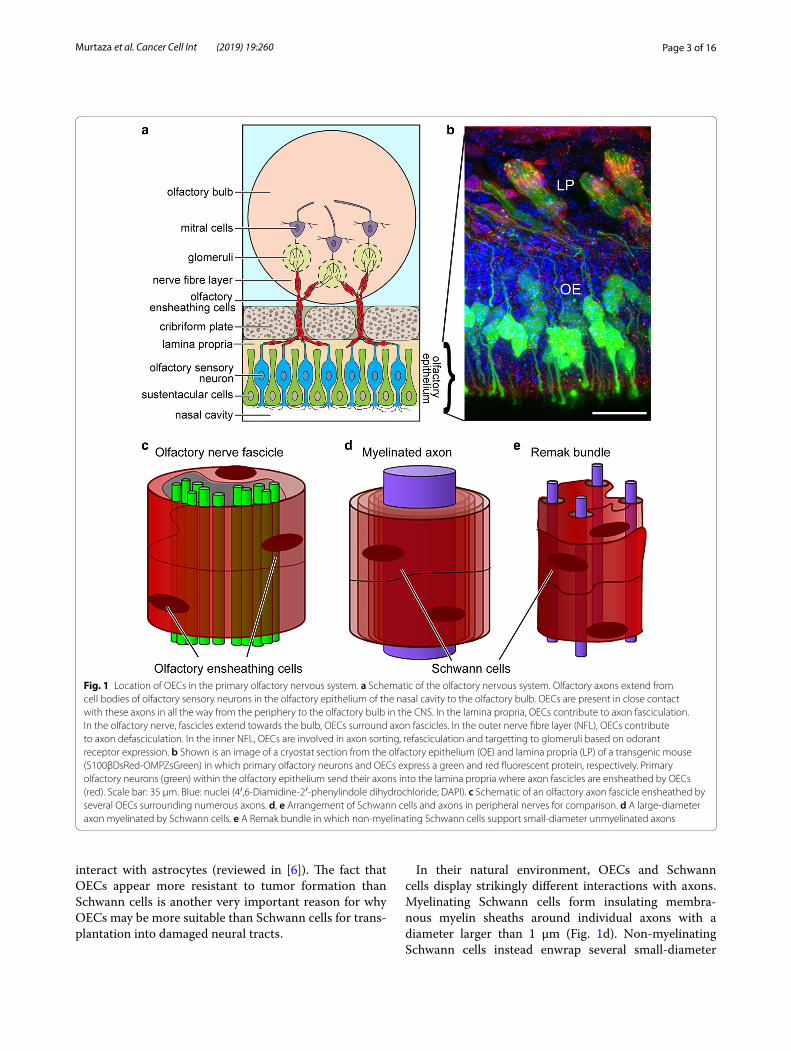
Biological functions of OECs in the primary olfactory nervous systemThe primary olfactory nervous system consists of [1] the olfactory nerve extending from the nasal cavity to the olfactory bulb in the brain, and [2] the outermost layer of the olfactory bulb termed the nerve fibre layer (NFL) (Fig. 1a). Olfaction exhibits the strongest association with memory and emotions amongst the senses in humans, and has an important role in distinguishing favourable from non-favourable or potentially dangerous surround-ings in other mammals and lower vertebrates. Therefore, olfaction has had massive impact on survival throughout evolution. However, primary olfactory sensory neurons are constantly exposed to irritants, toxins and pathogens entering the nasal cavity. Most likely for this reason, the primary olfactory nervous system has evolved to con-stantly regenerate itself, and is unique in that it under-goes lifelong neurogenesis. Olfactory sensory neurons live for approximately 1 month in rodents (the exact life-span of human olfactory sensory neurons remains unknown), and 1–3% of neurons are turned over daily [7]. The olfactory sensory neurons are continually replen-ished from progenitors in the olfactory epithelium. The continuous regeneration of the primary olfactory nerv-ous system is thought to be highly dependent on OECs, which are specialised glial cells with unique neurotrophic properties (reviewed in [6, 8–10]).
Olfactory sensory neurons extend dendrites, on which odorant receptors are localised, to the mucosal surface of the olfactory epithelium, and axons basally into the lamina propria. The axons of olfactory sensory neurons form fascicles (“bundles”), which together constitute the olfactory nerve, extend through the cribriform plate and reach their targets in the olfactory bulb [11–14] (Fig. 1a–c). When the fascicles reach the NFL in the olfactory bulb, the axons defasciculate, sort out and then refas-ciculate with axons expressing the same odorant recep-tor [15]. These now uniform fascicles extend to specific targets (glomeruli) in the olfactory bulb; each glomeru-lus is the target for axons expressing an individual type of
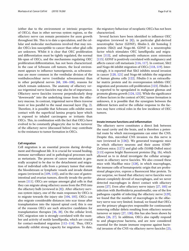
odorant receptor [16] (Fig. 1a, b). Thus, throughout life, new axons are continuously finding their way from the cell bodies in the olfactory epithelium all the way to their targets in the olfactory bulb. OECs are present in direct contact with olfactory sensory axons all the way from the lamina propria in the periphery to the NFL of the olfactory bulb. OECs give the olfactory axons structural support and have crucial roles in guiding and regulating the behaviour of the axons, which differ depending on anatomical location [17, 18] (Fig. 1a, b). In the olfactory nerve, OECs ensheath olfactory axon fascicles. The OECs do not myelinate olfactory axons; the fascicles instead consist of many unmyelinated axons surrounded by OECs [18] (Fig. 1c). This contrasts with most peripheral nerves which consist of both myelinated and unmyeli-nated fibers supported by myelinating and unmyelinating Schwann cells (Fig. 1d, e, respectively); discussed below. In the NFL of the olfactory bulb, OECs are also intimately associated with olfactory axons and are thought crucial for axon defasciculation, sorting and refasciculation [17, 18]. OECs secrete many neurotrophic factors, such as nerve growth factor (NGF), brain-derived neurotrophic factor (BDNF), various neuregulins and other neurotro-phins (reviewed in [6, 19–22]). Furthermore, OECs are the primary phagocytes in the olfactory nerve, respon-sible for clearing axonal debris resulting from the turno-ver of olfactory neurons and after injury to the olfactory nerve [23–27]. OECs also have important innate immune functions preventing pathogens from invading the CNS via the olfactory nerve [23, 28–31]. Due to their ability to promote growth and survival of neurons, as well as their unique ability to migrate long distances, OECs have been investigated as viable candidates for cell therapies for spinal cord injuries [32–43], neurodegenerative diseases [44–46] and peripheral nerve repair [47–50] with prom-ising but highly variable outcomes.
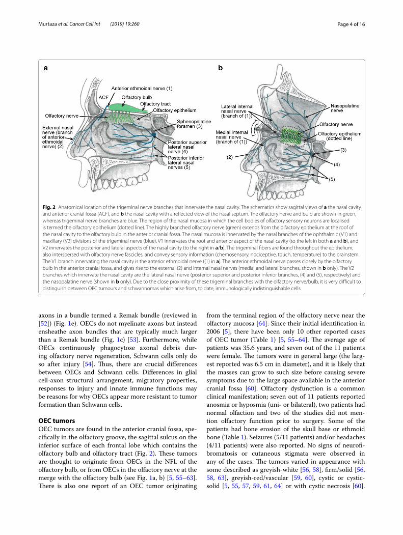
Similarities and differences between OECs and Schwann cellsSchwann cells are the glial cells of most peripheral nerves, including the trigeminal nerve which innervates the nasal cavity; many small trigeminal nerve branches are present in the same anatomical location as the olfac-tory nerve fascicles (Fig. 2; discussed below). OECs and Schwann cells share developmental origin (the neural crest [51]), as well as many similar morphological and molecular characteristics and functions; both cell types supply structural and neurotrophic support to axons. OECs and Schwann cells have both been considered for cell transplantation therapies, but OECs are considered preferable due to their ability to continuously promote neural regeneration of the olfactory nerve, their superior migratory and phagocytic properties and their ability to
Page 3 of 16Murtaza et al. Cancer Cell Int (2019) 19:260
interact with astrocytes (reviewed in [6]). The fact that OECs appear more resistant to tumor formation than Schwann cells is another very important reason for why OECs may be more suitable than Schwann cells for trans-plantation into damaged neural tracts.
In their natural environment, OECs and Schwann cells display strikingly different interactions with axons. Myelinating Schwann cells form insulating membra-nous myelin sheaths around individual axons with a diameter larger than 1 µm (Fig. 1d). Non-myelinating Schwann cells instead enwrap several small-diameter
Fig. 1 Location of OECs in the primary olfactory nervous system. a Schematic of the olfactory nervous system. Olfactory axons extend from cell bodies of olfactory sensory neurons in the olfactory epithelium of the nasal cavity to the olfactory bulb. OECs are present in close contact with these axons in all the way from the periphery to the olfactory bulb in the CNS. In the lamina propria, OECs contribute to axon fasciculation. In the olfactory nerve, fascicles extend towards the bulb, OECs surround axon fascicles. In the outer nerve fibre layer (NFL), OECs contribute to axon defasciculation. In the inner NFL, OECs are involved in axon sorting, refasciculation and targetting to glomeruli based on odorant receptor expression. b Shown is an image of a cryostat section from the olfactory epithelium (OE) and lamina propria (LP) of a transgenic mouse (S100βDsRed-OMPZsGreen) in which primary olfactory neurons and OECs express a green and red fluorescent protein, respectively. Primary olfactory neurons (green) within the olfactory epithelium send their axons into the lamina propria where axon fascicles are ensheathed by OECs (red). Scale bar: 35 µm. Blue: nuclei (4′,6-Diamidine-2′-phenylindole dihydrochloride; DAPI). c Schematic of an olfactory axon fascicle ensheathed by several OECs surrounding numerous axons. d, e Arrangement of Schwann cells and axons in peripheral nerves for comparison. d A large-diameter axon myelinated by Schwann cells. e A Remak bundle in which non-myelinating Schwann cells support small-diameter unmyelinated axons
Page 4 of 16Murtaza et al. Cancer Cell Int (2019) 19:260
axons in a bundle termed a Remak bundle (reviewed in [52]) (Fig. 1e). OECs do not myelinate axons but instead ensheathe axon bundles that are typically much larger than a Remak bundle (Fig. 1c) [53]. Furthermore, while OECs continuously phagocytose axonal debris dur-ing olfactory nerve regeneration, Schwann cells only do so after injury [54]. Thus, there are crucial differences between OECs and Schwann cells. Differences in glial cell-axon structural arrangement, migratory properties, responses to injury and innate immune functions may be reasons for why OECs appear more resistant to tumor formation than Schwann cells.
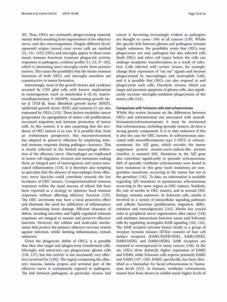
OEC tumorsOEC tumors are found in the anterior cranial fossa, spe-cifically in the olfactory groove, the sagittal sulcus on the inferior surface of each frontal lobe which contains the olfactory bulb and olfactory tract (Fig. 2). These tumors are thought to originate from OECs in the NFL of the olfactory bulb, or from OECs in the olfactory nerve at the merge with the olfactory bulb (see Fig. 1a, b) [5, 55–63]. There is also one report of an OEC tumor originating
from the terminal region of the olfactory nerve near the olfactory mucosa [64]. Since their initial identification in 2006 [5], there have been only 10 other reported cases of OEC tumor (Table 1) [5, 55–64]. The average age of patients was 35.6 years, and seven out of the 11 patients were female. The tumors were in general large (the larg-est reported was 6.5 cm in diameter), and it is likely that the masses can grow to such size before causing severe symptoms due to the large space available in the anterior cranial fossa [60]. Olfactory dysfunction is a common clinical manifestation; seven out of 11 patients reported anosmia or hyposmia (uni- or bilateral), two patients had normal olfaction and two of the studies did not men-tion olfactory function prior to surgery. Some of the patients had bone erosion of the skull base or ethmoid bone (Table 1). Seizures (5/11 patients) and/or headaches (4/11 patients) were also reported. No signs of neurofi-bromatosis or cutaneous stigmata were observed in any of the cases. The tumors varied in appearance with some described as greyish-white [56, 58], firm/solid [56, 58, 63], greyish-red/vascular [59, 60], cystic or cystic-solid [5, 55, 57, 59, 61, 64] or with cystic necrosis [60].
Fig. 2 Anatomical location of the trigeminal nerve branches that innervate the nasal cavity. The schematics show sagittal views of a the nasal cavity and anterior cranial fossa (ACF), and b the nasal cavity with a reflected view of the nasal septum. The olfactory nerve and bulb are shown in green, whereas trigeminal nerve branches are blue. The region of the nasal mucosa in which the cell bodies of olfactory sensory neurons are localised is termed the olfactory epithelium (dotted line). The highly branched olfactory nerve (green) extends from the olfactory epithelium at the roof of the nasal cavity to the olfactory bulb in the anterior cranial fossa. The nasal mucosa is innervated by the nasal branches of the ophthalmic (V1) and maxillary (V2) divisions of the trigeminal nerve (blue). V1 innervates the roof and anterior aspect of the nasal cavity (to the left in both a and b), and V2 innervates the posterior and lateral aspects of the nasal cavity (to the right in a/b). The trigeminal fibers are found throughout the epithelium, also interspersed with olfactory nerve fascicles, and convey sensory information (chemosensory, nociceptive, touch, temperature) to the brainstem. The V1 branch innervating the nasal cavity is the anterior ethmoidal nerve ((1) in a). The anterior ethmoidal nerve passes closely by the olfactory bulb in the anterior cranial fossa, and gives rise to the external (2) and internal nasal nerves (medial and lateral branches, shown in b only). The V2 branches which innervate the nasal cavity are the lateral nasal nerve (posterior superior and posterior inferior branches, (4) and (5), respectively) and the nasopalatine nerve (shown in b only). Due to the close proximity of these trigeminal branches with the olfactory nerve/bulb, it is very difficult to distinguish between OEC tumours and schwannomas which arise from, to date, immunologically indistinguishable cells
Page 5 of 16Murtaza et al. Cancer Cell Int (2019) 19:260
Tabl
e 1
Feat
ures
and
imm
unop
rofil
e of
the
publ
ishe
d ca
se re
port
s
Sex/
age
Sym
ptom
sLo
catio
nEn
hanc
emen
tTu
mou
r fea
ture
sPa
thol
ogy
Out
com
eM
arke
r pro
file
Refe
renc
e
Fem
ale
31Ri
ght-
side
d an
osm
ia,
gene
ralis
ed s
eizu
res
Ant
erio
r cra
nial
foss
a,
atta
ched
to th
e ol
fac-
tory
gro
ove
Het
erog
eneo
usIrr
egul
ar, a
vasc
ular
, cy
stic
-sol
id, c
apsu
-la
ted
tum
our w
ith
calc
ified
nod
ules
. 6.
5 cm
dia
met
er. B
one
eros
ion
Spin
dle-
shap
ed c
ells
in
a w
avy
cellu
lar
arra
ngem
ent.
Dis
-to
rted
and
twis
ted
nucl
ei
Com
plet
e re
mov
al.
Une
vent
ful
S100
+EM
A −
Leu7
−
[5]
Mal
e42
Nor
mal
olfa
ctor
y fu
nc-
tion,
gene
ralis
ed s
eizu
res
Ant
erio
r cra
nial
foss
a (le
ft s
ubfro
ntal
regi
on),
aris
ing
from
the
left
ol
fact
ory
bulb
Het
erog
eneo
usRo
und,
cys
tic-s
olid
fib
rous
tum
our
Spin
dle-
shap
ed c
ells
, fib
rous
cor
ds. C
urve
d ve
sicu
lar n
ucle
i with
ill
-defi
ned
cyto
plas
-m
ic m
argi
ns
Com
plet
e re
mov
al.
Une
vent
ful
S100
+EM
A –
GFA
P –
SMA
–Le
u7 –
[57]
Mal
e32
Olfa
ctor
y fu
nctio
n no
t m
entio
ned,
sei
zure
sA
nter
ior c
rani
al fo
ssa
(left
fron
tal b
ase)
Het
erog
eneo
usRo
und,
sol
id, g
reyi
sh-
whi
te tu
mou
r w
ith a
glis
teni
ng
appe
aran
ce a
nd
rubb
ery
cons
iste
ncy.
3.
6 ×
3.3
× 3
.9 c
m
Spin
dle-
shap
ed c
ells
, fib
rous
cor
ds. O
void
, el
onga
ted,
nor
mo-
chro
mat
ic, c
omm
a-sh
aped
nuc
lei.
Inte
rrup
ted
depo
sits
of
basa
l lam
ina
in th
e ce
llula
r mem
bran
e
Com
plet
e re
mov
al.
Une
vent
ful.
S100
+ (8
0% o
f cel
ls)
Leu7
–Ca
lretin
in –
Pod
opla
nin
–EM
A –
GFA
P –
[58]
Fem
ale
28A
nosm
ia, f
ocal
sei
zure
sA
nter
ior c
rani
al fo
ssa
Het
erog
eneo
usG
reyi
sh-w
hite
, irr
egul
ar, c
loud
y, s
olid
. 4 ×
3.5
× 2
.5 c
m
Wel
l-circ
umsc
ribed
tu
mou
r with
elo
n-ga
ted
spin
dle-
shap
ed
cells
, fibr
ous
cord
s. M
oder
ate
nucl
ear
pleo
mor
phis
m
Com
plet
e re
mov
al. A
nos-
mia
S100
+Sy
napt
ophy
sin +
EMA
–Le
u7 –
[56]
Fem
ale
30Ri
ght-
side
d an
osm
ia,
head
ache
Ant
erio
r cra
nial
foss
a,
intr
adur
al, e
xtra
-axi
al
spac
e an
d at
tach
ed
to th
e rig
ht c
ribrif
orm
pl
ate
Hom
ogen
ous
Roun
d, s
olid
4 c
m
diam
eter
.Ce
lls fo
rmed
pat
-te
rns
of c
ompa
ct
fasc
icul
ar A
nton
i A
area
s (re
sem
blin
g Sc
hwan
nom
a) w
ith
palis
adin
g nu
clei
Com
plet
e re
mov
al.
Olfa
ctor
y fu
nc-
tion
was
not
re
stor
ed
S100
+EM
A –
Leu7
–
[63]
Fem
ale
41A
nosm
ia, h
eada
che
Olfa
ctor
y m
ucos
a; o
lfac-
tory
cle
ft e
xten
ding
su
perio
rly to
the
olfa
c-to
ry g
roov
e
Het
erog
eneo
usIrr
egul
ar, c
ystic
tum
our.
Bone
def
ect i
n th
e sk
ull b
ase
Spin
dle-
shap
ed c
ells
w
ith e
osin
ophi
lic
cyto
plas
m a
nd
elon
gate
d or
wav
y nu
clei
with
occ
asio
nal
sym
plas
tic c
hang
es
Subt
otal
rese
c-tio
n. U
neve
ntfu
lS1
00 +
Neu
ron-
spec
ific
enol
ase +
Syna
ptop
hysi
n +
(wea
kly)
EMA
–Le
u7 –
[64]
Mal
e49
Hyp
osm
ia, v
isua
l im
pair-
men
tA
nter
ior c
rani
al fo
ssa
Hom
ogen
ous
Roun
d, c
ystic
-sol
id
tum
our,
erod
ing
the
right
crib
rifor
m p
late
Unk
now
nCo
mpl
ete
rem
oval
. U
neve
ntfu
l
S100
+EM
A –
Leu7
–
[55]
Page 6 of 16Murtaza et al. Cancer Cell Int (2019) 19:260
Tabl
e 1
(con
tinu
ed)
Sex/
age
Sym
ptom
sLo
catio
nEn
hanc
emen
tTu
mou
r fea
ture
sPa
thol
ogy
Out
com
eM
arke
r pro
file
Refe
renc
e
Mal
e20
Nor
mal
olfa
ctio
n,
head
ache
, gen
eral
ized
se
izur
es
Ant
erio
r cra
nial
foss
aH
eter
ogen
eous
The
tum
our g
rew
to
war
ds th
e le
ft
olfa
ctor
y gr
oove
and
co
mpr
esse
d th
e le
ft
front
al c
orte
x. G
reyi
sh-
red,
vas
cula
r tum
our.
Cyst
ic n
ecro
sis
insi
de
the
tum
our.
3.4 ×
2.6
×
5.0
cm
Spin
dle-
shap
ed c
ells
w
ere
pred
omin
antly
ar
rang
ed in
com
pact
fa
scic
les
or fi
brou
s co
rds
and
a fe
w c
ells
w
ere
arra
nged
in
who
rls
Com
plet
e re
mov
al, h
ypos
-m
ia
Vim
entin
+S1
00 +
EMA
–Le
u7 –
[60]
Fem
ale
45O
lfact
ory
func
tion
not
men
tione
d, fo
reig
n bo
dy s
ensa
tion
Ant
erio
r cra
nial
foss
a.H
eter
ogen
eous
Irreg
ular
, cys
tic tu
mou
r6.
2 ×
6.0
× 4
.0 c
mSp
indl
e-sh
aped
cel
ls.
Com
pact
, fas
cicu
lar
Ant
oni A
are
as a
s w
ell
as A
nton
i B a
reas
Com
plet
e re
mov
al.
Une
vent
ful
S100
+Le
u7 –
GFA
P –
EMA
–
[61]
Fem
ale
34H
ypos
mia
, diz
zine
ss,
emot
iona
l lab
ility
Ant
erio
r cra
nial
foss
aH
omog
enou
sW
ell-d
efine
d cy
stic
, gr
eyis
h-re
d m
ass,
3.1
cm d
iam
eter
Spin
dle
cells
with
eos
in-
ophi
lic p
roto
plas
m,
tadp
ole-
shap
ed
nucl
eus
Com
plet
e re
mov
al.
Une
vent
ful
Vim
entin
+S1
00 +
EMA
–G
FAP
–Le
u7 –
[59]
Fem
ale
40Le
ft-s
ided
ano
smia
, m
igra
ine,
hea
dach
esA
nter
ior c
rani
al fo
ssa,
ol
fact
ory
groo
ve a
dja-
cent
to th
e le
ft in
ferio
r an
terio
r fro
ntal
lobe
Het
erog
eneo
us3.
2 cm
dia
met
erSp
indl
e ce
ll ne
opla
sm
char
acte
rized
by
exte
nsiv
e pa
lisad
ing
and
prom
inen
t Ant
oni
A (V
eroc
ay b
odie
s)
and
Ant
oni B
are
as
Com
plet
e re
mov
al.
Left
-sid
ed a
nos-
mia
S100
+Ty
pe IV
col
lage
n +
Leu7
–EM
A –
[62]
+: p
ositi
ve; c
ell i
s ex
pres
sing
mar
ker;
–: n
egat
ive;
cel
l is
not e
xpre
ssin
g m
arke
r; S1
00: S
100
prot
ein
(glia
l mar
ker)
; Leu
7 (C
D57
or H
NK-
1): s
ugge
sted
mar
ker f
or S
chw
ann
cells
but
not
OEC
s; E
MA
: Epi
thel
ial m
embr
ane
antig
en; G
FAP:
Glia
l fibr
illar
y ac
idic
pro
tein
; SM
A: S
moo
th m
uscl
e ac
tin
Page 7 of 16Murtaza et al. Cancer Cell Int (2019) 19:260
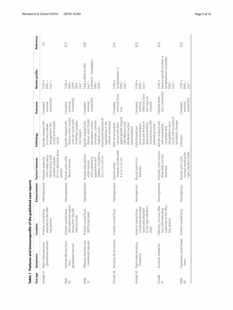
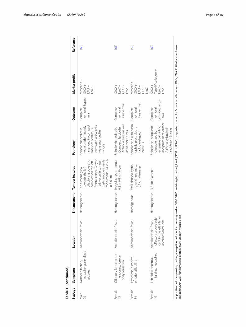
Surgical excision was performed as the main treatment. Excision of the tumor appeared curative with short fol-low-up times and no metastasis reported; outcomes were in general uneventful with 4/11 patients reporting new or recurring anosmia/hyposmia [56, 60, 62, 63]. Typical pathological characteristics of the excised tumors include spindle-shaped cells in fascicles [5, 57, 60–62] adjacent to looser paucicellular areas [61], similar to Antoni A and Antoni B areas, respectively, in schwannoma tumors [65], fibrous cords [56, 57, 60] and distorted nuclei [5, 59, 60, 64]. (Antoni A areas are highly cellular areas with nuclear palisades and associated Verocay bodies; Vero-cay bodies constitute two stacked rows of elongated palisading nuclei alternating with zones containing cyto-plasmic schwannoma cell extensions. Antoni B areas are localised adjacent to Antoni A areas and consist of loosely arranged cells in myxomatous tissue (tissue with mucoid substance) and microcysts [66]). Overall, very lit-tle is known about the clinical and immunohistochemi-cal characteristics of OEC tumors, which makes it very difficult to give a definite diagnosis. The immunohisto/cytochemical markers identified in the known cases are summarised in Table 1.
Why is it difficult to distinguish between OEC tumors and schwannomas?Overlapping anatomical location of the primary olfactory nervous system and trigeminal nerve branchesSchwannomas (nerve sheath tumors originating from Schwann cells) can arise from any peripheral or cranial nerve in which the glial cells are Schwann cells. The spo-radic schwannomas, which are compared to OEC tumors in this review, are distinct to nerve sheath tumors seen in the genetic conditions neurofibromatosis and schwan-nomatosis which are caused by germline mutations [67]. The most common location for schwannomas is the head and neck; approximately 3–4% of humans exhibit head or neck schwannomas on autopsy [68]. Schwannomas com-prise approximately 8% of all intracranial tumors [69]. Malignant schwannomas are uncommon but aggressive and comprise 2% of all sarcomas with a high metastatic potential and poor prognosis [70]. It is very difficult to distinguish OEC tumors from schwannomas and men-ingiomas, which can be present in the same anatomical areas as OEC tumors (Fig. 2) and cause similar symp-toms to OEC tumors, including anosmia [5, 58–60, 64]. In particular, it is difficult to distinguish between OEC tumors and schwannomas, since both tumors arise from glial cells with a shared developmental origin (the neu-ral crest, [51]), as well as many similar morphological and molecular characteristics (reviewed in [6]). The schwan-nomas that are so easily confused with OEC tumors are usually termed anterior cranial fossa schwannomas
or olfactory groove schwannomas (OGS) but can also occasionally be found in the nasal cavity and parana-sal sinuses (nasoethmoid schwannomas) [71–75]. These schwannomas are rare; to date, approximately 45 cases (without neurofibromatosis/schwannomatosis) have been reported in the literature [76, 77]. They are thought to originate from Schwann cells of the nasal branches of the ophthalmic (V1) and maxillary (V2) divisions of the trigeminal nerve (Fig. 2) [63, 78, 79]. These branches innervate the olfactory epithelium and underlying lam-ina propria, and pass closely to the olfactory bulb in the anterior cranial fossa, regions in which OECs are present (Fig. 2).
It has also been suggested that schwannomas can arise from the terminal nerve (cranial nerve zero) [78, 80, 81], a bilateral plexus of unmyelinated fascicles extending from the nasal epithelium via the cribriform plate and medial surface of the olfactory bulbs towards the preop-tic hypothalamic area [60]. The terminal nerve branches closely follow and intermingle with olfactory nerve fas-cicles, and the terminal nerve is often mistaken for the olfactory nerve in post-mortem humans. Whilst this nerve is well documented in many vertebrates, and it has been reported to exist in human embryos since the early 1900s, its existence in the adult human brain was not confirmed until the 1990s [82]. It is thought to have roles in gonadotropin-releasing hormone (GnRH) sign-aling and the hypothalamic-pituitary–gonadal (HPG) axis, but has also been suggested to be vestigial in adult-hood (reviewed in [83]). To date, the cellular nature of the glial cells of this nerve has not been studied, but as the nerve resembles many peripheral nerves, the termi-nal nerve glial cells are most likely Schwann cells [82, 83]. Further, developmental theories suggest that schwanno-mas in the nasal cavity/anterior cranial fossa arise from mesenchymal pial cells which transform into Schwann cells, or from aberrant neural crest cells [78, 80, 81, 84, 85]. In summary, OECs and Schwann cells share many similarities and are found in the same anatomical region (Fig. 2); thus, it is very difficult to distinguish between OEC tumors and schwannomas. This has led to specula-tion on the true origin and identity of schwannomas and OEC tumors.
Lack of OEC‑specific markersThe clinical and radiological features of OEC tumors and trigeminal nerve schwannomas are indistinguish-able. Instead, the two types of tumors are usually classi-fied immunocytochemically based on the expression of the marker Leu7 (reviewed in [78]). Leu7, also known as CD57 or HNK-1, is expressed by Schwann cells in sci-atic and trigeminal nerves [86, 87], but not by OECs. It has also been reported that cultured human [39, 88] and
Page 8 of 16Murtaza et al. Cancer Cell Int (2019) 19:260
rat [19] Schwann cells are Leu7-positive whilst OECs are Leu7-negative. Thus, gliomas of the nasal cavity and anterior cranial fossa/olfactory groove that do not express Leu7 are considered to be OEC tumors [5]; in all (11/11) case reports of OEC tumors, the lack of reac-tivity to Leu7 was used to conclude the diagnosis to be OEC tumor. Lack of Leu7 expression, however, does not necessarily mean that the tumor is definitely an OEC tumor, as ~ 20% of schwannomas are negative for Leu7 [89]. Recently, two cases of schwannoma-like tumors in the anterior cranial fossa were described to be immunon-egative for Leu7 but immunopositive for Schwann/2E, a marker for myelinating Schwann cells [90] and some schwannoma tumors [91]. Leu7 is expressed by Schwann cells in early development and then lost; when Schwann cells myelinate axons their expression of Leu7 is again up-regulated (reviewed in [78]). In addition to being expressed by myelinating Schwann cells, Leu7 is present in Schwann cells that have ingested myelin during Wal-lerian degeneration [86, 92]. However, cultured Schwann cells lose the expression of Leu7 once phagocytosed myelin debris becomes degraded [93]. To date, Leu7 expression has not been detected in human non-myeli-nating Schwann cells. A study in the adult canine trigem-inal nerve showed that whilst myelinating trigeminal Schwann cells express Leu7, non-myelinating Schwann cells do not [93]. One report shows expression of Leu7 in cultured rat non-myelinating Schwann cells [19], but these cells may have been exposed to myelin debris and thus the Leu7 immunoreactivity may have been label-ling phagocytosed material. Overall, it is likely that Leu7 expression by non-myelinating Schwann cells is low or non-existent. Thus, tumors arising from non-myelinating Schwann cells may very well be Leu7-negative. The per-centage of unmyelinated axons in the various branches of the human trigeminal nerve is not well characterized, except that the majority of axons are myelinated [94]. Counts of unmyelinated fibres are difficult as the axons are closely packed together in groups and it is difficult to distinguish individual axons [95]. Studies from the 1920s show that the trigeminal nerve contains ~ 10% unmyeli-nated axons in the cat [96], and ~ 20–40% in the dog [95]. The percentage of unmyelinated fibres is estimated to be 12–20% in human motor root [97], but may very well be higher in the sensory root which contains small, nocicep-tive unmyelinated C-fibers [98]. Regardless, the number of non-myelinating Schwann cells in the trigeminal nerve is significant and it can thus be expected that a significant proportion of schwannomas arise from these cells.
Expression of Leu7 may, conversely, also not necessar-ily mean that a tumor is a schwannoma and not an OEC tumor. One study shows that some OEC populations in the olfactory bulb of rats are in fact Leu7-positive [19],
further rendering Leu7 immunoreactivity as an inap-propriate marker to distinguish between the two types of tumors. Therefore, the identification of OEC tumors based solely on the absence of Leu7 immunoreactivity is inconclusive, and diagnostic tests should involve multiple markers rather than reliance on the absence of a single marker. To date, no markers that definitely distinguish between OECs and Schwann cells have been identified (reviewed in [6]). Schwann/2E expression has not yet been characterized in OECs, and regardless, Schwann/2E appears to be, like Leu7, a marker specific for myelinating Schwann cells [90].
Moreover, neoplastic transformation and tumorigen-esis is a dynamic process where cells within a tumor may no longer retain the same cellular properties or molecu-lar signature as the cells of origin. In the case of schwan-nomas, abnormal or lost axon-Schwann cell interactions, including myelination, has been suggested as being impli-cated in tumorigenesis [99–102] (discussed in more detail below). This, again, highlights the fact that Leu7, or other markers for myelinating Schwann cells, are not appropri-ate for diagnosis of schwannomas, as loss of expression of these markers is likely to accompany loss of myelination [93]. Due to the difficulties in using Leu7 as a marker of OEC versus Schwann cell tumors, and the lack of a suit-able panel of other markers to distinguish between OECs and Schwann cells, the possibility exists that some of the tumors diagnosed as OEC tumors may in fact have originated from Schwann cells. Thus, OEC tumors may be even rarer than the few cases to date reported in the literature.
Why are OEC tumors so rare?Local environment, plasticity and proliferationTwo-way communication between cells and their micro-environment is critical for tissue homeostasis and for tumor growth. According to the seed and soil cancer hypothesis, the fate of tumor-initiating cells (seed) is guided by the presence of favourable microenviron-ments (soil) [103]. The olfactory nerve is a neurogenic niche where olfactory neurons are replaced throughout life, and where axons continuously extend towards the olfactory bulb [7, 11–14]. The environment is frequently exposed to external insults and there is constant turnover of neurons; thus, this is a uniquely plastic region of the nervous system. Furthermore, OECs effectively respond to widespread injury of the olfactory nerve or olfactory bulb by proliferating [104, 105]. One may assume that a niche so permissive for proliferation is likely to have a higher probability of developing transformed cells, pre-cancerous lesions and tumors. Contrary to this expecta-tion, OEC tumors are extremely rare. It is plausible that the threshold for tumor initiation is higher in this niche
Page 9 of 16Murtaza et al. Cancer Cell Int (2019) 19:260
(either due to the environment or intrinsic properties of OECs), than in other nervous system regions, so the olfactory nerve can remain permissive for axon growth throughout life. This is to date a speculation, and the cel-lular and molecular mechanisms involved that may ren-der OECs less susceptible to cancer than other glial cells are unknown. Whilst it is clear that OEC proliferation and differentiation must be tightly regulated, the normal life-span of OECs, and the mechanisms regulating OEC proliferation/differentiation, has not been characterized. In the case of Schwann cell tumors, the local environ-ment appears to influence tumorigenesis, as schwanno-mas are more common in the vestibular division of the vestibulocochlear nerve (vestibular schwannoma) than in other peripheral nerves [68, 106–108], reasons for which remain unknown. The anatomy of olfactory ver-sus trigeminal nerve fascicles may also be of importance. Olfactory nerve fascicles traverse perpendicularly deep “downwards” into the underlying tissue from the olfac-tory mucosa. In contrast, trigeminal nerve fibers traverse more or less parallel to the nasal mucosal layer (Fig. 2). Therefore, it is possible that Schwann cells exhibit more contact with the superficial lamina propria layer which is exposed to inhaled carcinogens or irritants than OECs. This, in combination with the fact that OECs have evolved to be constantly phagocytic due to the turnover of the olfactory nerve (discussed below) may contribute to the resistance to tumor formation in OECs.
Cell migrationCell migration is an essential process during develop-ment and throughout life. It is crucial for wound healing, immune surveillance and in pathological processes such as metastasis. The process of cancer metastasis is gen-erally accepted to be due to the detachment and migra-tion of individual cells from a primary tumor that enter the bloodstream or lymphatic vessels and invade distant organs (reviewed in [109, 110]), and in the case of gastro-intestinal and ovarian tumors, directly invade the perito-neum [111]. OECs are unique amongst glial cells in that they can migrate along olfactory axons from the PNS into the olfactory bulb (reviewed in [8]). After olfactory nerv-ous system injury, one of the main responses by OECs is to migrate towards the injury site [104, 105]. OECs can also migrate considerable distances into scar tissue after transplantation into the injured spinal cord; this is one of the reasons OECs are such attractive candidates for transplantation therapies [36, 38]. On the cellular level, OEC migration rate is strongly correlated with the num-ber and activity of motile lamellipodia, which are crucial for contact-mediated migration [112–114]. Thus, OECs naturally exhibit strong capacity for migration. To date,
the migratory behaviour of neoplastic OECs has not been characterized.
Several factors have been identified to influence OEC migration (reviewed in [8]), in particular glial-derived neurotrophic factor (GDNF), fibulin-3, slit homolog 2 protein (Slit2) and Nogo-66. GDNF is a neurotrophic factor which stimulates OEC lamellipodia and migra-tion [113], and subsequently enhances axon extension [115]. GDNF is positively correlated with malignancy and affects cancer cell metastasis [116, 117]. In contrast, Slit2 and Nogo-66 inhibit migration of OECs [118, 119]. Inter-estingly, it is reported that Slit2 inhibits neural invasion in cancer [120, 121] and Nogo-66 inhibits the migration of human glioma cells [122]. Fibulin-3 is an extracellu-lar matrix protein and its overexpression inhibits OEC migration and promotes cell proliferation [123]. Fibulin-3 is reported to be upregulated in malignant gliomas and promote glioma growth [124, 125]. While the significance of these factors in the context of OEC tumor formation is unknown, it is possible that the synergism between the different factors and/or the cellular response to the fac-tors may have critical roles in the low incidence of OEC tumors.
Innate immune functions and inflammationThe olfactory nerve constitutes a direct link between the nasal cavity and the brain, and is therefore a poten-tial route by which microorganisms can enter the CNS. Despite this, microbial CNS invasion via this nerve is rare (reviewed in [126]). We generated transgenic mice in which olfactory neurons and their axons (OMP-ZsGreen mice; [127]) and glial cells (S100β-DsRed mice) [113] express bright fluorescent proteins (Fig. 1b), which allowed us to in detail investigate the cellular arrange-ment in olfactory nerve fascicles. We also crossed these mice with MacBlue mice [128], in which macrophages, the immune cells of hematopoietic origin that are profes-sional phagocytes, express a fluorescent blue protein. To our surprise, we found that olfactory nerve fascicles were almost completely devoid of macrophages, and we never detected macrophages in direct contact with olfactory axons [27]. Even after olfactory nerve injury [27, 105] or infection with Burkholderia pseudomallei, one of the few pathogens capable of infecting the olfactory nerve [129], we found that the number of macrophages in the olfac-tory nerve was very limited. Instead, we found that OECs are the primary phagocytes responsible for continuously removing cellular debris resulting from olfactory neuron turnover or injury [27, 130]; this has also been shown by others [26, 27]. In addition, OECs also rapidly respond to and phagocytose bacteria, and are now considered essential for the innate immune response against bacte-rial invasion of the CNS via olfactory nerve fascicles [23,
Page 10 of 16Murtaza et al. Cancer Cell Int (2019) 19:260
30]. Thus, OECs are constantly phagocytosing material, mainly debris resulting from regeneration of the olfactory nerve, and also microorganisms. Despite different devel-opmental origins (neural crest versus yolk sac myeloid [51, 131, 132]), OECs and microglia appear to share some innate immune functions (constant phagocytic activity, responses to pathogens, cytokine profile [11, 23, 27–30]), which is interesting since microglia rarely form primary tumors. This raises the possibility that the innate immune functions of both OECs and microglia somehow are counteractive to tumor formation.
Interestingly, most of the growth factors and cytokines secreted by CNS glial cells with known implications in tumorigenesis (such as interleukin 6 (IL-6), matrix-metalloproteinase 9 (MMP9), transforming growth fac-tor β (TGF-β), basic fibroblast growth factor (bFGF), epidermal growth factor (EGF) and tenascin-C) are also expressed by OECs [133]. These factors modulate cancer progression via upregulation of tumor cell proliferation, increased migration and immune protection of tumor cells. In this context, it is also surprising that the inci-dence of OEC tumors is so rare. It is possible that, from an evolutionary perspective, this microenvironment has adapted to preserve olfaction by employing mini-mal immune response during pathogen clearance. This is clearly reflected in the limited macrophage infiltra-tion of the olfactory nerve. Macrophages are implicated in tumor cell migration, invasion and metastasis making them an integral part of tumorigenesis and tumor-asso-ciated inflammation [134]. It is therefore also tempting to speculate that the absence of macrophages from olfac-tory nerve fascicles could contribute towards the low incidence of OEC tumors. Compartmentalized immune responses within the nasal mucosa of teleost fish have been reported as a strategy to optimize local immune responses without affecting olfactory function [135]. The OEC secretome may have a local protective effect and eliminate the need for infiltration of inflammatory cells, minimizing tissue damage. Efficient clearance of debris, invading microbes and highly regulated immune responses are integral to sustain and preserve olfactory function. However, the cellular and molecular mecha-nisms that protect the primary olfactory nervous system against infection, whilst limiting inflammation, remain unknown.
Given the phagocytic ability of OECs, it is possible that they also target and phagocytose transformed cells. Microglia and astrocytes can phagocytose glioma cells [136, 137], but this activity is not necessarily very effec-tive (reviewed by [138]). The region comprising the olfac-tory mucosa, lamina propria and terminal part of the olfactory nerve is continuously exposed to pathogens. The link between pathogens, in particular viruses, and
cancer is becoming increasingly evident as pathogens are thought to cause ~ 16% of all cancers [139]. Whilst the specific link between glioma and pathogens remains largely unknown, the possibility exists that OECs may phagocytose not only pathogens but also infected cells (both OECs and other cell types) before the cells can undergo neoplastic transformation as a result of infec-tion. Cells infected with certain viruses, for example, change their expression of “eat me” signals and become phagocytosed by macrophages and neutrophils [140], and it is possible that OECs can also respond to and phagocytose such cells. Oncolytic viruses, which can target and promote apoptosis of glioma cells, also signifi-cantly increase microglia-mediated phagocytosis of the tumor cells [141].
Comparison with Schwann cells and schwannomasWhilst this review focusses on the differences between OECs and schwannomas not associated with neurofi-bromatosis/schwannomatosis, it must be mentioned that schwannomas, including sporadic tumors, do have a strong genetic component. It is to date unknown if this is also the case for OEC tumors. In schwannomas asso-ciated with neurofibromatosis type 2 (NF2) or schwan-nomatosis, the Nf2 gene, which encodes the tumor suppressor protein moesin-ezrin-radixin-like protein (merlin), is mutated [68]. Mutations in the Nf2 gene also contribute significantly to sporadic schwannomas; 66% of sporadic vestibular schwannomas were found to have mutations in this gene (such mutations are non-germline mutations, occurring in the tumor but not in the germline) [142]. To date, no information is available regarding Nf2 mutations in spontaneous schwannomas occurring in the same region as OEC tumors. Similarly, the role of merlin in OEC tumors, and in normal OEC biology, remains unknown. In Schwann cells, merlin is involved in a variety of intracellular signaling pathways and cellular functions (proliferation, migration, differ-entiation and tumorigenesis) [143]. Merlin has crucial roles in peripheral nerve regeneration after injury [144] and mediates interactions between axons and Schwann cells by regulating neuregulin-ErbB signaling [102, 145]. The ErbB receptor tyrosine kinase family is a group of receptor tyrosine kinases (RTKs) consists of four cell surface receptors (ErbB1/EGFR/HER1, ErbB2/HER2, ErbB3/HER3, and ErbB4/HER4). ErbB receptors are mutated or overexpressed in many cancers [146]. In the rat, OECs show distinctly higher expression of ErbB2 and ErbB4, while Schwann cells express primarily ErbB2 and ErbB3 [147–150]. ErbB3, specifically, has been iden-tified as a biomarker for facial schwannomas in Tasma-nian devils [151]. In humans, vestibular schwannoma tissues have been shown to exhibit much higher levels of
Page 11 of 16Murtaza et al. Cancer Cell Int (2019) 19:260
phosphorylated ErbB3 in comparison to healthy paired nerves, and ErB inhibitors have been identified as a novel therapy for malignant schwannomas [152]. Furthermore, ErbB3/HER3 is now emerging as a novel selective thera-peutic cancer target [153]. Thus, differences in expression of ErbB receptors, in particular ErbB3, between OECs and Schwann cells may therefore contribute to differ-ential tendency to tumorigenesis between the two types of glial cells. Investigating the roles of merlin in normal OEC biology and in OEC tumor formation, as well as fur-ther characterizing the roles of ErbB receptors in periph-eral gliomas, is therefore important for understanding potential differences between the two types of tumors.
It is also possible that the reason OEC tumors are rarer than schwannomas are related to differences in the biology of OECs and Schwann cells, or to the microen-vironment in which the cells exist. The fact that the olfac-tory nerve continuously regenerates, whilst peripheral nerves only regenerate after injury, may also be crucial; peripheral nerve injuries, which lead to accumulation of myelin debris, have been implicated in tumorigenesis [99–102]. As discussed earlier, the olfactory nerve is a highly plastic environment in which OECs constantly phagocytose debris and respond to microorganisms. In contrast, peripheral nerves populated by Schwann cells do not undergo regeneration unless they have been injured, and Schwann cells in their natural environment do not often encounter microorganisms. After peripheral nerve injury, Schwann cells lose their contact with axons, become phagocytic “repair” Schwann cells [54] and pro-liferate [154]. Disrupted axon-Schwann cell contact has been implicated in schwannomagenesis [99–102]. Axonal injury has been shown to contribute towards a persistent regenerative “repair Schwann cell” response promoting schwannomagenesis, in particular in combination with mutations in the Nf2 gene [101]. As OECs continuously play an active role in neural regeneration, it is possible that they are less prone to pathological injury-related responses than Schwann cells. Again, this may be due to intrinsic cellular properties, or to the local environment of the olfactory nerve. We have demonstrated that the phagocytic activity of OECs but not Schwann cells can be strongly stimulated with curcumin [155, 156]. This sug-gests that the phagocytic machinery in the two cell types is regulated by different mechanisms, and perhaps OECs exhibit much greater scope for up-regulation of phago-cytic activity in response to nerve injury or infections than Schwann cells.
Denervated Schwann cells produce chemotactic cues that attract macrophages [157], which infiltrate peripheral nerve injury sites and have an essential role in Wallerian degeneration and regeneration [158]. As macrophages are strongly involved in tumorigenesis (reviewed in
[159]), it is possible that macrophages also have a role in schwannoma formation. One study shows a strong correlation between schwannomagenesis and the pres-ence of macrophages, in particular M2-polarised mac-rophages [101]. Interestingly, aspirin intake, which limits inflammation and macrophage infiltration, has been cor-related with slowed growth of schwannomas [160]. As discussed earlier, macrophages are mostly absent from the olfactory nerve fascicles, and macrophage invasion is very limited even after widespread injury. It is possible that differences in inflammatory responses between the olfactory nerve and other peripheral nerves populated by Schwann cells are crucial determinants of the likelihood of tumor formation. Furthermore, significant differences between OECs and Schwann cells in responses to bac-teria have been identified: OECs, but not Schwann cells, respond to gram-negative bacteria or lipopolysaccharide (LPS) with nuclear translocation of NFκβ and secre-tion of the chemokine Gro [30], suggesting that OECs exhibit more pronounced innate immune functions than Schwann cells. Interestingly, schwannoma cells are characterized by abnormal activation of NFκβ, which is normally suppressed by merlin, resulting in secretion of pro-inflammatory cytokines and macrophage recruit-ment (reviewed in [161]). Thus, it is possible that unique regulatory mechanisms in OECs, but not Schwann cells, allow the cells to respond to pathogens, clear cell debris and secrete pro-inflammatory cytokines without caus-ing excessive inflammation, macrophage infiltration and increased risk of tumor formation.
ConclusionsOEC tumors are difficult to distinguish from schwanno-mas, as the two types of tumors are found in the same anatomical location, cannot be distinguished radiologi-cally (CT/MRI) and originate from cells with numerous similarities. Currently, OEC tumors and schwannomas are classified based on Leu7 expression [5, 55–64]; how-ever, this marker is not suitable for distinguishing between the two glial cell tumor types. It is therefore essential to further characterize molecular differences between OECs/OEC tumors and Schwann cells/schwan-nomas. Regardless, OEC tumors are rare. The reasons for this are currently unknown but may relate to the fact that the primary olfactory nervous system constantly undergoes regeneration. OECs have evolved to support this regeneration by becoming a dynamic and respon-sive population of cells which perform distinct physi-ological functions in a context-dependent manner. OECs have unique functions in maintaining homeostasis in the olfactory system and they rapidly adapt and respond to new environmental cues. OECs are active phagocytes and innate immune cells, constantly removing cellular
Page 12 of 16Murtaza et al. Cancer Cell Int (2019) 19:260
debris and protecting the olfactory nerve against micro-bial invasion. Schwann cells, on the other hand, are not continuously phagocytosing debris or responding to microorganisms. Injury to peripheral nerves populated by Schwann cells leads to demyelination and macrophage attraction, processes suggested to contribute to schwan-noma. In contrast, the olfactory nerve is not myelinated, macrophages are largely absent from nerve fascicles and macrophage invasion after injury or infection is highly limited. These differences between peripheral nerves and the primary olfactory nervous system may be related to the likelihood of tumor formation. It is also possible that the local environment near the olfactory epithelium, nerve and bulb is not very permissive to tumor forma-tion, which would also explain why schwannomas in this region are rarer than, for example, in the vestibular nerve. Regardless, the fact that OECs appear resistant to neoplastic transformation is a further indication for using these cells in transplantation therapies for nervous sys-tem injuries.
In summary, the reasons for why OEC tumors are so rare remain unknown. Possible reasons include intrinsic cellular and molecular properties in OECs that (1) pre-vent transformation into tumor cells or limit responses to oncogenic stimuli, (2) tightly regulate proliferation and migration, and (3) allow phagocytosis of debris and microorganisms whilst limiting inflammatory responses. The local dynamic environment and structure of the olfactory nerve (in particular, lack of myelin) may also contribute. Future studies investigating interactions between OECs and immune cells, in particular mac-rophages, will shed more light on the role of OECs in inflammation and cancer. Understanding the functions of OECs under normal physiological conditions, as well as how they behave in inflammatory and tumor environ-ments, can offer insights into mechanisms initiating glio-magenesis. If there are unique factors that render OECs more resistant to tumor formation than other glial cells, these can be exploited in the future to provide therapeu-tic benefits to non-OEC microenvironments in the fight against cancer.
AbbreviationsOECs: olfactory ensheathing cells; CNS: central nervous system; PNS: peripheral nervous system; NFL: nerve fibre layer; NGF: nerve growth factor; BDNF: brain derived neurotrophic factor; OGS: olfactory groove schwanno-mas; GnRH: gonadotropin releasing hormone; HPG: hypothalamic pituitary gonadal; GDNF: glial derived neurotrophic factor; Slit2: slit homolog protein 2; IL6: interleukin 6; MMP9: matrix metalloproteinase 9; TGF-β: transforming growth factor β; bFGF: basic fibroblast growth factor; EGF: epidermal growth factor; NF-2: neurofibromatosis type 2; LPS: lipopolysaccharide.
AcknowledgementsNot applicable.
Authors’ contributionsMM conceived the original idea, performed the literature review and wrote the manuscript. AC contributed to the review of literature and edited the manuscript. AD contributed to the review of literature and edited the manu-script. RR designed the figures and edited the manuscript. AR contributed to the writing of the manuscript. BM conceived the original idea and edited the manuscript. JSJ conceived the original idea, helped shape the overall direc-tion of the manuscript and wrote the manuscript. JE conceived the original idea, wrote the manuscript and provided critical feedback to improve the presentation. All authors read and approved the final manuscript.
FundingThis work was supported by Grants from the Perry Cross Spinal Research Foun-dation to JE and JSJ, the Queensland Government Motor Accident Insurance Commission to JSJ and JE, the Australian Research Council (DP150104495) to JE and JSJ and the Clem Jones Foundation to JSJ.
Availability of data and materialsNot applicable.
Ethics approval and consent to participateNot applicable.
Consent for publicationNot applicable.
Competing interestsThe authors declare that they have no competing interests.
Author details1 Griffith Institute for Drug Discovery, Griffith University, Brisbane, QLD 4111, Australia. 2 Menzies Health Institute Queensland, Griffith University, Southport, QLD 4222, Australia. 3 Clem Jones Centre for Neurobiology and Stem Cell Research, Griffith University, Nathan 4111, Australia. 4 Department of Otolar-yngology-Head and Neck Surgery, Gold Coast University Hospital, 1 Hospital Boulevard, Southport, QLD 4215, Australia.
Received: 14 July 2019 Accepted: 1 October 2019
References 1. Dolecek TA, Propp JM, Stroup NE, Kruchko C. CBTRUS statistical report:
primary brain and central nervous system tumors diagnosed in the United States in 2005–2009. Neuro Oncol. 2012;14(Suppl 5):v1–49.
2. Omuro A, DeAngelis LM. Glioblastoma and other malignant gliomas: a clinical review. JAMA. 2013;310(17):1842–50.
3. Louis DN, Perry A, Reifenberger G, von Deimling A, Figarella-Branger D, Cavenee WK, et al. The 2016 World Health Organization classification of tumors of the central nervous system: a summary. Acta Neuropathol. 2016;131(6):803–20.
4. Hanani M. Satellite glial cells in sensory ganglia: from form to function. Brain Res Brain Res Rev. 2005;48(3):457–76.
5. Yasuda M, Higuchi O, Takano S, Matsumura A. Olfactory ensheathing cell tumor: a case report. J Neurooncol. 2006;76(2):111–3.
6. Barton MJ, St John JA, Clarke M, Wright A, Ekberg J. The glia response after peripheral nerve injury: a comparison between Schwann cells and olfactory ensheathing cells and their uses for neural regenerative therapies. Int J Mol Sci. 2017;18(2):E287.
7. Mackay-Sim A, Kittel P. Cell dynamics in the adult mouse olfac-tory epithelium: a quantitative autoradiographic study. J Neurosci. 1991;11(4):979–84.
8. Ekberg JA, Amaya D, Mackay-Sim A, St John JA. The migration of olfactory ensheathing cells during development and regeneration. Neurosignals. 2012;20(3):147–58.
9. Ekberg JA, St John JA. Crucial roles for olfactory ensheathing cells and olfactory mucosal cells in the repair of damaged neural tracts. Anat Rec (Hoboken). 2014;297(1):121–8.
Page 13 of 16Murtaza et al. Cancer Cell Int (2019) 19:260
10. Ekberg JA, St John JA. Olfactory ensheathing cells for spinal cord repair: crucial differences between subpopulations of the glia. Neural Regen Res. 2015;10(9):1395–6.
11. Chuah MI, West AK. Cellular and molecular biology of ensheathing cells. Microsc Res Tech. 2002;58(3):216–27.
12. Graziadei PP, Graziadei GA. Neurogenesis and neuron regeneration in the olfactory system of mammals. I. Morphological aspects of differen-tiation and structural organization of the olfactory sensory neurons. J Neurocytol. 1979;8(1):1–18.
13. Graziadei PP, Monti Graziadei GA. Neurogenesis and neuron regenera-tion in the olfactory system of mammals. III. Deafferentation and rein-nervation of the olfactory bulb following section of the fila olfactoria in rat. J Neurocytol. 1980;9(2):145–62.
14. Graziadei PP, Monti Graziadei GA. Neurogenesis and plasticity of the olfactory sensory neurons. Ann N Y Acad Sci. 1985;457:127–42.
15. Buck L, Axel R. A novel multigene family may encode odorant recep-tors: a molecular basis for odor recognition. Cell. 1991;65(1):175–87.
16. Mombaerts P, Wang F, Dulac C, Chao SK, Nemes A, Mendelsohn M, et al. Visualizing an olfactory sensory map. Cell. 1996;87(4):675–86.
17. Doucette R. Development of the nerve fiber layer in the olfactory bulb of mouse embryos. J Comp Neurol. 1989;285(4):514–27.
18. Doucette R. Glial influences on axonal growth in the primary olfactory system. Glia. 1990;3(6):433–49.
19. Barnett SC, Riddell JS. Olfactory ensheathing cells (OECs) and the treatment of CNS injury: advantages and possible caveats. J Anat. 2004;204(1):57–67.
20. Bartolomei JC, Greer CA. Olfactory ensheathing cells: bridging the gap in spinal cord injury. Neurosurgery. 2000;47(5):1057–69.
21. Ramon-Cueto A, Avila J. Olfactory ensheathing glia: properties and function. Brain Res Bull. 1998;46(3):175–87.
22. Roet KC, Verhaagen J. Understanding the neural repair-promoting properties of olfactory ensheathing cells. Exp Neurol. 2014;261:594–609.
23. Leung JY, Chapman JA, Harris JA, Hale D, Chung RS, West AK, et al. Olfactory ensheathing cells are attracted to, and can endocytose, bacteria. Cell Mol Life Sci. 2008;65(17):2732–9.
24. Wewetzer K, Kern N, Ebel C, Radtke C, Brandes G. Phagocytosis of O4(+) axonal fragments in vitro by p75(-) neonatal rat olfactory ensheathing cells. Glia. 2005;49(4):577–87.
25. He BR, Xie ST, Wu MM, Hao DJ, Yang H. Phagocytic removal of neuronal debris by olfactory ensheathing cells enhances neuronal survival and neurite outgrowth via p38MAPK activity. Mol Neurobiol. 2014;49(3):1501–12.
26. Su Z, Chen J, Qiu Y, Yuan Y, Zhu F, Zhu Y, et al. Olfactory ensheathing cells: the primary innate immunocytes in the olfactory pathway to engulf apoptotic olfactory nerve debris. Glia. 2013;61(4):490–503.
27. Nazareth L, Lineburg KE, Chuah MI, Tello Velasquez J, Chehrehasa F, St John JA, et al. Olfactory ensheathing cells are the main phagocytic cells that remove axon debris during early development of the olfactory system. J Comp Neurol. 2015;523(3):479–94.
28. Harris JA, West AK, Chuah MI. Olfactory ensheathing cells: nitric oxide production and innate immunity. Glia. 2009;57(16):1848–57.
29. Herbert RP, Harris J, Chong KP, Chapman J, West AK, Chuah MI. Cytokines and olfactory bulb microglia in response to bacterial chal-lenge in the compromised primary olfactory pathway. J Neuroinflam-mation. 2012;9:109.
30. Vincent AJ, Choi-Lundberg DL, Harris JA, West AK, Chuah MI. Bacteria and PAMPs activate nuclear factor kappaB and Gro production in a sub-set of olfactory ensheathing cells and astrocytes but not in Schwann cells. Glia. 2007;55(9):905–16.
31. Panni P, Ferguson IA, Beacham I, Mackay-Sim A, Ekberg JAK, St John JA. Phagocytosis of bacteria by olfactory ensheathing cells and Schwann cells. Neurosci Lett. 2013;539:65–70.
32. Tabakow P, Jarmundowicz W, Czapiga B, Fortuna W, Miedzybrodzki R, Czyz M, et al. Transplantation of autologous olfactory ensheath-ing cells in complete human spinal cord injury. Cell Transplant. 2013;22(9):1591–612.
33. Tabakow P, Raisman G, Fortuna W, Czyz M, Huber J, Li DQ, et al. Functional regeneration of supraspinal connections in a patient with transected spinal cord following transplantation of bulbar olfactory ensheathing cells with peripheral nerve bridging. Cell Transplant. 2014;23(12):1631–55.
34. Munoz-Quiles C, Santos-Benito FF, Liamusi MB, Ramon-Cueto A. Chronic spinal injury repair by olfactory bulb ensheathing glia and feasibility for autologous therapy. J Neuropathol Exp Neurol. 2009;68(12):1294–308.
35. Granger N, Blamires H, Franklin RJM, Jeffery ND. Autologous olfac-tory mucosal cell transplants in clinical spinal cord injury: a rand-omized double-blinded trial in a canine translational model. Brain. 2012;135:3227–37.
36. Boruch AV, Conners JJ, Pipitone M, Deadwyler G, Storer PD, Devries GH, et al. Neurotrophic and migratory properties of an olfactory ensheath-ing cell line. Glia. 2001;33(3):225–9.
37. Cloutier F, Kalincik T, Lauschke J, Tuxworth G, Cavanagh B, Meedeniya A, et al. Olfactory ensheathing cells but not fibroblasts reduce the duration of autonomic dysreflexia in spinal cord injured rats. Auton Neurosci. 2016;201:17–23.
38. Deng C, Gorrie C, Hayward I, Elston B, Venn M, Mackay-Sim A, et al. Survival and migration of human and rat olfactory ensheathing cells in intact and injured spinal cord. J Neurosci Res. 2006;83(7):1201–12.
39. Feron F, Perry C, Cochrane J, Licina P, Nowitzke A, Urquhart S, et al. Autologous olfactory ensheathing cell transplantation in human spinal cord injury. Brain. 2005;128(Pt 12):2951–60.
40. Gorrie CA, Hayward I, Cameron N, Kailainathan G, Nandapalan N, Sutharsan R, et al. Effects of human OEC-derived cell transplants in rodent spinal cord contusion injury. Brain Res. 2010;1337:8–20.
41. Kalincik T, Choi EA, Feron F, Bianco J, Sutharsan R, Hayward I, et al. Olfac-tory ensheathing cells reduce duration of autonomic dysreflexia in rats with high spinal cord injury. Auton Neurosci. 2010;154(1–2):20–9.
42. Lu J, Feron F, Mackay-Sim A, Waite PM. Olfactory ensheathing cells pro-mote locomotor recovery after delayed transplantation into transected spinal cord. Brain. 2002;125(Pt 1):14–21.
43. Mackay-Sim A, Feron F, Cochrane J, Bassingthwaighte L, Bayliss C, Davies W, et al. Autologous olfactory ensheathing cell transplantation in human paraplegia: a 3-year clinical trial. Brain. 2008;131(Pt 9):2376–86.
44. Hsieh J, Liu JW, Harn HJ, Hsueh KW, Rajamani K, Deng YC, et al. Human olfactory ensheathing cell transplantation improves motor function in a mouse model of type 3 spinocerebellar ataxia. Cell Transplant. 2017;26(10):1611–21.
45. Li Y, Chen L, Zhao Y, Bao J, Xiao J, Liu J, et al. Intracranial transplant of olfactory ensheathing cells can protect both upper and lower motor neurons in amyotrophic lateral sclerosis. Cell Transplant. 2013;22(Suppl 1):S51–65.
46. Shyu WC, Liu DD, Lin SZ, Li WW, Su CY, Chang YC, et al. Implantation of olfactory ensheathing cells promotes neuroplasticity in murine models of stroke. J Clin Invest. 2008;118(7):2482–95.
47. Cheng SY, Ruan HZ, Wu XG. Olfactory ensheathing cells enhance func-tional recovery of injured sciatic nerve. Zhongguo Xiu Fu Chong Jian Wai Ke Za Zhi. 2003;17(1):18–21.
48. Choi D, Raisman G. Disorganization of the facial nucleus after nerve lesioning and regeneration in the rat: effects of transplanting candidate reparative cells to the site of injury. Neurosurgery. 2005;56(5):1093–100 (Discussion‑100).
49. Paviot A, Guerout N, Bon-Mardion N, Duclos C, Jean L, Boyer O, et al. Efficiency of laryngeal motor nerve repair is greater with bulbar than with mucosal olfactory ensheathing cells. Neurobiol Dis. 2011;41(3):688–94.
50. Radtke C, Aizer AA, Agulian SK, Lankford KL, Vogt PM, Kocsis JD. Trans-plantation of olfactory ensheathing cells enhances peripheral nerve regeneration after microsurgical nerve repair. Brain Res. 2009;1254:10–7.
51. Barraud P, Seferiadis AA, Tyson LD, Zwart MF, Szabo-Rogers HL, Ruhr-berg C, et al. Neural crest origin of olfactory ensheathing glia. Proc Natl Acad Sci USA. 2010;107(49):21040–5.
52. Kaplan S, Odaci E, Unal B, Sahin B, Fornaro M. Development of the peripheral nerve. Int Rev Neurobiol. 2009;87:9–26.
53. Field P, Li Y, Raisman G. Ensheathment of the olfactory nerves in the adult rat. J Neurocytol. 2003;32(3):317–24.
54. Sulaiman W, Gordon T. Neurobiology of peripheral nerve injury, regen-eration, and functional recovery: from bench top research to bedside application. Ochsner J. 2013;13(1):100–8.
55. Al-Ghanem R, Ramos-Pleguezuelos FM, Perez-Darosa SI, Galicia-Bulnes JM, Cabrerizo-Carvajal F, El-Rubaidi OA. Olfactory ensheathing
Page 14 of 16Murtaza et al. Cancer Cell Int (2019) 19:260
cell tumour: case report and literature review. Neurocirugia (Astur). 2013;24(3):130–4.
56. Darie I, Riffaud L, Saikali S, Brassier G, Hamlat A. Olfactory ensheath-ing cell tumour: case report and literature review. J Neurooncol. 2010;100(2):285–9.
57. Ippili K, Ratnam BG, Gowrishankar S, Ranjan A, Lath R. Olfactory ensheathing cell tumor. Neurol India. 2009;57(1):76–8.
58. Lin SC, Chen MH, Lin CF, Ho DM. Olfactory ensheathing cell tumor with neurofibroma-like features: a case report and review of the literature. J Neurooncol. 2010;97(1):117–22.
59. Liu Y, Wei M, Yang K, Tan Z, Sun X, Li X, et al. Globose, cystic olfactory ensheathing cell tumor: a case report and literature review. Oncol Lett. 2016;12(5):3981–6.
60. Mu Q, Gao H, Liu P, Hu X, Zheng XU, Li P, et al. Olfactory ensheath-ing cell tumor: a case report and review of the literature. Oncol Lett. 2015;9(5):2078–84.
61. Qi X, Wan Y, Yan Q, Wang Y, Yang S. Cystic olfactory ensheathing cell tumor: a case report. Acta Neurol Belg. 2015;115(2):191–3.
62. Schild MH, Harrison WT, Cummings TJ. Olfactory ensheathing cell tumor: a case presentation. Clin Neuropathol. 2017;36(6):291–2.
63. Yamaguchi T, Fujii H, Dziurzynski K, Delashaw JB, Watanabe E. Olfactory ensheathing cell tumor: case report. Skull Base. 2010;20(5):357–61.
64. Ogino-Nishimura E, Nakagawa T, Mikami Y, Ito J. Olfactory ensheath-ing cell tumor arising from the olfactory mucosa. Case Rep Med. 2012;2012:426853.
65. Wippold FJ 2nd, Lubner M, Perrin RJ, Lammle M, Perry A. Neuropathol-ogy for the neuroradiologist: antoni A and Antoni B tissue patterns. AJNR Am J Neuroradiol. 2007;28(9):1633–8.
66. Joshi R. Learning from eponyms: Jose Verocay and Verocay bodies, Antoni A and B areas, Nils Antoni and schwannomas. Indian Dermatol Online J. 2012;3(3):215–9.
67. Kresak JL, Walsh M. Neurofibromatosis: a review of NF1, NF2, and schwannomatosis. J Pediatr Genet. 2016;5(2):98–104.
68. Hanemann CO, Evans DG. News on the genetics, epidemiology, medical care and translational research of Schwannomas. J Neurol. 2006;253(12):1533–41.
69. Auer RN, Budny J, Drake CG, Ball MJ. Frontal lobe perivascular schwan-noma. Case report. J Neurosurg. 1982;56(1):154–7.
70. Farid M, Demicco EG, Garcia R, Ahn L, Merola PR, Cioffi A, et al. Malig-nant peripheral nerve sheath tumors. Oncologist. 2014;19(2):193–201.
71. Dharia A, Karmody CS, Rebeiz EE. Schwannoma of the nasal cavity. Ear Nose Throat J. 2007;86(4):230–43.
72. Gupta R, Khurana N, Singh DK, Singh S. Schwannoma of nasal cavity with intracranial extension: a rare but interesting phenomenon in a benign neoplasm. Indian J Pathol Microbiol. 2008;51(3):447–8.
73. Mannan AA, Singh MK, Bahadur S, Hatimota P, Sharma MC. Solitary malignant schwannoma of the nasal cavity and paranasal sinuses: report of two rare cases. Ear Nose Throat J. 2003;82(8):634–40.
74. Wong E, Kong J, Oh L, Cox D, Forer M. Giant primary schwannoma of the left nasal cavity and ethmoid sinus. Case Rep Otolaryngol. 2016;2016:1706915.
75. Eichberg DG, Menaker SA, Buttrick SS, Gultekin SH, Komotar RJ. Nasoethmoid Schwannoma with Intracranial Extension: a case report and comprehensive review of the literature. Cureus. 2018;10(8):e3182.
76. Manto A, Manzo G, De Gennaro A, Martino V, Buono V, Serino A. An enigmatic clinical entity: a new case of olfactory schwannoma. Neuro-radiol J. 2016;29(3):174–8.
77. Sauvaget F, Francois P, Ben Ismail M, Thomas C, Velut S. Anterior fossa schwannoma mimicking an olfactory groove meningioma: case report and literature review. Neurochirurgie. 2013;59(2):75–80.
78. Figueiredo EG, Soga Y, Amorim RL, Oliveira AM, Teixeira MJ. The puz-zling olfactory groove schwannoma: a systematic review. Skull Base. 2011;21(1):31–6.
79. Viale EPA, Turtas S. Olfactory groove neurinomas. J Neurosurg Sci. 1973;17:193–6.
80. Amador AR, Santonja C, Del Pozo JM, Ortiz L. Olfactory schwannoma. Eur Radiol. 2002;12(4):742–4.
81. Ghobadifar MA. Schwannomas from olfactory nerve: a rare type. Indian J Surg Oncol. 2016;7(3):363–4.
82. Fuller GN, Burger PC. Nervus terminalis (cranial nerve zero) in the adult human. Clin Neuropathol. 1990;9(6):279–83.
83. Sonne J, Lopez-Ojeda W. Neuroanatomy, cranial nerve 0 (terminal nerve). Treasure Island: StatPearls; 2018.
84. Nagao S, Aoki T, Kondo S, Gi H, Matsunaga M, Fujita Y. Subfrontal schwannoma: a case report. No Shinkei Geka. 1991;19(1):47–51.
85. Redekop G, Elisevich K, Gilbert J. Fourth ventricular schwannoma. Case report. J Neurosurg. 1990;73(5):777–81.
86. Levi AD, Guenard V, Aebischer P, Bunge RP. The functional character-istics of Schwann cells cultured from human peripheral nerve after transplantation into a gap within the rat sciatic nerve. J Neurosci. 1994;14(3 Pt 1):1309–19.
87. Martini R, Bollensen E, Schachner M. Immunocytological localization of the major peripheral nervous system glycoprotein P0 and the L2/HNK-1 and L3 carbohydrate structures in developing and adult mouse sciatic nerve. Dev Biol. 1988;129(2):330–8.
88. Bianco JI, Perry C, Harkin DG, Mackay-Sim A, Feron F. Neurotrophin 3 promotes purification and proliferation of olfactory ensheathing cells from human nose. Glia. 2004;45(2):111–23.
89. Johnson MD, Glick AD, Davis BW. Immunohistochemical evaluation of Leu-7, myelin basic-protein, S100-protein, glial-fibrillary acidic-protein, and LN3 immunoreactivity in nerve sheath tumors and sarcomas. Arch Pathol Lab Med. 1988;112(2):155–60.
90. Arai H, Hirato J, Nakazato Y. A novel marker of Schwann cells and myelin of the peripheral nervous system. Pathol Int. 1998;48(3):206–14.
91. Bohoun CA, Terakawa Y, Goto T, Tanaka S, Kuwae Y, Ohsawa M, et al. Schwannoma-like tumor in the anterior cranial fossa immunonega-tive for Leu7 but immunopositive for Schwann/2E. Neuropathology. 2017;37(3):265–71.
92. Jauberteau MO, Jacque C, Preud’homme JL, Vallat JM, Baumann N. Human Schwann cells in culture: characterization and reactivity with human anti-sulfated glucuronyl glycolipid monoclonal IgM antibod-ies. Neurosci Lett. 1992;139(2):161–4.
93. Bock P, Beineke A, Techangamsuwan S, Baumgartner W, Wewetzer K. Differential expression of HNK-1 and p75(NTR) in adult canine Schwann cells and olfactory ensheathing cells in situ but not in vitro. J Comp Neurol. 2007;505(5):572–85.
94. Ezure H, Goto N, Nonaka N, Goto J, Tani H. Morphometric analy-sis of the human trigeminal nerve. Okajimas Folia Anat Jpn. 2001;78(2–3):49–53.
95. Windle WF. The distribution and probable significance of unmyeli-nated nerve fibers in the trigeminal nerve of the cat. J Comp Neurol. 1926;41(1):453–77.
96. Allen WF. Localization in the ganglion semilunare of the cat. J Comp Neurol. 1924;38(1):1–25.
97. Young RF, Stevens R. Unmyelinated axons in the trigeminal motor root of human and cat. J Comp Neurol. 1979;183(1):205–14.
98. Cruccu G, Pennisi E, Truini A, Iannetti GD, Romaniello A, Le Pera D, et al. Unmyelinated trigeminal pathways as assessed by laser stimuli in humans. Brain. 2003;126(Pt 10):2246–56.
99. Nakai Y, Zheng Y, MacCollin M, Ratner N. Temporal control of Rac in Schwann cell-axon interaction is disrupted in NF2-mutant schwan-noma cells. J Neurosci. 2006;26(13):3390–5.
100. Parrinello S, Noon LA, Harrisingh MC, Wingfield Digby P, Rosenberg LH, Cremona CA, et al. NF1 loss disrupts Schwann cell-axonal interactions: a novel role for semaphorin 4F. Genes Dev. 2008;22(23):3335–48.
101. Schulz A, Buttner R, Hagel C, Baader SL, Kluwe L, Salamon J, et al. The importance of nerve microenvironment for schwannoma develop-ment. Acta Neuropathol. 2016;132(2):289–307.
102. Schulz A, Kyselyova A, Baader SL, Jung MJ, Zoch A, Mautner VF, et al. Neuronal merlin influences ERBB2 receptor expression on Schwann cells through neuregulin 1 type III signalling. Brain. 2014;137(Pt 2):420–32.
103. Paget S. The distribution of secondary growths in cancer of the breast. 1889. Cancer Metastasis Rev. 1989;8(2):98–101.
104. Chehrehasa F, Ekberg JA, Lineburg K, Amaya D, Mackay-Sim A, St John JA. Two phases of replacement replenish the olfactory ensheathing cell population after injury in postnatal mice. Glia. 2012;60(2):322–32.
105. Chehrehasa F, Ekberg JA, St John JA. A novel method using intranasal delivery of EdU demonstrates that accessory olfactory ensheathing cells respond to injury by proliferation. Neurosci Lett. 2014;563:90–5.
Page 15 of 16Murtaza et al. Cancer Cell Int (2019) 19:260
106. Kleijwegt M, Ho V, Visser O, Godefroy W, van der Mey A. Real incidence of vestibular schwannoma? estimations from a national registry. Otol Neurotol. 2016;37(9):1411–7.
107. Propp JM, McCarthy BJ, Davis FG, Preston-Martin S. Descriptive epide-miology of vestibular schwannomas. Neuro Oncol. 2006;8(1):1–11.
108. Stangerup SE, Tos M, Thomsen J, Caye-Thomasen P. True incidence of vestibular schwannoma? Neurosurgery. 2010;67(5):1335–40 (Discus‑sion 40).
109. Chambers AF, Groom AC, MacDonald IC. Dissemination and growth of cancer cells in metastatic sites. Nat Rev Cancer. 2002;2(8):563–72.
110. Tarin D. Cell and tissue interactions in carcinogenesis and metastasis and their clinical significance. Semin Cancer Biol. 2011;21(2):72–82.
111. Mikula-Pietrasik J, Uruski P, Tykarski A, Ksiazek K. The peritoneal “soil” for a cancerous “seed”: a comprehensive review of the pathogenesis of intraperitoneal cancer metastases. Cell Mol Life Sci. 2018;75(3):509–25.
112. Windus LC, Chehrehasa F, Lineburg KE, Claxton C, Mackay-Sim A, Key B, et al. Stimulation of olfactory ensheathing cell motility enhances olfac-tory axon growth. Cell Mol Life Sci. 2011;68(19):3233–47.
113. Windus LC, Claxton C, Allen CL, Key B, St John JA. Motile membrane protrusions regulate cell-cell adhesion and migration of olfactory ensheathing glia. Glia. 2007;55(16):1708–19.
114. Windus LC, Lineburg KE, Scott SE, Claxton C, Mackay-Sim A, Key B, et al. Lamellipodia mediate the heterogeneity of central olfactory ensheath-ing cell interactions. Cell Mol Life Sci. 2010;67(10):1735–50.
115. Cao L, Su Z, Zhou Q, Lv B, Liu X, Jiao L, et al. Glial cell line-derived neu-rotrophic factor promotes olfactory ensheathing cells migration. Glia. 2006;54(6):536–44.
116. Fielder GC, Yang TW, Razdan M, Li Y, Lu J, Perry JK, et al. The GDNF fam-ily: a role in cancer? Neoplasia. 2018;20(1):99–117.
117. Huang SM, Chen TS, Chiu CM, Chang LK, Liao KF, Tan HM, et al. GDNF increases cell motility in human colon cancer through VEGF-VEGFR1 interaction. Endocr Relat Cancer. 2014;21(1):73–84.
118. Huang ZH, Wang Y, Su ZD, Geng JG, Chen YZ, Yuan XB, et al. Slit-2 repels the migration of olfactory ensheathing cells by triggering Ca2+ -dependent cofilin activation and RhoA inhibition. J Cell Sci. 2011;124(Pt 2):186–97.
119. Su Z, Cao L, Zhu Y, Liu X, Huang Z, Huang A, et al. Nogo enhances the adhesion of olfactory ensheathing cells and inhibits their migration. J Cell Sci. 2007;120(Pt 11):1877–87.
120. Gohrig A, Detjen KM, Hilfenhaus G, Korner JL, Welzel M, Arsenic R, et al. Axon guidance factor SLIT2 inhibits neural invasion and metastasis in pancreatic cancer. Cancer Res. 2014;74(5):1529–40.
121. Xu Y, Li WL, Fu L, Gu F, Ma YJ. Slit2/Robo1 signaling in glioma migration and invasion. Neurosci Bull. 2010;26(6):474–8.
122. Liao H, Duka T, Teng FY, Sun L, Bu WY, Ahmed S, et al. Nogo-66 and myelin-associated glycoprotein (MAG) inhibit the adhesion and migra-tion of Nogo-66 receptor expressing human glioma cells. J Neurochem. 2004;90(5):1156–62.
123. Vukovic J, Ruitenberg MJ, Roet K, Franssen E, Arulpragasam A, Sasaki T, et al. The glycoprotein fibulin-3 regulates morphology and motility of olfactory ensheathing cells in vitro. Glia. 2009;57(4):424–43.
124. Hu B, Nandhu MS, Sim H, Agudelo-Garcia PA, Saldivar JC, Dolan CE, et al. Fibulin-3 promotes glioma growth and resistance through a novel par-acrine regulation of Notch signaling. Cancer Res. 2012;72(15):3873–85.
125. Hu B, Thirtamara-Rajamani KK, Sim H, Viapiano MS. Fibulin-3 is uniquely upregulated in malignant gliomas and promotes tumor cell motility and invasion. Mol Cancer Res. 2009;7(11):1756–70.
126. Dando SJ, Mackay-Sim A, Norton R, Currie BJ, St John JA, Ekberg JA, et al. Pathogens penetrating the central nervous system: infection pathways and the cellular and molecular mechanisms of invasion. Clin Microbiol Rev. 2014;27(4):691–726.
127. Ekberg JA, Amaya D, Chehrehasa F, Lineburg K, Claxton C, Windus LC, et al. OMP-ZsGreen fluorescent protein transgenic mice for visualisation of olfactory sensory neurons in vivo and in vitro. J Neurosci Methods. 2011;196(1):88–98.
128. Sauter KA, Pridans C, Sehgal A, Bain CC, Scott C, Moffat L, et al. The MacBlue binary transgene (csf1r-gal4VP16/UAS-ECFP) provides a novel marker for visualisation of subsets of monocytes, macrophages and dendritic cells and responsiveness to CSF1 administration. PLoS ONE. 2014;9(8):e105429.
129. St John JA, Ekberg JA, Dando SJ, Meedeniya AC, Horton RE, Batzloff M, et al. Burkholderia pseudomallei penetrates the brain via destruction of the olfactory and trigeminal nerves: implications for the pathogenesis of neurological melioidosis. MBio. 2014;5(2):e00025.
130. Nazareth L, Tello Velasquez J, Lineburg KE, Chehrehasa F, St John JA, Ekberg JA. Differing phagocytic capacities of accessory and main olfac-tory ensheathing cells and the implication for olfactory glia transplanta-tion therapies. Mol Cell Neurosci. 2015;65:92–101.
131. Ginhoux F, Greter M, Leboeuf M, Nandi S, See P, Gokhan S, et al. Fate mapping analysis reveals that adult microglia derive from primitive macrophages. Science. 2010;330(6005):841–5.
132. Hambardzumyan D, Gutmann DH, Kettenmann H. The role of microglia and macrophages in glioma maintenance and progression. Nat Neuro-sci. 2016;19(1):20–7.
133. Gomez RM, Sanchez MY, Portela-Lomba M, Ghotme K, Barreto GE, Sierra J, et al. Cell therapy for spinal cord injury with olfactory ensheathing glia cells (OECs). Glia. 2018;66(7):1267–301.
134. Condeelis J, Pollard JW. Macrophages: obligate partners for tumor cell migration, invasion, and metastasis. Cell. 2006;124(2):263–6.
135. Sepahi A, Casadei E, Tacchi L, Munoz P, LaPatra SE, Salinas I. Tissue micro-environments in the nasal epithelium of rainbow trout (oncorhynchus mykiss) define two distinct CD8alpha + cell populations and establish regional immunity. J Immunol. 2016;197(11):4453–63.
136. Chang GH, Barbaro NM, Pieper RO. Phosphatidylserine-dependent phagocytosis of apoptotic glioma cells by normal human microglia, astrocytes, and glioma cells. Neuro Oncol. 2000;2(3):174–83.
137. Kopatz J, Beutner C, Welle K, Bodea LG, Reinhardt J, Claude J, et al. Siglec-h on activated microglia for recognition and engulfment of glioma cells. Glia. 2013;61(7):1122–33.
138. Sierra A, Abiega O, Shahraz A, Neumann H. Janus-faced microglia: ben-eficial and detrimental consequences of microglial phagocytosis. Front Cell Neurosci. 2013;7:6.
139. de Martel C, Ferlay J, Franceschi S, Vignat J, Bray F, Forman D, et al. Global burden of cancers attributable to infections in 2008: a review and synthetic analysis. Lancet Oncol. 2012;13(6):607–15.
140. Hashimoto Y, Moki T, Takizawa T, Shiratsuchi A, Nakanishi Y. Evidence for phagocytosis of influenza virus-infected, apoptotic cells by neutrophils and macrophages in mice. J Immunol. 2007;178(4):2448–57.
141. Fulci G, Dmitrieva N, Gianni D, Fontana EJ, Pan X, Lu Y, et al. Depletion of peripheral macrophages and brain microglia increases brain tumor titers of oncolytic viruses. Cancer Res. 2007;67(19):9398–406.
142. Hadfield KD, Smith MJ, Urquhart JE, Wallace AJ, Bowers NL, King AT, et al. Rates of loss of heterozygosity and mitotic recombination in NF2 schwannomas, sporadic vestibular schwannomas and schwannomato-sis schwannomas. Oncogene. 2010;29(47):6216–21.
143. Cooper J, Giancotti FG. Molecular insights into NF2/Merlin tumor sup-pressor function. FEBS Lett. 2014;588(16):2743–52.
144. Mindos T, Dun XP, North K, Doddrell RD, Schulz A, Edwards P, et al. Merlin controls the repair capacity of Schwann cells after injury by regulating Hippo/YAP activity. J Cell Biol. 2017;216(2):495–510.
145. Nave KA, Salzer JL. Axonal regulation of myelination by neuregulin 1. Curr Opin Neurobiol. 2006;16(5):492–500.
146. Wang Z. ErbB receptors and cancer. Methods Mol Biol. 2017;1652:3–35. 147. Dong Z, Brennan A, Liu N, Yarden Y, Lefkowitz G, Mirsky R, et al. Neu
differentiation factor is a neuron-glia signal and regulates survival, proliferation, and maturation of rat Schwann cell precursors. Neuron. 1995;15(3):585–96.
148. Meyer D, Birchmeier C. Multiple essential functions of neuregulin in development. Nature. 1995;378(6555):386–90.
149. Pollock GS, Franceschini IA, Graham G, Marchionni MA, Barnett SC. Neuregulin is a mitogen and survival factor for olfactory bulb ensheath-ing cells and an isoform is produced by astrocytes. Eur J Neurosci. 1999;11(3):769–80.
150. Thompson RJ, Roberts B, Alexander CL, Williams SK, Barnett SC. Comparison of neuregulin-1 expression in olfactory ensheathing cells, Schwann cells and astrocytes. J Neurosci Res. 2000;61(2):172–85.
151. Hayes DA, Kunde DA, Taylor RL, Pyecroft SB, Sohal SS, Snow ET. ERBB3: a potential serum biomarker for early detection and therapeutic target for devil facial tumour 1 (DFT1). PLoS ONE. 2017;12(6):e0177919.
Page 16 of 16Murtaza et al. Cancer Cell Int (2019) 19:260
• fast, convenient online submission
•
thorough peer review by experienced researchers in your field
• rapid publication on acceptance
• support for research data, including large and complex data types
•
gold Open Access which fosters wider collaboration and increased citations
maximum visibility for your research: over 100M website views per year •
At BMC, research is always in progress.
Learn more biomedcentral.com/submissions
Ready to submit your research ? Choose BMC and benefit from:
152. Bush ML, Burns SS, Oblinger J, Davletova S, Chang LS, Welling DB, et al. Treatment of vestibular schwannoma cells with ErbB inhibitors. Otol Neurotol. 2012;33(2):244–57.
153. Mujoo K, Choi BK, Huang Z, Zhang N, An Z. Regulation of ERBB3/HER3 signaling in cancer. Oncotarget. 2014;5(21):10222–36.
154. Yang DP, Zhang DP, Mak KS, Bonder DE, Pomeroy SL, Kim HA. Schwann cell proliferation during Wallerian degeneration is not necessary for regeneration and remyelination of the peripheral nerves: axon-depend-ent removal of newly generated Schwann cells by apoptosis. Mol Cell Neurosci. 2008;38(1):80–8.
155. Tello Velasquez J, Nazareth L, Quinn RJ, Ekberg JA, St John JA. Stimulat-ing the proliferation, migration and lamellipodia of Schwann cells using low-dose curcumin. Neuroscience. 2016;324:140–50.
156. Tello Velasquez J, Watts ME, Todorovic M, Nazareth L, Pastrana E, Diaz-Nido J, et al. Low-dose curcumin stimulates proliferation, migration and phagocytic activity of olfactory ensheathing cells. PLoS ONE. 2014;9(10):e111787.
157. Tofaris GK, Patterson PH, Jessen KR, Mirsky R. Denervated Schwann cells attract macrophages by secretion of leukemia inhibitory factor (LIF) and monocyte chemoattractant protein-1 in a process regulated by interleukin-6 and LIF. J Neurosci. 2002;22(15):6696–703.
158. Mueller M, Leonhard C, Wacker K, Ringelstein EB, Okabe M, Hickey WF, et al. Macrophage response to peripheral nerve injury: the quantitative contribution of resident and hematogenous macrophages. Lab Invest. 2003;83(2):175–85.
159. Allavena P, Mantovani A. Immunology in the clinic review series; focus on cancer: tumour-associated macrophages: undisputed stars of the inflammatory tumour microenvironment. Clin Exp Immunol. 2012;167(2):195–205.
160. Kandathil CK, Dilwali S, Wu CC, Ibrahimov M, McKenna MJ, Lee H, et al. Aspirin intake correlates with halted growth of sporadic vestibular schwannoma in vivo. Otol Neurotol. 2014;35(2):353–7.
161. Elmaci I, Altinoz MA, Sari R. Immune pathobiology of schwannomas: a concise review. J Neurol Surg A Cent Eur Neurosurg. 2018;79(2):159–62.
Publisher’s NoteSpringer Nature remains neutral with regard to jurisdictional claims in pub-lished maps and institutional affiliations.