von willebrand factor, angiodysplasia and angiogenesis
TRANSCRIPT
Mediterr J Hematol Infect Dis 2013; 5; Open Journal System
MEDITERRANEAN JOURNAL OF HEMATOLOGY AND INFECTIOUS DISEASES
www.mjhid.org ISSN 2035-3006
Review Article
Von Willebrand Factor, Angiodysplasia and Angiogenesis
Anna M. Randi,1 Mike A. Laffan
2 and Richard D. Starke
1
1
Cardiovascular Sciences, National Heart and Lung Institute, Faculty of Medicine, Hammersmith Campus,
Imperial College London, London, United Kingdom.
2 Department of Haematology, Hammersmith Campus, Imperial College London, London, United Kingdom.
Correspondence to: Anna M. Randi MD PhD. Imperial College London, NHLI Vascular Sciences, Hammersmith
Hospital, Du Cane Rd, London W12 0NN, United Kingdom. E-mail: [email protected]
Competing interests: The authors have declared that no competing interests exist.
Published: September 2, 2013
Received: July 17, 2013
Accepted: August 20, 2013 Citation: Mediterr J Hematol Infect Dis 2013, 5(1): e2013060, DOI: 10.4084/MJHID.2013.060
This article is available from: http://www.mjhid.org/article/view/12010
This is an Open Access article distributed under the terms of the Creative Commons Attribution License
(http://creativecommons.org/licenses/by/2.0), which permits unrestricted use, distribution, and reproduction in any medium,
provided the original work is properly cited.
Abstract.
The large multimeric glycoprotein Von Willebrand factor (VWF) is best known for its role in
haemostasis; however in recent years other functions of VWF have been identified, indicating that
this protein is involved in multiple vascular processes. We recently described a new role for VWF in
controlling angiogenesis, which may have significant clinical implications for patients with Von
Willebrand disease (VWD), a genetic or acquired condition caused by the deficiency or dysfunction
of VWF. VWD can be associated with angiodysplasia, a condition of degenerative blood vessels
often present in the gastrointestinal tract, linked to dysregulated angiogenesis. Angiodysplasia can
cause severe intractable bleeding, often refractory to conventional VWD treatments. In this review
we summarise the evidence showing that VWF controls angiogenesis, and review the angiogenic
pathways which have been implicated in this process. We discuss the possible mechanisms though
which VWF regulates angiopoietin-2 (Ang-2) and integrin αvβ3, leading to signalling through
vascular endothelial growth factor receptor-2 (VEGFR2), one of the most potent activators of
angiogenesis. We also review the evidence that links VWF with angiodysplasia, and how the newly
identified function of VWF in controlling angiogenesis may pave the way for the development of
novel therapies for the treatment of angiodysplasia in congenital VWD and in acquired conditions
such as Heyde syndrome.
Introduction. The presence of vascular abnormalities
in von Willebrand disease (VWD) was first described
in the 1960s, when Armand J. Quick, one of the
pioneers in the study of coagulation, reported the
presence of telangectasias, defined as skin and mucous
lesions consisting of dilated small blood vessels that
tend to bleed (rev in1). Since then, several groups have
reported the presence of vascular malformation in
VWD patients in various localizations, including nail
bed,2 skin, prostate and most frequently angiodysplasia
of the gastrointestinal tract.3 These lesions can be
responsible for severe, intractable bleeding which is
often not responsive to VWF replacement therapy and
thus represent a significant unmet clinical challenge.

Mediterr J Hematol Infect Dis 2013; 5: Open Journal System
Until recently, the pathological mechanism underlying
vascular malformations in VWD was unexplained.
However the recent discovery that von Willebrand
factor (VWF) regulates blood vessel formation4 has
shed new light on this syndrome and opened new
avenues for the treatment of angiodysplasia. In this
review we will summarise the process that led to this
discovery, its implications for vascular biology and for
the treatment of patients with VWD.
The Cellular and Molecular Basis of Angiogenesis.
Angiogenesis (the formation of new blood vessels from
pre-existing ones) is a complex process which involves
a cascade of events that require fine spatial and
temporal coordination (rev in5). The initial pro-
angiogenic stimulus, often a growth factor produced in
response to hypoxia, activates selected endothelial cells
(EC) in the pre-existing vascular plexus to undergo
changes in polarity and cytoskeletal remodelling,
inducing migration towards the source of the pro-
angiogenic stimulus. These cells, named tip cells,
maintain contact with the adjacent EC, called stalk
cells, which acquire a different phenotype.6 Stalk cells
proliferate to support the elongation of the new sprout.
Eventually tip cells come into contact with other tip
cells and through their thin finger-like protrusions
(filopodia) engage in a cell fusion process, which is
facilitated by tissue macrophages.7 Blood flow
eventually completes canalisation of the new vascular
sprout (rev in8). In order to become functional, blood
vessels undergo stabilization and maturation, with
active remodelling of the newly formed network,
recruitment of mural cells and deposition of
extracellular matrix.9 The process requires coordination
between EC and other vascular cells, in particular
pericytes and smooth muscle cells.
Growth factors driving the initiation of angiogenesis:
Vascular endothelial growth factor (VEGF). A large
and growing number of molecules involved in
regulating angiogenesis have been identified. Some are
crucial for the initiation and/or progression of the
process and their deficiency or dysregulation is
incompatible with vascular development. Many other
regulators, however, contribute to downstream steps in
this complex process; their defect may give rise to
dysfunctional vessels rather than complete disruption
of the vasculature (rev in5,10
). The best characterised
pro-angiogenic endothelial growth factor is vascular
endothelial growth factor (VEGF), a major regulator of
vasculogenesis and physiological angiogenesis during
embryogenesis, as well as physiological and
pathological angiogenesis in the adult (rev in5,11
). The
VEGF system is also required for lymphangiogenesis
(rev in12
). VEGF-A is the best characterised member of
a family which also includes VEGF-B, VEGF-C,
VEGF-D and placental-derived growth factor. These
bind to the VEGF receptors (R), of which 3 members
(VEGF-R1, -R2 and -R3) have been identified. The
complexity of the network is further enhanced by
splicing and proteolytic cleavage of the ligands (rev
in13
). The main receptor for VEGF in the vascular
endothelium is VEGFR2, which is critical for vascular
development as well as adult angiogenesis (rev in14
).
VEGF exerts many effects on the vascular
endothelium, including promoting proliferation,
migration and survival as well as increased
permeability (rev in 14
). Binding of VEGF-A to VEGF-
R2 on EC stimulates dimerization of the receptor and
autophosphorylation of specific intracellular tyrosine
residues, leading to activation of intracellular signalling
cascades, which lead to cell survival, permeability,
migration and/or proliferation.14
In vivo, VEGF
promotes angiogenesis; however overexpression of
VEGF leads to the formation of fragile capillaries, with
a disrupted structure, reminiscent of angiomas or
angiodysplasia.15,16
Growth factors controlling quiescence and vascular
stability: the Angiopoietins and Tie-2 system. Whilst
VEGF controls the early phases of the formation of a
new blood vessel, the system most clearly involved in
controlling the maturation and stability of new blood
vessels is that of Angiopoietins and the Tie-2 receptor.
Angiopoietin (Ang)-1 is produced by non-EC, such as
pericytes and mural cells that contribute to vascular
stability. Ang-1 binds to the tyrosine kinase receptor
Tie-2, which is mainly expressed on EC; Ang-1
signalling through Tie2 receptor promotes survival,
quiescence and stability of blood vessels. Ang-1 also
has anti-permeability and anti-inflammatory functions
(rev in17
). As ever, the picture is complicated by the
fact that in some experimental models Ang-1 has been
shown to promote cell migration and angiogenesis, in
apparent conflict with its pro-quiescence properties. An
interesting model has been put forward which proposes
that differences in the localization of Tie-2 receptors on
EC and their cell surface partners determines whether
this signalling pathway supports quiescence or
angiogenesis.18,19
VEGF and Ang-1 play essential and complementary
roles in vascular development and angiogenesis.
During embryogenesis, VEGF is required for the
formation of the initial vascular plexus, whilst Ang-1 is
necessary for the remodelling of this early vascular
network into mature blood vessels.20
A similar
interplay between these two systems seems to take
place during adult angiogenesis: both VEGF and Ang-1
are able to promote angiogenesis in vivo;21
however
VEGF causes vascular permeability and tissue oedema,
whilst Ang-1 contributes to the stabilization and the
maturation of growing blood vessels.22,23
Furthermore,
Mediterr J Hematol Infect Dis 2013; 5: Open Journal System
Ang-1 administration or overexpression in the dermal
compartment can protect from the potentially lethal
actions of VEGF as a consequence of uncontrolled
plasma leakage.24
Co-expression of VEGF and Ang-1
has recently been proposed as a strategy to generate
more stable new vessels.25
Another crucial regulator of the
quiescence/angiogenesis balance is Ang-2. Ang-2 is an
antagonistic ligand of Tie-2, which competitively
inhibits binding of Ang-1, priming the endothelium for
activation and vascular destabilisation. Ang-2 appears
to act synergistically with VEGF to promote
angiogenesis.26
Contrary to Ang-1, Ang-2 is
synthesised by EC and stored in organelles called
Weibel Palade Bodies (WPB), from where it can be
rapidly released upon cellular activation.27
So whilst
Ang-1 acts as an agonist of Tie-2, promoting structural
integrity of blood vessels, Ang-2 acts as a naturally
occurring antagonist, promoting vessel destabilisation
and growth, as well as inflammation.28
Depending on
the levels of other growth factors, such as VEGF-A,
Ang-2 can also promote vessel regression (rev in29
).
The angiopoietin-Tie-2 system is also an area of
intensive research for the development of modulatory
drugs (rev in30
).
Extracellular cues and cell adhesion receptors
controlling angiogenesis: integrin αvβ3. Molecular
interactions mediated by several adhesion receptors
and signalling complexes between cells need to be
coordinated to maintain the integrity of the vessel and
ultimately to stabilise the newly formed capillary. The
extracellular environment is crucial for physiological
development of the nascent sprout interaction; cell
surface receptors of the integrin family mediate
adhesion to and signalling by the extracellular matrix
(ECM). Integrins are heterodimeric transmembrane
proteins involved in the interaction of cells with their
extracellular environment. In response to extracellular
cues, integrins are able to transmit so called “outside-
in” signals to the cell leading to the activation of
signalling cascades via various pathways including
those of cellular adhesion and migration. The
extracellular conformation of integrins can also be
modulated by intracellular processes and transmit so
called “inside-out” signals leading to changes in the
way the receptor interacts with its extracellular matrix
environment and modulation of protease activity (rev
in31
). One integrin receptor in particular, αvβ3, which is
expressed on EC and is the best characterised
endothelial receptor for VWF, has been shown to play
a crucial role in angiogenesis and is a therapeutic target
for cancer. The expression of αvβ3 is up-regulated in
tumour associated blood vessels32
and drugs targeting
αvβ3 have shown some success in clinical trials (rev
in33
); however its role appears quite complex, since
deficiency of this integrin in the mouse has been linked
with increased VEGFR2-dependent angiogenesis.34
Interestingly αvβ3 can associate with VEGFR2 and
crosstalk between these receptors can stimulate
reciprocal activation (rev in35
). Ang-1 and -2 have been
shown to be able to regulate integrin mediated cell
adhesion36
and Ang-2 can modulate αvβ3 integrin
signalling.19,37
Angiodysplasia: Vascular Lesions Linked to
Abnormal Angiogenesis. Angiogenesis plays a crucial
role during embryonic development and in specific
processes during adulthood, such as wound healing and
the menstrual cycle. Excessive or insufficient
angiogenesis has been linked to a growing number of
diseases (rev in38
), and over the last few decades major
progress in the understanding of the cellular and
molecular basis of the process has been achieved. In
parallel to the scientific progress, there has also been
intense drug development activity in the search for
inhibitors or activators. The area of vascular
malformations, however, has received less attention
and the links with the pathways controlling
angiogenesis are poorly understood. The term
angiodysplasia defines vascular malformation, also
named ectasia, which affects submucosal veins,
mucosal venules and capillaries. The abnormal
vascular plexus is fragile and the architecture is
disrupted, with possible arteriovenous
communications. Angiodysplastic lesions are most
commonly observed in the gastrointestinal (GI) tract
and are the most common cause of occult GI bleeding
in subjects over 65. A firm diagnosis of angiodysplasia
may be difficult to achieve, partly because bleeding
may be intermittent and partly because not all lesions
are accessible to endoscopy. Although angiodysplasia
is most frequently located in the proximal large colon
(80% of lesions) which is visible by conventional
methods, 15% of lesions are located in the small bowel
and these may be either missed or require capsule
endoscopy, which is not universally available.
However, the use of capsule endoscopy has increased
the diagnostic yield in patients with obscure GI
bleeding to over 60% and as high as 93% in some
series, depending on patient selection. This is a
significant improvement over push enteroscopy, but in
a small number of cases the diagnosis is one of
exclusion based on the clinical picture of recurrent GI
blood loss.39
Despite the limited number of studies on the cellular
and molecular basis of angiodysplasia, a link between
angiodysplastic lesions and angiogenesis has been
identified. The expression of the angiogenic growth
factors VEGF and bFGF was found to be increased in
samples of angiodysplastic tissue isolated from patients
Mediterr J Hematol Infect Dis 2013; 5: Open Journal System
presenting with angiodysplasia.40,41
Also, increased
plasma levels of VEGF have been reported in patients
with hereditary haemorrhagic telangectasia (HHT),
who present with multiple angiodysplastic lesions,42
and patients with genetic or acquired VWD43
(see
below).
Von Willebrand Factor as a new Regulator of
Angiogenesis. Von Willebrand factor (VWF) is a large
multimeric plasma glycoprotein well known for its
crucial role in haemostasis, where it mediates platelet
adhesion to the endothelium and the sub-endothelial
matrix, and acts as a carrier for coagulation factor VIII
(FVIII) in plasma. Deficiency or dysfunction of VWF
causes von Willebrand disease (VWD), the most
common genetic bleeding disorder in man.
VWF is produced by EC and megakaryocytes; in
EC, VWF can be constitutively secreted or stored in
intracellular organelles called WPB, from where it can
be secreted in response to various stimuli (rev in44
).
Although platelets contain VWF, plasma VWF levels
have been shown to depend almost entirely on VWF
from endothelial cells.45
The pathways of VWF
synthesis, storage and secretion have been extensively
investigated (rev in46
). VWF drives the formation of
WPB, which contain numerous proteins (rev in47
). A
proteomic approach has recently identified more WPB
proteins.48
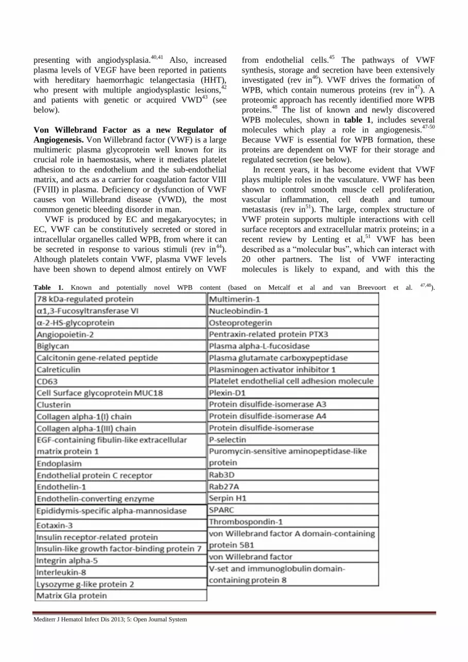
The list of known and newly discovered
WPB molecules, shown in table 1, includes several
molecules which play a role in angiogenesis.47-50
Because VWF is essential for WPB formation, these
proteins are dependent on VWF for their storage and
regulated secretion (see below).
In recent years, it has become evident that VWF
plays multiple roles in the vasculature. VWF has been
shown to control smooth muscle cell proliferation,
vascular inflammation, cell death and tumour
metastasis (rev in51
). The large, complex structure of
VWF protein supports multiple interactions with cell
surface receptors and extracellular matrix proteins; in a
recent review by Lenting et al,51
VWF has been
described as a “molecular bus”, which can interact with
20 other partners. The list of VWF interacting
molecules is likely to expand, and with this the
Table 1. Known and potentially novel WPB content (based on Metcalf et al and van Breevoort et al. 47,48).
Mediterr J Hematol Infect Dis 2013; 5: Open Journal System
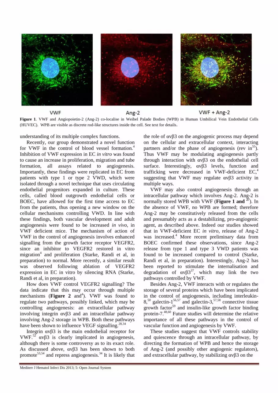
Figure 1. VWF and Angiopoietin-2 (Ang-2) co-localise in Weibel Palade Bodies (WPB) in Human Umbilical Vein Endothelial Cells
(HUVEC). WPB are visible as discrete rod-like structures inside the cell. See text for details.
understanding of its multiple complex functions.
Recently, our group demonstrated a novel function
for VWF in the control of blood vessel formation.4
Inhibition of VWF expression in EC in vitro was found
to cause an increase in proliferation, migration and tube
formation, all assays related to angiogenesis.
Importantly, these findings were replicated in EC from
patients with type 1 or type 2 VWD, which were
isolated through a novel technique that uses circulating
endothelial progenitors expanded in culture. These
cells, called blood outgrowth endothelial cells or
BOEC, have allowed for the first time access to EC
from the patients, thus opening a new window on the
cellular mechanisms controlling VWD. In line with
these findings, both vascular development and adult
angiogenesis were found to be increased in vivo, in
VWF deficient mice. The mechanism of action of
VWF in the control of angiogenesis involves enhanced
signalling from the growth factor receptor VEGFR2,
since an inhibitor to VEGFR2 restored in vitro
migration4 and proliferation (Starke, Randi et al, in
preparation) to normal. More recently, a similar result
was observed following ablation of VEGFR2
expression in EC in vitro by silencing RNA (Starke,
Randi et al, in preparation).
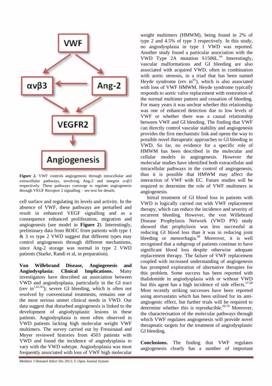
How does VWF control VEGFR2 signalling? The
data indicate that this may occur through multiple
mechanisms (Figure 2 and4). VWF was found to
regulate two pathways, possibly linked, which may be
controlling angiogenesis: an extracellular pathway
involving integrin αvβ3 and an intracellular pathway
involving Ang-2 storage in WPB. Both these pathways
have been shown to influence VEGF signalling.28,34
Integrin αvβ3 is the main endothelial receptor for
VWF.52
αvβ3 is clearly implicated in angiogenesis,
although there is some controversy as to its exact role.
As discussed above, αvβ3 has been shown to both
promote53,54
and repress angiogenesis.34
It is likely that
the role of αvβ3 on the angiogenic process may depend
on the cellular and extracellular context, interacting
partners and/or the phase of angiogenesis (rev in55
).
Thus VWF may be modulating angiogenesis partly
through interaction with αvβ3 on the endothelial cell
surface. Interestingly, αvβ3 levels, function and
trafficking were decreased in VWF-deficient EC,4
suggesting that VWF may regulate αvβ3 activity in
multiple ways.
VWF may also control angiogenesis through an
intracellular pathway which involves Ang-2. Ang-2 is
normally stored WPB with VWF (Figure 1 and 27
). In
the absence of VWF, no WPB are formed; therefore
Ang-2 may be constitutively released from the cells
and presumably acts as a destabilizing, pro-angiogenic
agent, as described above. Indeed our studies showed
that in VWF-deficient EC in vitro, release of Ang-2
was increased.4 More recent preliminary data from
BOEC confirmed these observations, since Ang-2
release from type 1 and type 3 VWD patients was
found to be increased compared to control (Starke,
Randi et al, in preparation). Interestingly, Ang-2 has
been reported to stimulate the internalisation and
degradation of αvβ337
, which may link the two
pathways controlled by VWF.
Besides Ang-2, VWF interacts with or regulates the
storage of several proteins which have been implicated
in the control of angiogenesis, including interleukin-
8,50
galectin-156,57
and galectin-3,57,58
connective tissue
growth factor59
and insulin-like growth factor binding
protein-7.48,60
Future studies will determine the relative
importance of all these pathways in the control of
vascular function and angiogenesis by VWF.
These studies suggest that VWF controls stability
and quiescence through an intracellular pathway, by
directing the formation of WPB and hence the storage
of Ang-2 (and possibly other angiogenic regulators),
and extracellular pathway, by stabilizing αvβ3 on the
Mediterr J Hematol Infect Dis 2013; 5: Open Journal System
Figure 2. VWF controls angiogenesis through intracellular and
extracellular pathways, involving Ang-2 and integrin v3
respectively. These pathways converge to regulate angiogenesis
through VEGF Receptor 2 signalling – see text for details.
cell surface and regulating its levels and activity. In the
absence of VWF, these pathways are perturbed and
result in enhanced VEGF signalling and as a
consequence enhanced proliferation, migration and
angiogenesis (see model in Figure 2). Interestingly,
preliminary data from BOEC from patients with type 1
& 3 vs type 2 VWD suggest that different types may
control angiogenesis through different mechanisms,
since Ang-2 storage was normal in type 2 VWD
patients (Starke, Randi et al, in preparation).
Von Willebrand Disease, Angiogenesis and
Angiodysplasia: Clinical Implications. Many
investigators have described an association between
VWD and angiodysplasia, particularly in the GI tract
(rev in1,61-63
); severe GI bleeding, which is often not
resolved by conventional treatments, remains one of
the most serious unmet clinical needs in VWD. Our
data suggest that disturbed angiogenesis is linked to the
development of angiodysplastic lesions in these
patients. Angiodysplasia is most often observed in
VWD patients lacking high molecular weight VWF
multimers. The survey carried out by Fressinaud and
Meyer reviewed histories from 4503 patients with
VWD and found the incidence of angiodysplasia to
vary with the VWD subtype. Angiodysplasia was most
frequently associated with loss of VWF high molecular
weight multimers (HMWM), being found in 2% of
type 2 and 4.5% of type 3 respectively. In this study,
no angiodysplasia in type 1 VWD was reported.
Another study found a particular association with the
VWD Type 2A mutation S1506L.64
Interestingly,
vascular malformations and GI bleeding are also
associated with acquired VWD, often in combination
with aortic stenosis, in a triad that has been named
Heyde syndrome (rev in65
), which is also associated
with loss of VWF HMWM. Heyde syndrome typically
responds to aortic valve replacement with restoration of
the normal multimer pattern and cessation of bleeding.
For many years it was unclear whether this relationship
was one of enhanced detection due to low levels of
VWF or whether there was a causal relationship
between VWF and GI bleeding. The finding that VWF
can directly control vascular stability and angiogenesis
provides the first mechanistic link and opens the way to
possible novel therapeutic approaches to GI bleeding in
VWD. So far, no evidence for a specific role of
HMWM has been described in the molecular and
cellular models in angiogenesis. However the
molecular studies have identified both extracellular and
intracellular pathways in the control of angiogenesis;
thus it is possible that HMWM may affect the
interaction of VWF with EC. Future studies will be
required to determine the role of VWF multimers in
angiogenesis.
Initial treatment of GI blood loss in patients with
VWD is logically carried out with VWF replacement
therapy, which can reduce the incidence and severity of
recurrent bleeding. However, the von Willebrand
Disease Prophylaxis Network (VWD PN) study
showed that prophylaxis was less successful at
reducing GI blood loss than it was in reducing joint
bleeding or menorrhagia.66
Moreover, it is well
recognised that a subgroup of patients continue to have
significant blood loss despite otherwise adequate
replacement therapy. The failure of VWF replacement
coupled with increased understanding of angiogenesis
has prompted exploration of alternative therapies for
this problem. Some success has been reported with
thalidomide in angiodysplasia with or without VWD
but this agent has a high incidence of side effects.67,68
Most recently striking successes have been reported
using atorvastatin which has been utilised for its anti-
angiogenic effect, but further trials will be required to
determine whether this is reproducible.69,70
Moreover,
the characterisation of the molecular pathways through
which VWF regulates angiogenesis will provide novel
therapeutic targets for the treatment of angiodysplastic
GI bleeding.
Conclusions. The finding that VWF regulates
angiogenesis clearly has a number of important

Mediterr J Hematol Infect Dis 2013; 5: Open Journal System
implications. Firstly, it provides a novel link between
VWD and angiodysplasia, which is likely to have
therapeutic implications for the future. Secondly, it
points the way to investigating the role of VWF in
normal development and healing but also in
pathological processes such as tumour growth, all of
which depend on angiogenesis. We anticipate that
these investigations will lead to novel agents to
modulate angiogenesis for therapeutic benefit. A
critical question for both of these problems will be
determining the relative roles of intra- and extra-
cellular VWF in regulation of angiogenesis. We
therefore remain some way from translation of these
exciting findings into clinical practice. Experience to
date suggests that replacement therapy does not always
correct the defect in angiodysplasia and it is unlikely
that simple infusion of VWF will be a panacea for
abnormal vasculature.
Acknowledgements. We would like to thank Dr.
Koralia Paschalaki for her major contribution on
BOEC cultures and for her support throughout the
studies. We also thank Dr. Elspeth Payne for her
contribution in establishing the BOEC technique in the
laboratory.
References:
1. Quick AJ. Telangiectasia: its relationship to the Minot-von
Willebrand syndrome. Am.J Med Sci. 1967;254:585-601. http://dx.doi.org/10.1097/00000441-196711000-00002
PMid:4862041
2. Koscielny JK, Latza R, Mursdorf S et al. Capillary microscopic and rheological dimensions for the diagnosis of von Willebrand
disease in comparison to other haemorrhagic diatheses.
Thromb.Haemost. 2000;84:981-988. PMid:11154145 3. Duray PH, Marcal JM, Jr., LiVolsi VA et al. Gastrointestinal
angiodysplasia: a possible component of von Willebrand's disease.
Hum.Pathol. 1984;15:539-544. http://dx.doi.org/10.1016/S0046-8177(84)80007-6
4. Starke RD, Ferraro F, Paschalaki KE et al. Endothelial von
Willebrand factor regulates angiogenesis. Blood 2011;117:1071-1080. http://dx.doi.org/10.1182/blood-2010-01-264507
PMid:21048155 PMCid:PMC3035068
5. Carmeliet P. Angiogenesis in health and disease. Nat.Med. 2003;9:653-660. http://dx.doi.org/10.1038/nm0603-653
PMid:12778163 6. Gerhardt H, Golding M, Fruttiger M et al. VEGF guides
angiogenic sprouting utilizing endothelial tip cell filopodia. J Cell
Biol 2003;161:1163-1177.
http://dx.doi.org/10.1083/jcb.200302047 PMid:12810700
PMCid:PMC2172999
7. Fantin A, Vieira JM, Gestri G et al. Tissue macrophages act as cellular chaperones for vascular anastomosis downstream of
VEGF-mediated endothelial tip cell induction. Blood
2010;116:829-840. http://dx.doi.org/10.1182/blood-2009-12-257832 PMid:20404134 PMCid:PMC2938310
8. Eilken HM, Adams RH. Dynamics of endothelial cell behavior in
sprouting angiogenesis. Curr.Opin.Cell Biol 2010;22:617-625. http://dx.doi.org/10.1016/j.ceb.2010.08.010 PMid: 20817428
9. Jain RK. Molecular regulation of vessel maturation. Nat.Med.
2003;9:685-693. http://dx.doi.org/10.1038/nm0603-685 PMid:12778167
10. Potente M, Gerhardt H, Carmeliet P. Basic and therapeutic aspects
of angiogenesis. Cell 2011;146:873-887. http://dx.doi.org/10.1016/j.cell.2011.08.039 PMid:21925313
11. Ferrara N. VEGF-A: a critical regulator of blood vessel growth.
Eur.Cytokine Netw. 2009;20:158-163. PMid:20167554 12. Lohela M, Bry M, Tammela T, Alitalo K. VEGFs and receptors
involved in angiogenesis versus lymphangiogenesis.
Curr.Opin.Cell Biol 2009;21:154-165. http://dx.doi.org/10.1016/j.ceb.2008.12.012 PMid:19230644
13. Ladomery MR, Harper SJ, Bates DO. Alternative splicing in
angiogenesis: the vascular endothelial growth factor paradigm. Cancer Lett. 2007;249:133-142.
http://dx.doi.org/10.1016/j.canlet.2006.08.015 PMid:17027147
14. Olsson AK, Dimberg A, Kreuger J, Claesson-Welsh L. VEGF receptor signalling - in control of vascular function.
Nat.Rev.Mol.Cell Biol 2006;7:359-371.
http://dx.doi.org/10.1038/nrm1911 PMid:16633338 15. Springer ML, Chen AS, Kraft PE, Bednarski M, Blau HM. VEGF
gene delivery to muscle: potential role for vasculogenesis in adults.
Mol.Cell 1998;2:549-558. http://dx.doi.org/10.1016/S1097-
2765(00)80154-9 16. Schwarz ER, Speakman MT, Patterson M et al. Evaluation of the
effects of intramyocardial injection of DNA expressing vascular
endothelial growth factor (VEGF) in a myocardial infarction model in the rat--angiogenesis and angioma formation. J.Am.Coll.Cardiol.
2000;35:1323-1330. http://dx.doi.org/10.1016/S0735-
1097(00)00522-2 17. Eklund L, Olsen BR. Tie receptors and their angiopoietin ligands
are context-dependent regulators of vascular remodeling. Exp.Cell
Res. 2006;312:630-641. http://dx.doi.org/10.1016/j.yexcr.2005.09.002 PMid:16225862
18. Fukuhara S, Sako K, Minami T et al. Differential function of Tie2
at cell-cell contacts and cell-substratum contacts regulated by angiopoietin-1. Nat.Cell Biol. 2008;10:513-526.
http://dx.doi.org/10.1038/ncb1714 PMid:18425120
19. Felcht M, Luck R, Schering A et al. Angiopoietin-2 differentially regulates angiogenesis through TIE2 and integrin signaling.
J.Clin.Invest 2012;122:1991-2005. http://dx.doi.org/10.1172/JCI58832 PMid:22585576
PMCid:PMC3366398
20. Suri C, Jones PF, Patan S et al. Requisite role of angiopoietin-1, a
ligand for the TIE2 receptor, during embryonic angiogenesis. Cell
1996;87:1171-1180. http://dx.doi.org/10.1016/S0092-
8674(00)81813-9 21. Suri C, McClain J, Thurston G et al. Increased vascularization in
mice overexpressing angiopoietin-1. Science 1998;282:468-471.
http://dx.doi.org/10.1126/science.282.5388.468 PMid:9774272 22. Senger DR, Galli SJ, Dvorak AM et al. Tumor cells secrete a
vascular permeability factor that promotes accumulation of ascites
fluid. Science 1983;219:983-985. http://dx.doi.org/10.1126/science.6823562 PMid:6823562
23. Thurston G, Suri C, Smith K et al. Leakage-resistant blood vessels
in mice transgenically overexpressing angiopoietin-1. Science 1999;286:2511-2514.
http://dx.doi.org/10.1126/science.286.5449.2511 PMid:10617467
24. Thurston G, Rudge JS, Ioffe E et al. Angiopoietin-1 protects the adult vasculature against plasma leakage. Nat.Med. 2000;6:460-
463. http://dx.doi.org/10.1038/74725 PMid:10742156
25. Tao Z, Chen B, Tan X et al. Coexpression of VEGF and angiopoietin-1 promotes angiogenesis and cardiomyocyte
proliferation reduces apoptosis in porcine myocardial infarction
(MI) heart. Proc.Natl.Acad.Sci.U.S.A 2011;108:2064-2069. http://dx.doi.org/10.1073/pnas.1018925108 PMid:21245320
PMCid:PMC3033313
26. Daly C, Eichten A, Castanaro C et al. Angiopoietin-2 functions as a Tie2 agonist in tumor models, where it limits the effects of VEGF
inhibition. Cancer Res. 2013;73:108-118.
http://dx.doi.org/10.1158/0008-5472.CAN-12-2064 PMid:23149917
27. Fiedler U, Scharpfenecker M, Koidl S et al. The Tie-2 ligand
angiopoietin-2 is stored in and rapidly released upon stimulation from endothelial cell Weibel-Palade bodies. Blood 2004;103:4150-
4156. http://dx.doi.org/10.1182/blood-2003-10-3685
Mediterr J Hematol Infect Dis 2013; 5: Open Journal System
PMid:14976056
28. Lobov IB, Brooks PC, Lang RA. Angiopoietin-2 displays VEGF-
dependent modulation of capillary structure and endothelial cell survival in vivo. Proceedings of the National Academy of Sciences
of the United States of America 2002;99:11205-11210.
http://dx.doi.org/10.1073/pnas.172161899 PMid:12163646 PMCid:PMC123234
29. Holash J, Wiegand SJ, Yancopoulos GD. New model of tumor
angiogenesis: dynamic balance between vessel regression and growth mediated by angiopoietins and VEGF. Oncogene
1999;18:5356-5362. http://dx.doi.org/10.1038/sj.onc.1203035
PMid:10498889 30. Augustin HG, Koh GY, Thurston G, Alitalo K. Control of vascular
morphogenesis and homeostasis through the angiopoietin-Tie
system. Nat.Rev.Mol.Cell Biol 2009;10:165-177. http://dx.doi.org/10.1038/nrm2639 PMid:19234476
31. Desgrosellier JS, Cheresh DA. Integrins in cancer: biological
implications and therapeutic opportunities. Nat Rev Cancer 2010;10:9-22. http://dx.doi.org/10.1038/nrc2748 PMid:20029421
32. Gladson CL, Cheresh DA. Glioblastoma expression of vitronectin
and the alpha v beta 3 integrin. Adhesion mechanism for transformed glial cells. J.Clin.Invest 1991;88:1924-1932.
http://dx.doi.org/10.1172/JCI115516 PMid:1721625
PMCid:PMC295768 33. Scaringi C, Minniti G, Caporello P, Enrici RM. Integrin inhibitor
cilengitide for the treatment of glioblastoma: a brief overview of
current clinical results. Anticancer Res. 2012;32:4213-4223. PMid:23060541
34. Reynolds LE, Wyder L, Lively JC et al. Enhanced pathological
angiogenesis in mice lacking beta3 integrin or beta3 and beta5 integrins. Nat.Med. 2002;8:27-34.
http://dx.doi.org/10.1038/nm0102-27 PMid:11786903
35. Somanath PR, Malinin NL, Byzova TV. Cooperation between integrin alphavbeta3 and VEGFR2 in angiogenesis. Angiogenesis.
2009;12:177-185. http://dx.doi.org/10.1007/s10456-009-9141-9
PMid:19267251 PMCid:PMC2863048 36. Carlson TR, Feng Y, Maisonpierre PC, Mrksich M, Morla AO.
Direct cell adhesion to the angiopoietins mediated by integrins.
J.Biol.Chem. 2001;276:26516-26525. http://dx.doi.org/10.1074/jbc.M100282200 PMid:11346644
37. Thomas M, Felcht M, Kruse K et al. Angiopoietin-2 stimulation of endothelial cells induces aVb3 integrin internalization and
degradation. Journal of Biological Chemistry 2010
http://dx.doi.org/10.1074/jbc.M109.097543
38. Carmeliet P. Angiogenesis in life, disease and medicine. Nature
2005;438:932-936. http://dx.doi.org/10.1038/nature04478
PMid:16355210 39. Regula J, Wronska E, Pachlewski J. Vascular lesions of the
gastrointestinal tract. Best.Pract.Res.Clin.Gastroenterol.
2008;22:313-328. http://dx.doi.org/10.1016/j.bpg.2007.10.026 PMid:18346686
40. Tan H, Chen H, Xu C et al. Role of vascular endothelial growth
factor in angiodysplasia: an interventional study with thalidomide. J.Gastroenterol.Hepatol. 2012;27:1094-1101.
http://dx.doi.org/10.1111/j.1440-1746.2011.06967.x
PMid:22098296 41. Junquera F, Saperas E, de T, I, Vidal MT, Malagelada JR.
Increased expression of angiogenic factors in human colonic
angiodysplasia. Am.J.Gastroenterol. 1999;94:1070-1076. http://dx.doi.org/10.1111/j.1572-0241.1999.01017.x
PMid:10201485
42. Cirulli A, Liso A, D'Ovidio F et al. Vascular endothelial growth factor serum levels are elevated in patients with hereditary
hemorrhagic telangiectasia. Acta Haematol. 2003;110:29-32.
http://dx.doi.org/10.1159/000072411 PMid:12975554 43. Gritti G, Cortelezzi A, Bucciarelli P et al. Circulating and
progenitor endothelial cells are abnormal in patients with different
types of von Willebrand disease and correlate with markers of angiogenesis. Am.J.Hematol. 2011;86:650-656.
http://dx.doi.org/10.1002/ajh.22070 PMid:21630316
44. Rondaij MG, Bierings R, Kragt A, van Mourik JA, Voorberg J. Dynamics and plasticity of Weibel-Palade bodies in endothelial
cells. Arterioscler.Thromb.Vasc.Biol. 2006;26:1002-1007.
http://dx.doi.org/10.1161/01.ATV.0000209501.56852.6c PMid:16469951
45. Kanaji S, Fahs SA, Shi Q, Haberichter SL, Montgomery RR.
Contribution of platelet vs. endothelial VWF to platelet adhesion
and hemostasis. J.Thromb.Haemost. 2012;10:1646-1652.
http://dx.doi.org/10.1111/j.1538-7836.2012.04797.x PMid:22642380 PMCid:PMC3419786
46. Michaux G, Cutler DF. How to roll an endothelial cigar: the
biogenesis of Weibel-Palade bodies. Traffic. 2004;5:69-78. http://dx.doi.org/10.1111/j.1600-0854.2004.00157.x
47. Metcalf DJ, Nightingale TD, Zenner HL, Lui-Roberts WW, Cutler
DF. Formation and function of Weibel-Palade bodies. J.Cell Sci. 2008;121:19-27. http://dx.doi.org/10.1242/jcs.03494
PMid:18096688
48. van Breevoort D, van Agtmaal EL, Dragt BS et al. Proteomic screen identifies IGFBP7 as a novel component of endothelial cell-
specific Weibel-Palade bodies. J.Proteome.Res. 2012;11:2925-
2936. http://dx.doi.org/10.1021/pr300010r PMid:22468712 49. Thomas M, Augustin HG. The role of the Angiopoietins in
vascular morphogenesis. Angiogenesis. 2009;12:125-137.
http://dx.doi.org/10.1007/s10456-009-9147-3 PMid:19449109 50. Petreaca ML, Yao M, Liu Y, Defea K, Martins-Green M.
Transactivation of vascular endothelial growth factor receptor-2 by
interleukin-8 (IL-8/CXCL8) is required for IL-8/CXCL8-induced endothelial permeability. Mol.Biol.Cell 2007;18:5014-5023.
http://dx.doi.org/10.1091/mbc.E07-01-0004 PMid:17928406
PMCid:PMC2096609 51. Lenting PJ, Casari C, Christophe OD, Denis CV. von Willebrand
factor: the old, the new and the unknown. J.Thromb.Haemost.
2012;10:2428-2437. http://dx.doi.org/10.1111/jth.12008 PMid:23020315
52. Cheresh DA. Human endothelial cells synthesize and express an
Arg-Gly-Asp-directed adhesion receptor involved in attachment to fibrinogen and von Willebrand factor. Proc.Natl.Acad.Sci.U.S.A
1987;84:6471-6475. http://dx.doi.org/10.1073/pnas.84.18.6471
PMid:2442758 PMCid:PMC299099 53. Brooks PC, Montgomery AM, Rosenfeld M et al. Integrin alpha v
beta 3 antagonists promote tumor regression by inducing apoptosis
of angiogenic blood vessels. Cell 1994;79:1157-1164. http://dx.doi.org/10.1016/0092-8674(94)90007-8
54. Brooks PC, Clark RA, Cheresh DA. Requirement of vascular
integrin alpha v beta 3 for angiogenesis. Science 1994;264:569-571. http://dx.doi.org/10.1126/science.7512751 PMid:7512751
55. Robinson SD, Hodivala-Dilke KM. The role of beta3-integrins in tumor angiogenesis: context is everything. Curr.Opin.Cell Biol.
2011;23:630-637. http://dx.doi.org/10.1016/j.ceb.2011.03.014
PMid:21565482
56. Thijssen VL, Postel R, Brandwijk RJ et al. Galectin-1 is essential
in tumor angiogenesis and is a target for antiangiogenesis therapy.
Proc.Natl.Acad.Sci.U.S.A 2006;103:15975-15980. http://dx.doi.org/10.1073/pnas.0603883103 PMid:17043243
PMCid:PMC1635112
57. Saint-Lu N, Oortwijn BD, Pegon JN et al. Identification of galectin-1 and galectin-3 as novel partners for von Willebrand
factor. Arterioscler.Thromb.Vasc.Biol. 2012;32:894-901.
http://dx.doi.org/10.1161/ATVBAHA.111.240309 PMid:22267483
58. Markowska AI, Liu FT, Panjwani N. Galectin-3 is an important
mediator of VEGF- and bFGF-mediated angiogenic response. J.Exp.Med. 2010;207:1981-1993.
http://dx.doi.org/10.1084/jem.20090121 PMid:20713592
PMCid:PMC2931172 59. Pi L, Shenoy AK, Liu J et al. CCN2/CTGF regulates neovessel
formation via targeting structurally conserved cystine knot motifs
in multiple angiogenic regulators. FASEB J. 2012;26:3365-3379. http://dx.doi.org/10.1096/fj.11-200154 PMid:22611085
PMCid:PMC3405264
60. Tamura K, Hashimoto K, Suzuki K et al. Insulin-like growth factor binding protein-7 (IGFBP7) blocks vascular endothelial cell
growth factor (VEGF)-induced angiogenesis in human vascular
endothelial cells. Eur.J.Pharmacol. 2009;610:61-67. http://dx.doi.org/10.1016/j.ejphar.2009.01.045 PMid:19374835
61. Warkentin TE, Moore JC, Anand SS, Lonn EM, Morgan DG.
Gastrointestinal bleeding, angiodysplasia, cardiovascular disease, and acquired von Willebrand syndrome. Transfus.Med.Rev.
2003;17:272-286. http://dx.doi.org/10.1016/S0887-7963(03)00037-
3 62. Makris M. Gastrointestinal bleeding in von Willebrand disease.
Thromb.Res. 2006;118 Suppl 1:S13-S17.

Mediterr J Hematol Infect Dis 2013; 5: Open Journal System
http://dx.doi.org/10.1016/j.thromres.2006.01.022
PMid:16542710
63. Fressinaud E, Meyer D. International survey of patients with von Willebrand disease and angiodysplasia. Thromb.Haemost.
1993;70:546. PMid:8259565
64. Castaman G, Federici AB, Tosetto A et al. Different bleeding risk in type 2A and 2M von Willebrand disease: a 2-year prospective
study in 107 patients. J.Thromb.Haemost. 2012;10:632-638.
http://dx.doi.org/10.1111/j.1538-7836.2012.04661.x PMid:22329792
65. Massyn MW, Khan SA. Heyde syndrome: a common diagnosis in
older patients with severe aortic stenosis. Age Ageing 2009;38:267-270. http://dx.doi.org/10.1093/ageing/afp019
PMid:19276092
66. Abshire TC, Federici AB, Alvarez MT et al. Prophylaxis in severe forms of von Willebrand's disease: results from the von Willebrand
Disease Prophylaxis Network (VWD PN). Haemophilia.
2013;19:76-81. http://dx.doi.org/10.1111/j.1365-2516.2012.02916.x PMid:22823000
67. Nomikou E, Tsevrenis V, Gafou A, Bellia M, Theodossiades G.
Type IIb von Willebrand disease with angiodysplasias and
refractory gastrointestinal bleeding successfully treated with thalidomide. Haemophilia. 2009;15:1340-1342.
http://dx.doi.org/10.1111/j.1365-2516.2009.02085.x
PMid:19702883 68. Bauditz J, Schachschal G, Wedel S, Lochs H. Thalidomide for
treatment of severe intestinal bleeding. Gut 2004;53:609-612.
http://dx.doi.org/10.1136/gut.2003.029710 PMid:15016759 PMCid:PMC1774015
69. Sohal M, Laffan M. Von Willebrand disease and angiodysplasia
responding to atorvastatin. Br.J.Haematol. 2008;142:308-309. http://dx.doi.org/10.1111/j.1365-2141.2008.07005.x
PMid:18510690
70. Alikhan R, Keeling D. Von Willebrand disease, angiodysplasia and atorvastatin. Br.J.Haematol. 2010;149:159-160.
http://dx.doi.org/10.1111/j.1365-2141.2009.08031.x
PMid:19995387