allosteric activation of adamts13 by von willebrand factor
TRANSCRIPT

Allosteric activation of ADAMTS13 by vonWillebrand factorJoshua Muiaa,1, Jian Zhua,1, Garima Guptaa, Sandra L. Haberichterb, Kenneth D. Friedmanb, Hendrik B. Feysc,Louis Deforched, Karen Vanhoorelbeked, Lisa A. Westfielda, Robyn Rothe, Niraj Harish Toliaf,g, John E. Heusere,and J. Evan Sadlera,f,2
Departments of aMedicine, eCell Biology and Physiology, fBiochemistry and Molecular Biophysics, and gMolecular Microbiology and Microbial Pathogenesis,Washington University School of Medicine, St. Louis, MO 63110; bBlood Research Institute, BloodCenter of Wisconsin, Milwaukee, WI 53201; cTransfusionResearch Center, Belgian Red Cross-Flanders, Ghent, Belgium; and dLaboratory for Thrombosis Research, KU Leuven Kulak, 8500 Kortrijk, Belgium
Edited* by David Ginsburg, University of Michigan Medical School, Ann Arbor, MI, and approved September 26, 2014 (received for review July 13, 2014)
The metalloprotease ADAMTS13 cleaves von Willebrand factor(VWF) within endovascular platelet aggregates, and ADAMTS13deficiency causes fatal microvascular thrombosis. The proximalmetalloprotease (M), disintegrin-like (D), thrombospondin-1 (T),Cys-rich (C), and spacer (S) domains of ADAMTS13 recognize acryptic site in VWF that is exposed by tensile force. Another sevenT and two complement C1r/C1s, sea urchin epidermal growthfactor, and bone morphogenetic protein (CUB) domains of un-certain function are C-terminal to the MDTCS domains. We findthat the distal T8-CUB2 domains markedly inhibit substratecleavage, and binding of VWF or monoclonal antibodies to distalADAMTS13 domains relieves this autoinhibition. Small angle X-rayscattering data indicate that distal T-CUB domains interact withproximal MDTCS domains. Thus, ADAMTS13 is regulated by sub-strate-induced allosteric activation, which may optimize VWFcleavage under fluid shear stress in vivo. Distal domains of otherADAMTS proteases may have similar allosteric properties.
hemostasis | metalloproteases | allosteric regulation
After vascular injury, platelets adhere to von Willebrandfactor (VWF) multimers bound to endothelial cell surfaces
and connective tissue. The force of flowing blood on a growingplatelet-rich thrombus stretches the central A2 domain of VWFand exposes a Tyr1605-Met1606 cleavage site for ADAMTS13 (Fig.1A) (1–5), a metalloprotease that severs VWF and releases ad-herent platelets. Deficiency of ADAMTS13 disrupts this feedbackregulatory mechanism and causes thrombotic thrombocytopenicpurpura (TTP), which is characterized by life-threatening micro-vascular thrombosis (3, 6, 7).The recognition and cleavage of VWF is a formidable challenge.
VWF and ADAMTS13 occur at ∼10 μg/mL and ∼1 μg/mL, re-spectively, compared with total plasma protein of ∼80,000 μg/mL.ADAMTS13 is constitutively active and has no known inhibitors invivo. Nevertheless, VWF is the only identified ADAMTS13 sub-strate, and VWF is resistant to cleavage until subjected to fluidshear stress (8), adsorbed on a surface (9), or treated with dena-turants (8, 10). This specificity depends on structural features ofboth ADAMTS13 and VWF that have not been characterized fully.The proximal metalloprotease (M), disintegrin-like (D), throm-
bospondin-1 (T), Cys-rich (C), and spacer (S) domains domains ofADAMTS13 bind to cryptic sites that are uncovered by unfoldingVWF domain A2 (11-15) (Fig. 1B), and these interactions are re-quired for efficient cleavage of VWF or peptide substrates. Moredistal ADAMTS13 domains bind to sites in or near VWF do-main D4 that are always available (16–18). Deletion of distalADAMTS13 domains impairs the cleavage of VWF multimersin vitro (16, 19) and increases VWF-dependent microvascularthrombosis in vivo (20) but accelerates the cleavage of peptidesubstrates (12, 13). In addition, ADAMTS13 cleaves guanidinehydrochloride-treated VWF multimers with an apparent Km of∼15 nM (21), which is 100-fold lower than the Km of ∼1.6–1.7 μM for peptide substrates that are based on the sequence of
VWF domain A2 (12, 14). These striking differences suggest thatdistal T or complement c1r/c1s, sea urchin epidermal growthfactor, and bone morphogenetic protein (CUB) domains regu-late ADAMTS13 activity. We have now shown that these distaldomains inhibit ADAMTS13, and binding to VWF relievesthis autoinhibition.
ResultsActivation of ADAMTS13 by Antibodies and Low pH. Evidence forallosteric regulation was obtained unexpectedly in the course ofanalyzing plasma samples from patients with TTP. The majorityof adult patients with acquired TTP have autoantibodies thatinhibit ADAMTS13 and reduce its activity in plasma to <5% ofnormal, but one patient proved to be a remarkable exception.When assayed with a fluorogenic ADAMTS13 substrate, VWF71(Fig. 1B) (22), patient BCW49 had high-titer autoantibodies thatparadoxically activated exogenous ADAMTS13 ∼threefold (Fig.1C). Activation occurred at pH 7.4, which is characteristic ofblood, but not at pH 6, which is used routinely for clinicalADAMTS13 assays (23). Furthermore, BCW49 plasma had noeffect on the activity of MDTCS at either pH 7.4 or pH 6.The loss of pH dependence for MDTCS suggested a regulatory
function for the distal domains that are missing from this trun-cated ADAMTS13 construct. We (22) and others (23, 24) haveobserved that full-length ADAMTS13 is most active at pH 6, withmarkedly decreased activity at pH 7.4 (Fig. 1D and Fig. S1). Thisphenomenon has been attributed to ionization of a Zn2+-bound
Significance
The blood protein von Willebrand factor (VWF) is required forplatelets to stop bleeding at sites of injury, and the metal-loprotease ADAMTS13 limits platelet adhesion by cleaving VWFonly when flowing blood stretches it, especially within a grow-ing thrombus. This feedback inhibition is essential becauseADAMTS13 deficiency causes fatal microvascular thrombosis.How ADAMTS13 recognizes VWF so specifically is not un-derstood. We now find that ADAMTS13 is folded roughly in halfso that its distal domains inhibit the metalloprotease domain.VWF relieves this autoinhibition and promotes its own de-struction by allosterically activating ADAMTS13. Thus, VWF isboth a substrate and a cofactor in this critical regulatory process.
Author contributions: J.M., J.Z., G.G., K.V., N.H.T., and J.E.S. designed research; J.M., J.Z.,G.G., S.L.H., K.D.F., H.B.F., L.D., K.V., L.A.W., and R.R. performed research; S.L.H., K.D.F., andH.B.F. contributed new reagents/analytic tools; J.M., J.Z., G.G., L.D., K.V., N.H.T., J.E.H., andJ.E.S. analyzed data; and J.M. and J.E.S. wrote the paper.
The authors declare no conflict of interest.
*This Direct Submission article had a prearranged editor.1J.M. and J.Z. contributed equally to this work.2To whom correspondence should be addressed. Email: [email protected].
This article contains supporting information online at www.pnas.org/lookup/suppl/doi:10.1073/pnas.1413282112/-/DCSupplemental.
www.pnas.org/cgi/doi/10.1073/pnas.1413282112 PNAS Early Edition | 1 of 6
BIOCH
EMISTR
Y
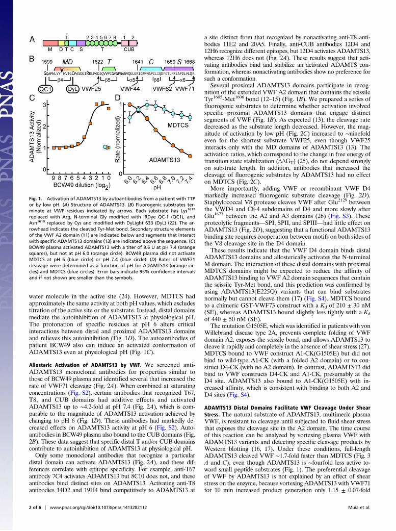
water molecule in the active site (24). However, MDTCS hadapproximately the same activity at both pH values, which excludestitration of the active site or the substrate. Instead, distal domainsmediate the autoinhibition of ADAMTS13 at physiological pH.The protonation of specific residues at pH 6 alters criticalinteractions between distal and proximal ADAMTS13 domainsand relieves this autoinhibition (Fig. 1D). The autoantibodies ofpatient BCW49 also can induce an activated conformation ofADAMTS13 even at physiological pH (Fig. 1C).
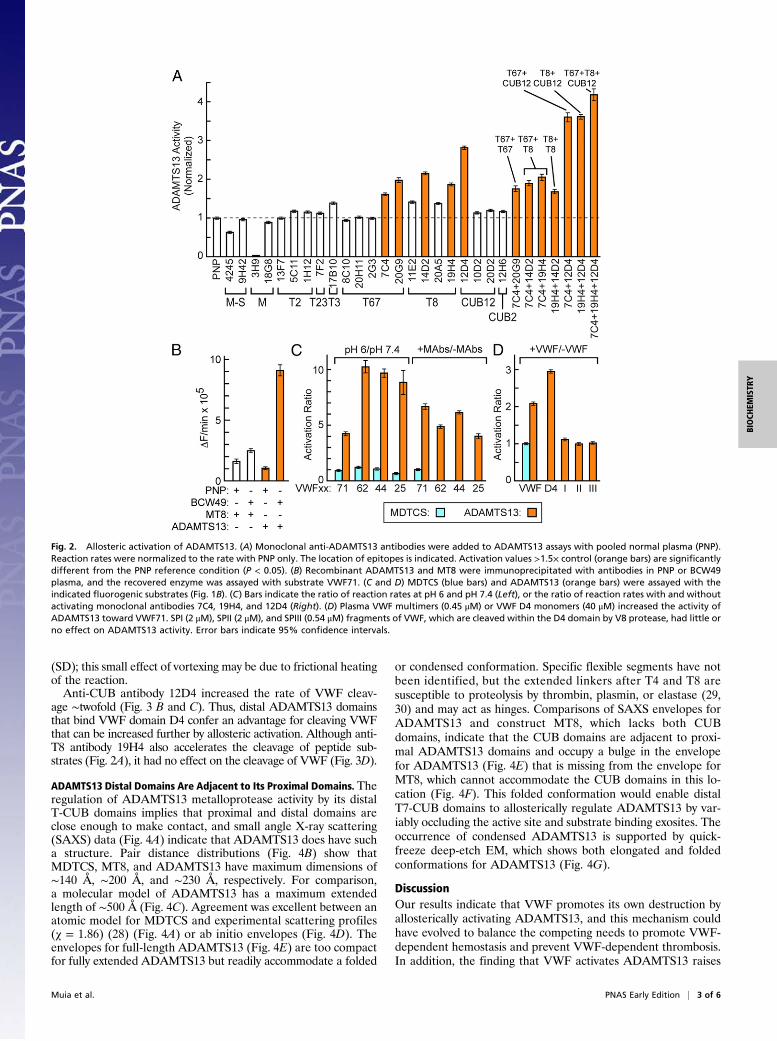
Allosteric Activation of ADAMTS13 by VWF. We screened anti-ADAMTS13 monoclonal antibodies for properties similar tothose of BCW49 plasma and identified several that increased therate of VWF71 cleavage (Fig. 2A). When combined at saturatingconcentrations (Fig. S2), certain antibodies that recognized T67,T8, and CUB domains had additive effects and activatedADAMTS13 up to ∼4.2-fold at pH 7.4 (Fig. 2A), which is com-parable to the magnitude of ADAMTS13 activation achieved bychanging to pH 6 (Fig. 1D). These antibodies had markedly de-creased effects on ADAMTS13 activity at pH 6 (Fig. S2). Auto-antibodies in BCW49 plasma also bound to the CUB domains (Fig.2B). These data suggest that specific distal T and/or CUB domainscontribute to autoinhibition of ADAMTS13 at physiological pH.Only some monoclonal antibodies that recognize a particular
distal domain can activate ADAMTS13 (Fig. 2A), and these dif-ferences correlate with epitope specificity. For example, anti-T67antibody 7C4 activates ADAMTS13 but 8C10 does not, and theseantibodies bind distinct sites on ADAMTS13. Activating anti-T8antibodies 14D2 and 19H4 bind competitively to ADAMTS13 at
a site distinct from that recognized by nonactivating anti-T8 anti-bodies 11E2 and 20A5. Finally, anti-CUB antibodies 12D4 and12H6 recognize different epitopes, but 12D4 activates ADAMTS13,whereas 12H6 does not (Fig. 2A). These results suggest that acti-vating antibodies bind and stabilize an activated ADAMTS con-formation, whereas nonactivating antibodies show no preference forsuch a conformation.Several proximal ADAMTS13 domains participate in recog-
nition of the extended VWF A2 domain that contains the scissileTyr1605-Met1606 bond (12–15) (Fig. 1B). We prepared a series offluorogenic substrates to determine whether activation involvedspecific proximal ADAMTS13 domains that engage distinctsegments of VWF (Fig. 1B). As expected (13), the cleavage ratedecreased as the substrate length decreased. However, the mag-nitude of activation by low pH (Fig. 2C) increased to ∼ninefoldeven for the shortest substrate VWF25, even though VWF25interacts only with the MD domains of ADAMTS13 (13). Theactivation ratios, which correspond to the change in free energy oftransition state stabilization (ΔΔGT) (25), do not depend stronglyon substrate length. In addition, antibodies that increased thecleavage of fluorogenic substrates by ADAMTS13 had no effecton MDTCS (Fig. 2C).More importantly, adding VWF or recombinant VWF D4
markedly increased fluorogenic substrate cleavage (Fig. 2D).Staphylococcal V8 protease cleaves VWF after Glu2129 betweenthe VWD4 and C8-4 subdomains of D4 and more slowly afterGlu1673 between the A2 and A3 domains (26) (Fig. S3). Theseproteolytic fragments—SPI, SPII, and SPIII—had little effect onADAMTS13 (Fig. 2D), suggesting that a functional ADAMTS13binding site requires cooperation between motifs on both sides ofthe V8 cleavage site in the D4 domain.These results indicate that the VWF D4 domain binds distal
ADAMTS13 domains and allosterically activates the N-terminalM domain. The interaction of these distal domains with proximalMDTCS domains might be expected to reduce the affinity ofADAMTS13 binding to VWF A2 domain sequences that containthe scissile Tyr-Met bond, and this prediction was confirmed byusing ADAMTS13(E225Q) variants that can bind substratesnormally but cannot cleave them (17) (Fig. S4). MDTCS boundto a chimeric GST-VWF73 construct with a Kd of 210 ± 30 nM(SE), whereas ADAMTS13 bound slightly less tightly with a Kdof 440 ± 50 nM (SE).The mutation G1505E, which was identified in patients with von
Willebrand disease type 2A, prevents complete folding of VWFdomain A2, exposes the scissile bond, and allows ADAMTS13 tocleave it rapidly and completely in the absence of shear stress (27).MDTCS bound to VWF construct A1-CK(G1505E) but did notbind to wild-type A1-CK (with a folded A2 domain) or to con-struct D4-CK (with no A2 domain). In contrast, ADAMTS13 didbind to VWF constructs D4-CK and A1-CK, presumably at theD4 site. ADAMTS13 also bound to A1-CK(G1505E) with in-creased affinity, which is consistent with binding to both A2 andD4 sites (Fig. S4).
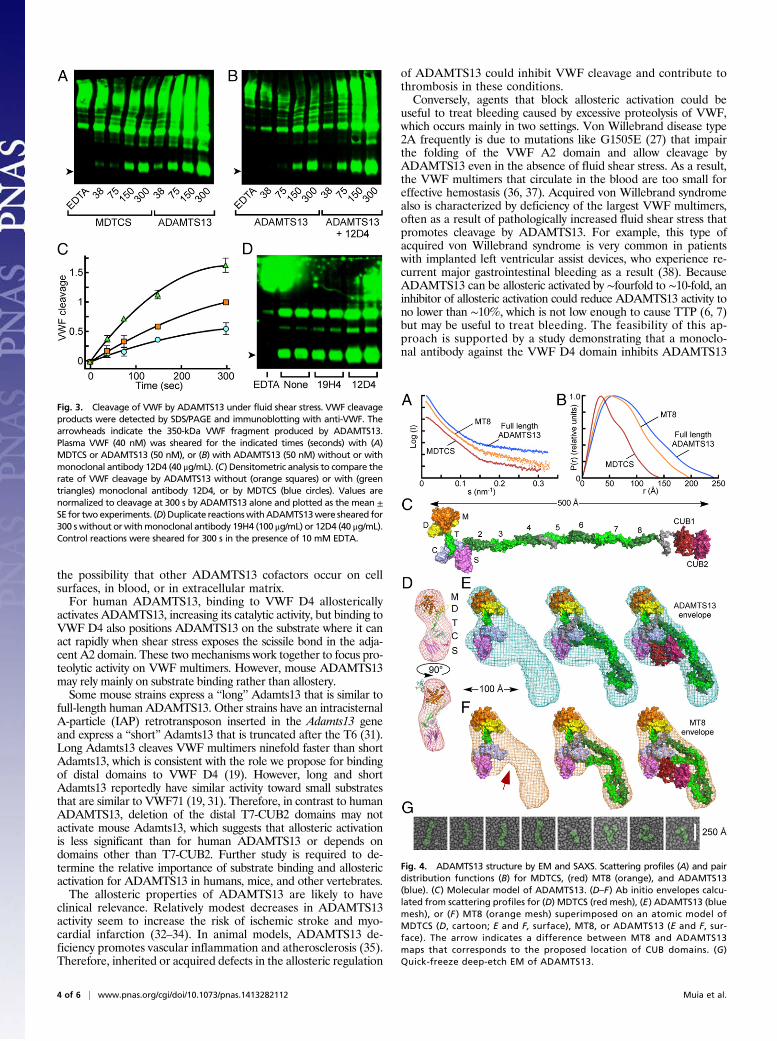
ADAMTS13 Distal Domains Facilitate VWF Cleavage Under ShearStress. The natural substrate of ADAMTS13, multimeric plasmaVWF, is resistant to cleavage until subjected to fluid shear stressthat exposes the cleavage site in the A2 domain. The time courseof this reaction can be analyzed by vortexing plasma VWF withADAMTS13 variants and detecting specific cleavage products byWestern blotting (16, 17). Under these conditions, full-lengthADAMTS13 cleaved VWF ∼1.7-fold faster than MDTCS (Fig. 3A and C), even though ADAMTS13 is ∼fourfold less active to-ward small peptide substrates (Fig. 1). The preferential cleavageof VWF by ADAMTS13 is not explained by an effect of shearstress on the enzyme, because vortexing ADAMTS13 with VWF71for 10 min increased product generation only 1.15 ± 0.07-fold
Fig. 1. Activation of ADAMTS13 by autoantibodies from a patient with TTPor by low pH. (A) Structure of ADAMTS13. (B) Fluorogenic substrates ter-minate at VWF residues indicated by arrows. Each substrate has Lys1617
replaced with Arg, N-terminal Gly modified with IRDye QC-1 (QC1), andAsn1610 replaced by Cys and modified with DyLight 633 (DyL) (22). The ar-rowhead indicates the cleaved Tyr-Met bond. Secondary structure elementsof the VWF A2 domain (11) are indicated below and segments that interactwith specific ADAMTS13 domains (13) are indicated above the sequence. (C)BCW49 plasma activated ADAMTS13 with a titer of 9.6 U at pH 7.4 (orangesquares), but not at pH 6.0 (orange circle). BCW49 plasma did not activateMDTCS at pH 6 (blue circle) or pH 7.4 (blue circle). (D) Rates of VWF71cleavage were determined as a function of pH for ADAMTS13 (orange cir-cles) and MDTCS (blue circles). Error bars indicate 95% confidence intervalsand if not shown are smaller than the symbols.
2 of 6 | www.pnas.org/cgi/doi/10.1073/pnas.1413282112 Muia et al.
(SD); this small effect of vortexing may be due to frictional heatingof the reaction.Anti-CUB antibody 12D4 increased the rate of VWF cleav-
age ∼twofold (Fig. 3 B and C). Thus, distal ADAMTS13 domainsthat bind VWF domain D4 confer an advantage for cleaving VWFthat can be increased further by allosteric activation. Although anti-T8 antibody 19H4 also accelerates the cleavage of peptide sub-strates (Fig. 2A), it had no effect on the cleavage of VWF (Fig. 3D).
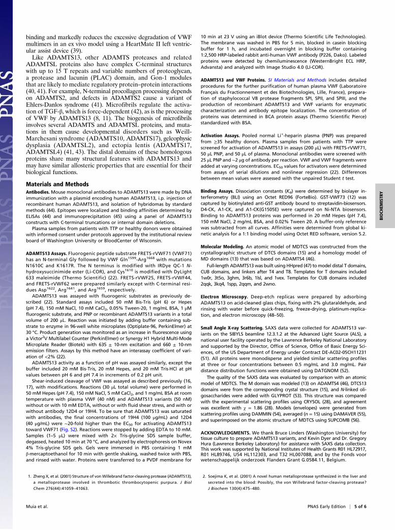
ADAMTS13 Distal Domains Are Adjacent to Its Proximal Domains. Theregulation of ADAMTS13 metalloprotease activity by its distalT-CUB domains implies that proximal and distal domains areclose enough to make contact, and small angle X-ray scattering(SAXS) data (Fig. 4A) indicate that ADAMTS13 does have sucha structure. Pair distance distributions (Fig. 4B) show thatMDTCS, MT8, and ADAMTS13 have maximum dimensions of∼140 Å, ∼200 Å, and ∼230 Å, respectively. For comparison,a molecular model of ADAMTS13 has a maximum extendedlength of ∼500 Å (Fig. 4C). Agreement was excellent between anatomic model for MDTCS and experimental scattering profiles(χ = 1.86) (28) (Fig. 4A) or ab initio envelopes (Fig. 4D). Theenvelopes for full-length ADAMTS13 (Fig. 4E) are too compactfor fully extended ADAMTS13 but readily accommodate a folded
or condensed conformation. Specific flexible segments have notbeen identified, but the extended linkers after T4 and T8 aresusceptible to proteolysis by thrombin, plasmin, or elastase (29,30) and may act as hinges. Comparisons of SAXS envelopes forADAMTS13 and construct MT8, which lacks both CUBdomains, indicate that the CUB domains are adjacent to proxi-mal ADAMTS13 domains and occupy a bulge in the envelopefor ADAMTS13 (Fig. 4E) that is missing from the envelope forMT8, which cannot accommodate the CUB domains in this lo-cation (Fig. 4F). This folded conformation would enable distalT7-CUB domains to allosterically regulate ADAMTS13 by var-iably occluding the active site and substrate binding exosites. Theoccurrence of condensed ADAMTS13 is supported by quick-freeze deep-etch EM, which shows both elongated and foldedconformations for ADAMTS13 (Fig. 4G).
DiscussionOur results indicate that VWF promotes its own destruction byallosterically activating ADAMTS13, and this mechanism couldhave evolved to balance the competing needs to promote VWF-dependent hemostasis and prevent VWF-dependent thrombosis.In addition, the finding that VWF activates ADAMTS13 raises
Fig. 2. Allosteric activation of ADAMTS13. (A) Monoclonal anti-ADAMTS13 antibodies were added to ADAMTS13 assays with pooled normal plasma (PNP).Reaction rates were normalized to the rate with PNP only. The location of epitopes is indicated. Activation values >1.5× control (orange bars) are significantlydifferent from the PNP reference condition (P < 0.05). (B) Recombinant ADAMTS13 and MT8 were immunoprecipitated with antibodies in PNP or BCW49plasma, and the recovered enzyme was assayed with substrate VWF71. (C and D) MDTCS (blue bars) and ADAMTS13 (orange bars) were assayed with theindicated fluorogenic substrates (Fig. 1B). (C) Bars indicate the ratio of reaction rates at pH 6 and pH 7.4 (Left), or the ratio of reaction rates with and withoutactivating monoclonal antibodies 7C4, 19H4, and 12D4 (Right). (D) Plasma VWF multimers (0.45 μM) or VWF D4 monomers (40 μM) increased the activity ofADAMTS13 toward VWF71. SPI (2 μM), SPII (2 μM), and SPIII (0.54 μM) fragments of VWF, which are cleaved within the D4 domain by V8 protease, had little orno effect on ADAMTS13 activity. Error bars indicate 95% confidence intervals.
Muia et al. PNAS Early Edition | 3 of 6
BIOCH
EMISTR
Y
the possibility that other ADAMTS13 cofactors occur on cellsurfaces, in blood, or in extracellular matrix.For human ADAMTS13, binding to VWF D4 allosterically
activates ADAMTS13, increasing its catalytic activity, but binding toVWF D4 also positions ADAMTS13 on the substrate where it canact rapidly when shear stress exposes the scissile bond in the adja-cent A2 domain. These two mechanisms work together to focus pro-teolytic activity on VWF multimers. However, mouse ADAMTS13may rely mainly on substrate binding rather than allostery.Some mouse strains express a “long” Adamts13 that is similar to
full-length human ADAMTS13. Other strains have an intracisternalA-particle (IAP) retrotransposon inserted in the Adamts13 geneand express a “short” Adamts13 that is truncated after the T6 (31).Long Adamts13 cleaves VWF multimers ninefold faster than shortAdamts13, which is consistent with the role we propose for bindingof distal domains to VWF D4 (19). However, long and shortAdamts13 reportedly have similar activity toward small substratesthat are similar to VWF71 (19, 31). Therefore, in contrast to humanADAMTS13, deletion of the distal T7-CUB2 domains may notactivate mouse Adamts13, which suggests that allosteric activationis less significant than for human ADAMTS13 or depends ondomains other than T7-CUB2. Further study is required to de-termine the relative importance of substrate binding and allostericactivation for ADAMTS13 in humans, mice, and other vertebrates.The allosteric properties of ADAMTS13 are likely to have
clinical relevance. Relatively modest decreases in ADAMTS13activity seem to increase the risk of ischemic stroke and myo-cardial infarction (32–34). In animal models, ADAMTS13 de-ficiency promotes vascular inflammation and atherosclerosis (35).Therefore, inherited or acquired defects in the allosteric regulation
of ADAMTS13 could inhibit VWF cleavage and contribute tothrombosis in these conditions.Conversely, agents that block allosteric activation could be
useful to treat bleeding caused by excessive proteolysis of VWF,which occurs mainly in two settings. Von Willebrand disease type2A frequently is due to mutations like G1505E (27) that impairthe folding of the VWF A2 domain and allow cleavage byADAMTS13 even in the absence of fluid shear stress. As a result,the VWF multimers that circulate in the blood are too small foreffective hemostasis (36, 37). Acquired von Willebrand syndromealso is characterized by deficiency of the largest VWF multimers,often as a result of pathologically increased fluid shear stress thatpromotes cleavage by ADAMTS13. For example, this type ofacquired von Willebrand syndrome is very common in patientswith implanted left ventricular assist devices, who experience re-current major gastrointestinal bleeding as a result (38). BecauseADAMTS13 can be allosteric activated by ∼fourfold to ∼10-fold, aninhibitor of allosteric activation could reduce ADAMTS13 activity tono lower than ∼10%, which is not low enough to cause TTP (6, 7)but may be useful to treat bleeding. The feasibility of this ap-proach is supported by a study demonstrating that a monoclo-nal antibody against the VWF D4 domain inhibits ADAMTS13
Fig. 3. Cleavage of VWF by ADAMTS13 under fluid shear stress. VWF cleavageproducts were detected by SDS/PAGE and immunoblotting with anti-VWF. Thearrowheads indicate the 350-kDa VWF fragment produced by ADAMTS13.Plasma VWF (40 nM) was sheared for the indicated times (seconds) with (A)MDTCS or ADAMTS13 (50 nM), or (B) with ADAMTS13 (50 nM) without or withmonoclonal antibody 12D4 (40 μg/mL). (C) Densitometric analysis to compare therate of VWF cleavage by ADAMTS13 without (orange squares) or with (greentriangles) monoclonal antibody 12D4, or by MDTCS (blue circles). Values arenormalized to cleavage at 300 s by ADAMTS13 alone and plotted as the mean ±SE for twoexperiments. (D) Duplicate reactionswithADAMTS13were sheared for300 swithout orwithmonoclonal antibody 19H4 (100 μg/mL) or 12D4 (40 μg/mL).Control reactions were sheared for 300 s in the presence of 10 mM EDTA.
Fig. 4. ADAMTS13 structure by EM and SAXS. Scattering profiles (A) and pairdistribution functions (B) for MDTCS, (red) MT8 (orange), and ADAMTS13(blue). (C) Molecular model of ADAMTS13. (D–F) Ab initio envelopes calcu-lated from scattering profiles for (D) MDTCS (red mesh), (E) ADAMTS13 (bluemesh), or (F ) MT8 (orange mesh) superimposed on an atomic model ofMDTCS (D, cartoon; E and F, surface), MT8, or ADAMTS13 (E and F, sur-face). The arrow indicates a difference between MT8 and ADAMTS13maps that corresponds to the proposed location of CUB domains. (G)Quick-freeze deep-etch EM of ADAMTS13.
4 of 6 | www.pnas.org/cgi/doi/10.1073/pnas.1413282112 Muia et al.
binding and markedly reduces the excessive degradation of VWFmultimers in an ex vivo model using a HeartMate II left ventric-ular assist device (39).Like ADAMTS13, other ADAMTS proteases and related
ADAMTSL proteins also have complex C-terminal structureswith up to 15 T repeats and variable numbers of proteoglycan,a protease and lacunin (PLAC) domain, and Gon-1 modulesthat are likely to mediate regulatory protein–protein interactions(40, 41). For example, N-terminal procollagen processing dependson ADAMTS2, and defects in ADAMTS2 cause a variant ofEhlers-Danlos syndrome (41). Microfibrils regulate the activa-tion of TGF-β, which is force-dependent (42), as is the processingof VWF by ADAMTS13 (8, 11). The biogenesis of microfibrilsinvolves several ADAMTS and ADAMTSL proteins, and muta-tions in them cause developmental disorders such as Weill-Marchesani syndrome (ADAMTS10, ADAMTS17), geleophysicdysplasia (ADAMTSL2), and ectopia lentis (ADAMTS17,ADAMTSL4) (41, 43). The distal domains of these homologousproteins share many structural features with ADAMTS13 andmay have similar allosteric properties that are essential for theirbiological functions.
Materials and MethodsAntibodies. Mouse monoclonal antibodies to ADAMTS13 were made by DNAimmunization with a plasmid encoding human ADAMTS13, i.p. injection ofrecombinant human ADAMTS13, and isolation of hybridomas by standardmethods (44). Epitopes were localized and binding affinities determined byELISAs (44) and immunoprecipitation (45) using a panel of ADAMTS13constructs with C-terminal truncations or internal domain deletions.
Plasma samples from patients with TTP or healthy donors were obtainedwith informed consent under protocols approved by the institutional reviewboard of Washington University or BloodCenter of Wisconsin.
ADAMTS13 Assays. Fluorogenic peptide substrate FRETS-rVWF71 (VWF71)has an N-terminal Gly followed by VWF Gln1599-Arg1668 with mutationsN1610C and K1617R. The N terminus is modified with IRDye QC-1 N-hydroxysuccinimide ester (Li-COR), and Cys1610 is modified with DyLight633 maleimide (Thermo Scientific) (22). FRETS-rVWF25, FRETS-rVWF44,and FRETS-rVWF62 were prepared similarly except with C-terminal resi-dues Asp1622, Arg1641, and Arg1659, respectively.
ADAMTS13 was assayed with fluorogenic substrates as previously de-scribed (22). Standard assays included 50 mM Bis-Tris (pH 6) or Hepes(pH 7.4), 150 mM NaCl, 10 mM CaCl2, 0.05% Tween-20, 1 mg/mL BSA, 1 μMfluorogenic substrate, and PNP or recombinant ADAMTS13 variants in a totalvolume of 200 μL. Reaction was initiated by adding buffer containing sub-strate to enzyme in 96-well white microplates (Optiplate-96, PerkinElmer) at30 °C. Product generation was monitored as an increase in fluorescence usinga Victor2V Multilabel Counter (PerkinElmer) or Synergy H1 Hybrid Multi-ModeMicroplate Reader (Biotek) with 635 ± 10-nm excitation and 660 ± 10-nmemission filters. Assays by this method have an interassay coefficient of vari-ation of <2% (22).
ADAMTS13 activity as a function of pH was assayed similarly, except thebuffer included 20 mM Bis-Tris, 20 mM Hepes, and 20 mM Tris·HCl at pHvalues between pH 6 and pH 7.4 in increments of 0.2 pH unit.
Shear-induced cleavage of VWF was assayed as described previously (16,17), with modifications. Reactions (30 μL total volume) were performed in50 mM Hepes (pH 7.4), 150 mM NaCl, 5 mM CaCl2, and 1 mg/mL BSA at roomtemperature with plasma VWF (40 nM) and ADAMTS13 variants (50 nM)without or with 10 mM EDTA, without or with fluid shear stress, and with orwithout antibody 12D4 or 19H4. To be sure that ADAMTS13 was saturatedwith antibodies, the final concentrations of 19H4 (100 μg/mL) and 12D4(40 μg/mL) were ∼20-fold higher than the EC50 for activating ADAMTS13toward VWF71 (Fig. S2). Reactions were stopped by adding EDTA to 10 mM.Samples (1–5 μL) were mixed with 2× Tris-glycine SDS sample buffer,degassed, heated 10 min at 70 °C, and analyzed by electrophoresis on Novex4% Tris-glycine SDS gels. Gels were immersed in PBS containing 1 mMβ-mercaptoethanol for 10 min with gentle shaking, washed twice with PBS,and rinsed with water. Proteins were transferred to a PVDF membrane for
10 min at 23 V using an iBlot device (Thermo Scientific Life Technologies).The membrane was washed in PBS for 5 min, blocked in casein blockingbuffer for 1 h, and incubated overnight in blocking buffer containing1:2,500 HRP-labeled rabbit anti-human VWF antibody (P226, Dako). Labeledproteins were detected by chemiluminescence (WesternBright ECL HRP,Advansta) and analyzed with Image Studio 4.0 (LI-COR).
ADAMTS13 and VWF Proteins. SI Materials and Methods includes detailedprocedures for the further purification of human plasma VWF (LaboratoireFrançais du Fractionnement et des Biotechnologies, Lille, France), prepara-tion of staphylococcal V8 protease fragments SPI, SPII, and SPIII, and theproduction of recombinant ADAMTS13 and VWF variants for enzymaticcharacterization and antibody epitope localization. The concentration ofproteins was determined in BCA protein assays (Thermo Scientific Pierce)standardized with BSA.
Activation Assays. Pooled normal Li+-heparin plasma (PNP) was preparedfrom ≥35 healthy donors. Plasma samples from patients with TTP werescreened for activation of ADAMTS13 in assays (200 μL) with FRETS-rVWF71,50 μL PNP, and 50 μL of plasma. Monoclonal antibodies were screened with25 μL PNP and ∼2 μg of antibody per reaction. VWF and VWF fragments wereadded at varying concentrations. EC50 values for activators were determinedfrom assays of serial dilutions and nonlinear regression (22). Differencesbetween mean values were assessed with the unpaired Student t test.
Binding Assays. Dissociation constants (Kd) were determined by biolayer in-terferometry (BLI) using an Octet RED96 (ForteBio). GST-VWF73 (12) wascaptured by biotinylated anti-GST antibody bound to streptavidin-biosensors.D4-CK, A1-CK, and A1-CK(G1505E) were captured on Ni-NTA biosensors.Binding to ADAMTS13 proteins was performed in 20 mM Hepes (pH 7.4),150 mM NaCl, 2 mg/mL BSA, and 0.02% Tween 20. A buffer-only referencewas subtracted from all curves. Affinities were determined from global ki-netic analysis for a 1:1 binding model using Octet RED software, version 5.2.
Molecular Modeling. An atomic model of MDTCS was constructed from thecrystallographic structure of DTCS domains (15) and a homology model ofMD domains (13) that was based on ADAMTS4 (46).
Full-length ADAMTS13was built using HHpred (47) tomodel distal T domains,CUB domains, and linkers after T4 and T8. Templates for T domains included1w0r, 3t5o, 3ghm, 3r6b, 1lsl, and 1vex. Templates for CUB domains included2qqk, 3kq4, 1spp, 2qqm, and 2wno.
Electron Microscopy. Deep-etch replicas were prepared by adsorbingADAMTS13 on acid-cleaned glass chips, fixing with 2% glutaraldehyde, andrinsing with water before quick-freezing, freeze-drying, platinum-replica-tion, and electron microscopy (48–50).
Small Angle X-ray Scattering. SAXS data were collected for ADAMTS13 var-iants on the SIBYLS beamline 12.3.1.2 at the Advanced Light Source (ALS), anational user facility operated by the Lawrence Berkeley National Laboratoryand supported by the Director, Office of Science, Office of Basic Energy Sci-ences, of the US Department of Energy under Contract DE-AC02-05CH11231(51). All proteins were monodisperse and yielded similar scattering profilesat three or four concentrations between 0.5 mg/mL and 3.0 mg/mL. Pairdistance distribution functions were obtained using DATGNOM (52).
The quality of the SAXS data was evaluated by comparison with an atomicmodel of MDTCS. The M domain was modeled (13) on ADAMTS4 (46), DTCS13domains were from the corresponding crystal structure (15), and N-linked oli-gosaccharides were added with GLYPROT (53). This structure was comparedwith the experimental scattering profiles using CRYSOL (28), and agreementwas excellent with χ = 1.86 (28). Models (envelopes) were generated fromscattering profiles using DAMMIN (54), averaged (n = 15) using DAMAVER (55),and superimposed on the atomic structure of MDTCS using SUPCOMB (56).
ACKNOWLEDGMENTS. We thank Bruce Linders (Washington University) fortissue culture to prepare ADAMTS13 variants, and Kevin Dyer and Dr. GregoryHura (Lawrence Berkeley Laboratory) for assistance with SAXS data collection.This work was supported by National Institutes of Health Grants R01 HL72917,R01 HL89746, U54 HL112303, and T32 HL007088, and by the Fonds voorwetenschappelijk onderzoek Flanders Grant G.0584.11, Belgium.
1. Zheng X, et al. (2001) Structure of vonWillebrand factor-cleaving protease (ADAMTS13),
a metalloprotease involved in thrombotic thrombocytopenic purpura. J Biol
Chem 276(44):41059–41063.
2. Soejima K, et al. (2001) A novel human metalloprotease synthesized in the liver and
secreted into the blood: Possibly, the von Willebrand factor-cleaving protease?
J Biochem 130(4):475–480.
Muia et al. PNAS Early Edition | 5 of 6
BIOCH
EMISTR
Y
3. Levy GG, et al. (2001) Mutations in a member of the ADAMTS gene family causethrombotic thrombocytopenic purpura. Nature 413(6855):488–494.
4. Fujikawa K, Suzuki H, McMullen B, Chung D (2001) Purification of human vonWillebrand factor-cleaving protease and its identification as a new member of themetalloproteinase family. Blood 98(6):1662–1666.
5. Gerritsen HE, Robles R, Lämmle B, Furlan M (2001) Partial amino acid sequence ofpurified von Willebrand factor-cleaving protease. Blood 98(6):1654–1661.
6. Furlan M, et al. (1998) von Willebrand factor-cleaving protease in thromboticthrombocytopenic purpura and the hemolytic-uremic syndrome. N Engl J Med339(22):1578–1584.
7. Tsai HM, Lian EC (1998) Antibodies to von Willebrand factor-cleaving protease inacute thrombotic thrombocytopenic purpura. N Engl J Med 339(22):1585–1594.
8. Tsai HM (1996) Physiologic cleavage of von Willebrand factor by a plasma protease isdependent on its conformation and requires calcium ion. Blood 87(10):4235–4244.
9. Majerus EM, Anderson PJ, Sadler JE (2005) Binding of ADAMTS13 to von Willebrandfactor. J Biol Chem 280(23):21773–21778.
10. Furlan M, Robles R, Lämmle B (1996) Partial purification and characterization ofa protease from human plasma cleaving von Willebrand factor to fragments pro-duced by in vivo proteolysis. Blood 87(10):4223–4234.
11. Zhang Q, et al. (2009) Structural specializations of A2, a force-sensing domain in theultralarge vascular protein von Willebrand factor. Proc Natl Acad Sci USA 106(23):9226–9231.
12. Gao W, Anderson PJ, Majerus EM, Tuley EA, Sadler JE (2006) Exosite interactionscontribute to tension-induced cleavage of von Willebrand factor by the antith-rombotic ADAMTS13 metalloprotease. Proc Natl Acad Sci USA 103(50):19099–19104.
13. Gao W, Anderson PJ, Sadler JE (2008) Extensive contacts between ADAMTS13 exositesand von Willebrand factor domain A2 contribute to substrate specificity. Blood112(5):1713–1719.
14. Zanardelli S, et al. (2006) ADAMTS13 substrate recognition of von Willebrand factorA2 domain. J Biol Chem 281(3):1555–1563.
15. Akiyama M, Takeda S, Kokame K, Takagi J, Miyata T (2009) Crystal structures of thenoncatalytic domains of ADAMTS13 reveal multiple discontinuous exosites for vonWillebrand factor. Proc Natl Acad Sci USA 106(46):19274–19279.
16. Zhang P, Pan W, Rux AH, Sachais BS, Zheng XL (2007) The cooperative activity be-tween the carboxyl-terminal TSP1 repeats and the CUB domains of ADAMTS13 iscrucial for recognition of von Willebrand factor under flow. Blood 110(6):1887–1894.
17. Feys HB, Anderson PJ, Vanhoorelbeke K, Majerus EM, Sadler JE (2009) Multi-stepbinding of ADAMTS-13 to vonWillebrand factor. J Thromb Haemost 7(12):2088–2095.
18. Zanardelli S, et al. (2009) A novel binding site for ADAMTS13 constitutively exposedon the surface of globular VWF. Blood 114(13):2819–2828.
19. ZhouW, Bouhassira EE, Tsai HM (2007) An IAP retrotransposon in themouse ADAMTS13gene creates ADAMTS13 variant proteins that are less effective in cleaving von Wille-brand factor multimers. Blood 110(3):886–893.
20. Banno F, et al. (2009) The distal carboxyl-terminal domains of ADAMTS13 are re-quired for regulation of in vivo thrombus formation. Blood 113(21):5323–5329.
21. Anderson PJ, Kokame K, Sadler JE (2006) Zinc and calcium ions cooperatively mod-ulate ADAMTS13 activity. J Biol Chem 281(2):850–857.
22. Muia J, et al. (2013) An optimized fluorogenic ADAMTS13 assay with increased sen-sitivity for the investigation of patients with thrombotic thrombocytopenic purpura.J Thromb Haemost 11(8):1511–1518.
23. Kokame K, Nobe Y, Kokubo Y, Okayama A, Miyata T (2005) FRETS-VWF73, a firstfluorogenic substrate for ADAMTS13 assay. Br J Haematol 129(1):93–100.
24. Di Stasio E, et al. (2008) Mechanistic studies on ADAMTS13 catalysis. Biophys J 95(5):2450–2461.
25. Wells JA (1990) Additivity of mutational effects in proteins. Biochemistry 29(37):8509–8517.
26. Girma JP, Chopek MW, Titani K, Davie EW (1986) Limited proteolysis of human vonWillebrand factor by Staphylococcus aureus V-8 protease: Isolation and partial char-acterization of a platelet-binding domain. Biochemistry 25(11):3156–3163.
27. Hassenpflug WA, et al. (2006) Impact of mutations in the von Willebrand factor A2domain on ADAMTS13-dependent proteolysis. Blood 107(6):2339–2345.
28. Svergun DI, Barberato C, Koch MHJ (1995) CRYSOL—a program to evaluate x-raysolution scattering of biological macromolecules from atomic coordinates. J ApplCryst 28:768–773.
29. Crawley JT, et al. (2005) Proteolytic inactivation of ADAMTS13 by thrombin andplasmin. Blood 105(3):1085–1093.
30. Hiura H, et al. (2010) Proteolytic fragmentation and sugar chains of plasma ADAMTS13purified by a conformation-dependent monoclonal antibody. J Biochem 148(4):403–411.
31. Banno F, Kaminaka K, Soejima K, Kokame K, Miyata T (2004) Identification of strain-specific variants of mouse Adamts13 gene encoding von Willebrand factor-cleavingprotease. J Biol Chem 279(29):30896–30903.
32. Crawley JT, Lane DA, Woodward M, Rumley A, Lowe GD (2008) Evidence that highvon Willebrand factor and low ADAMTS-13 levels independently increase the risk ofa non-fatal heart attack. J Thromb Haemost 6(4):583–588.
33. Bongers TN, et al. (2009) Lower levels of ADAMTS13 are associated with cardiovas-cular disease in young patients. Atherosclerosis 207(1):250–254.
34. Andersson HM, et al. (2012) High VWF, low ADAMTS13, and oral contraceptives in-crease the risk of ischemic stroke and myocardial infarction in young women. Blood119(6):1555–1560.
35. Gandhi C, Khan MM, Lentz SR, Chauhan AK (2012) ADAMTS13 reduces vascular in-flammation and the development of early atherosclerosis in mice. Blood 119(10):2385–2391.
36. Ginsburg D, Bowie EJW (1992) Molecular genetics of von Willebrand disease. Blood79(10):2507–2519.
37. Nichols WL, et al. (2008) von Willebrand disease (VWD): Evidence-based diagnosis andmanagement guidelines, the National Heart, Lung, and Blood Institute (NHLBI) ExpertPanel report (USA). Haemophilia 14(2):171–232.
38. Suarez J, et al. (2011) Mechanisms of bleeding and approach to patients with axial-flow left ventricular assist devices. Circ Heart Fail 4(6):779–784.
39. Rauch A, et al. (2014) Antibody-based prevention of von Willebrand factor degra-dation mediated by circulatory assist devices. Thromb Haemost 112(5):1014–1023.
40. Apte SS (2009) A disintegrin-like and metalloprotease (reprolysin-type) with throm-bospondin type 1 motif (ADAMTS) superfamily: functions and mechanisms. J BiolChem 284(46):31493–31497.
41. Le Goff C, Cormier-Daire V (2011) The ADAMTS(L) family and human genetic dis-orders. Hum Mol Genet 20(R2):R163–R167.
42. Shi M, et al. (2011) Latent TGF-β structure and activation. Nature 474(7351):343–349.43. Hubmacher D, Apte SS (2011) Genetic and functional linkage between ADAMTS su-
perfamily proteins and fibrillin-1: A novel mechanism influencing microfibril assemblyand function. Cell Mol Life Sci 68(19):3137–3148.
44. Feys HB, et al. (2006) ADAMTS-13 plasma level determination uncovers antigen ab-sence in acquired thrombotic thrombocytopenic purpura and ethnic differences.J Thromb Haemost 4(5):955–962.
45. Feys HB, et al. (2010) Thrombotic thrombocytopenic purpura directly linked withADAMTS13 inhibition in the baboon (Papio ursinus). Blood 116(12):2005–2010.
46. Mosyak L, et al. (2008) Crystal structures of the two major aggrecan degrading en-zymes, ADAMTS4 and ADAMTS5. Protein Sci 17(1):16–21.
47. Söding J, Biegert A, Lupas AN (2005) The HHpred interactive server for protein ho-mology detection and structure prediction. Nucleic Acids Res 33(Web Server issue):W244-8.
48. Heuser JE, et al. (1979) Synaptic vesicle exocytosis captured by quick freezing andcorrelated with quantal transmitter release. J Cell Biol 81(2):275–300.
49. Heuser JE (1983) Procedure for freeze-drying molecules adsorbed to mica flakes. J MolBiol 169(1):155–195.
50. Heuser J (1989) Protocol for 3-D visualization of molecules on mica via the quick-freeze, deep-etch technique. J Electron Microsc Tech 13(3):244–263.
51. Hura GL, et al. (2009) Robust, high-throughput solution structural analyses by smallangle X-ray scattering (SAXS). Nat Methods 6(8):606–612.
52. Svergun DI (1992) Determination of the regularization parameter in indirect-trans-form methods using perceptual criteria. J Appl Cryst 25(4):495–503.
53. Bohne-Lang A, von der Lieth CW (2005) GlyProt: In silico glycosylation of proteins.Nucleic Acids Res 33(Web Server issue):W214-9.
54. Svergun DI (1999) Restoring low resolution structure of biological macromoleculesfrom solution scattering using simulated annealing. Biophys J 76(6):2879–2886.
55. Volkov VV, Svergun DI (2003) Uniqueness of ab initio shape determination in small-angle scattering. J Appl Cryst 36(3-1):860–864.
56. Kozin MB, Svergun DI (2001) Automated matching of high- and low-resolution struc-tural models. J Appl Cryst 34(1):33–41.
6 of 6 | www.pnas.org/cgi/doi/10.1073/pnas.1413282112 Muia et al.
Supporting InformationMuia et al. 10.1073/pnas.1413282112SI Materials and MethodsADAMTS13 and VWF Proteins. Recombinant ADAMTS13 (1) wasobtained from Baxter Innovations. Purified human plasma VWF(Laboratoire Français du Fractionnement et des Biotechnologies,Lille, France) was chromatographed on a Superdex 200 equili-brated with 50 mM Hepes (pH 7.4), 150 mM NaCl, 5 mM CaCl2,and 1 μM ZnCl2 to remove traces of human serum albumin.Trace amounts of residual ADAMTS13 were removed by ad-sorption on monoclonal antibody 3H9-agarose.VWF fragments SPI, SPII, and SPIII were prepared by digestion
of plasma VWF with Staphylococcal V8 protease (Thermo Sci-entific Pierce) and purified by ion exchange chromatography (2).Human cDNA constructs encoding VWF domain D4
(Ser1873-Thr2255) (3), full-length mature ADAMTS13 (4), andADAMTS13 truncated after domain S (Ala685, MDTCS), T7(Arg1075, M-T7), or T8 (Ala1191, M-T8), with or without the mu-tation E225Q, were cloned into pTriEx-7 Ek/LIC (EMD Milli-pore). These constructs encode the following (underlined) mouseIgM signal peptide, StrepTag II, and enteropeptidase recognitionsequence followed by Ser1873 of VWF or Ala75 of ADAMTS13:MKFSWVMFFLMAVVTGVNSEVQASWSHPQFEKGAD-
DDDKMProteins were expressed in stably transfected T-REx 293 cells
(Invitrogen) grown in Freestyle serum-free medium containing1 μg/mL tetracycline (5). Conditioned medium was adsorbed onQ-Sepharose, and proteins were eluted with 25 mM Tris·HCl(pH 8.0) and 1 M NaCl. Pooled fractions containing VWF D4 orADAMTS13 variants were desalted on PD-10 equilibrated with100 mM Tris·HCl (pH 8.0) and 150 mM NaCl, adsorbed onStrepTactin-agarose, and eluted with buffer containing 2.5 mMdesthiobiotin.
ConstructD4-CK in vector pcDNA3.1 (Invitrogen) encodesVWFresidues Met1-Cys22 and Gly1874-Lys2813 followed by a 6xHis tag.Constructs A1-CK in vector pSV7D (6) and A1-CK(G1505E)in vector pcDNA3.1 encode VWF residues Met1-Cys22 andGlu1260-Lys2813 followed by a 6xHis tag. These three VWFconstructs contain the mutation C2773A to prevent C-terminaldimerization (7). A1-CK(G1505E) and D4-CK were expressed instably transfected HEK293 cells. A1-CK was expressed in tran-siently transfected BHK cells. Conditioned Freestyle serum-freemedium was adsorbed on Q-Sepharose, and proteins were elutedwith 20 mM Hepes (pH 7.4) and 1 M NaCl. Appropriate pooledfractions were dialyzed against 20 mMHepes (pH 7.4) and 150 mMNaCl. Imidazole (20 mM) was added, and proteins were ad-sorbed on HisPur Cobalt agarose (Thermo Scientific Pierce).After washing with 20 mM Hepes (pH 7.4), 150 mM NaCl, and20 mM imidazole, proteins were eluted with buffer containing300 mM imidazole.All proteins were further purified by chromatography on a
TSK-G2000SW or Superdex 200 and stored at −80 °C. Theconcentration of proteins was determined in BCA protein assays(Thermo Scientific Pierce) standardized with BSA.For epitope localization, ADAMTS13 constructs were cloned in
pcDNA4/TO with C-terminal V5 and (His)6 tags and expressedtransiently in T-REx 293 cells (8). C-terminal deletions were trun-cated after domain M (Gln289), D (Gly385), T1 (Glu439), C (Cys555),S (Ala685), T2 (Tyr745), T3 (Arg807), T7 (Arg1075), T8 (Ala1191),or CUB1 (Ala1291). Internal deletions lacked domain T2 (Trp686-Tyr745), T3 (Trp746-Arg807), T4 (Trp808-Ala894), T5 (His895-Pro952),T6 (Ala953-Arg1015), T7 (Trp1016-Arg1075), T8 (Trp1076-Ala1191), orCUB1 (Cys1192-Glu1298).
1. Plaimauer B, et al. (2002) Cloning, expression, and functional characterization of thevon Willebrand factor-cleaving protease (ADAMTS13). Blood 100(10):3626–3632.
2. Titani K, et al. (1986) Amino acid sequence of human von Willebrand factor. Bio-chemistry 25(11):3171–3184.
3. Matsushita T, Sadler JE (1995) Identification of amino acid residues essential for vonWillebrand factor binding to platelet glycoprotein Ib. Charged-to-alanine scanningmutagenesis of the A1 domain of human von Willebrand factor. J Biol Chem 270(22):13406–13414.
4. Zheng X, Nishio K, Majerus EM, Sadler JE (2003) Cleavage of von Willebrand factorrequires the spacer domain of the metalloprotease ADAMTS13. J Biol Chem 278(32):30136–30141.
5. Gao W, et al. (2012) Rearranging exosites in noncatalytic domains can redirect thesubstrate specificity of ADAMTS proteases. J Biol Chem 287(32):26944–26952.
6. Burke RL, et al. (1986) The functional domains of coagulation factor VIII:C. J Biol Chem261(27):12574–12578.
7. Katsumi A, Tuley EA, Bodó I, Sadler JE (2000) Localization of disulfide bonds in thecystine knot domain of human von Willebrand factor. J Biol Chem 275(33):25585–25594.
8. Majerus EM, Anderson PJ, Sadler JE (2005) Binding of ADAMTS13 to von Willebrandfactor. J Biol Chem 280(23):21773–21778.
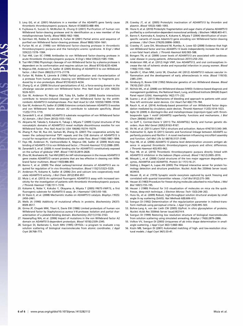
Fig. S1. Reaction time course for MDTCS and ADAMTS13 as a function of pH. Enzymes MDTCS and ADAMTS13 were assayed with substrate VWF71 at theindicated pH values.
Muia et al. www.pnas.org/cgi/content/short/1413282112 1 of 3
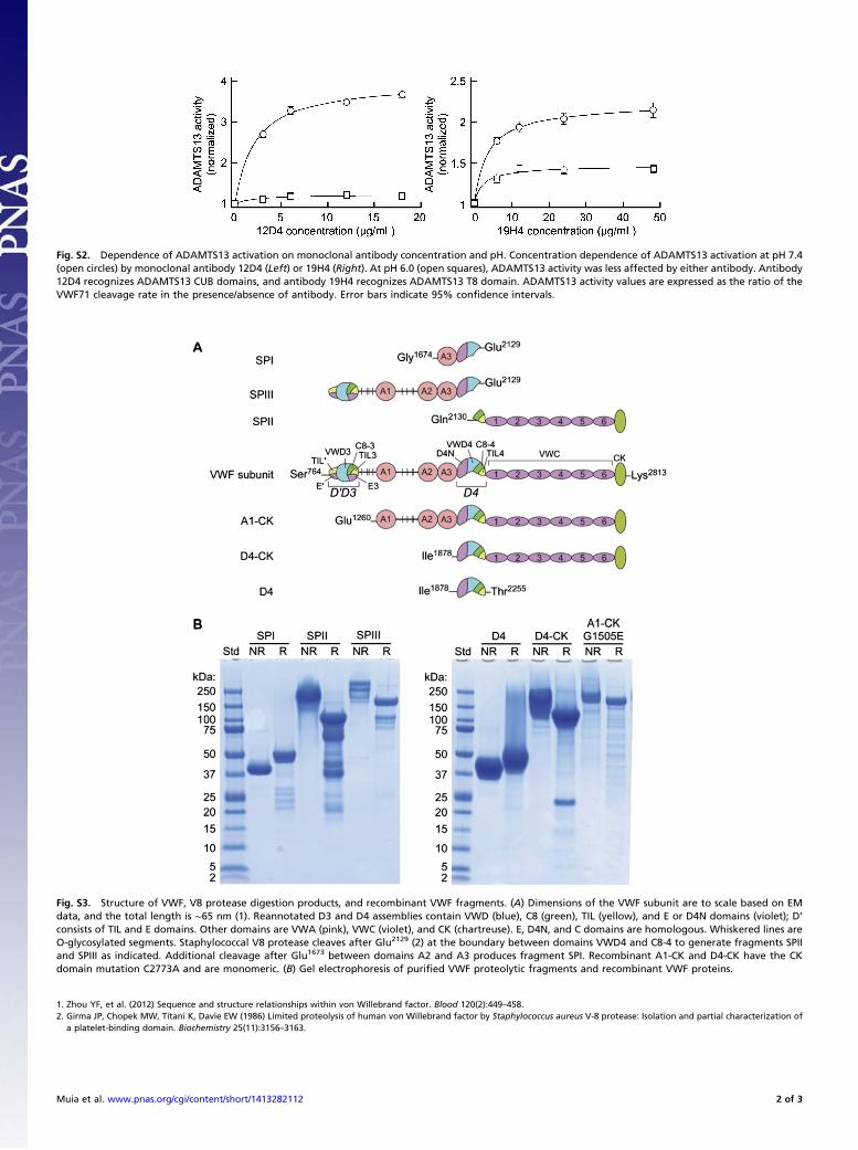
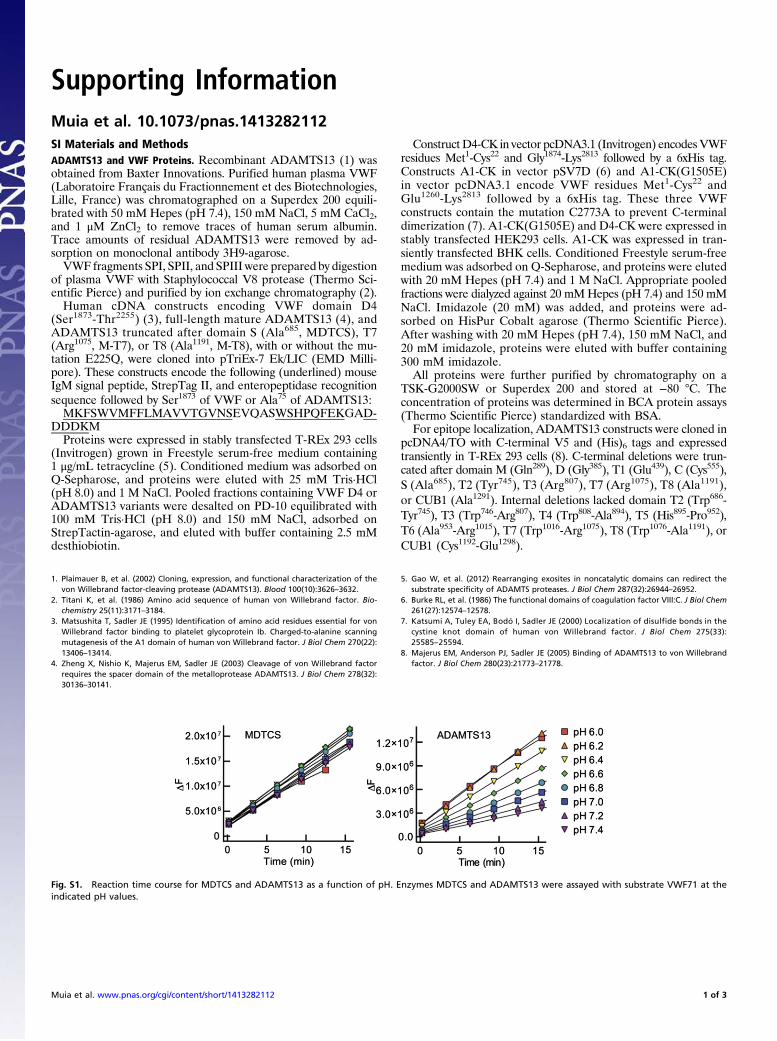
Fig. S2. Dependence of ADAMTS13 activation on monoclonal antibody concentration and pH. Concentration dependence of ADAMTS13 activation at pH 7.4(open circles) by monoclonal antibody 12D4 (Left) or 19H4 (Right). At pH 6.0 (open squares), ADAMTS13 activity was less affected by either antibody. Antibody12D4 recognizes ADAMTS13 CUB domains, and antibody 19H4 recognizes ADAMTS13 T8 domain. ADAMTS13 activity values are expressed as the ratio of theVWF71 cleavage rate in the presence/absence of antibody. Error bars indicate 95% confidence intervals.
Fig. S3. Structure of VWF, V8 protease digestion products, and recombinant VWF fragments. (A) Dimensions of the VWF subunit are to scale based on EMdata, and the total length is ∼65 nm (1). Reannotated D3 and D4 assemblies contain VWD (blue), C8 (green), TIL (yellow), and E or D4N domains (violet); D’consists of TIL and E domains. Other domains are VWA (pink), VWC (violet), and CK (chartreuse). E, D4N, and C domains are homologous. Whiskered lines areO-glycosylated segments. Staphylococcal V8 protease cleaves after Glu2129 (2) at the boundary between domains VWD4 and C8-4 to generate fragments SPIIand SPIII as indicated. Additional cleavage after Glu1673 between domains A2 and A3 produces fragment SPI. Recombinant A1-CK and D4-CK have the CKdomain mutation C2773A and are monomeric. (B) Gel electrophoresis of purified VWF proteolytic fragments and recombinant VWF proteins.
1. Zhou YF, et al. (2012) Sequence and structure relationships within von Willebrand factor. Blood 120(2):449–458.2. Girma JP, Chopek MW, Titani K, Davie EW (1986) Limited proteolysis of human von Willebrand factor by Staphylococcus aureus V-8 protease: Isolation and partial characterization of
a platelet-binding domain. Biochemistry 25(11):3156–3163.
Muia et al. www.pnas.org/cgi/content/short/1413282112 2 of 3
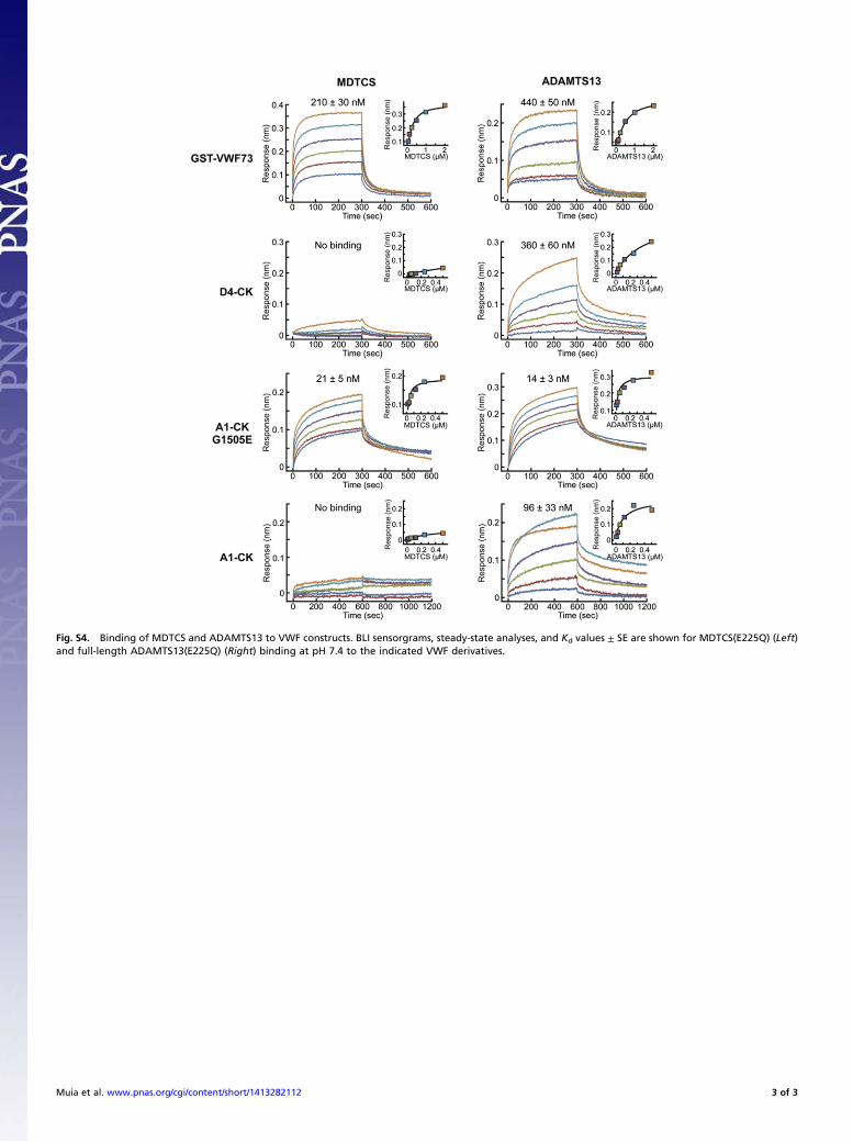
Fig. S4. Binding of MDTCS and ADAMTS13 to VWF constructs. BLI sensorgrams, steady-state analyses, and Kd values ± SE are shown for MDTCS(E225Q) (Left)and full-length ADAMTS13(E225Q) (Right) binding at pH 7.4 to the indicated VWF derivatives.
Muia et al. www.pnas.org/cgi/content/short/1413282112 3 of 3