vocal-cord only vs. complete laryngeal radiation (vocal)
TRANSCRIPT
STUDY PROTOCOL Open Access
Vocal-cord Only vs. Complete Laryngealradiation (VOCAL): a randomizedmulticentric Bayesian phase II trialHouda Bahig1*, David I. Rosenthal2*, Félix-Phuc Nguyen-Tan1, David C. Fuller2, Ying Yuan3,Katherine A. Hutcheson4, Apostolos Christopoulos5, Anthony C. Nichols6, Kevin Fung6, Olivier Ballivy1, Edith Filion1,Sweet Ping Ng7, Louise Lambert8, Jennifer Dorth9, Kenneth S. Hu10 and David Palma11
Abstract
Background: Radiotherapy, along with laser surgery, is considered a standard treatment option for patients withearly glottic squamous cell cancer (SCC). Historically, patients have received complete larynx radiotherapy (CL-RT)due to fear of swallowing and respiratory laryngeal motion and this remains the standard approach in manyacademic institutions. Local control (LC) rates with CL-RT have been excellent, however this treatment can carrysignificant toxicities include adverse voice and swallowing outcomes, along with increased long-term risk ofcerebrovascular morbidity. A recent retrospective study reported improved voice quality and similar local controloutcomes with focused vocal cord radiotherapy (VC-RT) compared to CL-RT. There is currently no prospectiveevidence on the safety of VC-RT. The primary objective of this Bayesian Phase II trial is to compare the LC of VC-RTto that of CL-RT in patients with T1N0 glottic SCC.
Methods: One hundred and fifty-five patients with T1a-b N0 SCC of the true vocal cords that are n ot candidate ordeclined laser surgery, will be randomized in a 1:3 ratio the control arm (CL-RT) and the experimental arm (VC-RT).Randomisation will be stratified by tumor stage (T1a/T1b) and by site (each site will be allowed to select onepreferred radiation dose regimen, to be used in both arms). CL-RT volumes will correspond to the conventional RTvolumes, with the planning target volume extending from the top of thyroid cartilage lamina superiorly to thebottom of the cricoid inferiorly. VC-RT volumes will include the involved vocal cord(s) and a margin accounting forrespiration and set-up uncertainty. The primary endpoint will be LC at 2-years, while secondary endpoints willinclude patient-reported outcomes (voice impairment, dysphagia and symptom burden), acute and late toxicityradiation-induced toxicity, overall survival, progression free survival, as well as an optional component of acousticand objective measures of voice analysis using the Consensus Auditory-Perceptual Evaluation of Voice.
(Continued on next page)
© The Author(s). 2021 Open Access This article is licensed under a Creative Commons Attribution 4.0 International License,which permits use, sharing, adaptation, distribution and reproduction in any medium or format, as long as you giveappropriate credit to the original author(s) and the source, provide a link to the Creative Commons licence, and indicate ifchanges were made. The images or other third party material in this article are included in the article's Creative Commonslicence, unless indicated otherwise in a credit line to the material. If material is not included in the article's Creative Commonslicence and your intended use is not permitted by statutory regulation or exceeds the permitted use, you will need to obtainpermission directly from the copyright holder. To view a copy of this licence, visit http://creativecommons.org/licenses/by/4.0/.The Creative Commons Public Domain Dedication waiver (http://creativecommons.org/publicdomain/zero/1.0/) applies to thedata made available in this article, unless otherwise stated in a credit line to the data.
* Correspondence: [email protected];[email protected] Oncology Department, Centre Hospitalier de l’Université deMontréal, 1051 Sanguinet, Montreal, QC H2X 3E4, Canada2Radiation Oncology Department, University of Texas MD Anderson CancerCenter, 1515 Holcombe, Houston, TX 77030, USAFull list of author information is available at the end of the article
Bahig et al. BMC Cancer (2021) 21:446 https://doi.org/10.1186/s12885-021-08195-8
(Continued from previous page)
Discussion: This study would constitute the first prospective evidence on the efficacy and safety of VC-RT in earlyglottic cancer. If positive, this study would result in the adoption of VC-RT as standard approach in early glottic cancer.
Trial registration: ClinicalTrials.gov Identifier: NCT03759431Registration date: November 30, 2018
Keywords: Glottic cancer, Larynx, Radiotherapy, Vocal cord, Local control
BackgroundRole of radiotherapy in early glottic cancerEach year, over 150,000 new cases of laryngeal cancerare diagnosed worldwide [1] and over a thousand newcases are diagnosed in Canada [2]. Treatment optionsfor early stage laryngeal cancer (T1–2N0) include trans-oral endoscopic microsurgery [3–5], radical radiotherapy(RT) [6–9] and, in rare cases, partial laryngectomy viaopen surgery [10–12]. Whereas endoscopic microsurgery(with or without laser) typically involves resection ofonly the tumor-bearing vocal cord with a narrow 1–2mm margin, current standard RT involves irradiation ofthe entire larynx [13–15]. Although prospective evidencecomparing RT to surgery is lacking, both treatmentoptions were reported to have equivalent oncologicaloutcomes in terms of local control (LC) and overallsurvival (OS) in several meta-analysis [16–18] andsystematic reviews [19–23]. Radical RT outcomes forearly glottic cancer are excellent with reported 5-yearLC rates varying between 85 and 95% and 5-year OSexceeding 90% [6–8, 24, 25]. Although it is often sug-gested that RT is associated with better voice preserva-tion compared to surgery, notably in larger tumors andtumors involving the anterior commissure [26], thisremains controversial [19]. In a first meta-analysisincluding 8 retrospective cohort studies, although 2studies reported improved voice preservation with RT,pooled results showed similar voice outcomes betweentransoral laser surgery and RT in T1 glottic cancers[27]. In a more recent meta-analysis including 14 stud-ies and comparing outcomes of surgery vs. RT for T1glottis cancers, while subjective voice assessments werecomparable, RT was associated with improved max-imum phonation time and decreased fundamental fre-quency [28]. Selection of optimal treatment modalitycurrently involves institutional expertise and elicitationof patient preferences, and although substantiated bydebatable evidence, also takes into account factorssuch as tumor size, location and histology [29–31]. Ina recent prospective study on the choice of treatmentin patients with T1–2N0 glottic cancer, it was reportedwhile 51% of patients are oriented directly towards RTby the medical team (i.e. deemed not suitable for sur-gery), a third of patients offered either transoral lasersurgery or RT opted for RT [32].
Toxicities of complete larynx radiotherapyDespite good LC, a substantial number of patientstreated with radical RT experience persistent voiceimpairment after treatment [33]. Several studies have re-ported mild-to-moderate voice impairments after earlyglottic cancer RT in the acute, subacute and chronicsettings [27, 34–36]. There is currently limited data onthreshold doses for functional voice preservation and, inthe context of current RT fields, this has been of littlepertinence considering that the entire larynx would re-ceive the full dose, leaving little room for organs at risksparing. Dornfeld et al. [37] found a strong correlationbetween quality of speech and doses to various struc-tures of the glottic and supraglottic larynx as well as thepharyngeal walls. Available dose-volume data suggestthat mean dose to the larynx above 45 Gy and meandose to the non-involved vocal cord above 50 Gy arepredictors of grade ≥ 2 laryngeal oedema and worse voiceoutcomes [38, 39].Other common toxicities of larynx RT for early glottic
cancer include increased risk of carotid artery stenosisand hypothyroidism. Cerebrovascular morbidity from ca-rotid artery stenosis has been documented in severalstudies in the setting of conventional RT [40–42]. In arecent SEER-database study, RT for treatment of earlyglottic cancer was associated with an increased risk ofmortality due to cerebrovascular events in comparisonto surgery [42]. Doses to the carotids between 35 and50 Gy have been associated with carotid vessels wallthickening [43]. In addition, rates of radiation-inducedhypothyroidism vary between 13 and 47% [35, 44], withhighest frequency at 1 year after treatment. Other severetoxicities of larynx RT include less than 1% risk of per-manent tracheostomy due persistent laryngeal oedemaand loss of functional larynx [35, 45] and less than 1%risk of persistent mild or moderate dysphagia [46].Importantly, laryngeal cancer patients have 22% risk ofdeveloping a secondary malignancy, with the widemajority originating from the upper aero-digestive tract[47]. In this context, their previous history of completelarynx RT limits their future therapeutic options.
Complete larynx radiation field and larynx motionThe historical standard for early glottic cancer remainsthe use of 3D-conformal radiotherapy, most commonly
Bahig et al. BMC Cancer (2021) 21:446 Page 2 of 11
using lateral opposing fields, with field size of 5 × 5 cm2
to 6 × 6 cm2 [13, 14] centered on the thyroid cartilage.Typically, the superior, posterior, inferior and anteriorborders of the field are 0.5–1.0 cm above the thyroidnotch, 1 cm behind the thyroid cartilage, below thecricoid and 1 cm beyond the patient’s external contour,respectively [13]. Although the use of intensity modulatedradiotherapy (IMRT) for early glottic cancer remains con-troversial, several institutions have adopted carotid-sparingIMRT planning [15, 48, 49]. Clinical outcomes fromcomplete larynx IMRT was published by the MemorialSloan-Kettering Cancer Center and showed excellent 3years LC [15]. The margins used in complete larynx IMRTremain conservative because of fear of geographical missassociated with internal motion of the larynx such asswallowing or breathing.Although swallowing motion is associated with large
larynx excursion up to 2 cm in the superior direction[50–53], swallowing motion was reported to be rare,rapid and easily suppressed by patients, and is thereforeconsidered to have negligible impact on RT dose deliv-ery [50, 53–56]. Although precaution should be taken toensure that the planning CT is not acquired while thepatient is swallowing (to avoid a risk of systematic errorthroughout treatment), additional margins to accountfor swallowing motion appear unnecessary [50]. On theother hand, respiratory motion in the order of severalmillimetres has also been described [54, 55]. Respiratorymotion reaching 6 mm in the superior-inferior directionand 2mm in the antero-posterior direction has previ-ously been described [50]. Such intra-fraction motioncannot be addressed by means of daily image guidanceand can be associated with a risk of tumor miss in thecontext of tighter treatment margin. In addition, occur-rence of a larynx shift in relation to the vertebral struc-tures, potentially resulting from anatomical changes overtime or set-up reproducibility, has also been described[50, 57]. The latter finding stresses the importance ofdaily imaging with laryngeal match rather than bonematch if tight margins are considered.
Vocal-cord only irradiationIn recent years, increasing interest in reducing RTtreatment volumes has emerged, with the objective ofreducing toxicity while maintaining LC. In fact, in theera of IMRT and image-guided radiotherapy (IGRT), itis appealing to mirror surgical approaches and evaluatevocal-cord radiotherapy (VC-RT). Focal vocal cord RTfor T1N0 glottic cancer has been assessed in dosimetricstudies which confirmed adequate target volume cover-age [58], homogeneity of planning target volume (PTV)coverage [50], as well as sparing of various laryngealstructures (including the contralateral vocal cord andarytenoid), the carotids and thyroid gland compared to
conventional RT [50, 59, 60]. The group from ErasmusMedical Center Cancer has reported clinical outcomesfrom a retrospective analysis of 30 patients with T1aN0glottic cancer treated with VC-RT [35]. They reportedexcellent 2 year LC of 100% as well as lower rates ofacute toxicities and improved voice outcomes comparedto a similar cohort of patients treated with CL-RT [35].In fact, VC-RT was associated with clinically significantvoice preservation compared to CL-RT immediatelyafter RT, at 6–12 weeks and at 6–12-18 months after RT[36]. By maximally avoiding structures involved in voicepreservation, VC-RT has the potential to reduce the rateand severity of acute and chronic toxicities andminimize voice impairment, while maintaining currentexcellent local control rates. While some institutionshave adopted a partial larynx RT approach in early glot-tic cancer, there is currently no prospective evidence onefficacy or safety of VC-RT and many academic institu-tions continue to consider treating the entire larynx asstandard of care. The hypothesis of the current phase IIstudy is that VC-RT would lead to non-inferior LCcompared to historical outcomes of CL-RT in patientswith T1N0 glottic cancer.
Methods/designStudy objectivesThe objective of this trial is to assess the efficacy andsafety of VC-RT, compared to CL-RT, in T1N0 glotticsquamous cell carcinoma.Primary endpoint:
� Local control at 2-year after the end of RT.
Local control will be defined as absence of biopsyproven recurrence within the larynx.Secondary endpoints:
� Patient-reported outcomes including dysphagia,voice impairment, and symptom burden.
� Acute and late toxicity radiation-induced toxicity� OS and progression free survival (PFS)� Acoustic and objective measures of voice analysis
using the Consensus Auditory-Perceptual Evaluationof Voice.
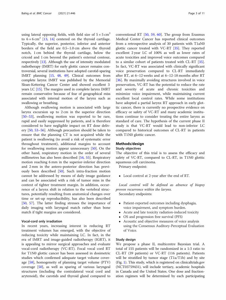
Study designWe propose a phase II, multicentre Bayesian trial. Atotal of 155 patients will be randomized in a 1:3 ratio toCL-RT (39 patients) or VC-RT (116 patients). Patientswill be stratified by tumor stage (T1a/T1b) and by site(Fig. 1). This study, which is registered on clinicaltrials.gov(NCT03759431), will include tertiary, academic hospitalsin Canada and the United States. One dose and fraction-ation regimen will be determined by each participating
Bahig et al. BMC Cancer (2021) 21:446 Page 3 of 11
center and will have to be the same in both arms. Thestudy is powered to compare LC of VC-RT compared tohistorical outcomes of CL-RT. The purpose of therandomization will be to generate data on secondary out-comes of voice impairment, dysphagia and quality of lifeas well as survival outcomes in the control arm.
Conditions for patient eligibility
� Age > 18 years� Stage T1a-b N0 of the true vocal cords planned for
definitive RT� Patient not candidate for laser surgery or declined
laser surgery� Biopsy-confirmed squamous cell carcinoma,
including verrucous carcinoma� Eastern Cooperative Oncology Group (ECOG)
performance status 0–2� Ability to provide written informed consent.� Ability to understand and read English or French at
a level adequate for completion of patient reportedoutcomes questionnaires
Conditions for patient ineligibility
� Previous irradiation of the head and neck region� Pregnancy or breastfeeding� Any medical condition that represents, in the
opinion of the investigator, a contraindication toradiotherapy or would prevent follow-up afterradiotherapy.
� Prior invasive malignancy (except non-melanomatous skin cancer) unless disease free for aminimum of 2 years.
Required pre-treatment evaluation
� History and Physical Examination, including:° Laryngoscopy, with detailed diagram of theprimary lesion confirmed by both the radiationoncologist and head and neck surgeon° Biopsy of the primary tumor
� Patient reported outcomes questionnaires includingthe Voice Handicap Index 10 (VHI-10), the MDAnderson Dysphagia Inventory (MDADI) and theMD Anderson Symptom Inventory Head and NeckModule (MDASI-HN).
InterventionIn both treatment arms, patients will be treated with aradical course of radiotherapy using a standard mildhypofractionation regimen of 5 fractions per week, over4 to 6 weeks. For pragmatic reasons, each participatinginstitution is allowed to use their standard of care doseand fractionation, but will have to be identical betweenthe standard and experimental arms at each institution.Each center will provide, at time study entrance, whatdose and fractionation will be used. This dose and frac-tionation will be the same for both arms and cannotchange over the course of the study. The study is strati-fied by participating institution, which means that it willbe stratified by dose/fractionation. In both treatmentarms, a daily volumetric imaging method will be re-quired for set-up verification, with match on the larynx.
Immobilization and simulationPatients will be positioned in a comfortable and repro-ducible position and will be immobilized in a thermo-plastic mask of the head and shoulders fixed to thetreatment Table. A 3-dimensional planning computedtomography (CT) scan (maximum slice thickness of 1.5mm) of the neck will be obtained. A planning magneticresonance imaging (MRI), co-registered to the planningCT to help define gross tumor volume (GTV), is recom-mended but optional.Clinicians should be aware of the risk of swallowing
during CT simulation. Patients should be instructed not
Fig. 1 Study Scheme of the VOCAL Trial. N = Number of patients
Bahig et al. BMC Cancer (2021) 21:446 Page 4 of 11
to swallow during the planning CT and MRI, as well asduring their radiation treatment. Co-registration of theplanning CT with a complementary planning volumetricimaging (such as MRI or contrast-enhanced CT) shouldbe performed in order to verify the position of thelarynx. If only one simulation imaging is obtained attime of planning, the position of the larynx should beverified on the first fraction of radiotherapy using avolumetric imaging method allowing 3D image recon-struction (Ex: Cone beam CT, CT on rail, MR-Linac).
VolumesThe GTV will be the same for both CL-RT and VC-RTand will be based physical examination and imaging(planning CT +/− MRI).
Standard arm (CL-RT) The RT volume in the standardarm is based on expected fields from conventional CL-RTvolume, with the planning target volume (PTV) extendingfrom the top of thyroid cartilage lamina superiorly, to thebottom of the cricoid inferiorly. The volumes below aredefined to lead to traditional volumes from conventionalCL-RT (Fig. 2).
� CTVCL-RT = GTV plus a manual expansionsuperiorly to include the cranial arytenoid cartilage,inferiorly to include 1–1.5 cm below true vocalcords, anteriorly to include the anterior commissure,posteriorly to include posterior commissure andarytenoid cartilage.
� PTV CL-RT = 1 cm circumferential expansion aroundCTV CL-RT in all directions, except posteriorly wherethe margin will be 0.5 cm. As the PTV of CL-RT isbased on previous fields of conventional RT, after
the addition of a 1 cm margin, the PTV CL-RT shouldextend to the top of the thyroid cartilage laminasuperiorly and to the bottom of the cricoidinferiorly; alternatively, the PTV CL-RT should bemanually expanded.
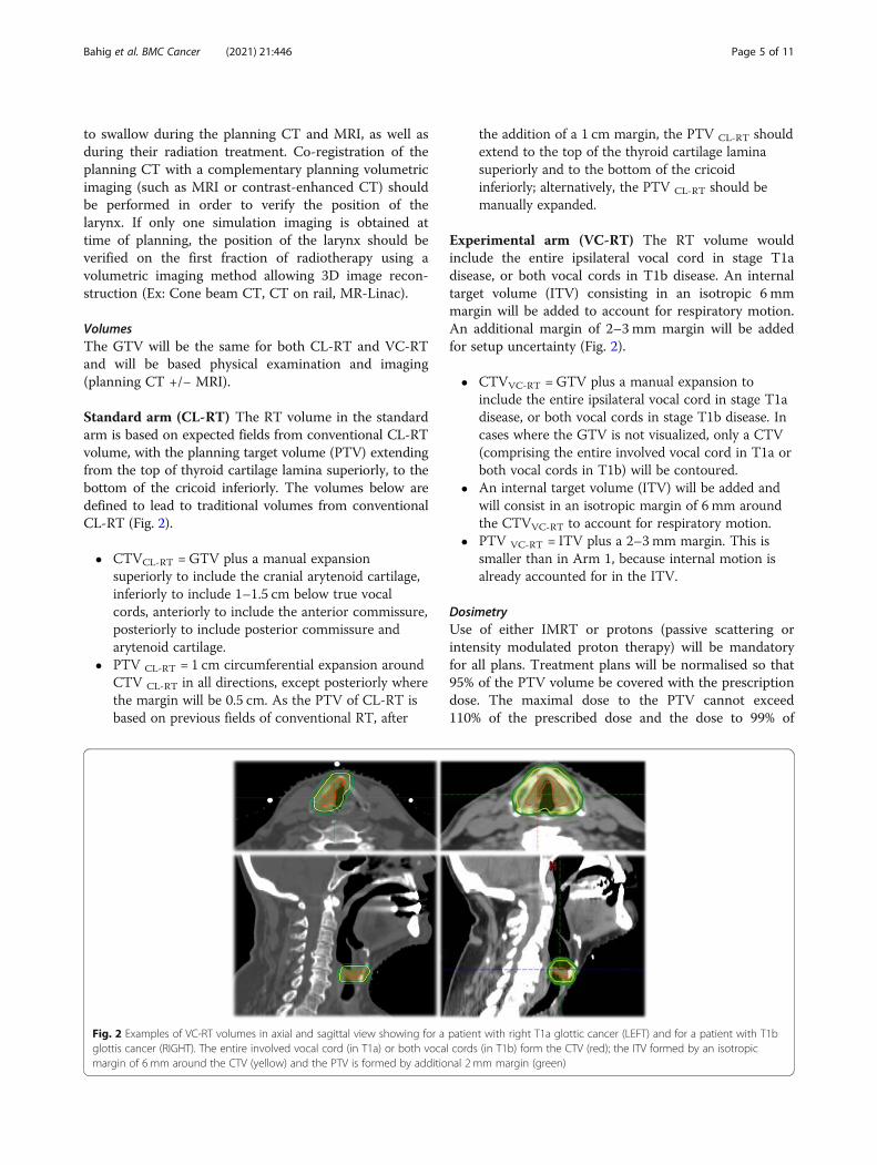
Experimental arm (VC-RT) The RT volume wouldinclude the entire ipsilateral vocal cord in stage T1adisease, or both vocal cords in T1b disease. An internaltarget volume (ITV) consisting in an isotropic 6 mmmargin will be added to account for respiratory motion.An additional margin of 2–3 mm margin will be addedfor setup uncertainty (Fig. 2).
� CTVVC-RT = GTV plus a manual expansion toinclude the entire ipsilateral vocal cord in stage T1adisease, or both vocal cords in stage T1b disease. Incases where the GTV is not visualized, only a CTV(comprising the entire involved vocal cord in T1a orboth vocal cords in T1b) will be contoured.
� An internal target volume (ITV) will be added andwill consist in an isotropic margin of 6 mm aroundthe CTVVC-RT to account for respiratory motion.
� PTV VC-RT = ITV plus a 2–3 mm margin. This issmaller than in Arm 1, because internal motion isalready accounted for in the ITV.
DosimetryUse of either IMRT or protons (passive scattering orintensity modulated proton therapy) will be mandatoryfor all plans. Treatment plans will be normalised so that95% of the PTV volume be covered with the prescriptiondose. The maximal dose to the PTV cannot exceed110% of the prescribed dose and the dose to 99% of
Fig. 2 Examples of VC-RT volumes in axial and sagittal view showing for a patient with right T1a glottic cancer (LEFT) and for a patient with T1bglottis cancer (RIGHT). The entire involved vocal cord (in T1a) or both vocal cords (in T1b) form the CTV (red); the ITV formed by an isotropicmargin of 6 mm around the CTV (yellow) and the PTV is formed by additional 2 mm margin (green)
Bahig et al. BMC Cancer (2021) 21:446 Page 5 of 11
PTV should be above 95% of the prescribed dose (D99 >95% of prescribed dose). Heterogeneity correction willbe required for dose calculation.
Critical structures delineation and constraintsCritical structures delineation will be based on previ-ously published international consensus guidelines [61].The latter reference provides a detailed atlas of eachstructure and should be consulted for additional preci-sions. Anatomic boundaries are details in Supplementarymaterial 1.
Standard arm (CL-RT)� Spinal cord: Not more than 0.03 cc of the planning
organ at risk (PRV) exceeds 42 Gy and not morethan 0.03 cc of the spinal cord receives > 40 Gy.
� Optimization of dose to other organs at risk (OAR)is not required. However, carotid-sparing isauthorized.
Experimental arm (VC-RT)� Spinal cord: Not more than 0.03 cc of the PRV
exceeds 42 Gy and not more than 0.03 cc of thespinal cord receives > 40 Gy.
� Additional dose specification goals that should nottake priority over PTV coverage:° Carotid arteries:▪ T1aIpsilateral carotid: Mean dose < 35 GyControlateral carotid: Mean dose < 15 Gy▪ T1b� Mean dose to carotids < 35 Gy
° Supraglottic larynx: Mean dose < 45 Gy° Contralateral vocal cord: Mean dose < 50 Gy
� Doses to other organs should be minimized anddocumented.
IGRTA volumetric imaging method allowing 3D image recon-struction (Ex: Cone beam CT, CT on rail, MR-Linac)will be required daily for set-up verification. Additionalimages (e.g. confirmatory kV X-ray) may be used assupplemental verification. Larynx soft-tissue matching is
required (as opposed to bony landmark [e.g. spine, baseof skull] alignment).
Study assessments
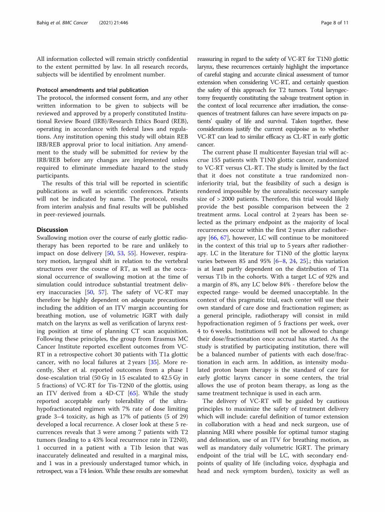
� There will be evaluation immediately post-treatment, as well as at 6, 12 and 24 months post-treatment as part of the study. Other follow-ups willbe as per institution standard of care, but suggestedschedule of assessments detailed in Table 1 isencouraged.
� Alternate follow-ups between the head and necksurgeon and the radiation oncologist will bemandatory.
� Physical examination will involve flexiblelaryngoscopy or/and videostrobe or/and mirrorexamination with adequate visualisation of vocalcords.
� Clinical follow-up will involve documentation of LCand survival status as well as reporting of CTCAEV4.03 toxicities.
� Patients will be asked to fill patient-reportedoutcome questionnaires at baseline and at each visit,as detailed in Table 1. The questionnaires will beadministered in the following order: VHI-10,MDADI and MDASI-HN. The questionnaires couldbe filled in paper format, or online using theanonymized CASTOR EDC system. Thequestionnaires will not be reviewed during the studyand will be analysed only at the pre-specifiedtimepoints.
� There will be an optional assessment of acoustic andobjective measures of voice analysis by a speechpathologist at baseline, immediately post-treatmentand at 6 months post treatment. The assessmentswill consist in a digital recording of a standard oralreading passage and a short monologue, whichwould later be analyzed for specific acousticvariables and microacoustic measures of thecycle-to-cycle variation of acoustic parameters (infrequency and intensity domains). The voicequality will be graded as per then GRBAS scale(grade, roughness, breathiness, asthenia, strain).
Table 1 Suggested Schedule of Assessments
Pre-RT End of RT 2moa 6mo 12 mo 24 mo 36moa 48moa 60moa
History (including TIA or stroke) & physical exam(including laryngoscopy)
x x x x x x x x x
Side effects x x x x x x x
TSHa x x x x x x
Questionnaire VHI −10 x x x x x x x x x
Questionnaire MDADI and MDASI-HN x x x x x x x x x
RT Radiotherapy, Mo monthsaOptional assessments
Bahig et al. BMC Cancer (2021) 21:446 Page 6 of 11
Statistical considerationsAccording to previous experience, expected LC afterCL-RT is estimated to be 92% [6, 8]. Assuming a non-inferiority margin of 8%, if LC in the VC-RT arm is≥84% at 2-years, we will deem the VC-RT arm accept-able. With a sample size of 111 patients, over 82% powerwill detect a margin difference of 8%, at a target signifi-cance level of 0.05 using a one-sided, exact binomial test(NCSS-PASS 2005). The purpose of the randomizationwill be to generate currently unavailable data on second-ary outcomes of voice impairment, dysphagia and qualityof life in the CL-RT arm. Considering a 5% attrition rate,155 patients will be randomized in a 1:3 ratio to CL-RT(39 patients) and VC-RT (116 patients) arms. Patientswill be stratified by stage (T1a/T1b) and by institution.An interim analysis will be conducted when 55
patients have a minimal follow-up of 6 months. We willmonitor the efficacy endpoint using the Bayesian optimalphase 2 (BOP2) design [62]. Specifically, let n denote theinterim sample size and N denote the maximum samplesize. Let peff denote the probability of efficacy (responserate) and define the null hypothesis H0 : peff < 0.84, repre-senting that the treatment is inefficacious. We will stopenrolling patients and claim that the treatment is notpromising if
Pr peff ≥0:84 j data� �
< λnN
� �α;
where λ =0.95 and α =1 are design parameters optimizedto minimize the chance of incorrectly claiming that anefficacious treatment is not promising (i.e., type II error)under the alternative hypothesis H1 : peff = 0.92, whilecontrolling the type I error rate at 0.05 (i.e., the chanceof incorrectly claiming that an inefficacious treatment ispromising is no more than 5%). Assuming a Beta(0.84,0.16) prior distribution for peff, if more than 10 pa-tients among the 55 patients develop recurrences, thetrial will be stopped in the interim analysis. Otherwise,the trial will be continued.
Analytic planA Bayesian regression model [63, 64] will be used tocompare the LC between the VC-RT and CL-RT armsat 2 years. The LC and OS time will be defined from endof RT date. The following recurrences will be consideredLC: in field, field margin and within larynx. Local recur-rences will be biopsy-proven when possible. Competingevents will include local recurrence, regional/distant re-currence and death prior to recurrence. LC will be esti-mated using both competing risk and Kaplan -Meier(KM) analysis. The analyses will be conducted using SASand R software. A Cox multivariable regression analysiswill be used to determine factors predictive of LC, PFSand OS. Chi-squared test or Fisher’s exact test will be
used to compare the difference in toxicity between arms.For the secondary endpoints of patient-reported out-comes (VHI-10, MDADI and MDASI-HN), trajectorytrends of scores between/among arms over time will beexplored using a generalized linear mixed model with arandom effect of time. Appropriate adjustments for co-variates will be considered.There will be two planned analysis: 1) an interim ana-
lysis and, 2) a final analysis. The interim analysis will bedone after 55 patients on the experimental arm have aminimum follow-up of 6 months. A short follow-up timeof 6 months was chosen for the interim analysis forsafety (i.e. for early detection of local recurrence trends).The trial may be interrupted early if VC-RT is deemednot promising on interim analysis. The final analysis willperformed 2 years after accrual is completed.
Data safety and monitoring boardA data safety and monitoring board (DSMB) from theCentre Hospitalier de l’Université de Montréal will beresponsible for assessing the safety data. The trial wouldbe interrupted early if higher than expected rates of localrecurrences are observed. The DSMB will reviewinterim/cumulative evidence of study related safety,consider factors external to the study when relevant in-formation becomes available and provide the sponsor arecommendation as to whether the study should: con-tinue without change, be modified, suspended or termi-nated. The DSMB will assess the safety data twice a year.An interim analysis will be planned after the first 55 pa-tients in the experimental arm have a 6-month follow-up. Using a Bayesian probabilistic model, the trial wouldbe interrupted early if higher than expected rates of localrecurrences are observed. As this trial involves only a re-duction of RT treatment volumes, we do not expect anyincrease in toxicities in patients treated on the experi-mental arm.
Subject discontinuation / withdrawalPatients may withdraw from the study prior to the com-pletion of study related procedures for the followingreasons:
� Patient withdraws consent for participation. Subjectsmay voluntarily discontinue participation in thestudy at any time.
� It is deemed in the patient’s best interest asdetermined by the attending/principal investigator.
Consent and confidentialityAll enrolled patients will be required to sign informedconsent before study entry. The principal investigator,the co-investigators and the research nurses will gatherand record all collected information in a research record.
Bahig et al. BMC Cancer (2021) 21:446 Page 7 of 11
All information collected will remain strictly confidentialto the extent permitted by law. In all research records,subjects will be identified by enrolment number.
Protocol amendments and trial publicationThe protocol, the informed consent form, and any otherwritten information to be given to subjects will bereviewed and approved by a properly constituted Institu-tional Review Board (IRB)/Research Ethics Board (REB),operating in accordance with federal laws and regula-tions. Any institution opening this study will obtain REBIRB/REB approval prior to local initiation. Any amend-ment to the study will be submitted for review by theIRB/REB before any changes are implemented unlessrequired to eliminate immediate hazard to the studyparticipants.The results of this trial will be reported in scientific
publications as well as scientific conferences. Patientswill not be indicated by name. The protocol, resultsfrom interim analysis and final results will be publishedin peer-reviewed journals.
DiscussionSwallowing motion over the course of early glottic radio-therapy has been reported to be rare and unlikely toimpact on dose delivery [50, 53, 55]. However, respira-tory motion, laryngeal shift in relation to the vertebralstructures over the course of RT, as well as the occa-sional occurrence of swallowing motion at the time ofsimulation could introduce substantial treatment deliv-ery inaccuracies [50, 57]. The safety of VC-RT maytherefore be highly dependent on adequate precautionsincluding the addition of an ITV margin accounting forbreathing motion, use of volumetric IGRT with dailymatch on the larynx as well as verification of larynx rest-ing position at time of planning CT scan acquisition.Following these principles, the group from Erasmus MCCancer Institute reported excellent outcomes from VC-RT in a retrospective cohort 30 patients with T1a glotticcancer, with no local failures at 2 years [35]. More re-cently, Sher et al. reported outcomes from a phase Idose-escalation trial (50 Gy in 15 escalated to 42.5 Gy in5 fractions) of VC-RT for Tis-T2N0 of the glottis, usingan ITV derived from a 4D-CT [65]. While the studyreported acceptable early tolerability of the ultra-hypofractionated regimen with 7% rate of dose limitinggrade 3–4 toxicity, as high as 17% of patients (5 of 29)developed a local recurrence. A closer look at these 5 re-currences reveals that 3 were among 7 patients with T2tumors (leading to a 43% local recurrence rate in T2N0),1 occurred in a patient with a T1b lesion that wasinaccurately delineated and resulted in a marginal miss,and 1 was in a previously understaged tumor which, inretrospect, was a T4 lesion. While these results are somewhat
reassuring in regard to the safety of VC-RT for T1N0 glotticlarynx, these recurrences certainly highlight the importanceof careful staging and accurate clinical assessment of tumorextension when considering VC-RT, and certainly questionthe safety of this approach for T2 tumors. Total laryngec-tomy frequently constituting the salvage treatment option inthe context of local recurrence after irradiation, the conse-quences of treatment failures can have severe impacts on pa-tients’ quality of life and survival. Taken together, theseconsiderations justify the current equipoise as to whetherVC-RT can lead to similar efficacy as CL-RT in early glotticcancer.The current phase II multicenter Bayesian trial will ac-
crue 155 patients with T1N0 glottic cancer, randomizedto VC-RT versus CL-RT. The study is limited by the factthat it does not constitute a true randomized non-inferiority trial, but the feasibility of such a design isrendered impossible by the unrealistic necessary samplesize of > 2000 patients. Therefore, this trial would likelyprovide the best possible comparison between the 2treatment arms. Local control at 2 years has been se-lected as the primary endpoint as the majority of localrecurrences occur within the first 2 years after radiother-apy [66, 67], however, LC will continue to be monitoredin the context of this trial up to 5 years after radiother-apy. LC in the literature for T1N0 of the glottic larynxvaries between 85 and 95% [6–8, 24, 25].; this variationis at least partly dependent on the distribution of T1aversus T1b in the cohorts. With a target LC of 92% anda margin of 8%, any LC below 84% - therefore below theexpected range- would be deemed unacceptable. In thecontext of this pragmatic trial, each center will use theirown standard of care dose and fractionation regimen; asa general principle, radiotherapy will consist in mildhypofractionation regimen of 5 fractions per week, over4 to 6 weeks. Institutions will not be allowed to changetheir dose/fractionation once accrual has started. As thestudy is stratified by participating institution, there willbe a balanced number of patients with each dose/frac-tionation in each arm. In addition, as intensity modu-lated proton beam therapy is the standard of care forearly glottic larynx cancer in some centers, the trialallows the use of proton beam therapy, as long as thesame treatment technique is used in each arm.The delivery of VC-RT will be guided by cautious
principles to maximize the safety of treatment deliverywhich will include: careful definition of tumor extensionin collaboration with a head and neck surgeon, use ofplanning MRI where possible for optimal tumor stagingand delineation, use of an ITV for breathing motion, aswell as mandatory daily volumetric IGRT. The primaryendpoint of the trial will be LC, with secondary end-points of quality of life (including voice, dysphagia andhead and neck symptom burden), toxicity as well as
Bahig et al. BMC Cancer (2021) 21:446 Page 8 of 11
objective measures of voice. If positive, this would resultin a paradigm shift in the approach to radiation fortreatment early glottic cancers in the institutions whereCL-RT remains the standard. On the contrary, if VC-RTdemonstrates inferior LC compared to CL-RT, the re-sults of this trial would strengthen the necessity to main-tain conservative margins until the possible loopholes inthe safety of VC-RT are better understood.
Abbrevations4D-CT: Four-dimensional computed tomography; BOP-2: Bayesian optimalphase 2; GRBAS: Grade, roughness, breathiness, asthenia, strain; CL-RT: Complete larynx radiotherapy; CT: Computed tomography;CTCAE: Common toxicity criteria for adverse events; CTV: Clinical targetvolume; DSMB: Data safety and monitoring board; ECOG: EasternCooperative Oncology Group; GTV: Gross tumor volume; HNC: Head andneck cancer; IGRT: Image-guided radiotherapy; IMRT: Intensity modulatedradiotherapy; IRB : Institutional Review Board; ITV: Internal target volume;KM: Kaplan meier; LC: Local control; MDADI: MD Anderson dysphagiainventory; MDASI-HN: MD Anderson Symptom inventory- Head and neckmodule; MRI: Magnetic resonance imaging; OS : Overall survival;PTV: Planning target volume; PRV: Planning organ at risk volume;REB: Research Ethics Boar; RT: Radiotherapy; VC-RT : Vocal cord radiotherapy;VHI-10: Voice handicap index-10
Supplementary InformationThe online version contains supplementary material available at https://doi.org/10.1186/s12885-021-08195-8.
Additional file 1.
AcknowledgementsNone.
Study statusOpen and currently accruing.
Authors’ contributionsHB, DP, DIR, CDF, FN, YY contributed to the conception and design of theprotocol, and initial drafting of the protocol. YY contributed to statisticaldesign. KAH, AC, CAN, KF, OB, EF, SPN, LL, JD, HSK contributed to conceptionand design of the protocol. All authors have read and approved themanuscript.
FundingNone.
Availability of data and materialsNot applicable.
Declarations
Ethics approval and consent to participateThe proposed study has been approved by the Centre Hospitalier del’Université de Montreal IRB. Informed written consent to participate in thestudy will be obtained from participants. The inclusion of Fig. 2 has beenapproved by our institutional institutional review board.
Consent for publicationNot applicable.
Competing interestsNone.
Author details1Radiation Oncology Department, Centre Hospitalier de l’Université deMontréal, 1051 Sanguinet, Montreal, QC H2X 3E4, Canada. 2Radiation
Oncology Department, University of Texas MD Anderson Cancer Center,1515 Holcombe, Houston, TX 77030, USA. 3Biostatistics Department,University of Texas MD Anderson Cancer Center, Houston, USA. 4Head andNeck Surgery Department, University of Texas MD Anderson Cancer Center,Houston, USA. 5Head and Neck Surgery Department, Centre Hospitalier del’Université de Montréal, Montreal, Canada. 6Department of Otolaryngology -Head and Neck Surgery, Western University, London, Ontario, Canada.7Radiation Oncology Department, Peter MacCallum Cancer Centre,Melbourne, Australia. 8Radiation Oncology Department, Centre Intégré deCancérologie de Laval, Laval, Canada. 9Radiation Oncology Department, CaseWestern Reserve University, Cleveland, USA. 10Radiation OncologyDepartment, NYU Langone Health, Newyork, USA. 11Radiation OncologyDepartment, Western University, London, Ontario, Canada.
Received: 22 June 2020 Accepted: 14 April 2021
References1. Ferlay JSI, Ervik M, Dikshit R, Eser S, Mathers C, et al. GLOBOCAN 2012 v1.0,
cancer incidence and mortality worldwide: IARC cancerbase no. 11. Lyon:International Agency for Research on Cancer; 2013.
2. 2016 CCSsSCoCS. Toronto, Ontario, Canada: Canadian Cancer Society. 2016.3. Grant DG, Salassa JR, Hinni ML, Pearson BW, Hayden RE, Perry WC.
Transoral laser microsurgery for carcinoma of the supraglottic larynx.Otolaryngol Head Neck Surg. 2007;136(6):900–6. https://doi.org/10.1016/j.otohns.2006.12.015.
4. Outzen KE, Illum P. CO2-laser therapy for carcinoma of the larynx. J LaryngolOtol. 1995;109(2):111–3. https://doi.org/10.1017/S0022215100129421.
5. Ambrosch P. The role of laser microsurgery in the treatment of laryngealcancer. Curr Opin Otolaryngol Head Neck Surg. 2007;15(2):82–8. https://doi.org/10.1097/MOO.0b013e3280147336.
6. Cellai E, Frata P, Magrini SM, Paiar F, Barca R, Fondelli S, et al. Radicalradiotherapy for early glottic cancer: results in a series of 1087 patients fromtwo Italian radiation oncology centers. I. the case of T1N0 disease. Int JRadiat Oncol Biol Phys. 2005;63(5):1378–86. https://doi.org/10.1016/j.ijrobp.2005.05.018.
7. Dinshaw KA, Sharma V, Agarwal JP, Ghosh S, Havaldar R. Radiation therapyin T1-T2 glottic carcinoma: influence of various treatment parameters onlocal control/complications. Int J Radiat Oncol Biol Phys. 2000;48(3):723–35.https://doi.org/10.1016/S0360-3016(00)00635-0.
8. Le QT, Fu KK, Kroll S, Ryu JK, Quivey JM, Meyler TS, et al. Influence offraction size, total dose, and overall time on local control of T1-T2 glotticcarcinoma. Int J Radiat Oncol Biol Phys. 1997;39(1):115–26. https://doi.org/10.1016/S0360-3016(97)00284-8.
9. Howell-Burke D, Peters LJ, Goepfert H, Oswald MJ. T2 glottic cancer.Recurrence, salvage, and survival after definitive radiotherapy. ArchOtolaryngol Head Neck Surg. 1990;116(7):830–5. https://doi.org/10.1001/archotol.1990.01870070078014.
10. Laccourreye O, Laccourreye L, Garcia D, Gutierrez-Fonseca R, Brasnu D,Weinstein G. Vertical partial laryngectomy versus supracricoid partiallaryngectomy for selected carcinomas of the true vocal cord classified asT2N0. Ann Otol Rhinol LaryngoL. 2000;109(10 Pt 1):965–71. https://doi.org/10.1177/000348940010901011.
11. Gallo A, Manciocco V, Simonelli M, Pagliuca G, D'Arcangelo E, deVincentiis M. Supracricoid partial laryngectomy in the treatment oflaryngeal cancer: univariate and multivariate analysis of prognosticfactors. Arch Otolaryngol Head Neck Surg. 2005;131(7):620–5. https://doi.org/10.1001/archotol.131.7.620.
12. Laccourreye O, Weinstein G, Brasnu D, Trotoux J, Laccourreye H. Verticalpartial laryngectomy: a critical analysis of local recurrence. Ann Otol RhinolLaryngol. 1991;100(1):68–71. https://doi.org/10.1177/000348949110000111.
13. Chatani M, Matayoski Y, Masaki N, Teshima T, Inoue T. Radiation therapy forearly glottic carcinoma (T1N0M0). The final results of prospectiverandomized study concerning radiation field. Strahlenther Onkol. 1996;172(3):169–72.
14. Protocol RTOG 95–12 ATea. A randomized study of hyperfractionationversus conventional fractionation in t2 squamous cell carcinoma of thevocal cord. 1996.
15. Zumsteg ZS, Riaz N, Jaffery S, Hu M, Gelblum D, Zhou Y, et al. Carotidsparing intensity-modulated radiation therapy achieves comparablelocoregional control to conventional radiotherapy in T1-2N0 laryngeal
Bahig et al. BMC Cancer (2021) 21:446 Page 9 of 11
carcinoma. Oral Oncol. 2015;51(7):716–23. https://doi.org/10.1016/j.oraloncology.2015.02.003.
16. Higgins KM, Shah MD, Ogaick MJ, Enepekides D. Treatment of early-stageglottic cancer: meta-analysis comparison of laser excision versusradiotherapy. J Otolaryngology Head Neck Surg. 2009;38(6):603–12.
17. Feng Y, Wang B, Wen S. Laser surgery versus radiotherapy for T1-T2N0glottic cancer: a meta-analysis. ORL J Otorhinolaryngol Relat Spec. 2011;73(6):336–42. https://doi.org/10.1159/000327097.
18. Mo HL, Li J, Yang X, Zhang F, Xiong JW, Yang ZL, et al. Transoral lasermicrosurgery versus radiotherapy for T1 glottic carcinoma: a systematicreview and meta-analysis. Lasers Med Sci. 2017;32(2):461–7. https://doi.org/10.1007/s10103-016-2103-8.
19. Yoo J, Lacchetti C, Hammond JA, Gilbert RW. Role of endolaryngeal surgery(with or without laser) versus radiotherapy in the management of early (T1)glottic cancer: a systematic review. Head Neck. 2014;36(12):1807–19. https://doi.org/10.1002/hed.23504.
20. Warner L, Chudasama J, Kelly CG, Loughran S, McKenzie K, Wight R, et al.Radiotherapy versus open surgery versus endolaryngeal surgery (with orwithout laser) for early laryngeal squamous cell cancer. Cochrane DatabaseSyst Rev. 2014;12:Cd002027.
21. Abdurehim Y, Hua Z, Yasin Y, Xukurhan A, Imam I, Yuqin F. Transoral lasersurgery versus radiotherapy: systematic review and meta-analysis fortreatment options of T1a glottic cancer. Head Neck. 2012;34(1):23–33.https://doi.org/10.1002/hed.21686.
22. Spielmann PM, Majumdar S, Morton RP. Quality of life and functional outcomesin the management of early glottic carcinoma: a systematic review of studiescomparing radiotherapy and transoral laser microsurgery. Clin Otolaryngol. 2010;35(5):373–82. https://doi.org/10.1111/j.1749-4486.2010.02191.x.
23. Dey P, Arnold D, Wight R, MacKenzie K, Kelly C, Wilson J. Radiotherapyversus open surgery versus endolaryngeal surgery (with or without laser) forearly laryngeal squamous cell cancer. Cochrane Database Syst Rev. 2002;2:CD002027.
24. Mendenhall WM, Werning JW, Hinerman RW, Amdur RJ, Villaret DB.Management of T1-T2 glottic carcinomas. Cancer. 2004;100(9):1786–92.https://doi.org/10.1002/cncr.20181.
25. Smee RI, Meagher NS, Williams JR, Broadley K, Bridger GP. Role ofradiotherapy in early glottic carcinoma. Head Neck. 2010;32(7):850–9.
26. Kinshuck AJ, Shenoy A, Jones TM. Voice outcomes for early laryngeal cancer.Curr Opin Otolaryngol Head Neck Surg. 2017;25(3):211–6. https://doi.org/10.1097/MOO.0000000000000363.
27. Greulich MT, Parker NP, Lee P, Merati AL, Misono S. Voice outcomesfollowing radiation versus laser microsurgery for T1 glottic carcinoma:systematic review and meta-analysis. Otolaryngol Head Neck Surg. 2015;152(5):811–9. https://doi.org/10.1177/0194599815577103.
28. Huang G, Luo M, Zhang J, Liu H. The voice quality after laser surgery versusradiotherapy of T1a glottic carcinoma: systematic review and meta-analysis.Onco Targets Ther. 2017;10:2403–10. https://doi.org/10.2147/OTT.S137210.
29. Dinapoli N, Parrilla C, Galli J, Autorino R, Micciche F, Bussu F, et al.Multidisciplinary approach in the treatment of T1 glottic cancer. The role ofpatient preference in a homogenous patient population. StrahlentherOnkol. 2010;186(11):607–13. https://doi.org/10.1007/s00066-010-2142-1.
30. Loughran S, Calder N, MacGregor FB, Carding P, MacKenzie K. Quality of lifeand voice following endoscopic resection or radiotherapy for early glotticcancer. Clin Otolaryngol. 2005;30(1):42–7. https://doi.org/10.1111/j.1365-2273.2004.00919.x.
31. Stoeckli SJ, Schnieper I, Huguenin P, Schmid S. Early glottic carcinoma:treatment according patient's preference? Head Neck. 2003;25(12):1051–6.https://doi.org/10.1002/hed.10323.
32. Zahoor T, Dawson R, Sen M, Makura Z. Transoral laser resection orradiotherapy? Patient choice in the treatment of early laryngeal cancer: aprospective observational cohort study. J Laryngol Otol. 2017;131(6):541–5.https://doi.org/10.1017/S0022215116010057.
33. Fung K, Yoo J, Leeper HA, Bogue B, Hawkins S, Hammond JA, et al. Effectsof head and neck radiation therapy on vocal function. J Otolaryngol. 2001;30(3):133–9. https://doi.org/10.2310/7070.2001.20192.
34. Sjogren EV, van Rossum MA, Langeveld TP, Voerman MS, van de Kamp VA,Friebel MO, et al. Voice outcome in T1a midcord glottic carcinoma: lasersurgery vs radiotherapy. Arch Otolaryngol Head Neck Surg. 2008;134(9):965–72. https://doi.org/10.1001/archotol.134.9.965.
35. Al-Mamgani A, Kwa SL, Tans L, Moring M, Fransen D, Mehilal R, et al.Single vocal cord irradiation: image guided intensity modulated
Hypofractionated radiation therapy for T1a Glottic Cancer: early clinicalresults. Int J Radiat Oncol Biol Phys. 2015;93(2):337–43. https://doi.org/10.1016/j.ijrobp.2015.06.016.
36. Jacobson BHJA, Grywalski C, et al. The voice handi- cap index (VHI)development and validation. Am J Speech- Lang Pat. 1997;6:66–70.
37. Dornfeld K, Simmons JR, Karnell L, Karnell M, Funk G, Yao M, et al. Radiationdoses to structures within and adjacent to the larynx are correlated withlong-term diet- and speech-related quality of life. Int J Radiat Oncol BiolPhys. 2007;68(3):750–7. https://doi.org/10.1016/j.ijrobp.2007.01.047.
38. Rancati T, Schwarz M, Allen AM, Feng F, Popovtzer A, Mittal B, et al.Radiation dose-volume effects in the larynx and pharynx. Int J RadiatOncol Biol Phys. 2010;76(3 Suppl):S64–9. https://doi.org/10.1016/j.ijrobp.2009.03.079.
39. Sanguineti G, Adapala P, Endres EJ, Brack C, Fiorino C, Sormani MP, et al.Dosimetric predictors of laryngeal edema. Int J Radiat Oncol Biol Phys. 2007;68(3):741–9. https://doi.org/10.1016/j.ijrobp.2007.01.010.
40. Plummer C, Henderson RD, O'Sullivan JD, Read SJ. Ischemic stroke andtransient ischemic attack after head and neck radiotherapy: a review. Stroke.2011;42(9):2410–8. https://doi.org/10.1161/STROKEAHA.111.615203.
41. Dorresteijn LD, Kappelle AC, Scholz NM, Munneke M, Scholma JT, Balm AJ,et al. Increased carotid wall thickening after radiotherapy on the neck. Eur JCancer. 2005;41(7):1026–30. https://doi.org/10.1016/j.ejca.2005.01.020.
42. Swisher-McClure S, Mitra N, Lin A, Ahn P, Wan F, O'Malley B, et al. Risk offatal cerebrovascular accidents after external beam radiation therapy forearly-stage glottic laryngeal cancer. Head Neck. 2014;36(5):611–6. https://doi.org/10.1002/hed.23342.
43. Martin JD, Buckley AR, Graeb D, Walman B, Salvian A, Hay JH. Carotid arterystenosis in asymptomatic patients who have received unilateral head-and-neck irradiation. Int J Radiat Oncol Biol Phys. 2005;63(4):1197–205. https://doi.org/10.1016/j.ijrobp.2005.04.017.
44. Mulholland GB, Zhang H, Nguyen NT, Tkacyzk N, Seikaly H, O'Connell D,et al. Optimal detection of hypothyroidism in early stage laryngeal cancertreated with radiotherapy. J Otolaryngol Head Neck Surg. 2015;44:34.
45. Mendenhall WM, Amdur RJ, Morris CG, Hinerman RW. T1-T2N0 squamouscell carcinoma of the glottic larynx treated with radiation therapy. J ClinOncol. 2001;19(20):4029–36. https://doi.org/10.1200/JCO.2001.19.20.4029.
46. Jones AS, Fish B, Fenton JE, Husband DJ. The treatment of early laryngealcancers (T1-T2 N0): surgery or irradiation? Head Neck. 2004;26(2):127–35.https://doi.org/10.1002/hed.10361.
47. Fujita M, Rudoltz MS, Canady DJ, Patel P, Machtay M, Pittard MQ, et al.Second malignant neoplasia in patients with T1 glottic cancer treated withradiation. Laryngoscope. 1998;108(12):1853–5. https://doi.org/10.1097/00005537-199812000-00016.
48. Rosenthal DI, Fuller CD, Barker JL Jr, Mason B, Garcia JA, Lewin JS, et al.Simple carotid-sparing intensity-modulated radiotherapy technique andpreliminary experience for T1-2 glottic cancer. Int J Radiat Oncol Biol Phys.2010;77(2):455–61. https://doi.org/10.1016/j.ijrobp.2009.04.061.
49. Chera BS, Amdur RJ, Morris CG, Mendenhall WM. Carotid-sparing intensity-modulated radiotherapy for early-stage squamous cell carcinoma of thetrue vocal cord. Int J Radiat Oncol Biol Phys. 2010;77(5):1380–5. https://doi.org/10.1016/j.ijrobp.2009.07.1687.
50. Bahig HN-T, Nguyen-Tan PF, Filion, E.; Roberge, D.; Thanomsack, P.; de Guise,Jacques.; Blais D,; Doucet R.; L Etourneau-Guillon L.; Lambert, L. Larynxmotion considerations in partial larynx volumetric modulated arc therapyfor early glottic cancer. J Med Imaging Radiat Oncol. 2017.
51. Leonard RJ, Kendall KA, McKenzie S, Goncalves MI, Walker A. Structuraldisplacements in normal swallowing: a videofluoroscopic study. Dysphagia.2000;15(3):146–52. https://doi.org/10.1007/s004550010017.
52. Dantas RO, Kern MK, Massey BT, Dodds WJ, Kahrilas PJ, Brasseur JG, et al.Effect of swallowed bolus variables on oral and pharyngeal phases ofswallowing. Am J Phys. 1990;258(5 Pt 1):G675–81.
53. Hamlet S, Ezzell G, Aref A. Larynx motion associated with swallowing duringradiation therapy. Int J Radiat Oncol Biol Phys. 1994;28(2):467–70. https://doi.org/10.1016/0360-3016(94)90073-6.
54. Bradley JA, Paulson ES, Ahunbay E, Schultz C, Li XA, Wang D. Dynamic MRIanalysis of tumor and organ motion during rest and deglutition and marginassessment for radiotherapy of head-and-neck cancer. Int J Radiat OncolBiol Phys. 2011;81(5):e803–12. https://doi.org/10.1016/j.ijrobp.2010.12.015.
55. van Asselen B, Raaijmakers CP, Lagendijk JJ, Terhaard CH. Intrafractionmotions of the larynx during radiotherapy. Int J Radiat Oncol Biol Phys.2003;56(2):384–90. https://doi.org/10.1016/S0360-3016(02)04572-8.
Bahig et al. BMC Cancer (2021) 21:446 Page 10 of 11
56. Bradley JD, Hope A, El Naqa I, Apte A, Lindsay PE, Bosch W, et al. Anomogram to predict radiation pneumonitis, derived from a combinedanalysis of RTOG 9311 and institutional data. Int J Radiat Oncol Biol Phys.2007;69(4):985–92. https://doi.org/10.1016/j.ijrobp.2007.04.077.
57. Gangsaas A, Astreinidou E, Quint S, Levendag PC, Heijmen B. Cone-beamcomputed tomography-guided positioning of laryngeal cancer patientswith large interfraction time trends in setup and nonrigid anatomyvariations. Int J Radiat Oncol Biol Phys. 2013;87(2):401–6. https://doi.org/10.1016/j.ijrobp.2013.06.2032.
58. Osman SO, Astreinidou E, Levendag PC, Heijmen BJ. Impact of geometricvariations on delivered dose in highly focused single vocal cord IMRT. ActaOncol. 2014;53(2):278–85. https://doi.org/10.3109/0284186X.2013.812793.
59. Osman SO, Astreinidou E, de Boer HC, Keskin-Cambay F, Breedveld S,Voet P, et al. IMRT for image-guided single vocal cord irradiation. Int JRadiat Oncol Biol Phys. 2012;82(2):989–97. https://doi.org/10.1016/j.ijrobp.2010.12.022.
60. Levendag PC, Teguh DN, Keskin-Cambay F, Al-Mamgani A, van Rooij P,Astreinidou E, et al. Single vocal cord irradiation: a competitive treatmentstrategy in early glottic cancer. Radiother Oncol. 2011;101(3):415–9. https://doi.org/10.1016/j.radonc.2011.05.026.
61. Brouwer CL, Steenbakkers RJ, Bourhis J, Budach W, Grau C, Gregoire V, et al.CT-based delineation of organs at risk in the head and neck region: DAHANCA, EORTC, GORTEC, HKNPCSG, NCIC CTG, NCRI, NRG oncology and TROGconsensus guidelines. Radiother Oncol. 2015;117(1):83–90. https://doi.org/10.1016/j.radonc.2015.07.041.
62. Zhou H, Lee JJ, Yuan Y. BOP2: Bayesian optimal design for phase II clinicaltrials with simple and complex endpoints. Stat Med. 2017;36(21):3302–14.https://doi.org/10.1002/sim.7338.
63. Gelman AaH J. Data analysis using regression and multilevel/hierarchicalmodels. Cambridge: Cambridge University Press; 2007.
64. McCullagh PaN JA. Generalized linear models. New York: Chapman andHall/CRC Press; 1989. https://doi.org/10.1007/978-1-4899-3242-6.
65. Sher DJ, Timmerman RD, Nedzi L, Ding C, Pham NL, Zhao B, et al. Phase 1fractional dose-escalation study of equipotent stereotactic radiation therapyregimens for early-stage Glottic larynx Cancer. Int J Radiat Oncol Biol Phys.2019;105(1):110–8. https://doi.org/10.1016/j.ijrobp.2019.03.010.
66. Mucha-Malecka A, Chrostowska A, Urbanek K, Malecki K. Prognostic factorsin patients with T1 glottic cancer treated with radiotherapy. StrahlentherOnkol. 2019;195(9):792–804. https://doi.org/10.1007/s00066-019-01481-2.
67. Lohynska R, Slavicek A, Bahanan A, Novakova P. Predictors of local failure inearly laryngeal cancer. Neoplasma. 2005;52(6):483–8.
Publisher’s NoteSpringer Nature remains neutral with regard to jurisdictional claims inpublished maps and institutional affiliations.
Bahig et al. BMC Cancer (2021) 21:446 Page 11 of 11