valley fever: danger lurking in a dust cloud
TRANSCRIPT

This article appeared in a journal published by Elsevier. The attachedcopy is furnished to the author for internal non-commercial researchand education use, including for instruction at the authors institution
and sharing with colleagues.
Other uses, including reproduction and distribution, or selling orlicensing copies, or posting to personal, institutional or third party
websites are prohibited.
In most cases authors are permitted to post their version of thearticle (e.g. in Word or Tex form) to their personal website orinstitutional repository. Authors requiring further information
regarding Elsevier’s archiving and manuscript policies areencouraged to visit:
http://www.elsevier.com/authorsrights

Author's personal copy
Review
Valley fever: danger lurking in a dust cloud
Larry Johnson a,b,1, Erin M. Gaab b,1, Javier Sanchez a,b, Phuong Q. Bui a,b, Clarissa J. Nobile a,b,Katrina K. Hoyer a,b, Michael W. Peterson c, David M. Ojcius a,b,*
a Department of Molecular Cell Biology, University of California, Merced, CA 95343, USAb Health Sciences Research Institute, University of California, Merced, CA 95343, USA
c Department of Internal Medicine, University of California San Francisco e Fresno, Fresno, CA 93703, USA
Received 1 April 2014; accepted 24 June 2014
Available online 16 July 2014
Abstract
Coccidioides immitis and Coccidioides posadasii contribute to the development of Valley Fever. The ability of these fungal pathogens toevade the host immune system creates difficulty in recognition and treatment of this debilitating infection. In this review, we describe the currentknowledge of Valley Fever and approaches to improve prevention, detection, and treatment.© 2014 Institut Pasteur. Published by Elsevier Masson SAS. All rights reserved.
Keywords: Innate immunity; Adaptive immunity; Fungal pathogen; Lung infection; Coccidioides
1. Introduction
Coccidioidomycosis is an infection caused by inhalingspores of the fungal species Coccidioides immitis or Cocci-dioides posadasii. The disease has commonly been termed“Valley Fever,” “San Joaquin Valley Fever,” “San JoaquinFever,” “desert fever,” and “desert rheumatism” [1]. A highincidence of coccidioidomycosis has been reported in thesouthwestern United States, Central America, and SouthAmerica [2,3]. The rise in cases has contributed to hospitali-zation costs totaling over $2 billion for those afflicted with theillness, which include individuals with symptoms rangingfrom mild local infections to disseminated disease [4].
Although inhalation of Coccidioides is the most commonmode of transmission, there are rare cases of transmissionthrough transplanted organs or inoculation by penetration of
the skin by a sharp object containing the fungus [3,5]. Whilemost infected individuals are asymptomatic, about 40% ofindividuals show flu-like symptoms, such as fever, cough,headache, skin rash, muscle aches, joint pain, and fatigue[1,3,6,7]. In most cases the immune system resolves theinfection without the need for medical intervention. However,without proper diagnosis, disseminated disease may occur,leading to increased severity of symptoms. Laboratory diag-nostic testing and clinical evaluation are the most effectivemeasures for determining coccidioidomycosis. Early detectionand antifungal drug treatments aid in slowing or inhibiting thedevelopment of disease and limit tissue damage, and mayprevent morbidity [2]. In this review, we aim to provide abetter understanding of coccidioidomycosis and to promoteawareness of these pathogenic fungi.
2. Valley fever
2.1. Geographic distribution of coccidioidomycosis
Two types of coccidioidomycosis-causing fungi exist: C.immitis and C. posadasii [8]. C. immitis is mainly endemic toCalifornia and is often referred to as the “Californian” strain,
* Corresponding author. Department of Molecular Cell Biology, University
of California, 5200 Lake Road, Merced, CA 95343, USA. Tel.: þ1 209 228
2948.
E-mail addresses: [email protected], [email protected]
(D.M. Ojcius).1 Both authors contributed equally to this work.
Microbes and Infection 16 (2014) 591e600www.elsevier.com/locate/micinf
http://dx.doi.org/10.1016/j.micinf.2014.06.011
1286-4579/© 2014 Institut Pasteur. Published by Elsevier Masson SAS. All rights reserved.

Author's personal copy
while C. posadasii is distinguished as the “non-Californian”strain [8]. However, C. immitis has also been isolated from soilin Venezuela and Washington State, where several patientswere suspected of contracting coccidioidomycosis [9,10].
There is a relatively low incidence rate of Valley Fever on anational scale in the United States and a variable status as areportable disease across endemic regions [11,12]. Cocci-dioides fungi have been found in the Western Hemisphere,mostly in hot, arid areas between latitudes of 40� north and40� south, including the southwestern United States, Mexico,and Central and South America (Fig. 1) [7,13,14]. Suspectedsites of infection have been described as dry plains, hills,prairies, and tropical desert brush land [9]. These areas tend tohave temperatures ranging from 5 �C to 45 �C, rainfall aver-aging between 125 and 500 mm, and altitudes between sealevel and 800 m above sea level [9]. With differences inreporting over time and between regions, it is difficult todetermine where the fungus was contracted, and which envi-ronments propagated the development of the disease [12].
2.2. Geographic distribution in Latin America
In 1892, one of the first described cases of coccidioido-mycosis was observed in a 36-year-old Argentinian soldier bya medical intern, Alejandro Posadas, in Buenos Aires [11,15].However, the more recent incidence and prevalence ofcoccidioidomycosis in Latin America is unclear [14]. Outsideof the United States, other endemic regions include Mexico,Central America, and South America. South American coun-tries that are confirmed to harbor the illness-causing fungusinclude Argentina, Colombia, Paraguay, and Venezuela,
though limited patient data exists to support these claims [9].The regions of Bolivia, Ecuador, and Peru are also potentialsites for harboring the fungus, but even less patient data isclearly documented for these regions.
2.3. Geographic distribution in North America
The highest rates of coccidioidomycosis cases in NorthAmerica have been reported in Arizona and California [16].The illness has also been reported in southern Nevada,southern Texas, Utah, New Mexico, and Washington[7,10,13,17]. Cases reported in Mexico generally tend tooriginate in the northern region [18]. However, the true inci-dence of the disease is not known, since coccidioidomycosiswas not a reportable disease in Mexico until recently [18].
2.3.1. CaliforniaMany of the endemic cases of coccidioidomycosis in the
United States are reported in California. The incidence ofhospitalizations in California at 0.89 (95% CI 0.79e0.99)/100,000 persons/year likely under represents the extent of thedisease in the San Joaquin Valley region, which contains only10% of California's population [19]. This underrepresentationmay be due to this region's population consisting of lower-income inhabitants, who are less likely to seek medical careexcept in severe cases of infection [19]. Initial reports ofcoccidioidomycosis in the United States were published in theSan Joaquin Valley of California in 1939 [1,20,21]. In the lastquarter century, dramatic increases in the reported incidence ofValley Fever in California have brought more public attentionto the disease [13,19].
Since the fungus is spread through dust, an increase in thenumber of reported cases tends to occur during the harvestingseason in endemic areas (Fig. 2). During World War II(WWII), several airfield training sites were built in the SanJoaquin Valley. The dusty sites were suspected to have causedan 8e25% rate of new infections in those employed by themilitary, making it the most common cause of hospitalizationat several Southwestern airbases [20]. A dust storm in 1977 inthe San Joaquin Valley and an earthquake in 1994 in North-ridge were also reported to have caused hundreds of cases of
Fig. 1. Geographic distribution of valley fever across the Americas.
Fig. 2. A tractor disrupting soil and creating a dust cloud, which potentially
could be spreading the fungal arthroconidia.
592 L. Johnson et al. / Microbes and Infection 16 (2014) 591e600
Author's personal copy
coccidioidomycosis in those areas of California [13]. A six-fold increase in the rates of reported cases occurred in Cali-fornia between 2000 and 2011 [16]. Whether this is due toincreased awareness of the illness, changes in the environ-ment, or other factors remains unknown.
2.3.2. ArizonaAbout 60%of endemic cases in theUnited States are reported
in Arizona (amounting to about 150,000 cases annually) [12].Only people experiencing symptoms are generally tested, sowhether the expression of symptoms in some individuals and notothers is due to a large concentration of the fungus or a higherrate of dissemination is unknown. During a six-month period ofWWII, as many as 50% of military personnel in Arizona un-dergoing the coccidioidomycosis skin test showed evidence ofinfection. At that time, Germany protested that exposure of itsprisoners of war to the fungus in work camps violated theGeneva Convention [22]. In recent years, the incidence ofcoccidioidomycosis has increased in the elderly, even when theincrease is adjusted for age [12]. This may be because olderadults are more likely to seek medical care (and receive adiagnosis of coccidioidomycosis) or have a greater suscepti-bility to developing symptoms, and/or may be due to theincreased influx of these elderly individuals from non-endemicregions into Arizona to retire, which could affect susceptibilityto the fungus (see below) [12].
2.4. Populations affected
Although coccidioidomycosis is most common in thesouthwestern United States, the Southwest's growing popula-tion and tourism industry may result in people from otherareas returning home with the disease before developingclinical symptoms [23]. One hypothesis is that people fromendemic regions develop immunity against the infection, andvisitors to endemic regions are more susceptible to infection.This idea is certainly possible, as individuals living in endemicregions are likely to have been exposed to the fungus, suc-cessfully cleared the infection, and developed antibodies.However, evidence for this hypothesis is confounded by thefact that the symptomatology of coccidioidomycosis is non-specific, which may prevent clinicians outside endemic areasfrom suspecting coccidioidomycosis [24]. Coccidioidomy-cosis increased from 21 to 91 cases per 100,000 between 1997and 2006 [12]. Coccidioidomycosis may manifest as acutepneumonia, chronic progressive pneumonia, pulmonary nod-ules and cavities, or as disseminated extrapulmonary non-meningeal disease and/or meningitis [23].
Ethnicity, disease status, and occupation have been asso-ciated with coccidioidomycosis incidence. Hospitalizationrates have been reportedly highest among the following groupsin the last few decades: African-Americans and Filipinos,males 50-years or older, pregnant individuals, acquired im-munodeficiency syndrome (AIDS) patients and other immu-nosuppressed individuals, and those working in certainoutdoor environments, such as construction workers[12,13,25].
2.4.1. EthnicityThe risk of developing disseminated coccidioidomycosis is
about 10e127 times greater in people of African-Americanand Filipino descent, due to a genetic component contrib-uting to the development of disseminated illness [13,25e29].Specific genes and blood groups are suspected to influencesusceptibility to severe coccidioidomycosis [30]. African-Americans are associated with increased rates of hospitaliza-tion [31]. People who identify as Native American and Asian-Pacific Islander have lower rates of dissemination than thosewho self-identify as white [31].
2.4.2. AgeAlthough coccidioidomycosis can occur at any stage of life,
the risk of developing coccidioidomycosis appears to increasewith age [31e33]. In the youngest age group (0e14 years old),the incidence of hospitalization is less than 1 per 100,000 [31].The rate of hospitalization increases to 7.2 per 100,000 in the50 years and older group [31]. As a result, coccidioidomycosishas not been well-described in children, despite it causing asubstantial disease burden in the children of Central Californiaand elsewhere [34].
2.4.3. Health statusIndividuals with primary immune deficiencies and women
in their third trimester of pregnancy are at high risk ofdeveloping disseminated coccidioidomycosis [13,16,31].Common concurrent conditions include: having an immuno-compromised state, AIDS, Hodgkin's disease, other lym-phomas, organ transplantation, and pregnancy [7,16]. Diabetespatients may also be at an increased risk of developing mul-tiple thin-walled chronic lung cavities as a residual effect ofinfection [35]. Although coccidioidomycosis may cause up to33% of the cases of community-acquired pneumonia in Ari-zona, less than 15% of these patients are tested for coccidi-oidomycosis, perhaps because many healthcare providers lackthe experience and knowledge to treat the illness [36].
2.4.4. OccupationIncreased exposure to Coccidioides is an occupational
hazard faced by individuals who work in outdoor environ-ments close to the soil and dust including: archaeologists,military personnel, construction workers (especially those inexcavation and pipeline or highway construction), cotton millworkers, and agricultural workers [13,25,37e40]. In partic-ular, personnel engaged in digging operations in dusty soil areat highest risk for infection [38]. Professions, lifestyles, andhobbies requiring travel to endemic areas also put individualsat risk of exposure to Coccidioides [38]. Containing andreducing human exposure to dust has been recommended as aprimary measure to reduce the risk of Valley fever [13].
2.5. Biology of pathogen
C. immitis and C. posadasii are dimorphic fungi of thephylum Ascomycota, in which most known human fungalpathogens belong. Proper biosafety protocols must be
593L. Johnson et al. / Microbes and Infection 16 (2014) 591e600

Author's personal copy
observed when working with Coccidioides as the arthroconi-dia are very stable, can be viable for years under dry condi-tions, and are capable of becoming airborne once they areformed. Under most laboratory conditions (Sabouraud-dextrose agar, brain-heart infusion agar, potato-dextrose agar,and blood agar), C. immitis and C. posadasii require 5e10days at room temperature to grow, forming a white highlyfilamentous aerial colony, which then turns tan [41]. Thiscolony contains predominantly arthroconidia and long sep-tated hyphae. Most soil fungi appear morphologically similarto C. immitis and C. posadasii at room temperature; however,only Coccidioides species are known to transition to theendosporulating spherule form (ranging in size from 10 to 100microns) at mammalian physiological temperatures in vitrounder inducing conditions and in vivo in animal infectionmodels. The most successful technique to induce spheruleformation in vitro is to culture the fungus in liquid modifiedConverse medium at 37e40 �C [42].
The sexual cycle of Coccidioides species has not yet beenelucidated. Although sexual structures have not been observedin the laboratory for Coccidioides species, there is molecularand genetic evidence to suggest the existence of a sexual cyclein Coccidioides. For example, molecular phylogenetic ana-lyses indicate that different Coccidioides strains have under-gone recombination (rather than clonal growth) [43e46]. Thiswork was also important in clarifying that C. immitis and C.posadasii, although very closely related, are distinct speciesundergoing separate sexual recombination events in nature.Subsequently, work on characterizing the mating type (MAT )locus, which is the genomic region regulating sexual repro-duction in the fungal kingdom, identified the structure of theMAT locus in C. posadasii and C. immitis [47,48]. Thesestudies found that C. posadasii and C. immitis MAT loci arearranged similarly to the MAT locus of Histoplasma capsu-latum, suggesting that they have a heterothallic sexual cycle
with alternating mating type genes found at a single locus.Indeed, population studies on C. posadasii and C. immitisisolates identified a 1:1 ratio of mating type alleles [48],providing further evidence for the existence of a sexual cyclein these species in nature.
2.5.1. Life cycleC. immitis and C. posadasii are similar in their develop-
ment and life cycle. The fungi have been reported to be foundclustered around animal burrows and ancient Indian burialsites in high concentrations [49,50]. A mammalian host, suchas a rodent, has been suggested to act as a carrier to spread thefungi throughout an endemic area [51]. Coccidioides is theonly alternating arthroconidia species to contribute to systemicdisease. Coccidioides species are dimorphic fungi with twodistinct life cycle phases: saprophytic and parasitic (Fig. 3)[3,52].
During the saprophytic phase the fungus resides in the soil,where the mycelia, or thread-like hyphae, feed off its sur-rounding environment of nonliving and organic matter, such asrodent corpses, in the soil [7,51]. As the environment changesdue to lack of nutrients or drying of the soil, the myceliaproduce arthroconidia in alternating cells, where the arthro-conidia are separated by dead cells [7,52,53]. Arthroconidiacan remain viable for years in the soil and continue togerminate new mycelia if growth conditions are favorable[3,7]. The fungi are also resistant to harsh conditions, such ashigh temperatures and high salinity, particularly in thearthroconidia form [54].
Soil disruptions, such as agricultural activities or naturaldisasters, can disarticulate arthroconidia and release Cocci-dioides into the air to be carried by the wind or spread duringdust storms [7,55,56]. Not only does this increase the distri-bution range of the spores, but also provides the opportunity toinfect additional hosts. Inhalation of the arthroconidia leads to
Fig. 3. Coccidioides immitis and Coccidioides posadasii life cycle in its two phases within the soil and host.
594 L. Johnson et al. / Microbes and Infection 16 (2014) 591e600
Author's personal copy
infection in humans, but has also been described to infecthorses, rodents, snakes, cats, and dogs [53,57e59].
Once inside the host's body, the fungus transitions into itsparasitic phase. The increased temperature and CO2 concen-tration in the host contribute to the transformation of arthro-conidia [3]. The barrel-shaped, 3e5 mm in size, arthroconidiabegin to modify their cell wall to form a spherule with the cellsinside rounding and swelling around a vacuole in the middle[3,53]. The structural changes distinguish the fungus in itsparasitic phase. Endospores begin to differentiate around thevacuole and expand the spherule for about 3e4 days [3,53].After developing hundreds of endospores, the spherule canrupture and spread its contents [3,53]. This results in furtherdistribution of infection throughout the body, allowing theparasite to repeat its life cycle.
2.6. Pathology and pathogenesis
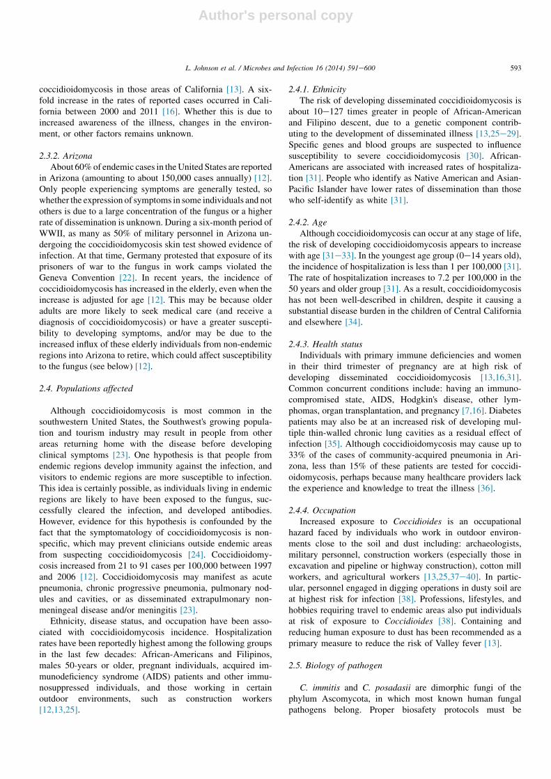
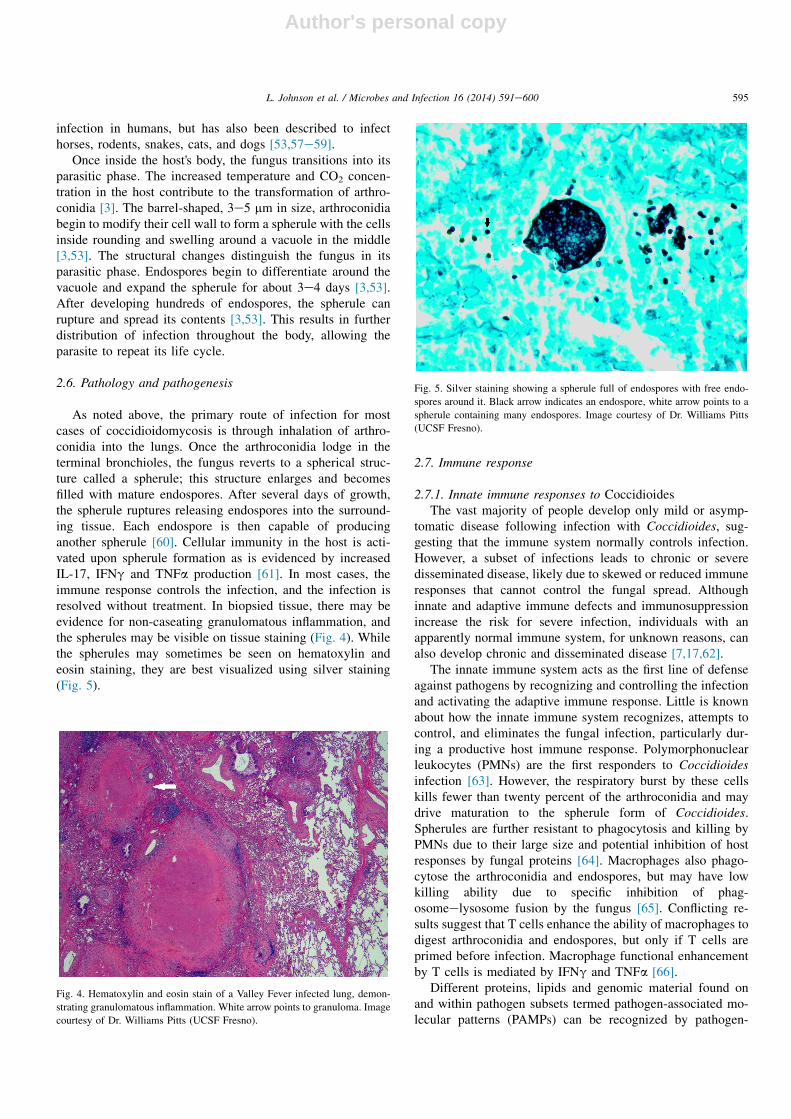
As noted above, the primary route of infection for mostcases of coccidioidomycosis is through inhalation of arthro-conidia into the lungs. Once the arthroconidia lodge in theterminal bronchioles, the fungus reverts to a spherical struc-ture called a spherule; this structure enlarges and becomesfilled with mature endospores. After several days of growth,the spherule ruptures releasing endospores into the surround-ing tissue. Each endospore is then capable of producinganother spherule [60]. Cellular immunity in the host is acti-vated upon spherule formation as is evidenced by increasedIL-17, IFNg and TNFa production [61]. In most cases, theimmune response controls the infection, and the infection isresolved without treatment. In biopsied tissue, there may beevidence for non-caseating granulomatous inflammation, andthe spherules may be visible on tissue staining (Fig. 4). Whilethe spherules may sometimes be seen on hematoxylin andeosin staining, they are best visualized using silver staining(Fig. 5).
2.7. Immune response
2.7.1. Innate immune responses to CoccidioidesThe vast majority of people develop only mild or asymp-
tomatic disease following infection with Coccidioides, sug-gesting that the immune system normally controls infection.However, a subset of infections leads to chronic or severedisseminated disease, likely due to skewed or reduced immuneresponses that cannot control the fungal spread. Althoughinnate and adaptive immune defects and immunosuppressionincrease the risk for severe infection, individuals with anapparently normal immune system, for unknown reasons, canalso develop chronic and disseminated disease [7,17,62].
The innate immune system acts as the first line of defenseagainst pathogens by recognizing and controlling the infectionand activating the adaptive immune response. Little is knownabout how the innate immune system recognizes, attempts tocontrol, and eliminates the fungal infection, particularly dur-ing a productive host immune response. Polymorphonuclearleukocytes (PMNs) are the first responders to Coccidioidesinfection [63]. However, the respiratory burst by these cellskills fewer than twenty percent of the arthroconidia and maydrive maturation to the spherule form of Coccidioides.Spherules are further resistant to phagocytosis and killing byPMNs due to their large size and potential inhibition of hostresponses by fungal proteins [64]. Macrophages also phago-cytose the arthroconidia and endospores, but may have lowkilling ability due to specific inhibition of phag-osomeelysosome fusion by the fungus [65]. Conflicting re-sults suggest that T cells enhance the ability of macrophages todigest arthroconidia and endospores, but only if T cells areprimed before infection. Macrophage functional enhancementby T cells is mediated by IFNg and TNFa [66].
Different proteins, lipids and genomic material found onand within pathogen subsets termed pathogen-associated mo-lecular patterns (PAMPs) can be recognized by pathogen-
Fig. 4. Hematoxylin and eosin stain of a Valley Fever infected lung, demon-
strating granulomatous inflammation. White arrow points to granuloma. Image
courtesy of Dr. Williams Pitts (UCSF Fresno).
Fig. 5. Silver staining showing a spherule full of endospores with free endo-
spores around it. Black arrow indicates an endospore, white arrow points to a
spherule containing many endospores. Image courtesy of Dr. Williams Pitts
(UCSF Fresno).
595L. Johnson et al. / Microbes and Infection 16 (2014) 591e600

Author's personal copy
recognition receptors (PRRs) on antigen presenting cells.Recognition of Coccidioides by a subset of these PRRs, Toll-like receptor 2 (TLR2) and TLR4, promotes TNFa productionand mediates a cell-mediated response to Coccidioides in vitro[67,68]. Another PRR important in recognition of fungal in-vasion, dectin-1, appears to promote innate immune cells todirect Th1 and Th17 effector responses in part by reducinginflammatory cytokine production [69]. Additionally, severalCoccidioides antigens have been identified with variableability to induce adaptive immune responses [68].
Dendritic cells (DC), as professional antigen presentingcells (APCs), are a critical link between innate and adaptiveimmune responses. Coccidioides antigens induce maturation,and activation of DCs and Coccidioides antigen-pulsed DCscan reverse the lymphocyte anergy found in disseminateddisease [70]. IL-12-induced Th1 responses are critical foreffective/productive immunity against Coccidioides infection.DCs are a major source of IL-12, and activated DCs presentantigen and costimulatory signals to naïve T cells; thus, DCsmight be expected to play an important role in host immuneresponses to Coccidioides. Together, the few studies evalu-ating DC responses during coccidioidomycosis suggest thatDC activation and antigen presentation is functional in patientswith disseminated disease [3]. However, it is unclear whetherthe DCs in these patients promote an effective or detrimentalresponse to Coccidioides. Perhaps the DCs in patients withdisseminated disease induce ineffective T helper effector re-sponses or promote immune tolerance. One study character-izing DC functions in mouse models of infection found higherTLR2, TLR4 and costimulatory molecule expression and IL-12 production in DCs of resistant mouse strains, comparedto susceptible mouse strains [71].
2.7.2. Adaptive immune responses to CoccidioidesCoccidioidomycosis induces both cell-mediated and hu-
moral immune responses. Protective immunity requires astrong Th1 skewed response resulting in production of IFNgand IgG2a antibodies. Asymptomatic immune patientsdemonstrate a strong delayed-type hypersensitivity (DTH)reaction and low levels of complement forming antibodies intheir serum, while severe disease is usually found in patientswith low DTH reactions and high titers of complementforming antibodies [70,72]. Recovery from severe disease isassociated with decreased complement forming antibodies andincreased DTH reaction [70]. Symptomatic patients develop Tcell anergy against Coccidioides that is generally specific forthis fungus and is reversible with disease remission [70,73,74].
It would therefore appear that humoral immunity plays aweak role in protection against this infection. As outlinedabove, the titer of complement forming antibodies correlateswell with disease severity. In further support, serum fromvaccinated mice does not protect from arthrospore infection,or depending upon the analysis, is less critical than T cells forprotection [75]. However, using a vaccine model, it was foundthat vaccine protection is less effective in the absence of Bcells, and that a B cell gene expression profile is associatedwith protection to arthroconidia [76]. Thus, the role of B cells
and antibodies in controlling infection or protection againstrepeat exposure remains unclear.
Further evidence supporting the critical role of T cells inimmunity to Coccidioides has been demonstrated using mousemodels of infection and evaluation of risk severity in patients.Mice lacking CD4þ or CD8þ T cells are more susceptible todisease, and T cells transfer protection to naive animals[77,78]. There is a high risk for dissemination and death inimmunosuppressed individuals during organ transplant, HIVinfection and neoplasia [79]. In HIV patients, the risk of severedisease increases with lower CD4þ T cell numbers [80].
The type of effector T cell response mounted againstCoccidioides appears to determine disease severity. Th1effector responses, particularly IFNg and TNFa production,are widely accepted as providing protective immunity againstCoccidioides that results primarily in mild or asymptomaticdisease. Assessment of T cell activation by CD69 expressionin coccidioidomycosis correlates with Th1 effector cytokinesand has been suggested to be a potential marker for measuringa productive cellular response to Coccidioides [81]. Incontrast, Th2 effector cytokine responses have largely beenassociated with more severe disease, perhaps in part due to theability of these cells to suppress macrophage activation andTh1 differentiation. While Th2-associated cytokines decreaseproductive immune responses against Coccidioides in mice, inpatients it is unclear if Th2 effectors have any direct effects onimmunity to Coccidioides, and overall Th2 cytokines are notproduced in response to Coccidioides antigens [3,82]. Th17effector responses have not yet been measured in patients withcoccidioidomycosis; however, evaluation of infection inimmunized mice indicates that disease susceptibility increaseswith the loss of Th17 functionality [83].
Regulatory T (Treg) cells are known to modulate immunityduring infection and their suppressive function can be bene-ficial or detrimental depending upon the site or stage of aninfection. Very little data exists evaluating the impact ofTreg cells on coccidioidomycosis. Reduced survival followinginfection in phagocytic NADPH oxidase-deficient micecorrelated with an expanded Treg cell population in the lung[84]. IL-10 producing cells, that may be secreted by Treg cellsor Th2 cells, have been found in clusters adjacent to gran-ulomata during coccidioidomycosis [85]. However, the roleand relative importance of IL-10 production in the granulomaand Treg presence in the lung is unknown.
2.7.3. Immune evasion by CoccidioidesMost pathogens utilize multiple mechanisms to escape host
immune detection, and Coccidioides expresses several docu-mented virulence factors that contribute to infection [86,87].As noted above, arthroconidia and spherules are highlyresistant to destruction by PMNs. The outer wall of arthro-conidia appears to protect from phagocytosis, as removing theouter wall increases uptake of the arthroconidia. In contrast,the immune system builds a response against the spheruleouter wall glycoprotein (SOWgp). Coccidioides endospores donot express SOWgp, thus avoiding immune detection by cellsresponsive to this antigen during a time when Coccidioides
596 L. Johnson et al. / Microbes and Infection 16 (2014) 591e600
Author's personal copy
can be more efficiently phagocytosed. The spherules arefurther protected from the immune system by the productionof proteases that digest antibodies. Finally, Coccidioides pro-duces urease and induces host production of arginase I, both ofwhich contribute to local tissue damage and enhance infection.Thus, while the host immune system fights to actively blockinfection or eliminate Coccidioides, the fungus has its ownmechanisms to circumvent, avoid or prevent immunesurveillance.
2.8. Diagnosis and treatment
2.8.1. DiagnosisEarly diagnosis of coccidioidomycosis is significant to
prevent disseminated disease, to reduce costs of hospitaliza-tions and treatment, and to avoid persistent infection leading totissue damage or death [2]. However, it is difficult to diagnoseearly infection for a couple of reasons. As described above,most individuals are asymptomatic. Some people exhibit flu-like symptoms, but do not seek medical evaluation becausetheir immune system resolves the infection over time withoutmedication. This also contributes to an underestimate of re-ported annual coccidioidomycosis cases. These limitations inself-diagnosis can lead to severe symptoms of chronic pneu-monia, meningitis, or bone and joint infection if the infectionbecomes a disseminated disease [3].
Individuals with coccidioidomycosis also have difficultiesobtaining proper diagnosis from clinicians and laboratorytesting. One assessment for patients who have persistent lunginfection is to obtain a radiographic examination. The resultsare frequently misinterpreted and patients are diagnosed withlung cancer, even though they may be infected with Cocci-dioides, which gives similar results on the X-ray [88]. Thereare other laboratory tests used to identify coccidioidomycosis,but they have limitations. Two commonly used diagnostictests, enzyme immunoassay testing and sputum testing, helpdetermine if a patient has been infected with Coccidioides.Enzyme immunoassay testing uses a patient's blood sample tomeasure Coccidioides antibodies. However, as many as 82%false-positive results for coccidioidomycosis have been re-ported with the antibody test [89]. These findings question theutility of the test for clinicians to diagnose the infection.Sputum culture requires a more invasive procedure to collectthe sample, but provides a more accurate result for diagnosis.Doctors can also evaluate patients by performing biopsies,joint effusions, or lumbar punctures [2].
2.8.2. TreatmentMost patients resolve coccidioidomycosis without need of
treatment. However, patients with chronic pulmonary ordisseminated disease may require antifungal therapy [2].Common antifungal drugs prescribed include: amphotericinB deoxycholate, lipid formulations of amphotericin, ketoco-nazole, fluconazole, and itraconazole [2]. Even thoughtreatment is beneficial in clearance of infection, it may comeat a cost to the patient, both financially and physically. Pa-tients who require long term medication of antifungal drugs
can spend up to $20,000 annually on top of hospital bills [2].Harmful side effects are associated with using antifungaldrugs, such as amphotericin B, which span from mildsymptoms of nausea, vomiting, fever, and hypoxia, to severeside effects of anemia, hypertension, hyperthermia, anddyspnea [90]. Routine follow-ups to the physician are rec-ommended after clearance of infection every 3e6 months forabout 2 years to prevent developing further complications ordisseminated disease [2]. However, chronically-infected pa-tients may need long term follow-up through examinationsand laboratory testing.
While there is limited data available for successful out-comes of treatment of immunocompromised individuals, onestudy demonstrated a significant reduction in relative risk fordeveloping symptomatic coccidioidomycosis among patientstreated with infliximab, a TNFa antagonist used for patientswith autoimmune disease [91].
2.8.3. VaccinesThe development of a successful coccidioidomycosis vac-
cine has long been a research goal despite numerous chal-lenges. A successful vaccine should show protective efficacyfor both immuno competent and immunocompromised in-dividuals [92]. Many vaccines developed to protect againstcoccidioidomycosis have failed to show conclusive results forvarious reasons, some of which we summarize below. Vac-cines containing killed organisms have shown optimum pro-tection in animal models but failed in human Phase III trialsdue to inadequate dosing. These vaccines also resulted inmajor side effects and pain, which presented an additionalissue. One potential solution for improving these killed vac-cines is to eliminate side effects while preserving the keyimmunogens by fractioning the vaccine components [93].Other vaccine trials in humans, which utilized auxotrophicmutants or attenuated live organisms, showed some survivaladvantages but failed to completely clear the fungus. Due tothe inherent potential risk of reversion to virulence of anattenuated mutant that exists for a live vaccine, recombinantproteins such as rAg2/Pra, rGel1, and CSA, have been pursuedfor vaccine trials. The majority of the recombinant antigens ofCoccidioides have failed to meet the benchmark of protectionrequired in murine vaccine trials [3]. Since these candidatevaccines failed to show necessary protection and ability tostimulate an effective immune response during animal testing,they will not be tested in humans.
Recent research has shown that vaccines with purifiedplasmid DNA provide superior protection against coccidioi-domycosis [92]. Currently, research is focused on pursuing apotential adjuvant vaccine designed to stimulate the appro-priate level of immune response [92]. Another line of researchfor a new potential vaccine involves complementary DNAexpression library immunization (ELI), which is underdevelopment along with use of parasitic cell wall proteins.Parasitic cell wall proteins can stimulate protective immunityagainst C. posadasii infection in mice and are considered themost protective antigens against coccidioidomycosis thusfar [94].
597L. Johnson et al. / Microbes and Infection 16 (2014) 591e600

Author's personal copy
3. Concluding remarks
Research and recognition of coccidioidomycosis has pro-gressed slowly since the first patient was diagnosed in 1892.However, increasing awareness of coccidioidomycosis cancontribute to improving methods to prevent and combat thisdisease. Prevention is the first step to managing this fungalinfection. One way to do so is by locating areas with highprevalence of Coccidioides. Because the fungus is endemic tocertain regions of the United States, researchers can study soilsampling in those areas. In turn, the public can be informed tobe cautious when a particular site contains high levels ofCoccidioides. Further improvement in technology of an on-sitetest for Coccidioides in the soil could add the benefit for turn-around time of evaluating a potential habitat for the fungus.Another preventive measure would be to understand theweather patterns during increased coccidioidomycosis cases.Dry periods after wet winters or summer have been associatedwith an increase in the rate of coccidioidomycosis cases forCalifornia and Arizona [53]. Collecting data on temperaturechanges and wind patterns, along with reports of coccidioi-domycosis, will be informative in assessing correlations be-tween environmental changes and infection rates. With thesefindings, individuals can consider seeking medical attention ifthey have symptoms related to Valley Fever during periods ofincreased likelihood of infection. Remaining indoors duringdust storms, for example, provides a preventive approachagainst exposure to arthroconidia.
Improving diagnostic testing and early detection can alsoincrease prevention of disseminated disease. Not only couldpatients benefit from early recognition, but also a more ac-curate number of reported cases could be tracked. Monitoringpatients during acute infection could in understanding themechanisms and progression of disease. This is critical infilling gaps in knowledge of Valley Fever for physicians. Oneuncertainty is whether a patient needs to be treated withantifungal drugs. If treatment is found to be needed, it is notknown which drug is most effective, which dosage should beadministrated, and what should be the duration of usage.Another question to be addressed is when a patient has clearedinfection, how long should the patient continue to be followed-up?
Further research in the development and proliferation of thefungus in the host can aid in our understanding of an effectiveapproach to clear the pathogen. It is unclear what regulatoryevents contribute to the transformation of arthroconidia intospherules after entry into the body. Characterizing thesignaling for the transformation could contribute to preventingearly infection. Studying the fungus could also contribute tothe development of a vaccine. A number of vaccines have beendeveloped and tested. Indeed, several coccidioidal antigenshave shown protective properties against the fungus in animalmodels. Thus far, however, a successful vaccine showing longterm immunity in humans has not yet been achieved [95e97].Overall, the approach to resolve this hidden danger in a dustcloud is to improve detection methods for Coccidioides,improve reporting of infection cases, and better understand the
immune response as a way of predicting which patients are atrisk for disseminated disease.
Conflict of interest
The authors declare that they have no conflict of interest.
Acknowledgments
We thank Trevor P. Hirst from the Health SciencesResearch Institute (HSRI) at UC Merced for acquiring thephotograph of the tractor and creating the map outlining thedistribution of coccidioidomycosis. We are grateful to Caro-line Chen for her contributions to the graphical design of thefungal life cycle. CJN and KH were supported by the NationalInstitutes of Health (NIH) grants K99AI100896 andR00HL090706, respectively. Research on Valley Fever at UCMerced is supported by the UC Merced Blum Center forDeveloping Economies, the UC Merced HSRI, a University ofCalifornia Presidential Chair, and Children's Hospital CentralCalifornia.
References
[1] Smith CE. Epidemiology of acute coccidioidomycosis with erythema
nodosum (“San Joaquin” or “Valley Fever”). Am J Public Health Nations
Health 1940;30:600e11.
[2] Galgiani JN, Ampel NM, Blair JE, Catanzaro A, Johnson RH,
Stevens DA, et al. Coccidioidomycosis. Clin Infect Dis
2005;41:1217e23.
[3] Cox RA, Magee DM. Coccidioidomycosis: host response and vaccine
development. Clin Microbiol Rev 2004;17:804e39.[4] Sondermeyer G, Lee L, Gilliss D, Tabnak F, Vugia D. Coccidioidomy-
cosis-associated hospitalizations, California, USA, 2000e2011. Emerg
Infect Dis 2013;19:1590e8.[5] Blair JE, Logan JL. Coccidioidomycosis in solid organ transplantation.
Clin Infect Dis 2001;33:1536e44.
[6] Williams PL, Sable DL, Mendez P, Smyth LT. Symptomatic coccidioi-
domycosis following a severe natural dust storm. An outbreak at the
Naval Air Station, Lemoore, Calif. Chest 1979;76:566e70.
[7] Hector RF, Laniado-Laborin R. Coccidioidomycosisda fungal disease of
the Americas. PLoS Med 2005;2:e2.
[8] Fisher MC, Koenig GL, White TJ, Taylor JW. Molecular and phenotypic
description of Coccidioides posadasii sp. nov., previously recognized as
the non-California population of Coccidioides immitis. Mycologia
2002;94:73e84.[9] Campins H. Coccidioidomycosis in South America. A review of its
epidemiology and geographic distribution. Mycopathol Mycol Appl
1970;41:25e34.
[10] Hurst S, Gade L, Marsden-Haug N, Engelthaler D, Hill H, Ralston C,
et al. Molecular detection and isolation of Coccidioides immitis from soil
in Washington State. In: Cocci study group 58th annual meeting,
Phoenix, Arizona; 2014.
[11] Posada A. Un nuevo caso de micosis fungoidea con psorosperm ias.
Anales del Circulo Medico Argent 1892;15:585e97.
[12] Sunenshine RH, Anderson S, Erhart L, Vossebrink A, Kelly PC,
Engelthaler D, et al. Public health surveillance for coccidioidomycosis in
Arizona. Ann N Y Acad Sci 2007;1111:96e102.[13] Kirkland TN, Fierer J. Coccidioidomycosis: a reemerging infectious
disease. Emerg Infect Dis 1996;2:192.
[14] Laniado-Laborin R. Expanding understanding of epidemiology of
coccidioidomycosis in the Western hemisphere. Ann N Y Acad Sci
2007;1111:19e34.
598 L. Johnson et al. / Microbes and Infection 16 (2014) 591e600
Author's personal copy
[15] Hirschmann JV. The early history of coccidioidomycosis: 1892-1945.
Clin Infect Dis 2007;44:1202e7.
[16] Sondermeyer G, Lee L, Gilliss D, Tabnak F, Vugia D. Coccidioidomy-
cosis-associated hospitalizations, California, USA, 2000e2011. Emerg
Infect Dis 2013;19.
[17] Stevens DA. Coccidioidomycosis. N Engl J Med 1995;332:1077e82.
[18] Laniado-Laborín R. Coccidioidomycosis and other endemic mycoses in
Mexico. Rev Iberoam Micol 2007;24:249.
[19] Seitz AE, Prevots DR, Holland SM. Hospitalizations associated with
disseminated coccidioidomycosis, Arizona and California, USA. Emerg
Infect Dis 2012;18:1476.
[20] Smith CE, Beard RR. Varieties of coccidioidal infection in relation to the
epidemiology and control of the diseases. Am J Public Health Nations
Health 1946;36:1394e402.
[21] Faber HK, Smith CE, Dickson EC. Acute coccidioidomycosis with er-
ythema nodosum in children. J Pediatr 1939;15:163e71.[22] Galgiani JN. Coccidioidomycosis: changing perceptions and creating
opportunities for its control. Ann N Y Acad Sci 2007;1111:1e18.
[23] Parish JM, Blair JE. Coccidioidomycosis. In: Mayo Clin Proceedings.
Elsevier; 2008. pp. 343e9.
[24] Chaturvedi V, Ramani R, Gromadzki S, Rodeghier B, Chang H-G,
Morse DL. Coccidioidomycosis in New York state. Emerg Infect Dis
2000;6:25.
[25] Hector RF, Rutherford GW, Tsang CA, Erhart LM, McCotter O,
Anderson SM, et al. The public health impact of coccidioidomycosis in
Arizona and California. Int J Environ Res Publ 2011;8:1150e73.
[26] Pappagianis D. Epidemiology of coccidioidomycosis. In: Curr Top Med
Mycol. Springer; 1988. pp. 199e238.
[27] Crum NF, Lederman ER, Stafford CM, Parrish JS, Wallace MR.
Coccidioidomycosis: a descriptive survey of a reemerging disease.
clinical characteristics and current controversies. J Med
2004;83:149e75.
[28] Pappagianis D. Coccidioidomycosis. Semin Dermatol 1993:301.
[29] Pappagianis D, Lindsay S, Beall S, Williams P. Ethnic background and
the clinical course of coccidioidomycosis. Am Rev Respir Dis
1979;120:959e61.
[30] Louie L, Ng S, Hajjeh R, Johnson R, Vugia D, Werner SB, et al. Influ-
ence of host genetics on the severity of coccidioidomycosis. Emerg Infect
Dis 1999;5:672.
[31] Hector R, Rutherford GW. The public health need and present status of a
vaccine for the prevention of coccidioidomycosis. Ann N Y Acad Sci
2007;1111:259e68.[32] Einstein HE, Johnson RH. Coccidioidomycosis: new aspects of epide-
miology and therapy. Clin Infect Dis 1993:349e54.
[33] Rosenstein NE, Emery KW, Werner SB, Kao A, Johnson R, Rogers D,
et al. Risk factors for severe pulmonary and disseminated coccidioido-
mycosis: Kern County, California, 1995e1996. Clin Infect Dis
2001;32:708e14.
[34] McCarty JM, Demetral LC, Dabrowski L, Kahal AK, Bowser AM,
Hahn JE. Pediatric coccidioidomycosis in central California: a retro-
spective case series. Clin Infect Dis 2013;56:1579e85.
[35] Baker EJ, Hawkins JA, Waskow EA. Surgery for coccidioidomycosis in
52 diabetic patients with special reference to related immunologic fac-
tors. J Thorac Cardiovasc Surg 1978;75:680e7.
[36] Chen S, Erhart LM, Anderson S, Komatsu K, Park B, Chiller T, et al.
Coccidioidomycosis: knowledge, attitudes, and practices among health-
care providers-Arizona, 2007. Med Mycol 2011;49:649e56.
[37] Petersen LR, Marshall SL, Barton C, Hajjeh RA, Lindsley MD,
Warnock DW, et al. Coccidioidomycosis among workers at an archeo-
logical site, northeastern Utah. Emerg Infect Dis 2004;10:637.
[38] Sipsas NV, Kontoyiannis DP. Occupation, lifestyle, diet, and invasive
fungal infections. J Infect 2008;36:515e25.
[39] Cummings KC, McDowell A, Wheeler C, McNary J, Das R, Vugia DJ,
et al. Point-source outbreak of coccidioidomycosis in construction
workers. Epidemiol Infect 2010;138:507e11.
[40] Gehlbach SH, Hamilton JD, Conant NF. Coccidioidomycosis: an occu-
pational disease in cotton mill workers. Arch Intern Med 1973;131:254.
[41] Welsh O, Vera-Cabrera L, Rendon A, Gonzalez G, Bonifaz A. Coccid-
ioidomycosis. Clin Dermatol 2012;30:573e91.
[42] Depiazzi LJ, Roberts WD, Hawkins CD, Palmer MA, Pitman DR,
McQuade NC, et al. Severity and persistence of footrot in Merino sheep
experimentally infected with a protease thermostable strain of Dichelo-
bacter nodosus at five sites. Aust Vet J 1998;76:32e8.
[43] Burt A, Carter DA, Koenig GL, White TJ, Taylor JW. Molecular markers
reveal cryptic sex in the human pathogen Coccidioides immitis. Proc Natl
Acad Sci U S A 1996;93:770e3.
[44] Fisher MC, Koenig G, White TJ, Taylor JW. A test for concordance
between the multilocus genealogies of genes and microsatellites in the
pathogenic fungus Coccidioides immitis. Mol Biol Evol
2000;17:1164e74.
[45] Koufopanou V, Burt A, Taylor JW. Concordance of gene genealogies
reveals reproductive isolation in the pathogenic fungus Coccidioides
immitis. Proc Natl Acad Sci U S A 1997;94:5478e82.[46] Koufopanou V, Burt A, Szaro T, Taylor JW. Gene genealogies, cryptic
species, and molecular evolution in the human pathogen Coccidioides
immitis and relatives (Ascomycota, Onygenales). Mol Biol Evol
2001;18:1246e58.
[47] Fraser JA, Stajich JE, Tarcha EJ, Cole GT, Inglis DO, Sil A, et al.
Evolution of the mating type locus: insights gained from the dimorphic
primary fungal pathogens Histoplasma capsulatum, Coccidioides immi-
tis, and Coccidioides posadasii. Eukaryot Cell 2007;6:622e9.
[48] Mandel MA, Barker BM, Kroken S, Rounsley SD, Orbach MJ. Genomic
and population analyses of the mating type loci in Coccidioides species
reveal evidence for sexual reproduction and gene acquisition. Eukaryot
Cell 2007;6:1189e99.
[49] Lacy GH, Swatek FE. Soil ecology of Coccidioides immitis at Amerin-
dian middens in California. Appl Microbiol 1974;27:379e88.[50] Maddy KT. The geographic distribution of Coccidioides immitis and
possible ecologic implications. Ariz Med 1958;15:178e88.
[51] Greene DR, Koenig G, FisherMC, Taylor JW. Soil isolation andmolecular
identification of Coccidioides immitis. Mycologia 2000;92:406e10.[52] Huppert M, Sun SH, Harrison JL. Morphogenesis throughout saprobic
and parasitic cycles of Coccidioides immitis. Mycopathologia
1982;78:107e22.
[53] Nguyen C, Barker BM, Hoover S, Nix DE, Ampel NM, Frelinger JA,
et al. Recent advances in our understanding of the environmental,
epidemiological, immunological, and clinical dimensions of coccidioi-
domycosis. Clin Microbiol Rev 2013;26:505e25.
[54] Egeberg RO, Elconin AE, Egeberg MC. Effect of salinity and tempera-
ture on Coccidioides immitis and three antagonistic soil saprophytes. J
Bacteriol 1964;88:473e6.
[55] Schieffelin JS, Torrellas M, Lartchenko S, Gill F, Garcia-Diaz J,
McGoey R. How natural disasters change natural patterns: coccidioido-
mycosis imported to New Orleans. J La State Med Soc 2013;165:145e9.
[56] Centers for Disease Control and Prevention, (CDC). Sources of coccid-
ioidomycosis (Valley fever); 2013.
[57] Ziemer EL, Pappagianis D, Madigan JE, Mansmann RA, Hoffman KD.
Coccidioidomycosis in horses: 15 cases (1975e1984). J Am Vet Med
Assoc 1992;201:910e6.
[58] Graupmann-Kuzma A, Valentine BA, Shubitz LF, Dial SM, Watrous B,
Tornquist SJ. Coccidioidomycosis in dogs and cats: a review. J Am Anim
Hosp Assoc 2008;44:226e35.
[59] Timm KI, Sonn RJ, Hultgren BD. Coccidioidomycosis in a Sonoran
gopher snake, Pituophis melanoleucus affinis. J Med Vet Mycol
1988;26:101e4.
[60] Saubolle MA, McKellar PP, Sussland D. Epidemiologic, clinical, and
diagnostic aspects of coccidioidomycosis. J Clin Microbiol
2007;45:26e30.
[61] Nesbit LA, Knox KS, Nguyen CT, Roesch J, Wheat LJ, Johnson SM,
et al. Immunological characterization of bronchoalveolar lavage fluid in
patients with acute pulmonary coccidioidomycosis. J Infect Dis
2013;208:857e63.
[62] Ampel NM. New perspectives on coccidioidomycosis. Proc Am Thorac
Soc 2010;7:181e5.
599L. Johnson et al. / Microbes and Infection 16 (2014) 591e600

Author's personal copy
[63] Drutz DJ, Huppert M. Coccidioidomycosis: factors affecting the host-
parasite interaction. J Infect Dis 1983;147:372e90.
[64] Frey CL, Drutz DJ. Influence of fungal surface components on the
interaction of Coccidioides immitis with polymorphonuclear neutrophils.
J Infect Dis 1986;153:933e43.[65] Beaman L, Holmberg CA. In vitro response of alveolar macrophages to
infection with Coccidioides immitis. Infect Immun 1980;28:594e600.
[66] Beaman L. Effects of recombinant gamma interferon and tumor necrosis
factor on in vitro interactions of human mononuclear phagocytes with
Coccidioides immitis. Infect Immun 1991;59:4227e9.
[67] Viriyakosol S, Fierer J, Brown GD, Kirkland TN. Innate immunity to the
pathogenic fungus Coccidioides posadasii is dependent on Toll-like re-
ceptor 2 and Dectin-1. Infect Immun 2005;73:1553e60.
[68] Ampel NM. The complex immunology of human coccidioidomycosis.
Ann N Y Acad Sci 2007;1111:245e58.
[69] Viriyakosol S, Jimenez Mdel P, Gurney MA, Ashbaugh ME, Fierer J.
Dectin-1 is required for resistance to coccidioidomycosis in mice. MBio
2013;4. e00597e12.
[70] Cox RA, Vivas JR. Spectrum of in vivo and in vitro cell-mediated im-
mune responses in coccidioidomycosis. Cell Immunol 1977;31:130e41.
[71] Awasthi S, Magee DM. Differences in expression of cell surface co-
stimulatory molecules, Toll-like receptor genes and secretion of IL-12
by bone marrow-derived dendritic cells from susceptible and resistant
mouse strains in response to Coccidioides posadasii. Cell Immunol
2004;231:49e55.
[72] Ampel NM. Measurement of cellular immunity in human coccidioido-
mycosis. Mycopathologia 2003;156:247e62.[73] Smith CE, Beard RR, Saito MT. Pathogenesis of coccidioidomycosis
with special reference to pulmonary cavitation. Ann Intern Med
1948;29:623e55.
[74] Ibrahim AB, Pappagianis D. Experimental induction of anergy to coc-
cidioidin by antigens of Coccidioides immitis. Infect Immun
1973;7:786e94.
[75] Beaman LV, Pappagianis D, Benjamini E. Mechanisms of resistance to
infection with Coccidioides immitis in mice. Infect Immun
1979;23:681e5.
[76] Magee DM, Friedberg RL, Woitaske MD, Johnston SA, Cox RA. Role of
B cells in vaccine-induced immunity against coccidioidomycosis. Infect
Immun 2005;73:7011e3.
[77] Beaman L, Pappagianis D, Benjamini E. Significance of T cells in
resistance to experimental murine coccidioidomycosis. Infect Immun
1977;17:580e5.
[78] Fierer J, Waters C, Walls L. Both CD4þ and CD8þ T cells can mediate
vaccine-induced protection against Coccidioides immitis infection in
mice. J Infect Dis 2006;193:1323e31.
[79] Centers for Disease Control and Prevention, (CDC). Coccidioidomycosis
(Valley fever) (Coccidioides spp.); 2012.
[80] Ampel NM, Dols CL, Galgiani JN. Coccidioidomycosis during human
immunodeficiency virus infection: results of a prospective study in a
coccidioidal endemic area. Am J Med 1993;94:235e40.
[81] Ampel NM, Kramer LA, Li L, Carroll DS, Kerekes KM, Johnson SM,
et al. In vitro whole-blood analysis of cellular immunity in patients with
active coccidioidomycosis by using the antigen preparation T27K. Clin
Diagn Lab Immunol 2002;9:1039e43.
[82] Corry DB, Ampel NM, Christian L, Locksley RM, Galgiani JN. Cytokine
production by peripheral blood mononuclear cells in human coccidioi-
domycosis. J Infect Dis 1996;174:440e3.
[83] Hung CY, Gonzalez A, Wuthrich M, Klein BS, Cole GT. Vaccine im-
munity to coccidioidomycosis occurs by early activation of three signal
pathways of T helper cell response (Th1, Th2, and Th17). Infect Immun
2011;79:4511e22.
[84] Gonzalez A, Hung CY, Cole GT. Absence of phagocyte NADPH oxidase
2 leads to severe inflammatory response in lungs of mice infected with
Coccidioides. Microb Pathog 2011;51:432e41.
[85] Li L, Dial SM, Schmelz M, Rennels MA, Ampel NM. Cellular immune
suppressor activity resides in lymphocyte cell clusters adjacent to gran-
ulomata in human coccidioidomycosis. Infect Immun 2005;73:3923e8.
[86] Hung CY, Xue J, Cole GT. Virulence mechanisms of Coccidioides. Ann
N Y Acad Sci 2007;1111:225e35.[87] Galgiani JN. Coccidioidomycosis. West J Med 1993;159:153e71.
[88] Petrini B, Skold CM, Bronner U, Elmberger G. Coccidioidomycosis
mimicking lung cancer. Respiration 2003;70:651e4.
[89] Kuberski T, Herrig J, Pappagianis D. False-positive IgM serology in
coccidioidomycosis. J Clin Microbiol 2010;48:2047e9.
[90] Laniado-Laborin R, Cabrales-Vargas MN. Amphotericin B: side effects
and toxicity. Rev Iberoam Micol 2009;26:223e7.
[91] Bergstrom L, Yocum DE, Ampel NM, Villanueva I, Lisse J, Gluck O,
et al. Increased risk of coccidioidomycosis in patients treated with tumor
necrosis factor alpha antagonists. Arthritis Rheum 2004;50:1959e66.
[92] Cole GT, Hung CY, Sanderson SD, Hurtgen BJ, Wuthrich M, Klein BS,
et al. Novel strategies to enhance vaccine immunity against coccidioi-
domycosis. PLoS Pathog 2013;9:e1003768.
[93] Yoon HJ, Clemons KV. Vaccines against coccidioides. Korean J Intern
Med 2013;28:403e7.[94] Tarcha EJ, Basrur V, Hung CY, Gardner MJ, Cole GT. Multivalent re-
combinant protein vaccine against coccidioidomycosis. Infect Immun
2006;74:5802e13.
[95] Pappagianis D. Evaluation of the protective efficacy of the killed Coc-
cidioides immitis spherule vaccine in humans. The Valley fever vaccine
study Group. Am Rev Respir Dis 1993;148:656e60.
[96] Levine HB, Miller RL, Smith CE. Influence of vaccination on respiratory
coccidiodial disease in cynomolgus monkeys. J Immunol
1962;89:242e51.
[97] Johnson SM, Lerche NW, Pappagianis D, Yee JL, Galgiani JN,
Hector RF. Safety, antigenicity, and efficacy of a recombinant coccidi-
oidomycosis vaccine in cynomolgus macaques (Macaca fascicularis).
Ann N Y Acad Sci 2007;1111:290e300.
600 L. Johnson et al. / Microbes and Infection 16 (2014) 591e600