valence state of europium doping ions during pulsed-laser deposition
TRANSCRIPT
Valence state of europium doping ions during pulsed-laser deposition
This article has been downloaded from IOPscience. Please scroll down to see the full text article.
2011 J. Phys. D: Appl. Phys. 44 375402
(http://iopscience.iop.org/0022-3727/44/37/375402)
Download details:
IP Address: 164.81.214.248
The article was downloaded on 20/12/2011 at 14:38
Please note that terms and conditions apply.
View the table of contents for this issue, or go to the journal homepage for more
Home Search Collections Journals About Contact us My IOPscience
IOP PUBLISHING JOURNAL OF PHYSICS D: APPLIED PHYSICS
J. Phys. D: Appl. Phys. 44 (2011) 375402 (9pp) doi:10.1088/0022-3727/44/37/375402
Valence state of europium doping ionsduring pulsed-laser depositionA Pillonnet1, A Pereira1, O Marty2 and C Champeaux3
1 Universite de Lyon, Lyon, F-69003, France; Universite Lyon 1; CNRS UMR 5620; Laboratoire dePhysico-Chimie des Materiaux Luminescents, Bat. Kastler, 10 rue Ampere, Villeurbanne, F-69622,France2 INL institut des Nanotechnologies de Lyon; UMR 5270 CNRS-UCBL-INSA-ECL; Bat Leon Brillouin;Universite de Lyon, France3 Science des Procedes Ceramiques et de Traitements de Surface; Faculte des Sciences et Techniques,CNRS UMR 6638, 123 avenue Albert Thomas, 87060 Limoges Cedex, France
E-mail: [email protected]
Received 21 April 2011, in final form 18 July 2011Published 31 August 2011Online at stacks.iop.org/JPhysD/44/375402
AbstractThe evolution of europium as a doping ion during the pulsed-laser deposition process ofEu : Al2O3 films has been studied. A decrease in oxygen pressure in the deposition chambergenerated the growth of γ -Al2O3 crystallites and a conversion of the 3+ to a 2+ valence stateof europium ions. Excitation-selective emission of Eu2+ and fluorescence line narrowing ofEu3+ revealed that two kinds of europium site families were created in the alumina matrix.Time-of-flight emission spectroscopy shows that oxygen came preferentially from the targetfor the studied range of pressure.
1. Introduction
Over recent years, the europium ion has received increasingattention due to its exceptional spectroscopic properties. Forexample, trivalent europium has been studied as an efficient redemitter [1] and it has been investigated for use in polychromaticdisplays and mercury free-lamps [2]. It has also been usedas an excellent structural probe, because its spectral anddynamic fluorescence properties are strongly related to itslocal environment [3–5]. Europium divalent ions are alsointeresting for their green–blue emission. For this purpose,reduction of Eu3+ to Eu2+ can be obtained by laser induction [6],chemical manipulation [7] or by annealing under a reducingatmosphere [8, 9].
The emission properties of the rare-earth ions can betuned by choosing the host matrices. To obtain luminescentenhancement, specific properties of the host matrices, suchas low phonon energy and high emission cross-section, areessential. Owing to its optical properties, alumina is nowa commonly used low-cost host material. In addition to itspotential for spectroscopic experiments, alumina exhibits goodchemical and mechanical properties that make it an interestingmaterial for various applications (e.g. catalytic). Severalcrystallographic phases of alumina are known, α-Al2O3 being
the most stable phase. However, Eu3+ ions have a larger sizethan Al3+ (∼0.95 Å and 0.53 Å, respectively). As a result, theincorporation of Eu3+ ions in alumina is challenging and it hasbeen demonstrated that the presence of europium ions preventsthe α-phase formation and promotes the thermal stability ofγ -Al2O3 [10, 11]. The latter remains stable up to ∼900 ◦C, andthe transformation from γ - into α-Al2O3 requires annealingat higher temperatures. It is thus expected that the methodused to synthesize Eu3+-doped alumina and the conditionsprevailing during the synthesis will have a strong influenceon the structure of the resulting material.
Most studies dealing with Eu-doped alumina have beenperformed on powders that were obtained by direct or indirectcombustion [9, 10, 12–14], Pechini methods [15] and solidstate reactions [15] to name only a few. Another interestingway to study the properties of this material and to obtainadditional information is to use Eu-doped alumina in the formof thin films. In most of these cases, Eu3+-doped alumina filmshave been fabricated by sol–gel processes [16–19]. However,Ishizaka et al [17] have shown that the existence of OH groupscan affect the luminescence properties of doped-alumina filmswith shorter decay times and quenching of luminescence.Moreover, chemical contamination can occur and a precisecontrol of the film thickness is difficult to achieve by thismethod.
0022-3727/11/375402+09$33.00 1 © 2011 IOP Publishing Ltd Printed in the UK & the USA
J. Phys. D: Appl. Phys. 44 (2011) 375402 A Pillonnet et al
Pulsed-laser deposition (PLD) is known as a versatile andpowerful method for the synthesis of high purity thin filmsand oxides of complex stoichiometry [20, 21]. The flexibilityof PLD allows for the deposition of films with radicallydifferent structural properties. Recent studies [22–24] havedemonstrated that the film properties can be tuned by adjustingthe kinetic energy of the ablated species. In particular,deposition at high kinetic energy values gives dense andsmooth films, whereas deposition at low kinetic energyvalues favours porous and textured films with nanoparticlesof controlled size. On the other hand, any change in themicro-nanostructure will affect the film properties (i.e. optical,electrochemical characteristics, etc) [24, 25]. However, upto now, only a few reports have appeared in the literatureconcerning PLD synthesis of europium-doped alumina films[26, 27]. In most of these cases, the studies were mainlyconcerned with erbium [28], thulium [28, 29] and chromium[30, 31] doped Al2O3 thin films or simple alumina filmswhich can be used as dielectric amorphous waveguiding films[25, 26, 32], matrix [5, 33–35] or porous alumina membranes[36], for example. In all the cases, to obtain the desiredoxide material properties, it is necessary to control the oxygenstoichiometry of the alumina film. Since in some cases anoxygen substrate contribution has been observed during thegrowth of oxide thin films [37], the key parameter to avoiddeficiencies in the oxygen content of the film is the backgroundatmosphere. In that respect, Espurescu et al [38] have providedclear evidence through optical emission spectroscopy that, inan O2 background, dissociation of molecular oxygen occursat the boundary of the plasma region, and therefore increasesthe possibility of oxide formation. Also, we assumed that theoxygen background could modify the valence state of dopingions during the deposition of europium-doped alumina films.This is important since the luminescent properties of Eu2+ andEu3+ differ from each other.
In previous studies, Al2O3 waveguides were grown byPLD and the analysis of their structure and composition werepresented in terms of the pressure during deposition and theannealing temperature. The stabilization of gamma phase byeuropium doping have also been presented. In this study,we present first the structure at nanometric scale of Eu-dopedalumina films prepared by PLD using x-ray diffraction (XRD)analysis and transmission electron spectroscopy. Second, thespectroscopic properties of these films were investigated, andthe sites of europium ions in grown films as compared with theinitial target were studied. Third, the plasma was analysed toinvestigate the factors affecting the valence state of europiumin the films during deposition.
2. Materials and methods
Europium-doped alumina films were obtained by PLD in anoxygen gas atmosphere. The Eu : Al2O3 target was ablatedwith a focused KrF laser beam (wavelength λ = 248 nm, laserpulse duration τ = 20 ns) operating at 10 Hz. The targets weremade from sintered powders, which were prepared by the sol–gel method from aluminium butoxide [18], the sol being dopedby Eu3+ ions with a 1% atomic ratio relative to aluminium. The
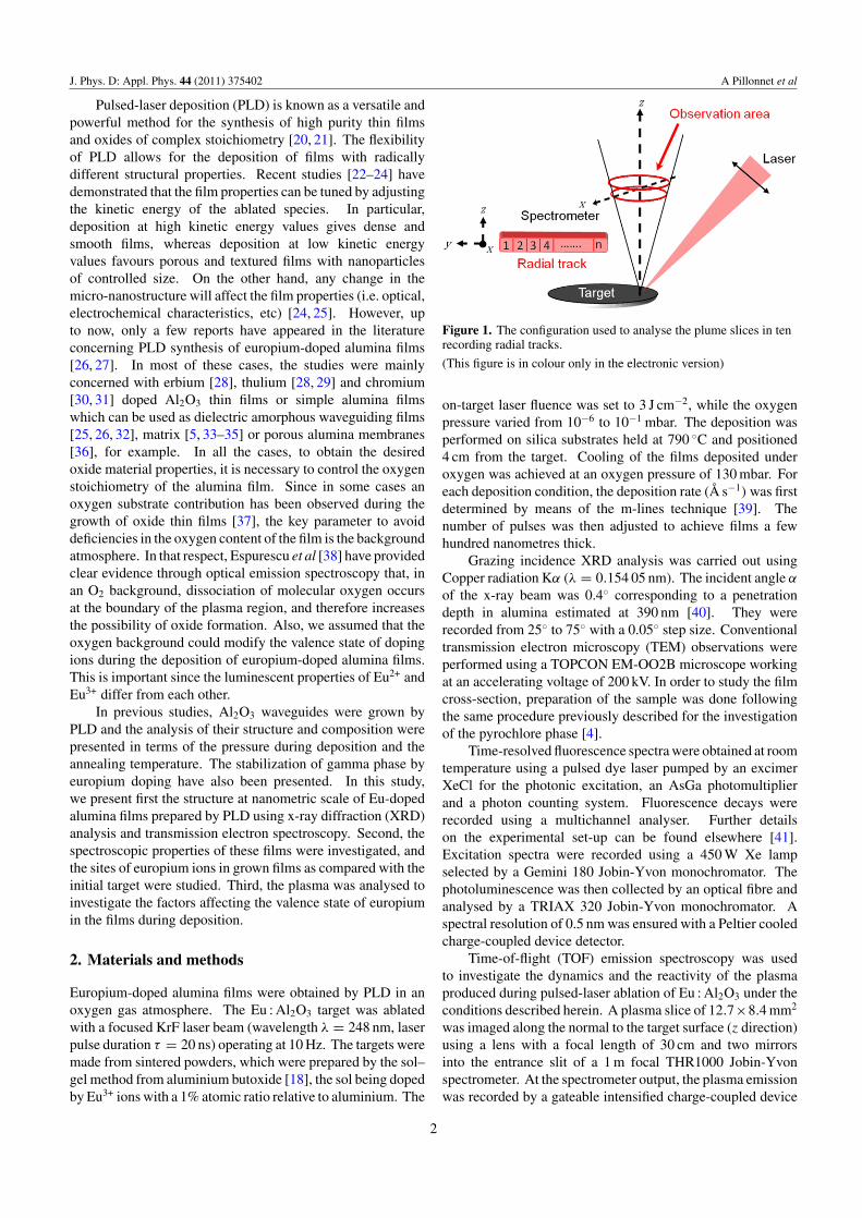
Figure 1. The configuration used to analyse the plume slices in tenrecording radial tracks.
(This figure is in colour only in the electronic version)
on-target laser fluence was set to 3 J cm−2, while the oxygenpressure varied from 10−6 to 10−1 mbar. The deposition wasperformed on silica substrates held at 790 ◦C and positioned4 cm from the target. Cooling of the films deposited underoxygen was achieved at an oxygen pressure of 130 mbar. Foreach deposition condition, the deposition rate (Å s−1) was firstdetermined by means of the m-lines technique [39]. Thenumber of pulses was then adjusted to achieve films a fewhundred nanometres thick.
Grazing incidence XRD analysis was carried out usingCopper radiation Kα (λ = 0.154 05 nm). The incident angle α
of the x-ray beam was 0.4◦ corresponding to a penetrationdepth in alumina estimated at 390 nm [40]. They wererecorded from 25◦ to 75◦ with a 0.05◦ step size. Conventionaltransmission electron microscopy (TEM) observations wereperformed using a TOPCON EM-OO2B microscope workingat an accelerating voltage of 200 kV. In order to study the filmcross-section, preparation of the sample was done followingthe same procedure previously described for the investigationof the pyrochlore phase [4].
Time-resolved fluorescence spectra were obtained at roomtemperature using a pulsed dye laser pumped by an excimerXeCl for the photonic excitation, an AsGa photomultiplierand a photon counting system. Fluorescence decays wererecorded using a multichannel analyser. Further detailson the experimental set-up can be found elsewhere [41].Excitation spectra were recorded using a 450 W Xe lampselected by a Gemini 180 Jobin-Yvon monochromator. Thephotoluminescence was then collected by an optical fibre andanalysed by a TRIAX 320 Jobin-Yvon monochromator. Aspectral resolution of 0.5 nm was ensured with a Peltier cooledcharge-coupled device detector.
Time-of-flight (TOF) emission spectroscopy was usedto investigate the dynamics and the reactivity of the plasmaproduced during pulsed-laser ablation of Eu : Al2O3 under theconditions described herein. A plasma slice of 12.7×8.4 mm2
was imaged along the normal to the target surface (z direction)using a lens with a focal length of 30 cm and two mirrorsinto the entrance slit of a 1 m focal THR1000 Jobin-Yvonspectrometer. At the spectrometer output, the plasma emissionwas recorded by a gateable intensified charge-coupled device
2
J. Phys. D: Appl. Phys. 44 (2011) 375402 A Pillonnet et al
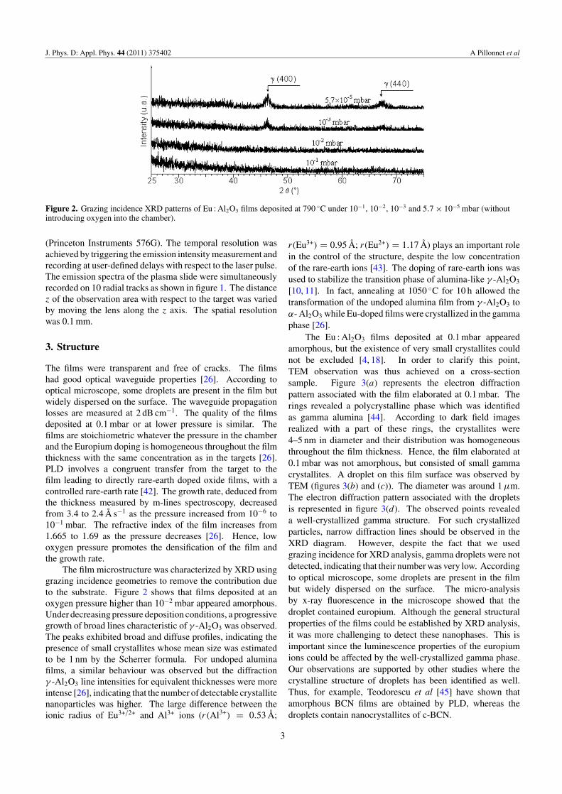
Figure 2. Grazing incidence XRD patterns of Eu : Al2O3 films deposited at 790 ◦C under 10−1, 10−2, 10−3 and 5.7 × 10⁻⁵ mbar (withoutintroducing oxygen into the chamber).
(Princeton Instruments 576G). The temporal resolution wasachieved by triggering the emission intensity measurement andrecording at user-defined delays with respect to the laser pulse.The emission spectra of the plasma slide were simultaneouslyrecorded on 10 radial tracks as shown in figure 1. The distancez of the observation area with respect to the target was variedby moving the lens along the z axis. The spatial resolutionwas 0.1 mm.
3. Structure
The films were transparent and free of cracks. The filmshad good optical waveguide properties [26]. According tooptical microscope, some droplets are present in the film butwidely dispersed on the surface. The waveguide propagationlosses are measured at 2 dB cm−1. The quality of the filmsdeposited at 0.1 mbar or at lower pressure is similar. Thefilms are stoichiometric whatever the pressure in the chamberand the Europium doping is homogeneous throughout the filmthickness with the same concentration as in the targets [26].PLD involves a congruent transfer from the target to thefilm leading to directly rare-earth doped oxide films, with acontrolled rare-earth rate [42]. The growth rate, deduced fromthe thickness measured by m-lines spectroscopy, decreasedfrom 3.4 to 2.4 Å s−1 as the pressure increased from 10−6 to10−1 mbar. The refractive index of the film increases from1.665 to 1.69 as the pressure decreases [26]. Hence, lowoxygen pressure promotes the densification of the film andthe growth rate.
The film microstructure was characterized by XRD usinggrazing incidence geometries to remove the contribution dueto the substrate. Figure 2 shows that films deposited at anoxygen pressure higher than 10−2 mbar appeared amorphous.Under decreasing pressure deposition conditions, a progressivegrowth of broad lines characteristic of γ -Al2O3 was observed.The peaks exhibited broad and diffuse profiles, indicating thepresence of small crystallites whose mean size was estimatedto be 1 nm by the Scherrer formula. For undoped aluminafilms, a similar behaviour was observed but the diffractionγ -Al2O3 line intensities for equivalent thicknesses were moreintense [26], indicating that the number of detectable crystallitenanoparticles was higher. The large difference between theionic radius of Eu3+/2+ and Al3+ ions (r(Al3+) = 0.53 Å;
r(Eu3+) = 0.95 Å; r(Eu2+) = 1.17 Å) plays an important rolein the control of the structure, despite the low concentrationof the rare-earth ions [43]. The doping of rare-earth ions wasused to stabilize the transition phase of alumina-like γ -Al2O3
[10, 11]. In fact, annealing at 1050 ◦C for 10 h allowed thetransformation of the undoped alumina film from γ -Al2O3 toα- Al2O3 while Eu-doped films were crystallized in the gammaphase [26].
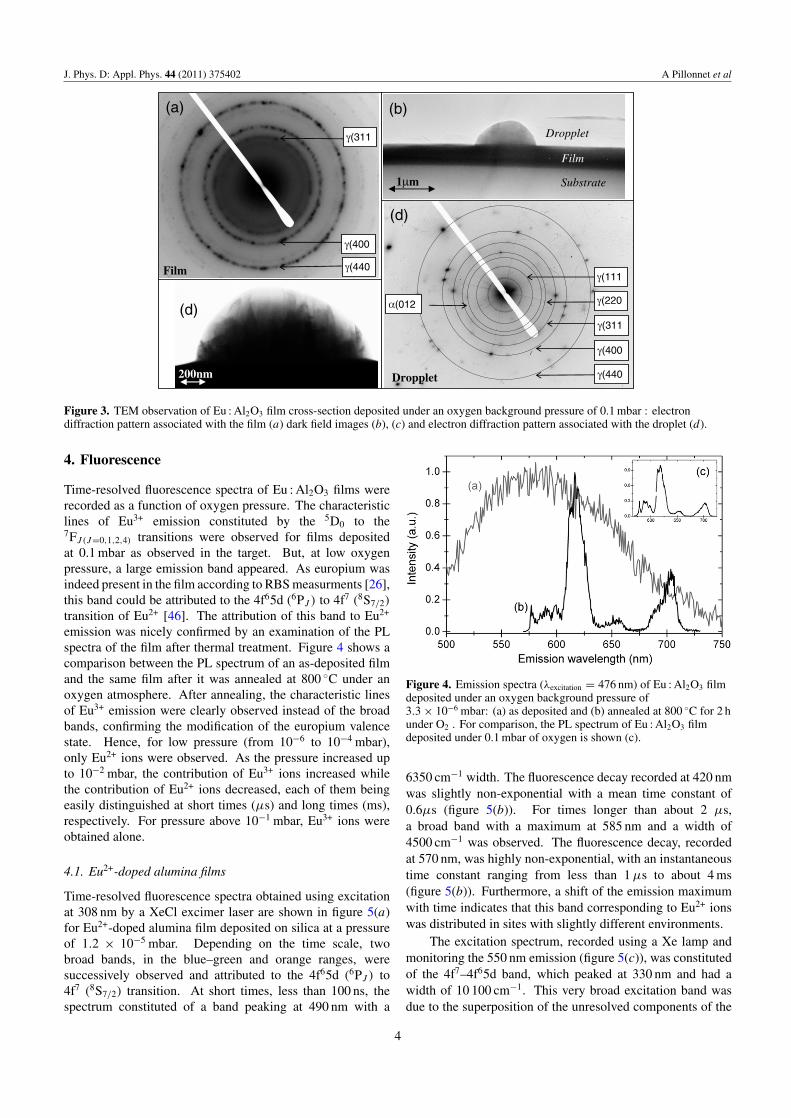
The Eu : Al2O3 films deposited at 0.1 mbar appearedamorphous, but the existence of very small crystallites couldnot be excluded [4, 18]. In order to clarify this point,TEM observation was thus achieved on a cross-sectionsample. Figure 3(a) represents the electron diffractionpattern associated with the film elaborated at 0.1 mbar. Therings revealed a polycrystalline phase which was identifiedas gamma alumina [44]. According to dark field imagesrealized with a part of these rings, the crystallites were4–5 nm in diameter and their distribution was homogeneousthroughout the film thickness. Hence, the film elaborated at0.1 mbar was not amorphous, but consisted of small gammacrystallites. A droplet on this film surface was observed byTEM (figures 3(b) and (c)). The diameter was around 1 µm.The electron diffraction pattern associated with the dropletsis represented in figure 3(d). The observed points revealeda well-crystallized gamma structure. For such crystallizedparticles, narrow diffraction lines should be observed in theXRD diagram. However, despite the fact that we usedgrazing incidence for XRD analysis, gamma droplets were notdetected, indicating that their number was very low. Accordingto optical microscope, some droplets are present in the filmbut widely dispersed on the surface. The micro-analysisby x-ray fluorescence in the microscope showed that thedroplet contained europium. Although the general structuralproperties of the films could be established by XRD analysis,it was more challenging to detect these nanophases. This isimportant since the luminescence properties of the europiumions could be affected by the well-crystallized gamma phase.Our observations are supported by other studies where thecrystalline structure of droplets has been identified as well.Thus, for example, Teodorescu et al [45] have shown thatamorphous BCN films are obtained by PLD, whereas thedroplets contain nanocrystallites of c-BCN.
3
J. Phys. D: Appl. Phys. 44 (2011) 375402 A Pillonnet et al
γ(311
γ(400
γ(440Film
(a)
Film
Substrat
Gouttelette
1µm Substrate
Film
Dropplet
(b)
200nm
(d)
γ(400
γ(440
α(012
γ(111
γ(311
γ(220
Dropplet
(d)
Figure 3. TEM observation of Eu : Al2O3 film cross-section deposited under an oxygen background pressure of 0.1 mbar : electrondiffraction pattern associated with the film (a) dark field images (b), (c) and electron diffraction pattern associated with the droplet (d).
4. Fluorescence
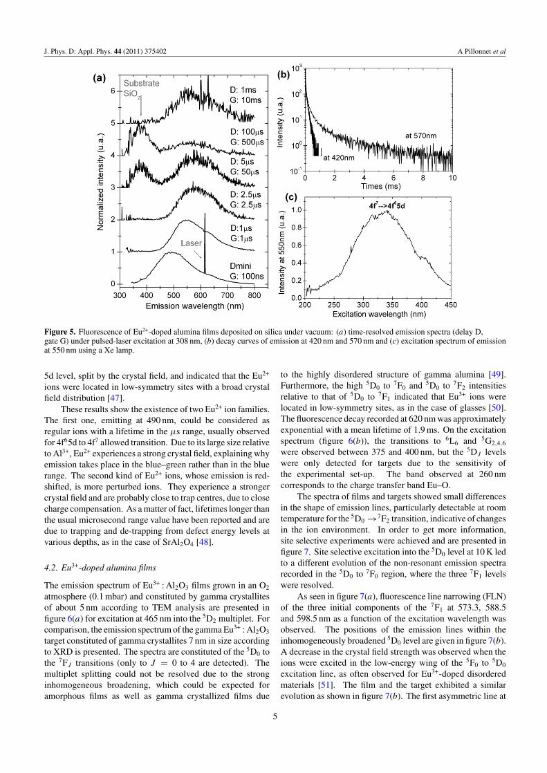
Time-resolved fluorescence spectra of Eu : Al2O3 films wererecorded as a function of oxygen pressure. The characteristiclines of Eu3+ emission constituted by the 5D0 to the7FJ (J=0,1,2,4) transitions were observed for films depositedat 0.1 mbar as observed in the target. But, at low oxygenpressure, a large emission band appeared. As europium wasindeed present in the film according to RBS measurments [26],this band could be attributed to the 4f65d (6PJ ) to 4f7 (8S7/2)
transition of Eu2+ [46]. The attribution of this band to Eu2+
emission was nicely confirmed by an examination of the PLspectra of the film after thermal treatment. Figure 4 shows acomparison between the PL spectrum of an as-deposited filmand the same film after it was annealed at 800 ◦C under anoxygen atmosphere. After annealing, the characteristic linesof Eu3+ emission were clearly observed instead of the broadbands, confirming the modification of the europium valencestate. Hence, for low pressure (from 10−6 to 10−4 mbar),only Eu2+ ions were observed. As the pressure increased upto 10−2 mbar, the contribution of Eu3+ ions increased whilethe contribution of Eu2+ ions decreased, each of them beingeasily distinguished at short times (µs) and long times (ms),respectively. For pressure above 10−1 mbar, Eu3+ ions wereobtained alone.
4.1. Eu2+-doped alumina films
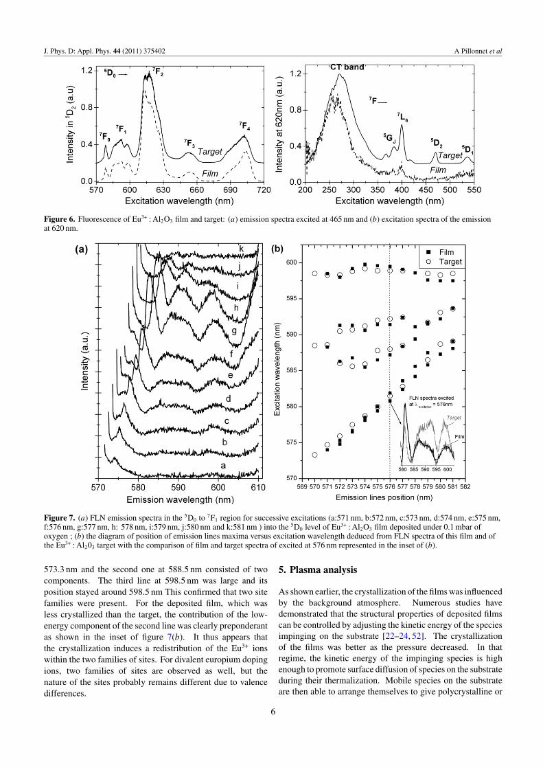
Time-resolved fluorescence spectra obtained using excitationat 308 nm by a XeCl excimer laser are shown in figure 5(a)for Eu2+-doped alumina film deposited on silica at a pressureof 1.2 × 10⁻⁵ mbar. Depending on the time scale, twobroad bands, in the blue–green and orange ranges, weresuccessively observed and attributed to the 4f65d (6PJ ) to4f7 (8S7/2) transition. At short times, less than 100 ns, thespectrum constituted of a band peaking at 490 nm with a
Figure 4. Emission spectra (λexcitation = 476 nm) of Eu : Al2O3 filmdeposited under an oxygen background pressure of3.3 × 10−6 mbar: (a) as deposited and (b) annealed at 800 ◦C for 2 hunder O2 . For comparison, the PL spectrum of Eu : Al2O3 filmdeposited under 0.1 mbar of oxygen is shown (c).
6350 cm−1 width. The fluorescence decay recorded at 420 nmwas slightly non-exponential with a mean time constant of0.6µs (figure 5(b)). For times longer than about 2 µs,a broad band with a maximum at 585 nm and a width of4500 cm−1 was observed. The fluorescence decay, recordedat 570 nm, was highly non-exponential, with an instantaneoustime constant ranging from less than 1 µs to about 4 ms(figure 5(b)). Furthermore, a shift of the emission maximumwith time indicates that this band corresponding to Eu2+ ionswas distributed in sites with slightly different environments.
The excitation spectrum, recorded using a Xe lamp andmonitoring the 550 nm emission (figure 5(c)), was constitutedof the 4f7–4f65d band, which peaked at 330 nm and had awidth of 10 100 cm−1. This very broad excitation band wasdue to the superposition of the unresolved components of the
4
J. Phys. D: Appl. Phys. 44 (2011) 375402 A Pillonnet et al
Figure 5. Fluorescence of Eu2+-doped alumina films deposited on silica under vacuum: (a) time-resolved emission spectra (delay D,gate G) under pulsed-laser excitation at 308 nm, (b) decay curves of emission at 420 nm and 570 nm and (c) excitation spectrum of emissionat 550 nm using a Xe lamp.
5d level, split by the crystal field, and indicated that the Eu2+
ions were located in low-symmetry sites with a broad crystalfield distribution [47].
These results show the existence of two Eu2+ ion families.The first one, emitting at 490 nm, could be considered asregular ions with a lifetime in the µs range, usually observedfor 4f65d to 4f7 allowed transition. Due to its large size relativeto Al3+, Eu2+ experiences a strong crystal field, explaining whyemission takes place in the blue–green rather than in the bluerange. The second kind of Eu2+ ions, whose emission is red-shifted, is more perturbed ions. They experience a strongercrystal field and are probably close to trap centres, due to closecharge compensation. As a matter of fact, lifetimes longer thanthe usual microsecond range value have been reported and aredue to trapping and de-trapping from defect energy levels atvarious depths, as in the case of SrAl2O4 [48].
4.2. Eu3+-doped alumina films
The emission spectrum of Eu3+ : Al2O3 films grown in an O2
atmosphere (0.1 mbar) and constituted by gamma crystallitesof about 5 nm according to TEM analysis are presented infigure 6(a) for excitation at 465 nm into the 5D2 multiplet. Forcomparison, the emission spectrum of the gamma Eu3+ : Al2O3
target constituted of gamma crystallites 7 nm in size accordingto XRD is presented. The spectra are constituted of the 5D0 tothe 7FJ transitions (only to J = 0 to 4 are detected). Themultiplet splitting could not be resolved due to the stronginhomogeneous broadening, which could be expected foramorphous films as well as gamma crystallized films due
to the highly disordered structure of gamma alumina [49].Furthermore, the high 5D0 to 7F0 and 5D0 to 7F2 intensitiesrelative to that of 5D0 to 7F1 indicated that Eu3+ ions werelocated in low-symmetry sites, as in the case of glasses [50].The fluorescence decay recorded at 620 nm was approximatelyexponential with a mean lifetime of 1.9 ms. On the excitationspectrum (figure 6(b)), the transitions to 6L6 and 5G2,4,6
were observed between 375 and 400 nm, but the 5DJ levelswere only detected for targets due to the sensitivity ofthe experimental set-up. The band observed at 260 nmcorresponds to the charge transfer band Eu–O.
The spectra of films and targets showed small differencesin the shape of emission lines, particularly detectable at roomtemperature for the 5D0 →7F2 transition, indicative of changesin the ion environment. In order to get more information,site selective experiments were achieved and are presented infigure 7. Site selective excitation into the 5D0 level at 10 K ledto a different evolution of the non-resonant emission spectrarecorded in the 5D0 to 7F0 region, where the three 7F1 levelswere resolved.
As seen in figure 7(a), fluorescence line narrowing (FLN)of the three initial components of the 7F1 at 573.3, 588.5and 598.5 nm as a function of the excitation wavelength wasobserved. The positions of the emission lines within theinhomogeneously broadened 5D0 level are given in figure 7(b).A decrease in the crystal field strength was observed when theions were excited in the low-energy wing of the 5F0 to 5D0
excitation line, as often observed for Eu3+-doped disorderedmaterials [51]. The film and the target exhibited a similarevolution as shown in figure 7(b). The first asymmetric line at
5
J. Phys. D: Appl. Phys. 44 (2011) 375402 A Pillonnet et al
Figure 6. Fluorescence of Eu3+ : Al2O3 film and target: (a) emission spectra excited at 465 nm and (b) excitation spectra of the emissionat 620 nm.
Figure 7. (a) FLN emission spectra in the 5D0 to 7F1 region for successive excitations (a:571 nm, b:572 nm, c:573 nm, d:574 nm, e:575 nm,f:576 nm, g:577 nm, h: 578 nm, i:579 nm, j:580 nm and k:581 nm ) into the 5D0 level of Eu3+ : Al2O3 film deposited under 0.1 mbar ofoxygen ; (b) the diagram of position of emission lines maxima versus excitation wavelength deduced from FLN spectra of this film and ofthe Eu3+ : Al203 target with the comparison of film and target spectra of excited at 576 nm represented in the inset of (b).
573.3 nm and the second one at 588.5 nm consisted of twocomponents. The third line at 598.5 nm was large and itsposition stayed around 598.5 nm This confirmed that two sitefamilies were present. For the deposited film, which wasless crystallized than the target, the contribution of the low-energy component of the second line was clearly preponderantas shown in the inset of figure 7(b). It thus appears thatthe crystallization induces a redistribution of the Eu3+ ionswithin the two families of sites. For divalent europium dopingions, two families of sites are observed as well, but thenature of the sites probably remains different due to valencedifferences.
5. Plasma analysis
As shown earlier, the crystallization of the films was influencedby the background atmosphere. Numerous studies havedemonstrated that the structural properties of deposited filmscan be controlled by adjusting the kinetic energy of the speciesimpinging on the substrate [22–24, 52]. The crystallizationof the films was better as the pressure decreased. In thatregime, the kinetic energy of the impinging species is highenough to promote surface diffusion of species on the substrateduring their thermalization. Mobile species on the substrateare then able to arrange themselves to give polycrystalline or
6
J. Phys. D: Appl. Phys. 44 (2011) 375402 A Pillonnet et al
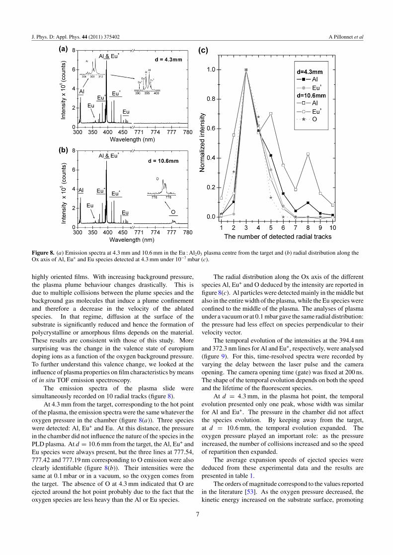
Figure 8. (a) Emission spectra at 4.3 mm and 10.6 mm in the Eu : Al203 plasma centre from the target and (b) radial distribution along theOx axis of Al, Eu+ and Eu species detected at 4.3 mm under 10−7 mbar (c).
highly oriented films. With increasing background pressure,the plasma plume behaviour changes drastically. This isdue to multiple collisions between the plume species and thebackground gas molecules that induce a plume confinementand therefore a decrease in the velocity of the ablatedspecies. In that regime, diffusion at the surface of thesubstrate is significantly reduced and hence the formation ofpolycrystalline or amorphous films depends on the material.These results are consistent with those of this study. Moresurprising was the change in the valence state of europiumdoping ions as a function of the oxygen background pressure.To further understand this valence change, we looked at theinfluence of plasma properties on film characteristics by meansof in situ TOF emission spectroscopy.
The emission spectra of the plasma slide weresimultaneously recorded on 10 radial tracks (figure 8).
At 4.3 mm from the target, corresponding to the hot pointof the plasma, the emission spectra were the same whatever theoxygen pressure in the chamber (figure 8(a)). Three specieswere detected: Al, Eu+ and Eu. At this distance, the pressurein the chamber did not influence the nature of the species in thePLD plasma. At d = 10.6 mm from the target, the Al, Eu+ andEu species were always present, but the three lines at 777.54,777.42 and 777.19 nm corresponding to O emission were alsoclearly identifiable (figure 8(b)). Their intensities were thesame at 0.1 mbar or in a vacuum, so the oxygen comes fromthe target. The absence of O at 4.3 mm indicated that O areejected around the hot point probably due to the fact that theoxygen species are less heavy than the Al or Eu species.
The radial distribution along the Ox axis of the differentspecies Al, Eu+ and O deduced by the intensity are reported infigure 8(c). Al particles were detected mainly in the middle butalso in the entire width of the plasma, while the Eu species wereconfined to the middle of the plasma. The analyses of plasmaunder a vacuum or at 0.1 mbar gave the same radial distribution:the pressure had less effect on species perpendicular to theirvelocity vector.
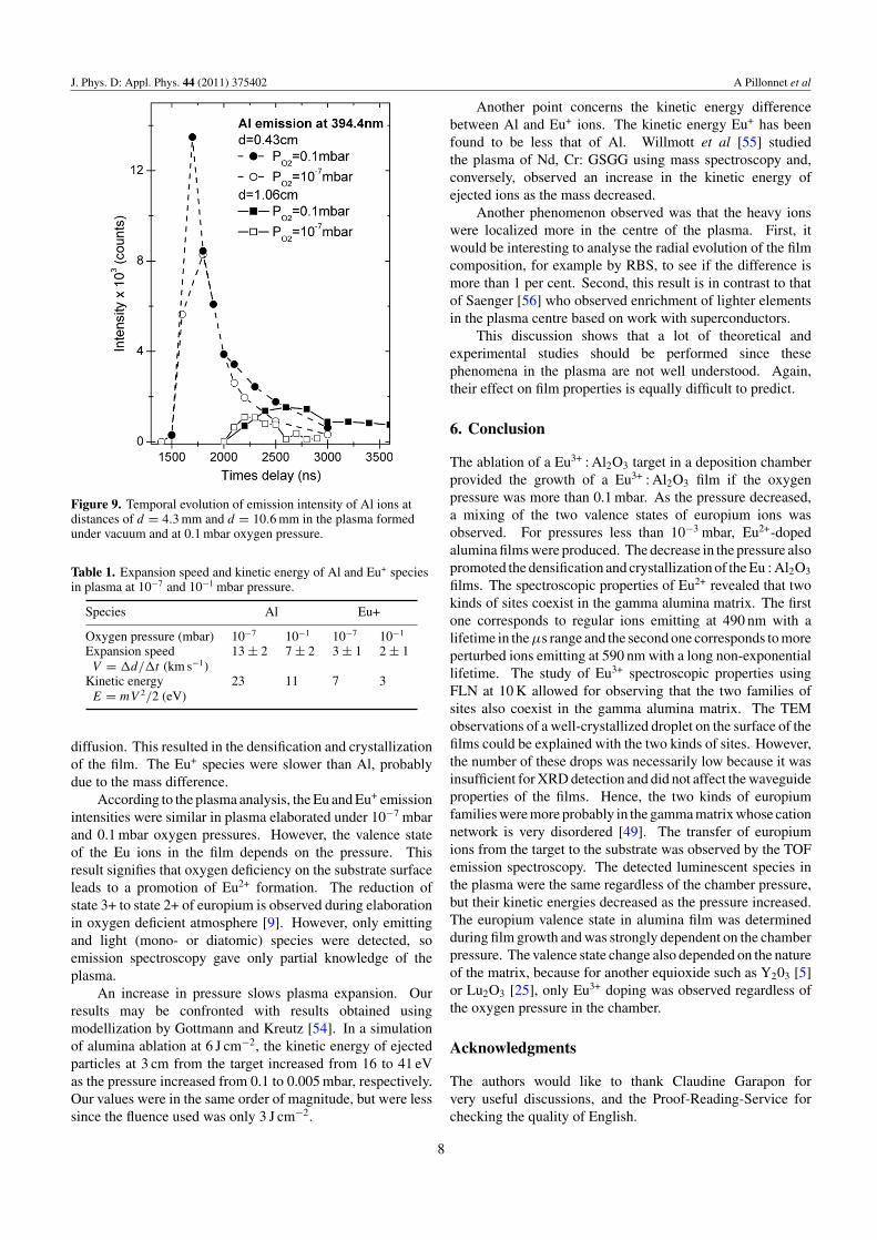
The temporal evolution of the intensities at the 394.4 nmand 372.3 nm lines for Al and Eu+, respectively, were analysed(figure 9). For this, time-resolved spectra were recorded byvarying the delay between the laser pulse and the cameraopening. The camera opening time (gate) was fixed at 200 ns.The shape of the temporal evolution depends on both the speedand the lifetime of the fluorescent species.
At d = 4.3 mm, in the plasma hot point, the temporalevolution presented only one peak, whose width was similarfor Al and Eu+. The pressure in the chamber did not affectthe species evolution. By keeping away from the target,at d = 10.6 mm, the temporal evolution expanded. Theoxygen pressure played an important role: as the pressureincreased, the number of collisions increased and so the speedof repartition then expanded.
The average expansion speeds of ejected species werededuced from these experimental data and the results arepresented in table 1.
The orders of magnitude correspond to the values reportedin the literature [53]. As the oxygen pressure decreased, thekinetic energy increased on the substrate surface, promoting
7
J. Phys. D: Appl. Phys. 44 (2011) 375402 A Pillonnet et al
Figure 9. Temporal evolution of emission intensity of Al ions atdistances of d = 4.3 mm and d = 10.6 mm in the plasma formedunder vacuum and at 0.1 mbar oxygen pressure.
Table 1. Expansion speed and kinetic energy of Al and Eu+ speciesin plasma at 10−7 and 10−1 mbar pressure.
Species Al Eu+
Oxygen pressure (mbar) 10−7 10−1 10−7 10−1
Expansion speed 13 ± 2 7 ± 2 3 ± 1 2 ± 1V = �d/�t (km s−1)
Kinetic energy 23 11 7 3E = mV 2/2 (eV)
diffusion. This resulted in the densification and crystallizationof the film. The Eu+ species were slower than Al, probablydue to the mass difference.
According to the plasma analysis, the Eu and Eu+ emissionintensities were similar in plasma elaborated under 10−7 mbarand 0.1 mbar oxygen pressures. However, the valence stateof the Eu ions in the film depends on the pressure. Thisresult signifies that oxygen deficiency on the substrate surfaceleads to a promotion of Eu2+ formation. The reduction ofstate 3+ to state 2+ of europium is observed during elaborationin oxygen deficient atmosphere [9]. However, only emittingand light (mono- or diatomic) species were detected, soemission spectroscopy gave only partial knowledge of theplasma.
An increase in pressure slows plasma expansion. Ourresults may be confronted with results obtained usingmodellization by Gottmann and Kreutz [54]. In a simulationof alumina ablation at 6 J cm−2, the kinetic energy of ejectedparticles at 3 cm from the target increased from 16 to 41 eVas the pressure increased from 0.1 to 0.005 mbar, respectively.Our values were in the same order of magnitude, but were lesssince the fluence used was only 3 J cm−2.
Another point concerns the kinetic energy differencebetween Al and Eu+ ions. The kinetic energy Eu+ has beenfound to be less that of Al. Willmott et al [55] studiedthe plasma of Nd, Cr: GSGG using mass spectroscopy and,conversely, observed an increase in the kinetic energy ofejected ions as the mass decreased.
Another phenomenon observed was that the heavy ionswere localized more in the centre of the plasma. First, itwould be interesting to analyse the radial evolution of the filmcomposition, for example by RBS, to see if the difference ismore than 1 per cent. Second, this result is in contrast to thatof Saenger [56] who observed enrichment of lighter elementsin the plasma centre based on work with superconductors.
This discussion shows that a lot of theoretical andexperimental studies should be performed since thesephenomena in the plasma are not well understood. Again,their effect on film properties is equally difficult to predict.
6. Conclusion
The ablation of a Eu3+ : Al2O3 target in a deposition chamberprovided the growth of a Eu3+ : Al2O3 film if the oxygenpressure was more than 0.1 mbar. As the pressure decreased,a mixing of the two valence states of europium ions wasobserved. For pressures less than 10−3 mbar, Eu2+-dopedalumina films were produced. The decrease in the pressure alsopromoted the densification and crystallization of the Eu : Al2O3
films. The spectroscopic properties of Eu2+ revealed that twokinds of sites coexist in the gamma alumina matrix. The firstone corresponds to regular ions emitting at 490 nm with alifetime in the µs range and the second one corresponds to moreperturbed ions emitting at 590 nm with a long non-exponentiallifetime. The study of Eu3+ spectroscopic properties usingFLN at 10 K allowed for observing that the two families ofsites also coexist in the gamma alumina matrix. The TEMobservations of a well-crystallized droplet on the surface of thefilms could be explained with the two kinds of sites. However,the number of these drops was necessarily low because it wasinsufficient for XRD detection and did not affect the waveguideproperties of the films. Hence, the two kinds of europiumfamilies were more probably in the gamma matrix whose cationnetwork is very disordered [49]. The transfer of europiumions from the target to the substrate was observed by the TOFemission spectroscopy. The detected luminescent species inthe plasma were the same regardless of the chamber pressure,but their kinetic energies decreased as the pressure increased.The europium valence state in alumina film was determinedduring film growth and was strongly dependent on the chamberpressure. The valence state change also depended on the natureof the matrix, because for another equioxide such as Y203 [5]or Lu2O3 [25], only Eu3+ doping was observed regardless ofthe oxygen pressure in the chamber.
Acknowledgments
The authors would like to thank Claudine Garapon forvery useful discussions, and the Proof-Reading-Service forchecking the quality of English.
8
J. Phys. D: Appl. Phys. 44 (2011) 375402 A Pillonnet et al
References
[1] Armelo L, Quici S, Barigelletti F, Accorsi G, Bottaro G,Cavazzini M and Tondello E 2010 Coord. Chem. Rev.254 487
[2] Wegh R T, Donker H, Oskam K D and Meijerink A 1999Science 283 663
[3] Cormier G, Capobianco J A, Morrison C A and Monteil A1993 Phys. Rev. B 48 16290
[4] Pillonnet A, LeBihan V, Massenelli B, Ledoux G, Marty O,Melinon P and Dujardin C 2010 ACS Mater. Interface2 1543
[5] Pillonnet A, Lancok J, Martinet C, Marty O, Bellessa J andGarapon C 2006 J. Phys. Condes. Matter 18 10043
[6] Lim K S, Lee S, Trinh M T, Kim S H, Lee M, Hamilton D Sand Gibson G N 2007 J. Lumin. 122–123 14
[7] Gaponenko N V, Davidson K A, Hamilton B, Skeldon P,Thompson G E, Zhou X and Pivin J C 2000 Appl. Phys.Lett. 76 1006
[8] Yang C C, Chen S Y and Liu D M 2006 J. Cryst. Growth293 113
[9] Rakov N and Maciel G S 2007 J. Lumin. 127 703[10] Ozuna O, Hirata G A and McKittrick J 2004 Appl. Phys. Lett.
84 1296[11] Loonj C K, Richardson J W Jr and Ozawa M 1997 J. Alloys
Compounds 250 356[12] Rakov N, Ramos F E, Hirata G and Xiao M 2003 Appl. Phys.
Lett. 83 272[13] Rakov N, Maciel G S, Lozano W and de Araujo C B 2007
J. Appl. Phys. 101 036102[14] Hirata G, Perea N, Tejeda M, Gonzalez-Ortega J A and
McKittrick J 2005 Opt. Mater. 27 1311[15] Monteiro M A F, Brito H F, Felinto M C F C M, Brito G E S,
Teotonio E E S, Vichi F M and Stefani R 2008 Micro.Mesoporous Mater. 108 237
[16] Gaponenko N V, Davidson K A, Hamilton B, Skeldon P,Thompson G E, Zhou X and Pivin J C 2000 Appl. Phys.Lett. 76 1006
[17] Ishizaka T, Nozaki R and Kurokawa Y 2002 J. Phys. Chem.Solids 63 613
[18] Pillonnet-Minardi A, Marty O, Bovier C, Garapon C andMugnier J 2001 Opt. Mater. 16 9
[19] Wrzyszcz J, Mista W, Hreniak D, Strek W, Zawaldzki M andGrabowska H 2002 J. Alloys Compounds. 341 358
[20] Chrisey D B and Hubler G H 1994 Pulsed Laser Deposition ofThin Films (New York: Wiley)
[21] Sujioka K, Meunier M and Pique A 2010 Laser PrecisionMicrofabrication (New York: Springer)
[22] Irissou E, Le Drogoff B, Chaker M and Guay D 2003 J. Appl.Phys. 94 4796
[23] Irissou E, Le Drogoff B, Chaker M, Trudeau M and Guay D2004 J. Mater. Res. 19 950
[24] Pereira A, Laplante F, Chaker M and Guay D 2007 Adv. Funct.Mater. 17 443
[25] Martinet C, Pillonnet A, Lancok J and Garapon C 2007J. Lumin. 126 807
[26] Pillonnet A, Garapon C, Champeaux C, Bovier C, Brenier R,Jaffrezic H and Mugnier J 1999 Appl. Phys. A 69 S735
[27] Wang G, Marty O, Garapon C, Pillonnet A and Zhang W 2004Appl. Phys. A 79 1599
[28] Xiao Z, Serna R and Afonso C N 2007 J. Appl. Phys.101 033112
[29] Xiao Z, Serna R, Xu F and Afonso C N 2008 Opt. Lett. 33 608[30] Pan C, Chen S-Y and Shen P 2008 J. Cryst. Growth 310 699[31] Pillonnet A, Garapon C, Champeaux C, Bovier C, Jaffrezic H
and Mugnier J 2000 J. Lumin. 87 1087[32] Suarez-Garcia A, Gonzalo J and Afonso C N 2003 Appl. Phys.
A 77 779[33] Serna R, Suarez-Garcia A, Afonso C N and Barbonneau D
2006 Nanotechnology 17 4588[34] Margueritat J, Gonzalo J, Afonso C N, Bachelier G, Mlayah A,
Laarakker A S, Murray D B and Saviot L 2007 Appl.Phys. A 89 369
[35] Haro-Poniatowski E, Serna R, Jimenez de Castro M,Suarez-Garcia A, Afonso C N and Vickridge I 2008Nanotechnology 19 485708
[36] Pereira A, Grojo D, Chaker M, Delaporte P, Guay D andSentis M 2008 Small 4 572
[37] Scneider C W, Espositio M, Marozau I, Conder K, Doebeli M,Hu Y, Mallepell M, Wokaun A and Lippert T 2010 Appl.Phys. Lett. 97 192107
[38] Epurescu G, Siegel J, Gonzalo J, Gordillo-Vazquez F J andAfonso C N 2005 Appl. Phys. Lett. 87 211501
[39] Ulrich R and Torge R 1973 Appl. Opt. 12 2901[40] Bigarre J, Fayeulle S, Treheux D and Moncoffre N 1997
J. Appl. Phys. 82 3740[41] Garapon C, Manaa H and Moncorge R 1991 J. Chem. Phys.
95 5501[42] Pons-Y-Moll O, Perriere J, Millon E, Defourneau R M,
Defourneau D, Vincent B, Essahlaoui A, Boudrioua A andSeiler W 2002 J. Appl. Phys. 92 4885
[43] Osaka M, Kimura M and Isogai A 1990 J. Less-Common Met.162 297
[44] Lippens B C and De Boer J H 1964 Acta Cryst. 17 1312[45] Teodorescu V S, Luches A, Dinu R, Zocco A, Ciobanu M F,
Martino M, Sandu V and Dinescu M 1999 Appl. Phys. A69 S667
[46] Blasse G and Grabmaier B C 1994 Luminescent Materials(Berlin: Spinger)
[47] Blasse G, Wanmaker W L, Ter Vrugt J W and Bril Philips A1968 Res. Rep. 23 189
[48] Jia W, Yuan H, Lu L, Liu H and Yen W M 1998 J. Lumin.76–77 424
[49] Zhou R S and Snyder R L 1991 Acta Cryst. B 47 617[50] Pucker G, Gatterer K, Fritzer H P, Bettinelli M and Ferrari M
1996 Phys. Rev. B 53 6225[51] Schmidt Th, Macfarlane R M and Volker S 1994 Phys. Rev. B
50 15707[52] Amoruso S, Schou J and Lunney J G 2006 Europhys. Lett.
76 436[53] Champeaux C 1992 PhD Thesis Limoges[54] Gottmann J and Kreutz E W 1999 Surf. Coat. Technol.
116 1189[55] Willmott P R, Manoravi P and Holliday K 2000 Appl. Phys. A
70 425[56] Sanger K L 1993 Proc. Adv. Mater. 2 63
9