urothelial transdifferentiation to prostate epithelia is mediated by paracrine tgf-β signaling
TRANSCRIPT

Urothelial transdifferentiation to prostate epithelia is mediated byparacrine TGF-β signaling
Xiaohong Lia, Yongqing Wanga, Ali-Reza Sharif-Afshara, Consolate Uwamariyaa, AndrewYia, Kenichiro Ishiia,b, Simon W. Haywarda, Robert J. Matusika, and Neil A. Bhowmicka,*aDepartment of Urologic Surgery, Vanderbilt-Ingram Cancer Center, Vanderbilt University,Nashville, TN 37232-2765, USAbDepartment of Nephro-Urologic Surgery and Andrology, Mie University Graduate School ofMedicine, Tsu, Mie, Japan
AbstractThe embryonic urogenital sinus mesenchyme (UGM) induces prostate epithelial morphogenesis indevelopment. The molecular signals that drive UGM-mediated prostatic induction have not beendefined. We hypothesized that the TGF-β signaling directed the prostatic induction. UGM from TGF-β type II receptor stromal conditional knockout mice (Tgfbr2fspKO) or control mice(Tgfbr2floxE2/floxE2)was recombined with wild-type adult mice bladder urothelial cells. The resultingurothelium associated with Tgfbr2floxE2/floxE2 UGM was instructively differentiated into prostaticepithelium, as expected. In contrast, the urothelium associated with Tgfbr2fspKO UGM permissivelymaintained the phenotype of bladder epithelial cells. Microarray analysis of UGM tissues suggestedthe down-regulation of multiple Wnt ligands and the up-regulation of the Wnt antagonist, Wif 1, bythe Tgfbr2fspKO UGM compared with Tgfbr2floxE2/floxE2 UGM. The overexpression of Wif-1 bywild-type UGM resulted in the inhibition of prostatic induction. These data suggest that the stromalTGF-β activity mediated by paracrine Wnt is necessary for the induction of prostatic differentiation.As Wnt ligands mediate differentiation and maintain the stem cell phenotype, the contribution ofmouse stem cells and somatic cells to prostatic epithelium in the tissue recombination models wastested. The directed differentiation of mouse embryonic stem cells by UGM is suggested by athreshold number of mouse stem cells required in prostatic differentiation. To determine thecontribution of somatic cells, the adult bladder epithelial compartment was labeled with green-fluorescent vital dye (CMFDA) and the stem-like cells marked by bromodeoxyuridine (BrdU) label-retention. The resulting prostatic epithelia of the tissue recombinants maintained the CMFDA dye,suggesting minimal cell division. Thus, the UGM can induce endoderm-derived epithelia and stemcells to form prostate through a transdifferentiation mechanism that requires stromal TGF-β signalingto mediate epithelial Wnt activity.
KeywordsUrogenital; Mesenchyme; Prostate; TGF-β; Wnt; Stem cell
1. IntroductionThe importance of mesenchymal cells in organogenesis is established in many organs includingthe prostate. The prostate develops from the embryonic urogenital sinus, having both
© 2008 International Society of Differentiation. Published by Elsevier Ltd. All rights reserved*Corresponding author. Tel.: +1 615 343 7140; fax: +1 615 322 5869. E-mail address: [email protected] (N.A. Bhowmick)..
NIH Public AccessAuthor ManuscriptDifferentiation. Author manuscript; available in PMC 2009 August 27.
Published in final edited form as:Differentiation. 2009 January ; 77(1): 95–102. doi:10.1016/j.diff.2008.09.012.
NIH
-PA Author Manuscript
NIH
-PA Author Manuscript
NIH
-PA Author Manuscript

mesodermally derived urogenital sinus mesenchyme (UGM) and endodermally derivedepithelial cells. In the presence of androgen, the epithelium undergoes proliferation anddifferentiation into luminal and basal subtypes. Concurrently, the UGM proliferates anddifferentiates into prostatic smooth muscle (Cunha et al., 1987; Hayward and Cunha, 2000).Tissue recombination models have been extensively used to examine mesenchymal-epithelialinteractions during the process of prostatic organ formation. They have provided importantinsights into the role of paracrine signaling. Yet, the mechanism for this mesenchymalprogramming is still not understood. The instructive nature of the UGM toward prostaticepithelial development suggests that paracrine factors are involved in this process (Shima etal., 1995). Many androgen-regulated cytokines and growth factors play important roles incontrolling proliferation and differentiation of both epithelial and stromal cells (Cunha et al.,1992, 1995; Thomson and Cunha, 1999; Hayward et al., 1998). The accumulation of TGF-β1protein is reported to localize in the mesenchyme of fetal and neonatal prostate (Timme et al.,1994). TGF-β signals by the binding of type I and II TGF-β receptors at the cell surface toactivate cytoplasmic signaling molecules including the Smad proteins (Massague and Gomis,2006). However, the role of TGF-β signaling on UGM inductivity is not known. We have usedthe tissue recombination model system to study the signaling mechanisms between the stromaland epithelial compartments in vivo.
The mechanisms gleaned through tissue recombination modeling often recapitulate nativetissue development (Thomson et al., 2002; Donjacour and Cunha, 1995). Tissue recombinationstudies illustrated the ability of UGM to instructively induce prostatic differentiation from anumber of endodermally derived epithelia including those of the prostate, bladder, and urethra(Boutin et al., 1991a, b; Cunha et al., 1983). When applied to human embryonic stem cells,UGM induces prostatic epithelium in the context of teratoma-like growth (Taylor et al.,2006). Using a similar model, we have reported bladder epithelial induction from mouseembryonic stem cells by embryonic bladder mesenchyme (Oottamasathien et al., 2007).Although it is established that the UGM can differentiate pluripotent embryonic stem cells, itis not known whether the differentiation of the adult bladder epithelia into prostate is a resultof the differentiation of resident tissue-specific stem cells or the adult urothelium itself. If itwere the latter, the studies would reveal the plasticity of non-transformed adult epithelia and,further, somatic cell differentiation in prostate development.
In the present study, we identify the role of TGF-β in UGM induction of prostatic differentiationthrough the use of tissue recombination techniques. We employed the TGF-β type II receptorconditional knockout mouse that has a loss of TGF-β responsivity in the mesenchymalcompartment (Tgfbr2fspKO) (Bhowmick et al., 2004). The differences between normal UGMfrom the Tgfbr2floxE2/floxE2 mouse and Tgfbr2fspKO UGM on instructing adult mouse bladderepithelial differentiation were compared. The results suggested that regulation of Wnt paracrinesignaling mediates the role of TGF-β in the inductive effects of the UGM on the adjacentepithelium. Further, the prostatic inductivity of the UGM was extended to transdifferentiationof mouse stem and somatic cells.
2. Materials and methods2.1. Animals and urogenital sinus
Tgfbr2floxE2/floxE2 and Tgfbr2fspKO mice bred on the C57Bl/6 background were generated asdescribed previously (Bhowmick et al., 2004). Tgfbr2floxE2/floxE2 mice having loxP sites atintrons 1 and 2 of Tgfbr2 were our control mice. Tgfbr2fspKO mice are homozygous conditionalknockout of the Tgfbr2 in fibroblasts. Adult male athymic nude mice and C57Bl/6 mice werepurchased from Harlan (Indianapolis, IN). All animal procedures were approved by theVanderbilt Institutional Animal Care and Use Committee.
Li et al. Page 2
Differentiation. Author manuscript; available in PMC 2009 August 27.
NIH
-PA Author Manuscript
NIH
-PA Author Manuscript
NIH
-PA Author Manuscript
The urogenital sinuses from mouse or rat embryos (E16 and E18 day, respectively) wereseparated under a dissecting microscope into epithelial and mesenchymal (UGM) componentsas described previously (Staack et al., 2003). Rat UGMs were pooled together and furtherdigested with collagenase into single cells (Hayward et al., 1999).
2.2. Tissue recombination and graftingUGM from Tgfbr2floxE2/floxE2 or Tgfbr2fspKO mice was recombined with 1/6 of the adultbladder epithelial cells (approximately 30,000 cells) from one mouse in a 50-μl rat-tail collagenand incubated overnight at 37 °C. For the recombination of rat UGM with mouse ES cells,each 50-μl collagen contained 300,000 rat UGM cells with 500-5000 ES cells. The ES cellswere maintained and cultured with a feeder layer of mitotically inactivated primary mouseembryonic fibroblasts as described before (Oottamasathien et al., 2007). The mouse-derivedtissue recombinants were grafted under the renal capsule of syngenic C57Bl/6, whereas thetissue recombinants containing rat UGM were xenografted under the renal capsule of athymicnude mice. The grafts were analyzed after 4 weeks following paraformaldehyde fixation andparaffin embedding.
2.3. Wif 1 lenti-virus production and transduction of UGMMouse Wif 1 cDNA clone in pCMV-SPORT6 vector (clone ID, 3984128) was purchased fromOpen Biosystems (Huntsville, AL). It was subcloned into pLenti6/V5-DEST Gateway vector(Invitrogen Life Technologies, Carlsbad, CA) through Gateway BP and LR recombinationreactions using Gateway Technology with Clonase II (Invitrogen) according to the usermanual. The Wif 1 lenti-virus was generated by transfection of Wif 1 in pLenti6/V5-DEST in293FT cells using ViraPower Packaging Mix from Invitrogen. Then the supernatants with viruswere collected and stored at -80 °C for further use.
Wif 1 overexpression UGMs were generated by incubation of UGMs fromTgfbr2floxE2/floxE2 mice embryos with Wif 1 lenti-virus-containing medium overnight. SinceUGMs lose the inductivity 2 or 3 days in vitro, the transduction rate was determined by twoparallel control experiments. In one, the UGMs were transduced with EGFP lenti-virusgenerated at the same time with Wif 1 lenti-virus, and the green fluorescence of the UGM wasobserved after 3-4 days. In the second experiment, primary cultured Tgfbr2floxE2/floxE2 miceprostate fibroblast cells were transduced with Wif 1 lenti-virus at the same condition as UGMs,then the cells were selected by blasticidin, and it was noticed that nearly 100% cells survived.
2.4. BrdU injection and uroepithelial cells preparationTgfbr2floxE2/floxE2 mice (7-9 weeks old) received one daily intraperitoneal injection of 0.1-ml10 mg/ml 5-bromo-2-deoxyuridine (BrdU, Sigma) in sterile saline for 12 days. The urinarybladders were excised from these mice 1 month after the last injection of BrdU (Kiel et al.,2007; Lawson et al., 2005; Taupin, 2007; Yan et al., 2007; Kempermann et al., 2003). Theurothelial cells of the bladder from either BrdU-injected or control mice were separated byEDTA incubation and micromanipulation (Oottamasathien et al., 2006). The bladder epitheliawere labeled with CellTracker CMFDA (5-chloromethylfluorescein diacetate, MolecularProbes, Eugene, OR) at 37 °C for 1 h and washed prior to tissue recombination (Cheng et al.,2005).
For counting of BrdU- and CMFDA-labeled urothelial cells, the cells were collected on a slideby Cytospin (Thermo Shandon, Pittsburgh, PA), 5 min at 1000 rpm. After fixation with ice-cold acetone, the cells were treated with 2 N HCl at room temperature for half an hour beforeincubation with BrdU antibody for immunofluorescent staining. Slides were evaluated usingepifluorescence microscopy.
Li et al. Page 3
Differentiation. Author manuscript; available in PMC 2009 August 27.
NIH
-PA Author Manuscript
NIH
-PA Author Manuscript
NIH
-PA Author Manuscript

We determined the threshold of CMFDA CellTracker dye detection following cell divisionunder fluorescence microscopy. Simply, a specified cell number was incubated with CMFDA,washed, and allowed to divide in culture. The cells were counted and visualized at regularintervals until the cells were not visible by epi-fluorescence microscopy.
2.5. Antibodies and immunohistochemistryTissue sections were deparaffinized with xylene and hydrated in graded ethanol. The tissueswere treated with hydrogen peroxide (1%) and antigen retrieval was performed by boiling inunmasking reagent (1:100, Vector Labs) prior to blocking. The primary antibodies used in thisstudy were as follows: biotin-conjugated anti-BrdU (Molecular Probes, 1:100), rabbitpolyclonal androgen receptor (N-20), p63 (H-137), Wif-1 (H-180), goat polyclonal FoxA1(C-20) (Santa Cruz Biotechnology, 1:1000), phospho-Smad2 (Ser465/467) (Cell Signaling,1:1000), mouse monoclonal anti-prostatic acid phosphatase (Sigma-Aldrich, 1:200), anduroplakin (a kind gift from Dr. T.T. Sun, New York University School of Medicine, Departmentof Dermatology, New York). All antibodies were diluted in PBS containing 10% fetal bovineserum (FBS) and incubated overnight at 4 °C. The secondary antibodies and detection reagents,from Dako (Dako, Carpinteria, CA), were applied the next day according to the manufacturer'sinstruction. The immunohistochemical stainings were visualized and captured using a NikonCoolscope (Nikon Instrument Inc., Melville, NY).
2.6. Microarray and real-time PCR confirmationTgfbr2floxE2/floxE2 UGMs from three embryos were pooled together as a common referencecontrol. Tgfbr2fspKO UGMs from three embryos were collected individually. They were placeddirectly in SuperAmplification lysis buffer provided by the Miltenyi Biotec Company (Auburn,CA), stored on dry ice, and immediately transported to the company for superamplification ofcDNA and microarray analysis using Agilent whole mouse genome oligomicroarray (44k).The three Tgfbr2fspKO UGM cDNA samples were directly compared with the pooledTgfbr2floxE2/floxE2 in three separate arrays. The response measures, average fold changes, fromdirect comparison within each array were extracted for each gene of three Tgfbr2fspKO UGMcDNA samples, and data were analyzed using the EASE software(http://david.abcc.ncifcrf.gov/ease/ease.jsp). To confirm the differential expression of genesof interest from the microarray analysis, reverse transcription and quantitative real-time PCRwere performed using iScript and iQ SYBR Green supermix, respectively (Bio-Rad, Hercules,CA). Relative quantitation was calculated by the ΔΔCt method normalized to GAPDH (Dorak,2006).
3. ResultsTissue recombination techniques have been used to study stromal-epithelial interactions andrecapitulate development of various tissues under the renal capsule. We have used tissuerecombination to study the role of stromal TGF-β signaling in epithelial differentiation.Recombinations of UGM from Tgfbr2floxE2/floxE2 15-18-day mouse embryos with adult mousewild-type bladder epithelia were grown under the renal capsules of syngeneic mice for 1 month.The grafts developed into glandular structures with surrounding stromal differentiation, asshown in Fig. 1. (A summary of all the tissue recombination grafts is shown in Table 1.) Theimmunohistochemical staining showed no expression of the urothelial differentiation marker,uroplakin. However, as expected, the expressions of prostate secretory protein differentiationmarkers, dorsal lateral prostate (mDLP), and prostate acid phosphatase (PAP) were detectedin the epithelium (Fig. 1). p63, a basal cell marker, showed the integrity of the glandularstructure of the grafted tissue. The staining for FoxA1 in the epithelium, a transcription factorin the cells developed from endoderm, confirmed the origin of the epithelium from urothelialcells (Lee et al., 2005). Additionally, phosphorylated Smad2, an indication of activated TGF-
Li et al. Page 4
Differentiation. Author manuscript; available in PMC 2009 August 27.
NIH
-PA Author Manuscript
NIH
-PA Author Manuscript
NIH
-PA Author Manuscript

β signaling, was localized to the nuclei of both stromal and epithelial cells. Together, the datawere consistent with previous reports that Tgfbr2floxE2/floxE2 UGM, similar to wild-type UGM,was inductive of bladder epithelial differentiation to prostatic epithelium.
Next we determined the role of TGF-β responsiveness of the UGM in the induction of prostatedifferentiation. UGM knocked out for the expression of Tgfbr2 was either obtained fromTgfbr2fspKO mouse embryos or generated by applying Creadenovirus on Tgfbr2floxE2/floxE2
UGM. The important difference between the two sources of UGM was the extent of Tgfbr2recombination. The Tgfbr2fspKO UGM had approximately 50% detectible recombination ofthe Tgfbr2 gene, and the ex vivo adenoviral-Cre transduction resulted in approximately 80%recombination as determined by phosphorylated-Smad2 expression (data not shown).However, both Tgfbr2fspKO and Cre-adenoviral UGM behaved in a similar manner in that theydid not yield glandular prostatic structures when recombined with bladder epithelia. Thegrafted tissues were histologically similar to transitional epithelia surrounding the luminalspace (Fig. 2). Immunohistochemical staining indicated uroplakin expression in the epithelium,suggesting the maintenance of urothelial differentiation. The lack of mDLP secretory proteinand PAP expression in the grafts further suggested the loss of prostatic induction by the Tgfbr2knockout UGM. Phosphorylated-Smad2 immuno-localization confirmed epithelial TGF-βsignaling, with substantial loss of stromal detection. The representative panels shown in Fig.2 were from Cre-adenovirus-transduced Tgfbr2floxE2/floxE2 mouse UGM. Thus, stromal TGF-β signaling played an important role in the UGM induction of prostatic differentiation frombladder urothelium.
To further identify the mechanism how TGF-β imparts UGM prostate identity on the adjacentepithelium, gene expression profiles were compared between the Tgfbr2floxE2/floxE2 andTgfbr2fspKO UGM. The Wnt signaling emerged from our analysis as a prime candidate to bethe primary regulated signaling pathway. Correspondingly, many Wnt ligands were down-regulated and the secreted Wnt antagonists (Wif 1, SFRP1, and SFRP2) were consistently up-regulated by the Tgfbr2fspKO UGM (Fig. 3A). Among those genes, Wnt2, Wnt4, Wnt10a,SFRP1, and SFRP2 and Wif 1 had greater than two-fold change and a significant p-value(p<0.0001) by microarray analysis. These changes were further validated by real-time PCRwith the exception of SFRP1 and SFRP2 (Fig. 3B). As many Wnt ligands are stromally secretedwith cognate receptors expressed by the epithelia, Wnts and their antagonists were goodcandidates for mediating stromal-epithelial interactions.
To determine the role of paracrine Wnt signaling in mediating prostate induction by the UGM,Wif 1 was overexpressed by wild-type UGM. Wif 1 is a pan-antagonist for both canonical andnon-canonical Wnt signaling by direct binding to Wnt ligands. Wif 1 lenti-virus-transducedUGM and control UGM were recombined with bladder epithelia and grafted in mice for 1month. Histology of recombinants generated using Wif 1-expressing UGM demonstrated themaintenance of bladder transitional epithelial differentiation expressing uroplakin and no PAP(Fig. 4). In contrast, the control UGM induced prostate luminal epithelial differentiation withassociated epithelia-expressed PAP and without uroplakin expression. Taken together, thesedata demonstrated that TGF-β signaling regulated the inductivity of UGM through inhibitionof Wnt signaling in the adjacent epithelium.
The importance of paracrine Wnt signaling in prostatic induction increased the possibility thatprostatic induction by the UGM is through directed differentiation of the residing stem cellsin the bladder epithelial compartment (Blank et al., 2008; Blanpain et al., 2007). To initiallydetermine the number of stem cells required in prostatic tissue induction by UGM, mouseembryonic stem (mES) cells were tested in tissue recombinant experiments in male SCID hostmice. We found that when 500 or 1000 mES cells were recombined with 300,000 rat UGMcells and xenografted for 6 weeks no tissue grew (Fig. 5A, B). However, under the same
Li et al. Page 5
Differentiation. Author manuscript; available in PMC 2009 August 27.
NIH
-PA Author Manuscript
NIH
-PA Author Manuscript
NIH
-PA Author Manuscript

conditions, 1500 and 5000 mES cells were induced to develop teratoma containing prostaticstructures (Fig. 5C, D). The prostatic structures comprised 10-20% of each graft. The prostaticphenotype was confirmed by immunohistochemistry staining of FoxA1, p63, AR, and mDLPsecretory protein (Fig. 5E-H). The representative pictures shown in Fig. 5E-H were from therecombinants using 1500 mES cells with UGM. Greater mES cell numbers (5000) grew intolarger tissue recombinants with similar structures and compositions (Fig. 5D). Theserecombination experiments indicate that the threshold number of mouse stem cells can beinduced to form prostate by the UGM.
To differentiate the contribution of adult stem cell differentiation from the alternativehypothesis of the transdifferentiation of urothelial cells, a set of experiments illustrated in Fig.6A were designed. BrdU label-retention methods were used to mark potential stem cells of thedonor mouse bladders 4 weeks prior to harvesting (Kempermann et al., 2003;Kiel et al.,2007). The bladder epithelia were further labeled with a green-fluorescent vital dye, CMFDA,following harvesting. The CMFDA fluorescent probe can be retained in living cells throughseveral generations. It can pass freely through cell membranes, but once inside the cell istransformed into a membrane-impermeable reaction product. The CMFDA dye can beinherited by daughter cells after cell division and is not transferred to adjacent cells in apopulation. In vitro, we have observed that the fluorescence can be detected up to five celldivisions, but was undetectable after six divisions. Following isolation from the bladder andcytospin, urothelial cells were counted. By this method an average of 200,000 epithelial cellswere isolated from one adult bladder. qThe number of BrdU label-retaining cells was about3600 (~1.8% of total). We recombined approximately 30,000 bladder epithelial cells,containing about 600 BrdU label-retaining cells, with a single-embryo UGM (approximately90,000 cells) to form each graft. If most of the urothelial cells transdifferentiated into prostate,after five or less divisions in 1 month, the green CMFDA vital dye would be visible in theharvested grafts. However, if only the stem cells of the bladder, originally only ~600 in number,differentiated to prostate, six or more cell divisions would be needed to develop into theprostatic tissues of the size observed. As the CMFDA concentration is diluted in each celldivision, most of the glandular cells would likely not have green CMFDA fluorescence. Theremay be only a few undifferentiated stem cells co-staining of BrdU and CMFDA in eitherscenario.
The recombinant tissues utilizing the CMFDA- and BrdU-labeled bladder epithelia wereharvested and analyzed by microscopy. Fig. 6A illustrates the red immunofluorescence forBrdU and green for CMFDA from the bladder epithelia used in the tissue recombinationexperiments. Immunohistochemistry illustrated the BrdU staining in the grafts of bladderepithelium alone (control, i.e., no mesenchyme) and the recombinant grafts with UGM (UGM+urothelium), respectively (Fig. 6B, C). The adult bladder epithelium-only grafts had a fewscattered BrdU-positive cells in the small pool of surviving epithelia, with no ductal structure.The UGM enabled prostatic structure formation with few cells maintaining BrdU staining inthe epithelial compartment. There was a reduction in apparent BrdU-labeled cells in the graftscompared to the approximate 600 BrdU-labeled stem cells in the starting material (see Fig.6A). To detect CMFDA fluorescence in the epithelia, the grafts were observed underfluorescent microscopy (Fig. 6D, E). The negative control of the bladder epithelium not labeledwith CMFDA had no significant green autofluorescence. However, the green fluorescence ofthe originally CMFDA-labeled bladder epithelia was still detectable in all of the resultingglandular prostate epithelium 4 weeks after grafting. The apparent reduction of BrdU label-retaining cells and the striking retention of CMFDA indicated that most of the adult bladderepithelia were transdifferentiated to prostatic epithelia with a likely contribution of stem cellsin prostatic induction by the UGM.
Li et al. Page 6
Differentiation. Author manuscript; available in PMC 2009 August 27.
NIH
-PA Author Manuscript
NIH
-PA Author Manuscript
NIH
-PA Author Manuscript

4. DiscussionThe role of TGF-β signaling in UGM prostate inductivity was revealed through tissuerecombination with adult bladder epithelium. The embryonic UGM is capable of inducingprostate-like morphogenesis in endodermally derived epithelial cells from fetal or adultbladder, urethra, or vagina (Cunha et al., 1983, 1987; Boutin et al., 1991a). Growth factorssuch as TGF-β, fibroblast growth factor-7 (FGF-7, also known as keratinocyte growth factoror KGF), insulin-like growth factors, Wnt ligands, epidermal growth factor, heparin-bindinggrowth factors, and platelet-derived growth factor have been correlated with epithelialdifferentiation of the prostate (Shima et al., 1995; Timme et al., 1994; Cunha et al., 1992,1995; Sugimura et al., 1996; Thomson et al., 1997). The ability of UGM to induce prostaticdifferentiation is likely to be regulated by more than one factor; these factors cross-talk atmultiple levels. The TGF-β family has been extensively studied and is believed to be importantin mediating the interaction between stromal cells and epithelium during development andpathogenesis. The UGM has been suggested to be the major site of TGF-β isoform expression,and accumulation of TGF-β1 protein was localized to the mesenchyme surrounding thedeveloping prostate buds throughout the perinatal period (Timme et al., 1994). The TGF-βtarget genes, such as uPA and c-myc, were also expressed more in the UGM compared to theurogenital epithelia (Timme et al., 1994; Haughney et al., 1998). Our present work illustratesthe necessity of TGF-β signaling in the UGM in prostate epithelial induction, and furtherimplicates Wnt epithelial activity in this induction.
The Wnt ligands are usually stromally secreted with the cognate receptors on adjacent epithelialcells. Once the Wnt ligands activate the frizzled receptors at the cell surface, Wnt signaling isinitiated through canonical or non-canonical pathways (Clevers, 2006; Kohn and Moon,2005). TGF-β and Wnt signaling cross-talk in development and cancer can be both cooperativeand antagonistic (Letamendia et al., 2001; Labbe et al., 2000, 2007; Attisano and Labbe,2004). However, our understanding of the interactions of these pathways at a paracrine levelis incomplete. In the presence of both Wnt and TGF-β, Smad3 and Smad4 bind to LEF1/TCFsto enhance β-catenin transcriptional activation of target genes (Labbe et al., 2000; Letamendiaet al., 2001). TGF-β is also reported to promote the physical interaction of smad7 with β-cateninand LEF1/TCF, and accumulates β-catenin in a p38 MAP kinase-dependent manner (Edlundet al., 2005). The studies described here suggest that TGF-β action on the stromal cells activatesWnt signaling, which then acts, presumably in a paracrine manner on adjacent epithelial cells,to elicit critical aspects of prostatic differentiation.
The comparison of the gene expression profiles between UGM from Tgfbr2floxE2/floxE2 andTgfbr2fspKO mice indicated the down-regulation of most Wnt ligands and the up-regulation ofWnt antagonists (Fig. 3). However, most Wnt signaling target genes, such as VEGF, MMPs,and CD44, were not changed (Supplemental Table). These data further implied that thedecreased Wnt signaling in the Tgfbr2fspKO UGM was not acting in an autocrine but probablyin a paracrine manner. The role for Wnt signaling was confirmed by antagonizing the pathwayin wild-type UGM by the transduction of Wif 1. Bladder epithelium was not converted toprostate when Wnt signaling was blocked in UGM tissues (Fig. 4). However, Tgfbr2fspKO micedevelop prostates, albeitwith a transformed phenotype (Bhowmick et al., 2004). It wouldsuggest that the signal intensity involved in prostate induction of embryonic urogenital sinusepithelia may differ from that required to re-program adult urothelia to the prostate. The roleof Wnt signaling in cancerous transformation of the prostate is well documented (Voeller etal., 1998;Bruxvoort et al., 2007;Chesire and Isaacs, 2003;Bierie et al., 2003). Interestingly, ourstudies indicate that the same pathway is necessary for the developmental differentiation ofthe prostate.
Li et al. Page 7
Differentiation. Author manuscript; available in PMC 2009 August 27.
NIH
-PA Author Manuscript
NIH
-PA Author Manuscript
NIH
-PA Author Manuscript

During prostatic development, the mesenchyme induces epithelial ductal morphogenesis anddifferentiation. In tissue recombination models involving heterotypic epithelia, the origin ofthe resultant epithelial component is unclear. Two possible explanations have been discussed:transdifferentiation from apparently “terminally” differentiated urothelium and, alternatively,induction of a stem cell population residing in the epithelium (Kinbara et al., 1996). Workshowing the formation of human prostate tissue from embryonic stem cells supported thepossibility of urothelial-resident stem cells differentiating to prostatic epithelia (Taylor et al.,2006). Our data with mES cells further supported this line of reasoning (Fig. 5). However, thedata also suggest that the number of stem-like label-retaining cells residing in the bladderepithelial compartment is not sufficient to develop into prostate alone (Figs. 5 and 6). Ourexperiment with mES cell-UGM recombinations was similar to previous publications withhuman ES cells, where a minimum number of stem cells were needed for prostatic glandulardevelopment (Taylor et al., 2006). Indeed, the UGM-mediated induction of mES cells and theadult label-retaining cells are not the same. The BrdU label-retention studies in fact normallyoverestimate the number of stem-like cells in the tissue (Kiel et al., 2007; Kurzrock et al.,2008). However, the prostatic specificity induced in tissue recombinants from a variety ofepithelial sources by UGM suggests common inductive mechanisms for this model and likelyin normal prostate development. The results with CMFDA vital dye-labeled cells are the mostpersuasive in supporting the alternative hypothesis of urothelial transdifferentiation, where thenewly developed prostatic epithelia maintained the dye. If the prostate was a result of stem celldifferentiation, much of the epithelium would have been replaced by cells with no visible dye.This indicated there was transdifferentiation of the urothelia itself. The contribution of theterminally differentiated apical umbrella cells of bladder epithelium in prostatictransdifferentiation is unlikely. The contribution of resident stem cells cannot be ruled out, butis apparently not the primary means of prostate formation in this model. Yamanaka andcolleagues reported the successful induction of pluripotent stem cells from both mouseembryonic and adult fibroblast cultures by defined factors (Aoi et al., 2008; Nakagawa et al.,2008; Takahashi et al., 2007a, b; Takahashi and Yamanaka, 2006). This implied that anydifferentiated cell can be reprogrammed to stem cells under certain factors, and thereprogrammed stem cells can be further induced to any tissues with the respective mesenchyme.Our studies further support the role of Wnt signaling activity in the reprogramming of adulturothelia to enable prostatic transdifferentiation. Although in natural prostatic developmenturothelial cells are not a contributor, our studies illustrate the capacity and mechanism of theUGM to transdifferentiate somatic cells.
Supplementary MaterialRefer to Web version on PubMed Central for supplementary material.
Appendix
Appendix A. Supplementary materialSupplementary data associated with this article can be found in the online version at doi:10.1016/j.diff.2008.09.012.
ReferencesAoi T, Yae K, Nakagawa M, Ichisaka T, Okita K, Takahashi K, Chiba T, Yamanaka S. Generation of
pluripotent stem cells from adult mouse liver and stomach cells. Science. 2008Attisano L, Labbe E. TGFbeta and Wnt pathway cross-talk. Cancer Metastasis Rev 2004;23:53–61.
[PubMed: 15000149]
Li et al. Page 8
Differentiation. Author manuscript; available in PMC 2009 August 27.
NIH
-PA Author Manuscript
NIH
-PA Author Manuscript
NIH
-PA Author Manuscript

Bhowmick NA, Chytil A, Plieth D, Gorska AE, Dumont N, Shappell S, Washington MK, Neilson EG,Moses HL. TGF-beta signaling in fibroblasts modulates the oncogenic potential of adjacent epithelia.Science 2004;303:848–851. [PubMed: 14764882](New York, NY)
Bierie B, Nozawa M, Renou JP, Shillingford JM, Morgan F, Oka T, Taketo MM, Cardiff RD, MiyoshiK, Wagner KU, Robinson GW, Hennighausen L. Activation of beta-catenin in prostate epitheliuminduces hyperplasias and squamous transdifferentiation. Oncogene 2003;22:3875–3887. [PubMed:12813461]
Blank U, Karlsson G, Karlsson S. Signaling pathways governing stem-cell fate. Blood 2008;111:492–503. [PubMed: 17914027]
Blanpain C, Horsley V, Fuchs E. Epithelial stem cells: turning over new leaves. Cell 2007;128:445–458.[PubMed: 17289566]
Boutin EL, Battle E, Cunha GR. The response of female urogenital tract epithelia to mesenchymalinductors is restricted by the germ layer origin of the epithelium: prostatic inductions. Differentiation1991a;48:99–105. [PubMed: 1773919]
Boutin EL, Sanderson RD, Bernfield M, Cunha GR. Epithelial-mesenchymal interactions in uterus andvagina alter the expression of the cell surface proteoglycan, syndecan. Dev. Biol 1991b;148:63–74.[PubMed: 1936576]
Bruxvoort KJ, Charbonneau HM, Giambernardi TA, Goolsby JC, Qian CN, Zylstra CR, Robinson DR,Roy-Burman P, Shaw AK, Buckner-Berghuis BD, Sigler RE, Resau JH, Sullivan R, Bushman W,Williams BO. Inactivation of Apc in the mouse prostate causes prostate carcinoma. Cancer Res2007;67:2490–2496. [PubMed: 17363566]
Cheng N, Bhowmick NA, Chytil A, Gorksa AE, Brown KA, Muraoka R, Arteaga CL, Neilson EG,Hayward SW, Moses HL. Loss of TGF-beta type II receptor in fibroblasts promotes mammarycarcinoma growth and invasion through upregulation of TGF-alpha-, MSP- and HGF-mediatedsignaling networks. Oncogene 2005;24:5053–5068. [PubMed: 15856015]
Chesire DR, Isaacs WB. Beta-catenin signaling in prostate cancer: an early perspective. Endocr.-Relat.Cancer 2003;10:537–560. [PubMed: 14713266]
Clevers H. Wnt/beta-catenin signaling in development and disease. Cell 2006;127:469–480. [PubMed:17081971]
Cunha GR, Fujii H, Neubauer BL, Shannon JM, Sawyer L, Reese BA. Epithelial-mesenchymalinteractions in prostatic development. I. Morphological observations of prostatic induction byurogenital sinus mesenchyme in epithelium of the adult rodent urinary bladder. J. Cell Biol1983;96:1662–1670. [PubMed: 6853597]
Cunha GR, Donjacour AA, Cooke PS, Mee S, Bigsby RM, Higgins SJ, Sugimura Y. The endocrinologyand developmental biology of the prostate. Endocr. Rev 1987;8:338–362. [PubMed: 3308446]
Cunha GR, Alarid ET, Turner T, Donjacour AA, Boutin EL, Foster BA. Normal and abnormaldevelopment of the male urogenital tract. Role of androgens, mesenchymal-epithelial interactions,and growth factors. J. Androl 1992;13:465–475. [PubMed: 1293128]
Cunha GR, Foster B, Thomson A, Sugimura Y, Tanji N, Tsuji M, Terada N, Finch PW, Donjacour AA.Growth factors as mediators of androgen action during the development of the male urogenital tract.World J. Urol 1995;13:264–276. [PubMed: 8580997]
Donjacour AA, Cunha GR. Induction of prostatic morphology and secretion in urothelium by seminalvesicle mesenchyme. Development 1995;121:2199–2207. [PubMed: 7635063](Cambridge,England)
Dorak, M. Real-Time PCR. Oxford; New York: 2006.Edlund S, Lee SY, Grimsby S, Zhang S, Aspenstrom P, Heldin CH, Landstrom M. Interaction between
Smad7 and beta-catenin: importance for transforming growth factor beta-induced apoptosis. Mol.Cell. Biol 2005;25:1475–1488. [PubMed: 15684397]
Haughney PC, Hayward SW, Dahiya R, Cunha GR. Species-specific detection of growth factor geneexpression in developing murine prostatic tissue. Biol. Reprod 1998;59:93–99. [PubMed: 9674998]
Hayward SW, Cunha GR. The prostate: development and physiology. Radiol. Clin. N. Am 2000;38:1–14. [PubMed: 10664663]
Li et al. Page 9
Differentiation. Author manuscript; available in PMC 2009 August 27.
NIH
-PA Author Manuscript
NIH
-PA Author Manuscript
NIH
-PA Author Manuscript

Hayward SW, Haughney PC, Rosen MA, Greulich KM, Weier HU, Dahiya R, Cunha GR. Interactionsbetween adult human prostatic epithelium and rat urogenital sinus mesenchyme in a tissuerecombination model. Differentiation 1998;63:131–140. [PubMed: 9697307]
Hayward SW, Haughney PC, Lopes ES, Danielpour D, Cunha GR. The rat prostatic epithelial cell lineNRP-152 can differentiate in vivo in response to its stromal environment. Prostate 1999;39:205–212.[PubMed: 10334110]
Kempermann G, Gast D, Kronenberg G, Yamaguchi M, Gage FH. Early determination and long-termpersistence of adult-generated new neurons in the hippocampus of mice. Development2003;130:391–399. [PubMed: 12466205]
Kiel MJ, He S, Ashkenazi R, Gentry SN, Teta M, Kushner JA, Jackson TL, Morrison SJ. Haematopoieticstem cells do not asymmetrically segregate chromosomes or retain BrdU. Nature 2007;449:238–242.[PubMed: 17728714]
Kinbara H, Cunha GR, Boutin E, Hayashi N, Kawamura J. Evidence of stem cells in the adult prostaticepithelium based upon responsiveness to mesenchymal inductors. Prostate 1996;29:107–116.[PubMed: 8700800]
Kohn AD, Moon RT. Wnt and calcium signaling: beta-catenin-independent pathways. Cell Calcium2005;38:439–446. [PubMed: 16099039]
Kurzrock EA, Lieu DK, Degraffenried LA, Chan CW, Isseroff RR. Label-retaining cells of the bladder:candidate urothelial stem cells. Am. J. Physiol. Renal Physiol. 2008
Labbe E, Letamendia A, Attisano L. Association of Smads with lymphoid enhancer binding factor 1/Tcell-specific factor mediates cooperative signaling by the transforming growth factor-beta and wntpathways. Proc. Natl. Acad. Sci. USA 2000;97:8358–8363. [PubMed: 10890911]
Labbe E, Lock L, Letamendia A, Gorska AE, Gryfe R, Gallinger S, Moses HL, Attisano L. Transcriptionalcooperation between the transforming growth factor-beta and Wnt pathways in mammary andintestinal tumorigenesis. Cancer Res 2007;67:75–84. [PubMed: 17210685]
Lawson DA, Xin L, Lukacs R, Xu Q, Cheng D, Witte ON. Prostate stem cells and prostate cancer. ColdSpring Harbor Symp. Quant. Biol 2005;70:187–196.
Lee CS, Friedman JR, Fulmer JT, Kaestner KH. The initiation of liver development is dependent on Foxatranscription factors. Nature 2005;435:944–947. [PubMed: 15959514]
Letamendia A, Labbe E, Attisano L. Transcriptional regulation by Smads: crosstalk between the TGF-beta and Wnt pathways. J. Bone Jt. Surg. Am 2001;83-A(Suppl 1):S31–S39.
Massague J, Gomis RR. The logic of TGFbeta signaling. FEBS Lett 2006;580:2811–2820. [PubMed:16678165]
Nakagawa M, Koyanagi M, Tanabe K, Takahashi K, Ichisaka T, Aoi T, Okita K, Mochiduki Y, TakizawaN, Yamanaka S. Generation of induced pluripotent stem cells without Myc from mouse and humanfibroblasts. Nat. Biotechnol 2008;26:101–106. [PubMed: 18059259]
Oottamasathien S, Williams K, Franco OE, Thomas JC, Saba K, Bhowmick NA, Staack A, Demarco RT,Brock JW III, Hayward SW, Pope J.C.t. Bladder tissue formation from cultured bladder urothelium.Dev. Dyn 2006;235:2795–2801. [PubMed: 16804891]
Oottamasathien S, Wang Y, Williams K, Franco OE, Wills ML, Thomas JC, Saba K, Sharif-Afshar AR,Makari JH, Bhowmick NA, DeMarco RT, Hipkens S, Magnuson M, Brock JW III, Hayward SW,Pope J.C.t. Matusik RJ. Directed differentiation of embryonic stem cells into bladder tissue. Dev.Biol 2007;304:556–566. [PubMed: 17289017]
Shima H, Tsuji M, Elfman F, Cunha GR. Development of male urogenital epithelia elicited by solublemesenchymal factors. J. Androl 1995;16:233–241. [PubMed: 7559156]
Staack A, Donjacour AA, Brody J, Cunha GR, Carroll P. Mouse urogenital development: a practicalapproach. Differentiation 2003;71:402–413. [PubMed: 12969333]
Sugimura Y, Foster BA, Hom YK, Lipschutz JH, Rubin JS, Finch PW, Aaronson SA, Hayashi N,Kawamura J, Cunha GR. Keratinocyte growth factor (KGF) can replace testosterone in the ductalbranching morphogenesis of the rat ventral prostate. Int. J. Dev. Biol 1996;40:941–951. [PubMed:8946242]
Takahashi K, Yamanaka S. Induction of pluripotent stem cells from mouse embryonic and adult fibroblastcultures by defined factors. Cell 2006;126:663–676. [PubMed: 16904174]
Li et al. Page 10
Differentiation. Author manuscript; available in PMC 2009 August 27.
NIH
-PA Author Manuscript
NIH
-PA Author Manuscript
NIH
-PA Author Manuscript

Takahashi K, Okita K, Nakagawa M, Yamanaka S. Induction of pluripotent stem cells from fibroblastcultures. Nat. Protoc 2007a;2:3081–3089. [PubMed: 18079707]
Takahashi K, Tanabe K, Ohnuki M, Narita M, Ichisaka T, Tomoda K, Yamanaka S. Induction ofpluripotent stem cells from adult human fibroblasts by defined factors. Cell 2007b;131:861–872.[PubMed: 18035408]
Taupin P. Protocols for studying adult neurogenesis: insights and recent developments. RegenerativeMed 2007;2:51–62.
Taylor RA, Cowin PA, Cunha GR, Pera M, Trounson AO, Pedersen J, Risbridger GP. Formation ofhuman prostate tissue from embryonic stem cells. Nat. Meth 2006;3:179–181.
Thomson AA, Cunha GR. Prostatic growth and development are regulated by FGF10. Development1999;126:3693–3701. [PubMed: 10409514]
Thomson AA, Foster BA, Cunha GR. Analysis of growth factor and receptor mRNA levels duringdevelopment of the rat seminal vesicle and prostate. Development 1997;124:2431–2439. [PubMed:9199369]
Thomson AA, Timms BG, Barton L, Cunha GR, Grace OC. The role of smooth muscle in regulatingprostatic induction. Development 2002;129:1905–1912. [PubMed: 11934856](Cambridge, England)
Timme TL, Truong LD, Merz VW, Krebs T, Kadmon D, Flanders KC, Park SH, Thompson TC.Mesenchymal-epithelial interactions and transforming growth factor-beta expression during mouseprostate morphogenesis. Endocrinology 1994;134:1039–1045. [PubMed: 8119140]
Voeller HJ, Truica CI, Gelmann EP. Beta-catenin mutations in human prostate cancer. Cancer Res1998;58:2520–2523. [PubMed: 9635571]
Yan L, Han Y, He Y, Xie H, Liu J, Zhao L, Wang J, Gao L, Fan D. Cell tracing techniques in stem celltransplantation. Stem Cell Rev 2007;3:265–269. [PubMed: 17990127]
Li et al. Page 11
Differentiation. Author manuscript; available in PMC 2009 August 27.
NIH
-PA Author Manuscript
NIH
-PA Author Manuscript
NIH
-PA Author Manuscript

Fig. 1.Tissue recombination experiment of Tgfbr2floxE2/floxE2 UGM with adult bladder epithelia.Bladder epithelia from wild-type C57Bl/6 mice were combined with UGM fromTgfbr2floxE2/floxE2 mouse embryo into collagen gel plug and grafted under the renal capsuleof synergistic mice for 1 month. The histology of the resulting grafts was visualized by H&Estaining at (A) 200× and (B) 400× magnification. Immunohistochemistry for the (C) endodermmarker FoxA1, (D) basal cell marker p63, (E) bladder urothelial marker uroplakin, (F)differentiated prostate epithelial markers mDLP secreted protein, and (G) PAP is shown inrepresentative pictures as indicated. (H) Immunohistochemical staining for phosphorylatedSmad2 indicated TGF-β signaling in both stromal and epithelial cells. Theimmunohistochemically stained sections were counterstained with hematoxylin (blue). Scalebar represents 200 μm (A) and 25 μm (B-H).
Li et al. Page 12
Differentiation. Author manuscript; available in PMC 2009 August 27.
NIH
-PA Author Manuscript
NIH
-PA Author Manuscript
NIH
-PA Author Manuscript

Fig. 2.Tissue recombination experiment of Tgfbr2fspKO UGM with adult bladder epithelia. Bladderepithelia from wild-type C57Bl/6 mice were combined with UGM from Tgfbr2fspKO mouseembryo into collagen gel plug and grafted under the renal capsule of synergistic mice for 1month. The histology of the resulting grafts was visualized by H&E staining at (A) 200× and(B) 400× magnification. Immunohistochemistry for the (C) endoderm marker FoxA1, (D) basalcell marker p63, (E) bladder urothelial marker uroplakin, (F) differentiated prostate epithelialmarkers mDLP secreted protein, and (G) PAP is shown in representative pictures as indicated.(H) Immunohistochemical staining for phosphorylated Smad2 indicated TGF-β signaling inthe epithelial compartment, diminished in the stromal cells. The immunohistochemicallystained sections were counterstained with hematoxylin (blue). Scale bar represents 200 μm (A)and 25 μm (B-H).
Li et al. Page 13
Differentiation. Author manuscript; available in PMC 2009 August 27.
NIH
-PA Author Manuscript
NIH
-PA Author Manuscript
NIH
-PA Author Manuscript
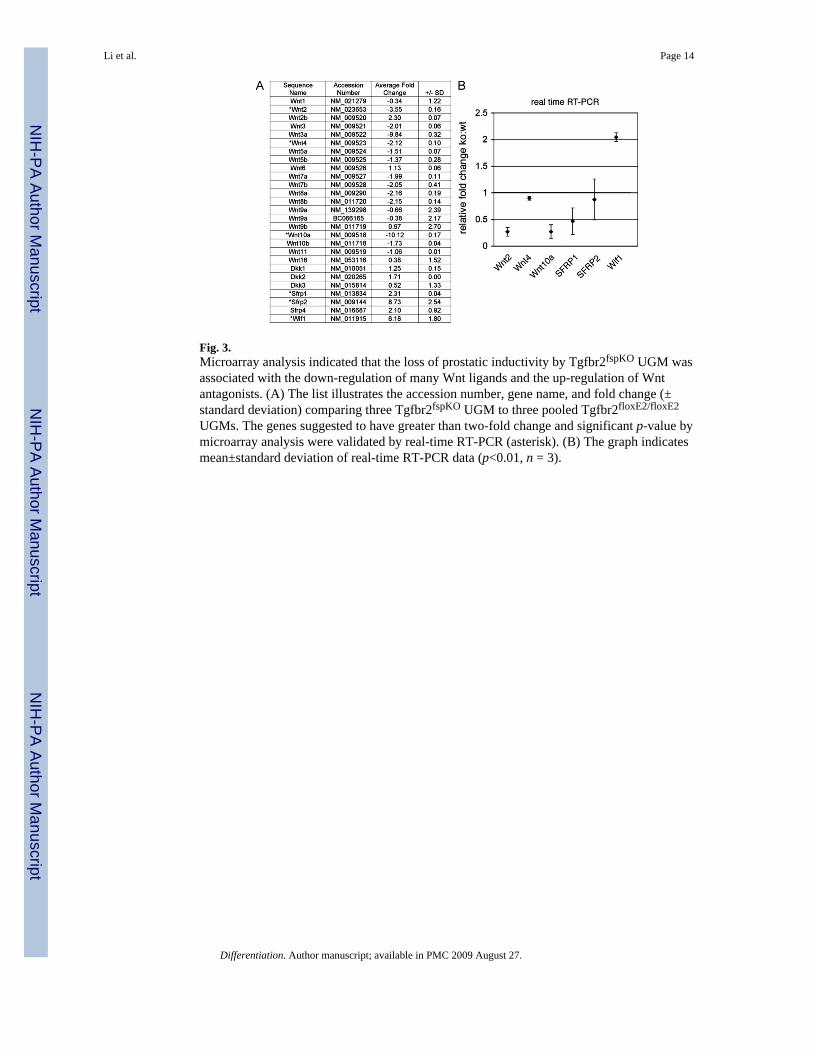
Fig. 3.Microarray analysis indicated that the loss of prostatic inductivity by Tgfbr2fspKO UGM wasassociated with the down-regulation of many Wnt ligands and the up-regulation of Wntantagonists. (A) The list illustrates the accession number, gene name, and fold change (±standard deviation) comparing three Tgfbr2fspKO UGM to three pooled Tgfbr2floxE2/floxE2
UGMs. The genes suggested to have greater than two-fold change and significant p-value bymicroarray analysis were validated by real-time RT-PCR (asterisk). (B) The graph indicatesmean±standard deviation of real-time RT-PCR data (p<0.01, n = 3).
Li et al. Page 14
Differentiation. Author manuscript; available in PMC 2009 August 27.
NIH
-PA Author Manuscript
NIH
-PA Author Manuscript
NIH
-PA Author Manuscript
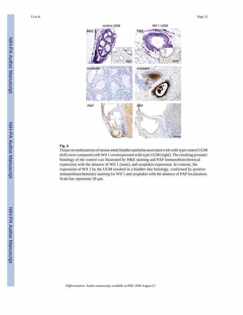
Fig. 4.Tissue recombinations of mouse adult bladder epithelia associated with wild-type control UGM(left) were compared with Wif 1-overexpressed wild-type UGM (right). The resulting prostatichistology of the control was illustrated by H&E staining and PAP immunohistochemicalexpression with the absence of Wif 1 (inset), and uroplakin expression. In contrast, theexpression of Wif 1 by the UGM resulted in a bladder-like histology, confirmed by positiveimmunohistochemistry staining for Wif 1 and uroplakin with the absence of PAP localization.Scale bar represents 50 μm.
Li et al. Page 15
Differentiation. Author manuscript; available in PMC 2009 August 27.
NIH
-PA Author Manuscript
NIH
-PA Author Manuscript
NIH
-PA Author Manuscript
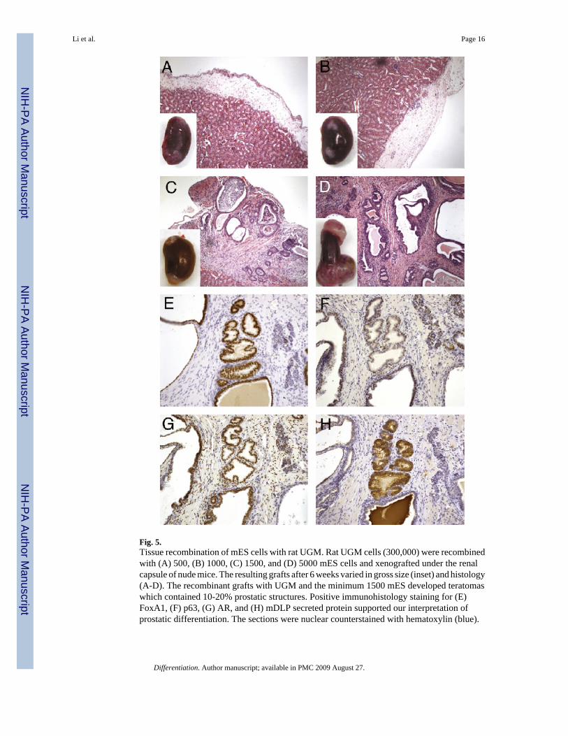
Fig. 5.Tissue recombination of mES cells with rat UGM. Rat UGM cells (300,000) were recombinedwith (A) 500, (B) 1000, (C) 1500, and (D) 5000 mES cells and xenografted under the renalcapsule of nude mice. The resulting grafts after 6 weeks varied in gross size (inset) and histology(A-D). The recombinant grafts with UGM and the minimum 1500 mES developed teratomaswhich contained 10-20% prostatic structures. Positive immunohistology staining for (E)FoxA1, (F) p63, (G) AR, and (H) mDLP secreted protein supported our interpretation ofprostatic differentiation. The sections were nuclear counterstained with hematoxylin (blue).
Li et al. Page 16
Differentiation. Author manuscript; available in PMC 2009 August 27.
NIH
-PA Author Manuscript
NIH
-PA Author Manuscript
NIH
-PA Author Manuscript

Fig. 6.Tissue recombination of Tgfbr2floxE2/floxE2 UGM with labeled adult bladder epithelia. (A) Thediagram illustrates the strategy for identifying BrdU label-retaining cells by the injection ofBrdU in wild-type donor mice for 12 consecutive days, then harvesting the bladder 30 dayslater. Following the isolation of the bladder epithelia, the cells were further labeled withCMFDA. Dual labeling was confirmed by cytospin and fluorescence detection. The panel onthe right illustrates urothelial cells immunostained for BrdU in red and green fluorescence ofCMFDA. Two outcomes of the tissue recombinants with the labeled bladder epithelia couldbe observed: (1) If majority of the resulting prostatic epithelium in the grafts maintaineddetectable green CMFDA-labeled dye, it would suggest urothelial transdifferentiation as only
Li et al. Page 17
Differentiation. Author manuscript; available in PMC 2009 August 27.
NIH
-PA Author Manuscript
NIH
-PA Author Manuscript
NIH
-PA Author Manuscript

a few cell divisions would be required (p5 cell divisions). (2) Alternatively, if only a few greencells were present maintaining BrdU (red), then it is likely that many more cell divisions (≥6)would be required, suggesting bladder stem cell induction as the primary means of prostatedifferentiation. Immunohistochemistry was used to detect BrdU in tissue recombinants after 1month of grafting for (B) the control graft with bladder epithelial cells only (no mesenchyme)and (C) in the tissue recombinants with both bladder epithelia and UGM. Arrowheads indicatecells positive for BrdU staining. (D) Fluorescence microscopy of the UGM+urothelium tissuerecombinant grafts showed little autofluorescence when the control epithelia were not labeledwith CMFDA. (E) However, tissue recombinants with bladder epithelia pre-labeled withCMFDA showed nearly all epithelial cells were green 1 month after grafting. The tissuerecombinants with UGM and bladder epithelial grafts in panels (C-E) had prostatic histology.
Li et al. Page 18
Differentiation. Author manuscript; available in PMC 2009 August 27.
NIH
-PA Author Manuscript
NIH
-PA Author Manuscript
NIH
-PA Author Manuscript

NIH
-PA Author Manuscript
NIH
-PA Author Manuscript
NIH
-PA Author Manuscript
Li et al. Page 19Ta
ble
1N
umbe
r, ty
pe, a
nd re
sults
of t
issu
e re
com
bina
nts
Wt
UG
MK
OU
GM
Wif
1U
GM
Rat
UG
M
BLE
40Pr
osta
te30
Bla
dder
8B
ladd
er
BLE
-labe
led
24Pr
osta
te
mES
22Te
rato
ma
Differentiation. Author manuscript; available in PMC 2009 August 27.