transforming growth factor-b1 mediates epithelial to mesenchymal transdifferentiation through a...
TRANSCRIPT
Molecular Biology of the CellVol. 12, 27–36, January 2001
Transforming Growth Factor-b1 Mediates Epithelial toMesenchymal Transdifferentiation through a RhoA-dependent MechanismNeil A. Bhowmick, Mayshan Ghiassi, Andrei Bakin, Mary Aakre,Christopher A. Lundquist, Michael E. Engel, Carlos L. Arteaga, andHarold L. Moses*
Vanderbilt-Ingram Cancer Center, Departments of Cancer Biology and Medicine, VanderbiltUniversity Medical Center, Nashville, Tennessee 37232
Submitted June 29, 2000; September 6, 2000; Accepted November 7, 2000Monitoring Editor: Carl-Henrik Heldin
Transforming growth factor-b1 (TGF-b) can be tumor suppressive, but it can also enhance tumorprogression by stimulating the complex process of epithelial-to-mesenchymal transdifferentiaion(EMT). The signaling pathway(s) that regulate EMT in response to TGF-b are not well understood.We demonstrate the acquisition of a fibroblastoid morphology, increased N-cadherin expression,loss of junctional E-cadherin localization, and increased cellular motility as markers for TGF-b–induced EMT. The expression of a dominant-negative Smad3 or the expression of Smad7 to levelsthat block growth inhibition and transcriptional responses to TGF-b do not inhibit mesenchymaldifferentiation of mammary epithelial cells. In contrast, we show that TGF-b rapidly activatesRhoA in epithelial cells, and that blocking RhoA or its downstream target p160ROCK, by theexpression of dominant-negative mutants, inhibited TGF-b–mediated EMT. The data suggest thatTGF-b rapidly activates RhoA-dependent signaling pathways to induce stress fiber formation andmesenchymal characteristics.
INTRODUCTION
Transforming growth factor-b1 (TGF-b) regulates growth,differentiation, and epithelial transformation in the multi-step processes of tumorigenesis, wound healing, and embry-ogenesis. Based on studies using cultured cells, transgenicmice, and human tumors, an emerging model suggests theTGF-b signaling pathway acts as a tumor suppressor, but itcan act as a promoter of tumor progression during the laterstages of tumorigenesis (McLeod et al., 1990; Han et al., 1993;Pierce et al., 1995; Cui et al., 1996; Oft et al., 1998; Portella etal., 1998). Although a role for TGF-b as an autocrine trans-forming and morphogenic factor in epithelial cells is wellestablished, our understanding of its intracellular signalingmechanisms is limited. This report identifies intracellularTGF-b effectors and new mechanistic insight into TGF-b–mediated fibroblastic conversion of mammary epithelialcells, a process likely involved in tumor invasion and me-tastasis.
TGF-b signals through an activated heteromeric complexof type I and type II serine/threonine kinase receptors(Wrana et al., 1994). Subsequent cytoplasmic signaling in-
volves the phosphorylation of ligand-specific SMAD pro-teins, Smad2 and/or Smad3, that act as intermediates fortranscriptional regulation and cell cycle arrest in conjunctionwith Smad4 in epithelial cells (reviewed in Kretzschmar andMassague, 1998). RhoA, Rac1, and Jun N-terminal kinase(JNK) can promote SMAD-mediated signaling, whereasnegative regulators of SMAD-mediated transcription in-clude Smad7, RhoB, and calmodulin (reviewed in Engel etal., 1998b). Smad7 specifically prevents the access and sub-sequent phosphorylation of Smad2 and Smad3 to the acti-vated TGF-b receptor complex before downstream amplifi-cation of the signaling cascade (Nakao et al., 1997).Additionally, the activation of the H-Ras oncogene leads tosuppression of SMAD signaling (Calonge and Massague,1999; Kretzschmar et al., 1999). Parallel TGF-b–mediatedtranscriptional regulation has been shown to include themitogen-activated protein kinase family (Atfi et al., 1997;Engel et al., 1999) and the potentiation of phosphatidylino-sitol 3-kinase (PI3-kinase) activity (Krymskaya et al., 1997;Higaki and Shimokado, 1999). The initiation of multiplesignaling pathways downstream of the activated receptorcomplex results in the pleotropic effects of TGF-b.
Acquisition of a spindle-shaped morphology, delocaliza-tion of E-cadherin from cell junctions, and elevated N-cad-herin expression are hallmarks of mesenchymal phenotypic
* Corresponding author. E-mail address: [email protected].
© 2001 by The American Society for Cell Biology 27
conversion of mammary epithelia in cell culture and intumor invasion (Nieman et al., 1999). As regulators of theactin cytoskeleton and cadherin junctions, the Rho family ofsmall GTPases is commonly implicated in these processes(Bishop and Hall, 2000). Microinjection of RhoA, Rac1, orCdc42 into fibroblasts triggers the formation of stress fibers,lamellipodia, or filopodia, respectively (Ridley and Hall,1992; Ridley et al., 1992). Rac1 and Cdc42 are involved in theestablishment and maintenance of epithelial intercellularadhesions (Braga et al., 1997; Hordijk et al., 1997; Takaishi etal., 1997; Zhong et al., 1997); in contrast, RhoA activation isimplicated in the reversion of the epithelioid phenotypetoward a migratory, fibroblastoid morpholology of NIH3T3cells (Sander et al., 1999). The activated GTP-bound form ofRhoA associates specifically with multiple protein kinases.Among these, p160 Rho-associated coiled-coil–containingprotein kinase (p160ROCK) regulates actin stress fiber forma-tion and integrin activation (Ishizaki et al., 1997). Thus, dataindicate an active role for small GTPases in the maintenanceand dynamic regulation of intercellular adhesion as well ashaving a cytoskeleton-independent role in cell transforma-tion, both alone and in the context of H-Ras activation.
Because TGF-b treatment of mammary epithelial cells re-capitulates this transition in culture, we explored the in-volvement of small GTPases and candidate downstreameffectors in TGF-b–induced EMT. We show here that EMTinduced by TGF-b requires RhoA signaling. Importantly, wedemonstrate that TGF-b rapidly activates RhoA in epithelialnontransformed mouse mammary cell line (NMuMG), minklung cell line (Mv1Lu), pancreatic tumor cell line (BxPc3),and primary mouse keratinocytes, but not in fibroblasticNIH3T3 cells or TGF-b type I receptor deficient mink lungcells (R1B).
MATERIALS AND METHODS
Reagents and ConstructsTGF-b1 was supplied by R&D Systems (Minneapolis, MN),LY294002 and curcumin was purchased from Sigma (St. Louis, MO).SCH51344 was a gift from Dr. C.C. Kumar (State University of NewYork at Stony Brook, NY) (Walsh et al., 1997) and LPA (1-oleoyl) wasfrom Avanti Polar Lipids (Alabaster, AL). The 3TP-Lux-reporterconstruct was obtained from J. Massague (Memorial Sloan-Ketter-ing Cancer Center, New York, NY). The cDNA constructs encodingSmad3-FLAG (Dr. Rik Derynck, University of California, San Fran-cisco, CA), Smad7-FLAG (Dr. Peter ten Dijke, Ludwig Institute forCancer Research, Uppsala, Sweden), JNK1APF, N17Rac1 (Dr. LynnCross, National Institutes of Health, Bethesda, MD), N19-RhoA (Dr.Lynn Cross), QL-RhoA (Dr. Lynn Cross), KD-IA p160ROCK (Dr.Shuh Narumiya, Kyoto University, Kyoto, Japan), and green fluo-rescent protein (GFP) (Dr. Fred H. Kant, Columbia University, NewYork, NY) were subcloned into the pBabe retroviral vector.
Thymidine Incorporation and Luciferase ReporterAssayNMuMG cells (American Type Culture Collection, Manassas, VA)stably expressing Smad3 and Smad7 as well as the parentalNMuMG cell line were plated in 24-well plates for thymidine in-corporation and 3TP-Lux reporter assays. To assay cell growth, cellswere treated for 24 h with TGF-b and loaded with [3H]thymidine2 h before harvesting. The cells were washed and lysates measuredwith a scintillation counter. Transcriptional activation was tested incells transfected with the 3TP-Lux luciferase (firefly) reporter con-
struct cDNA in conjunction with a cytomegalovirus-driven renelaluciferase plasmid (Promega, Madison, WI). Dual-luciferase assayswere performed on lysed cells as indicated by the manufacturer,Promega, and measured on a Monolight 2010 luminometer (Ana-lytical Luminescence Laboratory, San Diego, CA). The ratios offirefly and renela luciferase measurements were calculated in nor-malizing the reporter data in relative luminescent units.
Retroviral Transduction and Cell CulturePhoenix packaging cells (Kinsella and Nolan, 1996) were transfectedwith retroviral constructs with Superfect (Qiagen, Chatsworth, CA)according to manufacturer recommendations to produce culturesupernatants containing virus. We established matched isogenicclones from the NMuMG parent cell line. Each of the lines selectedfor further studies were TGF-b sensitive for growth inhibition andEMT (Bhowmick and Moses, unpublished data). NMuMG cellswere infected with virus by culturing the cells for 18 h in 1:1 Phoenixconditioned media: fresh DMEM, 10% fetal calf serum, 10 mg/mlinsulin, supplemented with 4 ng/ml Polybrene (Sigma). The cellswere subjected to various treatments, assaying for protein expres-sion, or cultured in puromycin-containing media for the establish-ment of stable cell lines 48 h after transduction.
Immunofluorescent DetectionCells grown on coverslips to be stained for E- or N-cadherin (Trans-duction Laboratories, Lexington, KY) were fixed in ice-cold 100%methanol and subsequently permeabilized in phosphate-bufferedsaline containing 0.1% Triton X-100 for 10 min each step. Nonspe-cific sites were blocked with 3% milk; diluted primary antibody(1:1000) was incubated for 1 h, and visualized using secondaryantibody conjugated to Cy2 or Cy3 (Sigma) fluorescence on a ZeissAxovert fluorescence microscope. F-actin was stained by fixing thecells in 4% paraformaldehyde followed by incubation with TexasRed-conjugated phalloidin (Molecular Probes).
Western Blotting and p160ROCK Kinase AssayCells transfected using Superfect (Qiagen) or retrovirally trans-duced were washed in ice-cold phosphate-buffered saline, lysed (in50 mM HEPES [pH 7.5], 150 mM NaCl, 0.2 mM vanadate, 1 mMMgCl2, 1 mM CaCl2, 10% glycerol, 1% NP-40, and 0.1% SDS), brieflysonicated, and clarified by centrifugation. Protein (10 mg) was sep-arated on a 10% SDS-polyacrylamide gel, transferred to nitrocellu-lose, and immunoblotted with appropriate antibodies. Respectivesecondary horseradish peroxidase-conjugated antibodies (Amer-sham Pharmacia Biotech, Piscataway, NJ) were used, and werevisualized with an enhanced chemiluminescence system (Amer-sham Pharmacia Biotech). Activity of p160ROCK was determined byimmunoprecipitating myc-tagged p160ROCK from 200 mg of total celllysate for incubation with 10 mg of histone, 5 mM [32P]ATP in 50mM HEPES (pH 7.4) and 0.5 mM MgCl2 for 8 min at 30°C. Thereaction was stopped by the addition of Laemmli sample buffer andseparated on a 15% polyacrylamide gel for visualization by autora-diography.
GTPase Activity AssaysThe biochemical activity assays were performed essentially as de-scribed previously by Reid et al. (Ren et al., 1999). A glutathioneS-transferase (GST) fusion protein of the Rho binding domain (RBD,a kind gift from Dr. Martin A. Schwartz, Scripps Institute, La JollaCA), rhotekin (Reid et al., 1996), was used (Ren et al., 1999). For eachmeasurement, one 100-mm dish of cells was lysed in 1% NP-40, 50mM Tris, pH 7.4, 10% glycerol, 100 mM NaCl, and 10 mM MgCl2.The GST-RBD precoupled to agarose-glutathione beads (Sigma) wasused to precipitate GTP-bound RhoA from cleared lysates of cellsfor 30 min at 4°C for subsequent immunoblotting for RhoA, similarto that described previously (Ren et al., 1999). A GST fusion protein
N.A. Bhowmick et al.
Molecular Biology of the Cell28
of the PAK3 binding domain (PBD, a kind gift from Dr. Gary M.Bokoch, Scripps Institute) was used to capture GTP-bound Rac1 andCdc42 for subsequent visualization by immunoblotting in a similarmanner (Benard et al., 1999).
RESULTS
EMT in mammary epithelial cells is characterized by theacquisition of spindle morphology and increased motility,with changes in cadherin expression and localization. Weused pharmacological and genetic approaches to affect can-didate-signaling pathways in TGF-b–mediated EMT.SMAD- and RhoGTPase-dependent pathways figuredprominently given their previously demonstrated involve-ment in TGF-b signal transduction.
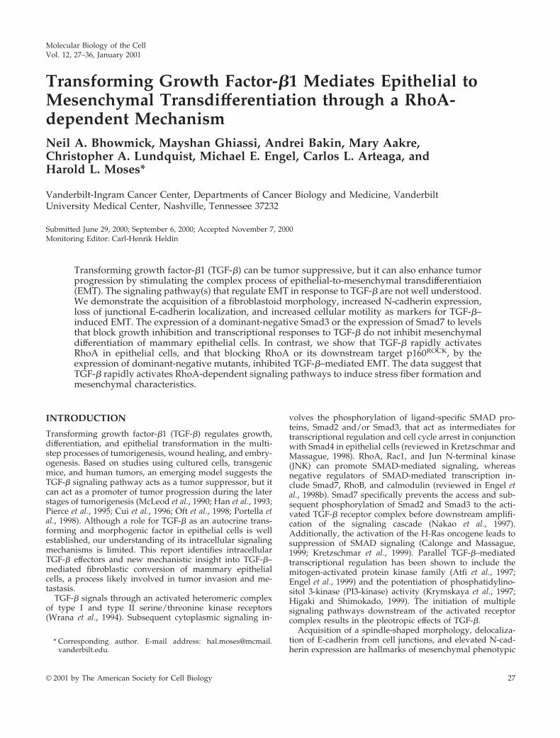
TGF-b–mediated morphological changes of NMuMG cellswere accompanied by a loss of E-cadherin junctional local-ization as reported previously (Miettinen et al., 1994; Piek etal., 1999) as well as our finding of the emergence of N-cadherin expression at cell margins (Figure 1). Recently, arelationship between N-cadherin expression with elevatedmotility and invasive characteristics of mammary tumorcells has been reported (Nieman et al., 1999). Interestingly,immunoblot analysis showed no change in E-cadherin orcadherin-associated b-catenin expression levels in responseto TGF-b. However, N-cadherin expression, absent fromepithelioid NMuMG cells, was observed after 3 h of TGF-btreatment and remained elevated through the 48-h timecourse examined (Figure 1A). Examination of TGF-b–treatedcells showed disruption of cell-cell adhesions and a changein cell morphology to a spindle shape in association with theloss of E-cadherin junctional localization and the appearanceof N-cadherin staining at the plasma membrane (Figure 1B).Concomitant acquisition of actin stress fibers was also ob-served (Piek et al., 1999) (see below). However, the cellsundergoing EMT remained sensitive to TGF-b growth inhi-bition during fibroblastic conversion (Figure 2C). These datasuggest that TGF-b initiates a program of EMT that corre-lates with changes in cadherin expression and localization.
Figure 1. NMuMG cells treated with TGF-b undergo EMT. (A)Immunoblotting of NMuMG cells incubated with 5 ng/ml TGF-bfor 0–48 h shows nearly equivalent cellular expression of E-cad-herin and b-catenin through the time course, whereas N-cadherinexpression is induced by 3 h of TGF-b treatment and remainselevated throughout the time course. (B) Epifluorescent and corre-sponding phase contrast images show that the fibroblastic cell mor-phology of cells treated with 5 ng/ml TGF-b for 24 h have a loss ofimmunodetectable E-cadherin at the cell junctions with a gain ofN-cadherin expression at cell margins.
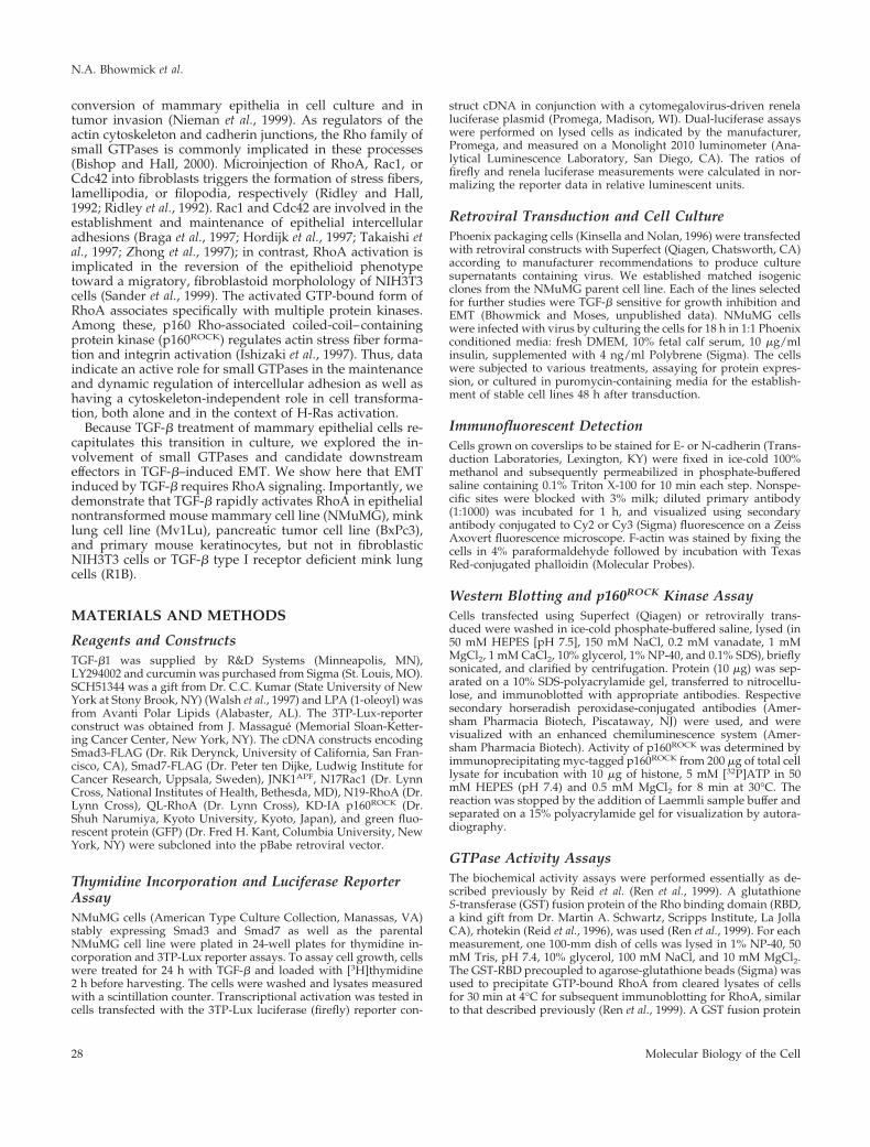
Figure 2. The down-regulation of SMAD signaling by retroviral transduction inhibits TGF-b–mediated responses. (A) Retroviral infectionof the GFP cDNA demonstrates the high transduction efficiency of the NMuMG cells. GFP-retrovirus infected cells were observed by phasecontrast and epifluorescence microscopy. (B) Expression of FLAG-tagged Smad3 or Smad7 was detected in retrovirally transduced NMuMGby immunoblotting for the epitope tag. (C) Cells stably transduced with GFP (open bar), Smad3 (gray bar), and Smad7 (closed bar) retroviruswere treated with TGF-b for 24 h and assayed for 3TP-Lux reporter activity (left) and thymidine incorporation (right).
TGF-b–mediated RhoA Activation in EMT
Vol. 12, January 2001 29
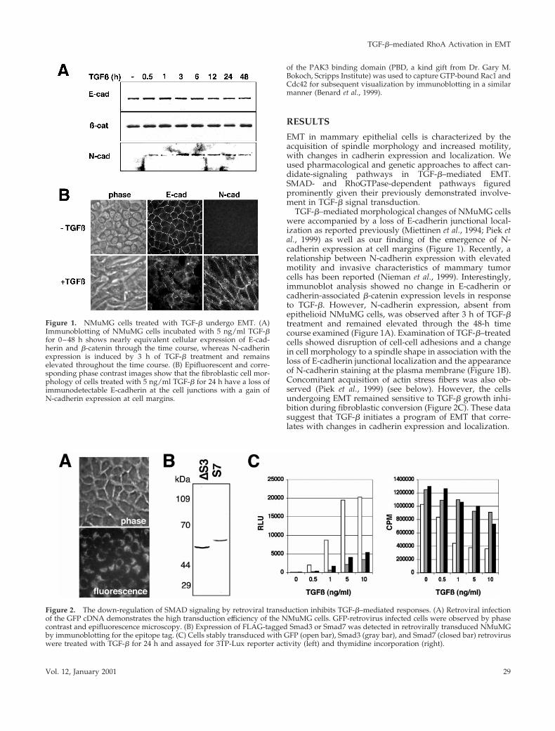
We initially addressed the role of SMADs in TGF-b–me-diated EMT by the overexpression of antagonists of theSMAD signaling pathway. Stable retroviral infections withGFP (control), FLAG-Smad3 (a Smad3 deletion of the C-terminal–activating phosphorylation site that behaves in adominant-negative manner; Zhang et al., 1996), and FLAG-Smad7 (an inhibitor of SMAD signaling; Nakao et al., 1997)cDNA were expressed and confirmed by epifluorescencemicroscopy and immunoblotting (Figure 2, A and B). Thetransduced cells displayed $80% inhibition of TGF-b tran-scriptional activation of the 3TP-Lux promoter, and dimin-ished responsiveness to TGF-b–mediated growth inhibitionas expected from previous reports (Zhang et al., 1996; Nakaoet al., 1997; Itoh et al., 1998) (Figure 2C). However, Smad3- orSmad7-expressing cells treated with TGF-b acquired a fibro-blastic morphology with a concomitant loss of junctionalE-cadherin staining and gain of plasma membrane N-cad-herin localization similar to control cells infected with ret-rovirus containing the GFP gene (Figure 3). These data sug-gest that TGF-b–induced EMT is unaffected by decreasedSMAD signaling as reflected by SMAD-dependent growthand transcriptional responses. Additionally, TGF-b–medi-ated stress fiber formation was not inhibited by the down-regulation of SMAD signaling.
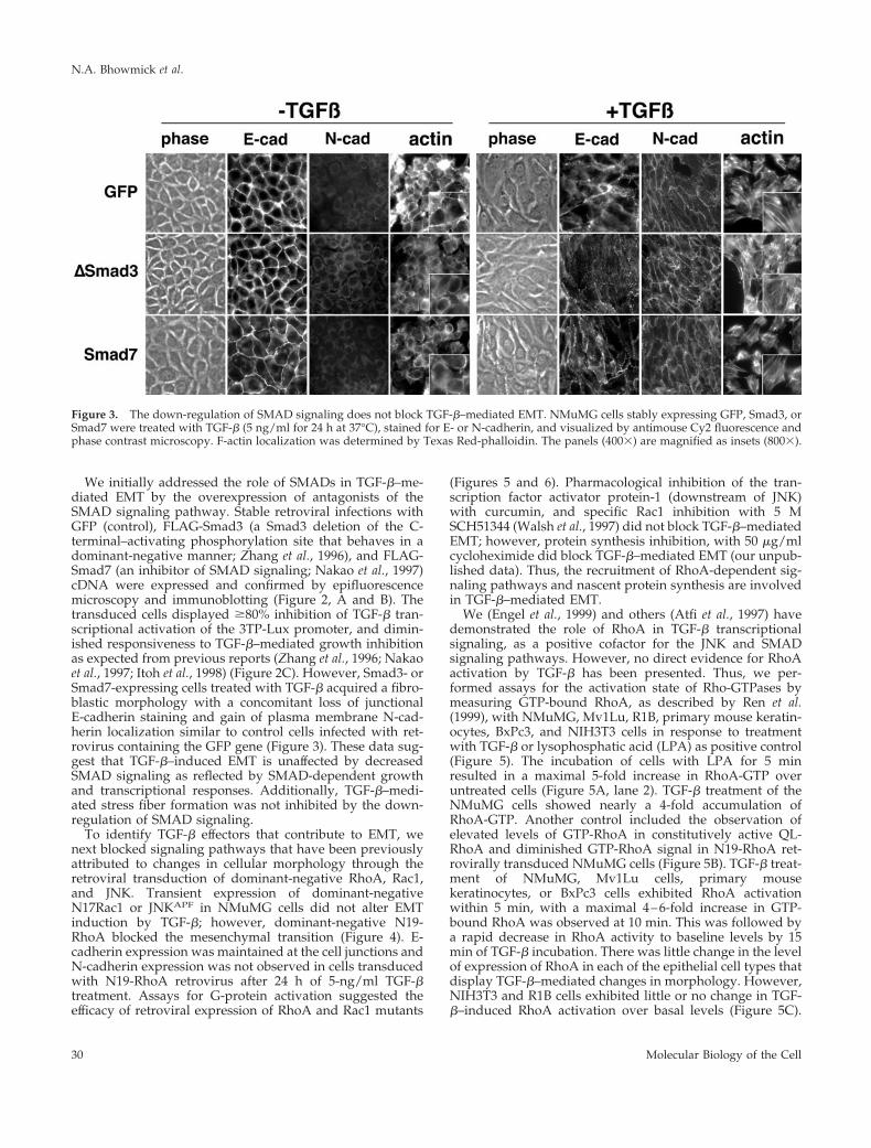
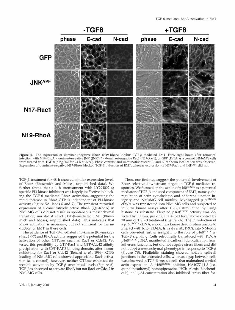
To identify TGF-b effectors that contribute to EMT, wenext blocked signaling pathways that have been previouslyattributed to changes in cellular morphology through theretroviral transduction of dominant-negative RhoA, Rac1,and JNK. Transient expression of dominant-negativeN17Rac1 or JNKAPF in NMuMG cells did not alter EMTinduction by TGF-b; however, dominant-negative N19-RhoA blocked the mesenchymal transition (Figure 4). E-cadherin expression was maintained at the cell junctions andN-cadherin expression was not observed in cells transducedwith N19-RhoA retrovirus after 24 h of 5-ng/ml TGF-btreatment. Assays for G-protein activation suggested theefficacy of retroviral expression of RhoA and Rac1 mutants
(Figures 5 and 6). Pharmacological inhibition of the tran-scription factor activator protein-1 (downstream of JNK)with curcumin, and specific Rac1 inhibition with 5 MSCH51344 (Walsh et al., 1997) did not block TGF-b–mediatedEMT; however, protein synthesis inhibition, with 50 mg/mlcycloheximide did block TGF-b–mediated EMT (our unpub-lished data). Thus, the recruitment of RhoA-dependent sig-naling pathways and nascent protein synthesis are involvedin TGF-b–mediated EMT.
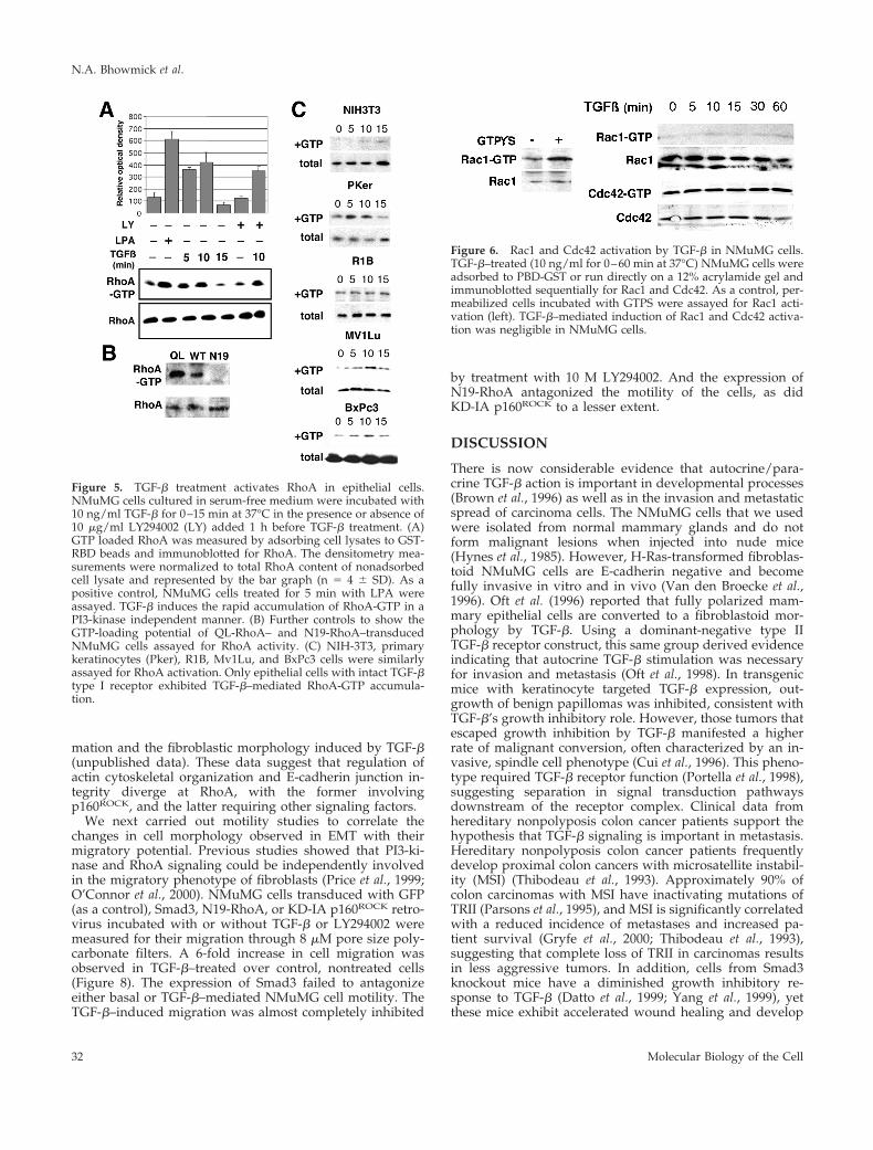
We (Engel et al., 1999) and others (Atfi et al., 1997) havedemonstrated the role of RhoA in TGF-b transcriptionalsignaling, as a positive cofactor for the JNK and SMADsignaling pathways. However, no direct evidence for RhoAactivation by TGF-b has been presented. Thus, we per-formed assays for the activation state of Rho-GTPases bymeasuring GTP-bound RhoA, as described by Ren et al.(1999), with NMuMG, Mv1Lu, R1B, primary mouse keratin-ocytes, BxPc3, and NIH3T3 cells in response to treatmentwith TGF-b or lysophosphatic acid (LPA) as positive control(Figure 5). The incubation of cells with LPA for 5 minresulted in a maximal 5-fold increase in RhoA-GTP overuntreated cells (Figure 5A, lane 2). TGF-b treatment of theNMuMG cells showed nearly a 4-fold accumulation ofRhoA-GTP. Another control included the observation ofelevated levels of GTP-RhoA in constitutively active QL-RhoA and diminished GTP-RhoA signal in N19-RhoA ret-rovirally transduced NMuMG cells (Figure 5B). TGF-b treat-ment of NMuMG, Mv1Lu cells, primary mousekeratinocytes, or BxPc3 cells exhibited RhoA activationwithin 5 min, with a maximal 4–6-fold increase in GTP-bound RhoA was observed at 10 min. This was followed bya rapid decrease in RhoA activity to baseline levels by 15min of TGF-b incubation. There was little change in the levelof expression of RhoA in each of the epithelial cell types thatdisplay TGF-b–mediated changes in morphology. However,NIH3T3 and R1B cells exhibited little or no change in TGF-b–induced RhoA activation over basal levels (Figure 5C).
Figure 3. The down-regulation of SMAD signaling does not block TGF-b–mediated EMT. NMuMG cells stably expressing GFP, Smad3, orSmad7 were treated with TGF-b (5 ng/ml for 24 h at 37°C), stained for E- or N-cadherin, and visualized by antimouse Cy2 fluorescence andphase contrast microscopy. F-actin localization was determined by Texas Red-phalloidin. The panels (4003) are magnified as insets (8003).
N.A. Bhowmick et al.
Molecular Biology of the Cell30
TGF-b treatment for 48 h showed similar expression levelsof RhoA (Bhowmick and Moses, unpublished data). Wefurther found that a 1 h pretreatment with LY294002 (aspecific PI3-kinase inhibitor) was largely ineffective in block-ing the TGF-b–mediated RhoA activation, suggesting therapid increase in RhoA-GTP is independent of PI3-kinaseactivity (Figure 5A, lanes 6 and 7). The transient retroviralexpression of a constitutively active RhoA (QL-RhoA) inNMuMG cells did not result in spontaneous mesenchymaltransition, nor did it effect TGF-b–mediated EMT (Bhow-mick and Moses, unpublished data). This indicates thatRhoA activation is necessary, but not sufficient for the in-duction of EMT in these cells.
The evidence of TGF-b–mediated PI3-kinase (Krymskayaet al., 1997) and RhoA activity suggested the potential for theactivation of other GTPases such as Rac1 or Cdc42. Wetested this possibility by GTP-Rac1 and GTP-Cdc42 affinityprecipitation with GST-PAK3 binding domain, after immu-noblotting for Rac1 or Cdc42 (Benard et al., 1999). GTPSloading of NMuMG cells showed appreciable Rac1 activa-tion (as a control); however, neither GTPase exhibited de-tectable activation by TGF-b over basal levels (Figure 6).TGF-b is observed to activate RhoA but not Rac1 or Cdc42 inNMuMG cells.
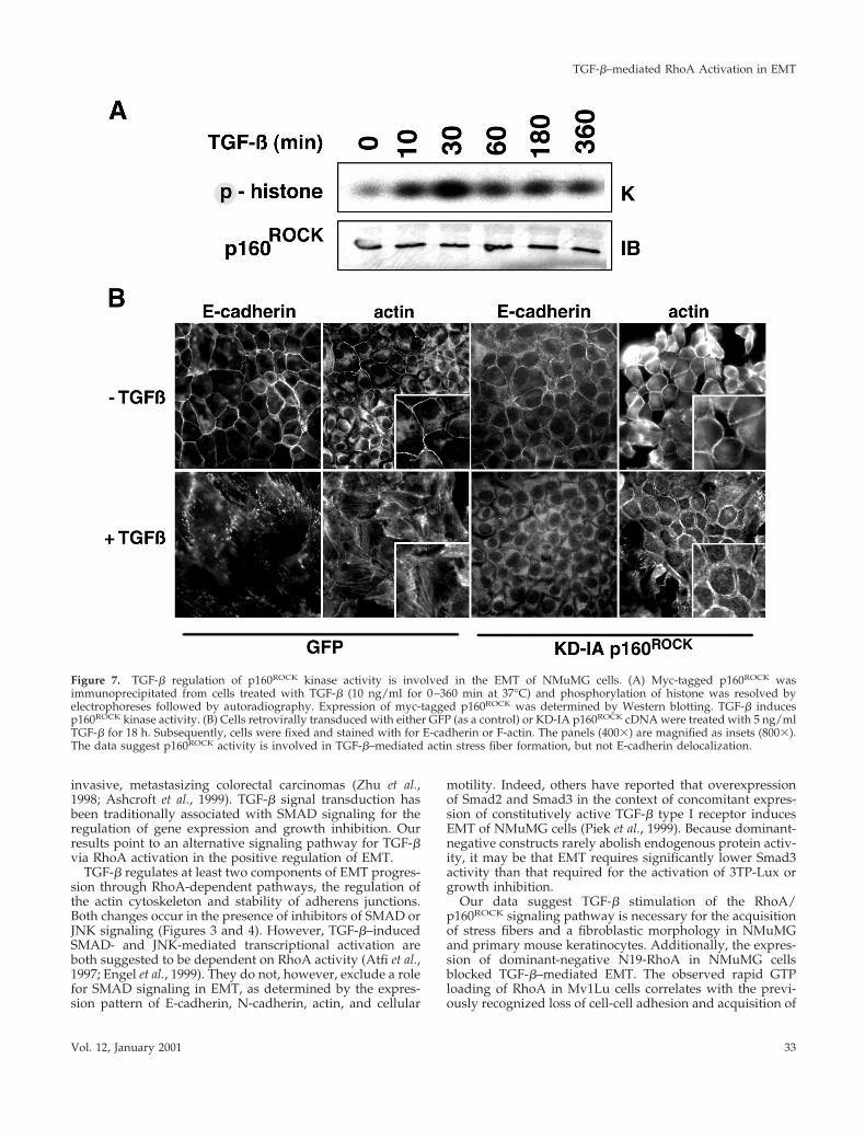
Thus, our findings suggest the potential involvement ofRhoA-selective downstream targets in TGF-b–mediated re-sponses. We focused on the action of p160ROCK as a potentialmediator of TGF-b–induced component of EMT, namely, theregulation of actin cytoskeleton and adherens junction in-tegrity and NMuMG cell motility. Myc-tagged p160ROCK
cDNA was transfected into NMuMG cells and subjected toin vitro kinase assays after TGF-b stimulation by usinghistone as substrate. Elevated p160ROCK activity was de-tected by 10 min, peaking at a 4-fold level above control by30 min of TGF-b treatment (Figure 7A). The introduction ofa p160ROCK cDNA, encoding a kinase dead protein unable tointeract with Rho (KD-IA; Ishizaki et al., 1997), into NMuMGcells provided further insight into the role of p160ROCK inTGF-b signaling. Cells retrovirally transduced with KD-IAp160ROCK cDNA manifested E-cadherin delocalization fromadherens junctions, but did not acquire stress fibers and didnot adopt a mesenchymal phenotype in response to TGF-b(Figure 7B). Phalloidin staining showed notable cell-celljunctions in the untreated cells, whereas a gap between cellswas observed in TGF-b–treated cells that maintained corticalactin expression. A p160ROCK inhibitor, HA1077 [1-5-(iso-quinolinesulfonyl)-homopiperazine HCl; Alexis Biochemi-cals], at 1 mM concentration also inhibited stress fiber for-
Figure 4. The expression of dominant-negative RhoA (N19-RhoA) inhibits TGF-b–mediated EMT. Forty-eight hours after retroviralinfection with N19-RhoA, dominant-negative JNK (JNKAPF), dominant-negative Rac1 (N17-Rac1), or GFP cDNA as a control, NMuMG cellswere treated with TGF-b (5 ng/ml for 24 h at 37°C). Phase contrast and immunofluorescent E- and N-cadherin localization was observed.Expression of dominant-negative N17-RhoA blocked TGF-b induction of EMT, whereas expression of N17-Rac1 and JNKAPF did not.
TGF-b–mediated RhoA Activation in EMT
Vol. 12, January 2001 31
mation and the fibroblastic morphology induced by TGF-b(unpublished data). These data suggest that regulation ofactin cytoskeletal organization and E-cadherin junction in-tegrity diverge at RhoA, with the former involvingp160ROCK, and the latter requiring other signaling factors.
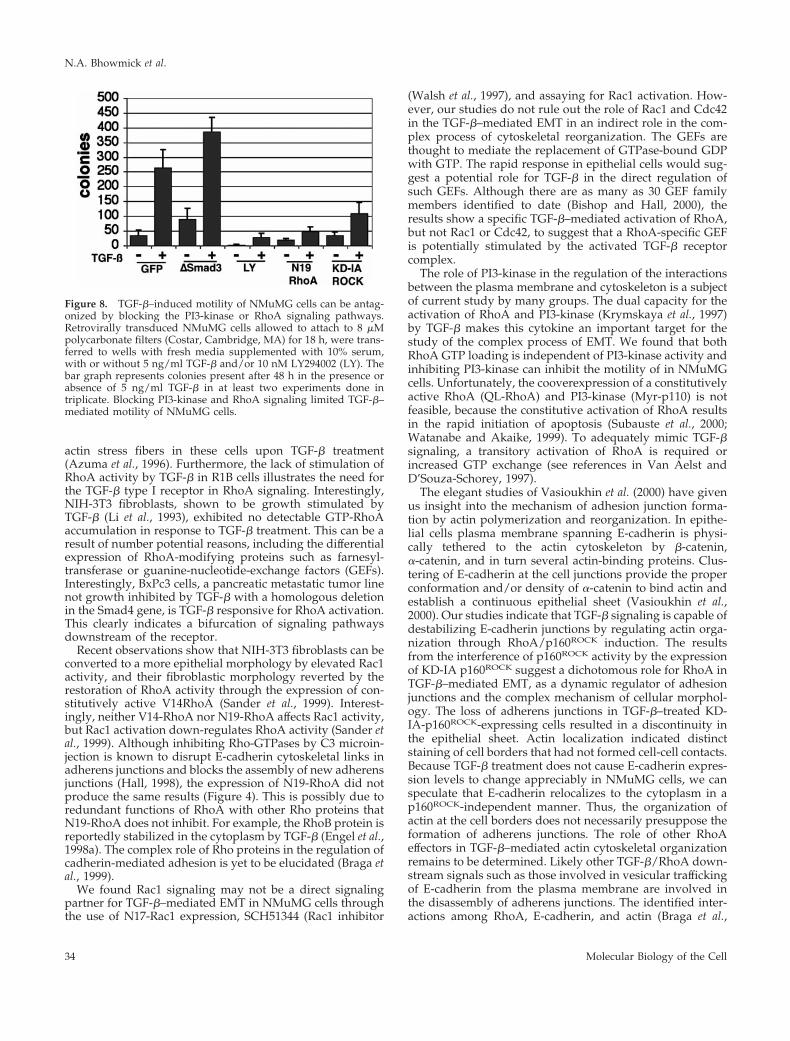
We next carried out motility studies to correlate thechanges in cell morphology observed in EMT with theirmigratory potential. Previous studies showed that PI3-ki-nase and RhoA signaling could be independently involvedin the migratory phenotype of fibroblasts (Price et al., 1999;O’Connor et al., 2000). NMuMG cells transduced with GFP(as a control), Smad3, N19-RhoA, or KD-IA p160ROCK retro-virus incubated with or without TGF-b or LY294002 weremeasured for their migration through 8 mM pore size poly-carbonate filters. A 6-fold increase in cell migration wasobserved in TGF-b–treated over control, nontreated cells(Figure 8). The expression of Smad3 failed to antagonizeeither basal or TGF-b–mediated NMuMG cell motility. TheTGF-b–induced migration was almost completely inhibited
by treatment with 10 M LY294002. And the expression ofN19-RhoA antagonized the motility of the cells, as didKD-IA p160ROCK to a lesser extent.
DISCUSSION
There is now considerable evidence that autocrine/para-crine TGF-b action is important in developmental processes(Brown et al., 1996) as well as in the invasion and metastaticspread of carcinoma cells. The NMuMG cells that we usedwere isolated from normal mammary glands and do notform malignant lesions when injected into nude mice(Hynes et al., 1985). However, H-Ras-transformed fibroblas-toid NMuMG cells are E-cadherin negative and becomefully invasive in vitro and in vivo (Van den Broecke et al.,1996). Oft et al. (1996) reported that fully polarized mam-mary epithelial cells are converted to a fibroblastoid mor-phology by TGF-b. Using a dominant-negative type IITGF-b receptor construct, this same group derived evidenceindicating that autocrine TGF-b stimulation was necessaryfor invasion and metastasis (Oft et al., 1998). In transgenicmice with keratinocyte targeted TGF-b expression, out-growth of benign papillomas was inhibited, consistent withTGF-b’s growth inhibitory role. However, those tumors thatescaped growth inhibition by TGF-b manifested a higherrate of malignant conversion, often characterized by an in-vasive, spindle cell phenotype (Cui et al., 1996). This pheno-type required TGF-b receptor function (Portella et al., 1998),suggesting separation in signal transduction pathwaysdownstream of the receptor complex. Clinical data fromhereditary nonpolyposis colon cancer patients support thehypothesis that TGF-b signaling is important in metastasis.Hereditary nonpolyposis colon cancer patients frequentlydevelop proximal colon cancers with microsatellite instabil-ity (MSI) (Thibodeau et al., 1993). Approximately 90% ofcolon carcinomas with MSI have inactivating mutations ofTRII (Parsons et al., 1995), and MSI is significantly correlatedwith a reduced incidence of metastases and increased pa-tient survival (Gryfe et al., 2000; Thibodeau et al., 1993),suggesting that complete loss of TRII in carcinomas resultsin less aggressive tumors. In addition, cells from Smad3knockout mice have a diminished growth inhibitory re-sponse to TGF-b (Datto et al., 1999; Yang et al., 1999), yetthese mice exhibit accelerated wound healing and develop
Figure 6. Rac1 and Cdc42 activation by TGF-b in NMuMG cells.TGF-b–treated (10 ng/ml for 0–60 min at 37°C) NMuMG cells wereadsorbed to PBD-GST or run directly on a 12% acrylamide gel andimmunoblotted sequentially for Rac1 and Cdc42. As a control, per-meabilized cells incubated with GTPS were assayed for Rac1 acti-vation (left). TGF-b–mediated induction of Rac1 and Cdc42 activa-tion was negligible in NMuMG cells.
Figure 5. TGF-b treatment activates RhoA in epithelial cells.NMuMG cells cultured in serum-free medium were incubated with10 ng/ml TGF-b for 0–15 min at 37°C in the presence or absence of10 mg/ml LY294002 (LY) added 1 h before TGF-b treatment. (A)GTP loaded RhoA was measured by adsorbing cell lysates to GST-RBD beads and immunoblotted for RhoA. The densitometry mea-surements were normalized to total RhoA content of nonadsorbedcell lysate and represented by the bar graph (n 5 4 6 SD). As apositive control, NMuMG cells treated for 5 min with LPA wereassayed. TGF-b induces the rapid accumulation of RhoA-GTP in aPI3-kinase independent manner. (B) Further controls to show theGTP-loading potential of QL-RhoA– and N19-RhoA–transducedNMuMG cells assayed for RhoA activity. (C) NIH-3T3, primarykeratinocytes (Pker), R1B, Mv1Lu, and BxPc3 cells were similarlyassayed for RhoA activation. Only epithelial cells with intact TGF-btype I receptor exhibited TGF-b–mediated RhoA-GTP accumula-tion.
N.A. Bhowmick et al.
Molecular Biology of the Cell32
invasive, metastasizing colorectal carcinomas (Zhu et al.,1998; Ashcroft et al., 1999). TGF-b signal transduction hasbeen traditionally associated with SMAD signaling for theregulation of gene expression and growth inhibition. Ourresults point to an alternative signaling pathway for TGF-bvia RhoA activation in the positive regulation of EMT.
TGF-b regulates at least two components of EMT progres-sion through RhoA-dependent pathways, the regulation ofthe actin cytoskeleton and stability of adherens junctions.Both changes occur in the presence of inhibitors of SMAD orJNK signaling (Figures 3 and 4). However, TGF-b–inducedSMAD- and JNK-mediated transcriptional activation areboth suggested to be dependent on RhoA activity (Atfi et al.,1997; Engel et al., 1999). They do not, however, exclude a rolefor SMAD signaling in EMT, as determined by the expres-sion pattern of E-cadherin, N-cadherin, actin, and cellular
motility. Indeed, others have reported that overexpressionof Smad2 and Smad3 in the context of concomitant expres-sion of constitutively active TGF-b type I receptor inducesEMT of NMuMG cells (Piek et al., 1999). Because dominant-negative constructs rarely abolish endogenous protein activ-ity, it may be that EMT requires significantly lower Smad3activity than that required for the activation of 3TP-Lux orgrowth inhibition.
Our data suggest TGF-b stimulation of the RhoA/p160ROCK signaling pathway is necessary for the acquisitionof stress fibers and a fibroblastic morphology in NMuMGand primary mouse keratinocytes. Additionally, the expres-sion of dominant-negative N19-RhoA in NMuMG cellsblocked TGF-b–mediated EMT. The observed rapid GTPloading of RhoA in Mv1Lu cells correlates with the previ-ously recognized loss of cell-cell adhesion and acquisition of
Figure 7. TGF-b regulation of p160ROCK kinase activity is involved in the EMT of NMuMG cells. (A) Myc-tagged p160ROCK wasimmunoprecipitated from cells treated with TGF-b (10 ng/ml for 0–360 min at 37°C) and phosphorylation of histone was resolved byelectrophoreses followed by autoradiography. Expression of myc-tagged p160ROCK was determined by Western blotting. TGF-b inducesp160ROCK kinase activity. (B) Cells retrovirally transduced with either GFP (as a control) or KD-IA p160ROCK cDNA were treated with 5 ng/mlTGF-b for 18 h. Subsequently, cells were fixed and stained with for E-cadherin or F-actin. The panels (4003) are magnified as insets (8003).The data suggest p160ROCK activity is involved in TGF-b–mediated actin stress fiber formation, but not E-cadherin delocalization.
TGF-b–mediated RhoA Activation in EMT
Vol. 12, January 2001 33
actin stress fibers in these cells upon TGF-b treatment(Azuma et al., 1996). Furthermore, the lack of stimulation ofRhoA activity by TGF-b in R1B cells illustrates the need forthe TGF-b type I receptor in RhoA signaling. Interestingly,NIH-3T3 fibroblasts, shown to be growth stimulated byTGF-b (Li et al., 1993), exhibited no detectable GTP-RhoAaccumulation in response to TGF-b treatment. This can be aresult of number potential reasons, including the differentialexpression of RhoA-modifying proteins such as farnesyl-transferase or guanine-nucleotide-exchange factors (GEFs).Interestingly, BxPc3 cells, a pancreatic metastatic tumor linenot growth inhibited by TGF-b with a homologous deletionin the Smad4 gene, is TGF-b responsive for RhoA activation.This clearly indicates a bifurcation of signaling pathwaysdownstream of the receptor.
Recent observations show that NIH-3T3 fibroblasts can beconverted to a more epithelial morphology by elevated Rac1activity, and their fibroblastic morphology reverted by therestoration of RhoA activity through the expression of con-stitutively active V14RhoA (Sander et al., 1999). Interest-ingly, neither V14-RhoA nor N19-RhoA affects Rac1 activity,but Rac1 activation down-regulates RhoA activity (Sander etal., 1999). Although inhibiting Rho-GTPases by C3 microin-jection is known to disrupt E-cadherin cytoskeletal links inadherens junctions and blocks the assembly of new adherensjunctions (Hall, 1998), the expression of N19-RhoA did notproduce the same results (Figure 4). This is possibly due toredundant functions of RhoA with other Rho proteins thatN19-RhoA does not inhibit. For example, the RhoB protein isreportedly stabilized in the cytoplasm by TGF-b (Engel et al.,1998a). The complex role of Rho proteins in the regulation ofcadherin-mediated adhesion is yet to be elucidated (Braga etal., 1999).
We found Rac1 signaling may not be a direct signalingpartner for TGF-b–mediated EMT in NMuMG cells throughthe use of N17-Rac1 expression, SCH51344 (Rac1 inhibitor
(Walsh et al., 1997), and assaying for Rac1 activation. How-ever, our studies do not rule out the role of Rac1 and Cdc42in the TGF-b–mediated EMT in an indirect role in the com-plex process of cytoskeletal reorganization. The GEFs arethought to mediate the replacement of GTPase-bound GDPwith GTP. The rapid response in epithelial cells would sug-gest a potential role for TGF-b in the direct regulation ofsuch GEFs. Although there are as many as 30 GEF familymembers identified to date (Bishop and Hall, 2000), theresults show a specific TGF-b–mediated activation of RhoA,but not Rac1 or Cdc42, to suggest that a RhoA-specific GEFis potentially stimulated by the activated TGF-b receptorcomplex.
The role of PI3-kinase in the regulation of the interactionsbetween the plasma membrane and cytoskeleton is a subjectof current study by many groups. The dual capacity for theactivation of RhoA and PI3-kinase (Krymskaya et al., 1997)by TGF-b makes this cytokine an important target for thestudy of the complex process of EMT. We found that bothRhoA GTP loading is independent of PI3-kinase activity andinhibiting PI3-kinase can inhibit the motility of in NMuMGcells. Unfortunately, the cooverexpression of a constitutivelyactive RhoA (QL-RhoA) and PI3-kinase (Myr-p110) is notfeasible, because the constitutive activation of RhoA resultsin the rapid initiation of apoptosis (Subauste et al., 2000;Watanabe and Akaike, 1999). To adequately mimic TGF-bsignaling, a transitory activation of RhoA is required orincreased GTP exchange (see references in Van Aelst andD’Souza-Schorey, 1997).
The elegant studies of Vasioukhin et al. (2000) have givenus insight into the mechanism of adhesion junction forma-tion by actin polymerization and reorganization. In epithe-lial cells plasma membrane spanning E-cadherin is physi-cally tethered to the actin cytoskeleton by b-catenin,a-catenin, and in turn several actin-binding proteins. Clus-tering of E-cadherin at the cell junctions provide the properconformation and/or density of a-catenin to bind actin andestablish a continuous epithelial sheet (Vasioukhin et al.,2000). Our studies indicate that TGF-b signaling is capable ofdestabilizing E-cadherin junctions by regulating actin orga-nization through RhoA/p160ROCK induction. The resultsfrom the interference of p160ROCK activity by the expressionof KD-IA p160ROCK suggest a dichotomous role for RhoA inTGF-b–mediated EMT, as a dynamic regulator of adhesionjunctions and the complex mechanism of cellular morphol-ogy. The loss of adherens junctions in TGF-b–treated KD-IA-p160ROCK-expressing cells resulted in a discontinuity inthe epithelial sheet. Actin localization indicated distinctstaining of cell borders that had not formed cell-cell contacts.Because TGF-b treatment does not cause E-cadherin expres-sion levels to change appreciably in NMuMG cells, we canspeculate that E-cadherin relocalizes to the cytoplasm in ap160ROCK-independent manner. Thus, the organization ofactin at the cell borders does not necessarily presuppose theformation of adherens junctions. The role of other RhoAeffectors in TGF-b–mediated actin cytoskeletal organizationremains to be determined. Likely other TGF-b/RhoA down-stream signals such as those involved in vesicular traffickingof E-cadherin from the plasma membrane are involved inthe disassembly of adherens junctions. The identified inter-actions among RhoA, E-cadherin, and actin (Braga et al.,
Figure 8. TGF-b–induced motility of NMuMG cells can be antag-onized by blocking the PI3-kinase or RhoA signaling pathways.Retrovirally transduced NMuMG cells allowed to attach to 8 mMpolycarbonate filters (Costar, Cambridge, MA) for 18 h, were trans-ferred to wells with fresh media supplemented with 10% serum,with or without 5 ng/ml TGF-b and/or 10 nM LY294002 (LY). Thebar graph represents colonies present after 48 h in the presence orabsence of 5 ng/ml TGF-b in at least two experiments done intriplicate. Blocking PI3-kinase and RhoA signaling limited TGF-b–mediated motility of NMuMG cells.
N.A. Bhowmick et al.
Molecular Biology of the Cell34
1999) suggest various methods of TGF-b/RhoA regulationof cadherin junctions.
In summary we show that 1) TGF-b activates RhoA andp160ROCK, 2) N19-RhoA blocks TGF-b–mediated EMT, and3) p160ROCK inhibition blocks actin cytoskeleton rearrange-ment and motility induced by TGF-b. These findings indi-cate a signaling cascade involving RhoA in TGF-b–inducedEMT. The biological activity of TGF-b–mediated RhoA sig-naling is not limited to its capacity to mediate the actinreorganization observed in many cell types. RhoA is cur-rently established as a positive regulatory factor in cell-cellcontacts, secretion, vesicular trafficking, and transformation.These studies further suggest TGF-b signaling pathways forgrowth inhibition and tumor suppression may be separablefrom those pathways involved in EMT. Hence, it may bepossible to generate antagonists of the EMT pathways usefulfor inhibiting tumor invasion and metastasis without block-ing the desirable tumor suppressive effects of TGF-b.
ACKNOWLEDGMENTS
We are grateful to Drs. William Grady, Brian Law, and Bart Lutter-bach for their critical reading of the manuscript. Fluorescence andphase contrast microscopy images were acquired through the use ofthe Vanderbilt University Medical Center Cell Imaging Core Re-source (supported by National Institutes of Health Grants CA-68485and DK-20593). This work was supported by a National Institutes ofHealth training grant CA-09592 and Department of Defense, USArmy Medical Research and Materiel Command Grant BC-991184(to N.A.B.), Public Health Service Grants CA-42572 and CA-85492(to H.L.M.), CA-62212 (to C.L.A.), Department of Defense, US ArmyMedical Research and Materiel Command Grant DAMD-17-98-1-8262 (to C.L.A.), and Vanderbilt-Ingram Cancer Center supportGrant CA-68485.
REFERENCES
Ashcroft, G.S., Yang, X., Glick, A.B., Weinstein, M., Letterio, J.L.,Mizel, D.E., Anzano, M., Greenwell-Wild, T., Wahl, S.M., Deng, C.,and Roberts, A.B. (1999). Mice lacking Smad3 show acceleratedwound healing and an impaired local inflammatory response. Nat.Cell Biol. 1, 260–266.
Atfi, A., Djelloul, S., Chastre, E., Davis, R., and Gespach, C. (1997).Evidence for a role of Rho-like GTPases and stress-activated proteinkinase/c-Jun N-terminal kinase (SAPK/JNK) in transforminggrowth factor b-mediated signaling. J. Biol. Chem. 272, 1429–1432.
Azuma, M., Tamatani, T., Fukui, K., Yuki, T., Motegi, K., and Sato,M. (1996). Different signaling pathways involved in transforminggrowth factor-b1-induced morphological change and type IV colla-gen synthesis in simian virus-40-immortalized normal human sali-vary gland duct and myoepithelial cell clones. Arch. Oral Biol. 41,413–424.
Benard, V., Bohl, B.P., and Bokoch, G.M. (1999). Characterization ofrac and cdc42 activation in chemoattractant-stimulated human neu-trophils using a novel assay for active GTPases. J. Biol. Chem. 274,13198–13204.
Bishop, A.L., and Hall, A. (2000). Rho GTPases and their effectorproteins. Biochem. J. 348, 241–255.
Braga, V.M., Del Maschio, A., Machesky, L., and Dejana, E. (1999).Regulation of cadherin function by Rho and Rac: modulation byjunction maturation and cellular context. Mol. Biol. Cell 10, 9–22.
Braga, V.M., Machesky, L.M., Hall, A., and Hotchin, N.A. (1997).The small GTPases Rho and Rac are required for the establishmentof cadherin-dependent cell-cell contacts. J. Cell Biol. 137, 1421–1431.
Brown, C.B., Boyer, A.S., Runyan, R.B., and Barnett, J.V. (1996).Antibodies to the type II TGF-b receptor block cell activation andmigration during atrioventricular cushion transformation in theheart. Dev. Biol. 174, 248–257.
Calonge, M.J., and Massague, J. (1999). Smad4/DPC4 silencing andhyperactive Ras jointly disrupt transforming growth factor-b anti-proliferative responses in colon cancer cells. J. Biol. Chem. 274,33637–33643.
Cui, W., Fowlis, D.J., Bryson, S., Duffie, E., Ireland, H., Balmain, A.,and Akhurst, R.J. (1996). TGF-1 inhibits the formation of benign skintumors, but enhances progression to invasive spindle carcinomas intransgenic mice. Cell 86, 531–542.
Datto, M.B., Frederick, J.P., Pan, L., Borton, A.J., Zhuang, Y., andWang, X.F. (1999). Targeted disruption of Smad3 reveals an essen-tial role in transforming growth factor b-mediated signal transduc-tion. Mol. Cell. Biol. 19, 2495–2504.
Engel, M.E., Datta, P.K., and Moses, H.L. (1998a). RhoB is stabilizedby transforming growth factor b and antagonizes transcriptionalactivation. J. Biol. Chem. 273, 9921–9926.
Engel, M.E., Datta, P.K., and Moses, H.L. (1998b). Signal transduc-tion by transforming growth factor-b: a cooperative paradigm withextensive negative regulation. J. Cell Biochem. Suppl. 31, 111–122.
Engel, M.E., McDonnell, M.A., Law, B.K., and Moses, H.L. (1999).Interdependent SMAD, and JNK signaling in transforming growthfactor-b-mediated transcription. J. Biol. Chem. 274, 37413–37420.
Gryfe, R., Kim, H., Hsieh, E.T., Aronson, M.D., Holowaty, E.J., Bull,S.B., Redston, M., and Gallinger, S. (2000). Tumor microsatelliteinstability and clinical outcome in young patients with colorectalcancer. N. Engl. J. Med. 342, 69–77.
Hall, A. (1998). Rho GTPases and the actin cytoskeleton. Science 279,509–514.
Han, E.K., Guadagno, T.M., Dalton, S.L., and Assoian, R.K. (1993). Acell cycle and mutational analysis of anchorage-independentgrowth: cell adhesion and TGF-b1 control G1/S transit specifically.J. Cell Biol. 122, 461–471.
Higaki, M., and Shimokado, K. (1999). Phosphatidylinositol 3-ki-nase is required for growth factor-induced amino acid uptake byvascular smooth muscle cells. Arterioscler. Thromb. Vasc. Biol. 19,2127–2132.
Hordijk, P.L., ten Klooster, J. P., van der Kammen, R. A., Michiels,F., Oomen, L. C., and Collard, J. G. (1997). Inhibition of invasion ofepithelial cells by Tiam1-Rac signaling. Science 278, 1464–1466.
Hynes, N.E., Jaggi, R., Kozma, S.C., Ball, R., Muellener, D., Weth-erall, N.T., Davis, B.W., and Groner, B. (1985). New acceptor cell fortransfected genomic DNA: oncogene transfer into a mouse mam-mary epithelial cell line. Mol. Cell. Biol. 5, 268–272.
Ishizaki, T., Naito, M., Fujisawa, K., Maekawa, M., Watanabe, N.,Saito, Y., and Narumiya, S. (1997). p160ROCK, a Rho-associatedcoiled-coil forming protein kinase, works downstream of Rho andinduces focal adhesions. FEBS Lett. 404, 118–124.
Itoh, S., Landstrom, M., Hermansson, A., Itoh, F., Heldin, C.H.,Heldin, N.E., and ten Dijke, P. (1998). Transforming growth factorb1 induces nuclear export of inhibitory Smad7. J. Biol. Chem. 273,29195–29201.
Kinsella, T.M., and Nolan, G.P. (1996). Episomal vectors rapidly andstably produce high-titer recombinant retrovirus. Hum Gene Ther.7, 1405–1413.
Kretzschmar, M., Doody, J., Timokhina, I., and Massague, J. (1999).A mechanism of repression of TGF-b/Smad signaling by oncogenicRas. Genes Dev. 13, 804–816.
Kretzschmar, M., and Massague, J. (1998). SMADs: mediators andregulators of TGF-b signaling. Curr. Opin. Genet. Dev. 8, 103–111.
TGF-b–mediated RhoA Activation in EMT
Vol. 12, January 2001 35
Krymskaya, V.P., Hoffman, R., Eszterhas, A., Ciocca, V., and Pan-ettieri, R.A., Jr. (1997). TGF-b1 modulates EGF-stimulated phospha-tidylinositol 3-kinase activity in human airway smooth muscle cells.Am. J. Physiol. 273, L1220–L1227.
Li, P.M., Fukazawa, H., Yamamoto, C., Mizuno, S., Tanaka, K., Hori,M., Yaginuma, S., Saito, T., and Uehara, Y. (1993). Method of iden-tifying inhibitors of oncogenic transformation: selective inhibition ofcell growth in serum-free medium. Oncogene 8, 1731–1735.
McLeod, C., Thornley, A., Veale, R., and Scott, E. (1990). The an-chorage-dependent and -independent growth of a human SCC cellline: the roles of TGF alpha/EGF and TGF b. Br. J. Cancer 61,267–269.
Miettinen, P.J., Ebner, R., Lopez, A.R., and Derynck, R. (1994).TGF-b induced transdifferentiation of mammary epithelial cells tomesenchymal cells: involvement of type I receptors. J. Cell Biol. 127,2021–2036.
Nakao, A., Afrakhte, M., Moren, A., Nakayama, T., Christian, J.L.,Heuchel, R., Itoh, S., Kawabata, M., Heldin, N.E., Heldin, C. H., andten Dijke, P. (1997). Identification of Smad7, a TGF-b-inducibleantagonist of TGF-b signaling. Nature 389, 631–635.
Nieman, M.T., Prudoff, R.S., Johnson, K.R., and Wheelock, M.J.(1999). N-cadherin promotes motility in human breast cancer cellsregardless of their E-cadherin expression. J. Cell Biol. 147, 631–644.
O’Connor, K.L., Nguyen, B.K., and Mercurio, A.M. (2000). RhoAfunction in lamellae formation and migration is regulated by thealpha64 integrin and cAMP metabolism. J. Cell Biol. 148, 253–258.
Oft, M., Heider, K.H., and Beug, H. (1998). TGF-b signaling isnecessary for carcinoma cell invasiveness and metastasis. Curr. Biol.8, 1243–1252.
Oft, M., Peli, J., Rudaz, C., Schwarz, H., Beug, H., and Reichmann,E. (1996). TGF-b1 and Ha-Ras collaborate in modulating the pheno-typic plasticity and invasiveness of epithelial tumor cells. GenesDev. 10, 2462–2477.
Parsons, R., Myeroff, L.L., Liu, B., Willson, J.K., Markowitz, S.D.,Kinzler, K.W., and Vogelstein, B. (1995). Microsatellite instabilityand mutations of the transforming growth factor b type II receptorgene in colorectal cancer. Cancer Res. 55, 5548–5550.
Piek, E., Moustakas, A., Kurisaki, A., Heldin, C., and ten Dijke, P.(1999). TGF-b type I receptor/ALK-5 and smad proteins mediateepithelial to mesenchymal transdifferentiation in NMuMG breastepithelial cells. J. Cell Sci. 112, 4557–4568.
Pierce, D.F., Jr., Gorska, A.E., Chytil, A., Meise, K.S., Page, D.L.,Coffey, R.J., Jr., and Moses, H.L. (1995). Mammary tumor suppres-sion by transforming growth factor b 1 transgene expression. Proc.Natl. Acad. Sci. USA 92, 4254–4258.
Portella, G., Cumming, S.A., Liddell, J., Cui, W., Ireland, H.,Akhurst, R.J., and Balmain, A. (1998). Transforming growth factor bis essential for spindle cell conversion of mouse skin carcinoma invivo: implications for tumor invasion. Cell Growth Differ. 9, 393–404.
Price, J.T., Tiganis, T., Agarwal, A., Djakiew, D., and Thompson,E.W. (1999). Epidermal growth factor promotes MDA-MB-231breast cancer cell migration through a phosphatidylinositol 39-ki-nase and phospholipase C-dependent mechanism. Cancer Res. 59,5475–5478.
Reid, T., Furuyashiki, T., Ishizaki, T., Watanabe, G., Watanabe, N.,Fujisawa, K., Morii, N., Madaule, P., and Narumiya, S. (1996).Rhotekin, a new putative target for Rho bearing homology to aserine/threonine kinase, PKN, and rhophilin in the rho-bindingdomain. J. Biol. Chem. 271, 13556–13560.
Ridley, A.J., and Hall, A. (1992). The small GTP-binding protein rhoregulates the assembly of focal adhesions and actin stress fibers inresponse to growth factors. Cell 70, 389–399.
Ridley, A.J., Paterson, H.F., Johnston, C.L., Diekmann, D., and Hall,A. (1992). The small GTP-binding protein rac regulates growthfactor-induced membrane ruffling. Cell 70, 401–410.
Sander, E.E., ten Klooster, J.P., van Delft, S., van der Kammen, R.A.,and Collard, J.G. (1999). Rac downregulates Rho activity: reciprocalbalance between both GTPases determines cellular morphology andmigratory behavior J. Cell Biol. 147, 1009–1022.
Subauste, M.C., Von Herrath, M., Benard, V., Chamberlain, C.E.,Chuang, T.H., Chu, K., Bokoch, G.M., and Hahn, K.M. (2000). Rhofamily proteins modulate rapid apoptosis induced by cytotoxic Tlymphocytes and Fas. J. Biol. Chem. 275, 9725–9733.
Takaishi, K., Sasaki, T., Kotani, H., Nishioka, H., and Takai, Y.(1997). Regulation of cell-cell adhesion by rac and rho small Gproteins in MDCK cells. J. Cell Biol. 139, 1047–1059.
Thibodeau, S.N., Bren, G., and Schaid, D. (1993). Microsatelliteinstability in cancer of the proximal colon. Science 260, 816–819.
Van Aelst, L., and D’Souza-Schorey, C. (1997). Rho GTPases andsignaling networks. Genes Dev. 11, 2295–2322.
Van den Broecke, C., Vleminckx, K., De Bruyne, G., Van Hoorde, L.,Vakaet, L., Van Roy, F., and Mareel, M. (1996). Morphotypic plas-ticity in vitro and in nude mice of epithelial mouse mammary cells(NMuMG) displaying an epithelioid (e) or a fibroblastic (f) morpho-type in culture. Clin. Exp. Metastasis 14, 282–296.
Vasioukhin, V., Bauer, C., Yin, M., and Fuchs, E. (2000). Directedactin polymerization is the driving force for epithelial cell-cell ad-hesion. Cell 100, 209–219.
Walsh, A.B., Dhanasekaran, M., Bar-Sagi, D., and Kumar, C.C.(1997). SCH 51344-induced reversal of RAS-transformation is ac-companied by the specific inhibition of the RAS and RAC-depen-dent cell morphology pathway. Oncogene 15, 2553–2560.
Watanabe, Y., and Akaike, T. (1999). Possible involvement ofcaspase-like family in maintenance of cytoskeleton integrity. J. CellPhysiol. 179, 45–51.
Wrana, J.L., Attisano, L., Wieser, R., Ventura, F., and Massague, J.(1994). Mechanism of activation of the TGF-b receptor. Nature 370,341–347.
Yang, X., Letterio, J.J., Lechleider, R.J., Chen, L., Hayman, R., Gu, H.,Roberts, A.B., and Deng, C. (1999). Targeted disruption of SMAD3results in impaired mucosal immunity and diminished T cell re-sponsiveness to TGF-b. EMBO J. 18, 1280–1291.
Zhang, Y., Feng, X., We, R., and Derynck, R. (1996). Receptor-associated Mad homologues synergize as effectors of the TGF-bresponse. Nature 383, 168–172.
Zhong, C., Kinch, M.S., and Burridge, K. (1997). Rho-stimulatedcontractility contributes to the fibroblastic phenotype of Ras-trans-formed epithelial cells. Mol. Biol. Cell 8, 2329–2344.
Zhu, Y., Richardson, J.A., Parada, L.F., and Graff, J.M. (1998). Smad3mutant mice develop metastatic colorectal cancer. Cell 94, 703–714.
N.A. Bhowmick et al.
Molecular Biology of the Cell36