atp-dependent paracrine intercellular communication in cultured bovine corneal endothelial cells
TRANSCRIPT

ATP-Dependent Paracrine Intercellular Communicationin Cultured Bovine Corneal Endothelial Cells
Priya Gomes,1 Sangly P. Srinivas,2 Johan Vereecke,1 and Bernard Himpens1
PURPOSE. Intercellular communication (IC) in nonexcitablecells is mediated through gap junctions and/or through therelease of paracrine mediators. This study was conducted toinvestigate adenosine-5� triphosphate (ATP)-dependent para-crine IC in the propagation of Ca2� waves in confluent mono-layers of cultured bovine corneal endothelial cells (BCECs).
METHODS. A Ca2� wave was induced by point mechanicalstimulation (PMS) of a single cell by indentation with a glassmicropipette (�1 �m tip) for �1 second. Dynamic changes in[Ca2�]i in the mechanically stimulated (MS) cell and in theneighboring (NB) cells were visualized with a confocal micro-scope, using a fluorescent dye. Normalized fluorescence (NF),calculated as the ratio of the average fluorescence of a cell tothe average under resting conditions, was used as a measure of[Ca2�]i. Expression of P2Y receptors and ecto-adenosinetriphosphatases (ATPases) was investigated by RT-PCR. ATPrelease in response to PMS was measured by luciferin-luciferase(LL) bioluminescence.
RESULTS. BCECs subjected to PMS showed a transient [Ca2�]i
increase. Under control conditions, the maximum NF in the MScell occurred within 600 ms, and the fluorescence returned tobaseline within 170 seconds. NB cells also presented a [Ca2�]i
increase with a transient characterized by decreasing maxi-mum NF and increasing latency as a function of the distancefrom the MS cell. These transients propagated as an intercellu-lar Ca2� wave to a distance of five or six NB cells away from theMS cell, covering areas (called active areas, AAs) up to77,000 � 3,200 �m2 (N � 21). The percentage of responsivecells (defined as cells showing maximum NF � 1.1) decreasedwith increasing distance from the MS cell. The Ca2� wavecrossed cell-free lanes. Pretreatment of cells with the nonse-lective purinergic receptor antagonist suramin (200 �M), ex-ogenous apyrases, which break down nucleotides (10 U/mL),or the PLC inhibitor U-73122 (10 �M) reduced the wave prop-agation, whereas the ecto-ATPase inhibitor ARL-67156 (100�M) significantly enhanced it. ATP-dependent LL biolumines-cence increased after PMS. RT-PCR showed mRNAs for P2Y1and P2Y2 receptors and ecto-ATPases in BCECs.
CONCLUSIONS. PMS of BCECs induces release of ATP and aconcomitant intercellular Ca2� wave, even in the absence ofdirect cell–cell contacts. The AA of the wave is modulated by
agents that affect P2Y receptor activity. Thus, PMS-inducedintercellular Ca2� wave propagation in BCECs involves ATP-dependent paracrine IC. (Invest Ophthalmol Vis Sci. 2005;46:104–113) DOI:10.1167/iovs.04-0846
The corneal endothelium plays an important role in cornealtransparency, which depends on the hydration state of the
stroma.1 The cells of this monolayer act as gatekeepers forentry of solutes and water into the stroma, and at the same timethey pump fluid from the stroma into the anterior chamber.2–4
Even though recent findings suggest a role of the cornealepithelium in fluid transport,5 it is the endothelial fluid trans-port that largely counterbalances the fluid leak into the stroma,leading to constant stromal hydration.2
Several investigations to date have unraveled G-protein–coupled receptors that are likely to influence the barrier integ-rity and pump functions of the corneal endothelium.6–14 Spe-cifically, recent studies have focused on the function ofpurinergic P1 and P2 receptors in the endothelium.6,9–11,14 Inrabbit corneal endothelial cells, adenosine has been shown toactivate the A2B subtype of P1 receptors, leading to stimulationof fluid transport and enhancement of barrier integrity.10,11
Adenosine-5� triphosphate (ATP), in contrast, has been shownto stimulate P2Y receptors, leading to activation of Cl� chan-nels,9 enhancement of regulatory volume decrease,14 and in-crease in cell proliferation.15 In other cell types, ATP is knownto be pleiotropic, with significant effects on ion channels,apoptosis, and barrier integrity.16–24 It has also been shownthat ATP forms a paracrine mediator of intercellular communi-cation (IC) in many cell types, including glial cells, epithelium,and vascular endothelium.25–31
In most tissues of the body, IC is essential for coordinatedcellular activity to maintain tissue homeostasis. As in excitablecells, many nonexcitable cells are also capable of IC throughtwo pathways: gap junctional intercellular communication(GJIC) and paracrine intercellular communication (PIC). InGJIC, second messengers and metabolites are exchangedthrough gap junctions formed by the docking of twohemichannels contributed by two adjacent cells.32 In contrastwith GJIC, PIC involves release of one or more diffusibleextracellular signaling molecules, enabling paracrine effects. Awidely investigated paracrine mediator is the endogenous P2agonist ATP. Several intra- and extracellular stimuli releaseATP, which then activates P2 receptors on the neighboringcells and thereby elicits a coordinated multicellular response.In the case of a Ca2� transient evoked by mechanical stimula-tion, intercellular propagation of the Ca2� wave is elicited bymeans of ATP release followed by diffusion of the messenger toneighboring cells and activation of their P2 receptors.
Although insight has been gained in the recent past aboutPIC and GJIC, their importance in a given cell type or theirengagement in response to a specific extracellular stress re-mains poorly understood. In the context of the corneal endo-thelium, the significance of IC to tissue homeostasis is notevident, although it is well known that point injuries anddefects of the monolayer occur, resulting in heterogeneity. ICmay be involved in the restoration of the integrity of theendothelial layer. The fact that corneal endothelial cells are
From the 1Laboratory of Physiology, KULeuven, Campus Gasthuis-berg, Leuven, Belgium; and the 2Indiana University, School of Optom-etry, Bloomington, Indiana.
Supported by National Eye Institute Grants EY11107 and EY14415(SPS); FWO-Vlaanderen Grants G.0218.03 and GOA/2004/07; IAP pro-gram 5/05 (BH, JV); and IRO KULeuven (PG).
Submitted for publication July 20, 2004; revised September 13,2004; accepted September 16, 2004.
Disclosure: P. Gomes, None; S.P. Srinivas, None; J. Vereecke,None; B. Himpens, None
The publication costs of this article were defrayed in part by pagecharge payment. This article must therefore be marked “advertise-ment” in accordance with 18 U.S.C. §1734 solely to indicate this fact.
Corresponding author: Johan Vereecke, Laboratory of Physiology,KULeuven, Campus Gasthuisberg O/N, B-3000 Leuven, Belgium;[email protected].
Investigative Ophthalmology & Visual Science, January 2005, Vol. 46, No. 1104 Copyright © Association for Research in Vision and Ophthalmology

nonproliferative suggests that the monolayer becomes senes-cent with reduced functional capacity vis-a-vis aging. Becausecellular senescence is known to influence the expression den-sity of certain connexins,33–36 it is likely that aged endothelialcells have reduced GJIC. This adds further impetus to examinePIC in the corneal endothelium.
The major objective of this study was to examine the role ofPIC in the intercellular Ca2� wave propagation in cornealendothelial cells. Using point mechanical stimulation (PMS) inbovine corneal endothelial cells (BCECs), this study showedintercellular Ca2� wave propagation mediated by PIC involvingP2Y receptors. Accordingly, both the intensity and distance ofthe propagation were reduced significantly by the activity ofectonucleotidases and P2Y antagonists.
MATERIALS AND METHODS
Cell Culture
Primary cultures of BCECs were established as previously de-scribed.14,37 The growth medium contained Dulbecco’s modified Ea-gle’s medium (cat no: 11960-044; Invitrogen-Gibco, Karlsruhe, Ger-many) supplemented with 10% fetal bovine serum (cat no. F7524;Sigma-Aldrich, Deisenhofen, Germany), 6.6% Glutamax (cat no.35050-038; Invitrogen-Gibco), and 1% antibiotic-antimycotic mixture(cat no. 15240-096; Invitrogen-Gibco). Cells were grown at 37°C in ahumidified atmosphere containing 5% CO2. Cells of the second andthird passages were harvested and seeded into two chambered glassslides (Laboratory-Tek, cat no: 155380; Nunc, Rosklide, Denmark) at adensity of 165,000 cells per chamber (4.2 cm2), unless otherwisestated. Cells were allowed to grow to confluence for 3 or 4 days beforeuse. Tightly packed cells of uniform size form highly ordered confluentmonolayers that closely resemble the arrangement of native cornealendothelium.38 The cells undergo terminal differentiation, show char-
acteristic polygonal morphology,38 and do not possess a fibroblast-likeappearance. The fresh or cultured cells were shown to possess severalexchangers, cotransporters, and channels that participate in cornealendothelial fluid transport.39,40 Fresh or cultured BCECs possess polar-ized ion transport proteins41–43 that mediate basolateral-to-apical fluidtransport.2
Measurement of [Ca2�]iCells were loaded with a Ca2�-sensitive dye (10 �M; Fluo-4 AM;Molecular Probes, Eugene, OR) for 30 minutes at 37°C. The dye wasexcited at 488 nm, and the resultant fluorescence emission was col-lected at 530 nm. Spatial changes in [Ca2�]i after PMS were measuredon a laser scanning fluorescence microscope (model LSM510; CarlZeiss Meditec, Jena, Germany) using a 40 objective, unless otherwisestated. Images were collected and stored on a computer. Polygonalregions of interest (ROIs) were drawn to define the borders of each cell(Fig. 1A). The central cell, labeled MS, was the mechanically stimulatedcell. The neighboring (NB) cells immediately surrounding the MS cellwere defined as neighboring cell layer 1 (NB1), the ones immediatelysurrounding the NB1 cells were defined as neighboring cell layer 2(NB2), and so on. Fluorescence was averaged over the area of eachROI. Normalized fluorescence (NF) was then obtained by dividing thefluorescence by the average fluorescence before PMS. IntercellularCa2� wave propagation was characterized by maximum NF, delay, andpercentage of responsive cells (%RCs), as well as the total surface areaof responsive cells (active area, AA) with NF � 1.1.
Reverse Transcription–PolymeraseChain Reaction
Total RNA was extracted from confluent cells grown on T25 flasks withan extraction kit (RNeasy; Qiagen, Valencia, CA). First-strand cDNAsynthesis was then performed (Advantage RT-for-PCR Kit; BD-Clon-
FIGURE 1. Definition of neighboring(NB) cell layers. (A) A confluentmonolayer. Hexagon labels: MS, themechanically stimulated cell; 1, in di-rect contact with the MS cell, repre-senting neighboring cell layer NB1; 2and 3, cell layers NB2 and NB3. (B) Amonolayer with a cell-free lane thatwas obtained by scratching off cellswith a needle. MS side, the sidewhere the MS cell is located; otherside, side across the cell-free lane.The cell-free lanes were 30 to 110�m wide.
TABLE 1. Primers Used for RT-PCR Amplification of P2Y1, P2Y2, CD39, and CD73
Gene Accession No. Primer Sequence (5� to 3�)Tm(°C) Homology
Size(bp)
P2Y1 X87628 Forward TCCCTAGGGAAAGCGCAGTC 64 Bovine 561Reverse GAACATCCAGATGGCCACGC 64
P2Y2 AF005153 Forward CCCCTGTGCTGTACTTCGTCAC 68 Bovine 274Reverse GCAGAGGACGAAGACAGTCAGC 70
CD39 AF005940 Forward GAAGGTGCCTATGGCTGGATTAC 55 Human/rat/mouse 870Reverse TGTTGGTCAGGTTCAGCATGTAG 55
CD73 AF034840 Forward AGTACCAGGGCACCATCTGGTTC 55 Human/rat/mouse 1317Reverse ATATCTTGGTCACCAGAGTCATG 55
IOVS, January 2005, Vol. 46, No. 1 Intercellular Communication in Corneal Endothelium 105

tech, Palo Alto, CA). PCR was performed with the following parame-ters: (1) initial denaturation at 95°C for 4 minutes, (2) denaturation at95°C for an additional minute, (3) annealing at 56°C to 58°C for 1minute, and (4) extension at 72°C for 2 minutes. Steps 2 through 4were repeated for �35 cycles. The final extension step lasted for 10minutes, and the final products were cooled to 4°C for �10 minutes.Negative control experiments were performed in the absence of re-verse transcriptase during the RT reaction. All PCR products, alongwith DNA ladders (Invitrogen-Gibco), were visualized on a 1%ethidium bromide–stained agarose gel and illuminated with UV light.The PCR products were confirmed by DNA sequencing. Table 1 is a listof the primers used for detecting P2Y receptors and ectonucleotidases.
ATP Measurements
The accumulation of released ATP in a solution bathing a monolayer ofBCECs was probed with the luciferin-luciferase (LL) bioluminescence
assay. Photons emitted as a result of the oxidation of luciferin byluciferase in the presence of ATP and O2 were detected by a photon-counting photomultiplier tube (H7360-01; Hamamatsu, HamamatsuCity, Japan), with a sensitive area 25 mm in diameter and positioned 20mm above the cells. Voltage pulses from the photomultiplier modulewere counted with a high-speed counter (PCI-6602; National Instru-ments Co., Austin, TX). The dark count of the photomultiplier tubewas �80 counts/s.
Mechanical Stimulation
The PMS of a single BCEC consisted of a short-lasting deformation ofthe cell by briefly touching �1% of the cell membrane with a glassmicropipette (tip diameter �1 �m) coupled to a piezoelectric nanopo-sitioner (P-280 piezo stage operated through an E463 amplifier/con-troller; PI Polytech, Karlsruhe, Germany) mounted on a micromanip-ulator.
FIGURE 2. Intercellular Ca2� wavepropagation elicited by mechanicalstimulation. Cells were loaded with afluorescent dye (Fluo-4 AM; Molecu-lar Probes, Eugene, OR) and a singlecell (MS cell) was subjected to PMS.(A) Pseudocolored fluorescence im-ages at different time points after me-chanical stimulation. The color scaleat the top left represents the fluores-cence changes induced by the[Ca2�]i changes. The first imageshows the fluorescence intensitiesbefore stimulation. The red arrow inthe second image (time � 8.5 sec-onds) identifies the MS cell. The timecourse of [Ca2�]i-sensitive fluores-cence of MS and a typical cell fromdifferent NB layers is shown as a linegraph at the top right of each panel.Note the delay in the onset of theCa2� increase in NB cell layers. (B)Left: the maximum NF in the MS celland in responsive NB cells. The num-ber of cells for MS and NB1 to NB4was 125, 851, 1546, 1767, and 1256,respectively. The number of MS cellscorresponds to the number of inde-pendent experiments. Right: the%RC for each of the NB cell layers.
FIGURE 3. Ca2� wave propagationin unconnected cells. (A) Fluores-cence images of Ca2� wave propaga-tion in sparsely seeded cells (left) andgraphs representing the time courseof NF in MS and two NB cells (right).Note that both cells A and B are dis-connected from the MS cell. (B) Ca2�
wave propagation in confluent cellswith cell-free lanes (left) and graphsrepresenting the time course of NF inMS and 4 different cells labeled A–Din the first image (right). Cells A andB are connected to the MS cell. CellsC and D are at the other side of thecell-free lane.
106 Gomes et al. IOVS, January 2005, Vol. 46, No. 1
Chemicals
6-Carboxyfluorescein diacetate (cat no. C1362) was obtained fromMolecular Probes. PBS (cat no. 14190-094) was obtained from Invitro-gen-Gibco. Suramin (cat no. S2671), apyrase VI (cat no. A6410),apyrase VII (cat no. A6535), ARL-67156 (6-N,N-diethyl-�,�-dibromom-ethylene-D-ATP, cat no: A265), U-73122 (cat no. U6756), U-73343 (catno. U6881), ATP assay mix (cat name: FLAAM), and ATP were obtainedfrom Sigma-Aldrich.
Data Analysis
All data are given as the mean � SEM. Comparisons of means betweengroups were performed by unpaired t-tests, with P � 0.05 considereda statistically significant difference. N indicates the number of indepen-dent experiments (the number of cells subjected to PMS) while nrepresents the total number of cells.
RESULTS
Intercellular Ca2� Wave Evoked by PMS
In the first series of experiments, we assessed the characteristicparameters of the Ca2� wave induced by mechanical stimula-tion. In response to PMS, the MS cell showed an initial increasein Ca2� that originated at the point of stimulation and spreadthroughout the MS cell. On reaching the boundaries of the MScell, the Ca2� increase was found to spread out to the NB cellsin a wave-like manner, as shown by fluorescence images inFigure 2A. The NF in the MS cell was higher than in the NBcells, and the NF in the NB cells decreased with increasingdistance from the MS cell. In control conditions, Ca2� tran-sients were observed up to approximately four to six cell layersaway from the MS cell. The time course of the Ca2� transients(i.e., NF versus t) in representative cells of the images is shownin the line plot in Figure 2A. Note that the decrease in maxi-mum NF, as well as an increase in the time delay of the NB cellswith increasing distance from the MS cell, is clearly evident.Figure 2B summarizes results from all experiments. A maxi-mum NF of 2.7 � 0.06 (N � 125) was obtained in the MS cell.After the peak, which was reached in approximately 4.1 � 0.2seconds. The NF in the MS cell showed a very gradual and slowdecline, returning to the basal value after 171 � 4 seconds afterapplication of the stimulus.
The maximum NFs in NB1, -2, -3, and -4 (representing NBcell layers 1, 2, 3, and 4) were 2.7 � 0.03 (n � 849), 2.4 � 0.02(n � 1469), 2.2 � 0.02 (n � 1430), and 2.1 � 0.03 (n � 778),respectively (Fig. 2B, left). The delay in response to PMS alsoincreased with increasing distance of the NB layer from the MScell. The response of the NB cells showed a delay of 1.8 � 0.06,4.8 � 0.07, 7.5 � 0.1, and 9.7 � 0.2 seconds (for NB1, -2, -3,and -4, respectively). The %RC for MS was 100%. %RC for NB1,
-2, -3, and -4 cells was 100%, 95%, 81%, and 62%, respectively,thus also decreasing as a function of the cell layer away fromthe MS cell (Fig. 2B, right). The average AA was 77,000 � 3,200�m2 (N � 21) in control conditions. These data, demonstratethe presence of a decremental, centrifugally propagating inter-cellular Ca2� wave in BCECs in response to PMS.
PIC in Cells without Cell–Cell Contacts
To find out whether PIC contributes to the wave propagation,we examined wave propagation in the absence of cell–cellcontacts. This was achieved by sparse seeding of cells (Fig. 3A)or by deliberate scratching of confluent monolayers (Fig. 3B) toyield cell-free lanes (Fig. 1B) with a width of 30 to 110 �m(average � 54 �m, N � 15). In both of these conditions, theCa2� wave was found to jump over the gaps between MS andNB cells, demonstrating that an extracellular messenger, re-leased by the MS cell, contributes to the propagation of theCa2� wave upon PMS.
P2Y Receptors in BCEC
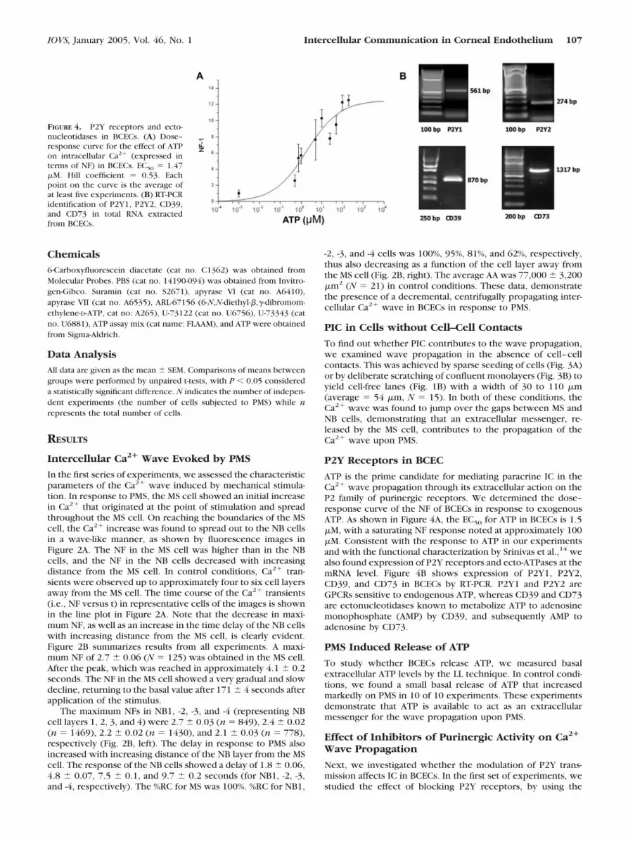
ATP is the prime candidate for mediating paracrine IC in theCa2� wave propagation through its extracellular action on theP2 family of purinergic receptors. We determined the dose–response curve of the NF of BCECs in response to exogenousATP. As shown in Figure 4A, the EC50 for ATP in BCECs is 1.5�M, with a saturating NF response noted at approximately 100�M. Consistent with the response to ATP in our experimentsand with the functional characterization by Srinivas et al.,14 wealso found expression of P2Y receptors and ecto-ATPases at themRNA level. Figure 4B shows expression of P2Y1, P2Y2,CD39, and CD73 in BCECs by RT-PCR. P2Y1 and P2Y2 areGPCRs sensitive to endogenous ATP, whereas CD39 and CD73are ectonucleotidases known to metabolize ATP to adenosinemonophosphate (AMP) by CD39, and subsequently AMP toadenosine by CD73.
PMS Induced Release of ATP
To study whether BCECs release ATP, we measured basalextracellular ATP levels by the LL technique. In control condi-tions, we found a small basal release of ATP that increasedmarkedly on PMS in 10 of 10 experiments. These experimentsdemonstrate that ATP is available to act as an extracellularmessenger for the wave propagation upon PMS.
Effect of Inhibitors of Purinergic Activity on Ca2�
Wave Propagation
Next, we investigated whether the modulation of P2Y trans-mission affects IC in BCECs. In the first set of experiments, westudied the effect of blocking P2Y receptors, by using the
FIGURE 4. P2Y receptors and ecto-nucleotidases in BCECs. (A) Dose–response curve for the effect of ATPon intracellular Ca2� (expressed interms of NF) in BCECs. EC50 � 1.47�M. Hill coefficient � 0.53. Eachpoint on the curve is the average ofat least five experiments. (B) RT-PCRidentification of P2Y1, P2Y2, CD39,and CD73 in total RNA extractedfrom BCECs.
IOVS, January 2005, Vol. 46, No. 1 Intercellular Communication in Corneal Endothelium 107

nonselective P2Y receptor antagonist suramin. In the presenceof 200 �M suramin,27,44 neither the NF (Fig. 5A) nor the %RC(Fig. 5B) of the MS cells (N � 42) was significantly differentfrom control condition (N � 53; Table 2). However, the %RCof NB cells was reduced markedly from NB2 onward when
compared with the controls (NB1: 92% vs. 99%, NB2: 59% vs.97%, NB3: 19% vs. 81%, NB4: 6% vs. 61%; Fig. 5B). Conse-quently, AA was reduced (Fig. 5C, Table 2).
We also studied the effect of nucleotide hydrolysis by ex-ogenous apyrase VI (which has a high ATPase/adenosinediphosphatase [ADPase] ratio) or apyrase VII (which preferen-tially hydrolyzes adenosine diphosphate [ADP]).27 Similar tothe effect of suramin, the presence of apyrase VI (10 U/mL; N� 25) or apyrase VII (10 U/mL; N � 17) resulted in an inhibi-tion of the Ca2� wave propagation (Fig. 5, Table 2). ApyraseVII caused a somewhat stronger inhibition in the outermostcell layers when compared to apyrase VI. The combination of5 U/mL apyrase VI and 5 U/mL apyrase VII had a cumulativeeffect, causing a more pronounced inhibition of the Ca2� wavethan either 10 U/mL apyrase VI or 10 U/mL apyrase VII (Fig. 5,Table 2), limiting the propagation to NB1. These experimentsdemonstrated that PIC, via an agonist of P2Y receptors, isinvolved in the IC upon PMS and provide evidence that bothATP and ADP are involved.
Ca2� Wave Propagation in the Presence ofInhibitors of Ectonucleotidase Activity
We next investigated the effect of inhibiting endogenous ec-tonucleotidase activity by application of 100 �M ARL-67156.45
Using a 10 objective, we saw that the presence of ARL-67156caused a threefold increase of AA of cells reached by the Ca2�
wave when compared to control conditions (840,000 �110,000 �m2; N � 6, vs. 210,000 � 25,000 �m2; N � 4; Fig. 6).In addition to a larger AA, the propagation of the wave is fasterand more intense, as seen in the line plots of Figure 6. This isthe result of higher concentrations of the nucleotides in theextracellular spaces surrounding NB cells in the presence ofARL-67156. These experiments demonstrated that PIC, via apurinergic mediator, is involved in the Ca2� wave propagation
FIGURE 5. Reduced Ca2� wave propagation in the presence of puri-nergic modulators. Cells were treated with suramin (200 �M), apyraseVI (10 U/mL), apyrase VII (10 U/mL), or apyrase VI�VII (5 U/mL each)for 30 minutes. The Ca2� wave propagation in response to PMS isrepresented in control (black bars; N � 53), suramin (hatched bars;N � 42), apyrase VI (light gray bars; N � 25), apyrase VII (dark graybars; N � 17), and apyrase VI�VII (dotted bars; N � 21). *P � 0.05versus control.
TABLE 2. Average Maximum NF, Percentage of Responsive Cells, and Average AA in MS and NB Cells
MS NB1 NB2 NB3 NB4Active Area(AA, �m2)
ControlNF 2.8 2.9 2.6 2.3 2.3
87,521 � 3,110
SEM 0.09 0.04 0.03 0.03 0.04n 53 367 650 750 578%RC 100 99 97 81 61
Suramin (200 �M)NF 2.7 2.4* 1.9* 1.5* 1.5*
23,303 � 1,865*
SEM 0.11 0.05 0.05 0.04 0.06n 42 292 462 598 468%RC 100 92 59 19 6
Apyrase VI (10 U/mL)NF 2.4* 2.4* 2.0* 1.8* 1.6*
25,659 � 4,012*
SEM 0.11 0.06 0.06 0.06 0.09n 25 163 233 254 159%RC 100 93 60 30 20
Apyrase VII (10 U/mL)NF 2.2* 2.3* 1.9* 1.6* 1.7*
16,903 � 1,886*SEM 0.20 0.10 0.10 0.10 0.20n 17 119 164 173 124%RC 100 88 59 21 2
Apyrase VI � VII (5 U/mL each)NF 2.1* 1.9* 1.2* 1.1* 1.1*
5,922 � 850*SEM 0.10 0.09 0.06 0.03 0.02n 21 130 262 287 196%RC 100 47 2 1 0
Data were collected during mechanical stimulation in control conditions and in cells treated withsuramin (200 �M), apyrase VI (10 U/mL), apyrase VII (10 U/mL), or apyrase VI � VII (5 U/mL each).
* P � 0.05 versus control.
108 Gomes et al. IOVS, January 2005, Vol. 46, No. 1

upon PMS and provide evidence that both ATP and ADP areinvolved.
Effect of Modulators of Purinergic Activity on theCa2� Wave across Cell-Free Lanes
The Ca2� wave propagation across cell-free lanes was largelyor completely blocked on pretreatment of the cells withsuramin (Fig. 7A, top row) or apyrases (Fig. 7A, middle row),whereas it was enhanced approximately fourfold in the pres-ence of ARL-67156 (Fig. 7A, bottom row). The AA of the rightside of the cell-free lane in these different conditions (Fig. 7B)is significantly different from the one in control condition.
Role of P2Y Receptors on Ca2� Release in MS andNB Cells
To study the role of P2Y in IC, and since P2Y receptors areknown to be coupled to PLC, we used the PLC inhibitorU-73122 (10 �M)46 and its negative control U-73343 (10 �M).Pretreatment with U-73122 (N � 19) caused a reduction of theCa2� transient in the MS cell and an inhibition of the Ca2�
wave in the NB cells (only 50% of the NB1 cells showed asignificant Ca2� increase), whereas its negative control,U-73343, did not have any effect on the IC (N � 26; Fig. 8).These results suggest that PMS resulted in activation of PLC inthe NB cells, and thus, that IP3-induced Ca2� release plays arole in the propagation of the intercellular Ca2� wave.
DISCUSSION
Characteristic Ca2� waves in cellular monolayers are a mani-festation of IC. In nonexcitable cells, Ca2� waves can bepropagated by PIC and/or GJIC. This study has investigated, forthe first time, intercellular Ca2� wave propagation in cornealendothelial cells with a focus on characterization of a PICpathway. Our major finding is that the propagation of a Ca2�
wave in response to PMS is sustained partially by ATP-mediatedPIC. The extent of propagation of the Ca2� wave is limited bythe ectonucleotidases present on the plasma membrane thatmetabolize ATP.
Ca2� Increase in MS Cells
BCECs responded to PMS by a [Ca2�]i transient (Fig. 2A). Sucha response has been recorded in several cell types, includingvascular endothelium, astrocytes, osteoblasts, and a wide vari-ety of epithelial cells.26,27,29,30,47–51 The response is character-ized by a rapid onset to a peak followed by slow decay towardthe baseline. The Ca2� transients evoked by PMS in our exper-iments result from mechanotransduction and are not due todamage of the plasma membrane. In the unusual cases ofmembrane damage, we noticed a precipitous loss of fluores-cent label (Fluo-4; Molecular Probes) from the MS cell. Exper-iments with such rapid fluorescence losses were promptlydiscarded from further consideration. The mechanisms for theCa2� increase in the MS cell, although not examined in detail,could be attributed to Ca2� influx through putative stretch-activated cation channels, release of Ca2� from endoplasmicreticulum (ER) stores secondary to activation of phospho-lipases, or autocrine effects of ATP release in response tomembrane stretch. The contribution of Ca2� release via apathway involving PLC and IP3 is supported by the finding thatexposure to U-73122 partially inhibited the Ca2� increase inthe MS cell (Fig. 8).
Contribution of PIC toward Ca2�
Wave Propagation
Analysis of the wave propagation in terms of NF and %RC in thelayers surrounding the MS cell offered several striking findings.The most noteworthy aspect is that the wave propagationcould also occur along pathways devoid of cell–cell contacts(Fig. 3). This is clear proof of the involvement of a paracrine
FIGURE 6. Enhanced Ca2� wavepropagation in the presence of anecto-ATPase inhibitor. Ca2� wavepropagation was characterized in re-sponse to PMS using the 10 insteadof the 40 objective. The fluores-cence images represent the timecourse of the Ca2� wave in controlconditions (top) and after pretreat-ment with 100 �M ARL-67156 for 30minutes. Two polygons were drawnon both time series of Ca2� waves,respectively representing the AA ob-served under control conditions(small ROI) and in the presence ofARL-67156 (large ROI). Bottom left:AA in both conditions; bottom right:the normalized fluorescence aver-aged over the small and the largeROIs as a function of time. Blacklines: Ca2� increase in control condi-tions; red lines: Ca2� increase afterpretreatment with ARL-67156. Notethat the average Ca2� increase ishigher and the propagation is fasterand more prolonged in time in thepresence of ARL-67156.
IOVS, January 2005, Vol. 46, No. 1 Intercellular Communication in Corneal Endothelium 109

factor. In support of this claim, and further suggesting aninvolvement of P2Y receptors, we found that the propagateddistance and the strength of the wave decreased significantly inthe presence of suramin or U-73122 (Table 2, Fig. 8). This wasfurther confirmed by receptor expression at the mRNA level,which indicated expression of P2Y1 and P2Y2 metabotropicreceptors (Fig. 4B). In conjunction with these claims, the factthat the paracrine factor is ATP and/or its metabolites is madeevident by the inhibitory effect of the exogenous apyrases onwave propagation. The finding that apyrase VII (which prefer-entially metabolizes ADP to AMP) was more potent thanapyrase VI (which preferentially metabolizes ATP to ADP) isconsistent with the fact that both ATP and ADP are agonists ofP2Y1 and P2Y2 receptors. In fact, ADP is noted to be potentcompared with ATP in activating the P2Y1 receptors.21 AMP is
not an agonist for either P2Y1 or P2Y2 receptors. The impor-tance of ADP in PIC is emphasized in the enhanced inhibitionof the wave propagation noted when cells were exposed to acombination of apyrase VI and apyrase VII (5 U/mL each).
Participation of GJIC in Ca2� Wave Propagation
Although no direct conclusions can be drawn in favor ofparticipation of GJIC in the observed wave propagation, it isimportant to note that U-73122, suramin, or apyrases did notcompletely inhibit the propagation. Srinivas et al.37 have noteda complete inhibition of P2Y activity in cells pre-exposed tothe PLC inhibitor U-73122. Moreover, connexin Cx43 has beenshown to be present in BCECs, as demonstrated by immuno-cytochemistry.52 Therefore, that there was not complete inhi-
FIGURE 7. Effect of PIC inhibitorson Ca2� wave propagation acrosscell-free lanes. (A) Fluorescence im-ages of the Ca2� wave in confluentcells with cell-free lanes. The differ-ent rows show the propagation inthe presence of 200 �M suramin (N� 15; top), apyrase VI�VII (5 U/mLeach; N � 13; middle), and 100 �MARL-67156 (n � 16; bottom). Arrowin the first image of each row: MScell. (B) AA of cells across the cell-free lane from five experiments ofeach of the different conditions.Dark gray bars: AA on the side ofPMS; light gray bars: AA on theother side of the cell-free lane. Darkgray open-ended bar signifies that inthe presence of ARL-67156 the AA onthe side of the PMS exceeded the sizeof the image.
110 Gomes et al. IOVS, January 2005, Vol. 46, No. 1

bition of the wave is suggestive of GJIC superimposed on PIC,which is supported by the presence of Cx43 in BCECs.52 Thiswas consistent with the fact that neither suramin nor theexogenous apyrases affected the dye-coupling as noted in flu-orescence recovery after photobleaching (FRAP) experimentsin cells loaded with BCECF-AM (data not shown).
Role of Ectonucleotidases
Because in several cell types purinergic receptor activity is wellknown to be modulated by the activity of putative ectonucle-otidases, we investigated their potential effect on PIC. Twoprominent ectoenzymes that promote nucleotide metabolismare CD39 (ATP diphosphohydrolase, E.C. 3.6.1.5) and CD73(5�-nucleotidase; E.C. 3.1.3.5).53 CD39 is capable of ATP/ADPmetabolism into AMP,54,55 whereas CD73 degrades AMP toadenosine.56,57 In BCECs, we found expression of both CD39and CD73 at the mRNA level (Fig. 4B). Motivated by thesefindings, we investigated the effects of a putative, nonspecificectonucleotidase inhibitor, ARL-67156, on Ca2� wave propa-gation. The drug enhanced both the distance and the strengthof the Ca2� wave propagation. More specifically, ARL-67156increased the AA of the Ca2� wave threefold, enhanced thevelocity of the propagation, and produced a three- to fourfoldincrease in Ca2�-sensitive fluorescence integrated over the AA.Expression of CD73 is also important because adenosine,which is known to activate the A2B subtype of P1 receptors incorneal endothelium, would result in elevated cAMP andthereby may promote GJIC.58,59
Significance of PIC in Corneal Endothelium
As a form of IC, the importance of PIC to endothelial homeosta-sis cannot be underestimated, despite expression of connexinsand functional GJIC, as evidenced by dye coupling.60–62 Inretinal pigmented epithelial cells (RPE cells), which expressdifferent isoforms of connexins, Ca2� wave propagation ispromoted by functional GJIC.63,64 However, similar experi-ments with calf pulmonary artery endothelial (CPAE) cellsindicated that expression of connexins and dye-coupling arenot sufficient for Ca2� wave propagation through gap junc-tions.27 Furthermore, Ca2� wave propagation in CPAE cellswas unaffected by the putative GJIC blockers.27 However,
suramin and exogenous apyrases abolished the Ca2� wave to asignificant extent, indicating an involvement of purinergic re-ceptors in Ca2� wave propagation. Unlike P2X receptors, P2Yare coupled to G�q/11. The results of PIC in BCECs reported inthis study are comparable to those found in CPAE cells, al-though the role of GJIC in Ca2� wave propagation is yet to befully examined.
In addition to the role of IC under resting conditions, it maybe useful to speculate on the potential of our findings in thecontext of phacoemulsification. As shown in several studies,despite the use of viscoelastic agents, functional decompensa-tion and loss of endothelial cells after phacoemulsificationappear to be inevitable.65–68 These effects are attributed tocompressive stresses on the endothelium by the bursting ofmicrobubbles induced by acoustic cavitation.69,70 Each ofthese bursts causes a transient point source of stress similar tothe PMS in this study. Therefore, it would be pertinent toexamine further whether the probable IC induced duringphaco-emulsification results in apoptosis in cells next to sitesof local injury (akin to the bystander effect known to bemediated by gap junctions71–73) or promotes a tissue-widedefensive response to overcome the acute mechanical stress.
In conclusion, this study has delineated an important rolefor ATP in the corneal endothelium as a paracrine factor incell–cell communication. Further investigation of the mecha-nism(s) of ATP release and putative roles of PIC would behelpful in understanding the pathophysiology of corneal endo-thelium.
References
1. Dikstein S, Maurice DM. The metabolic basis to the fluid pump inthe cornea. J Physiol. 1972;221:29–41.
2. Bonanno JA. Identity and regulation of ion transport mechanismsin the corneal endothelium. Prog Retin Eye Res. 2003;22:69–94.
3. Fischbarg J, Hernandez J, Liebovitch LS, Koniarek JP. The mecha-nism of fluid and electrolyte transport across corneal endothelium:critical revision and update of a model. Curr Eye Res. 1985;4:351–360.
4. Riley M. Pump and leak in regulation of fluid transport in rabbitcornea. Curr Eye Res. 1985;4:371–376.
5. Candia OA. Electrolyte and fluid transport across corneal, conjunc-tival and lens epithelia. Exp Eye Res. 2004;78:527–535.
6. Walkenbach RJ, Chao WT. Adenosine regulation of cyclic AMP incorneal endothelium. J Ocul Pharmacol. 1985;1:337–342.
7. Walkenbach RJ, Ye GS, Reinach PS, Boney F. Alpha 1-adrenocep-tors in the corneal endothelium. Exp Eye Res. 1992;55:443–450.
8. Jumblatt MM. Autocrine regulation of corneal endothelium byprostaglandin E2. Invest Ophthalmol Vis Sci. 1994;35:2783–2790.
9. Zhang Y, Xie Q, Sun XC, Bonanno JA. Enhancement of HCO3�
permeability across the apical membrane of bovine corneal endo-thelium by multiple signaling pathways. Invest Ophthalmol VisSci. 2002;43:1146–1153.
10. Riley MV, Winkler BS, Starnes CA, Peters MI. Adenosine promotesregulation of corneal hydration through cyclic adenosine mono-phosphate. Invest Ophthalmol Vis Sci. 1996;37:1–10.
11. Riley MV, Winkler BS, Starnes CA, et al. Regulation of cornealendothelial barrier function by adenosine, cyclic AMP, and proteinkinases. Invest Ophthalmol Vis Sci. 1998;39:2076–2084.
12. Wigham CG, Turner HC, Swan J, Hodson SA. Modulation of cor-neal endothelial hydration control mechanisms by Rolipram.Pflugers Arch. 2000;440:866–870.
13. Yasukura T, Inoue M, Irie T, et al. Adrenergic receptor-mediatedCl� transport in rabbit corneal endothelial cells. Jpn J Pharmacol.1995;67:315–320.
14. Srinivas SP, Yeh JC, Ong A, Bonanno JA. Ca2� mobilization inbovine corneal endothelial cells by P2 purinergic receptors. CurrEye Res. 1998;17:994–1004.
15. Cha SH, Hahn TW, Sekine T, et al. Purinoceptor-mediated calciummobilization and cellular proliferation in cultured bovine cornealendothelial cells. Jpn J Pharmacol. 2000;82:181–187.
FIGURE 8. Ca2� wave propagation in the presence of a PLC inhibitor.NF (top) and %RC (bottom) of BCECs in control conditions (blackbars) or treated with U-73122 (10 �M; light gray bars) or with theinactive analogue U-73343 (10 �M; dark gray bars) for 30 minutes.*P � 0.05 versus control.
IOVS, January 2005, Vol. 46, No. 1 Intercellular Communication in Corneal Endothelium 111

16. Leipziger J. Control of epithelial transport via luminal P2 receptors.Am J Physiol. 2003;284:F419–F432.
17. Jacobson KA, Jarvis MF, Williams M. Purine and pyrimidine (P2)receptors as drug targets. J Med Chem. 2002;45:4057–4093.
18. Inscho EW. P2 receptors in regulation of renal microvascularfunction. Am J Physiol. 2001;280:F927�F944.
19. Burnstock G, Williams M. P2 purinergic receptors: modulation ofcell function and therapeutic potential. J Pharmacol Exp Ther.2000;295:862–869.
20. Bailey MA, Hillman KA, Unwin RJ. P2 receptors in the kidney. JAuton Nerv Syst. 2000;81:264–270.
21. Ralevic V. P2 receptors in the central and peripheral nervoussystems modulating sympathetic vasomotor tone. J Auton NervSyst. 2000;81:205–211.
22. Jacobson KA, Hoffmann C, Kim YC, et al. Molecular recognition inP2 receptors: ligand development aided by molecular modelingand mutagenesis. Prog Brain Res. 1999;120:119–132.
23. Apasov S, Koshiba M, Redegeld F, Sitkovsky MV. Role of extracel-lular ATP and P1 and P2 classes of purinergic receptors in T-celldevelopment and cytotoxic T lymphocyte effector functions. Im-munol Rev. 1995;146:5–19.
24. Harden TK, Boyer JL, Nicholas RA. P2-purinergic receptors: sub-type-associated signaling responses and structure. Annu Rev Phar-macol Toxicol. 1995;35:541–579.
25. Anderson CM, Bergher JP, Swanson RA. ATP-induced ATP releasefrom astrocytes. J Neurochem. 2004;88:246–256.
26. Newman EA. Propagation of intercellular calcium waves inretinal astrocytes and Muller cells. J Neurosci. 2001;21:2215–2223.
27. Moerenhout M, Himpens B, Vereecke J. Intercellular com-munication upon mechanical stimulation of CPAE-endothelialcells is mediated by nucleotides. Cell Calcium. 2001;29:125–136.
28. Cotrina ML, Lin JH, Lopez-Garcia JC, et al. ATP-mediated gliasignaling. J Neurosci. 2000;20:2835–2844.
29. Homolya L, Steinberg TH, Boucher RC. Cell to cell communicationin response to mechanical stress via bilateral release of ATP andUTP in polarized epithelia. J Cell Biol. 2000;150:1349–1360.
30. Evans JH, Sanderson MJ. Intracellular calcium oscillations in-duced by ATP in airway epithelial cells. Am J Physiol. 1999;277:L30 –L41.
31. Osipchuk Y, Cahalan M. Cell-to-cell spread of calcium signalsmediated by ATP receptors in mast cells. Nature. 1992;359:241–244.
32. Goodenough DA, Paul DL. Beyond the gap: functions of unpairedconnexon channels. Nat Rev Mol Cell Biol. 2003;4:285–294.
33. Xie HQ, Hu VW. Modulation of gap junctions in senescent endo-thelial cells. Exp Cell Res. 1994;214:172–176.
34. Cotrina ML, Gao Q, Lin JH, Nedergaard M. Expression andfunction of astrocytic gap junctions in aging. Brain Res. 2001;901:55– 61.
35. Zhao W, Lin ZX, Zhang ZQ. Cisplatin-induced premature senes-cence with concomitant reduction of gap junctions in humanfibroblasts. Cell Res. 2004;14:60–66.
36. Del Monte U, Statuto M. Drop of connexins: a possible link be-tween aging and cancer? Exp Gerontol. 2004;39:273–275.
37. Srinivas SP, Ong A, Goon L, Bonanno JA. Lysosomal Ca2� stores inbovine corneal endothelium. Invest Ophthalmol Vis Sci. 2002;43:2341–2350.
38. MacCallum DK, Lillie JH, Scaletta LJ, et al. Bovine corneal endo-thelium in vitro: elaboration and organization and of a basementmembrane. Exp Cell Res. 1982;139:1–13.
39. Jelamskii S, Sun XC, Herse P, Bonanno JA. Basolateral Na�-K�-2Cl�
cotransport in cultured and fresh bovine corneal endothelium.Invest Ophthalmol Vis Sci. 2000;41:488–495.
40. Diecke FP, Wen Q, Sanchez JM, et al. Immunocytochemical local-ization of Na�-HCO3
� cotransporters and carbonic anhydrase de-pendence of fluid transport in corneal endothelial cells. Am JPhysiol. 2004;286:C1434–C1442.
41. Sun XC, Bonanno JA, Jelamskii S, Xie Q. Expression and localiza-tion of Na�-HCO3
� cotransporter in bovine corneal endothelium.Am J Physiol. 2000;279:C1648–C1655.
42. Sun XC, McCutheon C, Bertram P, et al. Studies on the expressionof mRNA for anion transport related proteins in corneal endothe-lial cells. Curr Eye Res. 2001;22:1–7.
43. Sun XC, Bonanno JA. Expression, localization, and functional eval-uation of CFTR in bovine corneal endothelial cells. Am J Physiol.2002;282:C673–C683.
44. Ralevic V, Burnstock G. Receptors for purines and pyrimidines.Pharmacol Rev. 1998;50:413–492.
45. Westfall TD, Menzies JR, Liberman R, et al. Release of a solubleATPase from the rabbit isolated vas deferens during nerve stimu-lation. Br J Pharmacol. 2000;131:909–914.
46. Yule DI, Williams JA. U73122 inhibits Ca2� oscillations in responseto cholecystokinin and carbachol but not to JMV-180 in rat pan-creatic acinar cells. J Biol Chem. 1992;267:13830–13835.
47. Churchill GC, Lurtz MM, Louis CF. Ca2� regulation of gap junc-tional coupling in lens epithelial cells. Am J Physiol. 2001;281:C972–C981.
48. Shiga H, Tojima T, Ito E. Ca2� signaling regulated by an ATP-dependent autocrine mechanism in astrocytes. Neuroreport.2001;12:2619–2622.
49. Stout CE, Costantin JL, Naus CC, Charles AC. Intercellular calciumsignaling in astrocytes via ATP release through connexinhemichannels. J Biol Chem. 2002;277:10482–10488.
50. Gomez P, Vereecke J, Himpens B. Intra- and intercellular Ca2�-transient propagation in normal and high glucose solutions in ROScells during mechanical stimulation. Cell Calcium. 2001;29:137–148.
51. Stalmans P, Himpens B. Confocal imaging of Ca2� signaling incultured rat retinal pigment epithelial cells during mechanical andpharmacologic stimulation. Invest Ophthalmol Vis Sci. 1997;38:176–187.
52. Mohay J, McLaughlin BJ. Corneal endothelial wound repair innormal and mitotically inhibited cultures. Graefes Arch Clin ExpOphthalmol. 1995;233:727–736.
53. Farahbakhsh NA. Ectonucleotidases of the rabbit ciliary body non-pigmented epithelium. Invest Ophthalmol Vis Sci. 2003;44:3952–3960.
54. Marcus AJ, Broekman MJ, Drosopoulos JH, et al. Metabolic controlof excessive extracellular nucleotide accumulation by CD39/ecto-nucleotidase-1: implications for ischemic vascular diseases. J Phar-macol Exp Ther. 2003;305:9–16.
55. Kittel A, Kaczmarek E, Sevigny J, et al. CD39 as a caveolar-associ-ated ectonucleotidase. Biochem Biophys Res Commun. 1999;262:596–599.
56. Napieralski R, Kempkes B, Gutensohn W. Evidence for coordi-nated induction and repression of ecto-5�-nucleotidase (CD73) andthe A2a adenosine receptor in a human B cell line. Biol Chem.2003;384:483–487.
57. Hashikawa T, Takedachi M, Terakura M, et al. Involvement ofCD73 (ecto-5�-nucleotidase) in adenosine generation by humangingival fibroblasts. J Dent Res. 2003;82:888–892.
58. Chanson M, White MM, Garber SS. cAMP promotes gap junctionalcoupling in T84 cells. Am J Physiol. 1996;271:C533–C549.
59. TenBroek EM, Lampe PD, Solan JL, et al. Ser364 of connexin43 andthe upregulation of gap junction assembly by cAMP. J Cell Biol.2001;155:1307–1318.
60. Watsky MA, Rae JL. Dye coupling in the corneal endothelium:effects of ouabain and extracellular calcium removal. Cell TissueRes. 1992;269:57–63.
61. Williams K, Watsky M. Gap junctional communication in the hu-man corneal endothelium and epithelium. Curr Eye Res. 2002;25:29–36.
62. Williams KK, Watsky MA. Bicarbonate promotes dye coupling inthe epithelium and endothelium of the rabbit cornea. Curr EyeRes. 2004;28:109–120.
63. Stalmans P, Himpens B. Properties of intra- and intercellular Ca2�-wave propagation elicited by mechanical stimulation in culturedRPE cells. Cell Calcium. 1999;25:391–399.
64. Himpens B, Stalmans P, Gomez P, et al. Intra- and intercellularCa2� signaling in retinal pigment epithelial cells during mechani-cal stimulation. FASEB J. 1999;13:S63–S68.
65. Bourne WM, McLaren JW. Clinical responses of the corneal endo-thelium. Exp Eye Res. 2004;78:561–572.
112 Gomes et al. IOVS, January 2005, Vol. 46, No. 1

66. Bourne RR, Minassian DC, Dart JK, et al. Effect of cataract surgeryon the corneal endothelium: modern phacoemulsification com-pared with extracapsular cataract surgery. Ophthalmology. 2004;111:679–685.
67. Pirazzoli G, D’Eliseo D, Ziosi M, Acciarri R. Effects of phacoemulsifi-cation time on the corneal endothelium using phacofracture andphaco chop techniques. J Cataract Refract Surg. 1996;22:967–969.
68. Edelhauser HF. The resiliency of the corneal endotheliumto refractive and intraocular surgery. Cornea. 2000;19:263–273.
69. Topaz M, Motiei M, Assia E, et al. Acoustic cavitation in
phacoemulsification: chemical effects, modes of action and cavi-tation index. Ultrasound Med Biol. 2002;28:775–784.
70. Pacifico RL. Ultrasonic energy in phacoemulsification: mechanicalcutting and cavitation. J Cataract Refract Surg. 1994;20:338–341.
71. Seymour C, Mothersill C. Cell communication and the “bystandereffect”. Radiat Res. 1999;151:505–506.
72. Wright EG. Commentary on radiation-induced bystander effects.Hum Exp Toxicol. 2004;23:91–94.
73. Edwards GO, Botchway SW, Hirst G, et al. Gap junction commu-nication dynamics and bystander effects from ultrasoft X-rays. Br JCancer. 2004;90:1450–1456.
IOVS, January 2005, Vol. 46, No. 1 Intercellular Communication in Corneal Endothelium 113