untitled - citeseerx
TRANSCRIPT


CONTRIBUTION OF PHAGES
TO THE
VIRULENCE OF PATHOGENIC MYCOBACTERIA
Nomakorinte Gcebe
Supervisor: Dr S. I Durbach
Co- Supervisor: Dr A. Michel
A dissertation submitted to the faculty of Science, University of the Witwatersrand,
in the fulfillment of the requirements for the degree of Master of Science.
Johannesburg, 2007
2
DECLARATION
I declare that this dissertation is my own, unaided work. It is being submitted for the
Degree of Master of Science in the University of Witwatersrand, Johannesburg. It has not
been submitted before for any degree or examination in any other University.
(Signature of Candidate)
Day of 2007
3

ABSTRACT
Bacteriophages have been known to contribute to the bacterial phenotype through
lysogenic conversion. Many virulence factors in pathogenic bacteria are phage encoded.
However, it is not known whether this is true in mycobacteria. A study by Pedulla et al,
(2003) looking at the genome sequences of 10 mycobacteriophages suggested that several
of the identified genes may have patho-adaptive potential. Perhaps paradoxically,
sequenced mycobacterial genomes revealed a paucity of recognizable prophages. To
initiate any enquiry into the contribution of prophages to the relevance of mycobacterial
disease, we set up some experiments to screen for the presence of prophages in
Mycobacterium bovis isolates from different outbreaks. We screened 27 isolates for
spontaneously induced phages by plaque assay using M. smegmatis, M. fortuitum, M.
scrofulaceum, and M. kansasii and an isolate (S2) from our lab as indicator strains.
However none of these formed reproducible plaques. Only three isolates formed plaques
that could not be propagated on any of the indicator strains used. To address if we could
enrich for induced prophages, we did some preliminary experiments to optimize
prophage induction, using a known lysogen (L5). Co-culturing of a lysogen with sensitive
cells was assessed at different concentrations. The result showed that there was no
difference in the rate of phage released between the co-cultured and the non-spiked
control cells. Since it is possible that we did not have a strain that is sensitive to M. bovis
phage(s), we checked, using the L5 lysogen, if any free phage could be detected from
solid culture, by Epiflourescence Microscopy (EFM). We were able to detect phage
particles in a titer of 102 as determined by plaque assay with EFM. We therefore screened
16 M. bovis isolates for any free phage, using the more sensitive EFM and no inducible
phages were detected. Since potential lysogens may be very stable, with minimal
induction, we decided to explore a molecular approach to screen for cryptic prophages.
Guessmers based on conserved regions in the L5 repressor, shared by other phage
genomes were designed .Out of 45 M .bovis isolates screened by PCR, nine produced
DNA bands of different sizes from each isolate. The sequences from the L5-M.
smegmatis mc2155 lysogen positive control were confirmed to be of the gp71 origins (L5
4

phage repressor).. Sequences from clone DNA from two isolates revealed existence of
different M. bovis AF2122/97 DNA specific binding proteins such as putative
transposases of the IS1553 element (Mb2968) , DNA helicases (Mb0884), transcriptional
regulators (Mb1160), and other M. bovis proteins such as CTP synthetase (Mb 1725),
spermidine synthetase (Mb2632), and a hypothetical protein Mb1618c. Interestingly, a
sequence DLLIRVNE which is conserved in L5 and other mycobacteriophage repressor
proteins was also conserved in some of the M. bovis DNA binding proteins. Hence this
protein might play an important role in DNA binding. An in-depth analysis of the whole
genome of M. bovis is needed in order to conclude if this sequence is of prophage origin.
5

ACKNOWLEDGEMENTS
I would like to thank Dr Anita Michel and the TB lab staff at OVI for supplying the M.
bovis isolates; Rodney Hull for helping me with the EFM; Digby Warner for helping with
gel extractions and purifications; Belinda Spillings with her advice and training on
working with phages; the Agricultural Research Council for the financial support.
To my lab partners at OVI TB lab, and SMEG lab; both past and present, and my
supervisors- Thank You.
6

CONTENTS Pages
DECLARATION……………………………………………………..3
ABSTRACT…………………………………………………………..4-5
ACKNOWLEDGEMENT…………………………………………….6
LIST OF FIGURES…………………………………………………...10
LIST OF TABLES…………………………………………………….11
LIST OF ABBREVIATION…………………………………………..12
CHAPTER ONE-INTRODUCTION
1.1 General Introduction………………………………………………13
1.2 Literature Review…………………………………………………14-33
1.3 Aim and Objectives………………………………………………..34
CHAPTER TWO-SCREENING OF M. bovis ISOLATES FOR
SPONTANEOUSLY INDUCED PHAGES
3.1 Introduction……………………………………………………….35-36
3.2 Aims and Objectives………………………………………………37
3.3 Methods……………………………………………………………38-43
3.4 Results……………………………………………………………...44-53
3.5 Discussion………………………………………………………….54-56
3.6 Conclusion………………………………………………………….57
CHAPTER THREE-A GENOMICS APPROACH TO SCREEN FOR
PROPHAGES IN M. bovis ISOLATES
4.1 Introduction…………………………………………………………58-59
4.2 Aims and objectives…………………………………………………60
4.3 Methods……………………………………………………………..61-67
7

4.4 Results…………………………………………………………………68-84
4.5 Discussion……………………………………………………………..85-86
4.6 Conclusion…………………………………………………………….87
CHAPTER FOUR-CONCLUDING REMARKS
Concluding Remarks………………………………………………………88-89
REFERENCES……………………………………………………………90-114
APPENDIX A
List of Chemicals……………………………………………………………115-116
APPENDIX B
General Media and Buffers…………………………………………………117-121
APPENDIX C
PGEM-T Easy vector map…………………………………………………122
APPENDIX D
NCBI BLASTX results ……………………………………………………123-126
Fig A : BLASTX results of a 679bp sequence from isolate TB 5151 revealing homology
with M. bovis transcriptional regulator protein (Mb1160)…………………123
Fig B: Amino acid sequence of the M. bovis Transcriptional regulator protein
(Mb1160)……………………………………………….……………………123
Fig C: BLASTX results of a 634bp sequence from isolate TB4850D revealing homology
with M. bovis DNA helicase protein (Mb0884)………………………………123
Fig D: Amino acid sequence of the the M. bovis DNA helicase protein
(Mb0884)………………………………………………………………………124
8

Fig E: BLASTX results of a 762bp sequence from isolate TB 4850D revealing homology
with M. bovis CTP synthetase protein (Mb1725)…………………………………..124
Fig F: Amino acid sequence of the M. bovis CTP synthetase protein
(Mb1725)……………………………………………………………………………124
Fig G: BLASTX results of a 561bp sequence from isolate TB 4850D revealing homology
with M. bovis IS1553 Transposase (Mb 2968)……………………………………..125
Fig H: Amino acid sequence of the IS1553 Transposase (Mb 2968)
of M. bovis..................................................................................................................125
Fig I: BLASTX results of a 327bp sequence from isolate TB 4850D revealing homology
with M. bovis Spermidine synthetase protein (Mb 2632)……………………………126
Fig J: Amino acid sequence of the M. bovis Spermidine synthetase protein (Mb 2632)
…………………………………………………………………………………...126
Fig K: BLASTX results of a 689bp sequence from isolate TB 5151D revealing homology
with a hypothetical protein (Mb 1618c) of M. bovis ………………………………….126
Fig L: Amino acid sequence of the M. bovis hypothetical protein
(Mb 1618c)……………………………………………………………………………..126
9

LIST OF FIGURES Pages
Fig 1: Nucleotide sequence comparison of 30 mycobacteriophages as
illustrated in a Dotter plot. (Hatfull et al., 2006)…………….30
Fig 2: Co- infection experiment…………………………………….45
Fig 3: Plaques from M. bovis isolates ………………………………48
Fig 4: Epiflourescence Microscopy photographs of M. bovis culture
filtrates………………………………………………………..52
Fig 5: Amino Acid multiple alignment of the mycobacteriophage L5
GP71 repressor with other repressor-like sequences from other
mycobacteriophages………………………………………….70
Fig 6: Amplification of the L5 GP71 by guessmer primers-1.5% TAE
agarose gel……………………………………………………..74
Fig 7a&b: Guessmer based screening of M. bovis isolates for prophages-1.5%
TAE agarose gel……………………………………………75&76
Fig 8: PCR confirmation of the quality and mycobacterial status of the
M. bovis DNA using hsp65 primers-1.5% TAE gel………………..
77
Fig 9a: EcoR1 digests of plasmids to release clones-1.5% TAE
agarose gel ……………………………………………………..80
Fig 9b: EcoR1 digests of plasmids to release clones-1.5% TAE gel…….81
10

LIST OF TABLES Pages
Table 1: Molecular determinants of virulence factors in M. bovis……23
Table 2: List of bacteria and phage strains used in this study…………38
Table 3: Screening for spontaneously induced phages from M. bovis
Isolates Using plaque assay…………………………………...47
Table 4: Screening for spontaneously induced phages from M. bovis
Isolates using EFM and comparing EFM results with plaque
assay ………………………………………………………….51
Table 5: List of oligos used for PCR.…………………………………..62
Table 6: Mycobacteriophages with identical repressor-like sequences…69
Table 7: Guessmer design………………………………………………72
Table 8: PCR based screening of M. bovis isolates for prophages, and the
hsp65 confirmation of the quality and mycobacterial status of the
M. bovis isolates DNA ………………………………………….78
Table 9: Summary of EcoR1 restriction digestion of plasmid DNA from
different clones…………………………………………………...82
Table 10: Summary of the homolog search results revealing the predicted
homolog proteins to the sequences and their location on the M.
bovis AF2122/97 genome……………………………………….84
11

LIST OF ABBREVIATIONS
ARC Agricultural Research Council
Cfu Colony forming units
dATP deoxyadenosine triphosphate
dCTP deoxycytosine triphosphate
dGTP deoxyguanosine triphosphate
dTTP deoxytyrosine triphosphate
EFM Epiflourescence Microscopy
KNP Kruger National Park
L5 Mycobacteriophage L5
MOI Multiplicity Of Infection
ORF Open Reading Frame
OVI Onderstepoort Veterinary Institute
PCR Polymerase Chain Reaction
Phage Bacteriophage
Pfu Plaque forming unit
SA South Africa
TB Tuberculosis
TEM Transmission Electron Microscopy
UV Ultra Violet
WHO World Health Organisation
12

CHAPTER ONE – INTRODUCTION AND LITERATURE REVIEW
1.1 INTRODUCTION
Tuberculosis is still a major global and public health problem, killing more people than
any other single infectious agent. Mycobacterium tuberculosis a facultative intracellular
pathogen is a leading cause of morbidity and mortality in AIDS patients. The situation is
worsened by the infection of these immuno-compromised people with Mycobacterium
avium complex strains. Co-infection of HIV with TB bacilli often leads to progression of
one of the diseases. In 2006 the World Health Organisation (WHO) reported that the
global TB incidence to be growing by 1% a year and about 2 billion people (third of the
world population) are infected with it. They reported that one out of ten infected people
will develop active TB. In addition, 20-50 cases of pulmonary TB, using smear
microscopy can be missed (Zink and Nerlich, 2004) especially in people co-infected with
TB and HIV. Misdiagnosis of TB can also happen because of normal radiographs in HIV
positive patients. Bovine tuberculosis, caused by Mycobacterium bovis remains an
important animal and zoonotic disease causing a significant loss in agriculture world
wide, and a public health hazard (Cosivi et al., 1998; Morris et al., 1994). In order to
eradicate the disease, it is very important to understand its pathology and what parts of its
genetic instructions make the bacillus so virulent.
Tuberculosis studies have lagged behind those of other infectious organisms, because of
the slow growth rate of the bacteria. However, recent advances in molecular biology, in
particular the elucidation of the genome sequences of both Mycobacterium bovis (Garnier
et al., 2003) and Mycobacterium tuberculosis H37Rv strain (Cole et al., 1998) as well as
the CDC155 strain ( Fleischmann et al., 2002) has contributed to a better understanding
of the pathology and some of the virulence attributes of these organisms.
13

1.2 LITERATURE REVIEW
The genus Mycobacterium
Mycobacteria are a group of Gram-positive bacteria, displaying diverse phenotypes. The
genus Mycobacterium contains more than 71 recognized or proposed species (Shinnick
and Good, 1994). The early classification of mycobacteria was based on growth rate,
pigmentation and clinical significance (Runyonn, 1959). A fundamental taxonomic
division was tied to growth rate. These can be divided into two main groups based on
their growth rate: the fast growers and the slow growers (Shinnick and Good, 1994).
Rapid or fast growers include environmental mycobacteria such as M. smegmatis
(Shinnick and Good, 1994). Mycobacterium fortuitum and M. chelonei, which can be
opportunistic pathogens, are also classified as rapid growers (Ward, 1975). Most species
in this group are free-living environmental organisms that rarely cause or never cause
diseases.
Among the slow growing mycobacteria are the ubiquitous and facultative pathogenic
Mycobacterium intracellulare and Mycobacterium avium, M. avium subsp
paratuberculosis, and M. sylvaticum which belong to the M. avium complex (MAC)
(Cangelosi et al., 2001; Mackintosh et al., 2004). MAC bacilli are a group of related
environmental mycobacteria. They are common in surface waters and soils and are often
isolated from water taps (Kansal et al., 1998; Cangelosi et al., 2001). The host to host
transmission mechanism is not known, but exposure to MAC through infected water is
common (Kansal et al., 1998; Cangelosi et al., 2001). Mycobacterium avium and M.
intracellulare are commonly associated with human infections by MAC (Wolinski, 1979;
Inderlied et al., 1993). Mycobacterium paratuberculosis causes Johne’s disease in
livestock and has been implicated in Chron’s disease in humans (Herman-Taylor, 2001).
Several pathogens in the M. tuberculosis complex which include M. tuberculosis, M.
bovis, M. microti, M. canettii, M. pinnipedii, M. africanum, M. caprae also belong to the
14

slow growing subclass (Brosch et al., 2002; Smith, 2006). This number is likely to
increase as new genetic differences between strains of the existing members are
identified. The M. tuberculosis complex is characterized by a 85%-99.9% similarity at
nucleotide level, and identical 16S rRNA gene sequences as determined by DNA
sequencing and related methods such as hybridization (Boddinghaus et al., 1990;
Sreevatsan et al., 1997; Van Soolingen et al., 1997). The existence of chromosomal
deletions together with single nucleotide polymorphism (SNP) and direct repeat content
(spoligotype) patterns allows discrimination between these bacteria (Kammerbeek et al.,
1997; Brosch et al., 2002; Mostowy et al., 2002; Smith et al., 2003). Synonymous single-
nucleotide polymorphism data suggests that M. bovis, the primary cause of bovine
tuberculosis, evolved at the same time as M. tuberculosis, the primary cause of human
tuberculosis (Sreevatsan et al., 1997). The study of deletions and insertions in the
genomes of M. tuberculosis complex provide a strong evidence for the independent
evolution of both M. bovis and M. tuberculosis from another precursor, possibly M.
canettii (Brosch et al., 2002). More recently sequence analysis of six housekeeping genes
(katG, gyrB, gyrA, rpoB, hsp65, sodA) revealed that human isolates of M. canettii from
East Africa represent an extant progenitor of an ancestral species named M.
prototuberculosis, from which M. tuberculosis complex evolved (Gutierrez et al., 2005.,
Smith, 2006).
The recent elucidation of the genome sequence M. bovis, the causative agent of
tuberculosis in a range of animals with zoonotic potential (Morris and Pfeifer, 1994;
Cosivi et al., 1998) compared with that of M. tuberculosis, the primary cause of human
tuberculosis, show that these bacilli are genetically similar at the nucleotide level (O’
Reilly et al., 1995, Cole et al., 1998; Garnier et. al., 2003). Mycobacterium bovis is also
the progenitor of the only available human TB vaccine, M. bovis bacillus Calmette-
Gừerin (BCG), a strain that was attenuated by serial passaging of M. bovis on potato
slices soaked in ox-bile and glycerol over 13 years .The precise mutations that led to the
attenuation of this strain are still unknown, though the key deletion of the region of
difference (RD1) appears to have played a role (Behr et al., 1999; Pym et al., 2002). This
hypothesis came about when Mahairas et al. (1996) compared the genomic sequences of
15
M. bovis and M. bovis BCG using subtractive hybridization, and found three regions of
difference (RD1 , RD2 and RD3) that were absent in BCG, but present in M. bovis
genomes. Later Behr et al. (1999) and Gordon et al. (2001) identified 16 large deletions
including RD1-RD3 that were present in M. tuberculosis but missing in M. bovis BCG.
Eleven of these 16 deletions were unique to M. bovis whereas the remaining 5 were
unique to M. bovis BCG. One of these (RD1) was absent to all the M. bovis BCG strains
currently used as vaccine for human tuberculosis.
Epidemiology and Pathology of Bovine Tuberculosis
The main hosts of M. bovis are both animals of agricultural importance and wild
mammals. Many wild life animals around the world have been infected by this pathogen
(Barlow, 1993; White and Harris, 1995; Schmit et al., 1997). Presently there are no
available methods for treating wild life population infected with the bacillus, and there is
no convincing evidence that these animals are able to resolve the pathogen naturally
(O’Reilly and Darbon, 1995; Michel et al., 2005). Once bovine tuberculosis has
established itself in a native free-ranging maintenance host, eradication of the disease
becomes highly unlikely (Michel et al., 2005).
The choice of suitable control measures for this disease depends on the primary
objectives of that particular ecosystem. However various implications have to be
considered when choosing a control strategy. These include the preservation of protected
species, the minimization of risk of transmission to domestic animals, and the potentially
devastating impact on population dynamics (Michel et al., 2005). In New Zealand for
instance, the control of bovine tuberculosis is made problematic by the presence of an
important wild life reservoir of the disease, notably brushtail possums (Coleman and
Cooke, 2001). The absence of effective vaccine strategies for bovine TB, particularly in
developing countries has resulted in a test and slaughter policy as the backbone of
national elimination programs for this disease (Cosivi et al., 1998). Currently the gold
standard for control of bovine tuberculosis internationally, relies on its diagnosis by the
16

intra-dermal tuberculin test. The main disadvantage of this test in Africa is that it requires
trained technicians for correct interpretation (Kleeberg, 1960; Cossivi et al., 1998).
Human infections by this organism in countries where there are no control measures
occur when infected, unpasteurized milk or meat is ingested, or when there is close
contact with animals that have the disease (Collins and Grange, 1987; Cosivi et al.,
1998). The incidence of this kind of tuberculosis in humans is much lower than the
disease caused by the human tubercle bacillus (Collins, 2000). Extra–pulmonary
tuberculosis is almost always due to drinking infected cow’s milk (Kazwala, 1999;
Coetzer et al., 2005). The AIDS epidemic has increased the risk of transmission of M.
bovis to humans (Grange et al., 1994). Human Immune Virus (HIV) infection results in
humans becoming more susceptible to all forms of tuberculosis. This not only poses a
risk of infection of other humans, but results in livestock being exposed to higher levels
of TB. Transmission of this bacillus from humans to animals may also occur. Humans
with open tuberculosis caused by M. bovis can transmit it to cattle by the aerogenous
route, spitting, coughing or urinating (Coetzer et al., 2005).
The first reference of bovine tuberculosis in cattle in South Africa was made by Hutcheon
in 1880 (Michel et al., 2005). A potential link between tuberculosis in livestock and game
was first suggested, when bovine tuberculosis was reported in kudu and small ungulates
in the Eastern Cape Province of South Africa (Paine and Martinaglia, 1929).
Subsequently the increasing economic importance of tuberculosis as a disease of cattle
led to the implementation of a national bovine tuberculosis control and eradication
scheme in South Africa (Paine and Martinaglia, 1929). Bovine tuberculosis was
apparently first introduced into African buffalo (Syncerus caffer) in the southern regions
of Kruger National Park (KNP, South Africa) in the 1960’s or 1980’s from domestic
cattle (Bengis et al., 1996).
Animal to animal (or animal to human) infections by M. bovis most often occur when
droplets carrying the bacterial cells are inhaled (Collins, 2000). This notion came about
because, when the disease develops, the associated granulomatous pathological changes
17

are seen mainly in the upper and lower respiratory tract (Neill et al., 1994) and because of
this pattern it is considered that infection most often follows aerosol exposure to M.
bovis. The congenital route is also important, and calves may be born with bovine
tuberculosis (Cousins et al., 2004), or oral infection can occur in nursing calves (Neill et
al., 1994). Infective bacilli gain access to macrophages via inhaled droplets containing
the organism. Bacteria not initially killed multiply in the phagosome of macrophages
(Adwell et al., 1997). The surviving bacterial particles from bactericidal attack by
activated macrophages will infect freshly recruited monocytes or macrophages. When the
pathogen reaches sufficient numbers, the cell dies and the released organisms are
ingested by other macrophages. The infected macrophages produce cytokines and
chemokines that attract other phagocytic cells including monocytes, other alveolar
macrophages and neutrophils which eventually form nodular granulomatous structure
called tubercle lesions (Dannenberg and Rook, 1994; Fenton and Vermeulen, 1996; van
Crevel et al., 2002). The organism is eventually disseminated to the lymph nodes and
blood stream. It is deposited in the liver, spleen, kidney, bone, brain, meninges, and other
parts of the lung. Infected animals can remain for years without any signs of infection,
even in advanced stages, until they are sent for slaughter (Collins et al., 2000).
An important characteristic of pathogenic bacteria is the ability to adapt to a wide range
of changing environmental conditions during the infection cycle. Mycobacterium bovis
demonstrate a remarkable ability to survive in diverse environments. It can survive for
years in stationary phase cultures in vitro. Outside its hosts this tubercle bacillus remains
viable in infective droplet nuclei (Mehrota and Bishai, 2001). Mycobacterium bovis can
survive for up to two years in soil or manure and possibly even longer in sputum (Kelly
et al., 1978; Tanner and Michel, 1999). The pathology and course of infection of M.
bovis and M. tuberculosis is predicted to be very similar (Hart and Sutherland, 1977). In
chronic infections, M. tuberculosis can remain in a latent state for years, undetected by
the host’s immune system but ready to switch to an active state once the opportunity
arises. This is often dependent on a compromised immune system (Mehrota and Bishai,
2001).
18

Virulence factors in M. bovis
Virulence factors in pathogenic mycobacteria are those attributes that enable the
bacterium to infect, survive and multiply in the host’s macrophages, resulting in disease
symptoms (Collins et al., 2001). The key to the mycobacteria’s virulence lies at least in
part with their ability to establish residence and proliferate inside the host’s macrophages
despite the antimicrobial properties of these cells (Dannenberg, 1993; Ernst, 1998). Even
though the host mounts a complex immune response, involving both innate and adaptive
components that often sequesters the pathogen in granulomas, pathogenic mycobacteria
are adept at establishing long-term infections that can manifest as acute or chronic
disease or sometimes be clinically asymptomatic with the potential of manifesting at a
later stage. An understanding of these factors may lead to strategies for the control of the
disease including development of effective vaccines and drugs.
There is no simple answer to ‘what makes M. bovis virulent? This bacillus does not
posses the classic bacterial attributes associated with pathogenecity, like toxins and
adherence proteins (Bigi et al., 2005). To define M. bovis virulence we need to find
factors that are important for the progression of animal TB. A number of structural and
physiological properties of this organism are being recognized as contributing to their
pathology and virulence (Collins et al., 2001). A review by Cole and Smith (1994)
pointed out that there are ongoing studies of these factors, although the studies are still in
their infancy possibly because of the pathogen’s slow growth rate. The most studied
virulence-associated attribute of M. bovis is its ability to persist in the host and some of
these factors have been reviewed by Smith (2003) and Cosma et al., (2003). They include
the bacterial cell envelope function; secretion; its mode of entrance into the host’s tissue;
cellular metabolism and transcriptional regulation (Smith, 2003; Cosma et al., 2003).
Furthermore, despite the delay in understanding of mycobacterial pathogenicity and
virulence, molecular analysis of this tubercle bacillus has undergone a major
advancement in the past few years, which has enhanced our knowledge of this bacterium.
The elucidation of the genomes of both M. bovis (Garnier et al., 2003) and M.
19

tuberculosis (Cole et al., 1998; Fleischmann et al., 2002) has allowed the contribution of
individual genes to the virulence of these organisms to be determined using comparative
genomics. The region RD1 has been identified as a putative virulence factor in both M.
tuberculosis and M. bovis (Behr et al., 1999; Pym et al., 2002). Of the nine genes in RD1,
Rv3875 of M. tuberculosis encoding Esat6 has been implicated in virulence, since a
mutant with a disruption in this gene showed attenuated growth in a guinea pig model
(Wards, et al., 2000). Further studies on RD1 might uncover more virulence determinants
in this region.
To find genes of selective importance in vivo, two approaches have been used to identify
genes that are important for virulence: expression screens and mutant screens (Cosma et
al., 2003). Expression screens yield important information about the environment. Genes
expressed solely in the context of the host can provide information about conditions the
bacteria face in the presence of the host’s immunity. Genes that are expressed in vitro and
repressed in vivo highlight activities that if expressed, subject the organisms to
eradication by the host (Cosma et al., 2003). Therefore there have been parallel efforts to
directly identify virulence genes by signature tagged mutagenesis (Tricass and Gicquel,
2000; Glickman et al., 2001; Ainsa et al., 2001). Various strategies have been developed
to make mutations in mycobacteria. As it was mentioned, the complete sequence of the
genome of M. bovis has allowed new genetic approaches into the studies of pathogenicity
and physiology of the bacillus. However important work has been done before the
elucidation of the genome sequence. These pre-genomic approaches largely dealt with
developing methods for creating mutations in specific genes. The choice of which gene to
use and ultimately inactivate in order to study virulence, was frequently based on the
existence of naturally occurring mutations in normally virulent strains that affected
pathogenicity (Collins et al.,1995; Wilson et al.,1995), or predicted virulence
determinants based on clues from other pathogenic bacteria (Smith et al., 1998). Current
genetic techniques for inactivating mycobacteria genes have been successful. Gene
disruption methods in mycobacteria can be divided into global and directed, but generally
require a selectable phenotype, usually resistance to antibiotic. Directed gene disruption
entails insertion of an antibiotic resistant cassette in the middle of the gene of interest,
20

followed by transformation of this DNA to mycobacteria as a linear or circular molecule.
The desired result is the allelic replacement of the chromosomal gene by the mutated one
(Smith, 2003). In M. tuberculosis gene disruption has been made by insertion of long
linear DNA (up to 40kb) (Balasubramanian et al., 1996), and shorter ones in the range of
4 kb (Aldovini et al., 1993; Reyrat et al., 1995). Global gene inactivation involves the
insertion of a foreign DNA, usually transposable elements in many sites in the
mycobacteria genome, ideally in a completely random manner (Smith, 2003).
At first, population genetic studies indicated that; unlike other bacteria, genetic exchange
in mycobacteria seems rare. In M. tuberculosis and M. bovis the unusual structure of
RecA, which encodes a key protein responsible for homologous recombination, DNA
repair and the regulation of the SOS response (Walker, 1984; Durbach et al., 1997 ), was
thought to be responsible for the low frequencies of homologous recombination
(McFadden, 1996). This notion came with the limited success of mutagenesis via allelic
exchange in these tubercle bacilli (Smith et al., 2003; Supply et al., 2003). The recA gene
in these mycobacteria is interrupted by an in-frame open reading Frame (ORF) encoding
an intein that is removed from a precursor protein by a protein-splicing reaction (Davies
et al., 1991; Davis et al., 1992). Subsequent experiments showed that the intein does not
affect RecA protein function or the frequency of double cross-over homologous
recombination events (Papavinasasundram et al., 1998). Gene knock-outs, using allelic
exchange have been successfully achieved in both fast growing and slow growing
mycobacteria. Recently, Krzywinska et al., (2004) demonstrated evidence for HGT in
natural populations of both fast growing M. smegmatis and slow growing pathogenic
mycobacteria species including M. bovis. Subsequently, gene knockout using allelic
exchange in M. smegmatis, M. bovis BCG and M. tuberculosis has been successfully
achieved, and it was demonstrated that the recombination frequencies in these organisms
were similar to other model bacteria, suggesting that the homologous recombination
machinery in fast and slow growing mycobacteria functions with comparable efficiency
(Krzywinska et al., 2004). In another study, homologous recombination was
demonstrated in M. bovis BCG, when a gene encoding orotidine -5’ monophosphate
decarboxylase (OMPDCase) was isolated from M. bovis BCG and transformed to an E.
21

coli mutant lacking this activity. A linear fragment of mycobacterial DNA containing
OMPD-Case gene (uraA) was then introduced to BCG cells (Aldovini et al., 1993).
Pavelka and Jacobs (1999) demonstrated that the recombination frequencies in M.
smegmatis, M. tuberculosis and M. bovis were in fact similar.
Virulence factors of M. bovis and M. tuberculosis are likely to be similar since these two
organisms share 99, 9% of their genes (O’ Reilly et al., 1995; Cole et al., 1998, Collins et
al., 2001; Garnier et. al., 2003). However as it was mentioned before, deletions in certain
RD regions, like deletion in RD9 and RD11 in the M. bovis genome, together with the
existence of SNP’s allows separation of these bacteria (Sreevatsan et al., 1997; Brosch et
al., 2002; Parsons et al., 2002). Thus far mutant analysis of mycobacteria has revealed at
least four broad categories as described by Cosma et al. (2003). These include mutants
that are replication-compromised at the onset of infection (e.g. erp, phoP, some
PG/PGRS loci); others are compromised only later in infection (e.g. pcaA, and icl);
mutants defective for dissemination (eg hbhA); and mutants that are capable of normal
replication that confer altered disease pathology (whiB3, sigH, and sigC) (Cosma et al.,
2003). Others group them according to their known or predicted functions, like cell
secretion and envelope function; enzymes involved in general cellular metabolism; and
transcriptional regulation (Smith, 2003).
Recently a new gene p36, which is an orthologous gene to erp in M. tuberculosis, has
been identified by Bigi et al., (2005) as a virulence factor in M. bovis. Erp/P36, which is
specific to pathogenic mycobacteria, is a surface located protein. Its function is not
known but has been shown to be a crucial virulence factor since the disruption of its gene
impaired multiplication of both virulent M. tuberculosis and M. bovis BCG in cultured
macrophages and immuno-competent mice. This was an indication that the p36 gene is
important for in vivo growth in M. bovis (Bigi et al., 2005). In Table 1, a few of these
virulence determinants are shown and are grouped according to their known or predicted
functions.
22
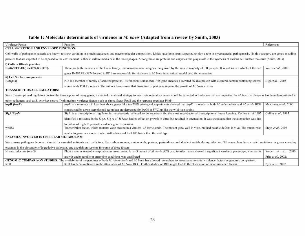
Table 1: Molecular determinants of virulence in M. bovis (Adapted from a review by Smith, 2003)Virulence Factor Function ReferencesCELL SECRETION AND ENVELOPE FUNCTION:
Cell walls of pathogenic bacteria are known to show variation in protein sequences and macromolecular composition. Lipids have long been suspected to play a role in mycobacterial pathogenesis. (In this category are genes encoding
proteins that are expected to be exposed to the environment , either in culture media or in the macrophages. Among these are proteins and enzymes that play a role in the synthesis of various cell surface molecule (Smith, 2003)
i) Culture filtrate proteinsEsat6/CFT-10,( Rv3874,Rv3875). These are both members of the Esat6 family, immuno-dominant antigens recognized by the sera in majority of TB patients. It is not known which of the two
genes Rv3875/Rv3874 located in RD1 are responsible for virulence in M. bovis in an animal model used for attenuation
Wards et al., 2000
ii) Cell Surface componentsP36(p36) P36 is a member of family of secreted proteins. Its function is unknown. P36 gene encodes a secreted 36 kDa protein with a central domain containing several
amino acids PGLTS repeats. The authors have shown that disruption of p36 gene impairs the growth of M. bovis in vivo.
Bigi et al., 2005
TRANSCRIPTIONAL REGULATORS:
Since Transcriptional regulators control the transcription of many genes, a directed mutational strategy to inactivate regulatory genes would be expected to find some that are important for M. bovis virulence as has been demonstrated in
other pathogens such as S. enterica, serova Typhimurium virulence factors such as sigma factor RpoS and the response regulator PhoP.hspR (hspR) hspR is a repressor of key heat shock genes like hsp70.Physiological experiments showed that hspR mutants in both M. tuberculosis and M. bovis BCG
constructed by a two step plasmid technique are depressed for hsp70 at 37ºC, unlike the wild type strains
McKinney et al., 2000
SigA/RpoV SigA is a transcriptional regulator in mycobacteria believed to be necessary for the most mycobacterial transcriptional house keeping. Collins et al 1995
identified a missense in the SigA. Sig A of M.bovis had no effect on growth in vitro, but resulted in attenuation. It was speculated that the attenuation was due
to failure of SigA to promote virulence gene expression.
Collins et al., 1995
whiB3 Transcription factor. whiB3 mutants were created in a virulent M. bovis strain. The mutant grew well in vitro, but had notable defects in vivo. The mutant was
unable to grow in a mouse model, with a bacterial load 105 lower than the wild type
Steyn et al., 2002
ENZYMES INVOLVED IN CELLULAR METABOLISM:
Since many pathogens become starved for essential nutrients and co-factors, like carbon sources, amino acids, purines, pyrimidines, and divalent metals during infection, TB researchers have created mutations in genes encoding
enzymes in the biosynthetic/degradative pathways, and acquisition systems for some of these factorsNitrate reductase (narG) Plays a role in anaerobic respiration in prokaryotes. A narG mutant of M. bovis BCG used to infect mice showed a significant virulence phenotype, whereas its
growth under aerobic or anaerobic conditions was unaffected
Weber et al.., 2000;
Fritz et al., 2002;GENOMIC COMPARISON STUDIES. The availability of the genomes of both M. tuberculosis and M. bovis has allowed researchers to investigate potential virulence factors by genomic comparison.RD1 RD1 has been implicated in the attenuation of M. bovis BCG. Further studies on RDI might lead to the elucidation of more virulence factors. Pym et al., 2002
23

Prophages and their Role in Bacterial Pathogenicity: Phage–Mediated Horizontal
Gene Transfer.
Bacteriophages are viruses that infect bacteria. They may take up two life cycles upon
entering the host: virulent phages only undergo a lytic life cycle, resulting in the lysis of
the host and release of progeny phages; whereas temperate phages can undergo both lytic
and lysogenic life cycles. Upon entering the host, temperate phages may incorporate their
DNA into the host’s genome, and form part of the genome and they are termed
prophages. Most of the prophage genes can remain dormant in the cell, until they are
induced to undergo lytic cycle. However, not all the prophage genes remain dormant.
Many of these phages express genes that have subtle effects on the phenotype of the host
bacterium (Cianciotto, 1989). Different bacteriophages bring about conversions to their
hosts, including increased virulence and pathogenicity (Waldor and Makalanos, 1996).
Phage genomes may themselves contain genes valuable to the host (Leitet et al., 2006).
In many toxin producing bacteria, pathogenicity and virulence have been shown to be
bacteriophage-mediated (Rajadhyaka and Rao, 1965.,Wagner and Waldor, 1996; and
Waldor and Makalanos, 1996), which emphasizes their consideration of their role in host
pathogenesis and the dissemination of toxin genes amongst different bacterial strains
(Wagner and Waldor, 2002). Vibrio cholera, Shiga–toxin producing E. coli,
Corynebacterium diphtheria, and Chlostridium botulinum, all depend on specific
prophage encoded toxin for causing specific disease (Kar et al., 1995; Brüssow et al.,
2004). Furthermore, Shiga toxin encoding phages of E. coli have shown that the
bacteriophage’s life cycle can exert control over virulence factor production by bacterial
pathogens. Shiga toxins (Stx1 and Stx2), are the principal virulence factors in
enterrohemmorhagic E. coli. Stx1 is reported to be located within prophage related to
lambda, which contain transcriptional units for various functions such as replication,
morphogenesis, and lysis that are coordinately expressed during specific intervals
following prophage induction due to regulatory influence of phage promoters, phage
repressor, transcriptional terminators and antiterminators (Wagner and Waldor, 2002).
24

Toxin genes are only a subset of the diverse virulence factors encoded by bacteriophages.
Phages can also contribute directly to bacterial virulence (Mavris et al., 1997; Guan et
al., 1999). In addition to conferring non-structural components as virulence factors,
structural components of the virion may also confer virulence attributes. In two Shigella
flexneri bacteriophages (SfV and SfII), O-antigen glycolysation was characterized (Huan
et al., 1997a, b; Mavris et al., 1997). A three gene cluster was found (gtrA, gtrB, and
gtrX) in both these O antigen glucolysation bacteriophages. The gtrA gene in phage SfV
encodes a highly hydrophobic protein of unknown function and gtrB in SfII has been
allocated a bactoprenol glucose transferase function. A study by Guan et al., (1999)
showing molecular characterization of phage SfX of Shigella flexneri also demonstrated
that the first gene (gtrA) of a three gene cluster to be most likely to encode a protein
involved in the translocation of lipid linked glucose across the cytoplasmic membrane,
leading to a modification on the bacterial cell surface. These authors also showed that the
second gene (gtrB) encodes a bactoprenol glucose transferase. As protective host immune
response to S. flexneri infection is directed against the O-antigen. This resulting serotype
conversion represents an important virulence factor for the bacteria (Guan et al., 1999).
Some phages encode regulatory factors that increase expression of the virulence genes
encoded by the phage, while others encode enzymes that alter bacterial components
related to pathogenicity (Wagner and Waldor, 2002). A study by Spanier et al., (1980)
showed bacteriophages to be involved in conversion of some streptococci strains to a
phagocytosis-resistant phenotype. The M protein is a streptococcal cell surface antigen
responsible for phagocytosis by human polymorphonuclear leuckocytes. In this study two
genes involved in the synthesis of anti-phagocytic M protein were found, one namely
mprA contributed by a prophage, and the second mprS contributed by the bacterium. The
genetic mechanism by which the wild type cells were converted to phagocytocis–resistant
phenotype were speculated to involve site specific inversion, deletion and insertion of a
controlling DNA segment in the prophage (Spanier et al., 1980).
25

As mobile elements, bacteriophages can also serve as vectors for transferring genes and
therefore potential virulence factors between bacteria. Comparative analysis of the whole
genome sequences of most bacteria suggests that many genes have been transferred
between prokaryotic species through horizontal gene transfer (HGT) (Ochman et al.,
2000). Phage mediated HGT occur via transduction. Generalized transduction is observed
with many bacteriophages. It was first demonstrated in Salmonella typhimurium with
phage P22 by Zinder and Lederberg (1952) and subsequently in E. coli with phages P1
(Lennox, 1955) and T1 (Drexler, 1970). Wilson (1979) later described a mutant of T4
that displayed the property of generalized transduction. In the process of generalized
transduction, after the empty heads are assembled, the phage DNA must be packaged, but
instead there is aberrant packaging of host DNA fragments at a finite frequency. This
results in delivery proficient phage particles that will deliver the bacteria DNA fragments
into another host. In pathogenic bacteria, HGT is thought to facilitate maintenance and
enhancement of virulence as well as the spread of antibiotic resistance (Brüssow et al.,
2004).
Phage encoded genes are not always transmissible due to the fact that integrated
prophages frequently become defective which may lead to them becoming cryptic since
the gene content will decay in the absence of selection. However some of the residual
gene content may be retained if it confers a selective advantage to its host. For instance
the non-transmissible stx genes in Shigella dysenteriae are adjacent to lambdoid phage-
like sequences interrupted by numerous insertions sequences, suggesting that the toxin
genes lie in a prophage that has been rendered defective by these insertion sequences
(McDenough et al., 1999). Defective prophages have been identified by analysis of
sequenced bacterial genomes. Comparative genomics has provided a tool for prophage
identification in sequenced bacterial genomes. Many sequences of bacterial pathogens
deposited in the public database contain prophage DNA integrated in the bacterial
chromosome. Other bacteria contain multiple prophages that constitute a large part of
their genomes (Canchaya et al., 2003). The most extreme cases are E. coli 0157:H7 strain
Sakai, a human pathogen. It contains 18 prophage genome elements, amounting to 16%
of its total genome content (Perna et al., 2001). Other cases are that of Streptococcus
26

pyogenes genomes of the sequenced M1, M18 and M3 strains with four to six prophages
each, which amounts to 12% of the bacterial DNA component (Smoot et al., 2002).
Furthermore, prophages have been demonstrated to play a role in inter-strain genetic
variation in several related bacterial species. Examples include Staphylococcus aureus
(Baba et al., 2002) and S. pyogenes (Smoot et al., 2002). When genomes from closely
related bacteria were compared in a dot plot analysis, prophage sequences frequently
accounted for a substantial proportion of the difference in the bacterial strains. Examples
are E. coli 0157:H7 and K12 strains (Perna et al.,2001); Listeria monocytogenes and
Listeria innocua; Salmonella enterica serovars Typhi and Typhimurium (McClelland et
al., 2001; Parkhill et al., 2001) Recently Srividhya et al. (2006) has used a Protein
Similarity Approach (PSA) to identify cryptic prophages. In this approach, using
information available from the integrated prophages of sequenced bacterial genomes,
they identified prophage e14 (a coliphage) homologs by similarity searches at protein
level. They took twenty three e14 proteins as query and used bacterial proteomes as
target (Srividhya et al., 2006). The e14 element is a very well characterized prophage
element, containing all the highly conserved prophage genes like the integrases,
excisionase, phage portal, cro type regulator, repressor and terminase genes elements
(Mehta et al., 2004). Using the PSA, Srividhya et al., (2006) identified probable
prophages in both pathogenic and non-pathogenic bacterial genomes. Some of these
prophages were reported in the literature and there were some with no literature reports.
The unreported prophages in the literature, included among the others, putative
prophages of M. bovis AF2122/97, and a prophage from S. pyogenes M18 strain.
The role of prophages is not limited to pathogenic bacteria, but some adaptations of non-
pathogenic bacteria to their ecological niche are believed to be prophage-mediated
(Brüssow and Hendrix, 2002). For instance, it is becoming clear that gut commensals and
pathogenic bacteria have much in common. Lactobacillus commensal bacteria are under
selective pressure of a T–cell independent mucosal IgA and respond by changing their
surface polysaccharide. Sequenced Lactobacillus commensals contain multiple prophage
genomes that showed lysogenic conversion genes related to those of the prophage of S.
pyogenes. In addition, the dairy strain Lactobacillus lactis IL1403 contains six prophage
27

genomes (Chopin et al., 2001). One therefore might suspect that these prophages
contribute to the evolutionary success of lactic acid bacteria living in strikingly distinct
environments (Brüssow and Hendrix, 2002).
HGT in pathogenic mycobacteria
Recombination between different strains of M. tuberculosis must be rare since the
bacteria rarely come into contact with different populations in the context of an infection
(Smith et al., 2003; Supply et al., 2003). Because of its isolated living space, lack of
migration between hosts, long generation time, and latent infections, members of the M.
tuberculosis complex remain paradigms of clonal evolution. Thus if recombination
occurs, it happened between identical individuals and hence leaves little detectable traces
(Smith et al., 2003). However, the presence of different gene delivery mechanisms and
functional homologous recombination machinery in M. bovis and other members of
mycobacterium species raise the possibilities of naturally occurring transfer and
recombination. It has been shown that mycobacterial plasmids can replicate within most
Mycobacterium species, so they can theoretically be spread horizontally, promoting gene
transfer between mycobacteria (Le Dantec et al., 2001; Kirby et al., 2002). Patients
simultaneously infected by two different strains of M. tuberculosis have been reported
(Shafer et al., 1995; Theisen et al., 1995; Yeh et al., 1999; Pavlic et al., 1999; Braden et
al., 2001; Das et al., 2004). A chromosomally coded conjugation system has recently
been identified in Mycobacterium smegmatis (Parsons et al., 1998; Wang et al., 2003)
which opened up the possibility of lateral DNA transfer, via conjugation with other
mycobacteria. Furthermore several plasmids were found in M. avium complex (Meissner
and Falkinham, 1986; Hellyer et al., 1991) among them plasmid pVT2, which is thought
to be conjugative (Kirby et al., 2002) and raises the possibility of conjugational transfer
in Mycobacterium avium complex. Evidence of recombination has been shown in M.
tuberculosis strains isolated from TB patients in East Africa (Gutierrez et al., 2005). In
these isolates, incongruence among gene phylogenies as well as mosaic gene sequences,
whose individual elements are retrieved in classical M. tuberculosis were detected.
Therefore, despite its apparent homogeneity, the M. tuberculosis genome appears to be a
28

composite assembly resulting from horizontal gene transfer events predating clonal
expansion (Gutierrez et al., 2005).
Possible Role of phages in HGT in Mycobacteria
Mycobacteriophages are extremely diverse in nature and carry highly mosaic genomes
(Pedulla, et al., 2003). Most of these viruses have been isolated from environmental
samples. While many bacteriophages have been isolated from environmental samples,
only 30 have been sequenced and characterized so far (Pedulla et al., 2003; Hatfull et al.,
2006). Genomic comparison of these phages at nucleotide level reveals considerable
overall diversity, with small groups having sequence similarity. The most numerous
phage genome clusters contains seven that are more closely related to each other than the
others phages. These include: L5, Bxb1, D29, Bethlehem, U2, BxZ2, and Che12. The
next numerous phage cluster contain six phages. These include Rosebush, Orion, PG1,
Cooper, Qyrzula and Pipefish. Another cluster is formed by phages Che8, PMC and Llij
(Hatfull et al., 2006). L5, Bxb1, D29 and TM4 have similar arrangement of structure and
assembly genes. The two most closely related genomes are those of L5 and D29, sharing
over 75% of their genes at amino acid level. More than 40% of the predicted protein
products of Bxb1 are related to those of L5 and D29 (Pedulla et al., 2003). The
nucleotide sequence comparison of these 30 sequenced mycobacteriophages is shown in
a dotter plot in Fig 1 below.
29

Fig 1: Nucleotide sequence comparison of 30 mycobacteriophages as illustrated in a Dotter plot using a
sliding window of 25bp. The lower triangle represents relationships at an elevated level of grey- scale
relative to the upper triangle, revealing weaker sequence relationships. DOI 10.1371/journal.pgen.
0020092.g001 (extracted from Hatfull et al., 2006)
Genomic characterization of 14 of these mycobacteriophages revealed within their
genomes many unexpected genes that were not previously thought to be phage encoded
(Pedulla et al., 2003). Some of these phage genes, for example gene 39 and 69 of phage
CjW1 and Omega respectively are reported to encode close homologs of M. leprae and
M. tuberculosis immuno-dominant antigen Lsr2 that is a potent stimulator of both cellular
and humoral immune responses. Even though the function of Lsr2 protein is not known it
30

is likely that these viruses could influence immune response of their host through the
introduction of these genes (Pedulla et al., 2003), hinting at a possible role of phages in
mycobacterial virulence. This also suggests the possible involvement of these phages in
genetic exchange between the prokaryotes, including gene transfer among mycobacteria.
In addition, the genome sequences of mycobacteriophages have shown that phage
genomes are extensively highly mosaic in nature with regions of obvious sequence
similarities interspread with segments that appear to be unrelated, suggesting extensive
horizontal genetic exchange among these viruses (Hendrix et al., 1999; Pedulla et al.,
2003). At first it was postulated that phages evolve by genetic exchange at special
intergenic sites, either through homologous recombination or by site specific mechanism
(Susskind and Botstein, 1978). However, the recent elucidation of the genomes of several
lambdoid phages has allowed the revelation of a different picture of mosaic formation
(Juhala et al., 2000; Hendrix, 2002). In this model, illegitimate recombination is believed
to take place quasi-randomly along the recombination genomes, generating an unholy
mélange of recombinant types, almost all of which will be defective for phage growth as
a consequence of their misplaced recombination. Natural selection eliminates all but the
tiny minority of recombinants in which biological function is intact, thus giving rise to a
observable population in which the sites of recombination are random (Juhala et al.,
2000; Hendrix, 2002). It is not clear as to what degree this picture of horizontal exchange
extends to other phage groups, however, the availability of the genomes of 14
mycobacteriophages provides an opportunity for the elucidation of the evolutionary
mechanisms that generate mosaicism in these mycobacteriophages (Pedulla et al., 2003).
Pedulla et al., (2003) postulated that one explanation is that, genetic modules are re-
assorted by homologous recombination at short conserved boundary or (linker)
sequences, as suggested initially by Susskind and Botstein (1978). An alternative model
is that, illegitimate exchange plays the major role, recombining viral and non-viral DNA
molecules in a sequence independent manner that generates mostly genomic trash that is
either incorrectly sized for packaging into capsid or lacks required genes. The viable
genomes that pass this filter for function and size will retain recombination boundaries
that have had minimal impact on gene function, occurring either at or close to gene
boundaries. The generated mosaic junctions will be subsequently re-assorted by
31

homologous recombination between flanking sequences (Pedulla et al., 2003). If the
mosaicism of phages is generated by illegitimate recombination, then the acquisition of
genes from the host is to be expected (Hatfull et al., 2006).
Even though there are no further reports of prophages capable of forming complete
phages that are infective in M. bovis and M. tuberculosis, the availability of the genome
sequences of both M. tuberculosis and M. bovis has allowed the identification of
prophage sequences in these tubercle bacilli. The genome of M. tuberculosis H37Rv
strain has been reported to contain two prophage like-elements, phiRv1 and phiRv2. Both
are relatively small (~10kb) but contain several phage related sequences (Hendrix et al.,
1999). PhiRv2 has at least two homologues of mycobacteriophages genes: integrase
(Rv2659c, which is related to integrases of phages L5 and D29) and a relative of L5 gene
36 (Rv2657c). It also contains a second recombinase, Rv2647, a homologue of the
prohead protease genes of phages HK97 (a coliphage) and Streptomyces phage ΦC31;
and a homologue of the actinophage RP3 (a temperate phage of Streptomyces) gene, in
addition to several others (Hendrix et al., 1999). In addition to this the phiRv1 also
encodes an active site specific recombinase, where an integrase of the serine recombinase
family catalyses recombination and excision, while an adjacent small Open Reading
Frame (ORF) controls the directionality of recombination (Canchaya et al., 2003).
Presumably the recombination functions associated with phiRv1 are active and may
therefore mediate the integration and / or excision of this DNA segment (Hendrix et al.,
1999). The phiRv2 prophage element also may be recombinationally active because the
attachment junctions (attL and attR) are present and appear to derive from a phage
attachment site (attP) that is structurally similar to the L5 attP site (Pena et al., 1997).
Although the two prophages appear too small and too deficient in terms of virion
structural genes to encode a complete virion, they could in fact be “complete” satellite
phages in the manner of coliphage P4 which uses the structural genes of another phage to
package its genome (Hendrix et al., 1999). Prophages phiRv1 and phiRv2 were identified
to be contained in regions RD3 and RD11 respectively in M. tuberculosis. In a study to
identify and differentiate between M. tuberculosis complex isolates by PCR deletion
analysis, the region RD3 was found to be present in most M. bovis isolates (Parsons et
32

al., 2002). The NCBI and the Sanger Institute databases also reveal the presence of
phiRv1 in the M. bovis AF2122/97 sequenced strain (www.ncbi.nlm.nih.gov;
www.sanger.co.za ). However as mentioned before, an avirulent M. bovis BCG strain has
neither phiRv1 nor does it have phiRv2. This strain is thought to have lost these prophage
sequences during its attenuation (Parsons et al., 2002) suggesting that these prophages
might play a role in the pathogenicity of both M. bovis and M. tuberculosis.
33

1.3 AIMS AND OBJECTIVES
Bacteriophages have been known to contribute to the bacterial phenotype through
lysogenic conversion. Many virulence factors in pathogenic bacteria are phage encoded.
However, it is not known whether this is true in mycobacteria. A study by Pedulla et al.,
(2003) looking at the genome sequence of 10 mycobacteriophages isolated at the time of
the study, suggested that several of the identified genes may have patho-adaptive
potential. Perhaps paradoxically, sequenced mycobacterial genomes revealed a paucity of
recognizable prophages. A study by Tageldin et al., (1981), which looked at 7 bovine
strains, only isolated two spontaneously inducible phages from two strains that had
different host range. To initiate an enquiry into the contribution of prophages to the
relevance of mycobacterial disease, we set up some experiments to screen for the
presence of inducible and or cryptic prophages in Mycobacterium bovis isolates from
different outbreaks. Using a well-characterized phage system (mycobacteriophage L5),
bacteriophage isolation methods were optimized and tested on the M. bovis isolates. Our
objectives therefore were:
Objectives
• To detect spontaneously induced prophages using more than one sensitive
indicator strains
• To screen for the presence of spontaneously induced prophages using non-
selective methods
• Identify prophages using a genomics approach
34

CHAPTER TWO- SCREENING FOR SPONTANEOUSLY INDUCED PHAGES
FROM M. BOVIS ISOLATES.
2.1 INTRODUCTION
Spontaneous phage induction is a process that results in the conversion of a prophage into
a lytic phage, without the use of chemical or physical inducing agents. It is the
characteristic of a temperate phage to either enter a lysogenic or dormant state or undergo
lytic infections. Prophages are integrated viral genomes in bacteria and range from fully
viable, to cryptic prophages that have undergone mutational decay and do not result in
lytic growth. Prophages can often be induced from their host by UV or chemical
treatment.
There are essentially two approaches to studying inducible phage populations associated
with bacteria. Those approaches that involve direct isolation and therefore require
sensitive strains (Tokunga et al., 1971; Tageldin et al., 1981; Kilic et al., 2001). The
other approach, involving microscopy, simply identifies the presence of phage, but does
not attempt to isolate it. These microscopy approaches more traditionally involved
transmission electron microscopy (TEM) (Rieber and Imaeda, 1969; Bergh et al., 1989;
Borsheim et al., 1990; Humphrey et al., 1999; Maggi and Breitsch, 2005), but currently,
Epiflourescence microscopy is becoming more popular (Suttle et al., 1990; Hara et al.,
1991; Proctor and Fuhrman, 1992; Noble and Fuhrman, 1998). In TEM, the bacterial
cells are harvested from liquid culture by centrifugation of the cultures (at 4,000×g)
(Humphrey et al., 1999) followed by suspension of the bacterial pellets in phage buffer
and negative staining for TEM to view any phage particles attached to the bacterial cells,
or ultracentrifugation of the supernatant (at 40,000×g) (Rieber and Imaeda, 1969)
followed by negative staining to view free phage particles in the supernatants. TEM
reveals the morphology and size of the bacteriophage (Rieber and Imaeda 1969;
Humphrey et al., 1999) where as EFM only reveals the presence of the phages in the
sample (Noble and Furhman, 1998). In EFM, phage particles are harvested from bacterial
cultures by centrifugation followed by filtration of the supernatants through a 0.02µm
35

pore size Al2O3 Anodisc 25 membrane filter and stained with SYBR Green 1 stain (Noble
and Fuhrman, 1998).
Phages have been isolated from pathogenic and non-pathogenic strains of mycobacteria.
In 1984 Timme et al. isolated bacteriophages from Mycobacterium avium,
Mycobacterium intracellulare and Mycobacterium scrofulaceum, after treatment of
bacterial cultures with mitomycin C. In addition lysogeny has been demonstrated in M.
fortuitum after treatment of the bacterium with mitomycin C and UV induction (Jones,
1973; Grange and Bird, 1975). In 1981 Tageldin et al. isolated two mycobacteriophages
from lysogenic Sudanese bovine strains. Out of seven bovine strains tested for
mycobacteriophage inductions, only two produced plaques. It was also shown that they
had different host range (Tageldin et al., 1981).
Most mycobacteriophage studies have employed the use of plaque assay procedures
which need sensitive indicator strains for direct isolation and propagation of these
phages. The already isolated mycobacteriophages may serve as useful controls for
optimizing some experimental parameters. In this study a well characterized
mycobacteriophage L5 has been used as a control in some experiments as it is easy to
work with, since even though it may form stable lysogens with M. smegmatis, as a
temperate phage, it is spontaneously inducible (Fullner and Hatfull, 1997; Lee et al.,
1991; Snapper et al., 1988). L5 lysogens spontaneously generate free phage particles, by
a way of prophage excision (Lewis and Hatfull, 2000). Prophage excision involves site-
specific recombination between the attachment junction attL and attR, and requires a
phage encoded excisionase protein in addition to integrase and host factor (Landy, 1989;
Lewis and Hatfull, 2000). Establishment of lysogeny involves integration of phage
genome to the mycobacterial host chromosomal genome through the integrase- mediated
site specific recombination event (Lewis and Hatfull, 2000). Maintenance of lysogeny by
temperate phage requires functions of repressor proteins (Lewis and Hatfull, 2000).
36

2.2 AIMS AND OBJECTIVES
Based on the low levels of phages seen in M. bovis using plaque assay (Tageldin et al.,
1981), we reasoned that the use of a single sensitive strain might be biased, and hence
could only select for phages that can infect that strain. In addition only a few studies as
far as we know have attempted to look at the use of more than one indicator strain. We
hence decided to screen for the presence of phages from M. bovis strains from different
outbreaks, using a plaque assay on different indicator strains (selective isolation), as well
as a non-selective epifluorescence microscopy. Epifluorescence microscopy allows for
the detection of spontaneously induced phages, irrespective of their host specificity. In
order to develop a control for these experiments, we used the well characterised L5 phage
system.
Our objectives therefore were:
• To test some approaches to attempt to increase the levels of phages in culture
filtrates using the well described and characterised phage L5.
• To screen for the presence of spontaneously liberated prophages by periodic
sampling of cultured M. bovis isolates using a standard plaque assay.
• To screen M. bovis isolates for spontaneously inducible prophages by non-
selective Epiflourescence microscopy.
37

2.3 METHODS
Bacteria and phage strains used in this chapter are listed in Table 2 below.
Table 2: List of strains used in this study
Bacteria StrainsName SourceM. bovis isolates Dr. Michel, Agricultural Research Council- TB
laboratory1
Indicator bacteria strainsM. fortuitum Dr Michel, Agricultural Research Council-TB
laboratoryM. kansasii Dr Michel, Agricultural Research Council-TB
laboratoryM. scrofulaceum Dr Michel, Agricultural Research Council-TB
laboratoryM. smegmatis- L5 lysogen Developed by Ms. Belinda Spillings, University of
the Witwatersrand, (Dr Durbach’s Lab)M. smegmatis mc2155 (wild type) Prof Dabbs, Wits UniversityMycobacterium ‘ S2’ isolate2 Dr Lukusa Kambulu, University of the
Witwatersrand (in Dr Durbach’s Lab)1
Phage strainsL5 phage Ms Spillings, University of the Witwatersrand
11 M. bovis isolates-. Their species and origin is indicated in Table 3 and 4 in the results section.2‘S2’- mycobacteria isolate =98% similar at amino acid level to M. smegmatis, characterized
by Dr Lukusa Kambulu
38

2.3.1 Liquid culturing of mycobacteria indicator strains
Different mycobacteria indicator strains which were maintained as liquid freezer stocks at
-70°C in 15% glycerol (v/v) were cultured in Middlebrook 7H9 medium (Difco)
(Appendix B) supplemented with 1mM CaCl2, 1mM MgCl2 (to facilitate phage
attachment); 0.03% glucose; 0.2% glycerol; 0.05% Tween 80 to reduce clumping
(Snapper et al., 1988) and were then incubated at 37ºC and shaken at 120rpm on a
platform shaker. Growth was monitored by optical density analysis at 600nm.
2.3.2 Liquid culturing of M. bovis isolate strains
Different M. bovis isolates from solid cultures in Löwenstein Jensen (LJ) medium
(National Health Laboratory Services) medium were cultured in Middlebrook 7H9
medium (Difco) supplemented with 0.03% glucose by dispensing 5ml aliquots of the
medium on the surface of the LJ slant with colonies. The medium containing M. bovis
was then poured into another 5ml of Middlebrook 7H9. The cultures were then incubated
at 37°C and were manually shaken gently for an average of 5 times daily.
2.3.3 Propagation of L5 and preparation of large scale L5 stocks (adapted from
Sambrook et al., 1989)
Phage plaques on bacterial indicator lawn were picked by immersing a blue pipette tip on
the plaque and sucking. The agar plug containing the phage was suspended in 1ml phage
buffer (see appendix B) and mixed by gentle pipetting. The suspensions were left at room
temperature to allow phage diffusion from the agar. They were centrifuged at 5000rpm in
a benchtop centrifuge for 2 minutes followed by filtration through a 0.45 µm non-
pyrogenic hydrophilic disposable filter (Ministart). For large scale phage stocks, the
phage suspensions were plated on 20 to 25 M. smegmatis mc2155 or M. fortuitum
indicator plates or overlays (see sections 2.3.5, and 2.3.6) and incubated at 37º C, until
plaques were seen. When plaques appeared, 5ml of phage buffer was poured onto the
39

plates and left overnight at 4ºC with intermittent gentle shaking to allow diffusion of the
phage particles into the phage buffer. The phage buffer was then harvested and placed in
50ml polypropylene Falcon tubes. Then 1ml of phage buffer was again added onto the
plates and stored for 15 min in a tilted position to allow the fluid to drain into a localised
area. The phage buffer from the plates was again removed and combined with the first
harvest. The suspension from the lysate stocks was centrifuged at 3500×g for five
minutes to pellet cell debris. The supernatant was transferred into 50ml Falcon tubes
(Lasec). Sodium chloride to a final concentration of 1M and 10% polyethylene glycol
(PEG) were added and gently shaken until dissolved. The solution was stirred at 4ºC
overnight in a platform shaker. The formed precipitate was centrifuged at 3500×g for 5
minutes at 4ºC. The phage pellet was resuspended in about 4 ml phage buffers, and mixed
gently by pipetting up and down. The volume of the suspended phage was measured, and
0.5g/ml (m/v) of Cesium Chloride (CsCl), was added on the phage suspension and mixed
to dissolve. The suspension was layered on top of a Cesium Chloride step gradient that
was prepared by layering solutions of CsCl dissolved in phage buffer to obtain densities
of 1.7g/ml, 1.5g/ml and 1.45g/ml on top of each other in a Beckman SW41 or SW28
clear polyethylene tube. The gradient with the phage suspensions were centrifuged in
Beckman SW28 or SW41 rotor at 22000rpm at 4ºC for 2 hours. The phage band between
0.45g/ml and 5g/ml CsCl densities , was collected by either puncturing the side of the
tube with the phage using a sterile needle or by inserting a sterile plastic syringe on top of
the centrifuge tube and sucking the phage band. The collected phage was dialyzed using a
dialysis bag (Seal air) that was soaked in phage buffer overnight at 4ºC. The phage stock
was screened for purity and viability and titer determined by plating on M. smegmatis
mc2155 indicator plates (see section 2.3.4). The phage stock maintained at 4ºC until
needed.
2.3.4 Preparation of M. bovis phage filtrate
Mycobacterium bovis filtrates were prepared by sampling 5ml from each liquid culture
periodically (up to 3 months of incubation) in sterile 50ml Falcon tubes (Lasec). They
40

were then centrifuged at 1088×g for 10min on a benchtop centrifuge (Heraeus). The
supernatants were filtered using a 0.45μm filter (see section 2.3.3).
2.3.5 Preparation of Indicator Plates for phage detection (Adapted from McNerney
et al., 2004)
Indicator plates were prepared by addition of a 10% (v/v) indicator bacterial culture
(OD600=0.8-1) in 1% of either Middlebrook 7H9 , Luria or Middlebrook 7H10 agar (See
Appendix B) that had been cooled to 45°C and supplemented with 0.03% glucose, 1mM
CaCl2, 1mM MgCl2 and 0.2% glycerol. A volume of 20ml of the agar was then poured
into 90mm Petri-dishes and allowed to solidify, and stored at 4ºC until required. Culture
filtrates containing potential phage (100μl) were spread plated on the indicator plates and
allowed to dry. The plates were incubated at 37ºC and checked daily for plaque
development.
2.3.6 Preparation of overlays for plaque assay: (Sambrook et al., 1989)
The bottom agar consisting of 1% of Luria broth agar (See appendix B) or Middlebrook
7H10 agar (see Appendix B) was prepared and 5ml of this agar was added in 90mm
Petri-dishes. For the top agar, 0.9% soft agar was prepared (see appendix B) and allowed
to cool to 45°C at room temperature. A volume of 100μl either purified L5 or culture
filtrate derived from M. bovis strains was added to 900μl of the indicator bacterial strain
that had been grown to an optical density at 600nm of 0.8 to 1 (section 2.3.1), followed
by addition of this mixture into 9ml of the soft agar in standard sterile glass test tube. The
agar was mixed and poured onto the bottom agar and allowed to solidify at room
temperature. The plates were then incubated at 37ºC, and monitored daily for plaque
development.
2.3.7 Co-culturing experiment
A liquid culture of an L5 lysogen of M. smegmatis mc2155 (OD600= 1.31) was aliquoted
into four equal volumes of 10ml each, in Falcon tubes (Lasec). Each culture was
41

inoculated with different concentrations of M. smegmatis mc2155 wild type cells
(OD600=1.31) at ratio of lysogen to wild type cells: 1:0, 1:1, 1:10, and 1:100. The co-
cultures were incubated at 37ºC, and sampled at different time intervals. The lysates were
filtered with a 0.45μm non-pyrogenic hydrophilic disposable filter (see section 2.3.4) and
monitored by standard plaque assay for plaque development as previously described (see
section 2.3.5).
2.3.8 Epiflourescence Microscopy (EFM) to detect free phage particles (adapted
from Noble and Fuhrman, 1998)
2.3.8.1 Preparation of culture filtrates
L5 lysogen and wild type M. smegmatis mc2155, or M. bovis isolates phage filtrates were
prepared by picking colonies that were freshly grown in Middlebrook 7H10 agar plates
(see Appendix B) or the LJ slants (National Health Laboratory services (NHLS) for M.
bovis, with a sterile inoculation loop and suspending this into 1ml phage buffer
(Appendix B). Each of the bacterial suspensions was mixed by repeat pipetting. The
bacterial cells were centrifuged at 13000rpm for 5 min, in a benchtop centrifuge followed
by filtration through a 0.45μm disposable filter as described previously (section 2.3.4).
The L5 phage titer from the lysogen was determined by plaque assay on M. smegmatis
indicator plates as described in section 2.3.5.
2.3.8.2 Preparation of SYBR Green 1 Stain (Adapted from Noble and Fuhrman,
1998)
SYBR Green 1 is reported as a viable tool in the enumeration of virus particles by EFM.
Double stranded DNA bacteriophage detection by this method is becoming more popular.
The principle of this method lies on the ability of SYBR Green 1 to penetrate the phage
capsid and intercalates on the nucleic acid. SYBR Green 1 has a proprietary formula and
its manufacturer (Molecular Probes) does not report its molecular weight or
concentration. The SYBR Green 1 (Molecular Probes) used for this study was diluted
42

1:10 of the supplied concentration in sterile de-ionized water with 0.2μm non-pyrogenic
hydrophilic (Ministart) filtered deionised water, and stored at -20ºC. From the 10% stock
solution a 2.5% working solution in sterile distilled water was prepared under subdued
light (final dilution of 2.5x10⁻³)(An optical density of 0.42 at 494nm when the stock was
diluted 1000, is how the concentrations were determined).
2.3.8.3 Specimen preparation (either purified L5, L5-lysogen or M. bovis culture
filtrates)
For purified L5, 500μl was diluted to 5ml with phage buffer. For L5-lysogen or M. bovis
contained on solid media, 500μl of culture filtrates (see preparation of culture filtrates in
section 2.3.8.1) were added into Falcon tubes with 5ml phage buffer. The suspensions
were each filtered separately onto a 0.02μm pore size Al2O3 Anodisc 25 membrane filters
(Whatman) backed by 0.8μm nitrocellulose filter paper (Millipore) at approximately
20kPa vacuum. The Anodisc membranes were filtered to dryness, removed with forceps
with vacuum still on.
2.3.8.4 Staining
Filtered material was stained by submerging the Anodisc filter in 100μl of SYBR Green
1 stain with sample-side facing up, as described in Noble and Furman (1998) for 15 min
in the dark. After the staining period, the filter was picked up and any remaining moisture
was then carefully wiped out by touching the back side of the membrane to a Kimwipe.
The Anodisc filter was mounted sample up, on a glass slide. A few drops of mounting
solution (50% Phosphate buffer saline (Appendix B), 50% glycerol, and 0.1% 2-
phenylenediamine (Sigma Chem, made freshly from a freshly made 10% aqueous stock
solution) was added onto the Anodisc filter and covered with a cover slip. The slides
were observed under a Zeiss Axiocam EF microscope, under blue excitation at 1000×
magnification. Representative fields were captured by a digital camera.
43

2.4 RESULTS
2.4.1 Isolation of spontaneously liberated phage
Before attempting to isolate spontaneously liberated phage directly from culture filtrates,
we first attempted a non-chemical approach to see if it would lead to increased levels of
spontaneously liberated phage in these culture filtrates.
2.4.1.1 Co-culturing a L5-M. smegmatis mc2155 lysogen with M. smegmatis mc2155
Spontaneous induction of prophages is generally low (10-2 to 10-5 phage per bacterium per
generation – depending on the particular phage-host system) (Chibani-Chennoufi, 2004).
In lysogenic cultures phage titers typically range between 104 and 105 pfu/ml in cultures
where bacterial counts are in the order of 109 cfu/ml. This demonstrates a very small
fraction of cells undergoing lysis, and this has no consequence to the majority of
population that is immune to super infection (Bossi et al., 2003). The situation has been
shown to change if the population is not clonal and contains bacteria from a non-
lysogenic strain (Bossi et al., 2003). They showed that co-cultures of certain Salmonella
strains carrying or lacking specific prophages undergo a swift composition changes as a
result of phage mediated killing bacteria and lysogenic conversion of survivors. What
happens is, i) Prophages are spontaneously induced in only a few lysogenic cell, ii) this
enhances the competitive fitness of the lysogen population as a whole, setting a selection
regime that forces maintenance and spread of viral DNA (Bossi et al, 2003), one of the
byproducts being an elevation of free phage in the culture medium, which therefore offers
itself as a potential method to amplify potentially low levels of phages in culture media.
In an attempt to see if this same approach may lead to a similar elevation of free phage in
mycobacteria, and therefore may prove to be an effective method to increase the levels of
free phage in the potentially lysogenic M. bovis outbreak strains we tested it with a
known lysogen. An L5-M. smegmatis mc2155 lysogen culture was co-cultured with its
44

non-lysogenic derivative at different ratios and tested for the levels of phage release by
plaque assay.
0 5 10 15 20 25 30
Sampling Time (H)
103
104
105
106
107
108
Numb
er of
plaq
ues
( pfu/ml)
Fig 2: Co-infection experiment showing the levels of phage released by the L5-M. smegmatis culture when
co-incubated with its non lysogenic counterpart. Plaques were obtained from plating duplicate samples
from each co-incubation at different ratios of lysogen : non-lysogen. The y-error bars represent the standard
errors obtained from three experiments. The different co-culture ratios are represented as = 1lys:
1non-lys, = 1lys: 10non-lsy =1lys: 100non-lys, = no non lys. Lys= lysogen, non-
lys=non lysogenic cells.
45

As can be seen in Fig 2, there is an initial rise in phage release in all co-cultures, followed
by a drop after ~3 hours. Significant increases in phage release at the l: 1 and 1:10 co-
infection cultures after 20 hours and 17 hours respectively were observed. As can be seen
in Fig. 2, and contrary to what was observed by Bossi et al, (2003), co-culturing of L5-
lysogen and non-lysogenic culture does not enhance the levels of spontaneously induced
L5.
We therefore did not explore this approach further as a method to isolate phages from the
M. bovis isolates.
2.4.1.2 Screening of M. bovis isolates for spontaneous phage induction using plaque
assay
Since the previous approaches did not significantly suggest that we would be able to
increase our yield of spontaneously liberated phage by co-incubation with potentially
sensitive hosts, we decided to directly screen for induced phage. In this assay we
screened for the presence of spontaneously inducible prophages from different M. bovis
isolates. Filtrates from these isolates were plated on different bacterial indicator plates,
and/or overlays and assayed for plaques. We reasoned that using different potentially
sensitive indicator strains would increase our likelihood of isolating phages. As can be
seen from Table 3, no plaques that could be propagated on the indicator strains were
isolated from 27 M. bovis isolates. Interestingly in some cases turbid plaques were
obtained after 3 months incubation that could not be propagated after the third serial
passaging on indicator plates (Fig. 3). The plaques appeared to be different in size. One
isolate TB 4031, as seen in Fig 3, produced big plaques that were approximately 5mm in
diameter with a titer of 102pfu/ml, compared to the other two isolates TB 2773 and TB
4328 (result not shown), which both produced plaques that were approximately 1.5mm in
diameter, with titers of 103 pfu/ml each. These three isolates were all from bovine
species, but from different origins, suggesting that they may be different isolates and
perhaps the plaques were different anti-mycobacterial entities too. The non- reproducing
nature of the plaques were not characteristic of phages, hence we thought, even though
this is unequivocally proven, that they might be bacteriocins.
46

Table 3: Screening for spontaneously induced phages from M. bovis isolates by plaque assay on different mycobacterial indicators.
M. bovis isolate number
Tissue origin and animal species Animal origin Mycobacterial Indicator Strains Plaque Amplification
M. smegmatis M. fortuitum M. scrofulaceum M. kansasiiTB 3984E Lung-Bovine Middleburg (SA) - - ND - -TB 4006 Retro-Bovine Stellenbosch (SA) - - ND - -TB 4016 Lung+ Liver- Bovine Middleburg (SA) - - ND - -TB 4023 Liver+ Lung- Bovine Ladysmith (SA) - - ND - -TB 4025 Thorax- Buffalo Hoedspruit (SA) - - ND - -TB 2332 Lung- Buffalo KNP (SA) - - ND - -TB 2338A Swabs-Buffalo KNP (SA) - - ND - -TB 2338B Swabs-Buffalo KNP (SA) - - ND - -TB 2891H No record - - ND - -TB 2630B Troncantor Bone-Lion KNP (SA) - - - - -TB 2682E Lung-Lion KNP (SA) - - - - -TB 2649C Lung- Buffalo KNP (SA) - - - - -TB 2773 Mediastinal lympnode- Bovine East London (SA) +
(3months)*(pfu/ml=103)+(3months) *(pfu/ml=103)
ND - -
TB 2804 Lympnodes -Warthog Mauriedale (SA) - - ND ND -TB 2836B Lung –Baboon Komatiepoort - - NP ND -TB 2881H No record - - ND ND -TB 2916H No record - - ND - -TB 3150 No record - - ND - -TB3878 Lympnodes- Bovine Vryheid (SA) - - ND - -TB 4031 Lympnodes- Bovine Nelspruit (SA) +
(3months)* (pfu/ml=101)+ (3 months)* (pfu/ml=101)
ND - -
TB 4328 Lympnodes- Bovine Nat. Dept of Agriculture (SA)
+ (3months)* (pfu/ml=103)
+ (3 months)* (pfu/ml=103)
ND - -
TB 1764 No record - - ND - -TB 4326 Lympnodes- Bovine Nat Dept of Agriculture (SA) - - ND - -TB 4330 Lympnodes- Bovine Nat Dept of Agriculture (SA) - - ND - -TB 4388 Lympnode-Buffalo Malelane - - ND - -TB 4329 Lympnode- Bovine Nat Dep of Agriculture (SA) - - ND - -TB 4043 Lung- Eland KNP (SA) - - ND - --=No plaques, ND= not done. -= no plaque amplification, + = plaques observed, *time of incubation of culture at which plaques were observed, retro= retropharyngeal
47

Isolation of spontaneously inducible prophages liberated from M. bovis that could be
propagated, using plaque assays with more than one sensitive strain was not achieved.
This assay is a selective method that will only select for those phages that can infect the
sensitive strains used. We reasoned that, if spontaneous phage induction from the isolates
did occur, we might not have indicator strains that are sensitive to the released phages,
hence we could have missed a certain proportion of the potential phages released.
Therefore a non-selective approach was followed to investigate if any detectable phages
are released from the M. bovis isolates.
48
BA
Fig 3: Plaques isolated from M. bovis isolates using plaque assay. A= Plaques from isolate TB4031, B=plaques from isolate TB2773.
b

2.4.1.3 Epiflourescence Microscopy (EFM) analysis
It is clear that the standard plaque assay methods for the isolation of spontaneously
inducible phages are biased, which could result in a high proportion of associated phages
being missed. We therefore attempted to identify if there were indeed any phages in the
culture filtrates of the M. bovis isolates by non–selective EFM that will allow us to
identify associated phages that have double-stranded DNA genomes (Vitzhum et al.,
1999; Zipper et al., 2004).
Since this approach has not been tested on mycobacteriophages, we first tested to see
whether we could detect the mycobacteriophage L5 using this approach. AL5-M.
smegmatis mc2155 lysogen culture grown on solid media was stained with SYBR Green I
and tested for the presence of any spontaneously induced L5 phage particles. M.
smegmatis mc2155 wild type cells were also prepared for staining, to serve as a negative
control. As can be seen from Fig. 4, the presence of free phage particles in L5 were
observed in a single field by EFM as approximately ~110 bright spots in a titer of 102
pfu/ml(Fig 4c.), which were not seen in the negative control (Fig 4f). Our analysis
therefore is that if EFM could detect 110 phage spots in 500µl=220spots/ml of phage
suspension (see 2.3.8.3 in the methods section), in a titer of 102 pfu/ml as determined by
plaque assay, therefore, a titer of 101pfu/ml, would be detected as 22 phage spots in a
milliliter by EFM, and a titer of 100 pfu/ml would give 2.2 phage spots/ml. This implies
that the EFM is so sensitive that it would detect two times the phage population detected
by plaque assay.
Since EFM, contrary to the plaque assay, is not selective, i.e., it does not require a
sensitive strain, which selects only phages that are capable of infecting only that strain,
we reasoned that if there were any phages that might be released, we may be able to
detect them, using EFM. Filtrates from solid cultures of M. bovis isolates were prepared,
stained with SYBR Green 1, and viewed under EFM as described in the materials and
methods (section 2.3.8). An M. smegmatis mc2155 lysogen of L5 was employed as a
49

positive control (102pfu/ ml as determined by plaque assay), and a wild type solid culture
of M. smegmatis mc2155, was used as a negative control.
Out of sixteen samples tested we also included two out of three that formed non-
reproducible plaques on M. smegmatis (See Table 3) of M. bovis tested for spontaneous
phage induction, by EFM, a single viral-like spot, similar to the L5 control was observed
in only one isolate: TB 4328. This isolate was also shown to form plaques with a titer of
103 pfu/ml on M. smegmatis and M. fortuitum (See table 3). Table 4 and figure 4a-f below
shows the EFM results from the isolates tested.
50

Table 4: EFM results to screen M. bovis isolates for spontaneous phage induction in
comparison to plaque assay results as previously determined.
Isolate number Tissue origin and animal species Animal origin EFM
result
Plaque assay result
TB 2773 Mediastal lympnodes-Bovine East London - + (3 months)*pfu/ml=103
TB 4328 Lymphnode- Bovine Nat. dept of agriculture ± + (3 months)*pfu/ml=103
TB 3150 No record - -TB 2916H No record - -TB 2804 Lympnode- Warthog Maurieedale (SA) - -TB 4036 Lympnodes- Bovine Middleburg - NDTB 4026 Mandible+ Retro- Bovine Middleburg - NDTB 4041 Lung- Beaver KNP (SA) - NDTB 4043 Lung- Eland KNP (SA) - -TB 5507D Prescap- Impala Malelani (SA) - NDTB 5196A Head- Buffalo KNP (SA) - NDTB 5119C Head- Buffalo KNP (SA) - NDTB 5151A Head- Buffalo KNP (SA) - NDTB 5082A Lung- Lion Hoedspruit (SA) - NDTB 4931C Left Retro- Buffalo KNP (SA) - NDTB 4898 No record - ND
±, potential viral-like spot; -, no viral spot, or plaque observed; +, plaques observed; retro,
retropharyngeal; prescap, prescapular; *, refers to time following inoculation where plaque was
observed.
51

Fig 4: EFM of M. bovis culture filtrates. a= isolate TB4812, b=isolate TB 4931, c= L5 lysogen, d= isolate
TB 4328,(→) points at a phage-like spot, e= isolate TB5082; f= M. smegmatis mc2155 wild type.
Based on these results, spontaneous prophage induction from these M. bovis, is either
very rare, or occurs below the levels of detection of our EFM assay. We reasoned that if
we could find any inducible prophages in M. bovis, we may still be able to explore the
presence of prophages that may be cryptic or simply undetectably low by using a
prophage-like DNA sequences in the genomes of our M. bovis isolates.
52
f
b
c
d
e
a

It is clear that EFM is a feasible microscopic technique for screening for the presence of
spontaneously induced phages in solid lysogenic cultures. This technique required a
phage titer of as little as 102 pfu/ml (as determined by plaque assay), in order to detect
individual phage in a single field. EFM can be used for direct screening and enumeration
of phage particles in solid lysogenic cultures.
53
2.5 DISCUSSION
In this chapter we attempted to test whether we could increase the detectable levels of
spontaneously liberated phage by co-incubating a lysogenic culture in the presence of a
non-lysogenic culture. We found no significant difference after 24 hours co-incubation
on the phage levels, even when high ratios of non-lysogen to lysogen were included. The
phages, irrespective of the presence of a non-lysogen, increase in the initial stages of the
incubation and reached a peak after approximately 3 hours. The lack of any difference in
behavior from the non-co-incubation control was not expected since even when the ratio
of the non-lysogenic cells to lysogens is high, there should be enough host cells for the
phage particles to infect, which should then lead to relatively more cells being induced, as
was observed by (Bossi et al., 2003). We therefore expected the rate of phage release in
co-cultures with higher ratio of host to lysogenic cells to be above that of the lower co-
culture ratio. One possible explanation for the results obtained is that since L5 forms very
stable lysogens with M. smegmatis cells (Snapper et al., 1988; Lee et al., 1991) only a
small fraction of the newly formed lysogens undergo lysis.
Spontaneously induced phages from M. bovis isolates that could be propagated for further
analysis could not be isolated by plaque assay methods. The possible reasons for this
failure could be the following: i) Even though we used more than one indicator strain
these cells may not have been sensitive for these phages, or ii) the M. bovis that we
screened contained prophages that were not spontaneously inducible . One of the reasons
for our inability to isolate phages from these isolates is due to the host specificity of
bacteriophages (Mediavilla et al., 2000; Hendrix, 2002). For example mycobacteriophage
Bxb1 infects M. smegmatis but does not form plaques on slowly growing strains,
(Mediavilla et al., 2000). Other mycobacteriophages like L5, TM4, and D29, however
have a broad host range ranging from slow growers and fast growers. Fullner and Hatfull
(1997) have shown that phage L5 infection of slow growing Mycobacterium bovis BCG
required increased levels of calcium ions, which are the conditions that differ from those
required from infection of M. smegmatis by L5, and M. bovis BCG by phage D29. This
difference in host specificity reflects the use of different receptor sites on the cell surface
(Mediavilla et al., 2000). It is clearly evident that the sensitivity of the host is a major
54

factor since Chen et al., (2001); and Lu et al., (2001) reported titers of usually 104-108
bacteriophage-like particles in marine ecosystems, as judged by either electron
microscopy or fluorescence microscopy, and only a very small fraction (<1%) could be
detected by plaque assay. Some studies revealed average bacteriophages concentration of
1.5x10⁸ particles per gram in some soils judged by microscopy and none of these visible
reproduced after extraction into liquid medium and inoculation into liquid cultures
(Ashelford et al., 2003; and Williamson et al., 2003). Phages of mycobacteria encounter a
cell wall that is rich in unusual lipids and sugar containing components which form a
formidable barrier which must be passed to gain access to the cell membrane (Barsom
and Hatfull, 1996). Phage resistance phenotype of M. smegmatis to phages L5 and D29,
were reported to result from and elevated expression of a multicopy phage resistance
gene (mpr) whose product may alter the structure of the host cell wall or membrane
thereby inhibiting effective injection of the phage DNA (Barsom and Hatfull, 1996).
Three isolates namely TB2773, TB4328, and TB4031 produced plaques with different
morphologies on both M. smegmatis and M. fortuitum that could not be reproduced by
amplification by serial passage. As can be seen in Table 3, the three isolates that
produced plaques originated from bovine isolates from different geographic origins. The
difference in plaque morphology may suggest that the causative agent may differ. These
plaques were thought, from their antimicrobial characteristic and non-reproducing
property, they might represent bacteriocins (Bradley et al., 1967, Jack et al., 1995)
Bacteriocins are antimicrobacterial peptides produced by bacteria that inhibit the growth
of the related species or even different strains of the same species (Saito et al., 1979).
Like bacteriophages, bacteriocins form plaques on bacterial lawns of their hosts, however
unlike phages; the bacteriocin plaques are not reproducible. (Bradley et al., 1967).
Mycobacteriocins (bacteriocins of mycobacteria) have been reported in both the slow
growing and fast growing mycobacteria. Smegmaticin (bacteriocin produced by M.
smegatis ), mycobacteriocin from M. fortuitum ATCC 6841/23012, M. parafortuitum
ATCC 19648, M. chelonei, M. phlei, M. chitinae strains have been reported to form
plaques on M. diernhorefi. (Saito et al., 1983). Anti-M. tuberculosis bacteriocins have
also been reported (Vasily et al., 2007).
55

Since we failed to isolate plaques that could be propagated we explored the possibility
that phage(s) may be released from M. bovis isolates, but that our indicator cells were
simply insensitive to these. The sensitivity of the method was estimated by comparing
the L5 phage titers as determined by EFM and plaque assay. When a titer of 102pfu/ml
(as determined by plaque assay) was analysed by EFM, results showed 220 particles per
milliliter of phage suspension in a representative field. We can therefore estimate that 100
pfu/ml should be detectable by EFM. Isolate TB4328 which formed plaques with a titer
of 103 pfu/ml on M. smegmatis and M. fortuitum produced a single spot using EFM. Even
though the EFM and the plaque assay results from this isolate could not be really
comparable since the conditions under which these two methods were explored were
different (with EFM a solid culture suspended in phage buffer was used, and with plaque
assay, a liquid culture in Middlebrook 7H9 medium), however if we were to compare
EFM and plaque assay, based on the estimated sensitivity of EFM and the titer of the
plaques we observed from isolate TB4328, we would expect to see about 2000 particles
from this isolate under EFM. Then we would conclude that the viral like spot observed in
from this isolate represents an artifact. The rest of the other isolates screened showed no
spontaneous phage induction (as determined by EFM). The fact that we only screened
cultures from solid media at only one time point might be a limiting factor to our assay.
56

2.6 CONCLUSION
We were unable to isolate phage (plaque assay) or conclusively identify spontaneously
release phage by non-selective EFM from M. bovis isolates. Even though we cannot
conclude that these isolates did not release any phages, no reproducible plaques were
produced on up to five different indicator plates. However three isolates from bovine
species from different geographic origins produced different plaque forming units that
could not be propagated for further analysis because of their non-reproducing
characteristic. These were thought to be bacteriocins, since they could not propagate
further on the indicator strains used. In addition, from EFM results which would detect
any free phage including those that could not be detected by plaque assay, we can
tentatively conclude that these M. bovis isolates lack prophages that are capable of
producing phage particles. The presence of a phage like spot determined by EFM from
one isolate was not enough to conclude that there was phage released, without any
supporting evidence like TEM and viral DNA analysis. In addition the relative sensitivity
of the method, as described by Chen et al., 2001; Lu et al., 2001 including the estimated
sensitivity results in this study when L5 was used, also gave doubts that this phage –like
spot was a phage particle. Our conclusion hence, taking into consideration the number of
M. bovis isolates tested for spontaneous phage induction, the number of indicator strains
used to isolate these phages, is that: spontaneous phage induction from this bacillus has
proven to be very rare.
57

CHAPTER THREE- A GENOMICS SCREEN FOR PROPHAGES IN M. bovis
ISOLATES
3.1 INTRODUCTION
Attempts to isolate and propagate phages from M. bovis isolates by spontaneous
induction failed. Possible reasons for this are that this bacillus might contain cryptic
prophages that are not capable of spontaneous excision from the bacterial cells; the
indicator strains used were not sensitive to the phages that might have been released; the
phages were very poorly induced.
Prophages can be available in many forms ranging from inducible phages, to cryptic
prophages showing deletions, insertions and rearrangements to prophage remnants that
have lost most of their phage genomes (Canchaya et al., 2003). Only a few prophage
genes are sufficiently conserved and distinct from bacterial genes, to serve as markers for
prophage sequences. Hence unambiguous detection of cryptic prophages is difficult as
they may be devoid of any cornerstone genes (Srividhya et al., 2006). However,
integrases are reported to be useful markers for identifying mobile DNA elements such as
prophages in bacterial genomes, and NCBI BLAST searches may be used to detect
integrase genes. However, it can be problematic to use an integrase as a prophage
signature, as it is not clear as to what qualifies an integrase as prophage related, as there
are many of these in bacterial genomes with diverse functions (Canchaya et al., 2003).
The integrase activity allows the insertion of open reading frames in the form of circular
cassettes at the recombination site. Integrase family members have the distinctive ability
to carry out a complete site-specific recombination reaction between two DNAs in the
absence of high energy cofactors (Kwon et al., 1997)
Other investigators have applied a prophage identification approach based on the codon
usage analysis (Moszer et al., 1999). This analysis however can be problematic as it
cannot clearly differentiate between prophage sequences and other horizontally acquired
elements (Canchaya et al., 2003). Our approach to identify prophage sequences in this
58

bacillus was to look for genes encoding for repressors as these are also a hallmark feature
of temperate phages and this afforded us a novel opportunity to see if we could use
repressor sequences to screen for prophages. Repressor proteins function in the
regulation of lysogeny maintenance and super infection immunity in diverse temperate
phages (Scott et al., 1978; Nesper et al., 1999; Jain and Hatfull, 2000). Out of 30
sequenced mycobacteriophages, repressor sequences are reported in two
mycobacteriophages in the NCBI database, namely, L5: gp71 and L1: cIts391
(www.ncbi.nlm.nih.gov), and in the literature in phage Bxb1: gp69 (Jain and Hatfull,
2000), Ms6:gp 6, Bxz2 gp: 74 (Sau et al., 2003), phage Ch12: gp80 (Gomathi et al.,
2007). Phage D29 a lytic phage that shares about 80% of its sequences with L5 is
believed to have lost its repressor functions (Ford et al., 1998; Hendrix et al., 1998),
which is evidenced as a deletion of about half of gene 71 in the phage D29 genome
when compared to the L5 genome (Ford et al., 1998). The observation that phage D29
does not have an intact gp71 thus explains its lytic properties.
Mycobacteriophage L5 is well characterized at the molecular level (Fullner and Hatfull,
1997). It is a double stranded tailed phage that contains a linear dsDNA genome of
52297bp for which the entire DNA sequence is known. Its repressor protein (GP71)
consists of 183-amino acids (Jain and Hatfull, 2000) and required for maintenance of
lysogeny and is sufficient to confer super-infection. L5 together with seven other
mycobacteriophages formed the basis for us to explore the use of repressor sequences as
prophage signatures in mycobacteria. In this chapter M. bovis isolates were screened for
prophages using mycobacteriophage repressor-like sequences to screen for these
prophages in an approach using guessmer primers.
59

3.2 AIMS AND OBJECTIVES
Since temperate phages contain repressor proteins, we attempted to look for conserved
sequences within the different mycobacteriophage repressor gene sequences in order to
use them to design guessmer primers to screen M. bovis isolates for the related sequences.
Our objectives therefore were:
• Design guessmer PCR primers based on conserved sequences in putative
mycobacteriophage repressors.
• Optimize guessmer PCR conditions using a L5 -M. smegmatis mc2155 lysogen.
• Screen M. bovis isolates for conserved sequences by PCR using guessmer primers
• Characterize the sequences
60

3.3 METHODS
3.3.1 Database searches
Homolog search for L5 GP71: Using a well characterized phage (L5) repressor protein
(GP71) sequence retrieved from NCBI database (Accession No: Q05286), an NCBI
BLASTP search which searches protein database using a protein query, was done to
search for homologs. The identified sequences were all aligned with L5 by a local
DNAMAN program using ClustalW algorithm to identify conserved sequences. The
nucleotide sequences were obtained using the protein sequences to query the NCBI
databases with the TBLASTN algorithm which searches translated nucleotide sequences.
This was followed by retrieval of the gene sequences of the different
mycobacteriophages, and these were aligned to facilitate design of the guessmer primers
as described in the results.
Guessmer PCR products homolog search: An NCBI BLASTX search, which searches a
protein database using translated query, was done to search for similar sequences, or
protein homologs, using an e-value (a measure of the reliability of the score which
measure the similarities of the queries to the obtained sequences) cut-off of 0.0001. The
complete protein sequences of these homologs were retrieved from the NCBI database.
Multiple sequence alignments: The DNA sequences from the guessmer PCR products
were translated using software contained within the DNAMAN suite of programs and
aligned by ClustalW.
61

3.3.2 PCR
Table 5 below shows the primer sequences (synthesized by Inqaba Biotech) used in this
study.
Table 5: List of Oligonucleotide primers sequences used for PCR.
Primer pair Oligo Sequences SOURCEGuessmers
RLRgp71
DLLgp71
5’MGVCTNMGDGATCAYGGCGARTAC,
3’TTCGTTSACYCKGATSARBAGRTC
This study
Hsp65
Hsp65 For
Hsp65 Rev
5’ACCAACGATGGTGTGTCCAT,
3’CTTGTCGAACCGCATACCCT
(Telenti et al., 1993)
3 The symbols in the guessmers represent: Y=pyrimidines (C/T), R=purines (A/G), N= C/G/A/T,
M=A/C, A = A/G/T, V=A/G/C, D=A/G/T, S= G/C, K=G/T, B= C/G/T.
3.3.2.1 Template preparation
The M. bovis DNA templates were supplied by the ARC (OVI). They were prepared by
suspending a colony grown in LJ slants in 100μl PCR grade water (Adcock Ingram). The
suspensions were then boiled for 25 minutes, allowed to cool followed by centrifugation
on a bench top Minispin centrifuge for 5 min at 12000rpm and stored at -20ºC until
required.
The L5-M smegmatis mc2155 lysogen, and M. smegmatis mc2155 DNA templates were
prepared by suspending a colony grown in Middlebrook 7H10 plates (see Appendix B),
in 100μl PCR grade water (Adcock Ingram). The suspensions were boiled for 5 min.,
allowed to cool followed by centrifugation on a bench top Minispin centrifuge for 5 min
at 12000rpm and stored at -20ºC until required.
62

3.3.2.2 PCR conditions
Guessmer PCR: The 50μl reaction mixture contained each a pair of guessmer primers
used at a final concentration of 4μM each, 1 x NH4 buffer [16mM NH4, 67mM Tris HCl
(pH 8.3), 0.01% Tween-20], 2mM MgCl2, 1.5 mM dNTP mix, 3.75 U Bioline BioTaqTM
DNA polymerase, and 2µl of DNA templates, which was prepared as described in section
3.3.2.1). The PCR reaction was carried out in a PCR Express (Omni Gene) thermocycler
as follows: (denaturation: 95ºC, 45 s; annealing: 30 s, 55 ºC; elongation: 72 ºC, 30 s for
40 cycles). The optimum annealing temperature (55 ºC) of the guessmers was determined
by gradient PCR.
Hsp 65 PCR: Two microlitres of cell lysate, which was prepared as described in section
3.3.2.1 was added to each reaction tube. The composition of the PCR mixture (50μl) was
2 μM (each) primer, 1 x NH4 buffer [16mM NH4, 67mM Tris HCl (pH 8.3), 0.01%
Tween-20], 2mM MgCl2, 1.5 mM dNTP mix and 3.75U Bioline BioTaqTM DNA
polymerase. The PCR reaction was carried in a PCR Express (Omi Gene) thermocycler
as follows: (denaturation: 95ºC, 45 s; annealing: 30 s, 59 ºC; elongation: 72 ºC, 30 s for
40 cycles).
3.3.2.3 Agarose gel electrophoresis
DNA samples were separated by electrophoresis in 1×TAE (see Appendix B) on a 1.5%
agarose gel at 90V. The resulting gels were photographed using a gel doc system
(BioDoc-It TM System)
3.3.3 DNA fragment gel extraction and purification
Appropriate DNA bands were purified from the agarose gel using GFX PCR DNA and
Gel band purification Kit (Amersham). The purification was done according to the
manufacturer’s specifications. Briefly the gel encompassing the DNA band was excised
and weighed. A capture buffer (acetate and chaotrope, concentrations not given by the
63

manufacturer) was then added and mixed by voterxing. The mixture was incubated at
60ºC until the agarose was dissolved and then briefly centrifuged at 13000rpm for 30 s in
a benchtop micro-centrifuge to diffuse DNA sample from the agarose. The sample was
then put in a resin-based column that binds DNA and then centrifuged as described
previously. The unbound material was then discarded and the column washed with a
buffer [10mM Tris- HCl (pH 8.0); 1mM EDTA] and centrifuged as described previously.
The DNA was eluted from the column with 20µl of sterile distilled water (sdH2O).
Recovery and yield of DNA was confirmed by agarose gel electrophoresis as described in
section 3.3.2.3.
3.3.4 Cloning of the PCR products
The PCR products were cloned into pGEM T Easy (Promega) vector to enable the use of
the primer sites situated on the flanks of the vector for sequencing (Appendix C). pGEM
T Easy vector contains 3’T overhangs at the insertion site providing a compatible
overhang for PCR products generated by thermostable polymerases, like the Taq
polymerase. It also contains the multiple restriction sites within the multiple cloning
regions (See Appendix C). The clones were transformed into E. coli MM294 competent
cells. The E. coli MM294 cells were obtained from Prof Dabbs’ lab from the University
of Witwatersrand.
3.3.4.1 Preparation of Competent E. coli MM294 Cells
Chemically competent cells of E. coli MM294 were freshly prepared by growing a 5ml
pre-culture. This was prepared by inoculating 5ml LB with a single colony from a LB
agar plate (see composition in Appendix B) and incubating at 37ºC overnight to allow
the culture to reach stationary phase. A secondary culture was prepared by inoculating
20ml of a pre-warmed LB containing 0.5% glucose with a 200µl of the pre-culture. The
culture was grown with aeration for ~ 2 hours at 37ºC till an optical density of ~0.4 at
540nm was reached. The cells were then incubated on ice for 5 min. followed by
centrifugation for 5 min. at 4 ºC, at 10,000 rpm. The supernatant was then discarded and
64

the cells resuspended in half the volume (10ml) of a transformation buffer (1.5mM
CaCl2.2H2O and 10mM Tris-HCl) and left on ice for at least 15 min. The suspension was
then centrifuged at 4ºC, at 10,000 rpm for 5 min. and again, the supernatant was
discarded. The cells were resuspended in 1.3ml of transformation buffer and left on ice
for 2 hours prior to transformation.
3.3.4.2 DNA ligations
DNA ligations were prepared as stipulated in the manufacturer’s manual (Promega
Corporation). Briefly, 5μl of 2×ligation buffer; 1μl pGEM-T Easy vector (50ng); PCR
product (~50ng-80ng); 1μl T4 DNA ligase (3units/µl); deionised water to a final reaction
volume of 10μl were mixed and incubated at room temperature for 1h followed by
overnight storage at -20ºC. The ligation reactions were them placed on ice for 10 min to
equilibrate the temperature with that of the competent cells.
3.3.4.3 Transformation conditions
To each ligation reaction, 50μl of competent cells was added and mixed by gentle
tapping. The mixtures were left on ice for 10min. They were then heat shocked for 90
sec at 44ºC. Aliquots of 0.5ml of LB were then added to each tube and incubated at 37ºC
for 60min. to allow phenotypic expression of Ampr. The cells were spread plated on
ampicilin (100μg/ml) containing plates and incubated at 37 ºC for 24h to develop
colonies.
3.3.5 Characterization of clones
The clones were characterized by: i) plasmid DNA extraction from the transformants to
confirm the presence of vector; ii) digestion of the plasmids to release the insert DNA;
iii) sequencing of insert DNA, and iv) analysis of the obtained sequences to determine
their identity and localization in the M. bovis genome.
65

3.3.5.1 Plasmid DNA extraction from transformants (adapted from Sambrook et al.,
1989)
Colonies that grew on ampicilin containing plates were picked and each was grown in
5ml ampicilin (100μg/ml)-containing LB medium, at 37ºC overnight in a shaker at
120rpm. The following day, 1.5 ml of the culture was transferred to sterile eppendorf
tube. The cells were pelleted by centrifugation in a bench top centrifuge at 12000rpm for
2 min. The supernatant was then discarded. Another 1.5 ml bacterial culture was added
and centrifuged again to increase the yield of the plasmid DNA. The supernatant was
again discarded and the pellet recentrifuged for 10s. The remaining medium was
carefully removed and discarded. A volume of 80μl of Solution 1 [50mM glucose, 25mM
Tris.Cl (pH 8.0), 10mM EDTA (pH 8.0)] was added and resuspended by pipetting. To
this mixture, 160μl of freshly prepared Solution II [0.2N NaOH, 1% SDS] was added and
mixed by inversion to lyse the bacterial cells and denature the proteins. The solution was
kept on ice for 3min. following which 120μl of solution III [3M potassium acetate,
glacial acetic acid (pH 4.8)] was added to precipitate the cell debris and chromosomal
DNA. The solution was then mixed by vortexing, followed by a further incubation on ice
for 10min. This was then centrifuged for 10min at 13000rpm at 4ºC to separate the
precipitated cell debris and the nucleic acid. The DNA containing supernatant was
transferred to a fresh sterile Eppendorf tube, and 1μl of a 10mg/ml RNAse A was added
to eliminate RNA and incubated at 37ºC for 60min. The plasmid DNA was then
precipitated by adding 450μl of isopropanol and centrifuged at 13000rpm for 10 min. at
room temperature in a bench top centrifuge to pellet the plasmid DNA. The supernatant
was discarded and the pellet washed with 1ml 70% ice-cold ethanol. The pellet was dried
and resuspended in 20μl of distilled water, and stored at -20ºC.
3.3.5.2 Plasmid DNA digestion
Plasmid DNA (~500ng to 800ng) (concentration estimated by comparison of the DNA
band intensities of the extracted plasmids with the control plasmid [puC18 whose
concentration was provided in the manufactures manual (~0.5mg/ml)] was digested with
66

EcoR1 enzyme [(0.025units) (Fermentas)] to release the inserts from the plasmid vector
(see vector map in Appendix C). The digestion reaction was carried out as follows: [5μl
plasmid DNA (~50ng-80ng), 1.5μl EcoR1 buffer [50mM Tris HCl (pH 7.5), 10mM,
MgCl2, 100mM NaCl, 0.02% Triton X-100, and 0.1 mg/ml BSA], and 0.5μl
EcoR1enzyme [0.025units (Fermentas)]. Each reaction mixture was incubated at 37ºC for
7 to 24h. The resulting digests were run on a 1.5% agarose gel in 1×TAE buffer at 90V as
previously described (see section 3.3.2.3). The gels were photographed and the migration
distance of the inserts measured to determine their sizes using a standard curve plotted for
the molecular weight marker standards that were run together with the digests.
3.3.5.3 Sequencing and sequence analysis
The plasmid DNA samples were sent to Inqaba Biotechnologies for sequencing using
automated sequencing. The sequencing primers used were M13 primers. The resulting
sequences were downloaded from the chromatogram files using Chromas and a homolog
search for similar sequences and multiple alignments of the sequences with the L5GP 71
were done as described in section 3.3.1.
67
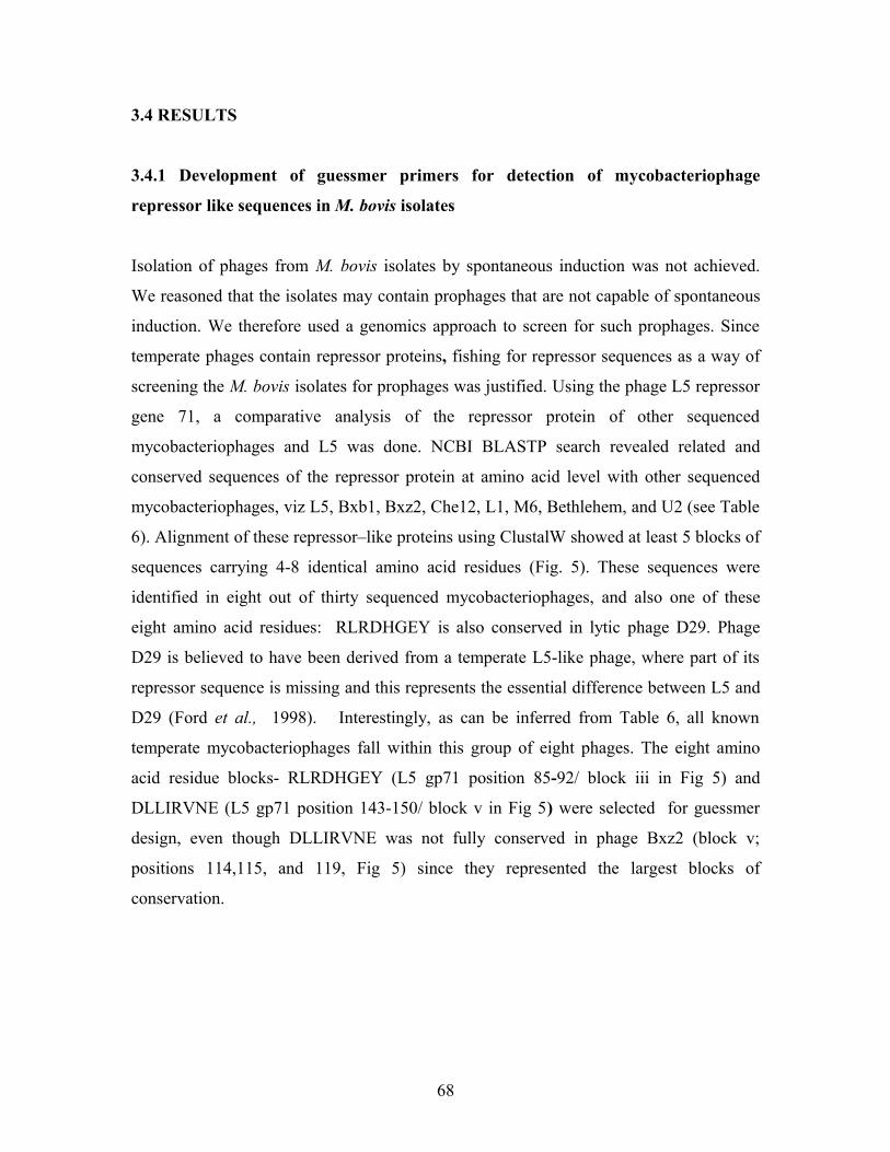
3.4 RESULTS
3.4.1 Development of guessmer primers for detection of mycobacteriophage
repressor like sequences in M. bovis isolates
Isolation of phages from M. bovis isolates by spontaneous induction was not achieved.
We reasoned that the isolates may contain prophages that are not capable of spontaneous
induction. We therefore used a genomics approach to screen for such prophages. Since
temperate phages contain repressor proteins, fishing for repressor sequences as a way of
screening the M. bovis isolates for prophages was justified. Using the phage L5 repressor
gene 71, a comparative analysis of the repressor protein of other sequenced
mycobacteriophages and L5 was done. NCBI BLASTP search revealed related and
conserved sequences of the repressor protein at amino acid level with other sequenced
mycobacteriophages, viz L5, Bxb1, Bxz2, Che12, L1, M6, Bethlehem, and U2 (see Table
6). Alignment of these repressor–like proteins using ClustalW showed at least 5 blocks of
sequences carrying 4-8 identical amino acid residues (Fig. 5). These sequences were
identified in eight out of thirty sequenced mycobacteriophages, and also one of these
eight amino acid residues: RLRDHGEY is also conserved in lytic phage D29. Phage
D29 is believed to have been derived from a temperate L5-like phage, where part of its
repressor sequence is missing and this represents the essential difference between L5 and
D29 (Ford et al., 1998). Interestingly, as can be inferred from Table 6, all known
temperate mycobacteriophages fall within this group of eight phages. The eight amino
acid residue blocks- RLRDHGEY (L5 gp71 position 85-92/ block iii in Fig 5) and
DLLIRVNE (L5 gp71 position 143-150/ block v in Fig 5) were selected for guessmer
design, even though DLLIRVNE was not fully conserved in phage Bxz2 (block v;
positions 114,115, and 119, Fig 5) since they represented the largest blocks of
conservation.
68

Table 6: Mycobacteriophage with identical repressor–like proteins, their origin and
function.
Mycobacteriophage Gene
product
Function of protein e-value Phage Lifestyle
L5 gp71 Phage repressor protein 2e-97 TemperateL1 cIts391 Temperature sensitive
repressor
1e-96 Temperate
MS6 gp6 Phage repressor protein* 2e-29 TemperateU2 gp68 Unassigned 2e-28 ND**Che12 gp80 Phage repressor* 4e-67 TemperateBxz2 gp74 Unassigned 6e-26 TemperateBxb1 gp69 Phage repressor* 2e-28 TemperateBethlehem Gp77 Unassigned 2e-28 ND** * , Putative; **, not determined
69

Fig 5: The alignment of the mycobacteriophage GP71 (L5) repressor-like amino acid sequences. The
amino acid sequences of the repressor proteins of Bxb1, L1, L5, Che12, Ms6, Bethlehem and U2 were
aligned by DNAMAN program (ClustalW). The blocks carrying identical, or nearly identical, amino acids
are shaded in black. The different phages are on the left, and the numbers on the right of the figure
represent the position of the amino acids within the repressor-like sequences. The numbers I, ii, iii, iv, v
show blocks of four or more identical amino acids.
70
31Bxb1 31Bethlehem 31U2 31Ms6 0Bxz2 39L1 39L5 39Che12
Consensus
.........MRTTREQLPRLSLEVIEALKATGETEADIAR
.........MRTTREQLPRLSLEVIEALKATGETEADIAR
.........MRTTREQLPRLSLEVIEALKATGETEADIAR
.........MRTTREHLPRLSLEVIEALKAAGETEADIAR
........................................MSGKIQHKAVVPAPSRIPLTL.SEIEDLRRKGFNQTEIAEMSGKIQHKAVVPAPSRIPLTL.SEIEDLRRKGFNQTEIAEMSGKMQNPARQQASSRQPL.IPSVIEDLRRKGYNQSEIAD
MMMM
VV
RRRR
TTTT
TTTT
RRRR
EEEE
LLLL
II
PPPP
PPP
RRRR
LLLL
SSSS
LLLL
EEEE
VVVV
V
IIII
III
EEEE
EEE
AAAA
LLLL
LLL
KKKK
RRR
AAAA
GGGG
GGG
EEEE
TTTT
EEEE
QQQ
AAAA
DDDD
EEE
IIII
III
AAAA
AAA
RRRR
71Bxb1 71Bethlehem 71U2 71Ms6 40Bxz2 79L1 79L5 79Che12
Consensus
MYGVTPQAVSWHVHTYGGKLTARQVIRREYPFKVPEPLSQMYGVTPQAVSWHVHTYGGKLTARQVIRREYPFKVPEPLSQMYGVTPQAVSWHVHTYGGKLTARQVIRREYPFKVPEPLSQMYGVTPQAVSWHVHTYGGKLTDRQVIRREYPFKVPEPLSQMYGVTRQYVSWIKHTYGGRLTPREEVLKEFPFDVPRNMGQLYGVTRQAVSWHKKTYGGRLTTRQIVQQNWPWDTRKPHDKLYGVTRQAVSWHKKTYGGRLTTRQIVQQNWPWDTRKPHDKMHGVTRQAVSWQKKTYGGRLTTRQIVQQAWPWKTGKGHDK
MMMMMLLM
YYYYYYY
g
GGGGGGGGv
VVVVVVVVt
TTTTTTTT
PPPP
q
QQQQQQQQ
AAAA
AAAv
VVVVVVVVs
SSSSSSSSw
WWWWWWWW
HHHH
HHQ
KKKK
HHHHH
t
TTTTTTTTy
YYYYYYYYg
GGGGGGGGg
GGGGGGGG
KKKKRRRRl
LLLLLLLLt
TTTTTTTT
r
RRRRRRRR
QQQQEQQQ
VVVV
III
IIIIVVVV
RRRR
RRRRK
EEEEENN
YYYYFWWWp
PPPPPPPP
FFFFFWWW
KKKK
K
VVVVV
PPPPP
EEEE
PPPP
PP
LLLLM
SSSSG
QQQQQ
110Bxb1 110Bethlehem 110U2 110Ms6 79Bxz2 119L1 119L5 119Che12
Consensus
CTPHKRLRDHGEYIATRGK.GMKEYKLKRLRSFYRMLRENCTPHKRLRDHGEYIATRGK.GMKEYKLKRLRSFYRMLRENCAPHKRLRDHGEYIATRGK.GMKEYKLKRLRSFYRMLRENCAPHKRLRDHGEYIATRGK.GMKDYKLKRLRSFYRMLRENTSPFKRLRDHGEYVATGGV.GMPDEKLKRLRSFYRKLRDESKAFQRLRDHGEYMRVGSFRTMSEDKKKRLLSWWKMLRDDSKAFQRLRDHGEYMRVGSFRTMSEDKKKRLLSWWKMLRDDAKGYQRLRDHGEYMRVGSFRTMSEDKEKRLKSWWKMLRDN
CCCC
PPPPPAA
HHHH
KKKKK
r
RRRRRRRRl
LLLLLLLLr
RRRRRRRRd
DDDDDDDDh
HHHHHHHHg
GGGGGGGGe
EEEEEEEEy
YYYYYYYY
IIIIVMMM
AAAAA
TTTTTGGGG
GGGGGSSS
KKKK
GGGGG
m
MMMMMMMM
KKKK
EEEDDEEE
YYYY
k
KKKKKKKK
LLLLL
k
KKKKKKKKr
RRRRRRRRl
LLLLLLLL
RRRRR
Ks
SSSSSSSS
FFFFFWWW
YYYYYWWW
RRRRRKKK
MMMM
MMMl
LLLLLLLLr
RRRRRRRR
EEEEDDDD
NNNNEDDN
148Bxb1 148Bethlehem 148U2 148Ms6 119Bxz2 156L1 156L5 157Che12
Consensus
NWVVEFDPNIPPIPGVSKRGGWAYRERQ.ESDED.LLIRVNWVVEFDPNIPPIPGVSKRGGWAYRERQ.ESDED.LLIRVNWVVEFDPNIPPIPGVSKRGGWAYRDRQ.ESDED.LLIRVNWVVEFDPNIPPIPGVSKRGGWAYRERQ.ESDED.LLIRVGLVLEFDPDIPPIDGVSAQGGWRYVEATPEERESGILIREDLVLEFDPSIELYEGMAG.GGFRYVPRG.IE.DDDLLIRVDLVLEFDPSIEPYEGMAG.GGFRYVPRG.IE.DDDLLIRVDWVLEFDPNIPPTPGVAPHGGFRYVTR..TEADDDLLIRV
NNNN
DDD
WWWWLLLWv
VVVVVVVV
VVVVLLLLe
EEEEEEEEf
FFFFFFFFd
DDDDDDDDp
PPPPPPPP
NNNND
Ni
IIIIIIII
PPPPP
P
PPPPP
PP
IIIII
PPPP
Pg
GGGGGGGG
VVVVVMMV
SSSSS
KKKK
RRRR
Hg
GGGGGGGGg
GGGGGGGG
WWWWWFFF
AAAA
y
YYYYYYYY
RRRR
EEDEE
RRRR
RRR
QQQQ
EEEEEEEEE
DDDD
EEEEEDDD
DDDD
DDD
LLLLILLLl
LLLLLLLLi
IIIIIIIIr
RRRRRRRR
VVVV
VVV
170Bxb1 170Bethlehem 170U2 169Ms6 140Bxz2 183L1 183L5 182Che12
Consensus
NEYTTLSEIGRHHIWRFPSVEPNEYTTLSEIGRHHIWRFPSVEPNEYTTLSEIGRHHIWRFPSVEPNEYTTLSEIG.HHIWRFPSVEPNEHSRITQKG.RMIWRFPPREPNEHTNLTAEG.ELLWSWPDDIEELLSEPNEHTNLTAEG.ELLWSWPDDIEELLSEPNEYTNLTDEG.EMLWCWPPDIEEILTn
NNNNNNNNe
EEEEEEEE
YYYY
Y
TTTT
TTT
TTTT
LLLLILLL
SSSS
EEEEQ
D
IIII
g
GGGGGGGG
HHHHR
HHHH
IIIIILLLw
WWWWWWWW
RRRRR
FFFFFWWWp
PPPPPPPP
SSSS
VVVV
EEEEE
PPPPP
i ii
iii
iv v

Table 7 shown below, represents the derived nucleotide sequences which was used to
design the following guessmer sequences, Forward:
MGVCTNMGDATCAYGGCGARTAC and Reverse:
TTCGTTSACYCKGATSARBAGRTC based on the resulting DNA consensus
sequences shown in Table 7.
71
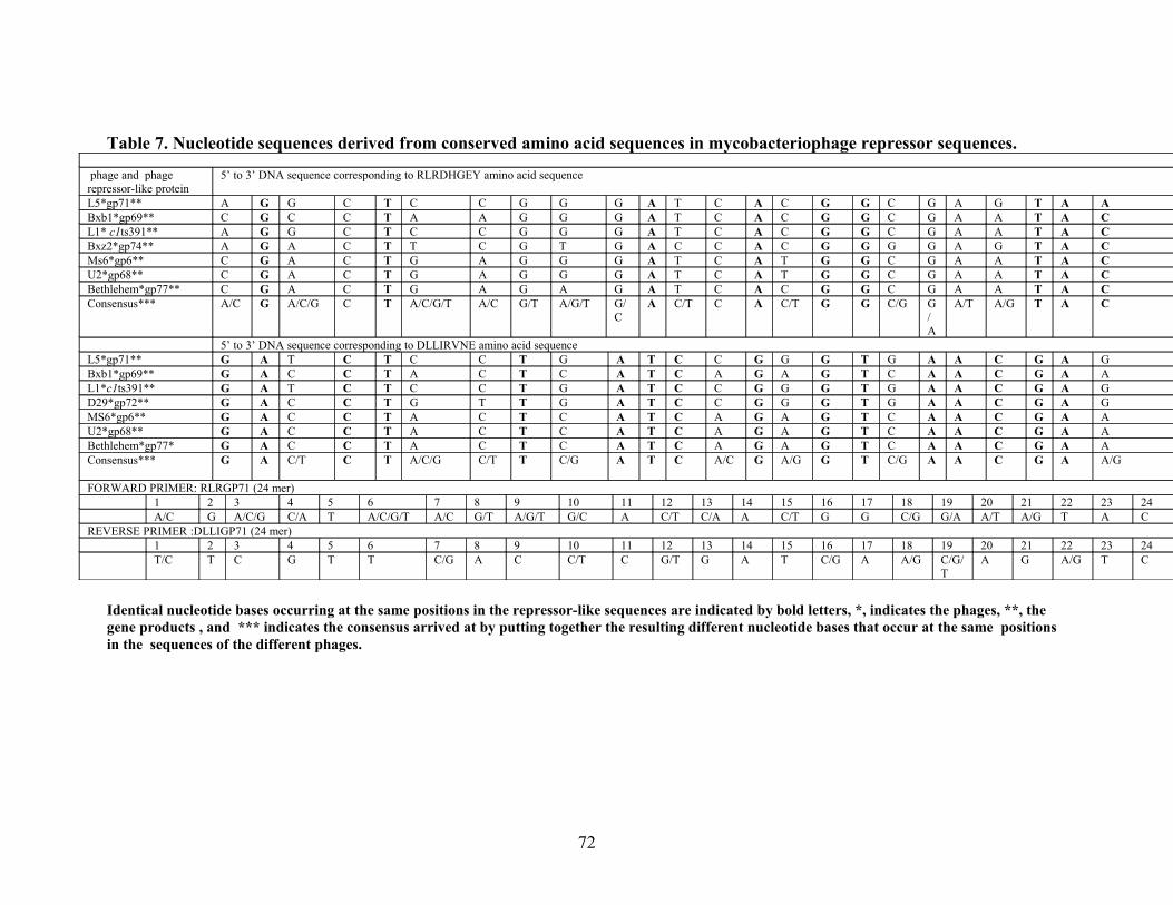
Table 7. Nucleotide sequences derived from conserved amino acid sequences in mycobacteriophage repressor sequences.
phage and phage repressor-like protein
5’ to 3’ DNA sequence corresponding to RLRDHGEY amino acid sequence
L5*gp71** A G G C T C C G G G A T C A C G G C G A G T A ABxb1*gp69** C G C C T A A G G G A T C A C G G C G A A T A CL1* c1ts391** A G G C T C C G G G A T C A C G G C G A A T A CBxz2*gp74** A G A C T T C G T G A C C A C G G G G A G T A CMs6*gp6** C G A C T G A G G G A T C A T G G C G A A T A CU2*gp68** C G A C T G A G G G A T C A T G G C G A A T A CBethlehem*gp77** C G A C T G A G A G A T C A C G G C G A A T A CConsensus*** A/C G A/C/G C T A/C/G/T A/C G/T A/G/T G/
CA C/T C A C/T G G C/G G
/A
A/T A/G T A C
5’ to 3’ DNA sequence corresponding to DLLIRVNE amino acid sequenceL5*gp71** G A T C T C C T G A T C C G G G T G A A C G A GBxb1*gp69** G A C C T A C T C A T C A G A G T C A A C G A AL1*c1ts391** G A T C T C C T G A T C C G G G T G A A C G A GD29*gp72** G A C C T G T T G A T C C G G G T G A A C G A GMS6*gp6** G A C C T A C T C A T C A G A G T C A A C G A AU2*gp68** G A C C T A C T C A T C A G A G T C A A C G A ABethlehem*gp77* G A C C T A C T C A T C A G A G T C A A C G A AConsensus*** G A C/T C T A/C/G C/T T C/G A T C A/C G A/G G T C/G A A C G A A/G
FORWARD PRIMER: RLRGP71 (24 mer)1 2 3 4 5 6 7 8 9 10 11 12 13 14 15 16 17 18 19 20 21 22 23 24A/C G A/C/G C/A T A/C/G/T A/C G/T A/G/T G/C A C/T C/A A C/T G G C/G G/A A/T A/G T A C
REVERSE PRIMER :DLLIGP71 (24 mer)1 2 3 4 5 6 7 8 9 10 11 12 13 14 15 16 17 18 19 20 21 22 23 24T/C T C G T T C/G A C C/T C G/T G A T C/G A A/G C/G/
TA G A/G T C
Identical nucleotide bases occurring at the same positions in the repressor-like sequences are indicated by bold letters, *, indicates the phages, **, the gene products , and *** indicates the consensus arrived at by putting together the resulting different nucleotide bases that occur at the same positions in the sequences of the different phages.
72

3.4.2 Amplification of L5 gp71 using guessmer primers, and PCR analysis
A L5-M.smegmatis mc2155 DNA was used to evaluate the guessmer primers. As can be
seen from Fig. 6, the PCR yielded a DNA band that appeared to be of the size anticipated
for the L5 gp71 (222bp), and the water control did not amplify. A M. smegmatis mc2155
wild type control was employed later when the M. bovis isolates were assessed. This
control also did not amplify as can be seen in Fig 7b, lane 2. The PCR product from the
L5-M. smegmatis mc2155 was sequenced and BLAST analysis revealed it to be 100%
identical to the region encompassing the conserved sequences in the gp71 L5 repressor
sequence.
3.4.3 Screening of M. bovis isolates for prophages using guessmer primers
Since we were able to amplify the correct L5 gp71 sequence using the L5-M.smegmatis
lysogen we then screened M. bovis isolates using the guessmer primers to evaluate the
presence of potential prophages. Out of 45 M. bovis isolates screened for prophage
repressor sequences, 9 produced amplification products (Fig 7a and 7b). As can be seen
in Fig 7a, 7b and Table 8, the amplicons from each positive sample produced DNA bands
of different sizes: ~1000bp, ~600bp, ~420bp, and ~220bp. Given that only 9 out of 45
isolates amplified, we investigated whether this was strain specific or whether it was due
to insufficient DNA content in the other 36 samples that did not amplify. Therefore to
confirm the quality and mycobacterial status of the M. bovis DNA templates that were
screened for prophages by guessmer PCR, we used hsp65 genus-specific primers which
is a genus-specific marker of mycobacteria (Telenti et al., 1993). As can be seen from
Fig. 8a and 8b, amplification using these primers resulted in DNA bands that appeared to
be of the expected size of 439bp (gel a, lanes 3-8; gel b, lanes 2-9 and gel c, lanes 1-4,
and 7-8 ) and were identical to the M. smegmatis positive control (gel b, lane 9).
Importantly a control lacking added template failed to amplify (Fig 8a, lane 1).
73

All 9 of the guessmer positive templates also yielded positive hsp65 amplicons (Fig 7
and 8; Table 8). Twenty isolates that were guessmer negative appeared to be hsp65
positive. We also noted that 11 isolates that did not amplify with the guessmers also did
not amplify with the hsp65 (Table 8), and we reasoned that in these samples there was
insufficient DNA of good quality. All guessmer positive products yielded a similar
amplicon profile that importantly was missing from both the no template control (Fig 7b;
lane 1) as well as the M. smegmatis mc2155 control (Fig 7b; lane 2). This amplification
profile is not common to all M. bovis isolates as not all hsp65-positive showed these
amplification products, suggesting perhaps some strain specific activity.
74
1 2 3
Fig 6: Amplification of the L5-GP71.PCR product of the L5-Msmegmatis lysogen .Lanes1=L5-lysogen,2=H
2O, 3=50bp O’Gene Ruler.
1031bp
500bp
200bp

75
Fig 7 A: Screening of M. bovis isolates for cryptic prophages using guessmer PCR. PCR Products were ran on 1.5% TAE agarose gel. Lanes 1-7= isolates: TB5109B, TB1066+, TB4931C, TB5149E, TB4760C, TB5151A, TB4812C, 8=50bp O’range Ruler
A
1031bp
500bp
200bp
1 2 3 4 5 6 7 8
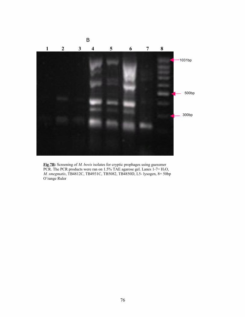
76
1 2 3 4 5 6 7 8B
1031bp
500bp
300bp
Fig 7B: Screening of M. bovis isolates for cryptic prophages using guessmer PCR. The PCR products were ran on 1.5% TAE agarose gel. Lanes 1-7= H2O, M. smegmatis, TB4812C, TB4931C, TB5082, TB4850D, L5- lysogen, 8= 50bp O’range Ruler

Fig8.:PCR based confirmation of the mycobacterial status of the M. bovis isolates using Hsp65 primers. PCR products were ran on a 1.5% agarose gel. A, Lane 1= H2O, lanes 2-9=isolates: TB5150, TB5149D, TB5952A, TB3867A,Tb 5149E, TB1755C, TB 3569,TB4760C, Lane 10= O’range DNA Ladder. B, Lane 1=H2O, Lanes 2-9=Isolates: TB499OC, TB4850D, TB4935, TB5149, TB5122, TB5751A, TB4931C, M. smegmatis mc2155, lane 10= 50bp O’range DNA Ladder. C, Lanes 1-9= TB4812J, TB3107A, TB5234A, TB3869G, TB1158Mtb, TB1066+, TB3522, TB4992B, TB4675, Lane 10= 50bp O’range DNA Ladder.
77

Table 8: Summary of PCR based screening of M. bovis isolates for prophages, and
hsp65 mycobacterial confirmation of the isolates.M.bovis isolate number Tissue origin and animal
species Animal Origin Guesmer
PCRHsp 65 PCR
TB4850D Thorax- Buffalo KNP (SA) + +TB4759C Mesentric gland- Bovine Ladysmith (SA) - +TB5149D Mandibles-Buffalo KNP (SA) + +TB4992B Thorax-Buffalo KNP (SA) - -TB4675 Mesentric gland-Porcine Ladysmith (SA) - -TB4893A Spleen- Monkey Mauritius - +TB4760C Medistal -Bovine Ladysmith (SA) - +TB4812J Axil-Buffalo KNP (SA) - +TB4804A Left Retropharyngeal-Buffalo KNP (SA) - +TB4860F Lung-Buffalo KNP (SA) - -TB4933A Left Retropharyngeal- Buffalo KNP (SA) - +TB4804F Thorax- Buffalo KNP (SA) - -TB4990C Carcass- Buffalo KNP (SA) - -TB4816F Thorax+ Lung- Buffalo KNP (SA) - +TB4812C Left Tonsil- Buffalo KNP (SA) - +TB4862B Right Retro-Buffalo KNP (SA) - -TB4779A Lung+ Bronchi- Bovine Ladysmith (SA) - -TB4865A Left Retropharyngeal- Buffalo KNP (SA) - +TB5149E Lung - Buffalo KNP (SA) - +TB4931C Left Retropharygeal-Buffalo KNP (SA) + +TB5149C Bone Marrow-Buffalo KNP (SA) + +TB1066B+ No record - +TB5151A Head- Buffalo KNP (SA) + +TB5109B Thorax- Buffalo KNP (SA) + +TB5082A Lung- Lion Hoedspruit (SA) + +TB5150A Head- Buffalo KNP (SA) - -TB1158Mtb No record - -TB1158Tfa No record - -TB3197 No record + +TB3569G No record - +TB5123D Left Retropharyngeal- Buffalo KNP (SA) + +TB4952C Sternum+ Lung-Buffalo KNP (SA) - +TB3670 No record - +TB5234B Muscle- Lion KNP (SA) - +TB5122A Head- Buffalo KNP (SA) - +TB4893D Mesentric gland- Monkey Mauritius - NdTB3869G No record - +TB1071B No record - NdTB1755C No record - +TB3107A No record - +TB3522 No record - +TB3534 No record - NdTB4935D Right tonsil-Buffalo KNP (SA) - +TB4936A Right Retropharyngeal- Buffalo KNP (SA) - NdTB4975B Spleen- Buffalo Cascades (SA) - -+= There was amplification, -= no amplification, Nd= PCR not done. KNP= Kruger National Park,
SA= South Africa,
78

3.4.4 Cloning and sequencing of the PCR products
In order to determine if our guessmer PCR amplified DNA were of prophage origin, we
determined the DNA sequences of some of the amplicons. We could not obtain readable
sequence information by sequencing directly with the guessmer primers, even though this
worked for PCR product derived from the L5-M. smegmatis lysogen (section 3.4.2). We
decided therefore to first clone the PCR products from the M. bovis DNA to enable the
use of the primer sites flanking the vector, for sequencing.
Since the DNA banding patterns of the M. bovis isolates tested for prophages appeared to
be similar, only two samples, namely TB 4850D and TB5151 (Fig7 , gel B, lane 6 and
Fig 7 gel A, lane6 respectively) were selected for cloning and subsequent sequencing of
the cloned insert. Four DNA bands from sample TB4850D (DNA samples of
approximately 1000bp, 600bp, 390bp and 220bp respectively), and one DNA band from
TB5151 (approximately 420bp in size) were selected for cloning and were separately
ligated into pGEM T- Easy vector as described in section 3.3.5.2. Transformants were
selected and analysed from each ligation, and were digested with EcoR1 which should
release the insert (see Appendix C) and puC18 plasmid was employed as a control for the
restriction digestion (9a). EcoR1 restriction enzyme digestion of the plasmid DNA from
different clones derived from the same ligation yielded different sized inserts (Fig 9a and
9b). This may have resulted from mixing of the DNA bands during DNA gel extraction
(section 3.3.3). Four clones from TB5151 (with approximate insert sizes: 420bp, 420bp,
420bp and 570bp respectively) and seven clones from TB4850D (with approximate insert
sizes 500bp, 640bp, 680bp, 930bp, 630bp, 250bp and 300bp respectively) were sent for
sequencing. The 220bp cloned PCR products from TB4850D (Results no shown) and that
of the L5-M.smegmatis mc2155 lysogen control (Fig 9a,) could not be observed from the
agarose gels following EcoR1 digestion and were not sent for sequencing. However the
sequence of the PCR product L5-M.smegmatis mc2155 lysogen was already determined
to be of the L5 GP71 origin (section 3.4.2).
79

Fig 9a: Restriction products of the plasmid DNA cloned with the PCR products from the guessmer PCR, dgested with EcoR1 showing the isolates , their PCR product sizes and the insert sizes. L5 lysogen indicates L5- M. smegmatis mc2155 lysogen. MWM= Molecular Weight Marker (50bp O’ range ruler.
80

1000bp
Fig 9b: Restriction products of the plasmid DNA cloned with DNA from guessmer PCR, digested with EcoR1 enzymes, showing isolate number, the approximate size of the PCR product and the insert sizes. MWM indicates Molecular Weight Marker (50bp O’ range Ruler)
81

Table 9: EcoR1 restriction enzyme digestion of plasmid DNA from different clones,
showing insert sizes and the original PCR product sizes.
Isolate number and
approximate size of the
PCR product cloned
Insert size (bp) Insert size
(bp)
Insert size
(bp)
Insert size
(bp)
Average
insert size
(bp)TB 5151 (420bp*) 420 420 420 570 457.5TB 4850D (1000bp*) 500 - - - 500TB 4850D (600bp*) 640 680 930 630 720TB 4850D (390bp*) - 250 300 - 275TB 4850D(220bp*) - - - - -L5-M.smegmatis
mc2155 lysogen
(220bp)*
- - - - -
- indicates that no DNA insert was observed, *=size of the original PCR product cloned.
3.4.5 Sequence analysis
In order to identify sequences similar to those that we had amplified and sequenced, we
searched the NCBI database for similar sequences using the NCBI BLASTX search
algorithm, which would allow us to search the translated database. As can be seen in
Table 10 and Appendix D, several proteins were identified and all of which were of
mycobacterial origin, and had the lowest e-values against M. bovis proteins, confirming
that our cloned PCR products were of Mycobacterium bovis origin. These proteins
include probable DNA helicases (Mb0884), transcriptional regulator (Mb1160c),
probable IS1533-derived transposases (Mb2968); a hypothetical protein (Mb1618c); a
CTP synthetase (Mb1725), and a spermidine synthetase (Mb2632) (BLASTX results:
Appendix D). Interestingly the majority of these proteins, i.e. DNA helicases (Mb0884),
transcriptional regulator (Mb1160c), probable IS1533-derived transposases (Mb2968) are
DNA binding of the helicase superfamily II, of the xenobiotic responsase element family
of the transcriptional regulators, and of the intergrases respectively
(www.ncbi.nlm.nih.gov/entrez). As can be seen in Appendix D these proteins showed
some identical sequences at amino acid level to the region between the sequences
82

RLRDHGEY and DLLIRVNE and interestingly as can be seen in Appendix D, some of
these sequences were identical to the primer sequences used. The CTP synthetase protein
showed a 75% and 50% identity to the DLLIRVNE, and RLRDHGEY guessmer
sequences respectively. The transcriptional regulator and the DNA helicase proteins
revealed 50% and 37.5% identity to the DLLIRVNE sequence respectively. The
hypothetical protein sequence (Mb1618c) showed a 37.5% and 25% identity to
RLRDHGEY and DLLIRVNE respectively. No sequence similarities to the primers
were observed on the spermidine synthetase ((Mb2632), and the IS1553 element.
With the observation that the shared sequence motif in most of these proteins and the
repressor-like proteins of the temperate mycobacteriophages is DLLIRVNE, we also did
a Sanger Institute BLASTX search using the DLLIRVNE sequence against the M. bovis
genome, in order to investigate the conservation of this sequence in the M. bovis genome
(www.sanger.ac.uk). Interestingly this sequence appeared to be also conserved (showing
87.5% conservation) in the glutamine- transport ATP binding abc transporter. A Prosite
database search to see if this sequence was identified previously was done using the
sequence DLLIRVNE as a search tool, and no hit was found indicating that a possible
novel DNA binding motif has been identified (http://www.expasy.ch/prosite/).
83

Table 10: Homolog search results revealing the predicted homolog proteins and their location on the M. bovis genomeIsolate Query
size (bp)Highest BLAST hit Other mycobacteria hit Other genus hit Genomic location Insert
size
(bp)TB4850D(1000bp*)
634 Probable DNA helicase [Mycobacterium.bovis AF2122/97][2e-69][122-538bp**]
Probable DNA helicase ercc3[ Mycobacterium tuberculosis H37Rv][2e-69]
Putative NDA helicase [norcadia farcinica ifm 10152[2e-57]
959285..960904*** ~500
TB4850D(600bp*)
762 Ctp synthetase [Mycobacterium bovis AF2122/97][2e-101][115-672bp**]
Ctp synthetase [Mycoabcterium tuberculosis CDC1551 [4e-101]
Ctp synthetase [rhodococcus sp. Rha1][2e-86]
1909166..1910926*** ~930
TB5151(420bp*)
679 Probable transcriptional regulator protein [Mycobacterium bovis AF2122/97][6e-80]116-571**]
Probable transcriptional regulator protein [Mycobacterium tuberculosis H37Rv][6e-80]
12544447..1255904*** ~420
TB5151(420bp*)
689 Hypothetical protein Mb1618c [Mycobacterium bovis AF2122/97][2e-77][207-376]
Hypothetical protein Mycobacterium tuberculosis CDC1551][2e-77]
1779105..1777768*** ~420
TB4850D(390bp*)
327 Spermidine synthetase [Mycobacterium bovis AF2122/97][2e-9][123-233**]
Hypothetical protein 3 [micropilis demolitor braccovirus][4e-10][2-112]
2896169..2897413*** ~250
TB4850D(390bp**)
561 IS1533 Transposase [Mycobacterium bovis AF2122/97][3e-17][187-312]
IS1533 transposase [Mycobacterium tuberculosis H37Rv]
3245037..3246725*** ~390
* indicates the original PCR product size, ** indicates the location of the query amino acid sequences within the BLAST hit, ***=location of the
proteins in the M. bovis genome
84

3.5 DISCUSSION
Guessmer PCR based genomic screening for M. bovis prophages using primers based on
the conserved sequences of repressor proteins of eight sequenced mycobacteriophages
were shown to amplify uniquely L5 repressor DNA in a L5 lysogen, yielded multiple
bands which occurred in several (30% - of the ones that had good quality hsp65+ DNA)
of the screened M. bovis samples. Sequence analysis of some of these bands revealed
DNA encoding homolog proteins to different M. bovis proteins, some of which had
affinity to DNA binding proteins.
One of the proteins identified in isolates (TB4850D, and TB5151) was a putative
transposase of the insertion sequence family IS1533 (Mb 2968); a putative transcriptional
regulators (Mb1160); a probable DNA helicase (Mb0884); a spermidine synthetase, a
CTP synthatase (Mb1725) as well as a hypothetical protein (Mb1618c) (Appendix D). An
overriding feature of several of the identified proteins is that they are implicated in DNA
binding (transcriptional regulator, IS1553 element, and the DNA helicase). As can be
seen in appendix D, the shared sequence motif is the DLLIRVNE sequence, which is also
conserved in the repressor-like proteins of mycobacteriophages Bxb1, Bxz2, L1, U2
Bethlehem, Che12 and Ms6. DLLIRVNE is also conserved in the phage D29, a lytic
phage that shares over 75% of its amino acid sequences with the L5 phage (Pedulla et al.,
2003). Interestingly 75%, 50%, 37.5%, and 25% of DLLIRVNE is conserved in the
following putative proteins respectively: CTP synthatase (Fig F, Appendix D), the
transcriptional regulator (Fig B, Appendix D), the DNA helicase (Fig D, Appendix D)
and the hypothetical protein (Mb1618c) (Fig K, Appendix D) of the sequences M. bovis
AF2122/97 strain and we predicted that these are the regions where our DLLIRVNE
guessmer primer bound. In addition, the BLASTX search for conservation of
DLLIRVNE in M. bovis, using the conserved DLLIRVNE as a query against M. bovis
AF2122/97 revealed the availability of the 87.5% of DLLIRVNE in probable glutamine-
transport ATP binding abc transporter, which is also DNA binding (www.sanger.ac.uk).
The function of the transcriptional regulator protein (Mb1160c) is not known, but this
protein is perceived to be a specific prokaryotic DNA binding protein belonging to the
85

xenobiotic responsase element family of the transcriptional regulators
(www.sanger.ac.uk;www.ncbi.nlm.nih.gov). This protein is also equivalent to Rv1129 of
M. tuberculosis. The conservation of DLLIRVNE in DNA binding proteins of M. bovis
and mycobacteriophages repressors may suggest that this sequence plays a very
important role in the DNA binding process. This sequence has not been recognized
before as a DNA binding motif in temperate mycobacteriophages. DLLIRVNE is located
at the C terminal domain of the mycobacteriophage repressor proteins and the C-terminal
domain of the temperate mycobacteriophage repressors such as Che12 and L5 is reported
to be responsible for dimerisation of the repressor monomers (Gomathi et al., 2007).
Only the helix turn helix motif located at the N- terminal has been reported as a DNA
binding motif in phages L5 (Donelly-Wu et al., 1993; Gomathi et al., 2007) Che12
(Gomathi et al., 2007), However it is possible that we might have identified a previously
unrecognized DNA binding region.. Another noteworthy observation is its high
conservation in the CTP synthetase, which functions for CTP synthesis; hence we also
think that this protein might be a DNA binding protein.
We could not conclude that this sequence (DLLIRVNE) is of prophage origin, since it is
only a small sequence and the NCBI data base does not contain prophage sequences.
86

3.6 CONCLUSION
In this chapter, our aim was to screen for mycobacteriophage related sequences, in M.
bovis isolates using conserved sequences from repressor proteins of different
mycobacteriophages. We were aware that the use of these sequences from repressor
proteins could be limiting as these phages with almost identical repressors seem to have a
common ancestry. However since all sequenced temperate phages known to us belong to
this group of 8/30 mycobacteriophage population, that seem to have identical repressor
sequences, with 2 blocks of 8 amino acids that is conserved, we reasoned that we could
use these sequences to look for their conservation in M. bovis prophages. We decided to
use repressor proteins as they are the hallmark of temperate phages instead of integrases,
which are enormous in many bacterial genomes.
Guessmers were designed from the two conserved sequences and M. bovis isolates were
screened for prophages using the designed guessmer primers. The BLAST results of the
sequences from the PCR products from this screen revealed different proteins of
mycobacterium origin, in particular M. bovis. Another noteworthy observation was that
some of these proteins were DNA binding. We also noted the conservation of the
DLLIRVNE sequence in these proteins which is also conserved in the
mycobacteriophages repressor-like sequences, and hence we came to a conclusion that
this sequence might play a very important role in DNA binding. Hence we also think that
the CTP synthetase of M. bovis in which this sequence is also very highly conserved
might have DNA binding affinity. We however could not conclude that this sequence
(DLLIRVNE) is of prophage origin, since there are no prophage sequences on the NCBI
database, and an in depth analysis of the whole M. bovis genome would be necessary to
confirm the origin of this sequence. Our conclusion therefore is that prophage sequences
were not identified in the M. bovis isolates, but a novel possible DNA binding motif
present in a number of proteins, indicating that they all bind DNA as part of their
function, has been identified.
87

CHAPTER FOUR: CONCLUDING REMARKS
The long term aim of this research was to determine the contribution of phages to the
virulence and pathogenicity of pathogenic M. bovis. The short term aim, which was the
aim of this study, was to isolate inducible or cryptic phages from M. bovis isolates and
characterize them. In this chapter we present the summary of the results and concluding
remarks.
Since the low frequency of spontaneous phage induction from M. bovis as shown by
Tageldin et al., (1981), and no further reports of phage isolation from this bacillus, it was
necessary for us to optimize methods for phage isolation, using a well-known phage
system. The study of the effect of co-cultivation of a lysogenic M. smegmatis mc2155 and
its wild type strain was motivated by the fact that most studies have shown that
spontaneous phage induction is very rare, occurring at a rate of 102-105 in a 109 bacterial
population, and the situation was proven different in Salmonella co-cultures. The result
we obtained with M. smegmatis co-cultures showed no significant difference in the rate
of phage release between the lysogen co-culture with wild type, and the lysogen control
cultivated clonally. We reasoned that this might be due to L5 forming stable lysogens
with M. smegmatis.
The detection of phage particles from solid lysogenic M. smegmatis mc2155 culture by
epiflourescence microscopy was motivated by the fact that some studies have shown that
plaque assay methods are biased, selecting only phages that can infect that sensitive
strain, leading to the detection of only a tiny fraction of phages, compared to non-
selective microscopic methods. Epiflourescence microscopy is this study was shown to
be more sensitive and feasible for phage detection in solid cultures requiring a titer of at
least 102 pfu/ml to detect twice the number of individual phage particles detected by
plaque assay.
Screening for spontaneously induced phages from M. bovis isolates by plaque assay using
5 different bacterial indicator strains was not achieved. In addition screening for
88

spontaneously induced phages from M. bovis isolates using a non-selective
epiflourescence microscopy, only revealed a viral like spot in one out of sixteen isolates.
However this result alone, without any supporting evidence from Transmission electron
microscopy and phage DNA isolation could not be used to conclude the presence of a
spontaneously induced phage in that isolate, since only one spot thought to be an artifact
was observed. Our conclusion therefore, based on plaque assay results and
epiflourescence microscopy, is that spontaneous phage induction in these isolates did not
occur.
The genomics approach to screen for the presence of cryptic phages using guessmers
designed from conserved regions in the mycobacteriophage repressor-like proteins was
motivated by the fact that, repressors are a hallmark of temperate phages. We came to the
realization that these phages sharing repressor-like sequences might actually be related or
come from a common ancestor. We also noted that characterized temperate
mycobacteriophages fall in this group of eight phages, which also made us to think that
may be all known temperate mycobacteriophages have identical repressors.
Amplification of the M. bovis DNA by the guesmers, followed by cloning and
sequencing of the PCR products was done. The obtained sequences were homologs of
different proteins in the sequenced M. bovis genome. Analysis of these sequences
revealed that they were DNA binding proteins, with sequences like DLLIRVNE or part
of, which was shared by the L5 and the other related mycobacteriophages. The function
of this sequence was not obvious. This sequence is highly conserved in temperate
mycobacteriophages and available in the obtained DNA binding protein sequences
including and a probable glutamine- transport ATP binding abc transporter protein, and a
CTP synthatase of the sequenced M. bovis genome, hence we concluded that this
sequence might play an important role in DNA binding. In order to conclude that these
obtained sequences in the M. bovis isolates in particular DLLIRVNE is of prophage
origin, a more in depth analysis of the whole genome is needed.
89

References
Aldovini, A., Husson, R.N., and Young, R.A. (1993). The uraA locus and homologous
recombination in Mycobacterium bovis BCG. Journal of bacteriology. 175: 7282-7289.
Aldwell, F.E., Wedlock, D.N., Buddle, B.M. (1997). Sequential activation of alveolar
macrophages by IFN [gamma] and LPS is required for enhanced growth inhibition of
virulent Mycobacterium bovis but not M. bovis BCG. Immunology and Cell Biology. 75:
161-166.
Ainsa, J.A., Martin, C., and Gicquel, B. (2001). Molecular approaches to tuberculosis.
Molecular Microbiology. 42: 561-570.
Artois, M., Delahay, R., Guberti, V., and Cheeseman, C. ( 2001). Control of Infectious
disease of wildlife in Europe. Veterinary Journal. 162: 141-157.
Asherford, K.E., Day, M.J., Fry, J.C. (2003). Elevated abundance of bacteriophage
infecting bacteria in soil. Applied Environmental Microbiology, 69: 285-289.
Atsumi, S., and Little, J.W. (2004. Regulatory circuit design and evolution using phage
lambda. Genes and development. 18: 2086-2094.
Baba, T., Takeuchi, F., Kuroda, M., Ito, I., Yuzawa, H., and Himaratsu, K. (2004).
The Staphylococcus aureus genome. Horwood publishing, Chichester, United Kingdom.
Balasubramanian, V., Pavelka, M.S., Bardarov, S.S., Martin, J., Weisbrod, T.R.,
McAdam, R.A., Bloom, B.R., and Jacobs, W.R. Jnr. (1996). Allelic exchange in
Mycobacterium tuberculosis with long linear substrates. Journal of Bacteriology. 178:
273-279.
90

Barlow, N.D.(1993). A model for the spread of bovine tuberculosis in New Zealand
possum populations. Journal of Applied Ecology. 28: 794-809.
Bardorov, S., Kriakov, J., Carriere, C., et al. (1997). Conditionally replicating
mycobacteriophages: a system for transposon delivery to Mycobacterium tuberculosis.
Proceeding of the National. Academy of Science. USA. 94: 10961-10966.
Barsom, E.K., and Hatfull, G. (1996). Characterisation of a Mycobacterium
smegamatis gene that confers resistance to phage L5 and D29 when overexpressed.
Molecular Microbiology . 21: 151-170.
Behr, M.A., Wilson, M.A., Gill, W.P., Salamon, H, Achoonik, G.K., Rane, S., et al.,
(1999). Comparative Genomics of BCG vaccines by whole genome DNA microarray.
Science. 284: 1520-1523.
Bengis, R.G., Kriek, N.P.J., Keet, D.F., Raat, J.P., de Vos, V., Huchzermeyer, H.
(1996). An outbreak of tuberculosis in a free living African buffalo (Syncerus caffer
SPARRMAN) population in Kruger National Park: a preliminary report. Onderstepoort
Journal of Veterinary Research. 63: 15-18.
Berg, O., Børsheim, K.Y., Bratbak,G., Heldal, M. (1989). High abundance of viruses
found in aquatic environments. Nature 340: 467-468.
Bigi, F., Gioffre, A., Klepp, L., Santangelo, M.P., Velisscovsky, C.A.,
Giambartolomei, G.H., Fossati, C.A., Romano, M.L., Mendum, T., McFadden, J.J.,
Cataldi, A. (2005). Mutation in P36 gene Mycobacterium bovis provokes the attenuation
of the bacillus in a mouse model. Tuberculosis, 85: 221-226.
Böddinghaus, B., Rogall, T., Blöcker, H., Flohi, T and Böttger, E.E (1990). Detection
and identification of mycobacteria by amplification of rRNA. Journal of Microbiology.
28: 1751-1759.
91

Børshei, K.Y., Bratbak,G., Heldal, M. (1990). Enumeration and biomass estimation of
planktonic bacteria and viruses by transmission electron microscopy. Applied
Environmental Microbiology. 56: 352-356.
Bossi, L., Fuentes, J.A., Mora, G., and Figueroa-Bossi, N. (2003). Prophage
contribution to bacterial population dynamics. Journal of Bacteriology, 185: 6467-6471.
Bowman, B.U Jnr., and Redmond, W.B. (1958). A temperate bacteriophage from
Mycobacterium. Butyricum. Bacteriology Proceedings. 233-238.
Brosch , R., Gordon, S.V., Marmiesse, M., Brodin, P., Buchrieser, C., Eiglmeir, K.,
Garnier, T., Gutierrez, C., Hewiston, G., Kremmer, K., Parsons, L.M., Pym, A.S.,
Samper, S., van Sooligen, D., and Cole, S.T. (2002). A new evolutionary scenario for
the Mycobacterium tuberculosis complex. Proceedings of the. National. Academy of
Science. USA. 99: 3684-3689.
Braden, C.R., Morlock, G.P., Woodley, C.L., Johnson, K.R., Colombel, A.C., Cave,
M.D., Yang, Z., Valway, S.E., Onorato, I.M., and Crawford, J.t. (2001).
Simultaneous infection with multiple strains of Mycobacterium tuberculosis. Clinical
Infectious Diseases. 33: 42-47.
Bradley, D.E. (1967). Ultrastructure of phages and bacteriocins. Bacteriology Reviews.
31: 230-314.
Brook, P.C., Movahedzadeh, F., and davis, E.O. (2001). Identification of some DNA
damage- inducible genes in Mycobacterium tuberculosis: apparent lack of correlation
with LexA binding. Journal of Bacteriology. 183: 4459-4467.
92

Brüssow, H., Canchaya, C., Hardt, W.D. (2004). Phages and the evolution of the
bacterial pathogens. From genomic arrangements to lysogenic conversion.
Microbiological Molecular Biology Reviews, 68: 560-602.
Brüssow, H., and Hendrix, R.W. (2002). Phage genomics: small is beautiful. Cell. 108:
13-16.
Canchaya, C., Proux, C., Fornous, A., Bruttin, A., and Brüssow, H. (2003). Prophage
genomics. Mocrobiology and Molecular Biology Reviews, 67: 238-276.
Cangelosi, G.A., Palermo, C.O., and Bermudez, L.E. (2001). Phenotypic
consequences of red-white colony type variation in Mycobacterium avium. Microbiology.
147: 527-533.
Casjens, S. (2003). Prophage and bacterial genomics: what have we learned so
far?.Molecular Microbiology. 49: 277-300.
Chibani-Chennoufi, S., Sidoti, J., Bruttin, A, Kutter, E., Sarker, S.A., and Brüssow,
H. (2004). Comparison of in vitro and in vivo bacteriolytic activity of Escherichia.
Coliphages: implications for phage therapy. Antimicrobial Agents.48: 2556-2569.
Chen, F., Lu, J.R., Binder, B.J., Liu, Y.C., and Hodson, R.E. (2001). Application of
digital image analysis and flow cytometry to enumerate marine viruses stained with
SYBR gold. Applied Environmental Microbiology, 68: 539-545.
Chopin, A., Bolotin, A., Sorokin, A., Ehlrich, S.D., and Chopin, M.C. (2001).
Analysis of six prophages in Lactobacillus lactis IL1403: different genetic structures of
temperate and virulent phage populations. Nucleic Acids Research. 29: 644-651
Cianciotto, N.P. (1986). Toxin gene carriage by bacteriophage in medically important
bacteria. Clinical Microbiology News. 8: 1
93

Cousins, DV.;Huchzermeyer, HFKA.;Griffin, JFT.;Brueckner, GK.;van Rensburg, IBJ.; Kriek, NPJ. (2004). Tuberculosis. In: Coetzer JAW, Tustin RC. , editor. Infectious Diseases of Livestock. Oxford University Press Southern Africa, Cape Town. pp. 1973–1993.
Cole, S.T., Brosch, R., Parkhill, J., Garnier, T., Churcher, C., Harris, D., Gordon,
S.V., Eiglmeier, K., Gas, S., and Barry, C.E., 3rd, et al., (1998). Deciphering the
biology of Mycobacterium tuberculosis from the complete genome sequence. Nature.
393: 537-544.
Cole, S.T., and Smith D.R. (1994). Toward mapping and sequencing of the genome of
Mycobacterium tuberculosis (Editors: B. R Bloom) In Tuberculosis: Pathogenesis,
Protection, and Control. American Society of Microbiology, Washington DC.
Coleman, J.D., Cooke, M.M. (2001). Mycobacterium bovis infection in wild life in New
Zealand. Tuberculosis (Edinb). 81: 191-202
Collins, C.H., and Grange, J.M. (1987). Zoonotic implications of Mycobacterium bovis
infection. Irish Veterinary Journal. 41: 363-366.
Collins, C.H. (2000). The bovine tubercle bacillus. British Journal of Biomedical
Science. 57: 234-240.
Collins, D.M. (2001). Virulence factors of Mycobacterium bovis. Tuberculosis, 81:
97-102.
Collins, D.M., Kawakami, R.P., deLisle, G.W., Pascopella, L., Bloom, B.R., and
Jacobs, W.R. (1995). Mutation in the principal sigma factor causes loss of virulence in a
strain of the Mycobacterium tuberculosis complex. Proc. Natl Acad. Sci USA. 92:
8036-8040.
94

Cook, S.M., Bartis, R.E., Pierson C.L., et al.(1994). Detection and characterization of
atypical mycobacteria by the polymerase chain reaction. Diagnosis Molecular Pathology.
3: 53-58
Cosivi , O., Grange, J.M., Daborn, C.J., Raviglione, M.C., Fujikura, T., Cousins, D.,
Robinson, R.A., Huchzermeyer, H.F.A.K., de Kantor, I., and Meslin, F.X. (1998).
Zoonotic tuberculosis due to Mycobacterium bovis in developing countries. Emerging
Infectious Disease.4: 59-70.
Dannenburg, A.M Jnr. (1993). Immuno-pathogenesis of pulmonary tuberculosis.
Hospital Practice. 28: 51-55.
Dannenburg, A.M. Jnr., and Rook, G.A.W. (Editors: Bloom B.R) (1994).
Pathogenesis, Protection and Control. In Tuberculosis, ASM Press, Washington DC.
459-483.
Das, S., Narayanan, S., Hari, L., Mohan, N.S., Somasundaram, S., Selvakumar, N.,
and Narayanan, P.R. (2004). Simultaneous infection with multiple strains of
Mycobacterium tuberculosis identified by restriction fragment length polymorphisism
analysis. International Journal of Tubercle Lung Diseases. 8: 267-270.
Davis, E.O., Sedgwick, S.G., and Colston, M.J. (1991). Novel structure of the recA
locus of Mycobacterium tuberculosis implies processing of the gene product. Journal of
Bacteriology. 173: 5653-5662.
Davis, E.O., Jenner, P.J., Brooks, P.C., Colston, M.J., and Sedgwick, S.G. (1992).
Protein splicing in the maturation of M. tuberculosis RecA protein: a mechanism for
tolerating a novel class intervening sequence. Cell. 71: 201-210.
95

Devallois, A., and Rastogi, N. (1997). Computer assisted analysis of Mycobacterium
avium fingerprints using insertion elements, IS1245 and IS1311 in Caribbean setting.
Research Microbiology. 14: 703-713.
Devallois, A., Goh, K.S., and Rastogi, N. (1997). Rapid identification of mycobacteria
to especies level by PCR-restriction fragment length polymorphism analysis of the hsp65
gene and preposition of an alorythm to differentiate 34 mycobacteria species. Journal of
Microbiology. 35: 2969-2973.
Dexter, H. (1970). Transduction of bacteriophage Mu by bacteriophage T1. Proceedings
of the National Academy of Science USA. 66: 1083-1088.
Donnelly-Wu, M.K., Jackobs, W.R. Jurnior., Hatfull, G.F. (1993). Superinfection
immunity of mycobacteriophages L5: applications for genetic manipulation of
mycobactera. Molecular Microbiology. 7: 407-417.
Durbach, S.I., Anderson, S.J., and Mizrahi, V. (1997). SOS induction in mycobacteria:
analysis of the DNA-binding activity of a LexA- like repressor and its role in DNA
damage induction of the recA gene from Mycobacterium smegmatis. Molecular
Microbiology. 26: 643-653.
Ernst, J.D. (1998). Macrophage receptors for Mycobacterium tuberculosis. Infection and
Immunity. 66: 1277-1281.
Fenton, M.J., and Vermeulen, M.W. (1996). Immunopathology of Tuberculosis. Roles
of Macrophages and Monocytes. Infection and Immunity. 64: 683-690.
Figueroa-Bossi, N., and Bossi, L. (2004). Resuscitation of a defective prophage in
Salmonella co cultures. Journal of Bacteriology, 186: 4038-4041.
96

Fleischmann, R.D., Alland, D., Eisen, J.A., Carpenter, L., White, O., Peterson, J.,
DeBoy, R., Dodson, R., Gwinn, M., Haft, D., et al. (2002). Whole Genome Comparison
of Mycobacterium tuberculosis Clinical and Laboratory Strains. Journal of Bacteriology.
184: 5479-5490.
Ford, M.E., Sarkis, G.J., Belanger, A.E., Hendrix, R.W., and Hatfull, G. (1998).
Genome structure of mycobacteriophage D29: Implications for phage Evolution. Journal
of Molecular Biology. 279: 143-164.
Fullner, K.J., and Hatfull, G.F. (1997). Mycobacteriophage L5 infection of
Mycobacterium bovis BCG: Implications for phage genetics in the slow growing
mycobacteria. Molecular Microbiology, 26: 755-766.
Fritz, C., Maass, S., Kreft, A., and Bange, F.C.(2002). Dependence of Mycobacterium
tuberculosis BCG on anaerobic nitrate reductase for persisatnce in tissue specific.
Infection and Immunity. 70: 286-291.
Garnier, T., Eiglmeier, K., Camus, J., Medina, N., Mansoor, H., Pryor, M., Duthoy,
S., Grondin, S., Lacroix, C., Monsempe, C., Simon, S., Harris, B., Atkin, R., Dogget,
J., Mayes, R., Keating, L., Wheeler, P., Parkhill, J., Barrell, B.G., Cole, S.T.,
Gordon, S.V., and Hewinsion, R.G. (2003). The complete genome sequence of
Mycobacterium bovis. Proceedings of the National Academy of Sciences, 100:
7877-7882.
Glaser, P., Frangeul, L., Buchrieser, C., Rusniok, C, Amed, A., et al.(2001)
Comparative genomics of Listeria Species. Science .294: 849-852.
Glickman, M.S., and Jacobs, W.R. Jnr. (2001). Microbial Pathogenisis of
Mycobacterium tuberculosis: dawn of a discipline. Cell. 104: 477-485.
97

Gomathi, N.S., Sameer, H., Kumar, V., Balaji, S., Dustackeer, V.N.a., Narayanan,
P.R. (2007). In Silico analysis of mycobacteriophage Che12 genome: Characterisation of
genes required to lysogenise Mycobacterium tuberculosis. Computational Biology and
Chemistry. 31: 82-91.
Gordon, S.V., Eiglmeier, K., Garnier, T., Brosch, R., Parkhill, J., Barrell, B., Cole,
S.T., and Hewinson, R.G. (2001). Genomics of Mycobacterium bovis. Tuberculosis
(Edinb). 81: 157-163.
Guan, S., Bastin, D.A., and Verma, N.K. (1999). Functional Analysis of the O- antigen
glucosylation gene cluster of Shigella flexneri bacteriophage Sfx. Microbiology.145:
1263-1273.
Gutierrez, M.C., Brisse, S., Brosch, R., Fabre, M., Omais, B., Marmiesse, M.,
Supply, P., and Vincent, V. (2005). Ancient origin and gene mosaism of the progenitor
of Mycobacterium tuberculosis. Public Library of Science: Pathology. 1: 5
Grange, J.M., and Bird, R.G. (1975). The nature and the incidence of lysogeny in
Mycobacterium fortuitum. Journal of Medical Microbiology, 8: 215-223.
Grange, J.M., and Yates, M.D. (1994). Zoonotic aspects of Mycobacterium bovis
infection. Veterinary Microbiology. 40: 137-151.
Hafner, B., Haag, H., Geiss, H.K., and Nolte, O. (2004). Different molecular methods
for the identification of rarely isolated non-tuberculous mycoabcteria and description of
new hsp65 restriction fragment length polymorphism. Molecular Cell Probes. 18: 59-65.
Hara, S., Terauchi K., Koike, I. (1991). Abundance of viruses in marine waters:
assessment by epiflourescence and transmission electron microscopy. Applied
Environmental Microbiology . 57: 2731-2734.
98

Hart, P.D., and Sutherland, I. (1977). BCG and vole bacillus vaccines in the prevention
of tuberculosis in adolescence and adult life. British Medical Journal. 2: 293-295
Hatfull, G.F. (2000). Molecular Genetics of Mycobacteriophages. In: Hatfull, G.F. and
Jacobs Jr., W.R., Editors, Molecular Genetics of the Mycobacteria, ASM Press,
Washington DC, pp 37–54 .
Hatfull, G., Pedulla, L.M., Jacobs-Sera, D., Cicho, P.M., Foley, A., Ford, M.E.,
Gonda, R.M., Houtz, J.M., et al., (2006). Exploring the Mycobacteriophage
Metaproteome: Phage genomics as an educational Platform. Public Library of Science:
Genetics. 2: 92.
Hellyer, T.J., Brown, I.N., Dale, J.W., and Easmon, C.S.(1991). Plasmid analysis of
Mycobacterium avium-intracellulare (MAI) isolated in the United Kingdom from
patients with and without AIDS. Journal of Medical Microbiology. 34: 225-231.
Hendrix, R.W. (2002). Bacteriophage: evolution of the majority. Theoretical.
Population. Biology. 61: 471-480.
Hendrix, R.W., Smith, M.C.M., Burns, R.N., Ford, M.E., and Hatfull, G.F. (1999).
Evolutionary relationships among diverse bacteriophages and prophages: all the world is
a phage. Proceedings of the. National. Academy of Sciences .USA, 96: 2192-2197.
Herman-Taylor, J. (2001). Protagonist: Mycobacterium avium subspecies
paratuberculosis is a cause of Chron’s disease. Gut. 49: 755-756.
Huan, P.T., Bastin, D.A., Whittle, B.L., Lindberg, A.A, and Verma, N.K. (1997a)
Molecular characterization of the genes involved in O-antigen modification, attachment,
integration and excision in Shigella flexneri bacteriophage SfV. Gene. 195: 217-227
99

Huan, P.T., Bastin, D.A., Whittle, B.L., Lindberg, A.A, and Verma, N.K. (1997b).
Shigella flexneri type-specific antigen V: Cloning, sequencing and characterization of the
glucosyl transferase gene of temperate bacteriophage SfV. Gene 195: 207-216.
Humphrey, S.B., Thad, B.S., and Neil, S.J. (1995). Mitomycin C induction of
bacteriopahges from Serpulina hyodysenteriae and Serpulina innocens. FEMS
Microbiology Letters. 134: 97-101.
Hutcheon, D. (1980). Tering: consumption, tables mesenterica. Annual Report. Colonial
Veterinary Surgeon. Cape of Good Hope.
http://www.epiflourescencemicroscopes.com
http://www.ncbi.nhm.nih
http://www.sanger.ac.uk
Inderlied, C.B., Kemper, C.A., and Bermudez, L.E. (1993). The Mycobacterium
avium complex. Clinical Microbiology Reviews.6: 266-310.
Jack, R.W., Tagg, J.R., and Ray, B. (1995). Bacteriocins of Gram-Positive Bacteria.
Microbiological Reviews. 59: 171-200.
Jain, S., and Hatfull, G.F (2000). Transcriptional regulation and immunity in
mycobacteriophage Bxb1. Molecular Biology. 38: 971-985.
Jones, W.D Jnr (1973). Studies on the bacteriophage of lysogenic M. fortuitum.
American Review of Respiratory Diseases. 108: 1438-1441.
1

Juhala, R.J., Ford, M.E., Duda, R.L., Youlton, A., Hatfull, G.F., and Hendrix, R.W.
(2000). Genome sequences of bacteriophages HK97 and HK022: pervasive genetic
mosaism in the lambdoid bacteriophages. Journal of Molecular Biology. 299: 27-51.
Kammerbeek, J., Schouls, L., Kolk, A., van Agterveld, M., van Soolingen, D.,
Kuijper, S., Bunscholen, A., Molhuizen, H., Shaw, R., Croyal, M., van Embden, J.,
(1997). Simultaneous detection and strain differentiation of Mycobacterium tuberculosis
for diagnosis and epidemiology. Journal of Clinical Microbiology. 35: 907-914.
Kansal, R.G., Gomez-Flores, R., and Mrhta, R.T. (1998). Change in colony
morphology influences the virulence as well as the biochemical properties of
Mycobacterium avium complex. Microbial Pathology. 25: 203-214.
Kapur, V., Whittam, T.S., and Musser, J.M. (1994). Is Mycobacterium tuberculosis
15,000 years old? Journal of Infectious Disease. 170: 1348-1349.
Kar, S., Mitra, S.N., Ghosh, R.K., Pajn, S., and Ghosh, A. (1995). Presence of
lysogenic mycobacteriophages in the outbreak strains of Vibrio Cholera 0139. Journal of
Medical Microbiology. 42: 399-403.
Kazwala, R.R. (1996). Molecular epidemiology of bovine tuberculosis in Tanzania. A
PhD thesis submitted to the University of Edinburg.
Kelly, W.R., and Collins, J.D. (1978). The health significance of some infectious agents
present in animal effluents. Veterinary Science Communication. 2: 95-103.
Kilic, A.O., Mitra, S.N., Ghosh, R.K., Pajn, S., and Gosh, A. (1995). Presence of
lysogenic bacteriophages in the outbreak strains of Vibrion cholerae 0139. Journal of
Medical Microbiology. 42: 399-403.
1

Kirby, C., Waring, A., Griffin, T.J., and Falkinham, J.O. (2002). Cryptic plasmids of
Mycobacterium avium. Tn552 to the rescue. Molecular Microbiology. 43: 173-186.
Kirchner, J., Lim, D., Witkin, E.M., Garvey, N., Roegner-Maniscalco, V. (1992). An
SOS- inducible defective retronophage (ΦR86) in Escherichia coli strain B. Molecular
Microbiology. 6: 2815-2824.
Kleeberg, H.H. (1960). The tuberculin test in cattle. Journal of South African Veterinary
Medical association. 31: 213-215.
Kocíncova, D., Sondèn, B., Bordat, Y., Pivert, E., de-Mendonca-Lima, L., Gicquel,
B., and Reyrat, J. (2004). The hydrophobic domain of Mycobacterial Erp protein is not
essential for virulence of Mycobacterium tuberculosis. Infection and Immunity.
2379-2382.
Kryzawinska, E., Kryzawinska, J., and Schorey, J. (2004). Naturally occurring
horizontal gene transfer and homologous recombination in Mycobacterium.
Microbiology. 150: 1707-1712.
Kwon, J.H., Trumalai R, Landy, A., and Ellenberger , T (1997). Flexibilityin DNA
Recombination: Structure of the Lambda Intergrase Catalytic Core. Science. 276:
126-131.
Landy, A. (1989). Dynamic, structural, and regulatory aspects of lambda site specific
recombination. Annual Reviews of Biochemistry. 58: 913-949.
Le Dantec, C., Winter, N., Gicquel, B., Vincent, V., and Picardeau, M. (2001).
Genomic sequence and transcriptional analysis of a 23 kilobase mycobacterial linear
plasmid: evidence for horizontal transfer and identification of plasmid maintenance
systems. Journal of Bacteriology. 183: 2157-2164.
1

Leitet, C., Riemann, L., and Hagström, A. (2006). Plasmids and prophages in baltic sea
bacterioplankton isolates. Journal of Marine Biological Association of United Kingdom.
86: 567-575.
Lee, M.H., Pascopella, L., Jacobs, W.R.Jnr., Hatfull, G.F. (1991). Site specific
integration of mycobacteriophage L5: integration proficient vectors for Mycobacterium
smegmatis, Mycobacterium tuberculosis, and bacilli Calmette-Guerin. Microbiology. 88:
3111-3115.
Lee, S., Kriakov, J., Vilcheze, C., Dai, Z., Hatfull, G.F., Jacobs, W.R., Jr. (2004).
Generalized transducing phage for mycobacteria. FEMS Microbiology Letters. 241:
271-276.
Lennox, E.S. (1955). Transduction of linked genetic characters of the host by
bacteriophage P1. Virology. 1: 190-206.
Lewis, J.A., and Hatfull, G.F. (2000). Identification and characterization of
mycobacteriophage L5 excisionase. Molecular Microbiology. 35: 350-360.
Lu,Z., Breidt, F.Jnr., Flemming< H.P., Altermann, E., and Klaenhammer, T.R.
(2003). Isolation and characterization of Lactobacillus plantarum bacteriophage, JL-1,
from cucumber fermentation. International Journal of Food Microbiology. 84: 225-235.
Lu, J., Chen, F., Hodson, R.E. (2001). Distribution, isolation, host specificity and
diversity of cyanophages infecting marine Synechococcus spp. In river estuaries. Applied
Environmental Microbiology. 67: 3285-3290.
Mackintosh, C.G., de Lisle, G.W., Collins, D.M., Griffin, J.F.T. (2004).
Mycobacterial diseases of deer. New Zealand Veterinary Journal.52: 163-174.
1

Mahairas, G.G., Sabo, P.J., Hickey, M.J., Singh, D.C., and Stover, C.K. (1996).
Molecular analysis of genetic dufferences between Mycobacterium bovis BCG and
virulent Mycobacterium bovis. Journal of Bacteriology. 178: 1274-1282.
Maggi, R.G., Breitschwerdt, E.B. (2005). Isolation of Bacteriophages from Bartonella
vinsonii subsp. Berkhoffii and the Characterisation of Pap31 Sequences from Bacterial
and Phage DNA. Journal of Molecular Microbiology and Biotechnology. 9: 44-51.
Mavris, M., Manning, P.A., and Morona, R. (1997). Mechanisms of bacteriophage SfII
mediated serotype conversion in Shigella flexneri. Molecular Microbiology. 26: 939-950.
McDenough, M.A., and Butterton, J.R. (1999). Spontaneous tandem amplification and
deletion of the Shiga toxin operon in Shigella dysenteriae. Molecular Microbiology. 34:
1058-1069.
McClelland, M., Sanderson, K.E., Spieth, J., Clifton, S.W., Latreille, P., Courtney,
L., Porwollik, S., Ali, J., Dante, M., Du, F., Hou, S., Layman, D., Leonard, S.,
Nguyen, C., Scott, K., Holmes, A., Grewal, N., Mulvaney, E., Ryan, E., Sun, H.,
Florea, L., Miller, W., Stoneking, T., Nhan, M., Waterston, R., and Wilson, R.K.
(2001). Complete Genome Sequence of Salmonella enterica serova Typphimurium LT2.
Nature. 413: 852-856.
McFadden, J. (1996). Recombination in mycobacteria. Molecular Microbiology. 21:
205-211.
McNerney, R., Kambashi,B.S., Kinkese, J., Tembwe, R., and Godfrey-Fausselt, R.
(2004). Development of a bacteriophage phage replication assay for diagnosis of
pulmonary tuberculosis. Journal of Clinical Microbiology. 42: 2115-2120
1

Mediavialla, J., Jain, S., Kriakov, J., Ford, M.E., Duda, R.L., Jacobs, W.R.,
Hendrix, R.W., and hatful, G.F. (2000). Genome organization and characterization of
mycobacteriophage Bxb1. Molecular Microbiology. 38: 955-970.
Mehrota, J., and Bishai, W.R. (2001). Regulation of virulence genes in Mycobacterium
tuberculosis. International Journal of Medical Microbiolgy. 291: 171-182.
Mehta, P., Casjens, S., and Krishnaswamy, S. (2004). Analysis of the lambdoid
prophage element e14 in the E. coli K12 genome. Biomed Central Microbiology. 4: 4
Meissner, P. S., and Falkinham, J.O., 3rd (1986). Plasmid DNA profiles as
epidemiological markers for clinical and environmental isolates of Mycobacterium
avium, Mycobacterium intracellulare, and Mycobacterium scrofulaceum. Journal of
Infectious Diseases. 153: 325-331.
Michel, A.L., Bengis, R.G., Keet, D.F., Hofmeyer, M., de Klerk, L.M., Cross, P.C.,
Jolles, A.E., Cooper, D., Whyte, I.J., Buss, P., and Godfroid, J. (2005). Wildlife
tuberculosis in South African conservation areas. Veterinary Microbiology. 112: 91-100.
Moreau, S., Leret, V., Le Marrec, C., Varangot, H., Ayaache, M., Bonnassie, S.,
Blanco, C., and Trautwetter, A. (1995). Prophage distribution in coryneform
bacteria.Research Microbiology .146: 493-505
Morris, R.S., and Pfeifer, D.U. (1994). The epidemiology of Mycobacterium bovis
infections. Veterinary Microbiology. 40: 153-177.
Morszeck, C. (2003). Strategies for Mycobacterial genetics. International Journal of
Medical Microbiology. 293: 251-259.
1

Mostowy, S., Cousins, D., Brinkman, J., Aranaz, A., and Behr, M.A. (2002).
Genomic Deletions Suggest a Phylogeny for Mycobacterium tuberculosis Complex. The
Journal of Infectious Diseases. 186: 74-80.
Moszer, L., Rocha, E.P., and Danchin, A. (1999). Codon usage and lateral gene
transfer in Bacillus subtilis. Current opinions: Microbiology. 2: 524-528.
Movahedzadeh, F., Colston, M.J., and Davis, E.O. (1997a). Characterisation of
Mycobacterium tuberculosis LexA: recognition of a Cheo (Bacillus type SOS) box.
Microbiology. 143: 929-936.
Neill, S.D., Pollock, J.M., Bryson, D.B., and Hanna, J. (1994). Pathogenisis of
Mycobacterium bovis infection in cattle. Veterinary Microbiology. 40: 41-52.
Nesper, J., Blass, J., Fountoulakis, M., and Reidl, J. (1999). Characterisation of the
major control region of Vibriocholera bacteriophage k139: immunity, exclusion, and
integration. Journal of bacteriology. 181: 2902-2913.
Noble, R.T., and Furman, J.A. (1998). Use of SYBR Green 1 for rapid epiflourescence
counts of marine viruses and bacteria. Aquatic Microbial Ecology. 14: 113-118.
Ochman, H.J., Lawrence, J.G., and Groismann, E.A. (2000). Lateral gene transfer and
the nature of bacterial innovation. Nature. 405: 299-304.
O’Reilly L.M., and Daborn, C.J. (1995) The epidemiology of Mycobacterium bovis
infections in animals and man: a review. Tubercle Lung Diseases. 76: 1-46.
Paine, R., and Martinaglia, G. (1929). Tuberculosis in wild buck living under natural
conditions. Journal. Computational. Pathology. Ther. XLII. 1: 1-8.
1

Papavinasasundaram, K.G., Anderson, C., Brooks, P.C, Thomas, N.A,
Movahedzadeh, F., Jenner, P.J., Colston, M.J., and Davis, E.O. (2001). Slow
induction of RecA by DNA damage in Mycobacterium tuberculosis. Microbiology. 147:
3271-3279.
Papavinasasundaram, K.G., Colston, M.J., and Davis, E.O. (1998). Construction and
complementation of a recA deletion mutant of Mycobacterium smegmatis reveals that the
intein in Mycobacterium tuberculosis recA does not affect RecA function. Molecular
Microbiology. 30: 525-534.
Parkhil, J., Wren, B.W., Thomson, N.R., Pikard, D., Wain, J., et al. (2001). Complete
genome sequence of multiple drug resistant Salmonella enterica serova Typhi CT18.
Nature. 413: 848-852.
Parsons, L.M., Jankowski, C.S., and Derbyshire, K.M. (1998). Conjugal transfer of
chromosomal DNA in Mycobacterium smegmatis. Molecular Microbiology. 28: 571-582.
Parsons, L.M., Brosch, R., Cole, S.I., Samokövi, A., Loder, A., Bretzel, G., van
Soolingen, D., Hale, Y.M., and Salfinger, M.C. (2002). Rapid and Simple approach for
identification of Mycobacterium tuberculosis Complex isolates by PCR – based genomic
deletion analysis. Journal of Clinical Microbiology. 40: 2339-2345.
Pavelka, M.S., and Jacobs, W.R.J. (1999). Comparison of the construction of unmarked
deletion mutations in Mycobacterium smegmatis, Mycobacterium bovis bacillus
Calmette-Guerin, and Mycobacterium tuberculosis H37Rv by allelic exchange. Journal
of Bacteriology. 181: 4780-4789.
Pavlic, M., Allerberger, F., Dierich, M.P., and Prodinger, W.M. (1999). Simultaneous
infection with two drug susceptible Mycobacterium tuberculosis strains in an immuno-
competent host. Journal of Clinical Microbiology. 37: 4156-4157.
1

Pedulla, M.I., Ford, M.E., Houtz, J.M., Karthikeyan, T., Wadsworth, C., Lewis,
J.A., Jabobs-Sera, D., Falbo, J., Gross, J., Pannunzio, N.R., et al., (2003). Origins of
Highly Mosaic Genomes. Mycobacteriophage genomics. Cell. 113: 171-182.
Perna, N.T. (2001). Genome sequence of enterrohemmorhagic Escherichia coli
0157:H7. Nature. 409: 529-533.
Plikaytis, B.B., Plikaytis, B.D., Yakrus, M.A., Butle, W.R., Woodley, C.L., Silcox,
V.A., and Shinnick, T.M. (1992). Differenciation of Slowly Growing Mycobacterium
Species Including Mycobacterium tuberculosis by Gene Amplification and Restriction
Fragment Length Polymorphism Analysis. Journal of Clinical Microbiology. 30:
1815-1822.
Proctor, L.M., Fuhrman, J.A. (1992). Mortality of marine bacteria in response to
enrichment of the virus size fraction from sea water. Marine Ecology Progress Seminars.
87: 283-293.
Pym, A.S., Brodin, P., Brosch, R., Huerre, M., Cole, S.T. (2002). Loss of RD1
contributed to the attenuation of the live tuberculosis vaccines, Mycobacterium bovis
BCG and Mycobacterium microtti. Molecular Microbiology. 46: 707-717.
Rajadhyaka, A.B., and Rao, S.S. (1965). The role of phages in the transduction of the
toxigenic factor in Corynebacterium diphtherieae. Journal of General Microbiology. 40:
421-429.
Reyrat, J.M., Berthet, F.X., and Gicquel , B. (1995). The urease gene of
Mycobacterium tuberculosis and its utilization for the demonstration of allelic exchange
in Mycobacterium bovis BCG. Proceedings of the. National. Academy.of Science. USA.
92: 8768-8772.
1

Rieber, M., Imaeda, T. (1969). Production of Tubules and Bacteriophage-like Particles
in Mycobacteria After Bacitracin Treatment. Journal of Bacteriology. 98: 821-823.
Roberts , J.W., and Devoret, R. (1983) (eds, Hendrix, R.W., et al.) Lysogenic
induction. In Lambda II. Cold Spring Harbor Laboratory Press. Cold Spring Harbor. pp
123-144.
Runyon, E.H. (1959). Anonymous mycobacteria in pulmonary disease. Medical Clinical
Journal North America. 43: 273-290.
Saito, H., Tomioka, H., Watanabe, T., and Yoneyama, T. (1983). Mycobacteriocins
produced by rapidly growing mycobacteria are Tween hydrolyzing esterases. Journal of
Bacteriology. 153: 1294-1300.
Saito, H., Watanabe, T., and Tomioka, H. (1979). Purification, Properties and
Cytotoxic effect of a Bacteriocin from Mycobacterium smegmatis. Antibacterial agents
and Chemotherapy. 15: 504-509.
Sambrook, J., Frisch, E.F., and Maniatis, T. (1989). Molecular Cloning: A laboratory
Manual, Cold Spring Harbour Laboratory Press. New York.
Sanger F, Nicklen S, Coulson AR.( 1977). DNA sequencing with chain-terminating inhibitors, Proceedings of the National Academy of Sciences. U S A. 74: 5463-5467
Sau. S., Chattoraj, P., Ganguly, T., Lee, C.Y., and Mandal, N.C. (2004). Cloning and
sequencing analysis of the repressor gene of temperate mycobacteriophage L1. Journal
of Biochemistry and Molecular Biology. 37: 254-259.
Schmitt, S.M., Fitzgerald, S.D., Cooley, T.M., Brunning, C.S., Fann, L., Sullivan, L.,
Berry, D., Carlson, T., Minnis, R.B., Payeur, J.B., and Sikarskie, J. (1997). Bovine
tuberculosis in free ranging white deer from Michigan. Journal of Wildlife Diseases. 33:
749-758.
1

Scott, J.R., West, B.W., and Laping, J.L. (1978). Superinfection, immunity and
prophage expression in phage P1.IV. The c1 repressor bypass function and the role of c4
repressor in immunity. Virology. 85: 587-600.
Shafer, R.W., Singh, S.P., Larkin, C., and Small, P.M. (1995). Exogenous infections
with multiple drug- resistant Mycobacterium tuberculosis in an immuno-competent
patient. Tubercle Lung Diseases. 76: 575-577.
Shinnick, T.M., and Good, R.C. (1994). Mycobacterial Taxonomy. European Journal
of Clinical Microbiology and Infectious Diseases. 13: 884-901.
Smith, I. (2003). Mycobacterium tuberculosis . Pathogenesis and Molecular determinant
of Virulence. Clinical Microbiology Reviews. 16: 463-496.
Smith, N.H. (2006). A Re-Evaluation of M. prototuberculosis. Public Library of
Science: Pathogens. 2: 0809-0811.
Smith, I., Dussurget, D., Rodriguez, G.M., Timm, J., Gomez, M., Dubnau, E., Gold,
B., and Manganelli, R. (1998). Extra- and intracellular expression of Mycobacterium
tuberculosis genes. Tubercle Lung Diseases. 79: 91-97.
Smoot, J.C. Barbian, K.D., van Gompel, J.J., Smoot, L.M., Chausse, M.S., Sylvia,
G.L. et al.(2002). Genome sequence and comparative microarray analysis of serotype
M18 group A Streptococcus strains associated with acute rheumatic fever outbreaks.
Proceedings of the National Academy of Sciences. USA. 99: 4668-4673.
Snapper, S.B., Lugosi, L., Jekkel, A., Melton, R.E., Kieser, T., Bloom, B.R., and
Jacobs, W.R. Jnr. (1988). Lysogeny and transformation in mycobacteria: stable
expression and foreign genes. Proceedings of the National Academy of Sciences. USA.
85: 6987-6991.
1

Spanier, J.G., and Clearly, P.P. (1980). Bacteriophage control of antiphagocytic
determinants in Group A Streptococci. Journal of Experimental Medicine. 152:
1393-1406.
Sreevatsan, S., Pan, X., Stockbauer, K.E., Connell, N.D., Kreiswith, B.N., Whittam,
T.S., and Musser, J.M. (1997). Restricted structural gene polymorphism in the
Mycobacterium tuberculosis complex indicates evolutionarily recent global
dissemination. Proceedings of the National Academy of Sciences. USA. 94: 9869-9874.
Srividhya, K.V., Rao, G.V., Raghavenderan, L., Mehta, P., Prilusky, J., Manicka, S.,
Sussman, J.L., and Krishnaswamy, S. (2006) Editors: Huang, D.S., Li, K., and Irwin
G.W. Database and Comparative Identification of Prophages. LNCIS. 344: 863-868.
Steyn, A.J., Collins, D.M., Hondalus, M.K., Jacobs, W.R.Jnr., Kawakami, R.P.,
Bloom, B.R. (2002). Mycobacterium tuberculosis WhiB3 interacts with RpoCV to affect
host survival but dispensable for in vivo growth. Proceedings of the National. Academy.
of Sciences. USA. 99: 3147-3152
Supply, P., Warren, R.M., Banuls, A.L., Lesjean, S., Spuy, G.D.V.D, Lewis, L-A.,
Tibayrenc, M., Helde, P.D.V., and Locht, C. (2003). Linkage disequilibrium between
minisatellite loci supports clonal evolution of Mycobacterium tuberculosis in a high
tuberculosis incidence area. Molecular Microbiology. 47: 529-538.
Susskind, M.M., and Botstein, D. (1978). Molecular genetics of bacteriophage P22.
Microbiology Reviews.42: 385-413.
Suttle, C.A., Chan, A.M., Cottrell, M.T. (1990). Infection of phytoplankton by viruses
and reduction of primary productivity. Nature 387: 467-469.
Tageldin, M.H., Hassan, A.M.E., and Mustafa, J.E., (1981). Spontaneous release of
mycophages from lysogenic bovine strains. Tubercle. 62: 263-269.
1

Tanner, M., and Michel, A.L.(1999). Investigation of the viability of Mycobacterium
bovis under different environmental conditions in the Kruger National Park.
Onderstepoort Journal of Veterinary Research. 66: 185-190.
Telenti, A., Marchesi, F., Balz, M., Bally, F., Bottger, E.C., and Bodmer, T. (1993).
Rapid identification of mycobacteria to the species level by polymerase chain reaction
and restriction enzyme analysis. Journal of Clinical Microbiology. 31: 175-178.
Theisen, A., Reichel, C., Gerdes, S.R., Haas, W.H., Rockstroh, J.K., Spengler, U.,
and Sauerbruch, T. (1995). Mixed strain infection with a drug sensitive and multidrug
resistant strain in Mycobacterium tuberculosis. Lancet. 345: 1512.
Timme, T.L., and Brennan, P.J. (1984). Induction of bacteriophages from members of
the Mycobacterium avium, Mycobacterium intracellulare and Mycobacterium
scrofulaceum serocomplex. Journal of Medical Microbiology. 130: 2059-2066.
Tokunga, T., Mizuguchi,Y., and Murohashi, T. (1964). Phage induction from
lysogenic mycobacteria. American Reviews of Respiratory Diseases. 90: 431-436.
Triccas, J.A., and Gicquel, B. (2000). Life on the inside: probing Mycobacterium
tuberculosis gene expression during infection. Immunological Cell Biology. 78: 311-317.
Van Crevel, R., Ottenhoff, T.M.N., van der Meer, J.W.M. (2002). Innate Immunity to
Mycobacterium tuberculosis. Clinical Microbiology Reviews. 15: 294-309.
Vasily, S., Vladimir, M., Boris, E., Edward, S., Yulia, S., Norman, S., Konstatin, M.,
Galina, S., Alla, S., Alexander, A. (2007). Antimycobacterial activity of bacteriocins
and their complexes with liposomes. Journal of Antimicrobial Chemotherapy. 59:
919-925.
1

Vitzthum, F., Geiger, G., Bisswanger, H., Brunner, H., and Bernhagen, J. (1999). A
quantitative fluorescence-based microplate assay for determination of double-stranded
DNA using SYBR Green I and a standard ultraviolet transilluminator gel imaging
system. Analytical Biochemistry. 276: 59-64.
Waldor, M., and Makalanos, J. (1996). Lysogenic conversion by filamentous phage
encoding cholera toxin. Science. 272: 1910-1914.
Walker, G.C. (1984). Mutagenesis and inducible responses to deoxyribonucleic acid
damage in Escherichia coli. Microbiology Reviews. 48: 60-93.
Wagner, P.L., and Waldor, M.K. (2002). Bacteriophage control of bacterial virulence.
Infection and Immunity. 70: 3985-3993.
Wang, J., Parsons, L.M., and Derbyshire, K.M. (2003). Unconventional conjugal
DNA transfer in mycobacteria. National Genetics. 34: 80-84.
Ward, J.M. (1975). M. fortuitum and M. chelonei-fast growing mycobacteria. A review
with case report. British Journal of Dermatology. 92: 453-459.
Wards, B.J., de Lisle, G.W., and Collins, D.M. (2000). An Esat 6 knockout mutant of
Mycobacterium bovis produced by homologous recombination will contribute to
development of live tuberculosis vaccine. Tubercle Lung Disease. 80: 185-189.
Weber, I., Fritz, C., Ruttkowskis, A., Kreft, A., and Bange, F.C.(2000). Anaerobic
nitrate reductase (narGHJ1) activity of Mycobacterium bovis BCG in vitro and its
contribution to virulence in immunodeficient mice. Molecular Microbiology. 35:
1017-1025.
White, P.C.L and Harris, S (1995b). Bovine tuberculosis in badger (Meles meles)
population in South West Englang: an assessment of past, present and possible future
1

control strategies using simulation modeling. Philosophical Transaction of the Royal
Society of London . 349: 415-432
Williamson, K.E., Wommack, K.E., Radoserich, M. (2003). Sampling natural viral
communities from soil for culture independent analysis. Applied Environmental
Microbiology. 69: 6628-6633.
Wilson, G.G., Young, K.K.Y., Edlin, G., and Konigsberg, W. (1979). High frequency
generalized transduction by bacteriophage T4. Nature Letters. 280: 80-82.
Wilson T.M., de Lisle, G.W., and D.M. Collins (1995). Effect of inhA and katG on
isoniazid resistance and virulence of Mycobacterium bovis. Molecular Microbiology. 15:
1009-1015.
Wolinsky, E. (1979). Nontuberculous mycobacteria and associated diseases. American
Review of Respiratory Diseases. 119: 107-159.
Yeh, R.W., Hopewell, P.C., and Daley, C.L. (1999). Simultaneous infection with two
strains of Mycobacterium tuberculosis identified by restriction fragment length
polymorphysim analysis. International Journal of Tubercle Lung Diseases. 3: 537-539.
Zinder, N.D., and Ledderberg, J. (1952). Genetic exchange in Salmonella 1. Journal of
Bacteriology. 64: 679-699.
Zink, A.R., and Nerlich, A.G. (2004). Molecular strain identification of Micobacterial
tuberculosis complex in archival tissue samples. Journal of clinical Pathology. 57:
1185-1192.
Zipper, H., Brunner, J., Bernhagen, J., and Vitzthum, F. (2004). Investigation on
DNA intercalation and surface binding by SYBR Green I, its structure determination and
methodological implications. Nucleic Acids Research. 32: 103.
1

APPENDIX A
Source of chemicals and suppliers
7H9 Middlebrook Broth Cat No 271310 Difco
7H10 Middlebrook Agar Cat No 262710 Difco
Ampicilin Cat No 171254 CalBiochem
BioTaqTM DNA Polymerase (5u/μl) Cat No BIO-2140 Bioline LTD
DNA Purification kit Cat No 27-9602-01 GelHealth
Löwenstein Jensen slants NHLS
Mitomycin C Cat No M0503 Sigma Aldrich
O’ Gene Ruler DNA ladder (50bp) Cat No SM1133 Fermentas Inc
Oligo Synthesis Inqaba Biotechnological Industries
PCR Grade water: Sabax injection Water Adcock Ingram
PGEMT-Easy Vector System Cat No A1360 Promega
RNAse A Cat No 109169 BoehringeMannheim
Restriction enzyme: EcoR1 Cat No ER0273 Fermentas
1

SYBR Green 1 Cat No 1988131 Roche
Sequencing Inqaba Biotechnical Industries
1

APPENDIX B
General Media and Buffers
Ampicilin (0.1g/ml)
Add 0.1g of ampicilin powder in 1ml of dH2O. Filter- sterilize and store at -20ºC.
Indicator plates
Per litre of indicator plate’s agar:
19g Middlebrook 7H10 agar
5ml Glycerol
900ml dH2O
Autoclave, 15min. Allow to cool and add filter sterilized glucose to a final concentration
of 0.03%, filter sterilsed Calcium Chloride, to a final concentration of 1mM, autoclaved
Magnesium Chloride to a final concentration of 1mM. Add a 100ml of fresh culture of
indicator bacterial strain and mix well
Luria Broth and Agar
Per litre of dH2O
10g Tryptone Powder
5g Yeast Extract
5g NaCl
For agar, 10g of Bacteriological agar. Autoclave 15-20 minutes.
1

Middlebrook 7H9 Broth
Per litre of dH2O
4.7g Middlebrook 7H9 powder
4ml Gycerol
Autoclave, 15 minutes. Allow to cool, and add filter sterilized glucose to a final
concentration of 0.03%.
Middlebrook 7H10 agar
Per litre of dH2O
19g Middlebrook 7H10 powder
5ml Glycerol
Autoclave,15 minutes. Allow to cool and add filter sterilized glucose to a final
concentration of 0.03%.
Mounting Solution
Per 900μl solution
445μl Phosphate Buffer Saline (PBS)
445μl Glycerol
10μl of 10%p- phenylenediamine (p-p-d)
Add all the reagents and mix thoroughly. Must always be freshly prepared.
Phage Buffer
Per litre dH2O
10mM Tris (pH=8)
10mM MgSO4
68mM NaCl
10mM CaCl2
1

Add about 800ml of dH2O to a mixture of Tris, MgS04 and NaCl. Adjust the pH to 7.8-8.
Add dH2O to a final volume of 1000ml and autoclave. Add filter sterilized Calcium
Chloride to a final concentration of 10mM. Store at room temperature or 4ºC.
Phosphate Buffer Saline (PBS)
Per litre dH2O
8g NaCl
0.2g KCl,
1.44g Na2HPO4
0.24g KH2PO4
Add about 800ml dH2O and mix. Adjust the pH to 7.4. Add dH2O to a final volume of
1000ml. Autoclave, 15-20 min. Allow to cool. Store at room temperature.
10% p-phenylenediamine (p-p-d)
Add 0.1g of p-p-d powder or granules to 1ml sdH2O. Heat gently in boiling water until
dissolved. Allow to cool and use immediately.
RNAse A (10mg/ml)
Per 1ml
Add 10mg of RNAse A powder in 1ml of 10mM Tris-HCl (pH=8), and 15mM
NaCl solution. Heat to 100ºC in a beaker of boiling water for 15min. Store at
-20ºC.
1

Soft Agar for Overlays
Prepare 0.9% Luria or 7H9 or 7H10 agar. Autoclave for 15-20 minutes
Solution 1
50mM glucose
25mM Tris. Cl (pH =8)
10mM EDTA (pH=8)
Autoclave for 15 minutes and store at 4 ºC
Solution II
0.2N NaOH (freshly prepared from a 10N stock)
1% SDS (Autoclaved)
Mix well but do not vortex. This is always freshly prepared
Solution III
For 100ml solution:
5M potassium Acetate (Autoclaved)
Glacial Acetic acid
sdH2O
Add 60ml of 5M potassium acetate, 11.5ml of glacial acetic acid and 28.5ml dH2O. The
resulting solution is 3M with respect to potassium and 5M with respect to acetate. Store
at 4ºC.
10x TAE Buffer
Per Litre of dH2O
48.4g Tris Base
1

11.42 ml Glacial Acetic acid
20ml of 1.5 M EDTA (pH=8)
Make up to a litre. Autoclave for 20 minutes.
Transformation Buffer
Per 100ml dH20
2.2g CaCl2. 2H20
1ml of 1M Tris.HCl (pH=8)
Add sdH2O to a final concentration of 100ml. Autoclave for 15minutes. Cool and store at
4ºC.
1

APPENDIX C
pGEM-T Easy Vector Map and sequence reference points
Fig extracted from: www.promega.com.
1
APPENDIX D
NCBI BLASTXQuery 194 ERMFTESGMRTGGLDIQLAELMRDRFGISVVIDDNLPDTAKRRYHPDTKVLRVAHWLMPG 373
ERMFTESGMRTGGLDIQLAELMRDRFGISVVIDDNLPDTAKRRYHPDTKVLRVAHWLMPG
Sbjct 177 ERMFTESGMRTGGLDIQLAELMRDRFGISVVIDDNLPDTAKRRYHPDTKVLRVAHWLMPG 236
Query 374 QRAFQIATQLALVGQSDLISSIVATDDQLSTEARGVARIGLANYFAGAFLLPYREFHRAA 553
QRAFQIATQLALVGQSDLISSIVATDDQLSTEARGVARIGLANYFAGAFLLPYREFHRAA
Sbjct 237 QRAFQIATQLALVGQSDLISSIVATDDQLSTEARGVARIGLANYFAGAFLLPYREFHRAA 296
Query 554 EQLRYDIDLL 583
EQLRYDIDLL
Sbjct 297 EQLRYDIDLL 306
Fig A: NCBI BLASTX results of a 679bp sequence from isolate TB5151 used as a query revealing homology with a transcriptional
regulator protein (Mb1160) of M, bovis AF2122/97 . The sequence shaded in red is identical to the guessmer primer sequence
DLLIRVNE .
MTRSNVLPVA RTYSRTFSGA RLRRLRQERG LTQVALAKAL DLSTSYVNQL ENDQRPITVP
61 VLLLLTERFD LSAQYFSSDS DARLVADLSD VFTDIGVEHA VSGAQIEEFV ARMPEVGHSL
121 VAVHRRLRAA TEELEGYRSR ATAETELPPA RPMPFEEVRD FFYDRNNYIH DLDMAAERMF
181 TESGMRTGGL DIQLAELMRD RFGISVVIDD NLPDTAKRRY HPDTKVLRVA HWLMPGQRAF
241 QIATQLALVG QSDLISSIVA TDDQLSTEAR GVARIGLANY FAGAFLLPYR EFHRAAEQLR
301 YDIDLLGRRF GVGFETVCHR LSTLQRPRQR GIPFIFVRTD KAGNISKRQS ATAFHFSRVG
361 GSCPLWVVHD AFAQPERIVR QVAQMPDGRS YFWVAKTTAA DGLGYLGPHK NFAVGLGCDL
421 AHAHKLVYST GVVLDDPSTE VPIGAGCKIC NRTSCAQRAF PYLGGRVAVD ENAGSSLPYS
481 STEQSV
Fig B: Amino acid sequence of the Transcriptional regulator (Mb 1160) protein of M, bovis AF2122/97, retrieved from the NCBI
database (accession number: NP_854816). The sequence shaded in red is the predicted binding site of the guessmer primer
(DLLIRVNE).
Query 122 HSDEEVLVIGAYLDQLDELGAELGAPVIQGSTRTSEREALFDAFRRGEVATLVVSKVANF 301
H DE+ LVIGAYLDQLDELGAELGAPVIQGSTRTSEREALFDAFRRGEVATLVVSKVANF
Sbjct 400 HPDEQTLVIGAYLDQLDELGAELGAPVIQGSTRTSEREALFDAFRRGEVATLVVSKVANF 459
Query 302 SIDLPEAAVAVQVSGTFGSRQEEAQRLGRILRPKADGGGAIFYSVVARDSLDAEYAAHRQ 481
SIDLPEAAVAVQVSGTFGSRQEEAQRLGRILRPKADGGGAIFYSVVARDSLDAEYAAHRQ
Sbjct 460 SIDLPEAAVAVQVSGTFGSRQEEAQRLGRILRPKADGGGAIFYSVVARDSLDAEYAAHRQ 519
Query 482 RFLAEQGYGYIIRDADDLL 538
RFLAEQGYGYIIRDADDLL
Sbjct 520 RFLAEQGYGYIIRDADDLL 538
Fig C: BLASTX results of a 634bp sequence from isolate TB4850D used as a query revealing homology with a DNA
helicase(Mb0884) protein of M, bovis AF2122/97 . The sequence shaded in red is part of the identical sequence used in the primers
123

MQSDKTVLLE VDHELAGAAR AAIAPFAELE RAPEHVHTYR ITPLALWNAR AAGHDAEQVV
61 DALVSYSRYA VPQPLLVDIV DTMARYGRLQ LVKNPAHGLT LVSLDRAVLE EVLRNKKIAP
121 MLGARIDDDT VVVHPSERGR VKQLLLKIGW PAEDLAGYVD GEAHPISLHQ EGWQLRDYQR
181 LAADSFWAGG SGVVVLPCGA GKTLVGAAAM AKAGATTLIL VTNIVAARQW KRELVARTSL
241 TENEIGEFSG ERKEIRPATI STYQMITRRT KGEYRHLELF DSRDWGLIIY DEVHLLPAPV
301 FRMTADLQSK RRLGLTATLI REDGREGDVF SLIGPKRYDA PWKDIEAQGW IAPAECVEVR
361 VTMTDSERMM YATAEPEERY RICSTVHTKI AVVKSILAKH PDEQTLVIGA YLDQLDELGA
421 ELGAPVIQGS TRTSEREALF DAFRRGEVAT LVVSKVANFS IDLPEAAVAV QVSGTFGSRQ
481 EEAQRLGRIL RPKADGGGAI FYSVVARDSL DAEYAAHRQR FLAEQGYGYI IRDADDLLGP
541 AI
Fig D : Amino acid sequence of DNA helicase (Mb0884) protein of M, bovis AF2122/97, retrieved from the NCBI database
(accession number NP_854542) . The sequence shaded in red is the predicted binding site of the primer sequence (DLLIRVNE) .
Query 672 GEYLGDTVQVIPHITDEIKRRILAMAQPDADGNRPDVVITEIGGTVGDIESQPFLEAARQ 493
GEYLGDTVQVIPHITDEIKRRILAMAQPDADGNRPDVVITEIGGTVGDIESQPFLEAARQ
Sbjct 112 GEYLGDTVQVIPHITDEIKRRILAMAQPDADGNRPDVVITEIGGTVGDIESQPFLEAARQ 171
Query 492 VRHYLGREDVFFLHVSLVPYLAPSGELKTKPTQHSVAALRSIGITPDALILRCDRDVPEA 313
VRHYLGREDVFFLHVSLVPYLAPSGELKTKPTQHSVAALRSIGITPDALILRCDRDVPEA
Sbjct 172 VRHYLGREDVFFLHVSLVPYLAPSGELKTKPTQHSVAALRSIGITPDALILRCDRDVPEA 231
Query 312 LKNKIALMCDVDIDGVISTPDAPSIYDIPKVLHREELDAFVVRRLNLPFRDVDWTEWDDL 133
LKNKIALMCDVDIDGVISTPDAPSIYDIPKVLHREELDAFVVRRLNLPFRDVDWTEWDDL
Sbjct 232 LKNKIALMCDVDIDGVISTPDAPSIYDIPKVLHREELDAFVVRRLNLPFRDVDWTEWDDL 291
Query 132 LIRVNE 115
L RV+E
Sbjct 292 LRRVHE 297
Fig E: NCBIBLASTX results of a 762bp sequence from isolate TB4850D used as a query revealing homology with CTP Symthetase
(Mb 1725) protein of M, bovis AF2122/97 . The sequence shaded in red is identical to the primer
MRKHPQTATK HLFVSGGVAS SLGKGLTASS LGQLLTARGL HVTMQKLDPY LNVDPGTMNP
61 FQHGEVFVTE DGAETDLDVG HYERFLDRNL PGSANVTTGQ VYSTVIAKER RGEYLGDTVQ
121 VIPHITDEIK RRILAMAQPD ADGNRPDVVI TEIGGTVGDI ESQPFLEAAR QVRHYLGRED
181 VFFLHVSLVP YLAPSGELKT KPTQHSVAAL RSIGITPDAL ILRCDRDVPE ALKNKIALMC
241 DVDIDGVIST PDAPSIYDIP KVLHREELDA FVVRRLNLPF RDVDWTEWDD LLRRVHEPHE
301 TVRIALVGKY VELSDAYLSV AEALRAGGFK HRAKVEICWV ASDGCETTSG AAAALGDVHG
361 VLIPGGFGIR GIEGKIGAIA YARARGLPVL GLCLGLQCIV IEAARSVGLT NANSAEFDPD
421 TPDPVIATMP DQEEIVAGEA DLGGTMRLGS YPAVLEPDSV VAQAYQTTQV SERHRHRYEV
481 NNAYRDKIAE SGLRFSGTSP DGHLVEFVEY PPDRHPFVVG TQAHPELKSR PTRPHPLFVA
541 FVGAAIDYKA GELLPVEIPE IPEHTPNGSS HRDGVGQPLP EPASRG
Fig F: Amino acid sequence of CTP Symthetase (Mb 1725) protein of M, bovis AF2122/97, retrieved from the NCBI database
(accession number NP_855378) . The sequence shaded are the predicted binding sites of the guessmer primers.
124

Query 187 TYVAGEIGQCDFWFPDVVVPVGYGQVRTATALPVLTMVCGYS 312 TYVAGEI QCDFWFP + +PVG+GQ RTA LPVLTMVC YSSbjct 112 TYVAGEIAQCDFWFPPIELPVGFGQTRTAKQLPVLTMVCAYS 153Query 129 SWRVRELRPLYLPPDPASR 185 S RV ELRP+YLPPDPASRSbjct 92 SARVAELRPVYLPPDPASR 110Fig G: NCBI BLASTX results of a 561bp sequence from isolate TB4850D used as a query revealing homology with Insertion
sequence element (IS) 1553 Transposase (Mb 2968) protein of M, bovis AF2122/97 .
MLTVEDWAEI RRLHRAEGLP IKMIARVLGI SKNTVKSALE SNQQPKYERA PQGSIVDAVE
61 PRIRELLQAY PTMPATVIAE RIGWERSIRV LSARVAELRP VYLPPDPASR TTYVAGEIAQ
121 CDFWFPPIEL PVGFGQTRTA KQLPVLTMVC AYSRWLLAML LPSRCAEDLF AGWWRLIEAL
181 GAVPRVLVWD GEGAIGRWRG GRSELTTECQ AFRGTLAAKV LICRPADPEA KGLIERAHDY
241 LERSFLPGRV FASPADFNAQ LGAWLALVNT RTRRALGCAP TDRIGADRAA MLSLPPVAPA
301 TGWCTSLRLP RDHYVRCDSN DYSVHPGVIG HRVLVRADLE RVHVFCDGEL VADHERIWAV
361 HQTVSDPAHV EAAKVLRRRH FSAASPVVEP QVQVRSLSDY DDALGVDIDG GVA
Fig H : Amino acid sequence of IS 1553 (Mb 2968) protein of M, bovis AF2122/97, retrieved from the NCBI database
(NP_856613) .
125

Query 233 E*VVQVELDPAVIELARTTLRDVNAGSLDNPRVLAVI 123
E + VQVELDPAVIELARTTLRDVNAGSLDNPRV VISbjct 327 EQIVQVELDPAVIELARTTLRDVNAGSLDNPRVHVVI 363Fig I : NCBI BLASTX results of a 327bp sequence from isolate TB4850D used as a query revealing homology with Spermidine
synthetase (Mb2632) protein of M, bovis AF2122/97 .
MTSTRQAGEA TEASVRWRAV LLAAVAACAA CGLVYELALL TLAASLNGGG IVATSLIVAG
61 YIAALGAGAL LIKPLLAHAA IAFIAVEAVL GIIGGLSAAA LYAAFAFLDE LDGSTLVLAV
121 GTALIGGLVG AEVPLLMTLL QRGRVAGAAD AGRTLANLNA ADYLGALVGG LAWPFLLLPQ
181 LGMIRGAAVT GIVNLAAAGV VSIFLLRHVV SGRQLVTALC ALAAALGLIA TLLVHSHDIE
241 TTGRQQLYAD PIIAYRHSAY QEIVVTRRGD DLRLYLDGGL QFCTRDEYRY TESLVYPAVS
301 DGARSVLVLG GGDGLAAREL LRQPGIEQIV QVELDPAVIE LARTTLRDVN AGSLDNPRVH
361 VVIDDAMSWL RGAAVPPAGF DAVIVDLRDP DTPVLGRLYS TEFYALAARA LAPGGLMVVQ
421 AGSPYSTPTA FWRIISTIRS AGYAVTPYHV HVPTFGDWGF ALARLTDIAP TPAVPSTAPA
481 LRFLDQQVLE AATVFSGDIR PRTLDPSTLD NPHIVEDMRH GWD
Fig J: Amino acid sequence of Spermidine synthase (Mb 2632) protein of M, bovis AF2122/97 tretrieved from the NCBI database
(accession number: NP-856278) .
Query 645GGRGNSISLSDHGEYAPDLDIVGAVLGSPVGDLGHTFRRLNGTllaglpalvvaaLQHSY 466 GG ++ + GEYAPDLDIVGAVLGSPVGDLGHTFRRLNGTLLAGLPALVVAALQHSYSbjct 207 GGLASAWAAEACGEYAPDLDIVGAVLGSPVGDLGHTFRRLNGTLLAGLPALVVAALQHSY 266
Query 465 PGLARVIKEHANDEGRQLLEQLTEMTTVDAVIRMAGRDMGDFLDEPLEDILSTPEVSHVF 286 PGLARVIKEHANDEGRQLLEQLTEMTTVDAVIRMAGRDMGDFLDEPLEDILSTPEVSHVFSbjct 267 PGLARVIKEHANDEGRQLLEQLTEMTTVDAVIRMAGRDMGDFLDEPLEDILSTPEVSHVF 326
Query 285 GDTKLGSAVPTPPVLIVQAVHDYLIDVSDIDALADSYTAGGANVTYHRDL 136 GDTKLGSAVPTPPVLIVQAVHDYLIDVSDIDALADSYTAGGANVTYHRDLSbjct 327 GDTKLGSAVPTPPVLIVQAVHDYLIDVSDIDALADSYTAGGANVTYHRDL 376
Fig K : NCBI BLASTX results of a 689bp sequence from isolate TB5151 used as a query revealing homology with a hypothetical
protein (Mb1618c) of M, bovis AF2122/97 . The sequence shaded in red is identical to the primer sequences used
MVEPGNLAGA TGAEWIGRPP HEELQRKVRP LLPSDDPFYF PPAGYQHAVP GTVLRSRDVE
61 LAFMGLIPQP VTATQLLYRT TNMYGNPEAT VTTVIVPAEL APGQTCPLLS YQCAIDAMSS
121 RCFPSYALRR RAKALGSLTQ MELLMISAAL AEGWAVSVPD HEGPKGLWGS PYEPGYRVLD
181 GIRAALNSER VGLSPATPIG LWGYSGGGLA SAWAAEACGE YAPDLDIVGA VLGSPVGDLG
241 HTFRRLNGTL LAGLPALVVA ALQHSYPGLA RVIKEHANDE GRQLLEQLTE MTTVDAVIRM
301 AGRDMGDFLD EPLEDILSTP EVSHVFGDTK LGSAVPTPPV LIVQAVHDYL IDVSDIDALA
361 DSYTAGGANV TYHRDLFSEH VSLHPLSAPM TLRWLTDRFA GKPLTDHRVR TTWPTIFNPM
421 TYAGMARLAV IAAKVITGRK LSRRPL
Fig L: Amino acid sequence of hypothetical (Mb 1618c) of M, bovis AF2122/97 retrieved from NCBI database, (accession number
NP-855271) . The sequences shaded in red is the predicted binding site of the primers .
126

127