typing of pantoea agglomerans isolated from colonies of honey bees (apis mellifera) and...
TRANSCRIPT
Apidologie 40 (2009) 40–54 Available online at:c© INRA/DIB-AGIB/ EDP Sciences, 2009 www.apidologie.orgDOI: 10.1051/apido/2008062
Original article
Typing of Pantoea agglomerans isolated from coloniesof honey bees (Apis mellifera) and culturability of selected
strains from honey*
Igor Loncaric1−3, Helmut Heigl2, Elisabeth Licek3, Rudolf Moosbeckhofer2,Hans-Jürgen Busse1, Renate Rosengarten1
1 Institute of Bacteriology, Mycology and Hygiene, University of Veterinary Medicine, Veterinaerplatz 1,1210 Vienna, Austria
2 Institute for Apiculture, Austrian Agency for Health and Food Safety (AGES), Spargelfeldstrasse 191,1226 Vienna, Austria
3 Clinic for Avian, Reptile and Fish Medicine, University of Veterinary Medicine, Veterinaerplatz 1,1210 Vienna, Austria
Received 6 December 2007 – Revised 15 September 2008 – Accepted 1 October 2008
Abstract – Pantoea agglomerans is a possible biocontrol agent against fire blight (Erwinia amylovora) anda facultative pathogen of humans. Isolates were gathered from flowers, pollen loads, honey sacs, and freshlystored nectar (FSN). Under artificial inoculation conditions, strains completely lost their culturability at24 ◦C after 120 h of incubation in honey and 156 h in honey solution, respectively. None of tested strainscould be cultivated from FSN, honey, or honey solution after 48 h at temperatures higher then 28 ◦C. Atdifferent time intervals, culturable population levels at 35 ◦C and 24 ◦C were significantly higher in blossomhoney or its solution than in blossom and honeydew honey or its solution. Our results indicated that P.agglomerans is widely spread throughout honey bee’s environment. Strains lost culturability after shortperiods of incubation in honey or honey solution. In samples of honey and royal jelly from test colonies, noculturable P. agglomerans isolates could be detected.
bacterial diversity / strain traceability / pollen analysis / genomic fingerprinting / Erwinia amylovora
1. INTRODUCTION
Pantoea agglomerans, previously named asErwinia herbicola or Enterobacter agglomer-ans (Gavini et al., 1989) is a member of theEnterobacteriaceae and ubiquitous in nature.It was isolated from plant surfaces, seeds, wa-ter, animals, and humans (Gavini et al., 1989).P. agglomerans is of commercial interest as abiological control agent of the major posthar-vest pathogens on pome and citrus fruits, es-pecially against fire blight (Costa et al., 2002).
Corresponding author: I. Loncaric,[email protected]*Manuscript editor: Klaus HartfelderOnline material is available at:http://www.apidologie.org
P. agglomerans is harmless to apple and peartrees and is able to protect them against inva-sion of the Erwinia amylovora (Wright et al.,2001). Fire blight (E. amylovora) and its oc-currence has been reported in more than 40countries around the world (Ordax et al.,2006). It is a very serious and destructivedisease of pome fruits and many ornamen-tal plants from the Rosaceae family (Ordaxet al., 2006). Several strains of P. agglomer-ans have been selected as a biocontrol agentagainst E. amylovora (Ishimaru et al., 1988;Wilson et al., 1990; Vanneste et al., 1992;Wodzinski et al., 1994;Wright and Beer, 1996;Kearns and Hale, 1996; Pusey, 1997; Vannesteet al., 2002; Özaktan and Bora, 2004). Con-trol of fire blight with antagonistic strains has
Article published by EDP Sciences

Pantoea agglomerans isolates from honey bees 41
been achieved either by spraying a suspensionof the antagonistic strains onto apple, pear,Asian pear, and hawthorn blossoms, or by us-ing honey bees (Apis mellifera) as a vector(Vanneste, 1996). Honeybees also play an im-portant role as a secondary colonizer of bio-control agents in orchards and to surroundingplants (Nuclo et al., 1998).
As P. agglomerans is also considered to bean opportunistic pathogen of humans and ani-mals, there is additional current interest in thisbacterium. Themost common infection causedby P. agglomerans is septic arthritis or synovi-tis (Kratz et al., 2003). Other reported infec-tions caused by P. agglomerans include ostitis(Laporte et al., 2002), polymicrobial peritoni-tis (Lau et al., 2005), peritonitis (Lim et al.,2006), and sepsis after rotavirus gastroenteri-tis (Cicchetti et al., 2006), and it is also con-sidered to be a strong allergen (Dutkiewicz,1997). Recently, Cruz et al. (2007) reportedabout 53 pediatric cases of P. agglomerans in-fections. However, until present there are noeasy and reliable tests to determine whetherany particular strain of P. agglomerans haspathogenic or non-pathogenic characteristics(Wright and Beer, 2006). Therefore, there arecontroversial opinions about the use of P. ag-glomerans strains for biocontrol purposes.
One of the crucial points in this context isto avoid any risks for the quality of hive prod-ucts and their consumers. So, in the run-upto a potential application of selected P. ag-glomerans strains from abroad for fireblightcontrol in Austria, information about the nat-ural occurrence of P. agglomerans on bee for-age plants,diversity of strains, their traceabilityfrom blossoms to beehive, and their culturabil-ity from bee products (e.g. honey) is needed tosupport risk assessment.
Heissenberger (2004) and Heissenbergeret al. (2006) reported about isolation and char-acterization of different P. agglomerans strainsfrom different fire blight host plants in Austria.But until present there are no Austrian reportsabout intraspecies diversity of P. agglomer-ans strains isolated from honeybees, honey,or non-host plant blossoms visited by hon-eybees after the blooming period of appleand pear. There are also no reports about the
culturability of P. agglomerans from bee for-age and bee products.
The focus of this study was therefore toinvestigate (i) the intraspecies diversity of P.agglomerans from pollen loads, honey sacs,freshly stored nectar, royal jelly, and honey,and (ii) the traceability of particular P. ag-glomerans strains from collected blossoms tocolonies of honeybees by means of pollenanalysis. Because of the importance of honeyas a natural product for human consumption,the time dependent culturability of selectedP. agglomerans strains from artificially spikedsamples of honey and honey solution was alsoevaluated.
2. MATERIALS AND METHODS
2.1. Bacterial isolates
Adult honey bee workers (Apis mellifera L.)were collected from two bee hives (hives 169and 186) of the Institute for Apiculture, Aus-trian Agency for Health and Food Safety (AGES),13 times between June 18th and October 2nd.Freshly stored nectar was collected 6 times duringthe same period when found present in the combcells. Honey bees were killed by freezing at –25 ◦Cfor at least 20 minutes.
For preparation of honey sacs, the abdomen wasseparated from the thorax of the frozen bees. Afterthawing, the first and second abdominal segmentswere removed using two pairs of #5 Dumont for-ceps (A. Dumont & Fils, Autils, Switzerland) toexpose the honey sac. Only full honey sacs weretested. The honey sac was then removed and placedin a sterile 15 mL glass tube. The bulk sample of40 full honey sacs was mixed with 3 mL of 0.9%NaCl and vortexed for 60 s. 200 μL of this mixturewere transferred on blood agar (Columbia bloodagar base (Oxoid), supplemented with 5% defibri-nated sheep blood) and Peptone-Yeast-Extract agar(PYE) (l−1: 3.0 g pepton from casein (ROTH), 3.0 gyeast extract (Merck), 15.0 g agar (Oxoid), pH 7.2)and incubated at 28 ◦C for 48 h. Additionally, pollenanalysis was performed from 15 honeysac contents.
For the isolation of P. agglomerans from pollenloads, honey bees were collected at the same time asfor extraction of honey sacs. 30 pollen loads, show-ing the same color, were mixed with 3 mL of 0.9%NaCl and vortexed for 60 s. 100 μL of this mixturewas diluted with 900 μL of 0.9% NaCl, and 100 μL
42 I. Loncaric et al.
of dilution was plated and incubated as describedfor honey sacs. Additionally, pollen analysis wasperformed from 5 pollen loads, showing identicalcolor.
Isolation of P. agglomerans from FSN: beforesampling, a sweetness test of FSN was performedby testing 3 drops of collected fluid. Positive reac-tion was characterized if the drops were sweet andnegative if the drops were not sweet. If the sweet-ness test was positive, one sample per hive was col-lected and monitored for bacteria. A total of 5 mL ofFSN was sampled from nectar-filled cells from thebrood nest area, or from cells of the honey storessurrounding the brood area of a comb. All sampleswere taken from different combs. Five mL of col-lected nectar was mixed with 5 mL of sterile wa-ter and vortexed for 30 s 200 μL of diluted nectarwas directly inoculated on blood agar plates as wellas on PYE agar and incubated at 28 ◦C for 48 h.One mL of freshly stored nectar was used for pollenanalysis (see below).
For the isolation of bacteria from honey, themethod described by Bakonyi et al. (2003) to detectthe causative agent of American foulbrood (Paeni-bacillus larvae) was slightly modified to keep non-spore forming bacteria alive and viable. 200 μL ofdiluted honey was directly inoculated on two bloodagar plates. The plates were incubated as describedfor honey sacs. Additionally honey was screened forpollen (see below).
During the sampling period, a total of 13 queencells were collected, from which the royal jelly wasobtained and tested for the presence of P. agglom-erans. Royal jelly was collected from each individ-ual queen cell and mixed with 1 mL of 0.9% NaCl.200 μL of this dilution was plated and incubatedas described above. In all, 13 queen cells were col-lected from both bee hives.
In order to isolate bacteria from blossoms, a totalof 21 different plants were collected simultaneouslywith honey bee probes. Their blossoms were exam-ined for presence of P. agglomerans. Blossoms usedin this study were collected within a radius of 150 mfrom hives. The sampling took place from differentplants in a meadow and a small plot of Phaceliatanacetifolia on which honey bees had been seen orpollen have been found either in honey sacs, pollenloads, or freshly stored nectar during the last honeybees sampling (usually a week before). Blossomswere collected every time from the same place withthe exception of Taraxacum officinale, which weresampled from different spots of the meadow. Blos-soms were washed in 20 mL of 0.9% NaCl and
mixed under agitation 200 rpm for 30 min 100 μLof washing solution was transferred to agars and in-cubated as described above.
2.2. Pollen analysis
Preparation of FSN and honey sac content forpollen analyses: 0.5 mL of freshly stored nectaror diluted honey sac content was filled into a cen-trifuge tube (capacity 50 mL) and 15 mL of distilledwater was added. The solution was agitated untilhomogeneity and centrifuged for 10 min at 1000 g.The supernatant liquid was decanted, and the sedi-ment with some remaining water transferred with amicropipette onto an object holder covering an areaof 15 × 15 mm. After drying on a heating plate at40 ◦C, a drop of liquified glycerine jelly was addedand the preparation covered with a coverslip. Af-ter hardening, microscopic examination was carriedout under 1000 × magnification.
Preparation of pollen loads: five pollen loadswere mixed with a couple drops of distilled wa-ter. Using a micropipette, the suspension was trans-ferred to object holder. The following steps were asdescribed for FSN.
Pollen analysis from honey was performed asdescribed by Louveaux et al. (1970). For pollenidentification, reference material as well as the dataset from the pollen database (PONET) of the Insti-tute for Apiculture was used (URL: http://www15.ages.at:7778/pls/pollen/pollen_suche).
2.3. Preselection of P. agglomeransisolates
Preselection was performed based on some dif-ferential characteristics for P. agglomerans as de-scribed by Gavini et al. (1989). After incuba-tion, 3 yellow pigmented colony forming units(CFUs) were randomly picked from blood agarplate. The isolates were subcultivated on PYE andMcConkey agar (Oxoid) and incubated at 28 ◦Cfor 48 h for further analyses. After incubation iso-lates that had grown well and showed character-istic colony morphology on McConkey agar weretested as following: the Gram reaction, as de-scribed by Gerhard et al. (1994); cell morphologywas observed under a light microscope (1000 ×magnification); oxidase activity, using Bactident-Oxidase test strips (Merck) according to the man-ufacturer instructions; catalase activity, using 3%
Pantoea agglomerans isolates from honey bees 43
H2O2; reduction nitrate to nitrite test was performedin nitrate broth (Merck). After incubation at 28 ◦Cfor 48 h equal volumes of API test reagents (BioMerieux) NIT1 and NIT2 were added and eval-uated; indole production, using DMACA Indolereagent (BBL) according to the manufacturer in-structions; the glucose oxidation-fermentation testwas done in Cap-o-test (Milian) tubes containing2 mL of Hottinger bouillon (l−1: 10.0 g peptonfrom casein (ROTH), 2.5 g NaCl, 1.25 g K2HPO4,40 mL bromthymolblue 0.1%, pH 8.0) and 1 mLof 1% glucose; Voges-Proskauer reaction (MR-VPmedium). All isolates, which showed yellow orwhite colony morphology, Gram-negative behav-ior, non-spore-forming rods, no oxidase activity butcatalase activity, reduction of nitrate to nitrite, noindole formation, production of acid from glucoseby oxidation as well as by fermentation and pos-itive Voges-Proskauer reaction, were subjected tocomparison of their protein patterns after sodiumdodecyl sulfate-polyacrylamide gel electrophoresis(SDS-PAGE) of whole-cell proteins.
2.4. Sodium dodecylsulfate-polyacrylamide gelelectrophoresis (SDS-PAGE)of whole-cell proteins
For SDS-PAGE analyses (Laemmli, 1970), iso-lates were grown on PYE agar at 28 ◦C for 48 h.One loop of biomass was scraped off of agar platesand suspended in 55 μL sterile water. 1 mL of 10%trichloracetic acid (TCA) was added to biomass,vortexed and incubated for 5 min at room temper-ature, centrifuged at 10000 rpm for 1 min, thenwashed in 1 mL of 90% ice cold acetone contain-ing 20 mM HCl, incubated for 30 min at –25 ◦Cand washed for one more time without incubation.After centrifugation at 10000 rpm for 1 min, the fi-nal acetone supernatant was removed and the pelletswere air dried. 0.2 mL of sample buffer containing1% SDS, 9 M urea, 25 mM Tris-HCl pH 6.8, 1 mMEDTA, 0.7 M 2-mercaptoethanol, 0.01% bromphe-nol blue was mixed with the pellets, boiled for 4 minin thermo mixer under agitation of 1300 rpm, andcentrifuged at 10000 rpm for 10 minutes. Onceit had completely cooled, 4–6 μL of the boiledsample was loaded onto a gel, 0.75 mm in thick-ness (4% stacking gel and 12.5% running gel), andelectrophoresed overnight at 6 mA/gel using Pro-tean II (Bio-Rad) electrophoresis units. The gel wasstained with Coomassie brilliant blue R-250 and
destained in a solution containing 10% acetic acidin distilled water until the bands were clearly vis-ible to the naked eye. The molecular size markerRoti�-Mark Standard (Roth) was included in eachrun. The gel was dried in a gel dryer, and the bandswere visually compared. Isolates showing signifi-cant similarities with the reference strains of P. ag-glomerans were selected for genomic analysis.
2.5. DNA extraction (supplementarydata Appendix I)
After incubation of bacteria for 48 h at 28 ◦Con PYE agar, bacterial DNA was extracted as de-scribed by Loncaric et al. (2008), except that weused a shorter step of heating cells (20 min insteadof 45 min) after adding MD1 solution.
2.6. Random amplificationof polymorphic DNA (RAPD)analysis and PCR conditions
For RAPD-PCR (Williams et al., 1990), nine10-nucleotide random primers were tested in or-der to obtain a specific fingerprint profile ofeach P. agglomerans isolate. A preliminary screen-ing was conducted using eight P. agglomeransstrains, which showed dissimilarities in their pro-tein profiles. Primer Opl-11 (5’-ACGATGAGCC-3’) (Operon Technologies) was selected, producinga robust, reproducible, and unique profile of eachstrain tested and applied for genomic fingerprintingof all isolates. Amplification reactions with singleRAPD primers were performed in a GeneAmp PCRSystem Thermocycler (Perkin Elmer) in a 15 μLreaction volume containing 7.5 μL of a REDTaqReadyMix PCR Reaction Mix (20 mM Tris/HCl,100 mM KCl, 3 mM MgCl2, 0.4 mM dNTP), with0.45 units of Taq DNA polymerase (Sigma), 1–3 ngof template DNA and 12.5 pmol of primer. Thermalcycling parameters were as follows: initial denatu-ration at 94 ◦C for 5 min; 45 cycles of denaturationat 94 ◦C for 1 min annealing at 36 ◦C for 1 min,and extension at 72 ◦C for 2 min and a final ex-tension step at 72 ◦C for 5 min. Each RAPD assaywas performed twice to check the consistency ofthe method. PCR products were analyzed by elec-trophoresis in 1.5% agarose gel in TAE buffer runat 5 V/cm with 100 bp ladder (ROTH, Invitrogen)and detected by staining with ethidium bromide(0.5 μg mL−1) under UV light and photographed.
44 I. Loncaric et al.
2.7. Characterization of strainsby the enterobacterial repetitiveintergenic consensus PCR(ERIC-PCR)
The primers used for ERIC-PCR were describedby Versalovic et al. (1991). The PCR reaction wasset up in a GeneAmp PCR System Thermocycler(Applied Biosystem) in a 15 μL reaction volumecontaining 7.5 μL of a REDTaq ReadyMix PCRRe-action Mix (20 mM Tris/HCl, 100 mM KCl, 3 mMMgCl2, 0.4 mM dNTP), with 0.45 units of TaqDNA polymerase (Sigma), 10–20 ng of templateDNA and 10 pmol of each primer for ERIC-PCR.DNA amplifications were performed with an initialdenaturation (7 min at 95 ◦C) followed by 30 cy-cles of denaturation (1 min at 94 ◦C), annealing1 min at 52 ◦C, and extension 8 min at 65 ◦C witha final extension 15 min at 65 ◦C. Amplification re-actions were determined twice to corroborate the re-producibility of the method. PCR products were an-alyzed and visualized as described for RAPD-PCR.
2.8. Cluster analysis
Relationships were estimated on the basis of pat-terns of all isolates obtained after RAPD-PCR withOpl-11 primer and ERIC-PCR. Data were analyzedconsidering the presence (1) or the absence (0) ofbands for every isolate. All bands were analyzed.Variations in band intensity were not consideredto be different. The binary data sets were fed intoDendroUPGMA, a dendrogram construction utility(DendroUPGMA, S. Garcia- Vallvé, Biochemistryand Biotechnology Department, Universitat Rovirai Virgili, Tarragona, Spain, [http://genomes.urv.es/UPGMA/], Garcia-Vallvé et al., 1999). Using Den-droUPGMA program Jaccard‘s coefficient (S) sim-ilarity matrixes were then calculated. The distance(d) between two strains is calculated with the for-mula d = 1 − S . A d value of 0 indicates that thetwo isolates have identical RAPD- or ERIC-PCRproducts, and a value of 1 indicates that the two iso-lates have no markers in common. A distance ma-trix of pair-wise comparisons between isolates wasconstructed. The relationship between isolates wasanalyzed with the unweighted pair group methodwith arithmetic averages (UPGMA) using the sameprogram. For each dendrogram, the cophenetic cor-relation coefficient (CCC) was calculated. The CCCmeasures the agreement between the similarity val-ues implied by the dendrogram and those of the
original similarity matrix. The adequacy of a den-drogram is indicated by high CCC, and if CCC isover 0.8 the dendrogram is likely to be fairly sat-isfactory in this respect (Sneath and Sokal, 1973).The dendrogram tree was viewed and drawn usingthe TREEVIEW program (Page, 1996).
2.9. Diversity analysis
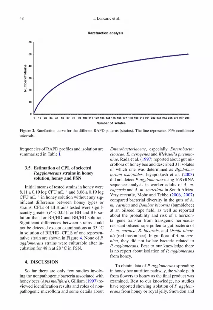
Two different statistical analyses were used toevaluate whether total diversity was covered byscreening 301 isolates using RAPD-PCR method.The coverage (C) [C = 1 − (n1/N) × 100, where n1is the number of strains which occurred only oncein our culture collection and N is the total numberof isolates (Ravenschlag et al., 1999). In addition, ararefaction analysis was performed to determine thenumber of unique OTUs as a proportion of the esti-mated total diversity. Calculations were performedusing the freeware program Analytic Rarefactionversion 1.3 (Holland, 2003), [http://www.uga.edu/strata/software/Software.html]. The program usesthe rarefaction equations described by Hurlbert(1971) and Heck et al. (1975).
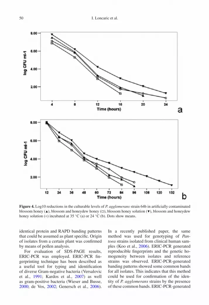
2.10. Estimation of culturablepopulation levels (CPL) of selectedP. agglomerans strains in honey
In order to test culturability in honey in vitro,four strains of P. agglomerans were selected basedon their interaction with the growth of the Austrianlocal strain Erwinia amylovora 295/93 in vitro. Se-lected P. agglomerans strains 64b and 280b couldsuppress E. amylovora growth in vitro and strains700b and 1376b were ineffective (Heissenberger,2004). Type strain of P. agglomerans DSM 3493T
was also included in the study. Examinations wereperformed with two types of honey not originatedfrom tested beehives: blossom honey (BH – mois-ture 17.2%, pH 3.6, dominant pollen group: Car-damine pratensis, Tilia platyphyllos, Allium sp.) orblossom and honeydew honey (BH/HD – moisture17.4%, pH 3.1, dominant pollen group: Prunus sp.Malus sp. – details see supplementary data Ap-pendix I). Two types of tests were conducted: (a)with pure honeys; (b) with honey solutions. Cultur-ability from pure honey and solutions was tested af-ter artificial inoculation with selected P. agglomer-ans strains.
Culturability in pure honey was tested in a sterile50 mL centrifuge tube by mixing 20 g of each honey

Pantoea agglomerans isolates from honey bees 45
with 1 mL of bacterial water suspension (1 OD600approx. 2 × 10⁹ CFU mL−1) of each strain whichwas incubated for 48 h at 28 ◦C on PYE. Tests wereperformed at 24 ◦C, the temperature in the outer re-gions of a comb (Ritter, 1982), and 35 ◦C, the cen-tral colony temperature if brood is present (South-wick, 1991). The temperature intervals were basedon results of pretests performed with P. agglomer-ans DSM 3493T and BH. The honey and honey so-lution samples incubated at 24 ◦C and at 35 ◦C wereplated every 12 h and 4 h, respectively. After mix-ing 0.1 mL of solution were serial diluted (1:10) andplated in triplicate on PYE agar and incubated for72 h at 28 ◦C.
For the honey solution, 20 g of honey was vor-texed thoroughly with 2 mL of sterile water untilhomogeneity was obtained, then 1 mL of the bac-terial suspension was added, incubated, and platedas described above. Initial number of CFU mL−1
of honey or honey solution was counted after mix-ing bacterial suspension with honey or honey so-lution by plating each sample tested in serial dilu-tions, where 0.1 mL was plated on 6 PYE plates.Initial number of CFUmL−1of honey or honey solu-tion was estimated by dividing obtained CFUs with15.2 for honey and 17.2 for honey solution, whichare the total amounts of mL in artificially contam-inated honey or honey solution. Specific gravity ofhoney is about 1.4 (Krell, 1996), 20 g ≈ 14.2 mLof honey + 1 mL of bacterial suspension for cul-turability test in pure honey or 1 mL of bacterialsuspension + 2 mL of sterile H2O.
2.11. Estimation of culturablepopulation levels (CPL) of selectedP. agglomerans strains in FSN
FSN was collected from two beehives used forthis study. Culturability in FSN was tested by mix-ing 5 mL of nectar (characteristics: pH < 5, domi-nant pollen content: Trifolium repens) with 0.2 mLOD600 = 1 (approx. 2×10⁹ CFU/mL) of each strainused for CPL-experiments in this study in sterile15 mL centrifuge tubes. Tests were performed at28 ◦C in two independent experiments and sampleswere prepared as described for honey experimentsand plated in triplicate on PYE agar every 4 h. Thebacterial growth on PYE agar was controlled after72 h of incubation at 28 ◦C. CFUs were not counted.
2.12. Statistical analysis
Numbers of CFU ml−1 were logarithm trans-formed to achieve a normal distribution, and allresults were expressed as log (CFU). Data were sub-jected to statistical analysis using SPSS for Win-dows. The differences among the strains and amongtwo honey types incubated were analyzed usingANOVA when the mean counts were greater than0.5 log CFU mL−1. In order to compare the differ-ence between each group, Bonferroni multiple com-parison was applied.
3. RESULTS
3.1. P. agglomerans isolates and pollenanalysis
A total of 307 P. agglomerans like CFUswere recovered and undertaken further exami-nations. Origin and frequencies of isolates aregiven in Table I. Detailed characteristics ofisolates and results of pollen analysis are sum-marized in Appendix II (supplementary data).No culturable bacteria could be detected inroyal jelly and two honey samples originatingfrom test hives.
3.2. Analysis of whole-cell proteinprofiles by SDS-PAGE
With this technique, 50 different profilesrelating to P. agglomerans reference strainscould be identified within wild isolates (Ap-pendix III (supplementary data)). Comparisonof the protein patterns demonstrated that 301isolates that showed obvious similarities withthe protein profiles of two reference strainsconfirmed the preliminary identification basedon physiological characteristics. Differencesbetween protein profiles of P. agglomeransstrains were primarily observed approximatelyin the range 35 and 66 kD. Six P. agglomerans-like CFUs, showing two different protein pro-files, had no similarities with reference strainsand were therefore excluded of further studies.

46 I. Loncaric et al.
Table I. Origin of Pantoea agglomerans strains utilized in this work, groups based on ERIC-PCR patternwith frequency of RAPD-PCR profiles and isolates within a group.
ERIC Group No. of RAPD profiles No. of isolates Origin1 1 1 Pl2 12 96* B, Hs, Pl, N3 2 9 B, Hs, Pl4 1 4 B, Hs, Pl5 1 1 Pl6 1 1 B7 1 3 B, Hs, Pl8 3 15 B, Hs, Pl9 1 3 B, Hs, Pl10 1 4 B, Pl11*12 1 5 B, Hs, Pl13 1 16 B, Hs, Pl, N14 1 3 B, Pl15 1 4 B16 1 3 B, N, Pl17 2 14 B18 1 2 B19 1 3 B, Pl20 4 33 B, Hs, Pl21 3 4 B, Hs, Pl22 1 13 B, Hs, Pl23 1 7 B, Hs, Pl24 1 1 Hs25 1 2 Pl26 1 3 B, Pl27 2 2 B28 1 2 B, Pl29 1 2 Pl30 1 44 B, Hs, Pl, N31 1 1 Pl
B: blossom; Hs: honey sac; Pl: pollen loads; N: nectar.* Excl. DSM 3493T.** DSM 1619.
3.3. RAPD PCR and cluster analysis
Employing RAPD primer Opl-11 each ofthe 50 isolates exhibited a unique genomic fin-gerprint. Bands with identical electrophoreticbehavior were only rarely observed, demon-strating that the employed primer is mostuseful for differentiation among strains of P.agglomerans and may be also useful for epi-demiological studies. The number of RAPD-PCR generated banding patterns employingOpl-11 was identical that obtained by SDS-PAGE (n = 50) (Fig. 1). From each group
of isolates sharing the same fingerprints ob-tained after SDS-PAGE and RAPD PCR withOpl-11, one isolate was selected as a represen-tative strain for cluster analysis. Similarities ofthe isolates in the RAPD-PCR banding pat-terns with the pattern of P. agglomerans DSM3493T were in the range between 0 and 50%.Between E. amylovora and all P. agglomeransstrains, there were no genetic similarities. Thegenetic distance between all strains of P. ag-glomerans was > 0.17. The dendrogram hasa cophenetic correlation r = 0.754 (data notshown). Diversity coverage reaches a value of

Pantoea agglomerans isolates from honey bees 47
Figure 1. RAPD fingerprints of the 52 Pantoea agglomerans strains. Lane: P corresponds to P. agglomeransDSM 3493T, D to P. agglomerans DSM 1619. The lane numbers correspond to the isolate numbers shownin Appendix II (supplementary data); M1 1 kb DNA leader; M2 1 kb DNA leader.
98.34%, indicating that nearly the total diver-sity was covered. Rarefaction analysis (Fig. 2)also revealed that continuing sampling of iso-lates would have yielded very few new strains,as documented by the modest slope of rarefac-tion curve.
3.4. ERIC PCR analysisof P. agglomerans strains
A total of 31 different ERIC banding pat-terns were observed among 52 strains exam-
ined using ERIC PCR (Fig. 3). All strainsshared at least 3 of the 6 major bands of ap-proximately 0.5, 0.7 and 2.6 kb in size, whichindicates a high degree of relatedness and con-firms the identification of the isolates as mem-bers of P. agglomerans. No similarities wereobserved between patterns of P. agglomeransstrains tested and E. amylovora 295/93. AllP. agglomerans isolates were grouped at alevel higher than 57% using UPGMA method.The dendrogram has a cophenetic correlationr = 0.820 (Appendix IV). All selected strainsgrouped based on their ERIC-PCR profile with
48 I. Loncaric et al.
Figure 2. Rarefaction curve for the different RAPD patterns (strains). The line represents 95% confidenceintervals.
frequencies of RAPD profiles and isolation aresummarized in Table I.
3.5. Estimation of CPL of selectedP.agglomerans strains in honeysolution, honey and FSN
Initial means of tested strains in honey were8.11± 0.19 log CFU mL−1 and 8.06± 0.19 logCFU mL−1 in honey solution without any sig-nificant difference between honey types orstrains. CPLs of all strains tested were signif-icantly greater (P < 0.05) for BH and BH so-lution than for BH/HD and BH/HD solution.Significant differences between strains couldnot be detected except examinations at 35 ◦Cin solution of BH/HD. CPLS of one represen-tative strain are shown in Figure 4. None of P.agglomerans strains were culturable after in-cubation for 48 h at 28 ◦C in FSN.
4. DISCUSSION
So far there are only few studies involv-ing the nonpathogenic bacteria associated withhoney bees (Apis mellifera). Gilliam (1997) re-viewed identification results and roles of non-pathogenic microflora and some details about
Enterobacteriaceae, especially Enterobactercloacae, E. aerogenes and Klebsiella pneumo-niae. Rada et al. (1997) reported about gut mi-croflora of honey bee and described 31 isolatesof which one was determined as Bifidobac-terium asteroides. Jeyaprakash et al. (2003)did not detect P. agglomerans using 16S rRNAsequence analysis in worker adults of A. m.capensis and A. m. scutellata in South Africa.Very recently, Mohr and Tebbe (2006, 2007)compared bacterial diversity in the guts of A.m. carnica and Bombus bicornis (bumblebee)at an oilseed rape field, as well as reportedabout the probability and risk of a horizon-tal gene transfer from transgenic herbicide-resistant oilseed rape pollen to gut bacteria ofA. m. carnica, B. bicornis, and Osmia bicor-nis (red mason bee). In gut flora of A. m. car-nica, they did not isolate bacteria related toP. agglomerans. Best to our knowledge thereis no report about isolation of P. agglomeransfrom honey.
To obtain data of P. agglomerans spreadingin honey bee nutrition pathway, the whole pathfrom flowers to honey as the final product wasexamined. Best to our knowledge, no studieshave reported showing isolation of P. agglom-erans from honey or royal jelly. Snowdon and

Pantoea agglomerans isolates from honey bees 49
Figure 3. ERIC fingerprints of the 52 Pantoea agglomerans strains. Lane: P corresponds to P. agglomeransDSM 3493T ; D to P. agglomerans DSM 1619. The lane numbers correspond to the isolate numbers shownin Appendix II (supplementary data); M1 1 kb DNA leader; M2 1 kb DNA leader.
Cliver (1996), and results described by Rallet al. (2003), indicated that no vegetative formsof causative agents of human bacterial diseaseshave been found in honey.
The technique of SDS PAGE has proven tobe a rapid and cost-efficient method for thecomparison of large groups of bacteria andit can be used for an initial step in polypha-sic characterization (de Vos, 2002). Thecomparison of protein fingerprints obtained bySDS-PAGE is suitable for identification anddifferentiation of closely related bacteria (Potet al., 1994; Lyra et al., 1997; Esteban et al.,2003; de Vos, 2002). Therefore, consideringthe fact that all our isolates have almost iden-tical protein fingerprints with the type strainof P. agglomerans DSM 3493T and reference
strain DSM 1619, it can be deduced that theybelong to this species.
The RAPD fingerprinting technique hasbeen described to be a powerful typing methodfor many bacterial species (Mbwana et al.,2006). In our work, the use of the RAPDtechnique allowed discrimination among dif-ferent strains of P. agglomerans and yieldedreproducible results in different assays. Theresults presented here allow us to concludethat RAPD analysis can be a useful tool forthe genotyping of P. agglomerans. Our resultssuggest that the strains circulating in a honeybee’s environment are unevenly distributedon plants, bees, and two hives tested. Elevenstrains, which were isolated three or moretimes from blossoms, honey sacs, pollen loads,and/or FSN during sampling period showed
50 I. Loncaric et al.
Figure 4. Log10 reductions in the culturable levels of P. agglomerans strain 64b in artificially contaminatedblossom honey (�), blossom and honeydew honey (�), blossom honey solution (�), blossom and honeydewhoney solution (◦) incubated at 35 ◦C (a) or 24 ◦C (b). Dots show means.
identical protein and RAPD banding patternsthat could be assumed as plant specific. Originof isolates from a certain plant was confirmedby means of pollen analysis.
For evaluation of SDS-PAGE results,ERIC-PCR was employed. ERIC-PCR fin-gerprinting technique has been described asa useful tool for typing and identificationof diverse Gram-negative bacteria (Versalovicet al., 1991; Kardos et al., 2007) as wellas gram-positive bacteria (Wieser and Busse,2000; de Vos, 2002; Genersch et al., 2006).
In a recently published paper, the samemethod was used for genotyping of Pan-toea strains isolated from clinical human sam-ples (Koo et al., 2006). ERIC-PCR generatedreproducible fingerprints and the genetic ho-mogeneity between isolates and referencestrains was observed. ERIC-PCR-generatedbanding patterns showed some common bandsfor all isolates. This indicates that this methodcould be used for confirmation of the iden-tity of P. agglomerans strains by the presenceof these common bands. ERIC-PCR-generated
Pantoea agglomerans isolates from honey bees 51
fingerprints are in agreement with the SDS-PAGE results and indicate that the isolates infact are members of P. agglomerans. The den-drograms obtained in the present study showedno association between isolates with regard totime of isolation or origin. A possible expla-nation for that is that the foraging radius ofhoney bees can cover distances up to 2000 mfrom hives (Seeley, 1997). Considering thisfact and result of our study, which provedthat almost all tested plants were contaminatedwith P. agglomerans, it could be assumed thathoney bees can come into contact with themost P. aggomerans strains present on bee-visited plants in the foraging area. As honey isknown to have a variety of antimicrobial prop-erties (Molan, 1992a,b), several tests were per-formed during this study to evaluate the CPLof selected P. agglomerans strains in FSN andripe, extracted honey. Until present, no infor-mation is available concerning the estimationof CPL of P. agglomerans in freshly storednectar (FSN) and honey. However, several au-thors describe the given survival rates of someother bacterial species (Tysset and Durand,1973; Beyme et al., 1975; Tysset et al., 1979;deWael et al., 1990; Alexandrova et al., 2002).Because of differences in species tested, incu-bation temperatures and bacterial concentra-tion, a direct comparison with those reportswas not possible. Based on our in vitro stud-ies, it can be concluded that the time withinwhich P. agglomerans could be recovered –expressed by the culturability on PYE agar– is considerably shorter than the period be-tween nectar collection and honey harvest-ing. According to Horn (1992), honey ripen-ing takes at least 1–3 days and the honey isusually harvested at the end of blooming pe-riod, which could last at least one week ormore. It is important to mention that some bac-teria under stress may enter into the viable-but-nonculturable (VBNC) state (Ordax et al.,2006). Honey with its antimicrobial properties(e.g., high osmolarity) could probably inducea VBNC state. Thus, to obtain exact survivalrates and to avoid false negative results, furtherinvestigations are necessary to clarify whetheror not strains of P. agglomerans could enterinto the VBNC state.
In this study, we demonstrated that P. ag-glomerans strains are widely spread in a honeybee’s environment and on flowers visited bybees. Strains were highly genetically diversewith good diversity coverage. From all sam-ples of bee visited flowers, at least one isolateof P. agglomerans could be detected. Some ob-tained strains seem to be plant-specific. Be-cause of relatively small numbers of isolatesin our study, further studies about this issue arenecessary to confirm or reject the hypothesis oforigin specificity to certain plant species. Wealso demonstrated that P. agglomerans strainscompletely lost their culturability in artificiallycontaminated FSN and honey within 48 h attemperatures higher than 28 ◦C as well as120 h in honey and 156 h in honey solutionat 24 ◦C, respectively. Further studies undernatural conditions are necessary to clarify thepossible role of P. agglomerans as a food con-taminant, prior to its use for biocontrol in thefield.
ACKNOWLEDGEMENTS
This work was supported in part by internalfunds of the Institute of Bacteriology, Mycologyand Hygiene, University of Veterinary Medicine Vi-enna, Institute for Apiculture, Austrian Agency forHealth and Food Safety (AGES) as well as Clinicfor Avian, Reptile and Fish Medicine, Universityof Veterinary Medicine Vienna. We are gratefulto Marianne Keck for providing DNA of Erwiniaamylovora strain 295/93 used in this study and toEwald B. M. Denner and Birgit Heissenberger forproviding reference strains of Pantoea agglomer-ans. We thank Sandra Buczolits, Gabriele Roth-müller, Irmgard Derakhshifar and Hermann Pech-hacker for helpful comments and suggestions, andChristine Schramm and Katharina Etter for techni-cal assistance.
Caractérisation de la diversité des bactéries Pan-toea agglomerans isolées de colonies d’abeillesdomestiques (Apis mellifera) et possibilité decroissance dans le miel de lignées sélectionnées.
diversité bactérienne / traçabilité des souches /analyse pollinique / empreinte génomique / Er-winia amylovora
52 I. Loncaric et al.
Zusammenfassung – Typisierungen von Panto-ea agglomerans isoliert aus Kolonien von Honig-Bienen (Apis mellifera), sowie Untersuchung derKultivierbarkeit ausgewählter Stämme in Ho-nig. Ziel dieser Studie war die Untersuchung derDiversität von Pantoea agglomerans und seinerRückverfolgbarkeit von Trachtpflanzen zum Bie-nenvolk, sowie die Abschätzung der kultivierbarenKeimzahl (CPL) dieses Bakteriums in Honig, Ho-niglösung und frisch eingelagertem Nektar (FSN).P. agglomerans ist ein möglicher Kandidat zur bio-logischen Bekämpfung von Feuerbrand (Erwiniaamylovora),wurde aber auch als fakultativ human-pathogener Keim beschrieben.Blüten verschiedener Pflanzen, Pollenhöschen, Ho-nigblaseninhalt und frisch eingelagerter Nektarwurden gesammelt, aufbereitet und das gewonne-ne Probenmaterial auf Agarplatten inkubiert, umIsolate von P. agglomerans zu gewinnen. Zur Be-wertung der Diversität wurden SDS-PAGE, RAPD-and ERIC-PCR eingesetzt. Die Abschätzung derVerwandtschaft der Isolate erfolgte mittels Cluster-Analyse.Zur Untersuchung der Kultivierbarkeit von P. ag-glomerans in Honig, Honiglösung und frisch einge-lagertem Nektar wurden diese mit einer wässrigenSuspension von 5 ausgewählten Stämmen des Bak-teriums künstlich inokuliert und sorgfältig durch-mischt. In bestimmten Intervallen wurden Probenentnommen, auf Agarplatten ausplattiert und die-se bebrütet. Für jeden Stamm wurde die Zahl derkultivierbaren koloniebildenden Einheiten (KBE)ermittelt.Von Blüten verschiedener Pflanzenarten, Pollen-höschen, Honigblaseninhalten und frisch eingela-gertem Nektar konnten einzelne Isolate von P.agglomerans gewonnen werden, nicht aber ausHonig- und Weiselfuttersaftproben der Testvöl-ker. Auf Basis der Koloniemorphologie, Pigmen-tierung, biochemischen Eigenschaften und demVergleich der Proteinmuster mit Referenzstämmennach Natriumsulfat-Polyacrylamid Gel Elektropho-rese (SDS-PAGE) wurden 301 Isolate ausgewählt.Von diesen zeigten 50 Stämme unterschiedliche Ei-weißprofile. Die Analyse mittels RAPD-PCR er-brachte die gleiche Profilanzahl PCR (Abb. 1). EineIdentifizierung der isolierten Stämme erfolgte mitERIC-PCR (Abb. 2).Aus den künstlich inokulierten Proben konnte aufPYE Agar bei 24 ◦C und einer Inkubationsdauervon mehr als 120 h (Honig) bzw. 156 h (Honiglö-sung) keiner der Teststämme reisoliert werden. BeiTemperaturen über 28 ◦C war bereits nach 48 h Be-brütungsdauer keine Rückisolation der Teststämmeaus Honig, Honiglösung oder frisch eingelagertemNektar mehr möglich.Sowohl bei 35 ◦C als auch bei 24 ◦C war die kul-tivierbare Keimzahl der Teststämme zu verschie-denen Zeitintervallen in „Blütenhonig“ (BH) si-gnifikant höher (P < 0, 05) als in „Blüten- mitWaldhonig“, oder ihren Lösungen (Abb. 4).
Gestützt auf diese Ergebnisse kann geschlossenwerden, dass die Zeit, in der die getesteten P. agglo-merans Stämme aus Honig kultiviert werden konn-ten, beträchtlich kürzer ist, als die Zeitspanne, diein der imkerlichen Praxis zwischen dem Sammelndes Nektars und der Ernte des Honigs üblicherwei-se eingehalten wird. Im Falle eines Einsatzes vonP. agglomerans als biologisches Mittel zur Feuer-brandbekämpfung in blühenden Obstanlagen kanndies ein wichtiger Punkt sein.
Bakterielle Vielfalt / Stamm-Rückverfolgbarkeit/ Pollenanalyse / genetischer Fingerabdruck / Er-winia amylovora
REFERENCES
Alexandrova M., Porrini C., Bazzi C., CarpanaE., Bigliardi M., Sabatini A.G. (2002) Erwiniaamylovora longevity in beehives, beehive productsand honeybees, Acta Hortic. (ISHS) 590, 201–205.
Bakonyi T., Derakhshifar I., Grabensteiner E.,Nowotny N. (2003) Development and evaluationof PCR assays for the detection of Paenibacilluslarvae in honey samples: comparison with iso-lation and biochemical characterization, Appl.Environ. Microbiol. 69, 1504–1510.
Beyme D., Ficke W., Kleinhempel H., Schaefe,H.J., Bremer R. (1975) Modellversuche zurÜberlebensdauer von Erwinia amylovora imVerdauungstrakt der Honigbiene, im Honig undan Teilen des Bienenstockes, Arch. Phytopathol.u. Pflanzenschutz 11, 203–211.
Cicchetti R., Iacobini M., Midolla F., Papoff P.,Mancuso M., Moretti C. (2006) Pantoea agglom-erans sepsis after rotavirus gastroenteritis, Pediatr.Infect. Dis J. 25, 280–281.
Costa E., Usall J., Teixido N., Delgado J., Vinas I.(2002) Water activity, temperature, and pH ef-fects on growth of the biocontrol agent Pantoeaagglomerans CPA-2, Can. J. Microbiol. 4, 1082–1088.
Cruz A.T., Cazacu A.C., Allen C.H. (2007) Pantoeaagglomerans - a plant pathogen causing humandisease, J. Clin. Microbiol. 45, 1989–1992.
de Vos P. (2002) Nucleic acid analysis and SDS-PAGE of whole-cell proteins in Bacillus taxon-omy, in: Berkeley R., Heyndrickx M., Logan N.,de Vos P. (Eds.), Applications and systematicsof Bacillus and relatives, Blackwell Publishing,Oxford, pp. 141–159.
de Wael L., Greef M., Laere O. (1990) The honeybeesas a possible vector of Erwinia amylovora (Burr.)Winslow et al., Acta Hortic. (ISHS) 273, 107–114.
Dutkiewicz J. (1997) Bacteria and fungi in organic dustas potential health hazard, Ann Agric. Environ.Med. 4, 11–16.
Esteban J., Molleja A., Cabria F., Soledad JimenezM. (2003) SDS-PAGE for identification of species
Pantoea agglomerans isolates from honey bees 53
belonging to the Mycobacterium fortuitum com-plex, Clin. Microbiol. Infect. 9, 327–331.
Garcia-Vallvé S., Palau J., Romeu A. (1999)Horizontal gene transfer in glycosyl hydro-lases inferred from codon usage in Escherichiacoli and Bacillus subtilis, Mol. Biol. Evol. 16,1125–1134.
Gavini F., Mergaert J., Beji A., Mielcarek C., IzardD., Kersters K., De Ley J. (1989) Transferof Enterobacter agglomerans (Beijerinck 1888)Ewing and Fife 1972 to Pantoea gen. nov. asPantoea agglomerans comb. nov. and descriptionof Pantoea dispersa sp. nov., Int. J. Syst. Bacteriol.39, 337–345.
Genersch E., Forsgren E., Pentikainen J., AshiralievaA., Rauch S., Kilwinski J., Fries I. (2006)Reclassification of Paenibacillus larvae subsp.pulvifaciens and Paenibacillus larvae subsp. lar-vae as Paenibacillus larvae without subspeciesdifferentiation, Int. J. Syst. Evol. Microbiol. 56,501–511.
Gerhard P., Murray R.E., Wood W.A., Krieg R. (1994)Methods for general and molecular bacteriology.American Society for Microbiology, WashingtonDC.
Gilliam M. (1997) Identification and roles of non-pathogenic microflora associated with honey bees,FEMS Microbiol. Lett. 155, 1–10.
Heck K.L. Jr., Van Belle G., Simberloff D. (1975)Explicit calculation of the rarefaction diversitymeasurement and the determination of sufficientsample size, Ecology 56, 1459–1461.
Heissenberger B. (2004) Untersuchungen zur biolo-gischen Feuerbrandbekämpfung mit Pantoea ag-glomerans, einer erregerverwandten Bakterienart.Diploma thesis, University of Natural Resourcesand Applied Life Sciences, Vienna, Austria.
Heissenberger B., Spornberger A., Loncaric I.,Moosbeckhofer R., Keck M., Foissy H. (2006)In vitro studies on fire blight control by bac-terial antagonists, Proc. 1st Int. Symp. onBiological Control of Bacterial Plant Diseases,in: Zeller W., Ulrich C. (Eds.), Mitteilungen ausder Biologischen Bundesanstalt für Land- undForstwirtschaft Berlin-Dahlem 408, pp. 281–282.
Holland S.M. (2003). Analytic rarefaction, version 1.3.University of Georgia, Athens, [online] http://www.uga.edu/strata/software/Software.html (ac-cessed on 9 October 2008).
Horn H. (1992) Das grosse Honigbuch: Entstehung,Gewinnung, Zusammensetzung, Qualität,Gesundheit und Vermarktung, EhrenwirthVerlag, München.
Hurlbert S.H. (1971). The nonconcept of species diver-sity: a critique and alternative parameters, Ecology52, 577–586.
Ishimaru C.A., Klos E.J., Brubaker R.R. (1988)Multiple antibiotic production by Erwinia her-bicola, Phytopathology 78, 746–750.
Jeyaprakash A., Hoy M.A., Allsopp M.H. (2003)Bacterial diversity in worker adults of Apismellifera capensis and Apis mellifera scutellata
(Insecta: Hymenoptera) assessed using 16S rRNAsequences, J. Invertebr. Pathol. 84, 96–103.
Kardos G., Nagy J., Antal M., Bistyak A., Tenk M.,Kiss I. (2007) Development of a novel PCR assayspecific for Riemerella anatipestifer, Lett. Appl.Microbiol. 44, 145–148.
Kearns L.P., Hale C.N. (1996) Partial characteriza-tion of an inhibitory strain of Erwinia herbicolawith potential as a biocontrol agent for Erwiniaamylovora, the fire blight pathogen, J. Appl.Bacteriol. 81, 369–374.
Koo H.S., Kim J.S., Eom J.S., You J.Y., Park J.Y.,Kim H.S., Song W., Cho H.C., Lee K.M. (2006)Pseudooutbreak of Pantoea species bacteremia as-sociated with contaminated cotton pledgets, Am.J. Infect. Control 34, 443–446.
Kratz A., Greenberg D., Barki Y., Cohen E., LifshitzM. (2003) Pantoea agglomerans as a cause of sep-tic arthritis after palm tree thorn injury; case reportand literature review, Arch. Dis. Child 88, 542–544.
Krell R. (1996) Value-added products from bee-keeping: Physical characteristics of honey, FAOAgricultural services bulletin No. 124, Rome,[online] http://www.fao.org/docrep/w0076e/w0076e00.htm#con (accessed on 9 October2008).
Laemmli U.K. (1970) Cleavage of structural proteinsduring the assembly of the head of bacteriophageT4, Nature 15, 227, 680–685.
Laporte C., Demachy M.C., Thevenin-Lemoine C.(2002) Ostéite tibiale à Pantoea agglomeransau décours d’une fracture ouverte stade IIIB dejambe, Rev. Chir. Orthop. Réparatrice Appar. Mot.88, 625–627.
Lau K.K., Ault B.H., Jones D.P. (2005) Polymicrobialperitonitis including Pantoea agglomerans fromteething on a catheter, South. Med. J. 98, 580–581.
Lim P.S., Chen S.L., Tsai C.Y., Pai M.A. (2006)Pantoea peritonitis in a patient receiving chronicambulatory peritoneal dialysis, Nephrology(Carlton) 11, 97–99.
Loncaric I., Donat C., Antlinger B., OberlerchnerJ.T., Heissenberger B., Moosbeckhofer R. (2008)Strain-specific detection of two Aureobasidiumpullulans strains, fungal biocontrol agents of fireblight by new, developed multiplex-PCR, J. Appl.Microbiol. 104, 1433–1441.
Louveaux J., Maurizio A., Vorwohl G. (1970)Internationale Kommission für Bienenbotanik derI.U.B.S. - Methodik der Melissopalynologie,Apidologie 1, 193–209; English version in BeeWorld, 1970, 51, 125–138.
Lyra C., Hantula J., Vainio E., Rapala J., RouhiainenL., Sivonen K. (1997) Characterization ofcyanobacteria by SDS-PAGE of whole-cellproteins and PCR/RFLP of the 16S rRNA gene,Arch. Microbiol. 168, 176–184.
Mbwana J., Bolin I., Lyamuya E., Mhalu F.,Lagergard T. (2006) Molecular characterizationof Haemophilus ducreyi isolates from differ-ent geographical locations, J. Clin. Microbiol.44, 132–137.
54 I. Loncaric et al.
Mohr K.I., Tebbe C.C. (2006) Diversity and phylotypeconsistency of bacteria in the guts of three beespecies (Apoidea) at an oilseed rape field, Environ.Microbiol. 8, 258–272.
Mohr K.I., Tebbe C.C. (2007) Field study results on theprobability and risk of a horizontal gene transferfrom transgenic herbicide-resistant oilseed rapepollen to gut bacteria of bees, Appl. Microbiol.Biotechnol. 75, 573–582.
Molan P.C. (1992a) The antibacterial activity of honey:1. The nature of the antibacterial activity, BeeWorld 73, 5–28.
Molan P.C. (1992b) The antibacterial activity of honey:2. Variation in the potency of the antibacterial ac-tivity, Bee World 73, 59–76.
Nuclo R.L., Johnson K.B., Sugar D., Stockwell V.O.(1998) Secondary colonization of pear blossomsby two bacterial antagonists of the fire blightpathogen, Plant Dis. 82, 661–668.
Ordax M., Marco-Noalesm E., Lopez M.M., BioscaE.G. (2006) Survival strategy of Erwiniaamylovora against copper: induction of theviable-but-nonculturable state, Appl. Environ.Microbiol. 72, 3482–3488.
Özaktan H., Bora T. (2004) Biological control offire blight in pear orchards with a formulationof Pantoea agglomerans strain Eh 24, Braz. J.Microbiol. 35, 224–229.
Page R.D.M. (1996) TREEVIEW: An application todisplay phylogenetic trees on personal computers,Comp. Appl. Biosci. 12, 357–358.
PONET- pollen database, Institute for Apiculture,Austrian Agency for Health and Food Safety,[online] http://www15.ages.at:7778/pls/pollen/pollen_suche (accessed on 9 October 2008).
Pot B., Vandamme P., Kersters K. (1994) Analysis ofelectrophoretic whole-organism protein finger-prints, in: Goodfellow M., O’Donnell A.G. (Eds.),Modern microbiological methods, Chemicalmethods in prokaryotic systematic, Chichester,England, John Wiley and Sons, pp. 493–521.
Pusey P.L. (1997) Crab apple blossoms as amodel system for fire blight biocontrol research,Phytopathology 87, 1096–1102.
Rada V., Machoa M., Huk J., Marounek M., DuskovaD. (1997) Microflora in the honeybee digestivetract: counts, characteristics and sensitivity to vet-erinary drugs, Apidologie 28, 357–365.
Rall V.L., Bombo A.J., Lopes T.F., Carvalho L.R.,Silva M.G. (2003) Honey consumption in the stateof Sao Paulo: a risk to human health? Anaerobe 9,299–303.
Ravenschlag K., Sahm K., Pernthaler J., Amann R.(1999) High bacterial diversity in permanentlycold marine sediments, Appl. Environ. Microbiol.65, 3982–3989.
Ritter W. (1982) Experimenteller Beitrag zurThermoregulation des Bienenvolks (Apis melliferaL.), Apidologie 13, 169–195.
Seeley T.D. (1997) Honigbienen - Im Mikrokosmosdes Bienenstocks, Birkhäuser Verlag, Basel.
Sneath P.H.A., Sokal R.R. (1973) NumericalTaxonomy, W.H. & Freeman, San Francisco.
Snowdon J.A., Cliver D.O. (1996) Microorganisms inhoney, Int. J. Food Microbiol. 31, 1–26.
Southwick E.E. (1991) The colony as a thermoregulat-ing superorganism, in: Goodman L.J., Fisher R.C.(Eds.), The behavior and physiology of bees, CABInternational, Oxon, UK, pp. 28–47.
Tysset C., Durand C. (1973) On the survival of somegram negative, non-sporulated bacteria in com-mercial honey, Bull. Acad. Vet. Fr. 46, 191–196.
Tysset C., Haas P., Durand C. (1979) Survival of somemycobacteria in honey stored at room tempera-ture, Bull. Acad. Vet. Fr. 52, 447–452.
Vanneste J.L. (1996) Honey bees and epiphytic bacte-ria to control fire blight, a bacterial disease of ap-ple and pears, Biocontrol News Inform. 17, 67N–78N.
Vanneste J.L., Cornish D.A., Yu J., Voyle M.D. (2002)P10C: a new biological control agent for controlof fire blight which can be sprayed or distributedusing honey bees, Acta Hortic. (ISHS) 590, 231–235.
Vanneste J.L., Yu J., Beer S.V. (1992) Role of an-tibiotic production by Erwinia herbicola Eh252in biological control of Erwinia amylovora, J.Bacteriol. 174, 2785–2796.
Versalovic J., Koeuth T., Lupski J.R. (1991)Distribution of repetitive DNA sequences ineubacteria and application to fingerprintingof bacterial genomes, Nucleic Acids Res. 19,6823–6831.
Wieser M., Busse H.J. (2000) Rapid identificationof Staphylococcus epidermidis, Int. J. Syst. Evol.Microbiol. 50, 1087–1093.
Williams J.G., Kubelik A.R., Livak K.J., Rafalski J.A.,Tingey S.V. (1990) DNA polymorphisms ampli-fied by arbitrary primers are useful as geneticmarkers, Nucleic Acids Res. 18, 6531–6535.
Wilson M., Epton H.A.S., Sigee D.C. (1990)Biological control of fire blight of hawthorn(Crategus monogyna) with Erwinia herbicola un-der protected conditions, Plant Pathol. 39, 301–308.
Wodzinski R.S., Umholtz T.E., Rundle J.R., BeerS.V. (1994) Mechanisms of inhibition of Erwiniaamylovora by E. herbicola in vitro and in vivo, J.Appl. Bacteriol. 76, 22–29.
Wright S.A.I., Beer S.V. (1996) The role of antibi-otics in biological control of fire blight by Erwiniaherbicola strain EH318, Acta Hortic. (ISHS) 411,309–312.
Wright S.A.I., Beer S.V. (2006) Pantoea agglomer-ans, a biocontrol agent and ubiquitous microor-ganism – friend or foe? Proc. 1st Int. Symp. onBiological Control of Bacterial Plant Diseases,in: Zeller W., Ulrich C. (Eds.), Mitteilungen ausder Biologischen Bundesanstalt für Land- undForstwirtschaft Berlin-Dahlem, 408, pp. 334–338.
Wright S.A., Zumoff C.H., Schneider L., BeerS.V. (2001) Pantoea agglomerans strain EH318produces two antibiotics that inhibit Erwiniaamylovora in vitro, Appl. Environ. Microbiol. 67,284–292.