tumor-infiltrating b lymphocyte profiling identifies igg-biased
TRANSCRIPT
CANCER RESEARCH | TUMOR BIOLOGYAND IMMUNOLOGY
Tumor-Infiltrating B Lymphocyte Profiling IdentifiesIgG-Biased, Clonally ExpandedPrognostic Phenotypes inTriple-Negative Breast CancerRobert J. Harris1,2,3, Anthony Cheung1,2,4, Joseph C.F. Ng5, Roman Laddach1,2,6, Alicia M. Chenoweth1,2,4,Silvia Crescioli1,2, Matthew Fittall1,2,4, Diana Dominguez-Rodriguez1,2, James Roberts1,2,6, Dina Levi4,Fangfang Liu4, Elena Alberts1,2,4, Jelmar Quist4, Aida Santaolalla7,8, Sarah E. Pinder9,10, Cheryl Gillett9,10,Niklas Hammar7, Sheeba Irshad9, Mieke Van Hemelrijck7,8, Deborah K. Dunn-Walters11, Franca Fraternali5,James F. Spicer9, Katie E. Lacy1,2, Sophia Tsoka6, Anita Grigoriadis4, Andrew N.J. Tutt4,12, andSophia N. Karagiannis1,2,4
ABSTRACT◥
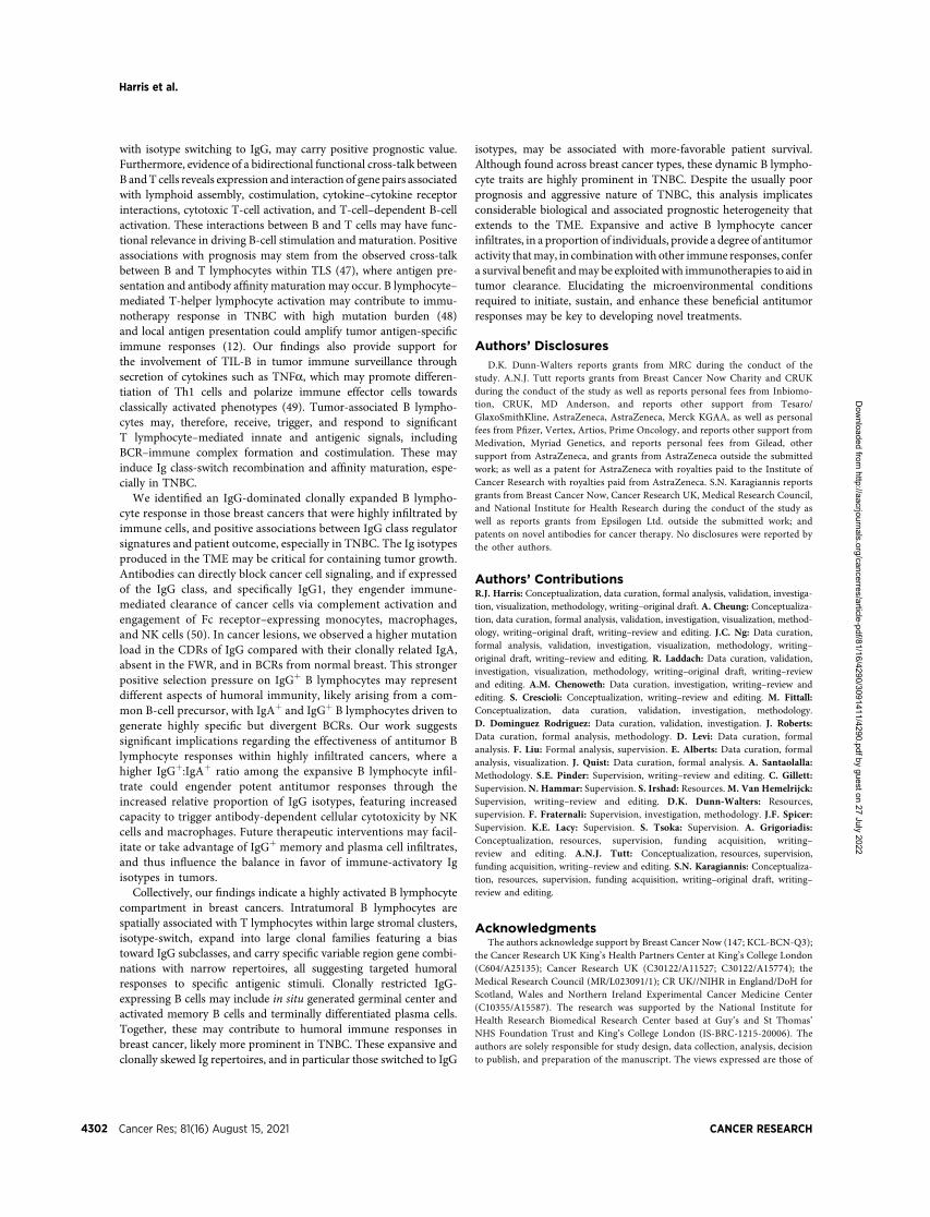
In breast cancer, humoral immune responses may contribute toclinical outcomes, especially in more immunogenic subtypes. Here,we investigated B lymphocyte subsets, immunoglobulin expression,and clonal features in breast tumors, focusing on aggressive triple-negative breast cancers (TNBC). In samples from patients withTNBC and healthy volunteers, circulating and tumor-infiltrating Blymphocytes (TIL-B) were evaluated. CD20þCD27þIgD� isotype-switched B lymphocytes were increased in tumors, compared withmatched blood. TIL-B frequently formed stromal clusters with Tlymphocytes and engaged in bidirectional functional cross-talk,consistent with gene signatures associated with lymphoid assembly,costimulation, cytokine–cytokine receptor interactions, cytotoxicT-cell activation, and T-cell–dependent B-cell activation. TIL-B–upregulated B-cell receptor (BCR) pathway molecules FOS andJUN, germinal center chemokine regulator RGS1, activationmarkerCD69, and TNFa signal transduction via NFkB, suggesting BCR–immune complex formation. Expression of genes associated with Blymphocyte recruitment and lymphoid assembly, includingCXCL13, CXCR4, andDC-LAMP,was elevated in TNBC comparedwith other subtypes and normal breast. TIL-B–rich tumors showedexpansion of IgG but not IgA isotypes, and IgG isotype switchingpositively associated with survival outcomes in TNBC. Clonalexpansion was biased toward IgG, showing expansive clonal fam-ilies with specific variable region gene combinations and narrowrepertoires. Stronger positive selection pressure was present in the
complementarity determining regions of IgG compared with theirclonally related IgA in tumor samples. Overall, class-switched Blymphocyte lineage traits were conspicuous in TNBC, associatedwith improved clinical outcomes, and conferred IgG-biased, clon-ally expanded, and likely antigen-driven humoral responses.
Significance: Tumor-infiltrating B lymphocytes assemble inclusters, undergoing B-cell receptor–driven activation, prolifera-tion, and isotype switching. Clonally expanded, IgG isotype-biasedhumoral immunity associates with favorable prognosis primarily intriple-negative breast cancers.
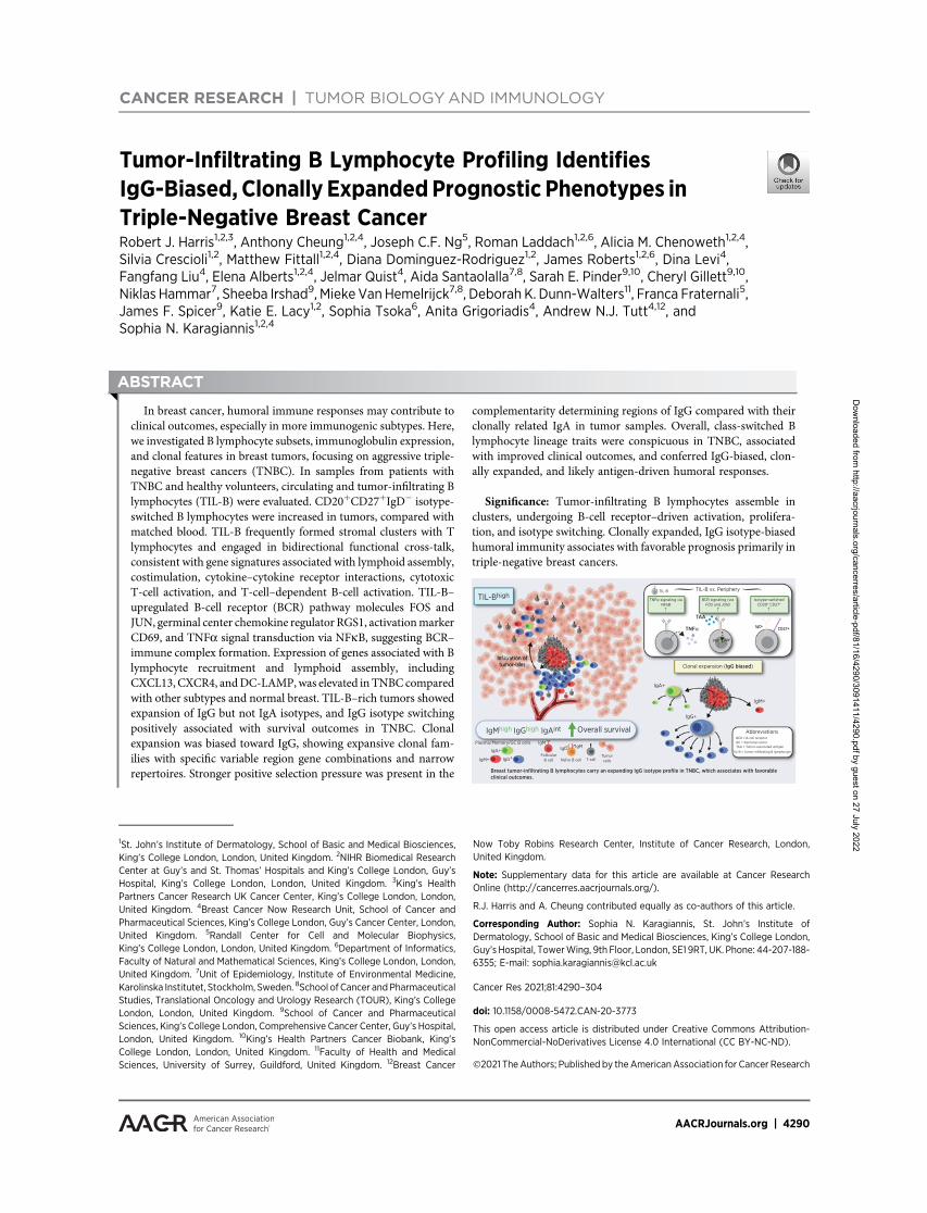
Breast tumor-infiltrating B lymphocytes carry an expanding IgG isotype profile in TNBC, which associates with favorableclinical outcomes.
TIL-Bhigh
IgMhigh IgGhigh IgAint Overall survival AbbreviationsBCR = B-cell receptorGC = Germinal centerTAA = Tumor-associated antigen
TIL-B = Tumor-infiltrating B lymphocyte
Plasma/Memory/GC B cells
IgA+
IgA+
IgM+
IgM+
IgM
FollicularB cell T-cellNaÏve B cell
TIL-B vs. Periphery
BCR signaling (viaFOS and JUN)
Isotype-switchedCD20+ CD27+
↑
TIL-B
TAA
Clonal expansion (lgG biased)
FOS JUN
IgD- CD27+TNFα
TNFα signaling viaNFkB
Tumorcells
IgMIgD
IgG+
IgG+
Infiltration oftumor-Islet
GC
GC
↑ ↑
1St. John’s Institute of Dermatology, School of Basic and Medical Biosciences,King’s College London, London, United Kingdom. 2NIHR Biomedical ResearchCenter at Guy’s and St. Thomas’ Hospitals and King’s College London, Guy’sHospital, King’s College London, London, United Kingdom. 3King’s HealthPartners Cancer Research UK Cancer Center, King’s College London, London,United Kingdom. 4Breast Cancer Now Research Unit, School of Cancer andPharmaceutical Sciences, King’s College London, Guy’s Cancer Center, London,United Kingdom. 5Randall Center for Cell and Molecular Biophysics,King’s College London, London, United Kingdom. 6Department of Informatics,Faculty of Natural and Mathematical Sciences, King’s College London, London,United Kingdom. 7Unit of Epidemiology, Institute of Environmental Medicine,Karolinska Institutet, Stockholm, Sweden. 8School of Cancer andPharmaceuticalStudies, Translational Oncology and Urology Research (TOUR), King’s CollegeLondon, London, United Kingdom. 9School of Cancer and PharmaceuticalSciences, King’s College London, Comprehensive Cancer Center, Guy’s Hospital,London, United Kingdom. 10King’s Health Partners Cancer Biobank, King’sCollege London, London, United Kingdom. 11Faculty of Health and MedicalSciences, University of Surrey, Guildford, United Kingdom. 12Breast Cancer
Now Toby Robins Research Center, Institute of Cancer Research, London,United Kingdom.
Note: Supplementary data for this article are available at Cancer ResearchOnline (http://cancerres.aacrjournals.org/).
R.J. Harris and A. Cheung contributed equally as co-authors of this article.
Corresponding Author: Sophia N. Karagiannis, St. John’s Institute ofDermatology, School of Basic and Medical Biosciences, King’s College London,Guy’s Hospital, TowerWing, 9th Floor, London, SE1 9RT, UK. Phone: 44-207-188-6355; E-mail: [email protected]
Cancer Res 2021;81:4290–304
doi: 10.1158/0008-5472.CAN-20-3773
This open access article is distributed under Creative Commons Attribution-NonCommercial-NoDerivatives License 4.0 International (CC BY-NC-ND).
�2021 TheAuthors; Published by the American Association for Cancer Research
AACRJournals.org | 4290
Dow
nloaded from http://aacrjournals.org/cancerres/article-pdf/81/16/4290/3091411/4290.pdf by guest on 27 July 2022

IntroductionInitiation of effective adaptive immunity may contribute to
tumor growth restriction through specific antigen-directed res-ponses. The T lymphocyte component of antitumor immunity hasreceived significant attention (1). In contrast, B lymphocytes,especially the memory and isotype-switched B lymphocyte com-partments, and their expressed antibody profiles remain onlypartially elucidated. Emerging findings suggest that aspects ofhumoral immune responses may correlate with improved clinicaloutcomes via B lymphocyte tumor-infiltration and expression ofantibodies in lesions or in the circulation (2, 3). These could differacross tumor types, potentially offering opportunities for stratifi-cation and for guiding therapy options.
Breast cancer is one of the most frequently diagnosed malignancies,divided into biological, and differential therapy-associated subtypesbased on estrogen receptor (ER), progesterone receptor (PR), andHER2 expression, with specific prognostic and predictive biomarkerimplications. Triple-negative breast cancers (TNBC) do not expressany of these markers and demonstrate the least-favorable prognosisdue to both an aggressive phenotype and limited targeted therapies (4).Although breast cancer has not traditionally been regarded as a typicalimmunogenic malignancy, emerging studies report the presence andpotential clinical significance of tumor-infiltrating immune cells forclinical outcomes (5). Paradoxically, despite an overall poor prognosisof patients with TNBC, immune infiltration is more pronouncedcompared with other breast cancer types. Consistent with an immu-nogenic tumor microenvironment (TME), some patients with TNBCmay benefit from anti-programmed death-ligand 1 (PD-L1) and anti–PD-1 immunotherapy with atezolizumab in combination with che-motherapy (6). TNBCs are characterized by immunologically variableand compartmentalized tumors with structural features in the tumor–immune interphase and large variability across individuals, mandatingthe need for patient stratification for therapy selection (7). The mostthoroughly studied effector cells within the breast cancer setting areCD8þ cytotoxic T lymphocytes and natural killer (NK) cells (8).However, tumor-infiltrating B lymphocytes (TIL-B) aggregating with-in tertiary lymphoid structures (TLS; ref. 9) may have an antigen-educated phenotype (10) and autoantibodies are thought to triggertumor cell clearance (11). TIL-Bs might also serve as antigen-presenting cells to promote antitumor Th responses (12). Therefore,it is increasingly recognized that humoral immune responses may beimportant contributors to breast cancer outcome, especially in moreimmunogenic TNBCs.
The interaction between the immune system and malignant cells,therefore, constitutes amajor focus of current translational and clinicalinvestigation (13). Recent studies have provided evidence of TIL-Bsand tumor-reactive immunoglobulin (Ig) in several solid tumors,including in breast cancer, and TIL-Bs have been reported to respondto B-cell receptor stimulation and produce Igs ex vivo (14–16).
Here, in peripheral blood and cancer lesions of patients withbreast cancer, specifically in individuals with TNBC, we performedflow cytofluorimetric, transcriptomic, immunofluorescence, single-cell RNA-sequencing (scRNA-seq), and long-read Ig repertoirestudies to evaluate isotype-switched and memory B lymphocytesubsets, Ig isotype distribution, and clonal expansion profiles.
Materials and MethodsClinical sample collection and cohort descriptions
A collection of internal and external cohorts of healthy volunteer(HV) and patient samples, including the unique accession numbers,
are summarized in Supplementary Table S1 and SupplementaryMaterials and Methods. All internal King’s College London (KCL)samples were collected with informed written consent, in accordancewith theHelsinki Declaration [study design was approved by theGuy’sResearch Ethics Committee (REC No. 07/H0804/131), Guy’s and St.Thomas’NHS Foundation Trust]. Peripheral bloodmononuclear cellswere isolated using Ficoll–Paque PLUS density gradient centrifuga-tion, and single cells from breast tissues were isolated as describedpreviously in Supplementary Materials and Methods.
Gene expression profiling of lymphocyte and lymphoidassembly markers
Gene expression (GEx) levels were analyzed from internal Guy’sHospital and The Cancer Genome Atlas (TCGA) Breast cohorts (KCLand TCGA GEx cohorts), and compared between PAM50 (basal-like,HER2, luminal A, luminal B, and normal-like) and TNBC subtypes(basal-like 1/2, immunomodulatory, mesenchymal, mesenchymalstem-like, and luminal androgen receptor) according to classificationdescribed previously in Bras�o-Maristany and colleagues (17). A Blymphocyte metagene signature was analyzed from published Nano-String data of primary and metastatic breast cancers (GSE102818;ref. 18). Kaplan–Meier (KM) plotter tool was used to generate survivalplots (19). CIBERSORT was applied to evaluate na€�ve B, plasma, andmemory B cells identified among 22 immune subsets in the KCL GExcohort (20). Univariate Cox proportional hazards regression modelswere used to investigate the prognostic importance of these immunesubsets in high and low TIL-infiltrated tumors (semiquantitative TILclassification was performed by a trained histopathologist using tissuemicroarrays; Supplementary Materials and Methods). B lymphocytefunction–associated gene setswere identified fromgene ontology (GO)database (SupplementaryMaterials andMethods). A lymphoid assem-bly–associated gene signature was compiled from a set of knownmarkers (21).
scRNA-seq analysisAnalyses were performed on a published scRNA-seq dataset
(GSE114725; ref. 22; single-cell cohort) using R package Seurat (23).Dimensionality reduction was performed using Uniform ManifoldApproximation and Projection (UMAP) and cells were clusteredusing the Louvain algorithm (23). Ig isotypes were detected basedupon heavy-chain gene expression. Differentially expressed genesidentified by Seurat were used to perform gene set enrichmentanalysis (GSEA) using the fgsea package. Gene sets were obtainedfrom Broad Institute Molecular Signature Database using R packagemsigdbr (24). CellPhoneDB v2.0 was used to analyze B-cell–T-cellinteractions (25).
Immunohistochemical/immunofluorescence evaluations of TIL-B distribution and surface Ig expression
Three sections per tissue sample were stained with fluorescentlylabeled antibodies conferring three panels: TIL classification (DAPI/CD20/CD3/PanCK), na€�ve B lymphocyte identification (DAPI/CD20/IgD), and Ig isotype expression (DAPI/CD27/IgG/IgA/IgM). Anti-bodies used are detailed in Supplementary Table S2. TIL-B struc-tural features were evaluated (Supplementary Materials and Meth-ods) following TIL working group guidelines (26), guided by trainedpathologists. Olympus VS120-S5 and Nikon TE 2000-U micro-scopes were used for imaging.
Long-read Ig repertoire analysisIg repertoire analysis was performed from cDNA synthesized
from breast tissues with the 50 RACE template switch method
Expanded Tumor-Infiltrating IgGþ B Cells in TNBC
AACRJournals.org Cancer Res; 81(16) August 15, 2021 4291
Dow
nloaded from http://aacrjournals.org/cancerres/article-pdf/81/16/4290/3091411/4290.pdf by guest on 27 July 2022
(Supplementary Materials and Methods). Full-length Ig cDNAswere PCR-amplified with primers containing unique molecularbarcodes. Purified DNA samples were sequenced using PacBioSingle Molecule, Real-Time (SMRT) Sequencing platform (27).Redundant sequences with identical molecular barcodes wereremoved. Ig genes and complementarity determining region (CDR)3 sequences were determined using IMGT/HighV-QUEST (28). Relat-edness among sequences were estimated using BRepertoire webser-ver (29) “Clonotype clustering” function, after partitioning all CDR3DNA sequences by the sample and the V gene family used (30).Selection pressure analysis was performed using R packageshazam (31).
Data availabilityThe R code used to analyze scRNA-seq data from GSE114725 (22)
can be accessed from https://codeocean.com/capsule/7488142. The Rcode to analyze BCR Repertoire data collected from breast tissues canbe accessed at https://codeocean.com/capsule/4153741.
Statistical analysesGraphPad Prism and R were used for statistical analyses of
paired and unpaired datasets. Data are presented as mean � SEM.P values reported as: �, P < 0.05; ��, P < 0.01; ���, P < 0.001; ����, P <0.0001 and all tests were two-sided.
Further details can be found in Supplementary Materials andMethods.
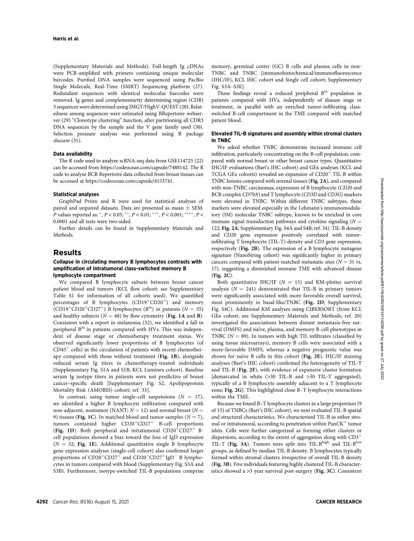
ResultsCollapse in circulating memory B lymphocytes contrasts withamplification of intratumoral class-switched memory Blymphocyte compartment
We compared B lymphocyte subsets between breast cancerpatient blood and tumors (KCL flow cohort; see SupplementaryTable S1 for information of all cohorts used). We quantifiedpercentages of B lymphocytes (CD19þCD20þ) and memory(CD19þCD20þCD27þ) B lymphocytes (Bm) in patients (N ¼ 55)and healthy subjects (N ¼ 48) by flow cytometry (Fig. 1A and B).Consistent with a report in melanoma (32), we identified a fall inperipheral Bm in patients compared with HVs. This was indepen-dent of disease stage or chemotherapy treatment status. Weobserved significantly lower proportions of B lymphocytes (ofCD45þ cells) in the circulation of patients with recent chemother-apy compared with those without treatment (Fig. 1B), alongsidereduced serum Ig titers in chemotherapy-treated individuals(Supplementary Fig. S1A and S1B, KCL Luminex cohort). Baselineserum Ig isotype titers in patients were not predictive of breastcancer–specific death [Supplementary Fig. S2, ApolipoproteinMortality Risk (AMORIS) cohort; ref. 33].
In contrast, using tumor single-cell suspensions (N ¼ 17),we identified a higher B lymphocyte infiltration compared withnon-adjacent, nontumor (NANT; N ¼ 12) and normal breast (N ¼9) tissues (Fig. 1C). In matched blood and tumor samples (N ¼ 7),tumors contained higher CD20þCD27þ B-cell proportions(Fig. 1D). Both peripheral and intratumoral CD20þCD27þ B-cell populations showed a bias toward the loss of IgD expression(N ¼ 32; Fig. 1E). Additional quantitative single B lymphocytegene expression analyses (single-cell cohort) also confirmed largerproportions of CD20þCD27þ and CD20þCD27þIgD� B lympho-cytes in tumors compared with blood (Supplementary Fig. S3A andS3B). Furthermore, isotype-switched TIL-B populations comprise
memory, germinal center (GC) B cells and plasma cells in non-TNBC and TNBC [immunohistochemical/immunofluorescence(IHC/IF), KCL IHC cohort and Single cell cohort; SupplementaryFig. S3A–S3E].
These findings reveal a reduced peripheral Bm population inpatients compared with HVs, independently of disease stage ortreatment, in parallel with an enriched tumor-infiltrating class-switched B-cell compartment in the TME compared with matchedpatient blood.
Elevated TIL-B signatures and assembly within stromal clustersin TNBC
We asked whether TNBC demonstrate increased immune cellinfiltration, particularly concentrating on the B-cell population, com-pared with normal breast or other breast cancer types. QuantitativeIHC/IF evaluations (Bart’s IHC cohort) and GEx analyses (KCL andTCGA GEx cohorts) revealed an expansion of CD20þ TIL-B withinTNBC lesions compared with normal tissues (Fig. 2A), and comparedwith non-TNBC carcinomas, expression of B lymphocyte (CD20 andBCR complex CD79A) and T lymphocyte (CD3D and CD3G) markerswere elevated in TNBC. Within different TNBC subtypes, thesemarkers were elevated especially in the Lehmann’s immunomodula-tory (IM) molecular TNBC subtype, known to be enriched in coreimmune signal transduction pathways and cytokine signaling (N ¼122; Fig. 2A; Supplementary Fig. S4A and S4B; ref. 34). TIL-B densityand CD20 gene expression positively correlated with tumor-infiltrating T lymphocyte (TIL-T) density and CD3 gene expression,respectively (Fig. 2B). The expression of a B lymphocyte metagenesignature (NanoString cohort) was significantly higher in primarycancers compared with patient-matched metastatic sites (N ¼ 31 vs.17), suggesting a diminished immune TME with advanced disease(Fig. 2C).
Both quantitative IHC/IF (N ¼ 15) and KM-plotter survivalanalysis (N ¼ 241) demonstrated that TIL-B in primary tumorswere significantly associated with more favorable overall survival,most prominently in basal-like/TNBC (Fig. 2D; SupplementaryFig. S4C). Additional KM analyses using CIBERSORT (from KCLGEx cohort, see Supplementary Materials and Methods; ref. 20)investigated the associations between distant metastasis-free sur-vival (DMFS) and na€�ve, plasma, and memory B-cell phenotypes inTNBC (N ¼ 89). In tumors with high TIL infiltrates (classified byusing tissue microarrays), memory B cells were associated with amore-favorable DMFS, whereas a negative prognostic value wasshown for na€�ve B cells in this cohort (Fig. 2E). IHC/IF staininganalyses (Bart’s IHC cohort) confirmed the heterogeneity of TIL-Tand TIL-B (Fig. 2F), with evidence of expansive cluster formation[demarcated in white (>30 TIL-B and >30 TIL-T aggregated),typically of a B lymphocyte assembly adjacent to a T lymphocytezone; Fig. 2G]. This highlighted close B–T lymphocyte interactionswithin the TME.
Because we found B–T lymphocyte clusters in a large proportion (9of 15) of TNBCs (Bart’s IHC cohort), we next evaluated TIL-B spatialand structural characteristics. We characterized TIL-B as either stro-mal or intratumoral, according to penetration within PanCKþ tumorislets. Cells were further categorized as forming either clusters ordispersions, according to the extent of aggregation along with CD3þ
TIL-T (Fig. 3A). Tumors were split into TIL-Bhigh and TIL-Blow
groups, as defined by median TIL-B density. B lymphocytes typicallyformed within stromal clusters irrespective of overall TIL-B density(Fig. 3B). Five individuals featuring highly clustered TIL-B character-istics showed a >5 year survival post-surgery (Fig. 3C). Consistent
Harris et al.
Cancer Res; 81(16) August 15, 2021 CANCER RESEARCH4292
Dow
nloaded from http://aacrjournals.org/cancerres/article-pdf/81/16/4290/3091411/4290.pdf by guest on 27 July 2022

with the B–T lymphocyte cluster formation observed, we foundelevated expression of B lymphocyte recruitment and lymphoidassembly marker genes (CXCL13, CXCR4, and DC-LAMP) in TNBCcompared with both non-TNBC and normal breast, and within theTNBC cohort the highest expression of these genes was detected inIM tumors (Fig. 3D; Supplementary Fig. S4D, KCL and TCGA GEx
cohorts). Higher expression of these signatures were also associatedwith significantly improved odds of overall survival (10-year follow-up) in basal-like/TNBC (Fig. 3E, KM plotter cohort).
Therefore, substantial TIL-B densities in TNBC typically formclusters along with T lymphocytes. TIL-B marker (CD20 and CD79A)and GEx features conferring B lymphocyte recruitment and lymphoid
Figure 1.
Flow cytometric analyses reveal reduced circulating CD20þCD27þmemory and amplification of tumor-infiltrating CD20þCD27þIgD� class-switched subsets amongB lymphocytes. A, Gating strategy for identification of B lymphocytes and memory (Bm) lymphocytes derived from peripheral blood mononuclear cell (examplepatient peripheral blood mononuclear cell shown). B,Quantification of total circulating B cells (top) and Bm cells (bottom) as percentage of CD45þ cells in HV (N¼48) and patient (N ¼ 55) peripheral blood (KCL flow cohort; Supplementary Table S1 for patient information), stratified according to stage and treatment status.C, Quantification of B lymphocytes (CD20þ) from single cell suspensions of normal breast (N ¼ 9), NANT (N ¼ 12), and cancer tissue (N ¼ 17) samples.D,Quantification ofmatched patient circulating- and tumor-infiltrating CD20þCD27þB cells (matched samples of 7 patients).E,Quantification of CD20þCD27þIgD�
B cells in HV (N¼ 17), patient peripheral blood (N¼ 7), and cancers (N¼ 8) of total CD20þCD27þ B cells. Statistical significance was determined using the Studentt test. ns, nonsignificant; � , P < 0.05; �� , P < 0.01; ���, P < 0.001; ���� , P < 0.0001.
Expanded Tumor-Infiltrating IgGþ B Cells in TNBC
AACRJournals.org Cancer Res; 81(16) August 15, 2021 4293
Dow
nloaded from http://aacrjournals.org/cancerres/article-pdf/81/16/4290/3091411/4290.pdf by guest on 27 July 2022
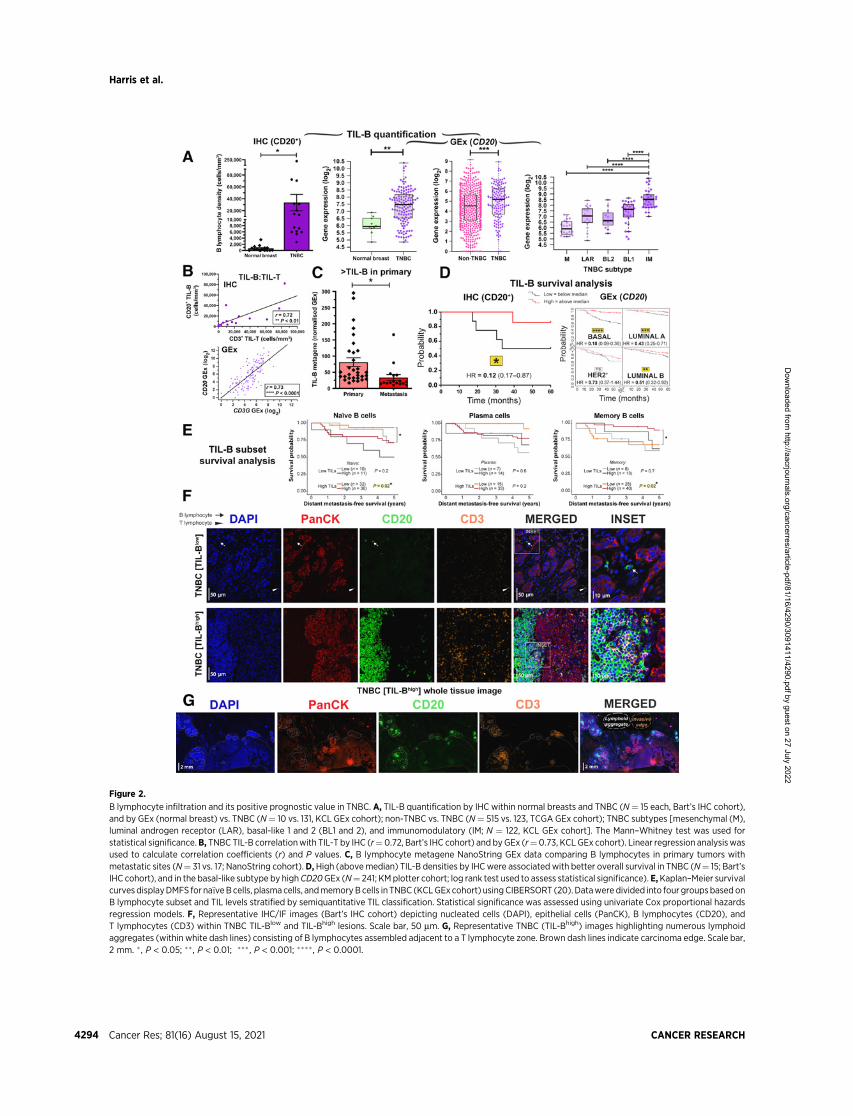
Figure 2.
B lymphocyte infiltration and its positive prognostic value in TNBC. A, TIL-B quantification by IHC within normal breasts and TNBC (N¼ 15 each, Bart’s IHC cohort),and by GEx (normal breast) vs. TNBC (N¼ 10 vs. 131, KCL GEx cohort); non-TNBC vs. TNBC (N¼ 515 vs. 123, TCGA GEx cohort); TNBC subtypes [mesenchymal (M),luminal androgen receptor (LAR), basal-like 1 and 2 (BL1 and 2), and immunomodulatory (IM; N ¼ 122, KCL GEx cohort]. The Mann–Whitney test was used forstatistical significance.B, TNBC TIL-B correlationwith TIL-T by IHC (r¼0.72, Bart’s IHC cohort) and byGEx (r¼0.73, KCL GEx cohort). Linear regression analysiswasused to calculate correlation coefficients (r) and P values. C, B lymphocyte metagene NanoString GEx data comparing B lymphocytes in primary tumors withmetastatic sites (N¼ 31 vs. 17; NanoString cohort).D,High (abovemedian) TIL-B densities by IHCwere associated with better overall survival in TNBC (N¼ 15; Bart’sIHC cohort), and in the basal-like subtype by highCD20GEx (N¼ 241; KMplotter cohort; log rank test used to assess statistical significance). E,Kaplan–Meier survivalcurves displayDMFS for na€�veBcells, plasma cells, andmemoryBcells in TNBC (KCLGEx cohort) usingCIBERSORT (20). Datawere divided into four groupsbased onB lymphocyte subset and TIL levels stratified by semiquantitative TIL classification. Statistical significance was assessed using univariate Cox proportional hazardsregression models. F, Representative IHC/IF images (Bart’s IHC cohort) depicting nucleated cells (DAPI), epithelial cells (PanCK), B lymphocytes (CD20), andT lymphocytes (CD3) within TNBC TIL-Blow and TIL-Bhigh lesions. Scale bar, 50 mm. G, Representative TNBC (TIL-Bhigh) images highlighting numerous lymphoidaggregates (within white dash lines) consisting of B lymphocytes assembled adjacent to a T lymphocyte zone. Brown dash lines indicate carcinoma edge. Scale bar,2 mm. � , P < 0.05; �� , P < 0.01; ��� , P < 0.001; ����, P < 0.0001.
Harris et al.
Cancer Res; 81(16) August 15, 2021 CANCER RESEARCH4294
Dow
nloaded from http://aacrjournals.org/cancerres/article-pdf/81/16/4290/3091411/4290.pdf by guest on 27 July 2022

Figure 3.
Occurrence of B lymphocytes in stromal clusters. A, Representative IHC/IF images (Bart’s IHC cohort) highlighting key TIL characteristics: clustered TIL versusdispersed TIL and stromal TIL (outside tumor nests) versus intratumoral TIL (within tumor nests). Scale bar, 50 mm. B, Quantitative assessment of TIL-B spatial andstructural characteristicswithin TNBC (N¼ 15; left) intratumoral versus stromal; clustered versus dispersed (right). Patients ranked according to CD20þ TIL-B density(TNBC1 ¼ highest), and overall survival data are indicated. Inset, patient samples split at median density into high and low TIL-B groups and the percentage ofintratumoral (left) or clustered (right) B lymphocytes were analyzed. Statistical significance was determined using the Student t test. C, Characterization of TNBCTIL-B profile as stromal clustered, intratumoral clustered, stromal dispersed, or intratumoral dispersed. Overall survival data are indicated. D, GEx data for lymphoidassembly marker genes CXCL13 and DC-LAMP: normal breast versus TNBC (N ¼ 10 vs. 131, KCL GEx cohort; left); non-TNBC vs. TNBC (N ¼ 515 vs. 123, TCGA GExcohort; middle); TNBC subtypes (N¼ 122, KCL GEx cohort; right). E, Survival analysis in KM Plotter of determined ER�HER2�/basal surrogate, HER2þ, luminal A, andluminal B subtype KM plotter surrogate subgroups (KM plotter cohort; ref. 19). These indicate that expression of lymphoid cell assembly genes carries positiveprognostic value in TNBC/basal-like and luminal B subtypes. Individual genes were evaluated in combination with each other gene (left) and gene set as a whole(right). ns, nonsignificant; �, P < 0.05; �� , P < 0.01; ���� , P < 0.0001.
Expanded Tumor-Infiltrating IgGþ B Cells in TNBC
AACRJournals.org Cancer Res; 81(16) August 15, 2021 4295
Dow
nloaded from http://aacrjournals.org/cancerres/article-pdf/81/16/4290/3091411/4290.pdf by guest on 27 July 2022
assembly were elevated compared with non-TNBC and normal breasttissues, and associated with more favorable survival in basal-like/TNBC subtype.
TIL-Bs are activated via the B-cell receptorWe sought direct evidence of active roles for B lymphocytes in the
TME, using a previously published dataset (single-cell cohort). Weapplied dimensionality reduction (UMAP) to single-cell B lymphocytepopulations and revealed distinct CD20þ and CD27þ TIL-B popula-tions compared with those in the circulation (blood (N¼ 1,476 B cells)and tumors (N¼ 1,021 B cells) of eight patients; Fig. 4A). Downstreamdifferential expression gene (DEG) analysis identified several upre-gulated genes in TIL-B: FOS and JUN, molecules downstream of theBCR complex pathway (35); RGS1, germinal center B lymphocyteregulator of chemokine receptor signaling (36); and the lymphocyteactivation marker CD69, triggered through cross-linking of surface Ig(Fig. 4B; ref. 37). Hallmark enrichment analysis (38) revealed signif-icantly enhanced expression among TIL-B for genes controlling TNFasignaling via NFkB, hypoxia, and UV response pathways, in compar-ison with circulating B lymphocytes (Fig. 4C). Several genes, known topositively regulate B lymphocyte activation, proliferation, and differ-entiation functions were upregulated in TNBC compared with normalbreast (Supplementary Fig. S5, KCL and TCGAGEx cohorts). Survivalanalysis of these gene signatures revealed positive associations withoverall survival (10-year follow up), most pronounced in basal-like/TNBC (Fig. 4D, KM plotter cohort). We next evaluated B-cell–T-cellinteractions in the TME (single-cell cohort) using CellPhoneDB (25).We identified cell communication pathways associated with lymphoidassembly (CXCL12, CCL19, CCL21, and CXCL13), cytokine signaling(IL4, IL6, IL13, IL15, IL17, and IFNg), costimulation (CD28 and ICOS)and immune activation (CD40 and CD226). These findings support abidirectional functional cross-talk between tumor-infiltrating B and Tlymphocytes (Fig. 4E).
These findings indicate the presence of distinct B lymphocytepopulations between patient blood and cancers and evidence ofTIL-B antigen–Ig complexing, BCR pathway stimulation, and evi-dence of B-cell–T-cell cross-talk. Key functional attributes for theinitiation of B lymphocyte responses are associated with improvedpatient outcomes, especially in TNBC.
IgGþ B lymphocyte densities are elevated in tumors, andIgG isotype switching predicts positive survivaloutcomes in TNBC
We studied B lymphocyte densities according to Ig isotype expres-sion in primary breast cancers by quantitative IHC/IF evaluations ofIgD, IgM, IgA, and IgG isotype-expressing B lymphocytes [Bart’s IHCcohort; normal breast tissue (N¼ 10), TNBC (N¼ 14)]. Na€�ve (IgDþ)B lymphocytes were found at low densities in normal breast, TIL-Blow
and TIL-Bhigh tumors (Supplementary Fig. S6A). Higher IgGþ andIgMþ B lymphocyte densities were found in TIL-Bhigh and, to a lesserextent, in TIL-Blow cancers compared with normal breast (Fig. 5A andB). In contrast, IgAþ B lymphocyte densities were consistent amongtissue compartments, highlighting an overall bias toward increasedIgGþ:IgAþ B lymphocyte ratio within cancers compared with normalbreast, and within TIL-Bhigh compared with TIL-Blow cancers (Fig. 5Cand D).
IHC/IF showed the dominating IgAþ B lymphocyte profile presentin normal breast is contained within normal lobules and their periph-ery (Supplementary Fig. S6B). Although TIL-Blow cancers lacked largeB lymphocyte follicles and contained small IgM/IgA/IgG plasma cellzones, TIL-Bhigh cancers typically contained denser IgM/IgA/IgG
plasma cell zones surrounding expansive IgMþ follicular B lym-phocyte clusters with defined germinal centers (Fig. 5E). Theresulting B lymphocyte assembly in these TIL-Bhigh tumors sharessome structural similarities with those in tonsil tissues. IgG and IgAexpression were accompanied with CD27 upregulation, validatingthe differentiated, isotype-switched status of these cells (Supple-mentary Fig. S7A–S7C). We further confirmed that intratumoralIgGþ cells comprised both CD20þ as well as CD138þ (plasma) cellpopulations, the latter being low or negative for CD20 expression(Supplementary Figs. S7D and S3C).
Because TNBCs feature high levels of IgG-expressing B lympho-cytes, we studied Ig isotypes (Ig heavy chain) using a published scRNA-seq dataset (single-cell cohort; N¼ 1,021 TIL-B across eight patients).UMAP applied to single-cell B lymphocyte populations revealeddistinct TIL-B populations in TNBC compared with non-TNBCsamples. This analysis also revealed enhanced isotype-switching toIgG1, IgG3, IgG4 and IgA2 subclasses in TNBC (Fig. 6A and B).UMAP confirmed differential isotype-expressing B lymphocyte com-partments in TNBC compared with non-TNBC for IgM and IgG1based on clustering (Fig. 6C). Heatmaps of single CD20þCD27þ Blymphocyte IgCH expression showed a propensity toward non-switched IgM transcripts in blood in contrast with high levels ofisotype-switched IgG and IgA B lymphocytes in tumor samples(Supplementary Fig. S8), and higher levels of isotype-switching inTNBC compared with non-TNBC tumor samples (Fig. 6D). Thesesupport an active tumor-resident humoral response, different to theequivalent response in the circulation, and likely driven by inflam-matory and possibly antigenic signals that may support Ig class-switchrecombination in the breast cancer microenvironment, especially inTNBC.
Survival analysis of isotype-switching gene signatures revealed apositive association with 10-year overall survival for IgG, but not forIgA, isotype switching, most pronounced in basal-like/TNBC (Fig. 6E,KM plotter cohort). Moreover, as expected, several genes involved inthe mechanism of and/or positively regulating isotype switching wereupregulated in TNBC compared with normal breast (SupplementaryFig. S9, KCL and TCGA GEx cohorts).
These findings indicate a shift in favor of IgGþ B lymphocytes inTIL-Bhigh TNBC and point to IgG isotype switching as a contributor tothe overall positive role of B lymphocyte responses to breast cancers.
IgG-biased, clonally expanded, and Ig repertoires in breastcancer
Using long-read sequencing, we next generated a dataset of Igheavy-chain repertoires (N ¼ 7,670) from two TNBCs, two ERþ
cancers, and one normal breast tissue (KCL sequencing cohort) andperformed sequence clustering analyses to define clonotypes andcompare the distributions of Ig isotypes following B lymphocyte clonalexpansion. We observed consistently larger clonal family sizes withinthe 10 largest clones of cancers compared with normal breast (Fig. 7A,top). B lymphocyte sequences from ERþ cancers featured a hetero-geneous IgG subclass expansion with few IgA clones. In contrast,TNBCs showed clones with coexisting IgG1 and IgA1 subclasses,suggested intraclone isotype switching (Fig. 7A, bottom). Kolmo-gorov–Smirnov analyses revealed that IgG and IgA clonal familyfrequency distributions were significantly different between carcino-mas and normal breast (Fig. 7B). Accordingly, IgG and IgA wereclonally expanded within cancers (Supplementary Fig. S10A andS10B), and on average IgGþ B lymphocytes belonged to larger clonalfamilies than IgAþ cells (Fig. 7C). These data point to an inherent biastoward the preferential clonal expansion of IgG isotypes within
Harris et al.
Cancer Res; 81(16) August 15, 2021 CANCER RESEARCH4296
Dow
nloaded from http://aacrjournals.org/cancerres/article-pdf/81/16/4290/3091411/4290.pdf by guest on 27 July 2022
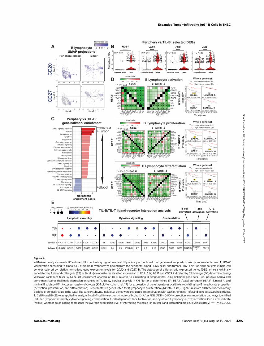
Figure 4.
scRNA-seq analysis reveals BCR-driven TIL-B activatory signatures, and B lymphocyte functional trait gene markers predict positive survival outcome. A, UMAPvisualization according to global GEx of single B lymphocytes pooled from the peripheral blood (1,476 cells) and tumors (1,021 cells) of eight patients (single-cellcohort), colored by relative normalized gene expression levels for CD20 and CD27. B, The detection of differentially expressed genes [DEG; on cells originallyannotated by Azizi and colleagues (22) as B cells] demonstrates elevated expression of FOS, JUN, RGS1, and CD69, indicated by fold change (FC; determined usingWilcoxon rank sum test). C, Gene set enrichment analysis of TIL-B relative to circulating B lymphocytes using hallmark gene sets. Red, positive normalizedenrichment scores (hallmark expression enhanced in TIL-B). D, Survival analysis in KM Plotter of determined ER�HER2�/basal surrogate, HER2þ, luminal A, andluminal B subtype KM plotter surrogate subgroups (KM plotter cohort; ref. 19) for expression of gene signatures positively regulating key B lymphocyte properties(activation, proliferation, and differentiation). Representative genes listed for B lymphocyte proliferation (44 total in set). Signatures from all three functions carrypositive prognostic value in the basal-like cancer subtype. Individual geneswere evaluated in combinationwith each other gene (left) and gene set as awhole (right).E, CellPhoneDB (25) was applied to analyze B-cell–T-cell interactions (single-cell cohort). After FDR (FDR < 0.001) correction, communication pathways identifiedincluded lymphoid assembly, cytokine signaling, costimulation, T-cell–dependent B-cell activation, and cytotoxic T lymphocyte (CTL) activation. Circle sizes indicateP value, whereas color-coding represents the average expression level of interacting molecule 1 in cluster 1 and interacting molecule 2 in cluster 2. ���� , P < 0.0001.
Expanded Tumor-Infiltrating IgGþ B Cells in TNBC
AACRJournals.org Cancer Res; 81(16) August 15, 2021 4297
Dow
nloaded from http://aacrjournals.org/cancerres/article-pdf/81/16/4290/3091411/4290.pdf by guest on 27 July 2022
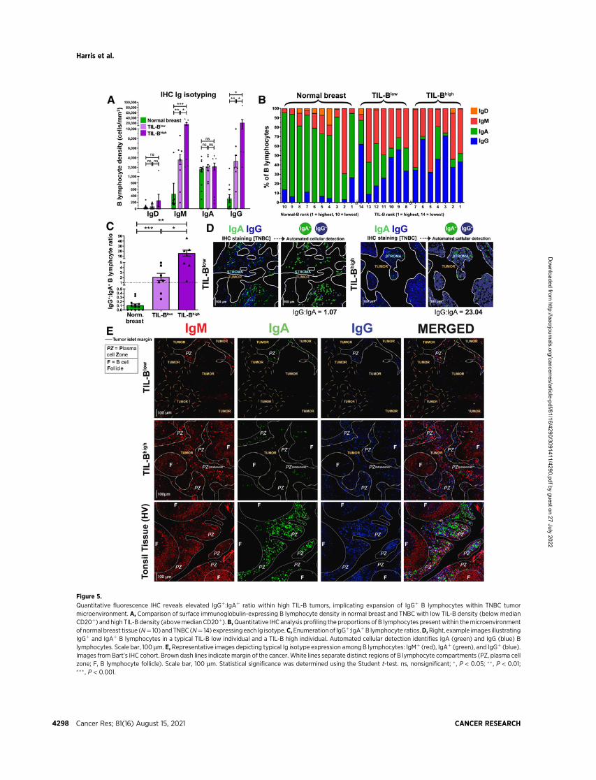
Figure 5.
Quantitative fluorescence IHC reveals elevated IgGþ:IgAþ ratio within high TIL-B tumors, implicating expansion of IgGþ B lymphocytes within TNBC tumormicroenvironment. A, Comparison of surface immunoglobulin-expressing B lymphocyte density in normal breast and TNBC with low TIL-B density (below medianCD20þ) and high TIL-B density (abovemedian CD20þ).B,Quantitative IHC analysis profiling the proportions of B lymphocytes presentwithin themicroenvironmentof normal breast tissue (N¼ 10) andTNBC (N¼ 14) expressingeach Ig isotype.C,Enumerationof IgGþ:IgAþB lymphocyte ratios.D,Right, example images illustratingIgGþ and IgAþ B lymphocytes in a typical TIL-B low individual and a TIL-B high individual. Automated cellular detection identifies IgA (green) and IgG (blue) Blymphocytes. Scale bar, 100 mm. E, Representative images depicting typical Ig isotype expression among B lymphocytes: IgMþ (red), IgAþ (green), and IgGþ (blue).Images from Bart’s IHC cohort. Brown dash lines indicate margin of the cancer. White lines separate distinct regions of B lymphocyte compartments (PZ, plasma cellzone; F, B lymphocyte follicle). Scale bar, 100 mm. Statistical significance was determined using the Student t-test. ns, nonsignificant; � , P < 0.05; �� , P < 0.01;��� , P < 0.001.
Harris et al.
Cancer Res; 81(16) August 15, 2021 CANCER RESEARCH4298
Dow
nloaded from http://aacrjournals.org/cancerres/article-pdf/81/16/4290/3091411/4290.pdf by guest on 27 July 2022

Figure 6.
Single-cell RNA-seq data analysis reveals favor of IgG isotypes in TNBC, while isotype-switching gene markers predict positive survival outcome. A, UMAPvisualization of B lymphocyte populations in non-TNBC versus TNBC (single-cell cohort). B, Percentage of each Ig isotype based upon raw data of Ig heavy-chain(Student t test). C, UMAP visualization for IgM and IgG1 isotypes colored by relative normalized gene expression levels (N¼ 1,021 cells).D, IgCH switch transcripts ofsingle B lymphocytes (CD19þCD27þ/CD20þCD27þ/CD22þCD27þ single cells) were analyzed in non-TNBC and TNBC tissues and demonstrated more Ig isotype-switching events in the TNBC samples. E, Survival analysis in KM Plotter of determined ER�HER2�/basal surrogate, HER2þ, luminal A, and luminal B subtype KMplotter surrogate subgroups (KM plotter cohort; ref. 19) for expression of gene signatures conferring isotype switching and those positively regulating isotypeswitching (IgG and IgA isotype switching). Signatures from all three functions carry positive prognostic value in basal-like cancer. Individual geneswere evaluated incombination with each other gene (left) and the gene set as a whole (right). ns, nonsignificant; ���, P < 0.001; ���� , P < 0.0001.
Expanded Tumor-Infiltrating IgGþ B Cells in TNBC
AACRJournals.org Cancer Res; 81(16) August 15, 2021 4299
Dow
nloaded from http://aacrjournals.org/cancerres/article-pdf/81/16/4290/3091411/4290.pdf by guest on 27 July 2022

Figure 7.
B lymphocyte repertoire analyses of immunoglobulin isotype switching and clonal expansion in breast cancers. A, A total of 7,670 immunoglobulin heavy-chainsequences were analyzed (KCL sequencing cohort). Top 10 clones determined by B lymphocyte repertoire long read data analyses. Clonotypes were estimated viaclustering CDR3 sequences. Top, bars depict sizes of clones and their breakdown by isotypes. Bottom, isotypes present in each clone are indicated by dots. Verticallines signify co-occurrence of isotypes in the same clone.B,Clone size frequency distribution of IgM/IgA/IgG sequences in normal breast (1,771 sequences) andbreastcancer (5,899 sequences). Kolmogorov–Smirnov analysis highlights significant differences in clone size frequency distributions.C,Mean sequences per clone of IgM/IgA/IgG isotypes. IgG and, to a lesser extent, IgA isotypes are clonally expanded, whereas on average, IgG isotypes have significantly larger clone sizes than IgA.D,Comparisons of IgAand IgGvariable usage ofV–J, D–J, andV–Dgenes extracted fromnormal breast, ERþ cancer, andTNBC. For eachgeneusage combination, dotsize is proportional to the frequency before clonal expansion. Dot colors correspond to fold change in the number of sequences following clonal expansion, indicatingthe preference of B lymphocytes with that specific VDJ combination to be clonally expanded. E, Selection pressure in clonally related IgA and IgG. Clonally relatedsequences are represented as paired observations (gray lines), and selection pressure was considered separately for the complementarity determining regions andframework regions. Sequences are grouped into normal breast and breast cancer (containing two TNBC and two ERþ samples). Paired Wilcoxon tests wereconducted, and P values were corrected (Benjamini–Hochberg) for multiple comparisons. ns, nonsignificant; � , P < 0.05; �� , P < 0.01.
Harris et al.
Cancer Res; 81(16) August 15, 2021 CANCER RESEARCH4300
Dow
nloaded from http://aacrjournals.org/cancerres/article-pdf/81/16/4290/3091411/4290.pdf by guest on 27 July 2022
breast cancers. When we compared variable region gene usage, wefound specific V(D)J genes whose combined usage was overrepre-sented in clonally expanded IgG and IgA sequences in cancers.Specific combinatorial gene usages appeared to expand in TNBCbut not ERþ lesions, and vice versa, suggestive of clonally restricted,likely antigen-focused, Ig repertoires (Fig. 7D; SupplementaryFig. S10C). Furthermore, Ig light chain repertoire transcripts fromsingle CD20þCD27þ B lymphocytes from two matched blood andtumor samples (single-cell cohort, Supplementary Fig. S10D)showed similarities in kappa and lambda chain V genes (IGKV,IGLV) and J genes (IGKJ, IGLJ), pointing to potential commonclonal origins. Furthermore, we observed a stronger positive selec-tion pressure in the CDRs of IgG compared with their clonallyrelated IgA in tumor samples, whereas such a relationship wasabsent in the framework region (FWR), and in BCRs from normalbreast (Fig. 7E). This hints at different aspects of B lymphocyteresponses where IgA expression may act as an early response andIgG may confer higher affinity antigen-driven responses.
Large clonal families, isotype switching within clonally expandedTIL-B, and a bias for IgG subclasses accumulating mutations onspecific variable region gene combinations, together suggest a maturehumoral immune response driven toward specific antigenic stimuli inbreast cancer. Further understanding of these features may revealtherapeutic targets, prognostic biomarkers, and patient subpopula-tions upon whom to focus adaptive immune response-enhancingtherapies.
DiscussionClinical outcomes in patients with cancer may be influenced by
the initiation of effective antitumoral adaptive T lymphocyteresponses, but these are likely to be significantly more effectivewhen launched in combination with humoral immunity, includinginduction of isotype-switched B lymphocytes and secretion ofantibodies. We undertook flow cytometric, IHC/IF, bulk GEx,scRNA-seq, and long-read Ig sequencing analyses to investigateactivated, memory and isotype-switched B lymphocytes in breastcancers, including the more-aggressive and more-immunogenicTNBC subtypes. Consistent with previous reports (39, 40), theTIL-B compartment in tumor stroma is typically organized intoclusters of lymphocytes, including isotype-switched memory, GC Bcells, and plasma cells. Enhanced isotype-switched B lymphocytes,with an IgG-isotype bias may be part of the humoral clonalexpansion mechanism, especially pronounced in TIL-Bhigh TNBC.The accompanied narrow mature Ig variable region repertoires andenhanced BCR signaling in TIL-B strongly signify antigen-drivenresponses. This dynamic humoral immune profile is especiallyassociated with immunogenic TNBC and indicative of more favor-able patient outcomes.
We found a depleted memory CD19þCD20þCD27þ B lympho-cyte repertoire in patients’ peripheral blood regardless of stageor treatment history for their carcinoma. Moreover, this effectappears to be exacerbated in patients with breast cancer who haveundergone chemotherapy, possibly owing to the depletion of Blymphocytes during treatment, which may irreversibly diminishlong-lived memory populations. Accordingly, patients receivingchemotherapy have lower total serum Ig titers, indicative of adepleted circulating antibody-secreting B lymphocyte population.In contrast, our flow cytometric analyses revealed an upregulatedCD20þCD27þIgD� B-cell compartment among TIL-B. In agree-ment, single-cell transcriptomic analyses point to a bias toward
non-switched IgM transcripts in blood memory CD20þCD27þ Blymphocytes and higher levels of isotype-switched IgG and IgACD20þCD27þ B lymphocytes in tumor samples. Consistent with animmunogenic signature in TNBCs, we found higher levels of isotypeswitching shown in single CD20þCD27þ B lymphocyte transcriptsin TNBC samples compared with non-TNBC tumors. These may bedriven by a combination of antigen exposure and inflammation inthe breast cancer microenvironment that promotes Ig class-switchrecombination.
The TIL-B population is largely assembled in clusters (>30 TIL-Band >30 TIL-T aggregated) and positively associates with overallsurvival. Although evident across breast cancer types, transcrip-tomic analysis suggests that TIL-B infiltration is more pronouncedin TNBC compared with non-TNBC. TNBC, especially the IMmolecular subtype, featured elevated expression of B lymphocyterecruitment and lymphoid cell assembly (CXCL13, CXCR4,DC-LAMP) genes, compared with normal breast. Enhanced localexpression of CXCL13 in arthritic synovial fluids can draw circu-lating B lymphocytes to inflammation sites (41) and B lymphocytesmay traffic from the blood towards lymphoid tissues via CXCR4stimulation (42). The evident expression of these signals within theTME may recruit B lymphocytes, including Bm, from the peripheryto cancer lesions. Consistent with this, we report reduced circulat-ing and enhanced intratumoral CD20þCD27þ B-cell compartmentsin patients. Such chemoattractant signals may also promote local Blymphocyte assembly into B–T clusters, in line with our observa-tions of close proximity and strong correlation between tumor-infiltrating B and T lymphocytes. Although the B–T clusters wedescribe are not entirely equivalent to TLSs identified in routinehistology (43), within these clusters, B–T lymphocytic cross-talkcan lead to B lymphocyte activation, Ig isotype-switching and localclonal expansion (39, 44).
Alongside IHC evidence of spatial B–T association, an active andfunctional B lymphocyte compartment is also indicated by BCRsignaling and isotype switching. Analyses of scRNA-seq data dem-onstrated that TIL-B are phenotypically distinct to those in blood,featuring upregulated BCR complex pathway molecules FOS andJUN, germinal center chemokine regulator RGS1, lymphocyte acti-vation marker CD69 and TNFa signaling via NFkB. These implicateactive BCR engagement by immune complexes. In concordance, Igrepertoire analyses revealed IgG-skewed clonal family expansionwith clonally restricted Ig variable regions. We detected significantlylarger IgG and to a lesser extent IgA, clones with narrow variableregion repertoires in cancers compared with normal breast, indic-ative of antigen-focused clonal expansion. Together, these suggest adynamic expanded IgG-biased humoral response focused toward asmall repertoire of antigenic stimuli in the TME the greaterunderstanding of which has the potential to inform therapeuticand biomarker strategies in patients.
Several studies reported that TIL-B carry positive prognostic val-ue (5, 26), whereas others found no significant effect (45), or evenpoorer survival with CD138þ plasma cell infiltrates (46). However, theextent towhichB lymphocyte activitymay correlatewith prognosis hasnot been addressed. In ourTNBC cohort, we report positive prognosticassociations of memory, but not of na€�ve or plasma, B lymphocyteinfiltration in high-TIL tumors. This may point to positive contribu-tions of memory B lymphocytes as part of the humoral response inimmunogenic breast cancers and especially in TNBC. In concordance,our findings indicate that genes that positively regulate B lymphocytefunctions, particularly those involved in activation, proliferation,differentiation, and isotype switching, and especially genes associated
Expanded Tumor-Infiltrating IgGþ B Cells in TNBC
AACRJournals.org Cancer Res; 81(16) August 15, 2021 4301
Dow
nloaded from http://aacrjournals.org/cancerres/article-pdf/81/16/4290/3091411/4290.pdf by guest on 27 July 2022
with isotype switching to IgG, may carry positive prognostic value.Furthermore, evidence of a bidirectional functional cross-talk betweenB andT cells reveals expression and interaction of gene pairs associatedwith lymphoid assembly, costimulation, cytokine–cytokine receptorinteractions, cytotoxic T-cell activation, and T-cell–dependent B-cellactivation. These interactions between B and T cells may have func-tional relevance in driving B-cell stimulation and maturation. Positiveassociations with prognosis may stem from the observed cross-talkbetween B and T lymphocytes within TLS (47), where antigen pre-sentation and antibody affinity maturation may occur. B lymphocyte–mediated T-helper lymphocyte activation may contribute to immu-notherapy response in TNBC with high mutation burden (48)and local antigen presentation could amplify tumor antigen-specificimmune responses (12). Our findings also provide support forthe involvement of TIL-B in tumor immune surveillance throughsecretion of cytokines such as TNFa, which may promote differen-tiation of Th1 cells and polarize immune effector cells towardsclassically activated phenotypes (49). Tumor-associated B lympho-cytes may, therefore, receive, trigger, and respond to significantT lymphocyte–mediated innate and antigenic signals, includingBCR–immune complex formation and costimulation. These mayinduce Ig class-switch recombination and affinity maturation, espe-cially in TNBC.
We identified an IgG-dominated clonally expanded B lympho-cyte response in those breast cancers that were highly infiltrated byimmune cells, and positive associations between IgG class regulatorsignatures and patient outcome, especially in TNBC. The Ig isotypesproduced in the TME may be critical for containing tumor growth.Antibodies can directly block cancer cell signaling, and if expressedof the IgG class, and specifically IgG1, they engender immune-mediated clearance of cancer cells via complement activation andengagement of Fc receptor–expressing monocytes, macrophages,and NK cells (50). In cancer lesions, we observed a higher mutationload in the CDRs of IgG compared with their clonally related IgA,absent in the FWR, and in BCRs from normal breast. This strongerpositive selection pressure on IgGþ B lymphocytes may representdifferent aspects of humoral immunity, likely arising from a com-mon B-cell precursor, with IgAþ and IgGþ B lymphocytes driven togenerate highly specific but divergent BCRs. Our work suggestssignificant implications regarding the effectiveness of antitumor Blymphocyte responses within highly infiltrated cancers, where ahigher IgGþ:IgAþ ratio among the expansive B lymphocyte infil-trate could engender potent antitumor responses through theincreased relative proportion of IgG isotypes, featuring increasedcapacity to trigger antibody-dependent cellular cytotoxicity by NKcells and macrophages. Future therapeutic interventions may facil-itate or take advantage of IgGþ memory and plasma cell infiltrates,and thus influence the balance in favor of immune-activatory Igisotypes in tumors.
Collectively, our findings indicate a highly activated B lymphocytecompartment in breast cancers. Intratumoral B lymphocytes arespatially associated with T lymphocytes within large stromal clusters,isotype-switch, expand into large clonal families featuring a biastoward IgG subclasses, and carry specific variable region gene combi-nations with narrow repertoires, all suggesting targeted humoralresponses to specific antigenic stimuli. Clonally restricted IgG-expressing B cells may include in situ generated germinal center andactivated memory B cells and terminally differentiated plasma cells.Together, these may contribute to humoral immune responses inbreast cancer, likely more prominent in TNBC. These expansive andclonally skewed Ig repertoires, and in particular those switched to IgG
isotypes, may be associated with more-favorable patient survival.Although found across breast cancer types, these dynamic B lympho-cyte traits are highly prominent in TNBC. Despite the usually poorprognosis and aggressive nature of TNBC, this analysis implicatesconsiderable biological and associated prognostic heterogeneity thatextends to the TME. Expansive and active B lymphocyte cancerinfiltrates, in a proportion of individuals, provide a degree of antitumoractivity thatmay, in combinationwith other immune responses, confera survival benefit andmay be exploited with immunotherapies to aid intumor clearance. Elucidating the microenvironmental conditionsrequired to initiate, sustain, and enhance these beneficial antitumorresponses may be key to developing novel treatments.
Authors’ DisclosuresD.K. Dunn-Walters reports grants from MRC during the conduct of the
study. A.N.J. Tutt reports grants from Breast Cancer Now Charity and CRUKduring the conduct of the study as well as reports personal fees from Inbiomo-tion, CRUK, MD Anderson, and reports other support from Tesaro/GlaxoSmithKline, AstraZeneca, AstraZeneca, Merck KGAA, as well as personalfees from Pfizer, Vertex, Artios, Prime Oncology, and reports other support fromMedivation, Myriad Genetics, and reports personal fees from Gilead, othersupport from AstraZeneca, and grants from AstraZeneca outside the submittedwork; as well as a patent for AstraZeneca with royalties paid to the Institute ofCancer Research with royalties paid from AstraZeneca. S.N. Karagiannis reportsgrants from Breast Cancer Now, Cancer Research UK, Medical Research Council,and National Institute for Health Research during the conduct of the study aswell as reports grants from Epsilogen Ltd. outside the submitted work; andpatents on novel antibodies for cancer therapy. No disclosures were reported bythe other authors.
Authors’ ContributionsR.J. Harris: Conceptualization, data curation, formal analysis, validation, investiga-tion, visualization, methodology, writing–original draft. A. Cheung: Conceptualiza-tion, data curation, formal analysis, validation, investigation, visualization, method-ology, writing–original draft, writing–review and editing. J.C. Ng: Data curation,formal analysis, validation, investigation, visualization, methodology, writing–original draft, writing–review and editing. R. Laddach: Data curation, validation,investigation, visualization, methodology, writing–original draft, writing–reviewand editing. A.M. Chenoweth: Data curation, investigation, writing–review andediting. S. Crescioli: Conceptualization, writing–review and editing. M. Fittall:Conceptualization, data curation, validation, investigation, methodology.D. Dominguez Rodriguez: Data curation, validation, investigation. J. Roberts:Data curation, formal analysis, methodology. D. Levi: Data curation, formalanalysis. F. Liu: Formal analysis, supervision. E. Alberts: Data curation, formalanalysis, visualization. J. Quist: Data curation, formal analysis. A. Santaolalla:Methodology. S.E. Pinder: Supervision, writing–review and editing. C. Gillett:Supervision. N. Hammar: Supervision. S. Irshad: Resources.M. Van Hemelrijck:Supervision, writing–review and editing. D.K. Dunn-Walters: Resources,supervision. F. Fraternali: Supervision, investigation, methodology. J.F. Spicer:Supervision. K.E. Lacy: Supervision. S. Tsoka: Supervision. A. Grigoriadis:Conceptualization, resources, supervision, funding acquisition, writing–review and editing. A.N.J. Tutt: Conceptualization, resources, supervision,funding acquisition, writing–review and editing. S.N. Karagiannis: Conceptualiza-tion, resources, supervision, funding acquisition, writing–original draft, writing–review and editing.
AcknowledgmentsThe authors acknowledge support by Breast Cancer Now (147; KCL-BCN-Q3);
the Cancer Research UK King’s Health Partners Center at King’s College London(C604/A25135); Cancer Research UK (C30122/A11527; C30122/A15774); theMedical Research Council (MR/L023091/1); CR UK//NIHR in England/DoH forScotland, Wales and Northern Ireland Experimental Cancer Medicine Center(C10355/A15587). The research was supported by the National Institute forHealth Research Biomedical Research Center based at Guy’s and St Thomas’NHS Foundation Trust and King’s College London (IS-BRC-1215-20006). Theauthors are solely responsible for study design, data collection, analysis, decisionto publish, and preparation of the manuscript. The views expressed are those of
Harris et al.
Cancer Res; 81(16) August 15, 2021 CANCER RESEARCH4302
Dow
nloaded from http://aacrjournals.org/cancerres/article-pdf/81/16/4290/3091411/4290.pdf by guest on 27 July 2022
the author(s) and not necessarily those of the NHS, the NIHR or the Departmentof Health. The authors acknowledge the Breast Cancer Now Tissue Bank incollecting and making available samples used in the generation of this publication.They acknowledge the Biomedical Research Center Immune Monitoring CoreFacility at Guy’s and St Thomas’ NHS Foundation Trust and the Nikon ImagingCenter at Kings College London for assistance.
The costs of publication of this article were defrayed in part by the payment of pagecharges. This article must therefore be hereby marked advertisement in accordancewith 18 U.S.C. Section 1734 solely to indicate this fact.
Received November 19, 2020; revised March 23, 2021; accepted June 14, 2021;published first June 15, 2021.
References1. Erdag G, Schaefer JT, Smolkin ME, Deacon DH, Shea SM, Dengel LT, et al.
Immunotype and immunohistologic characteristics of tumor-infiltratingimmune cells are associated with clinical outcome in metastatic melanoma.Cancer Res 2012;72:1070–80.
2. Ladanyi A, Kiss J,MohosA, Somlai B, LiszkayG,GildeK, et al. Prognostic impactof B-cell density in cutaneous melanoma. Cancer Immunol Immunother 2011;60:1729–38.
3. Garaud S, Zayakin P, Buisseret L, Rulle U, Silina K, de Wind A, et al. Antigenspecificity and clinical significance of IgG and IgA autoantibodies producedin situ by tumor-infiltrating B cells in breast cancer. Front Immunol 2018;9:2660.
4. Lehmann BD, Pietenpol JA, Tan AR. Triple-negative breast cancer: molecularsubtypes and new targets for therapy. AmSocClinOncol Educ Book 2015:e31–9.DOI: 10.14694/EdBook_AM.2015.35.e31.
5. Loi S, Michiels S, Salgado R, Sirtaine N, Jose V, Fumagalli D, et al. Tumorinfiltrating lymphocytes are prognostic in triple negative breast cancer andpredictive for trastuzumab benefit in early breast cancer: results from theFinHER trial. Ann Oncol 2014;25:1544–50.
6. Schmid P, Rugo HS, Adams S, Schneeweiss A, Barrios CH, Iwata H, et al.Atezolizumab plus nab-paclitaxel as first-line treatment for unresectable, locallyadvanced or metastatic triple-negative breast cancer (IMpassion130): updatedefficacy results from a randomised, double-blind, placebo-controlled, phase 3trial. Lancet Oncol 2020;21:44–59.
7. Keren L, Bosse M, Marquez D, Angoshtari R, Jain S, Varma S, et al. A structuredtumor-immune microenvironment in triple negative breast cancer revealed bymultiplexed ion beam imaging. Cell 2018;174:1373–87.
8. Ramakrishnan R, Assudani D, Nagaraj S, Hunter T, Cho HI, Antonia S, et al.Chemotherapy enhances tumor cell susceptibility to CTL-mediated killingduring cancer immunotherapy in mice. J Clin Invest 2010;120:1111–24.
9. Seow DYB, Yeong JPS, Lim JX, Chia N, Lim JCT, Ong CCH, et al. Tertiarylymphoid structures and associated plasma cells play an important role in thebiology of triple-negative breast cancers. Breast Cancer Res Treat 2020;180:369–77.
10. Singh M, Al-Eryani G, Carswell S, Ferguson JM, Blackburn J, Barton K, et al.High-throughput targeted long-read single cell sequencing reveals the clonal andtranscriptional landscape of lymphocytes. Nat Commun 2019;10:3120.
11. Scott AM, Wolchok JD, Old LJ. Antibody therapy of cancer. Nat Rev Cancer2012;12:278–87.
12. Rossetti RAM, Lorenzi NPC, Yokochi K, Rosa M, Benevides L, Margarido PFR,et al. B lymphocytes can be activated to act as antigen presenting cells to promoteanti-tumor responses. PLoS ONE 2018;13:e0199034.
13. Schumacher TN, Schreiber RD. Neoantigens in cancer immunotherapy. Science2015;348:69–74.
14. Hu X, Zhang J, Wang J, Fu J, Li T, Zheng X, et al. Landscape of B cell immunityand related immune evasion in human cancers. Nat Genet 2019;51:560–7.
15. Helmink BA, Reddy SM, Gao J, Zhang S, Basar R, Thakur R, et al. B cells andtertiary lymphoid structures promote immunotherapy response. Nature 2020;577:549–55.
16. Garaud S, Buisseret L, Solinas C, Gu-Trantien C, deWind A, Van den Eynden G,et al. Tumor infiltrating B cells signal functional humoral immune responses inbreast cancer. JCI Insight 2019;5:e129641.
17. Braso-Maristany F, Filosto S, Catchpole S, Marlow R, Quist J, Francesch-Domenech E, et al. PIM1 kinase regulates cell death, tumor growth andchemotherapy response in triple-negative breast cancer. Nat Med 2016;22:1303–13.
18. Szekely B, Bossuyt V, Li X, Wali VB, Patwardhan GA, Frederick C, et al.Immunological differences between primary and metastatic breast cancer.Ann Oncol 2018;29:2232–9.
19. Gyorffy B, Lanczky A, Eklund AC, Denkert C, Budczies J, Li Q, et al. An onlinesurvival analysis tool to rapidly assess the effect of 22,277 genes on breast cancer
prognosis usingmicroarray data of 1,809 patients. Breast Cancer Res Treat 2010;123:725–31.
20. Newman AM, Liu CL, Green MR, Gentles AJ, Feng W, Xu Y, et al. Robustenumeration of cell subsets from tissue expression profiles. Nat Methods 2015;12:453–7.
21. Dieu-Nosjean MC, Goc J, Giraldo NA, Sautes-Fridman C, Fridman WH.Tertiary lymphoid structures in cancer and beyond. Trends Immunol 2014;35:571–80.
22. Azizi E, Carr AJ, Plitas G, Cornish AE, Konopacki C, Prabhakaran S, et al. Single-cell map of diverse immune phenotypes in the breast tumor microenvironment.Cell 2018;174:1293–308.
23. Butler A, Hoffman P, Smibert P, Papalexi E, Satija R. Integrating single-celltranscriptomic data across different conditions, technologies, and species.Nat Biotechnol 2018;36:411–20.
24. Subramanian A, Tamayo P, Mootha VK, Mukherjee S, Ebert BL, Gillette MA,et al. Gene set enrichment analysis: a knowledge-based approach for inter-preting genome-wide expression profiles. Proc Natl Acad Sci U S A 2005;102:15545–50.
25. Efremova M, Vento-Tormo M, Teichmann SA, Vento-Tormo R. CellPhoneDB:inferring cell–cell communication from combined expression of multi-subunitligand-receptor complexes. Nat Protoc 2020;15:1484–506.
26. Salgado R, Denkert C, Demaria S, Sirtaine N, Klauschen F, Pruneri G, et al. Theevaluation of tumor-infiltrating lymphocytes (TILs) in breast cancer: recom-mendations by an International TILsWorking Group 2014. AnnOncol 2015;26:259–71.
27. WuYC, Kipling D, Dunn-Walters D. Assessment of B-cell repertoire in humans.Methods Mol Biol 2015;1343:199–218.
28. Brochet X, Lefranc MP, Giudicelli V. IMGT/V-QUEST: the highly customizedand integrated system for IG and TR standardized V-J and V-D-J sequenceanalysis. Nucleic Acids Res 2008;36:W503–8.
29. Margreitter C, Lu HC, Townsend C, Stewart A, Dunn-Walters DK, Fraternali F.BRepertoire: a user-friendly web server for analysing antibody repertoire data.Nucleic Acids Res 2018;46:W264–W70.
30. Townsend CL, Laffy JM, Wu YB, Silva O’Hare J, Martin V, Kipling D, et al.Significant differences in physicochemical properties of human immunoglobulinkappa and lambda CDR3 regions. Front Immunol 2016;7:388.
31. Yaari G, Uduman M, Kleinstein SH. Quantifying selection in high-throughput immunoglobulin sequencing datasets. Nucleic Acids Res 2012;40:e134.
32. Carpenter EL, Mick R, Rech AJ, Beatty GL, Colligon TA, Rosenfeld MR, et al.Collapse of the CD27þ B-cell compartment associated with systemic plasma-cytosis in patients with advanced melanoma and other cancers. Clin Cancer Res2009;15:4277–87.
33. Walldius G, Malmstrom H, Jungner I, de Faire U, Lambe M, VanHemelrijck M, et al. Cohort profile: the AMORIS cohort. Int J Epidemiol2017;46:1103–i.
34. Lehmann BD, Bauer JA, Chen X, Sanders ME, Chakravarthy AB, Shyr Y,et al. Identification of human triple-negative breast cancer subtypes andpreclinical models for selection of targeted therapies. J Clin Invest 2011;121:2750–67.
35. Yin Q, Wang X, McBride J, Fewell C, Flemington E. B-cell receptor activationinduces BIC/miR-155 expression through a conserved AP-1 element. J BiolChem 2008;283:2654–62.
36. Moratz C, Kang VH, Druey KM, Shi CS, Scheschonka A, MurphyPM, et al. Regulator of G protein signaling 1 (RGS1) markedlyimpairs Gi alpha signaling responses of B lymphocytes. J Immunol2000;164:1829–38.
37. D’Arena G, Musto P, Nunziata G, Cascavilla N, Savino L, Pistolese G. CD69expression in B-cell chronic lymphocytic leukemia: a new prognostic marker?Haematologica 2001;86:995–6.
Expanded Tumor-Infiltrating IgGþ B Cells in TNBC
AACRJournals.org Cancer Res; 81(16) August 15, 2021 4303
Dow
nloaded from http://aacrjournals.org/cancerres/article-pdf/81/16/4290/3091411/4290.pdf by guest on 27 July 2022
38. Liberzon A, Birger C, Thorvaldsdottir H, Ghandi M, Mesirov JP, Tamayo P. TheMolecular Signatures Database (MSigDB) hallmark gene set collection. Cell Syst2015;1:417–25.
39. Maletzki C, JahnkeA,OstwaldC,Klar E, Prall F, LinnebacherM. Ex-vivo clonallyexpanded B lymphocytes infiltrating colorectal carcinoma are of matureimmunophenotype and produce functional IgG. PLoS ONE 2012;7:e32639.
40. Silina K, Rulle U, Kalnina Z, Line A. Manipulation of tumour-infiltrating B cellsand tertiary lymphoid structures: a novel anti-cancer treatment avenue?Cancer Immunol Immunother 2014;63:643–62.
41. Armas-Gonzalez E, Dominguez-Luis MJ, Diaz-Martin A, Arce-Franco M,Castro-Hernandez J, Danelon G, et al. Role of CXCL13 and CCL20 in therecruitment of B cells to inflammatory foci in chronic arthritis. Arthritis Res Ther2018;20:114.
42. Okada T, Ngo VN, Ekland EH, Forster R, LippM, Littman DR, et al. Chemokinerequirements for B-cell entry to lymph nodes and Peyer’s patches. J Exp Med2002;196:65–75.
43. Pavoni E, Monteriu G, Santapaola D, Petronzelli F, Anastasi AM, Pelliccia A,et al. Tumor-infiltrating B lymphocytes as an efficient source of highly specificimmunoglobulins recognizing tumor cells. BMC Biotechnol 2007;7:70.
44. Buisseret L, Desmedt C, Garaud S, Fornili M, Wang X, Van den Eyden G,et al. Reliability of tumor-infiltrating lymphocyte and tertiary lymphoid
structure assessment in human breast cancer. Mod Pathol 2017;30:1204–12.
45. Song IH, Heo SH, BangWS, Park HS, Park IA, Kim YA, et al. Predictive value oftertiary lymphoid structures assessed by high endothelial venule counts in theneoadjuvant setting of triple-negative breast cancer. Cancer Res Treat 2017;49:399–407.
46. Mohammed ZM, Going JJ, Edwards J, Elsberger B, McMillan DC. Therelationship between lymphocyte subsets and clinico-pathological determi-nants of survival in patients with primary operable invasive ductal breastcancer. Br J Cancer 2013;109:1676–84.
47. Garnelo M, Tan A, Her Z, Yeong J, Lim CJ, Chen J, et al. Interaction betweentumour-infiltrating B cells and T cells controls the progression of hepatocellularcarcinoma. Gut 2017;66:342–51.
48. Hollern DP, Xu N, Thennavan A, Glodowski C, Garcia-Recio S, Mott KR, et al.B cells and T follicular helper cells mediate response to checkpoint inhibitors inhigh mutation burden mouse models of breast cancer. Cell 2019;179:1191–206.
49. Lund FE. Cytokine-producing B lymphocytes-key regulators of immunity.Curr Opin Immunol 2008;20:332–8.
50. Cheung A, Opzoomer J, Ilieva KM, Gazinska P, Hoffmann RM, Mirza H, et al.Anti-folate receptor alpha-directed antibody therapies restrict the growth oftriple-negative breast cancer. Clin Cancer Res 2018;24:5098–111.
Cancer Res; 81(16) August 15, 2021 CANCER RESEARCH4304
Harris et al.
Dow
nloaded from http://aacrjournals.org/cancerres/article-pdf/81/16/4290/3091411/4290.pdf by guest on 27 July 2022