transcriptome and secretome analysis of intra-mammalian life
TRANSCRIPT
Transcriptome and secretome analysis of intra-mammalian life-stagesof the emerging helminth pathogen, Calicophoron daubneyi revealsadaptation to a unique host environment.Huson, K., Atcheson, E., Oliver, N., Best, P., Barley, J. P., Hanna, R., McNeilly, T., Fang, Y., Haldenby, S.,Paterson, S., & Robinson, M. (2021). Transcriptome and secretome analysis of intra-mammalian life-stages ofthe emerging helminth pathogen, Calicophoron daubneyi reveals adaptation to a unique host environment.Molecular and Cellular Proteomics. https://doi.org/10.1074/mcp.RA120.002175
Published in:Molecular and Cellular Proteomics
Document Version:Publisher's PDF, also known as Version of record
Queen's University Belfast - Research Portal:Link to publication record in Queen's University Belfast Research Portal
Publisher rightsCopyright 2021 the authors.This is an open access article published under a Creative Commons Attribution License (https://creativecommons.org/licenses/by/4.0/),which permits unrestricted use, distribution and reproduction in any medium, provided the author and source are cited.
General rightsCopyright for the publications made accessible via the Queen's University Belfast Research Portal is retained by the author(s) and / or othercopyright owners and it is a condition of accessing these publications that users recognise and abide by the legal requirements associatedwith these rights.
Take down policyThe Research Portal is Queen's institutional repository that provides access to Queen's research output. Every effort has been made toensure that content in the Research Portal does not infringe any person's rights, or applicable UK laws. If you discover content in theResearch Portal that you believe breaches copyright or violates any law, please contact [email protected].
Download date:23. Jul. 2022
RESEARCH
Transcriptome and Secretome Analysis of Intra-Mammalian Life-Stages of Calicophoron daubneyiReveals Adaptation to a Unique Host EnvironmentAuthorsKathryn M. Huson, Erwan Atcheson, Nicola A. M. Oliver, Philip Best, Jason P. Barley,Robert E. B. Hanna, Tom N. McNeilly, Yongxiang Fang, Sam Haldenby, Steve Paterson, andMark W. Robinson
Correspondence Graphical Abstract
2021, Mol Cell Proteomics 20, 10005© 2021 THE AUTHORS. Published bMolecular Biology. This is an open alicenses/by/4.0/).https://doi.org/10.1074/mcp.RA120.0
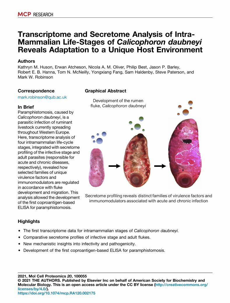
In BriefParamphistomosis, caused byCalicophoron daubneyi, is aparasitic infection of ruminantlivestock currently spreadingthroughout Western Europe.Here, transcriptome analysis offour intramammalian life-cyclestages, integratedwith secretomeprofiling of the infective stage andadult parasites (responsible foracute and chronic diseases,respectively), revealed howselected families of uniquevirulence factors andimmunomodulators are regulatedin accordance with flukedevelopment and migration. Thisanalysis allowed the developmentof the first coproantigen-basedELISA for paramphistomosis.
Highlights
• The first transcriptome data for intramammalian stages of Calicophoron daubneyi.
• Comparative secretome profiles of infective stage and adult flukes.
• New mechanistic insights into infectivity and pathogenicity.
• Development of the first coproantigen-based ELISA for paramphistomosis.
5y Elsevier Inc on behalf of American Society for Biochemistry andccess article under the CC BY license (http://creativecommons.org/
02175
RESEARCH
Transcriptome and Secretome Analysis of Intra-Mammalian Life-Stages of Calicophorondaubneyi Reveals Adaptation to a Unique HostEnvironmentKathryn M. Huson1, Erwan Atcheson1, Nicola A. M. Oliver1, Philip Best1, Jason P. Barley2,Robert E. B. Hanna2, Tom N. McNeilly3, Yongxiang Fang4, Sam Haldenby4,Steve Paterson4, and Mark W. Robinson1,*
Paramphistomosis, caused by the rumen fluke, Cal-icophoron daubneyi, is a parasitic infection of ruminantlivestock, which has seen a rapid rise in prevalencethroughout Western Europe in recent years. After inges-tion of metacercariae (parasite cysts) by the mammalianhost, newly excysted juveniles (NEJs) emerge and invadethe duodenal submucosa, which causes significant pa-thology in heavy infections. The immature flukes thenmigrate upward, along the gastrointestinal tract, andenter the rumen where they mature and begin to produceeggs. Despite their emergence, and sporadic outbreaksof acute disease, we know little about the molecularmechanisms used by C. daubneyi to establish infection,acquire nutrients, and avoid the host immune response.Here, transcriptome analysis of four intramammalian life-cycle stages, integrated with secretome analysis of theNEJ and adult parasites (responsible for acute andchronic diseases, respectively), revealed how theexpression and secretion of selected families of virulencefactors and immunomodulators are regulated in accor-dance with fluke development and migration. Our datashow that while a family of cathepsins B with varying S2subsite residues (indicating distinct substrate specific-ities) is differentially secreted by NEJs and adult flukes,cathepsins L and F are secreted in low abundance byNEJs only. We found that C. daubneyi has an expandedfamily of aspartic peptidases, which is upregulated inadult worms, although they are under-represented in thesecretome. The most abundant proteins in adult flukesecretions were helminth defense molecules that likelyestablish an immune environment permissive to flukesurvival and/or neutralize pathogen-associated molecularpatterns such as bacterial lipopolysaccharide in themicrobiome-rich rumen. The distinct collection of mole-cules secreted by C. daubneyi allowed the developmentof the first coproantigen-based ELISA for para-mphistomosis which, importantly, did not recognize
From the 1School of Biological Sciences, Queen’s University Belfast, BBiosciences Institute, Belfast, Northern Ireland; 3Disease Control Depafor Genomic Research, University of Liverpool, Liverpool, England
*For correspondence: Mark W. Robinson, [email protected].
© 2021 THE AUTHORS. Published by Elsevier Inc on behalf of American Society for BioThis is an open access article under the CC BY license (http://creativecommons.org/lice
antigens from other helminths commonly found as coin-fections with rumen fluke.
Infections by parasitic fluke are an important animal healthand production concern for livestock producers worldwide. Inthe United Kingdom, and throughout Europe, the liver fluke(Fasciola hepatica) has historically been a major focus forproducers. In recent years, however, there has been a sharpincrease in both the incidence and severity of rumen fluke (theparamphistome Calicophoron daubneyi) infections in bothsheep and cattle (1). Paramphistomes are endemic to sub-tropical and tropical regions and are believed to have beencarried into Western Europe from North Africa via the move-ment of ruminant livestock (2). Although the exact reasons forthe rise in rumen fluke infections in Europe are not fully un-derstood, the increase in warm wet summers and mild win-ters—conditions that favor Galba truncatula, the snailintermediate host of C. daubneyi—is thought to be a majorcontributing factor (3).Acute, clinical paramphistomosis is caused when grazing
stock ingest large numbers of metacercariae from pasture,which then excyst en masse in the duodenum (4). The newlyexcysted juvenile (NEJ) flukes then migrate into the intestinalsubmucosa causing significant damage to the host tissue (5).Large areas of damaged small intestine may hemorrhage,causing significant blood loss and hypoalbuminemia,frequently resulting in mortality at this point (6, 7). After aperiod spent feeding on the host tissue in the small intestine,immature paramphistomes migrate to the rumen where theymature, and infections become patent (8). Although chronicinfections are generally seen as well tolerated, postmortemobservations have noted both rumenitis and abomasitis ininfected animals (9, 10), along with atrophy of the rumenpapillae (1, 10).
elfast, Northern Ireland; 2Veterinary Sciences Division, Agri-Food andrtment, Moredun Research Institute, Edinburgh, Scotland; and 4Centre
Mol Cell Proteomics (2021) 20 100055 1chemistry and Molecular Biology.nses/by/4.0/). https://doi.org/10.1074/mcp.RA120.002175
The Transcriptome and Secretome of C. daubneyi
Despite a prevalence of 55% to 77% (1), clinical disease isstill relatively rare in the United Kingdom/Ireland. However,fatal disease outbreaks, linked to significant immature parasiteburdens, have been reported in both sheep and cattle inrecent years (4, 6, 10–13). Control of fluke infection currentlyrelies on anthelmintic drugs. While several drugs show effi-cacy against liver fluke, only one anthelmintic (oxyclozanide) iseffective against rumen fluke (14, 15). This makes correctdiagnosis imperative. However, detection of rumen flukeinfection currently requires either examination of animals atpostmortem or labor-intensive fecal egg counts that onlydetect chronic infection due to the presence of egg-producingadult flukes. Thus, the development of new tools for morerapid diagnosis of C. daubneyi (including acute infection) isrequired.Compared with other helminths of veterinary importance,
C. daubneyi remains a poorly studied species. We have muchto learn about its basic biology and interactions with theruminant host, particularly the infective juvenile stages (1).Here, we have performed the first transcriptomic analysis of allfour major intramammalian life-cycle stages of C. daubneyi.These data, coupled with secretome analysis of the infectiveand adult stages that are associated with acute and chronicdiseases, respectively, have provided a greater understandingof the key biochemical and molecular mechanisms underpin-ning C. daubneyi infectivity, migration, and developmentwithin its ruminant host. Our data reveal how the parasiteregulates expression and secretion of a collection of mole-cules required for tissue invasion, nutrition, and modulation ofthe host immune response according to fluke developmentand exposure to different host microenvironments (i.e., theduodenum and rumen). Owing to the unique composition ofC. daubneyi secretions compared with those of F. hepatica(often found as coinfections), the diagnostic potential of thesemolecules was investigated. Accordingly, we present the firstELISA-based assay capable of detecting C. daubneyi antigensin fecal samples from naturally infected cattle. Our datarepresent an important foundation for future studies aimed atunderstanding rumen fluke infectivity and the development offuture treatment options and diagnostic tests.
EXPERIMENTAL PROCEDURES
Experimental Design and Statistical Rationale
For transcriptomics studies, 3 biological replicates of each life-cyclestage were prepared for Illumina RNA-Seq, with 1 replicate used forPacBio sequencing for use as full-length transcript scaffold data duringassembly. Three biological replicates were also used for the proteomicsstudy of excretory/secretory (E/S) products analyzed by LC-MS/MS.ELISA data are representative of three independent experiments.
Source of Parasite Material and RNA Extraction
To obtain RNA samples, C. daubneyi adults and newly migratedparasites were collected from the rumen of a naturally-infected cow ina local abattoir (Dungannon, Northern Ireland). Individual adults and
2 Mol Cell Proteomics (2021) 20 100055
small groups (approx. 6) of newly migrated flukes were rinsed briefly insterile warm (39 ◦C) PBS and immediately placed into TRIzol reagent(Life Technologies) to preserve mRNA. Immature parasites wereremoved from the duodenum of a recently deceased bovine hostduring postmortem examination, and approximately 200 mg of para-site material placed directly into TRIzol reagent. Rumen fluke meta-cercariae (Ridgeway Research) were excysted in vitro and the resultingNEJ parasites cultured in RPMI-1640 medium for 24 h as describedpreviously (16). The culture media was removed, and TRIzol reagentwas added directly to groups of 500 NEJs. All samples were stored inTRIzol at −80 ◦C until RNA extraction. Samples were homogenized inTRIzol, and total RNA was extracted following the manufacturer’sprotocol. RNA yields were quantified using a NanoDrop spectropho-tometer and shipped to the Centre for Genomic Research (Universityof Liverpool, UK) for library construction. Before sequencing, therumen fluke species was confirmed by PCR using C. daubneyi–spe-cific primers targeting an 885-bp region of the cytochrome oxidase 1gene; Cd_Cox1F 5’-TGGAGAGTTTGGCGTCTTTT-3’ and Cd_Cox1R5’-CCATCTTCCACCTCATCTGG-3’ as previously described (17).
PacBio Iso-Seq Library Preparation and Sequencing
Full-length cDNA was made from 1 μg of total RNA from eachsample using the TeloPrime full-length cDNA amplification kit fromLexogen. The final second strand product was amplified for 20 cyclesand converted to a PacBio SMRTbell library using the SMRTbellTemplate Prep Kit 1.0. Each sample was run on a single Sequel SMRTcell v2 using MagBead loading and 10-h movie times. PacBio readswere analyzed using the Iso-seq pipeline (SMRT Link version4.0.0.190159), with default parameters. Each sample was processedindependently. Subsequently, high- and low-quality full-length iso-forms from each sample were clustered using CD-HIT (18). Clusterrepresentatives were polished with Arrow (19) and reclustered with cd-hit-est, as above but with more relaxed parameters to limit over-clustering (-aL 0.97). These two steps were iteratively performed togenerate a set of 27,949 isoforms, which were subsequently filtered toremove any sequences that (a) had a mean base quality score of lessthan 50 or (b) had more than 1% of bases with a quality score below10. This yielded a final set of 27,254 isoforms.
Annotation of Transcripts
Putative proteins sequences were obtained for isoforms usingTransDecoder (version 3.0.1; (20)), selecting for a minimum amino acidsequence length of 50. Sequences were annotated with input for theTrinotate pipeline (version 3.1.0; (20)), according to the proposedguidelines from the authors. The BLAST (21) was used to alignnucleotide and protein sequences against UniProt (release 2017_11)and TrEMBL and against proteins encoded by F. hepatica genemodels available at WormBase ParaSite (ftp://ftp.ebi.ac.uk/pub/databases/wormbase/parasite/releases/WBPS2/species/fasciola_hepatica/PRJEB6687/fasciola_hepatica.PRJEB6687.WBPS2.protein.fa.gz). All BLAST alignments which yielded e-values of 1 x 10⁻⁵ or lowerwere reported. Predicted amino acid sequences were compared withPfam-A (release 31.0) (22), using hmmscan (version 3.1b1; (23)). Signalpeptides and transmembrane domains were predicted using SignalP(version 4.1; (24)) and TMHMM (version 2.0; (25)).
Illumina RNA-Seq Library Preparation and Sequencing
Triplicate total RNA samples (from C. daubneyi NEJs, immatureintestinal flukes, newly migrated flukes, and mature adults) weretreated with Turbo DNase (Life Technologies) to remove all contami-nating DNA. 1 μg total RNA was selected for poly A using NEBNextPoly(A) mRNA Magnetic Isolation Module. RNA-Seq libraries wereprepared from the enriched material using the NEBNext Ultra
The Transcriptome and Secretome of C. daubneyi
Directional RNA Library Prep Kit for Illumina using 12 cycles ofamplification. Final libraries were pooled in equimolar amounts usingthe Qubit and Bioanalyzer data and checked by quantitative PCR fortemplate loading calculations. DNA was diluted to a loading concen-tration of 300 pM. The RNA libraries were sequenced on an IlluminaHiSeq 4000 platform with version 1 chemistry using sequencing bysynthesis (SBS) technology to generate 2 x 150 bp paired-end reads.
RNA-Seq Data Processing and Quality Filtering
Base calling and de-multiplexing of indexed reads was performedby CASAVA version 1.8.2 (Illumina). Sequences were trimmed usingCutadapt, version 1.2.1 (26), to remove any adaptor sequencesmatching with 3 or more bases. The reads were further trimmed toremove low-quality bases, using Sickle, version 1.200, with a mini-mum window quality score of 20. Illumina reads were aligned to thefinal Iso-Seq isoforms using Bowtie2, version 2.1.0 (27), accepting allglobal concordantly paired alignments.
Differential Expression Analysis
Read alignment data were used as input to BitSeq (28), to estimateraw expression values for each transcript in the dataset. Differentialtranscript expression analysis was applied to the read count data,which was conducted in the R environment, using the DESeq2package (29). Transcripts per million (TPM) values were calculated bydividing the counts to each transcript by the length of the transcriptand then normalized further to generate counts per million reads.Transcripts were clustered according to their log-fold changes inexpression between sample groups, using k-means clustering func-tions in R.
Discovery and Characterization of C. daubneyi Gene Families
C. daubneyi gene families and specific homologs of interest wereidentified using BlastP (1.0 x 10⁻⁶ e-value cut-off) run locally withBioEdit (30) using published query sequences from other trematodespecies (F. hepatica, Fasciola gigantica, Opisthorchis viverrini, Clo-norchis sinensis, and Schistosoma spp.). Duplicate and truncatedsequences were manually removed from the BLAST hits, and tran-script TPM values were extracted for the remaining transcripts at eachof the 4 life-cycle stages to allow differential expression (DE) analysisacross the intramammalian stages of the C. daubneyi lifecycle andused to produce heatmaps via the heatmapper.ca expression tool (31).Helical wheel analysis was conducted on C. daubneyi HDM familymembers (http://tcdb.org/progs/helical_wheel.php) to identify amphi-pathic regions.
Neighbor-joining phylogenetic trees were constructed for selectedprotein sequences using MEGA6 (32). The bootstrap consensus treeinferred from 1000 replicates is taken to represent the evolutionaryhistory of the taxa analyzed (33). Branches corresponding to partitionsreproduced in less than 50% bootstrap replicates are collapsed. Thepercentage of replicate trees in which the associated taxa clusteredtogether in the bootstrap test (1000 replicates) is shown next to thebranches. The evolutionary distances were computed using thePoisson correction method (34) and are in the units of the number ofamino acid substitutions per site.
Collection of Parasite E/S Products
To prepare secretions, adult rumen flukes were thoroughly washed(3 x 5 min) with warm (39 ◦C) sterile PBS to void their gut contents andto remove any host contaminants. The integrity and movement of theflukes were monitored by microscopy and any found to be damagedor dead were discarded before culture. Flukes were then maintained inRPMI-1640 culture medium containing 0.1% glucose, 100 U penicillin,and 100 μg/ml streptomycin (Sigma-Aldrich), at 1 worm/ml for 5 h at
39 ◦C. Whole E/S products were also collected from C. daubneyi NEJs(in groups of 500 parasites) as described previously (16). ExcystedNEJs were cultured in 1-ml RPMI-1640 supplemented with 100 IUmL−1 penicillin and 100-mg mL−1 streptomycin for 24 h at 39 ◦C. Theculture media was recovered and centrifuged for 30 min at 1500g toremove large debris. Adult and NEJ E/S fractions were concentrated(approx. 10-fold) using Amicon Ultra Centrifugal Filter Units with a 3-kDa molecular weight cutoff (Millipore, UK) and stored at –80 ◦C un-til further analysis.
E/S proteins were reduced with 2-mM DTT in 50-mM NH4HCO3 at60 ◦C for 20 min and alkylated with 5-mM iodoacetamide at roomtemperature (RT) (18–21 ◦C) in the dark for 30 min. E/S samples wereincubated with 100 ng/μl sequencing grade trypsin (Promega) over-night at 37 ◦C. The digestions were stopped by the addition of TFA toa final concentration of 0.1%.
MS analysis of C. daubneyi Proteins
E/S proteins were analyzed in biological triplicate. Tryptic peptideswere dried in a vacuum centrifuge and reconstituted with 10 μl of 0.1%TFA before analysis by LC-MS/MS. Peptides is 5 μl of the resultingsuspension were purified using an Acclaim PepMap 100 column (C18,100 μM x 2 cm) before delivery to an analytical column (Eksigent C18-CL NanoLC Column, 3 μm; 75 μm x 15 cm) equilibrated in 5%acetonitrile/0.1% formic acid (FA). Elution was carried out with a lineargradient of 5 to 35% buffer B in buffer A for 30 min (buffer A: 0.1% FA;buffer B: acetonitrile, 0.1% FA) at a flow rate of 300 nl/min. Peptideswere analyzed using an LTQ OrbiTrap Velos Pro (Thermo Scientific)operating in an information-dependent acquisition mode using a top15 method. MS spectra were acquired in the Orbitrap analyzer with amass range of 335 to 1800 m/z, with a resolution of 60,000 in theOrbitrap. Collision-induced dissociation peptide fragments were ac-quired in the ion trap with a collision energy of 35, activation energy of0.25, and 10-ms activation time, with a default charge state of 2 forfragment ions. Orbitrap Velos RAW data files were extracted andconverted to Mascot generic files (.mgf) for database searching usingMascot v2.4.1 (Matrix Science, London, UK).
Database Searching
All MS/MS samples were analyzed using Mascot (version 2.4.1,Matrix Science, London, UK). Mascot was set up to search theLIMS12524_20171212 C. daubneyi transcriptome database (version1.0, 48,899 entries) assuming the digestion enzyme strict trypsin with1 missed cleavage allowed. The transcript data can be accessed viathe European Nucleotide Archive (www.ebi.ac.uk/ena) under acces-sion number PRJEB28150. Mascot was searched with a fragment ionmass tolerance of 0.60 Da and a parent ion tolerance of 10.0 parts permillion. Carbamidomethylation of cysteine was specified in Mascot asa fixed modification. Gln->pyro-Glu of the N-terminus, deamidation ofasparagine and glutamine, oxidation of methionine, dioxidation ofmethionine, and acetylation of the N-terminus were specified inMascot as variable modifications.
Criteria for Protein Identification and Quantitation
Scaffold (version Scaffold_4.4.5, Proteome Software Inc., Portland,OR) was used to validate MS/MS–based peptide and protein identi-fications. Peptide identifications were accepted if they could beestablished at greater than 95.0% probability by the Scaffold Localfalse discovery rate algorithm. Protein identifications were accepted ifthey could be established at greater than 99.0% probability andcontained at least 2 identified peptides. Protein probabilities wereassigned by the ProteinProphet algorithm (35). Proteins that containedsimilar peptides and could not be differentiated based on MS/MSanalysis alone were grouped to satisfy the principles of parsimony.
Mol Cell Proteomics (2021) 20 100055 3
The Transcriptome and Secretome of C. daubneyi
Proteins sharing significant peptide evidence were grouped intoclusters. In addition, a label-free quantitative analysis was performedin Scaffold for those proteins (NEJ vs adult), with at least two uniquepeptides, that were present in all three biological replicates. Theexponentially modified protein abundance index (emPAI) was used asa quantitative method with a t-test (Benjamini-Hochberg false dis-covery rate correction; significance level, p < 0.05) as a statisticalmethod. For quantitation, missing values were replaced with a mini-mum value (default of zero), and normalization was performed withzero as a minimum value.
Coproantigen ELISA
The coproantigen ELISA was tested against fecal samples takenfrom cattle naturally infected with C. daubneyi and with fecal eggcounts ranging from 12 to 299 eggs per gram. Adult E/S productswere used as a positive control. Fecal supernatants were preparedusing a method adapted from Teimoori et al (36). Briefly, 1-g feces washomogenized in 3-ml lysis buffer (20-mM Tris HCl, 0.5% SDS, 8 Murea) and rotated overnight at RT (18–21 ◦C). The extracts werecentrifuged at 8000g for 10 min, with the supernatant further centri-fuged at 10,000g for 5 min and the final supernatant stored at −20 ◦Cuntil use. Affinity-purified antibodies (anti–C. daubneyi E/S proteinantibody [α-CdE/S]) were raised in a rabbit against whole E/S proteinscollected from adult C. daubneyi maintained in vitro (Eurogentec). Aproportion of these antibodies were biotinylated (biotinylated anti–C.daubneyi E/S protein antibody). The 96-well flat-bottom microtiterplates (Immulon 2HB, Thermo Scientific) were incubated with 50-μl(2 μg/ml) α-CdE/S Ig diluted in PBS per well and incubated overnightat 4 ◦C. Plates were washed six times in PBS containing 0.5% Tween20 and then blocked for 1 h at RT in 1% skimmed milk/PBS-Tween.Fifty μl of the fecal supernatants was then added, at a dilution of1:27 in PBS, and incubated for 2 h at RT. Plates were washed asbefore, and 50-μl (2 μg/ml) biotinylated anti–C. daubneyi E/S proteinantibody added per well and incubated for 1 h at RT. Plates werewashed as before, and 50 μl of ExtrAvidin–alkaline phosphataseconjugate (Sigma) (1:1500 dilution) was added per well. Plates werewashed as before prior to the addition of 100 μl per well of SIGMA-FAST p-nitrophenyl phosphate substrate (Sigma-Aldrich). After incu-bation at RT for 30 min, plates were read at 405 nm using a POLARstarOmega microplate reader (BMG LABTECH).
For assessment of specificity, 96-well plates were coated with 2 μg/ml of whole-worm extracts or E/S products (50 μl/well) fromF. hepatica and a range of gastrointestinal nematode parasites that arecommonly found in ruminants in the United Kingdom/Ireland (Tela-dorsagia circumcincta, Ostertagia ostertagi, Haemonchus contortus,Trichostrongylus axei, and Trichostrongylus vitrinus) and incubatedovernight at RT. Plates were washed and blocked as described above,and then 2 μg/ml of α-CdE/S (50 μl/well) was applied for 2 h at RT.Plates were washed as before, and 50 μl per well of anti-rabbitimmunoglobulin G/alkaline phosphatase conjugate (Sigma) wasapplied at a 1:1500 dilution. Plates were washed, developed, and readas described above. For all ELISAs, samples were analyzed in tripli-cate, and results are the representative of at least three independentexperiments. ELISA results were considered positive for rumen flukeinfection if returning an optical density greater than the mean plus twoSDs of uninfected control samples. This cutoff was also used to defineno cross-reactivity with the other species tested.
Immunofluorescence Microscopy and Whole-Mount Preparations
Adult C. daubneyi and F. hepatica were fixed with 4% para-formaldehyde in PBS (Sigma-Aldrich) overnight at 4 ◦C and subse-quently embedded in JB-4 resin (Sigma-Aldrich). Semithin sections, 2-μm thick, were cut on a pyramitome and mounted on clean glassslides. For immunofluorescence, JB-4 sections were washed with
4 Mol Cell Proteomics (2021) 20 100055
PBS and then incubated in 10 μg/ml of affinity-purified polyclonalα-CdE/S in an antibody diluent (AbD: PBS containing 0.2% (v/v) TritonX-100) overnight at 4 ◦C. As a negative control, comparable sectionswere incubated in rabbit preimmune serum. The sections were thenwashed three times in AbD before incubation in a 1:100 dilution of thesecondary antibody, FITC-conjugated goat anti-rabbit immunoglob-ulin G (Sigma-Aldrich), in AbD for 1 h at RT. After three washes in PBS,the sections were mounted in glycerol containing 10% (v/v) PBS and0.1 M propyl gallate (Sigma-Aldrich) and then viewed under a LeicaDM2500 fluorescent microscope.
For whole-mount preparations, flukes were flat-fixed in formolacetic alcohol solution for 3 to 5 days at RT and then incubated in 1%(w/v) carmine (Sigma) for 1.5 h at RT. Flukes were then destained in70% acidified ethanol overnight and dehydrated in a series of alco-hols. The samples were then incubated overnight with HistoChoiceclearing agent (Sigma) and mounted on glass slides using DPXmounting media (Sigma). Flukes were viewed using a Leica DM2500microscope.
RESULTS
The Intramammalian Life-Cycle Stages of C. daubneyi
The intramammalian life cycle of C. daubneyi involves fourmajor developmental stages: (1) the NEJ flukes that emergefrom ingested metacercariae and migrate into the duodenalsubmucosa, (2) the immature worms that actively feed on hosttissue in the intestine, (3) small newly migrated flukes thathave recently completed their migration to the rumen from theduodenum, and (4) mature adult flukes that are well estab-lished in the rumen. Macroscopic/microscopic examination ofthese stages shows their developmental progression (Fig. 1).The NEJs are typically 150 μm in length and already bear theoral and posterior suckers characteristic of paramphistomes.Histological examination revealed that while no reproductivestructures were present, subtegumental muscle bundles and adeveloping bifurcated gut were observed (data not shown).The immature flukes (2–3 mm in length) are generally found inassociation with the duodenal mucosa though some remainwithin the intestinal lumen in very heavy infections. A promi-nent bifurcated gut was clearly visible in carmine-stainedwhole-mount worms as were genital anlagen correspondingto the rudimentary testes (anterior and posterior) and ovary.No vitellaria was observed at this stage (Fig. 1).The newly migrated fluke (typically 5–7 mm in length) are
found in the rumen attached to the rumen papillae, wall, orcontents. Live worms are bright red in appearance and sharesimilar morphology to adult fluke but are much smaller in size.In whole-mount preparations, the testes and ovary appearlarger and more developed, and vitelline follicles have nowappeared around the periphery of the fluke body. Despite theirsmall size, eggs were often observed in the uterus of newlymigrated flukes (Fig. 1). The eggs produced by newly migratedC. daubneyi were viable and released highly motile miracidiaduring in vitro egg hatch assays performed as describedpreviously (37). Adult C. daubneyi (typically 1.5–2.0 cm inlength) appear mid to pale pink with redder coloration aroundboth the anterior and posterior suckers. They are found
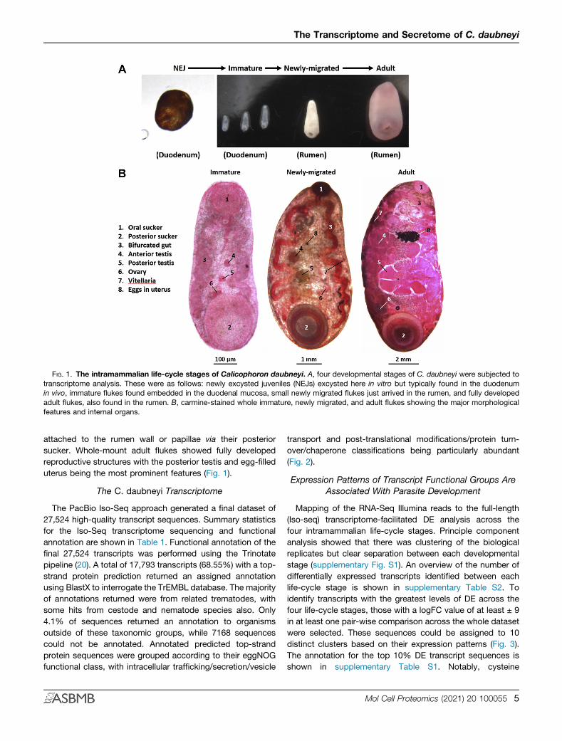
FIG. 1. The intramammalian life-cycle stages of Calicophoron daubneyi. A, four developmental stages of C. daubneyi were subjected totranscriptome analysis. These were as follows: newly excysted juveniles (NEJs) excysted here in vitro but typically found in the duodenumin vivo, immature flukes found embedded in the duodenal mucosa, small newly migrated flukes just arrived in the rumen, and fully developedadult flukes, also found in the rumen. B, carmine-stained whole immature, newly migrated, and adult flukes showing the major morphologicalfeatures and internal organs.
The Transcriptome and Secretome of C. daubneyi
attached to the rumen wall or papillae via their posteriorsucker. Whole-mount adult flukes showed fully developedreproductive structures with the posterior testis and egg-filleduterus being the most prominent features (Fig. 1).
The C. daubneyi Transcriptome
The PacBio Iso-Seq approach generated a final dataset of27,524 high-quality transcript sequences. Summary statisticsfor the Iso-Seq transcriptome sequencing and functionalannotation are shown in Table 1. Functional annotation of thefinal 27,524 transcripts was performed using the Trinotatepipeline (20). A total of 17,793 transcripts (68.55%) with a top-strand protein prediction returned an assigned annotationusing BlastX to interrogate the TrEMBL database. The majorityof annotations returned were from related trematodes, withsome hits from cestode and nematode species also. Only4.1% of sequences returned an annotation to organismsoutside of these taxonomic groups, while 7168 sequencescould not be annotated. Annotated predicted top-strandprotein sequences were grouped according to their eggNOGfunctional class, with intracellular trafficking/secretion/vesicle
transport and post-translational modifications/protein turn-over/chaperone classifications being particularly abundant(Fig. 2).
Expression Patterns of Transcript Functional Groups AreAssociated With Parasite Development
Mapping of the RNA-Seq Illumina reads to the full-length(Iso-seq) transcriptome-facilitated DE analysis across thefour intramammalian life-cycle stages. Principle componentanalysis showed that there was clustering of the biologicalreplicates but clear separation between each developmentalstage (supplementary Fig. S1). An overview of the number ofdifferentially expressed transcripts identified between eachlife-cycle stage is shown in supplementary Table S2. Toidentify transcripts with the greatest levels of DE across thefour life-cycle stages, those with a logFC value of at least ± 9in at least one pair-wise comparison across the whole datasetwere selected. These sequences could be assigned to 10distinct clusters based on their expression patterns (Fig. 3).The annotation for the top 10% DE transcript sequences isshown in supplementary Table S1. Notably, cysteine
Mol Cell Proteomics (2021) 20 100055 5
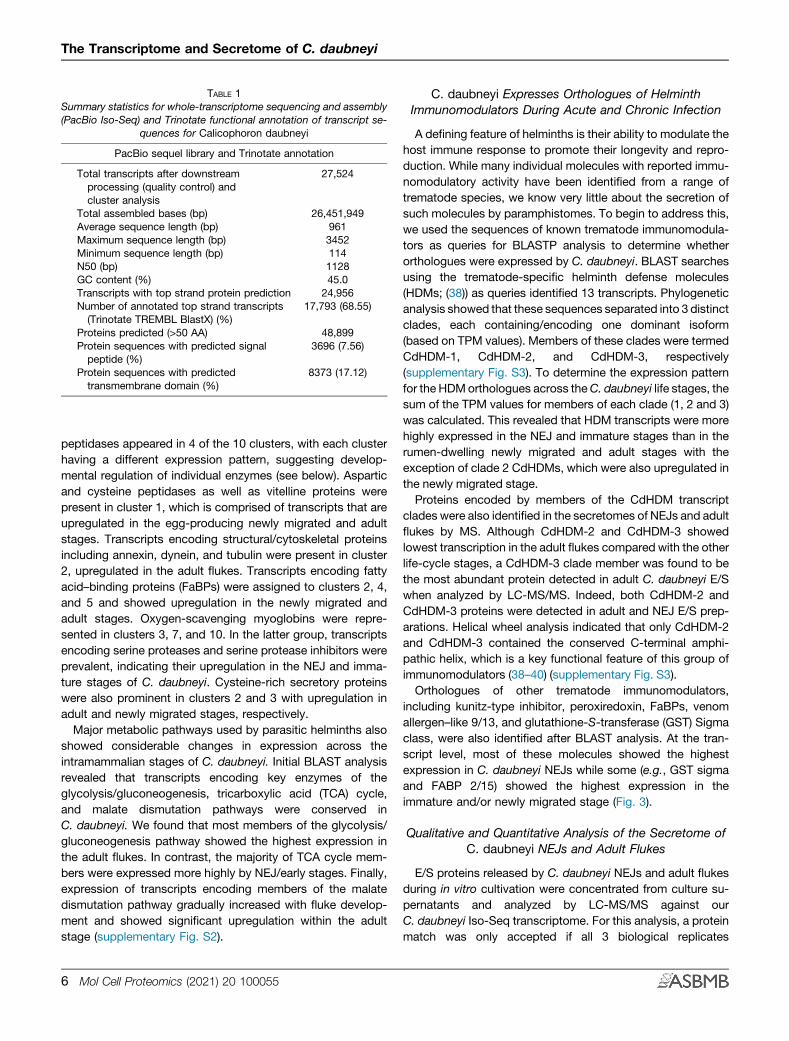
TABLE 1Summary statistics for whole-transcriptome sequencing and assembly(PacBio Iso-Seq) and Trinotate functional annotation of transcript se-
quences for Calicophoron daubneyi
PacBio sequel library and Trinotate annotation
Total transcripts after downstreamprocessing (quality control) andcluster analysis
27,524
Total assembled bases (bp) 26,451,949Average sequence length (bp) 961Maximum sequence length (bp) 3452Minimum sequence length (bp) 114N50 (bp) 1128GC content (%) 45.0Transcripts with top strand protein prediction 24,956Number of annotated top strand transcripts(Trinotate TREMBL BlastX) (%)
17,793 (68.55)
Proteins predicted (>50 AA) 48,899Protein sequences with predicted signalpeptide (%)
3696 (7.56)
Protein sequences with predictedtransmembrane domain (%)
8373 (17.12)
The Transcriptome and Secretome of C. daubneyi
peptidases appeared in 4 of the 10 clusters, with each clusterhaving a different expression pattern, suggesting develop-mental regulation of individual enzymes (see below). Asparticand cysteine peptidases as well as vitelline proteins werepresent in cluster 1, which is comprised of transcripts that areupregulated in the egg-producing newly migrated and adultstages. Transcripts encoding structural/cytoskeletal proteinsincluding annexin, dynein, and tubulin were present in cluster2, upregulated in the adult flukes. Transcripts encoding fattyacid–binding proteins (FaBPs) were assigned to clusters 2, 4,and 5 and showed upregulation in the newly migrated andadult stages. Oxygen-scavenging myoglobins were repre-sented in clusters 3, 7, and 10. In the latter group, transcriptsencoding serine proteases and serine protease inhibitors wereprevalent, indicating their upregulation in the NEJ and imma-ture stages of C. daubneyi. Cysteine-rich secretory proteinswere also prominent in clusters 2 and 3 with upregulation inadult and newly migrated stages, respectively.Major metabolic pathways used by parasitic helminths also
showed considerable changes in expression across theintramammalian stages of C. daubneyi. Initial BLAST analysisrevealed that transcripts encoding key enzymes of theglycolysis/gluconeogenesis, tricarboxylic acid (TCA) cycle,and malate dismutation pathways were conserved inC. daubneyi. We found that most members of the glycolysis/gluconeogenesis pathway showed the highest expression inthe adult flukes. In contrast, the majority of TCA cycle mem-bers were expressed more highly by NEJ/early stages. Finally,expression of transcripts encoding members of the malatedismutation pathway gradually increased with fluke develop-ment and showed significant upregulation within the adultstage (supplementary Fig. S2).
6 Mol Cell Proteomics (2021) 20 100055
C. daubneyi Expresses Orthologues of HelminthImmunomodulators During Acute and Chronic Infection
A defining feature of helminths is their ability to modulate thehost immune response to promote their longevity and repro-duction. While many individual molecules with reported immu-nomodulatory activity have been identified from a range oftrematode species, we know very little about the secretion ofsuch molecules by paramphistomes. To begin to address this,we used the sequences of known trematode immunomodula-tors as queries for BLASTP analysis to determine whetherorthologues were expressed by C. daubneyi. BLAST searchesusing the trematode-specific helminth defense molecules(HDMs; (38)) as queries identified 13 transcripts. Phylogeneticanalysis showed that these sequences separated into 3 distinctclades, each containing/encoding one dominant isoform(based on TPM values). Members of these clades were termedCdHDM-1, CdHDM-2, and CdHDM-3, respectively(supplementary Fig. S3). To determine the expression patternfor the HDMorthologues across theC. daubneyi life stages, thesum of the TPM values for members of each clade (1, 2 and 3)was calculated. This revealed that HDM transcripts were morehighly expressed in the NEJ and immature stages than in therumen-dwelling newly migrated and adult stages with theexception of clade 2 CdHDMs, which were also upregulated inthe newly migrated stage.Proteins encoded by members of the CdHDM transcript
clades were also identified in the secretomes of NEJs and adultflukes by MS. Although CdHDM-2 and CdHDM-3 showedlowest transcription in the adult flukes compared with the otherlife-cycle stages, a CdHDM-3 clade member was found to bethe most abundant protein detected in adult C. daubneyi E/Swhen analyzed by LC-MS/MS. Indeed, both CdHDM-2 andCdHDM-3 proteins were detected in adult and NEJ E/S prep-arations. Helical wheel analysis indicated that only CdHDM-2and CdHDM-3 contained the conserved C-terminal amphi-pathic helix, which is a key functional feature of this group ofimmunomodulators (38–40) (supplementary Fig. S3).Orthologues of other trematode immunomodulators,
including kunitz-type inhibitor, peroxiredoxin, FaBPs, venomallergen–like 9/13, and glutathione-S-transferase (GST) Sigmaclass, were also identified after BLAST analysis. At the tran-script level, most of these molecules showed the highestexpression in C. daubneyi NEJs while some (e.g., GST sigmaand FABP 2/15) showed the highest expression in theimmature and/or newly migrated stage (Fig. 3).
Qualitative and Quantitative Analysis of the Secretome ofC. daubneyi NEJs and Adult Flukes
E/S proteins released by C. daubneyi NEJs and adult flukesduring in vitro cultivation were concentrated from culture su-pernatants and analyzed by LC-MS/MS against ourC. daubneyi Iso-Seq transcriptome. For this analysis, a proteinmatch was only accepted if all 3 biological replicates
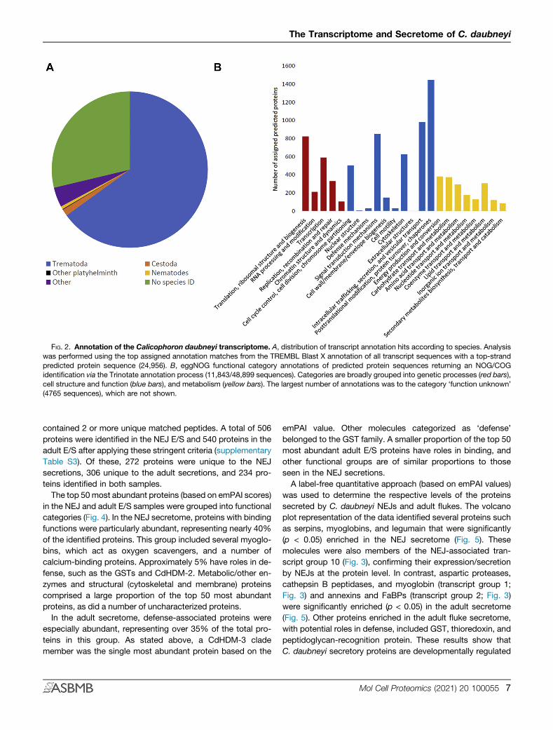
FIG. 2. Annotation of the Calicophoron daubneyi transcriptome. A, distribution of transcript annotation hits according to species. Analysiswas performed using the top assigned annotation matches from the TREMBL Blast X annotation of all transcript sequences with a top-strandpredicted protein sequence (24,956). B, eggNOG functional category annotations of predicted protein sequences returning an NOG/COGidentification via the Trinotate annotation process (11,843/48,899 sequences). Categories are broadly grouped into genetic processes (red bars),cell structure and function (blue bars), and metabolism (yellow bars). The largest number of annotations was to the category ‘function unknown’(4765 sequences), which are not shown.
The Transcriptome and Secretome of C. daubneyi
contained 2 or more unique matched peptides. A total of 506proteins were identified in the NEJ E/S and 540 proteins in theadult E/S after applying these stringent criteria (supplementaryTable S3). Of these, 272 proteins were unique to the NEJsecretions, 306 unique to the adult secretions, and 234 pro-teins identified in both samples.The top 50most abundant proteins (based on emPAI scores)
in the NEJ and adult E/S samples were grouped into functionalcategories (Fig. 4). In the NEJ secretome, proteins with bindingfunctions were particularly abundant, representing nearly 40%of the identified proteins. This group included several myoglo-bins, which act as oxygen scavengers, and a number ofcalcium-binding proteins. Approximately 5% have roles in de-fense, such as the GSTs and CdHDM-2. Metabolic/other en-zymes and structural (cytoskeletal and membrane) proteinscomprised a large proportion of the top 50 most abundantproteins, as did a number of uncharacterized proteins.In the adult secretome, defense-associated proteins were
especially abundant, representing over 35% of the total pro-teins in this group. As stated above, a CdHDM-3 clademember was the single most abundant protein based on the
emPAI value. Other molecules categorized as ‘defense’belonged to the GST family. A smaller proportion of the top 50most abundant adult E/S proteins have roles in binding, andother functional groups are of similar proportions to thoseseen in the NEJ secretions.A label-free quantitative approach (based on emPAI values)
was used to determine the respective levels of the proteinssecreted by C. daubneyi NEJs and adult flukes. The volcanoplot representation of the data identified several proteins suchas serpins, myoglobins, and legumain that were significantly(p < 0.05) enriched in the NEJ secretome (Fig. 5). Thesemolecules were also members of the NEJ-associated tran-script group 10 (Fig. 3), confirming their expression/secretionby NEJs at the protein level. In contrast, aspartic proteases,cathepsin B peptidases, and myoglobin (transcript group 1;Fig. 3) and annexins and FaBPs (transcript group 2; Fig. 3)were significantly enriched (p < 0.05) in the adult secretome(Fig. 5). Other proteins enriched in the adult fluke secretome,with potential roles in defense, included GST, thioredoxin, andpeptidoglycan-recognition protein. These results show thatC. daubneyi secretory proteins are developmentally regulated
Mol Cell Proteomics (2021) 20 100055 7
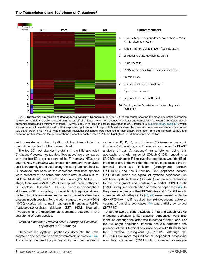
FIG. 3. Differential expression of Calicophoron daubneyi transcripts. The top 10% of transcripts showing the most differential expressionacross our sample set were selected using a cut-off of at least a 9-log fold change in at least one comparison between C. daubneyi devel-opmental stages and a minimum average TPM value of 2 in at least one stage. This returned 2470 transcripts (supplementary Table S1), whichwere grouped into clusters based on their expression pattern. A heat map of TPM values scaled by transcript values (where red indicates a lowvalue and green a high value) was produced. Individual transcripts were matched to their BlastX annotation from the Trinotate output, andcommon proteins/protein family annotations present in each cluster (1–10) are highlighted. TPM, transcripts per million.
The Transcriptome and Secretome of C. daubneyi
and correlate with the migration of the fluke within thegastrointestinal tract of the ruminant host.The top 50 most abundant proteins in the NEJ and adult
C. daubneyi secretomes (as described above) were comparedwith the top 50 proteins secreted by F. hepatica NEJs andadult flukes. F. hepatica was chosen for comparative analysisas it is frequently found coinfecting the same ruminant host asC. daubneyi and because the secretions from both specieswere collected at the same time points after in vitro culture,24 h for NEJs (41) and 5 h for adult flukes (42). At the NEJstage, there was a 24% (12/50) overlap with actin, cathepsinB, enolase, fasciclin-1, FaBPs, fructose-bisphosphatealdolase, GST, myoglobin, nucleoside diphosphate kinase,protein disulfide isomerase, serpin, and superoxide dismutasepresent in both species. For the adult stages, there was a 20%(10/50) overlap with annexin, cathepsin B, enolase, FaBPs,fructose-bisphosphate aldolase, GST, HDM, legumain,myoglobin, and triosephosphate isomerase detected in thesecretome of both species.
Cysteine Peptidase Families Have Undergone SelectiveExpansion in C. daubneyi
Cathepsin-like cysteine peptidases dominate the tran-scriptome and secretome of many trematode species (43, 44).Accordingly, we used the primary amino acid sequences of
8 Mol Cell Proteomics (2021) 20 100055
cathepsins B, D, F, and L from Schistosoma mansoni,O. viverrini, F. hepatica, and C. sinensis as queries for BLASTanalysis of our C. daubneyi transcriptome. Using thisapproach, a single transcript (Cdaub_01,253) encoding a53.0-kDa cathepsin F–like cysteine peptidase was identified.InterPro analysis showed that the molecule possessed the N-terminal proteinase inhibitor (prosegment) domain(IPR013201) and the C-terminal C1A peptidase domain(IPR000668), which are typical of cysteine peptidases. Anadditional cystatin domain (SSF5440) was present N-terminalto the prosegment and contained a partial QXVXG motif(QAPGG) required for inhibition of cysteine peptidases (45). Inthe prosegment region, the ERFNAQ-like and E/DXGTA motifscharacteristic of cathepsin Fs (46, 47) were present, while theGXNXFXD-like motif required for pH-dependent autopro-cessing of cysteine peptidases (48) was partially conserved(GITPFSD).A further two transcripts (Cdaub_01489 and Cdaub_08378)
encoding cathepsin L-like cysteine peptidases were alsoidentified although the latter was truncated at the 5` end. Forthe full-length sequence, InterPro analysis confirmed thepresence of the C-terminal peptidase domain (IPR000668) andthe N-terminal prosegment (IPR013201). Although theGXNXFXD-like motif required for pH-dependent processingwas fully conserved (GVNEFSD), conserved asparagine
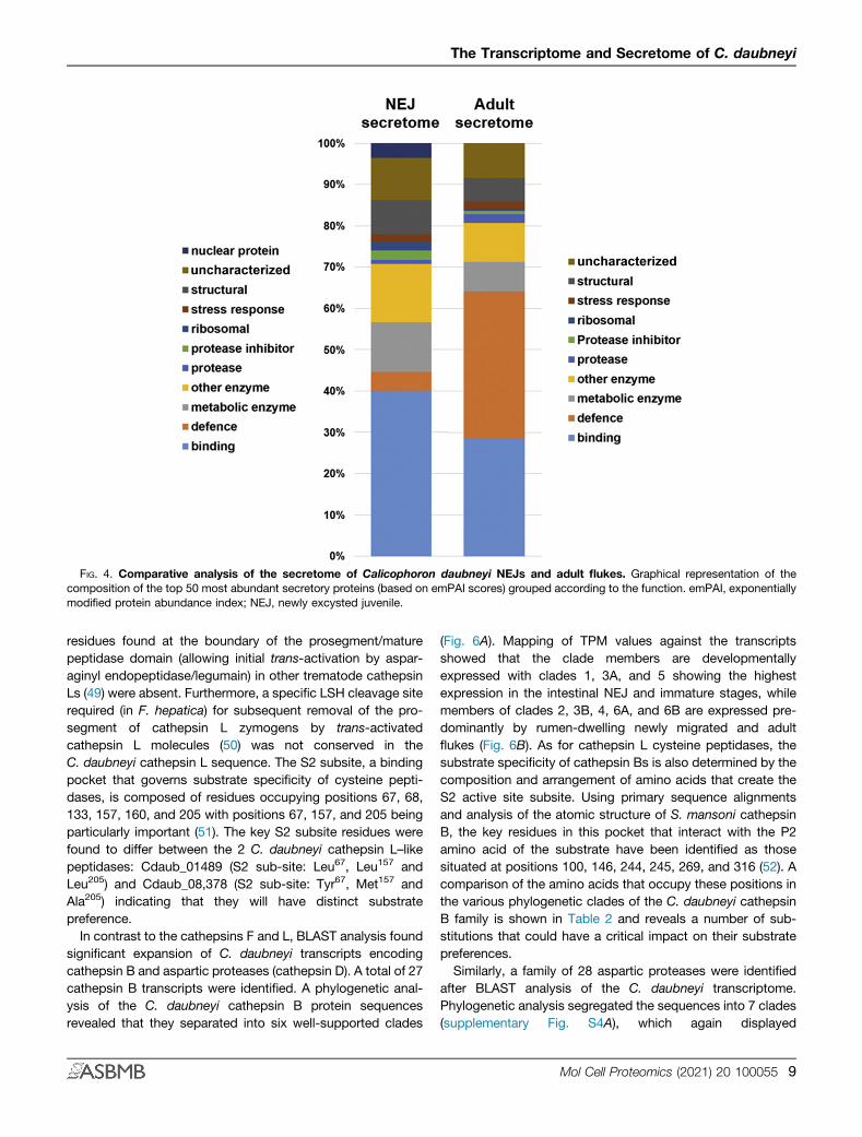
FIG. 4. Comparative analysis of the secretome of Calicophoron daubneyi NEJs and adult flukes. Graphical representation of thecomposition of the top 50 most abundant secretory proteins (based on emPAI scores) grouped according to the function. emPAI, exponentiallymodified protein abundance index; NEJ, newly excysted juvenile.
The Transcriptome and Secretome of C. daubneyi
residues found at the boundary of the prosegment/maturepeptidase domain (allowing initial trans-activation by aspar-aginyl endopeptidase/legumain) in other trematode cathepsinLs (49) were absent. Furthermore, a specific LSH cleavage siterequired (in F. hepatica) for subsequent removal of the pro-segment of cathepsin L zymogens by trans-activatedcathepsin L molecules (50) was not conserved in theC. daubneyi cathepsin L sequence. The S2 subsite, a bindingpocket that governs substrate specificity of cysteine pepti-dases, is composed of residues occupying positions 67, 68,133, 157, 160, and 205 with positions 67, 157, and 205 beingparticularly important (51). The key S2 subsite residues werefound to differ between the 2 C. daubneyi cathepsin L–likepeptidases: Cdaub_01489 (S2 sub-site: Leu67, Leu157 andLeu205) and Cdaub_08,378 (S2 sub-site: Tyr67, Met157 andAla205) indicating that they will have distinct substratepreference.In contrast to the cathepsins F and L, BLAST analysis found
significant expansion of C. daubneyi transcripts encodingcathepsin B and aspartic proteases (cathepsin D). A total of 27cathepsin B transcripts were identified. A phylogenetic anal-ysis of the C. daubneyi cathepsin B protein sequencesrevealed that they separated into six well-supported clades
(Fig. 6A). Mapping of TPM values against the transcriptsshowed that the clade members are developmentallyexpressed with clades 1, 3A, and 5 showing the highestexpression in the intestinal NEJ and immature stages, whilemembers of clades 2, 3B, 4, 6A, and 6B are expressed pre-dominantly by rumen-dwelling newly migrated and adultflukes (Fig. 6B). As for cathepsin L cysteine peptidases, thesubstrate specificity of cathepsin Bs is also determined by thecomposition and arrangement of amino acids that create theS2 active site subsite. Using primary sequence alignmentsand analysis of the atomic structure of S. mansoni cathepsinB, the key residues in this pocket that interact with the P2amino acid of the substrate have been identified as thosesituated at positions 100, 146, 244, 245, 269, and 316 (52). Acomparison of the amino acids that occupy these positions inthe various phylogenetic clades of the C. daubneyi cathepsinB family is shown in Table 2 and reveals a number of sub-stitutions that could have a critical impact on their substratepreferences.Similarly, a family of 28 aspartic proteases were identified
after BLAST analysis of the C. daubneyi transcriptome.Phylogenetic analysis segregated the sequences into 7 clades(supplementary Fig. S4A), which again displayed
Mol Cell Proteomics (2021) 20 100055 9
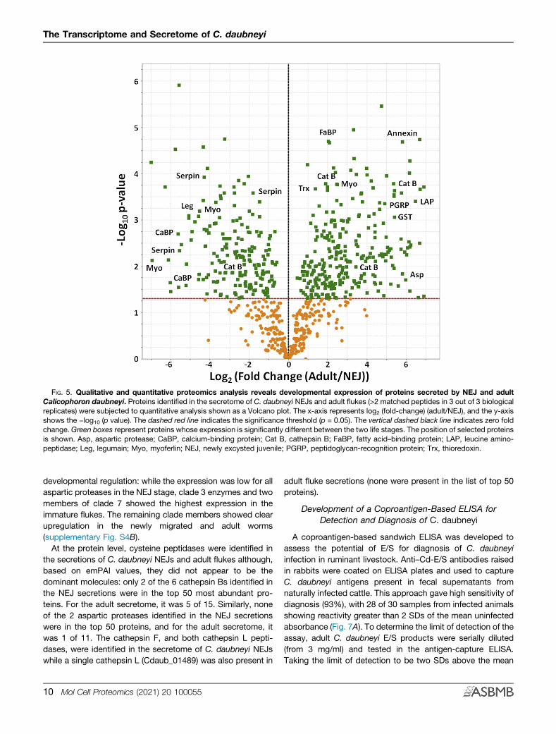
FIG. 5. Qualitative and quantitative proteomics analysis reveals developmental expression of proteins secreted by NEJ and adultCalicophoron daubneyi. Proteins identified in the secretome of C. daubneyi NEJs and adult flukes (>2 matched peptides in 3 out of 3 biologicalreplicates) were subjected to quantitative analysis shown as a Volcano plot. The x-axis represents log2 (fold-change) (adult/NEJ), and the y-axisshows the −log10 (p value). The dashed red line indicates the significance threshold (p = 0.05). The vertical dashed black line indicates zero foldchange. Green boxes represent proteins whose expression is significantly different between the two life stages. The position of selected proteinsis shown. Asp, aspartic protease; CaBP, calcium-binding protein; Cat B, cathepsin B; FaBP, fatty acid–binding protein; LAP, leucine amino-peptidase; Leg, legumain; Myo, myoferlin; NEJ, newly excysted juvenile; PGRP, peptidoglycan-recognition protein; Trx, thioredoxin.
The Transcriptome and Secretome of C. daubneyi
developmental regulation: while the expression was low for allaspartic proteases in the NEJ stage, clade 3 enzymes and twomembers of clade 7 showed the highest expression in theimmature flukes. The remaining clade members showed clearupregulation in the newly migrated and adult worms(supplementary Fig. S4B).At the protein level, cysteine peptidases were identified in
the secretions of C. daubneyi NEJs and adult flukes although,based on emPAI values, they did not appear to be thedominant molecules: only 2 of the 6 cathepsin Bs identified inthe NEJ secretions were in the top 50 most abundant pro-teins. For the adult secretome, it was 5 of 15. Similarly, noneof the 2 aspartic proteases identified in the NEJ secretionswere in the top 50 proteins, and for the adult secretome, itwas 1 of 11. The cathepsin F, and both cathepsin L pepti-dases, were identified in the secretome of C. daubneyi NEJswhile a single cathepsin L (Cdaub_01489) was also present in
10 Mol Cell Proteomics (2021) 20 100055
adult fluke secretions (none were present in the list of top 50proteins).
Development of a Coproantigen-Based ELISA forDetection and Diagnosis of C. daubneyi
A coproantigen-based sandwich ELISA was developed toassess the potential of E/S for diagnosis of C. daubneyiinfection in ruminant livestock. Anti–Cd-E/S antibodies raisedin rabbits were coated on ELISA plates and used to captureC. daubneyi antigens present in fecal supernatants fromnaturally infected cattle. This approach gave high sensitivity ofdiagnosis (93%), with 28 of 30 samples from infected animalsshowing reactivity greater than 2 SDs of the mean uninfectedabsorbance (Fig. 7A). To determine the limit of detection of theassay, adult C. daubneyi E/S products were serially diluted(from 3 mg/ml) and tested in the antigen-capture ELISA.Taking the limit of detection to be two SDs above the mean
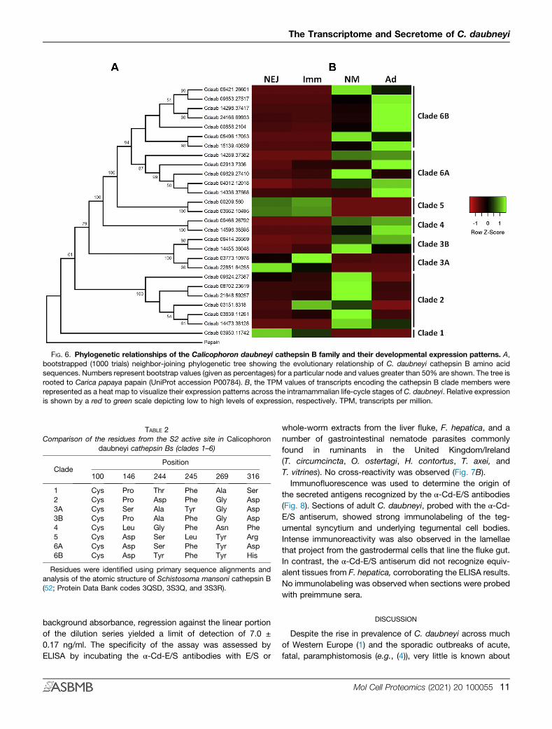
FIG. 6. Phylogenetic relationships of the Calicophoron daubneyi cathepsin B family and their developmental expression patterns. A,bootstrapped (1000 trials) neighbor-joining phylogenetic tree showing the evolutionary relationship of C. daubneyi cathepsin B amino acidsequences. Numbers represent bootstrap values (given as percentages) for a particular node and values greater than 50% are shown. The tree isrooted to Carica papaya papain (UniProt accession P00784). B, the TPM values of transcripts encoding the cathepsin B clade members wererepresented as a heat map to visualize their expression patterns across the intramammalian life-cycle stages of C. daubneyi. Relative expressionis shown by a red to green scale depicting low to high levels of expression, respectively. TPM, transcripts per million.
TABLE 2Comparison of the residues from the S2 active site in Calicophoron
daubneyi cathepsin Bs (clades 1–6)
CladePosition
100 146 244 245 269 316
1 Cys Pro Thr Phe Ala Ser2 Cys Pro Asp Phe Gly Asp3A Cys Ser Ala Tyr Gly Asp3B Cys Pro Ala Phe Gly Asp4 Cys Leu Gly Phe Asn Phe5 Cys Asp Ser Leu Tyr Arg6A Cys Asp Ser Phe Tyr Asp6B Cys Asp Tyr Phe Tyr His
Residues were identified using primary sequence alignments andanalysis of the atomic structure of Schistosoma mansoni cathepsin B(52; Protein Data Bank codes 3QSD, 3S3Q, and 3S3R).
The Transcriptome and Secretome of C. daubneyi
background absorbance, regression against the linear portionof the dilution series yielded a limit of detection of 7.0 ±0.17 ng/ml. The specificity of the assay was assessed byELISA by incubating the α-Cd-E/S antibodies with E/S or
whole-worm extracts from the liver fluke, F. hepatica, and anumber of gastrointestinal nematode parasites commonlyfound in ruminants in the United Kingdom/Ireland(T. circumcincta, O. ostertagi, H. contortus, T. axei, andT. vitrines). No cross-reactivity was observed (Fig. 7B).Immunofluorescence was used to determine the origin of
the secreted antigens recognized by the α-Cd-E/S antibodies(Fig. 8). Sections of adult C. daubneyi, probed with the α-Cd-E/S antiserum, showed strong immunolabeling of the teg-umental syncytium and underlying tegumental cell bodies.Intense immunoreactivity was also observed in the lamellaethat project from the gastrodermal cells that line the fluke gut.In contrast, the α-Cd-E/S antiserum did not recognize equiv-alent tissues from F. hepatica, corroborating the ELISA results.No immunolabeling was observed when sections were probedwith preimmune sera.
DISCUSSION
Despite the rise in prevalence of C. daubneyi across muchof Western Europe (1) and the sporadic outbreaks of acute,fatal, paramphistomosis (e.g., (4)), very little is known about
Mol Cell Proteomics (2021) 20 100055 11
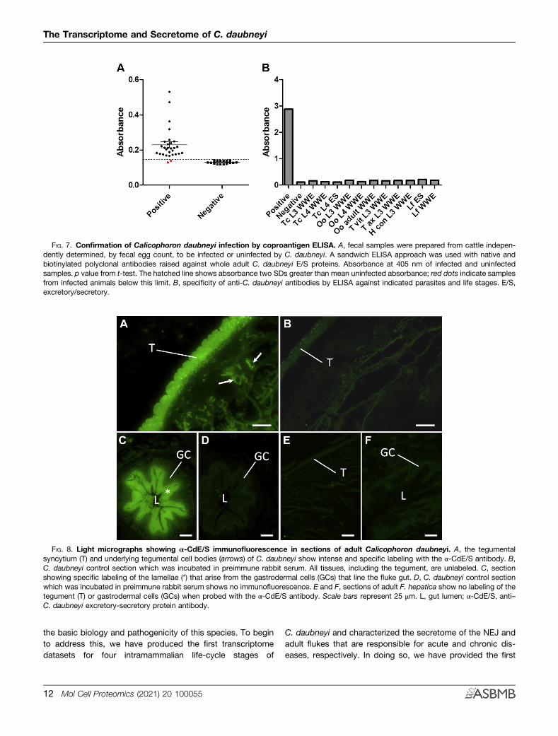
FIG. 7. Confirmation of Calicophoron daubneyi infection by coproantigen ELISA. A, fecal samples were prepared from cattle indepen-dently determined, by fecal egg count, to be infected or uninfected by C. daubneyi. A sandwich ELISA approach was used with native andbiotinylated polyclonal antibodies raised against whole adult C. daubneyi E/S proteins. Absorbance at 405 nm of infected and uninfectedsamples. p value from t-test. The hatched line shows absorbance two SDs greater than mean uninfected absorbance; red dots indicate samplesfrom infected animals below this limit. B, specificity of anti-C. daubneyi antibodies by ELISA against indicated parasites and life stages. E/S,excretory/secretory.
FIG. 8. Light micrographs showing α-CdE/S immunofluorescence in sections of adult Calicophoron daubneyi. A, the tegumentalsyncytium (T) and underlying tegumental cell bodies (arrows) of C. daubneyi show intense and specific labeling with the α-CdE/S antibody. B,C. daubneyi control section which was incubated in preimmune rabbit serum. All tissues, including the tegument, are unlabeled. C, sectionshowing specific labeling of the lamellae (*) that arise from the gastrodermal cells (GCs) that line the fluke gut. D, C. daubneyi control sectionwhich was incubated in preimmune rabbit serum shows no immunofluorescence. E and F, sections of adult F. hepatica show no labeling of thetegument (T) or gastrodermal cells (GCs) when probed with the α-CdE/S antibody. Scale bars represent 25 μm. L, gut lumen; α-CdE/S, anti–C. daubneyi excretory-secretory protein antibody.
The Transcriptome and Secretome of C. daubneyi
the basic biology and pathogenicity of this species. To beginto address this, we have produced the first transcriptomedatasets for four intramammalian life-cycle stages of
12 Mol Cell Proteomics (2021) 20 100055
C. daubneyi and characterized the secretome of the NEJ andadult flukes that are responsible for acute and chronic dis-eases, respectively. In doing so, we have provided the first
The Transcriptome and Secretome of C. daubneyi
description of how various metabolic, virulence, and invasivefactors are regulated with parasite development and haveused these molecules to develop the first ELISA-based cop-roantigen test for diagnosis of C. daubneyi infection oflivestock.Transcriptome data are currently available for two para-
mphistome species, Paramphistomum cervi and C. daubneyi(53, 54). However, these studies have been limited to the adultlife-cycle stage, which are often described as “well-tolerated”by their ruminant hosts because they are responsible forchronic paramphistomosis, which is largely subclinical (13).Here we present a comparative transcriptome analysis of theadult stage with the previously uncharacterized NEJs (infec-tive stage), immature flukes, and newly migrated flukes(migratory stages). We initially used the PacBio platform togenerate full-length transcripts for all four life-cycle stageswithout the need for troublesome assembly of short reads.PacBio sequencing identified a total of 24,956 full-lengthtranscripts with a top-strand protein coding region. Thismethod almost certainly avoids transcript overestimation, withthe number of transcripts described here for C. daubneyiapproximately half of those reported for other trematodeparasites using de novo assembly (e.g. (53–56)) but similar tothose which have been produced using genome-guided ap-proaches (57–59).Changes in gene transcription were followed across the four
intramammalian developmental stages of C. daubneyi usingTPM values derived from RNA-Seq libraries (in biologicaltriplicate for each stage). Principal component analysisrevealed clustering of the replicates but clear separation be-tween life-cycle stages. This demonstrates that each life-cyclestage displays distinct patterns of temporal gene expressionthat correlate with both their development/maturity and nichewithin the mammalian host. DE analysis revealed that the mosthighly regulated transcripts (i.e., those showing at least a 9-logfold change in at least one comparison between stages) weredominated by a few functional groups. Some of the moststriking transcriptional changes involved upregulation in boththe newly migrated and adult stages. These included tubulinsand dynein, both key structural components of the axonemeof trematode spermatozoa (60, 61), and the vitelline proteinsthat comprise trematode eggshells (62). This temporalexpression pattern of reproduction-associated genes followsthe appearance of the reproductive organs during C. daubneyidevelopment with the anterior and posterior testes, ovary, andvitellaria first seen (all together) by the newly migrated stage.Despite the clear size difference between the newly migratedand adult rumen fluke, both stages produced eggs. Thus, it islikely that egg production begins rapidly upon the arrival of themigrating flukes into the rumen, similar to the almost imme-diate onset of egg production observed when juvenileF. hepatica enter the bile duct from the liver parenchyma (61).A total of 7.5% of our predicted protein sequences
possessed an N-terminal signal peptide for secretion via the
endoplasmic reticulum/Golgi pathway, which is comparablewith other trematodes such as C. sinensis (6.5%) andO. viverrini (6.9%) (56). Indeed, the majority of the most highlyregulated transcripts encoded proteins found in C. daubneyisecretions after the LC-MS/MS analysis. Moreover, ourquantitative proteomics analysis showed that many proteinsincluding myoglobins, serpins, cysteine peptidases, asparticpeptidases, GSTs, and FaBPs were developmentally regu-lated, which corroborates the transcriptome analysis. Thesecretomes of two key stages in the C. daubneyi intra-mammalian life cycle were analyzed: the infectious NEJs thatare responsible for acute disease in the duodenum and themature adult worms found in the rumen during chronicinfection. Proteins with binding function dominated the se-cretions of both stages, notably 14-3-3 proteins, calcium-binding proteins, FaBPs, ferritin, and myoglobin isoforms.Although some of these molecules have reported immuno-modulatory functions in other trematodes (63–65), their ca-pacity for binding would also allow them to function as carriersto facilitate uptake of various molecules from the hostmicroenvironment in vivo. Interestingly, ferritin was enriched inadult fluke secretions compared with those of the NEJ. Ferritinis believed to act as an iron carrier after extracellular hemo-globin digestion in the schistosome gut (66). Because adultC. daubneyi are not thought to feed directly on host blood(because of the absence of supporting pathology) (1) secretedferritin may sequester iron from rumen fluid or have alternativeimmunomodulatory roles as described for C. sinensis (67).Similarly, myoglobin was highly expressed by C. daubneyi atthe transcript level, and different isoforms were found to beenriched in NEJ and adult fluke secretions. Myoglobins act asoxygen scavengers and are thought to be important forparasite metabolism through oxygen transport/storage (68,69). Trematode myoglobins have some of the highest recor-ded affinities for oxygen (70), which would be of particularvalue in the largely anaerobic rumen (71).Metacercariae are nonfeeding stages and must rely upon
endogenous glycogen stores to support them during excyst-ment and initial establishment of infection. In F. hepatica, therelative expression levels of key enzymes that regulateglycogen synthesis and degradation indicate that meta-cercarial glycogen stores are rapidly used up after excystment(41). Similarly, fructose-1,6-bisphosphatase (involved inglycogen synthesis) and phosphofructokinase (involved inglycogen degradation) were both upregulated in C. daubneyiNEJs, indicating that the fluke is expending pre-existingglycogen stores while simultaneously replenishing them dur-ing early infection. Conceivably, this provides the NEJ withsufficient energy to establish infection (e.g., to support activesecretory processes and locomotion required during pene-tration into the duodenal mucosa) while creating new reservesto metabolize during the long migration toward the rumen.Trematodes undergo drastic changes in oxygen availability
as they progress through their life cycle. Metacercariae, and
Mol Cell Proteomics (2021) 20 100055 13
The Transcriptome and Secretome of C. daubneyi
other free-living stages, readily obtain oxygen from the envi-ronment. However, once infections become established andflukes migrate within host tissues, they begin to encounterenvironmental hypoxia (72, 73), which coincides with a switchfrom aerobic energy metabolism (TCA cycle) to anaerobicmalate dismutation (74). Our transcriptome analysis indicatesthat C. daubneyi follows this metabolic strategy with TCAcycle members expressed most highly by the NEJ stage andbecoming gradually downregulated as the parasite matures tothe adult stage. Coincident with the steady decline of the TCApathway is the upregulation of members of the malate dis-mutation pathway with parasite development. Such gradualchange of metabolic pathways could suggest the existence ofmultiple populations of mitochondria in C. daubneyi each withdifferent respiratory activities/tissue locations as have beendescribed for the lung fluke, Paragonimus westermani (75).This would provide the metabolic plasticity required asC. daubneyi moves from aerobic conditions within theduodenal submucosa toward an increasingly anaerobic envi-ronment (i.e., the rumen) and ensure that efficient ATP pro-duction is maintained to support the enormous energydemands of egg production that begins after this migration.While the biochemistry of C. daubneyi mitochondria requirefurther investigation, Huson et al. (54) did report the produc-tion of propionate (a major excretory product of malate dis-mutation) by adult C. daubneyi during in vitro culture, whichsupports the current transcriptome data.After excystment in the duodenum, NEJs must respond
rapidly to adapt to their new environment and to counter inev-itable attack by the host immune system (41). At the transcrip-tional level,C. daubneyi orthologues of trematode proteins withknown immunomodulatory function including kunitz-type in-hibitor, peroxiredoxin, FaBPs, venom allergen–like proteins,and GSTs were largely upregulated in the NEJ stage. InF. hepatica, and other trematodes, these proteins act, via avariety of mechanisms, to establish a Th2 immune environmentthat is permissive to parasite survival and reproduction (76–81).While the host immune response to C. daubneyi infection (andto what extent the parasite can manipulate this) has yet to becharacterized in detail, the NEJ stage is clearly equipped withsufficient armory to avoid the immune response and establishinitial infection in the duodenal mucosa. However, field obser-vations and experimental studies indicate that previous expo-sure of livestock to rumen fluke provides protection (routinely>99%) against massive infections that would typically causeacute (and often fatal) paramphistomosis (7, 82–84). Critically,these studies showed that immunization with adult worms(delivered orally to establish infection directly in the rumen) wasnot effective and that protective immunity requires exposure ofthe duodenum to NEJ-derived antigen (7). This is in starkcontrast to liver fluke, where there is limited evidence of naturalacquired immunity in cattle and less so in sheep (85). As a result,attempts to vaccinate livestock against liver fluke have beenlargely unsuccessful, despite a considerable research effort
14 Mol Cell Proteomics (2021) 20 100055
(86). Thus, rumen fluke may be more amenable to vaccinalcontrol than liver fluke infection, and our proteomics analysis ofNEJ secretions has unveiled a range of early-stage moleculesthat represent potential candidates for vaccine development.The single most abundant protein found in the adult fluke
secretome was CdHDM-3. This represents one of the threeisoforms of the HDMs, a family of trematode-specific proteinswith diverse immunomodulatory functions; F. hepatica HDM-1has been shown to protect mice against lipopolysaccharideinduced inflammation by significantly reducing the release ofinflammatory cytokines from macrophages (38) and to sup-press antigen processing and presentation in macrophagesvia inhibition of lysosomal vacuolar ATPase (39). This is thefirst time HDMs have been reported in suborder Pronoce-phalata and the first time it has been identified in the secre-tions of C. daubneyi, presumably not being resolved on the 2-DE used by Huson et al. (54) because of its size (~6 kDa).While the function of HDMs during C. daubneyi infection isunknown, two of the isoforms were predicted to possess a C-terminal amphipathic helix, which is the key functional deter-minant of the lipopolysaccharide-binding/immunomodulatoryproperties of HDMs from other trematodes (38–40). Thus,C. daubneyi may use HDMs to simultaneously maintain a Th2immune environment and to protect against pathogen-associated molecular patterns in the microbiome-richrumen—both of which would serve to sustain chronic in-fections that can persist in livestock for many years (7).In trematodes, cathepsin-like cysteine peptidases play
central roles in virulence, infection, tissue migration, andmodulation of host immune responses (44). While cathepsin Bpeptidases are expressed by most species, there is cleardivergence in the use of other types of cathepsins: Fasciolaspp. and Schistosoma spp. predominantly express cathepsinsL, whereas cathepsins F are the major peptidases expressedby Clonorchis spp., Paragonimus spp., and Opisthorchis spp(43). Our transcriptome and phylogenetic analysis showedthat the C. daubneyi cathepsin B family has undergoneconsiderable expansion and has diverged into six clades withvarying S2 subsite residues. Because the composition andarrangement of amino acids that create the S2 subsite withinthe cathepsin active site determine the specificity of theenzyme (51, 87), it is likely that the C. daubneyi cathepsin Bclade members represent a repertoire of enzymes with over-lapping/complementary substrate specificities that arecapable of degrading a variety of host macromolecules theyencounter as they develop and migrate within the host. Insupport of this, the clade members were found, at both thetranscript and protein levels, to be developmentally regulatedwith clades 1, 3A, and 5 expressed/secreted predominantly bythe NEJs/immature stage and clades 2, 3B, 4, and 6expressed/secreted by newly migrated/adult flukes.C. daubneyi was also found to have a large aspartic pepti-
dase family that could be segregated into distinct cladeslargely expressed by the adult flukes. Despite this, aspartic
The Transcriptome and Secretome of C. daubneyi
peptidases were not particularly abundant in the secretome.This mirrors O. viverrini which, despite having 27 asparticpeptidase genes, secrete only low levels of the enzyme (58,88). Although it is not currently possible to predict the activesite composition of aspartic peptidases, it is likely that, giventheir expansion and clear developmental regulation, they toowill have varying substrate preferences. In blood-feedinghelminths, aspartic peptidases usually form part of a multi-enzyme cascade, with endopeptidase and exopeptidase ac-tivity, to degrade host hemoglobin (89). While the source ofnutrition for C. daubneyi has yet to be confirmed (e.g., hosttissue, rumen fluid, rumen microorganisms), such an enzymecascade is possible because other digestive enzymesincluding aminopeptidases, carboxypeptidases, andchymotrypsin-like peptidases were all found in the secretome.This would be in contrast to F. hepatica, which relies solely onan expanded repertoire of cathepsin L peptidases, whichcomprise >80% of the total soluble protein secreted by adultfluke, to degrade hemoglobin (43). Here, we found only twotranscripts encoding cathepsin L (and one encoding cathepsinF), which were secreted by NEJs in low abundance. Whilethese retain key active site residues, indicating that they willform functional enzymes, they lack conserved asparagines atthe junction of the prosegment and mature domain. Thissuggests that they are not trans-activated to the matureenzyme by asparaginyl endopeptidase (legumain; althoughthese were also found in C. daubneyi secretions) but could beactivated by cathepsin B as reported for O. viverrini cathepsinF (90).Owing to their abundance in liver fluke secretions,
cathepsin L is the target antigen for most coproantigen-based ELISA tests developed for F. hepatica infection(91, 92). The MM3 mAb used for antigen capture inF. hepatica coproantigen assays does not cross-react withrumen fluke antigens either on histological sections or byELISA (93). Our secretome analysis has now confirmed thatthis is because C. daubneyi secrete very little cathepsin L,mostly from the NEJ stage. Thus, while specific tests forfasciolosis are available (which would be of particular valuein areas where paramphistomosis also occurs, either singlyor as coinfections with F. hepatica), there are currently nosuch tests for rumen fluke infection. As a result of thesignificant differences in the profile of molecules secretedby both species, we investigated the diagnostic value ofC. daubneyi E/S antigens in a coproantigen-based ELISA.The results demonstrate that nanogram amounts ofC. daubneyi E/S antigen could be captured from fecalextracts with sufficient sensitivity to distinguish betweennaturally infected and uninfected cattle. Importantly, theanti–CdE/S serum did not cross-react with somatic orsecretory antigens from a range of helminth species thatcommonly share the same ruminant hosts as C. daubneyi.The use of this coproantigen ELISA would offer a nonin-vasive means of diagnosing current rumen fluke infection
(antigens only persist for the duration of infection) andcould also be used for the detection of drug resistance(94); because the current treatment is reliant on a singledrug (oxyclozanide), this seems inevitable (1).In this report, we have generated the first transcriptome
resources for multiple intramammalian life-cycle stages ofC. daubneyi. By integrating this with secretome analysis ofthe most clinically relevant stages, we provide a compre-hensive, and dynamic, overview of initial infection in theduodenum, migration within the gastrointestinal tract, andfinal maturation upon arrival in the rumen. In particular, ouranalysis of the molecules expressed/secreted by the NEJstage provides a valuable framework for studies aimed atbetter understanding infectivity and how drugs or vaccinescould be developed to prevent infection and/or limitdamaging intestinal pathology associated with acute para-mphistomosis. Because prepatent rumen fluke infections arethe primary cause of clinical disease (4), our NEJ secretomedata have yielded a range of putative early-stage antigensthat could allow timely diagnosis and treatment for thisemerging infection.
DATA AVAILABILITY
The transcriptome datasets supporting the conclusionsof this article are available in the European Nucleotide Archive(ENA) repository under accession number PRJEB28150,http://www.ebi.ac.uk/ena/data/view/PRJEB28150. The MSproteomics data have been deposited to the Proteo-meXchange Consortium via the PRIDE partner repository withthe dataset identifier PXD014550 and 10.6019/PXD014550.
Supplemental data—This article contains supplementaldata.
Funding and additional information—This work was supportedby an Industrial Partnership Award (to M. W. R.) from theBiotechnology and Biological Sciences Research Council (BB/N017757/1) with additional financial support from AgriSearchand AHDB Beef & Lamb. N. A. M. O. was supported by apostgraduate studentship from the Department for the Econ-omy (DfE), Northern Ireland.
Author contributions—M. W. R. and S. P. designedresearch; K. M. H., E. A., N. A. M. O., Y. F., S. H., and M. W. R.performed research; J. P. B., R. E. B. H., and T. N. M.,contributed new reagents or analytic tools; K. M. H., E. A.,N. A. M. O., Y. F., S. H., P. B., and M. W. R. analyzed data;K. M. H., E. A. and M. W. R. wrote the paper.
Conflict of interest—The authors declare no competinginterests.
Abbreviations—The abbreviations used are: AbD, antibodydiluent; DE, differential expression; E/S, excretory/secretory;
Mol Cell Proteomics (2021) 20 100055 15
The Transcriptome and Secretome of C. daubneyi
emPAI, exponentially modified protein abundance index; FA,formic acid; FaBP, fatty acid–binding protein; HDM, helminthdefense molecule; NEJ, newly excysted juvenile; TCA, tricar-boxylic acid; TPM, transcripts per million; α-CdE/S, anti–C. daubneyi excretory/secretory protein antibody.
Published, MCPRO Papers in Press, February 11, 2021, https://doi.org/10.1074/mcp.RA120.002175
REFERENCES
1. Huson, K. M., Oliver, N. A. M., and Robinson, M. W. (2017) Para-mphistomosis of ruminants: An emerging parasitic disease in Europe.Trends Parasitol. 33, 836–844
2. Taylor, M. A. (2013) Parasite control in sheep: A risky business. SmallRumin. Res. 110, 88–92
3. Skuce, P. J., Morgan, E. R., van Dijk, J., and Mitchell, M. (2013) Animalhealth aspects of adaptation to climate change: Beating the heat andparasites in a warming Europe. Animal 7, 333–345
4. O’Shaughnessy, J., Garcia-Campos, A., McAloon, C. G., Fagan, S., deWaal, T., McElroy, M., Casey, M., Good, B., Mulcahy, G., Fagan, J.,Murphy, D., and Zintl, A. (2018) Epidemiological investigation of a severerumen fluke outbreak on an Irish dairy farm. Parasitology 145, 948–952
5. Devos, J., Vassiloglou, B., Amenna-Bernard, N., and Marcotty, T. (2013)Paramphistomosis in sheep; natural infection of lambs by Calicophorondaubneyi. Rev. Med. Vet. (Toulouse) 164, 528–535
6. Anonymous. Regional veterinary laboratories report. Vet. Irel. J. 7, (2016),25–30
7. Horak, I. G. (1971) Paramphistomiasis of Domestic ruminants. Adv. Para-sitol. 9, 33–72
8. Sanabria, R. E. F., and Romero, J. R. (2008) Review and update of para-mphistomosis. Helminthologia 45, 64–68
9. Fuertes, M., Perez, V., Benavides, J., Gonzalez-Lanza, M. C., Mezo, M.,Gonzalez-Warleta, M., Giraldez, F. J., Fernandez, M., Manga-Gonzalez,M. Y., and Ferreras, M. C. (2015) Pathological changes in cattle naturallyinfected by Calicophoron daubneyi adult flukes. Vet. Parasitol. 209, 188–196
10. Mason, C., Stevenson, H., Cox, A., and Dick, I. (2012) Disease associatedwith immature paramphistome infection in sheep. Vet. Rec. 170, 343–344
11. Anonymous. (2017) Northern Ireland disease surveillance report, October toDecember 2016. Vet. Rec. 180, 112–116
12. Foster, A. P., Otter, A., O’Sullivan, T., Cranwell, M. P., Twomey, D. F., Millar,M. F., and Taylor, M. A. (2008) Rumen fluke (paramphistomosis) in Britishcattle. Vet. Rec. 162, 528
13. Millar, M., Colloff, A., and Scholes, S. (2012) Disease associated withimmature paramphistome infection. Vet. Rec. 171, 509–510
14. Rolfe, P. F., and Boray, J. C. (1987) Chemotherapy of paramphistomosis incattle. Aust. Vet. J. 64, 328–332
15. Paraud, C., Gaudin, C., Pors, I., and Chartier, C. (2009) Efficacy of oxy-clozanide against the rumen fluke Calicophoron daubneyi in experimen-tally infected goats. Vet. J. 180, 265–267
16. Huson, K. M., Wild, C., Fenn, C., and Robinson, M. W. (2017) Optimizedconditions for the in vitro excystment of Calicophoron daubneyi meta-cercariae. Parasitology 145, 1015–1019
17. Martínez-Ibeas, A. M., Gonzalez-Warleta, M., Martínez-Valladares, M.,Castro-Hermida, J. A., Gonzalez-Lanza, C., Minambres, B., Ferreras, C.,Mezo, M., and Manga-Gonzalez, M. Y. (2013) Development and validationof a mtDNA multiplex PCR for identification and discrimination of Cal-icophoron daubneyi and Fasciola hepatica in the Galba truncatula snail.Vet. Parasitol. 195, 57–64
18. Li, W., and Godzik, A. (2006) Cd-hit: A fast program for clustering andcomparing large sets of protein or nucleotide sequences. Bioinformatics22, 1658–1659
19. Chin, C. S., Alexander, D. H., Marks, P., Klammer, A. A., Drake, J., Heiner,C., Clum, A., Copeland, A., Huddleston, J., Eichler, E. E., Turner, S. W.,and Korlach, J. (2013) Nonhybrid, finished microbial genome assembliesfrom long-read SMRT sequencing data. Nat. Methods 10, 563–569
20. Haas, B. J., Papanicolaou, A., Yassour, M., Grabherr, M., Blood, P. D.,Bowden, J., Couger, M. B., Eccles, D., Li, B., Lieber, M., Macmanes, M.D., Ott, M., Orvis, J., Pochet, N., Strozzi, F., et al. (2013) De novo
16 Mol Cell Proteomics (2021) 20 100055
transcript sequence reconstruction from RNA-seq using the Trinity plat-form for reference generation and analysis. Nat. Protoc. 8, 1494–1512
21. Altschul, S. F., Gish, W., Miller, W., Myers, E. W., and Lipman, D. J. (1990)Basic local alignment search tool. J. Mol. Biol. 215, 403–410
22. Finn, R. D., Coggill, P., Eberhardt, R. Y., Eddy, S. R., Mistry, J., Mitchell, A.L., Potter, S. C., Punta, M., Qureshi, M., Sangrador-Vegas, A., Salazar, G.A., Tate, J., and Bateman, A. (2016) The Pfam protein families database:Towards a more sustainable future. Nucleic Acids Res. 44, D279–D285
23. Eddy, S. R. (2011) Accelerated profile HMM searches. PLOS Comput. Biol.7, e1002195
24. Petersen, T. N., Brunak, S., von Heijne, G., and Nielsen, H. (2011) SignalP 4.0: discriminating signal peptides from transmembrane regions. Nat.Methods 8, 785–786
25. Krogh, A., Larsson, B., von Heijne, G., and Sonnhammer, E. L. L. (2001)Predicting transmembrane protein topology with a hidden markov model:Application to complete genomes. J. Mol. Biol. 305, 567–580
26. Martin, M. (2011) Cutadapt removes adapter sequences from high-throughput sequencing reads. EMBnet.journal. 17, 10–12
27. Langmead, B., and Salzberg, S. L. (2012) Fast gapped-read alignment withBowtie 2. Nat. Methods 9, 357–359
28. Glaus, P., Honkela, A., and Rattray, M. (2012) Identifying differentiallyexpressed transcripts from RNA-seq data with biological variation. Bio-informatics 28, 1721–1728
29. Love, M. I., Huber, W., and Anders, S. (2014) Moderated estimation of foldchange and dispersion for RNA-seq data with DESeq2. Genome Biol. 15,550
30. Hall, T. (1999) BioEdit: A user-friendly biological sequence alignment editorand analysis program for Windows 95/98/NT. Nucleic Acids Symp. Ser.41, 95–98
31. Babicki, S., Arndt, D., Marcu, A., Liang, Y., Grant, J. R., Maciejewski, A., andWishart, D. S. (2016) Heatmapper: Web-enabled heat mapping for all.Nucleic Acids Res. 44, W147–W153
32. Tamura, K., Stecher, G., Peterson, D., Filipski, A., and Kumar, S. (2013)MEGA6: Molecular evolutionary genetics analysis version 6.0. Mol. Biol.Evol. 30, 2725–2729
33. Felsenstein, J. (1985) Confidence limits on phylogenies: An approach usingthe bootstrap. Evolution 39, 783–791
34. Zuckerkandl, E., and Pauling, L. (1965) Molecules as documents of evolu-tionary history. J. Theor. Biol. 8, 357–366
35. Nesvizhskii, A. I., Keller, A., Kolker, E., and Aebersold, R. (2003) A statisticalmodel for identifying proteins by tandem mass spectrometry. Anal.Chem. 75, 4646–4658
36. Teimoori, S., Arimatsu, Y., Laha, T., Kaewkes, S., Sereerak, P., Sripa, M.,Tangkawattana, S., Brindley, P. J., and Sripa, B. (2017) Chicken IgY-based coproantigen capture ELISA for diagnosis of human opisthorch-iasis. Parasitol. Int. 66, 443–447
37. Fairweather, I., McShane, D. D., Shaw, L., Ellison, S. E., O’Hagan, N. T.,York, E. A., Trudgett, A., and Brennan, G. P. (2012) Development of anegg hatch assay for the diagnosis of triclabendazole resistance in Fas-ciola hepatica: Proof of concept. Vet. Parasitol. 183, 249–259
38. Robinson, M. W., Donnelly, S., Hutchinson, A. T., To, J., Taylor, N. L.,Norton, R. S., Perugini, M. A., and Dalton, J. P. (2011) A family of helminthmolecules that modulate innate cell responses via molecular mimicry ofhost antimicrobial peptides. Plos Pathog. 7, e1002042
39. Robinson, M. W., Alvarado, R., To, J., Hutchinson, A. T., Dowdell, S. N.,Lund, M., Turnbull, L., Whitchurch, C. B., O'Brien, B. A., Dalton, J. P., andDonnelly, S. (2012) A helminth cathelicidin-like protein suppresses anti-gen processing and presentation in macrophages via inhibition of lyso-somal vATPase. FASEB J. 26, 4614–4627
40. Thivierge, K., Cotton, S., Schaefer, D. A., Riggs, M. W., To, J., Lund, M. E.,Robinson, M. W., Dalton, J. P., and Donnelly, S. M. (2013) Cathelicidin-like helminth defence molecules (HDMs): Absence of cytotoxic, anti-microbial and anti-protozoan activities imply a specific adaptation toimmune modulation. Plos Negl. Trop. Dis. 7, e2307
41. Cwiklinski, K., Jewhurst, H., McVeigh, P., Barbour, T., Maule, A. G., Tort, J.,O'Neill, S. M., Robinson, M. W., Donnelly, S., and Dalton, J. P. (2018)Infection by the helminth parasite Fasciola hepatica requires rapid regu-lation of metabolic, virulence, and invasive factors to Adjust to itsmammalian host. Mol. Cell. Proteomics 17, 792–809
42. Cwiklinski, K., de la Torre-Escudero, E., Trelis, M., Bernal, D., Dufresne, P.J., Brennan, G. P., O'Neill, S., Tort, J., Paterson, S., Marcilla, A., Dalton, J.
The Transcriptome and Secretome of C. daubneyi
P., and Robinson, M. W. (2015) The extracellular vesicles of the helminthpathogen, Fasciola hepatica: Biogenesis pathways and cargo moleculesinvolved in parasite pathogenesis. Mol. Cell. Proteomics 14, 3258–3273
43. Robinson, M. W., Tort, J. F., Lowther, J., Donnelly, S. M., Wong, E., Xu, W.,Stack, C. M., Padula, M., Herbert, B., and Dalton, J. P. (2008) Proteomicsand phylogenetic analysis of the cathepsin L protease family of the hel-minth pathogen Fasciola hepatica. Mol. Cell. Proteomics 7, 1111–1123
44. Cwiklinski, K., Donnelly, S., Drysdale, O., Jewhurst, H., Smith, D., De MarcoVerissimo, C., Pritsch, I. C., O'Neill, S., Dalton, J. P., and Robinson, M. W.(2019) The cathepsin-like cysteine peptidases of trematodes of the genusFasciola. Adv. Parasitol. 104, 113–164
45. Auerswald, E. A., Genenger, G., Assfalg-Machleidt, I., Machleidt, W., Engh,R. A., and Fritz, H. (1992) Recombinant chicken egg white cystatin vari-ants of the QLVSG region. Eur. J. Biochem. 209, 837–845
46. Deussing, J., Tisljar, K., Papazoglou, A., and Peters, C. (2000) Mousecathepsin F: cDNA cloning, genomic organization and chromosomalassignment of the gene. Gene 251, 165–173
47. Kang, T. H., Yun, D. H., Lee, E. H., Chung, Y. B., Bae, Y. A., Chung, J. Y.,Kang, I., Kim, J., Cho, S. Y., and Kong, Y. (2004) A cathepsin F of adultClonorchis sinensis and its phylogenetic conservation in trematodes.Parasitology 128, 195–207
48. Vernet, T., Berti, P. J., de Montigny, C., Musil, R., Tessier, D. C., Menard, R.,Magny, M. C., Storer, A. C., and Thomas, D. Y. (1995) Processing of thepapain precursor. The ionization state of a conserved amino acid motifwithin the Pro region participates in the regulation of intramolecularprocessing. J. Biol. Chem. 270, 10838–10846
49. Dalton, J. P., Brindley, P. J., Donnelly, S., and Robinson, M. W. (2009) Theenigmatic asparaginyl endopeptidase of helminth parasites. Trends Par-asitol. 25, 59–61
50. Stack, C. M., Donnelly, S., Lowther, J., Xu, W., Collins, P. R., Brinen, L. S.,and Dalton, J. P. (2007) The major secreted cathepsin L1 protease of theliver fluke, Fasciola hepatica: A Leu-12 to Pro-12 replacement in thenonconserved C-terminal region of the prosegment prevents completeenzyme autoactivation and allows definition of the molecular events inprosegment removal. J. Biol. Chem. 282, 16532–16543
51. Turk, D., Guncar, G., Podobnik, M., and Turk, B. (1998) Revised definition ofsubstrate binding sites of papain-like cysteine proteases. Biol. Chem.379, 137–147
52. Jílkova, A., Rezacova, P., Lepsík, M., Horn, M., Vachova, J., Fanfrlík, J.,Brynda, J., McKerrow, J. H., Caffrey, C. R., and Mares, M. (2011)Structural basis for inhibition of cathepsin B drug target from the humanblood fluke, Schistosoma mansoni. J. Biol. Chem. 286, 35770–35781
53. Choudhary, V., Garg, S., Chourasia, R., Hasnani, J. J., Patel, P. V., Shah, T.M., Bhatt, V. D., Mohapatra, A., Blake, D. P., and Joshi, C. G. (2015)Transcriptome analysis of the adult rumen fluke Paramphistomum cervifollowing next generation sequencing. Gene 570, 64–70
54. Huson, K. M., Morphew, R. M., Allen, N. R., Hegarty, M. J., Worgan, H. J.,Girdwood, S. E., Jones, E. L., Phillips, H. C., Vickers, M., Swain, M.,Smith, D., Kingston-smith, A. H., and Brophy, P. M. (2018) Polyomic toolsfor an emerging livestock parasite, the rumen fluke Calicophoron daub-neyi; identifying shifts in rumen functionality. Parasit. Vectors 11, 617
55. Cantacessi, C., Mulvenna, J., Young, N. D., Kasny, M., Horak, P., Aziz, A.,Hofmann, A., Loukas, A., and Gasser, R. B. (2012) A Deep Exploration ofthe transcriptome and “Excretory/secretory” Proteome of adult Fas-cioloides magna. Mol. Cell. Proteomics 11, 1340–1353
56. Young, N. D., Campbell, B. E., Hall, R. S., Jex, A. R., Cantacessi, C., Laha,T., Sohn, W. M., Sripa, B., Loukas, A., Brindley, P. J., and Gasser, R. B.(2010) Unlocking the transcriptomes of two carcinogenic parasites, Clo-norchis sinensis and Opisthorchis viverrini. Plos Negl. Trop. Dis. 4, e719
57. Huang, Y., Chen, W., Wang, X., Liu, H., Chen, Y., Guo, L., Luo, F., Sun, J.,Mao, Q., Liang, P., Xie, Z., Zhou, C., Tian, Y., Lv, X., Huang, L., et al.(2013) The carcinogenic liver fluke, Clonorchis sinensis: New assembly,Reannotation and analysis of the genome and characterization of tissuetranscriptomes. PLoS One 8, e54732
58. Protasio, A. V., Tsai, I. J., Babbage, A., Nichol, S., Hunt, M., Aslett, M. A., deSilva, N., Velarde, G. S., Anderson, T. J. C., Clark, R. C., Davidson, C.,Dillon, G. P., Holroyd, N. E., LoVerde, P. T., Lloyd, C., et al. (2012)A systematically improved high quality genome and transcriptome of thehuman blood fluke Schistosoma mansoni. Plos Negl. Trop. Dis. 6, e1455
59. Young, N. D., Nagarajan, N., Lin, S. J., Korhonen, P. K., Jex, A. R., Hall, R.S., Safavi-Hemami, H., Kaewkong, W., Bertrand, D., Gao, S., Seet, Q.,
Wongkham, S., Teh, B. T., Wongkham, C., Intapan, P. M., et al. (2014) TheOpisthorchis viverrini genome provides insights into life in the bile duct.Nat. Commun. 5, 4378
60. Stitt, A. W., and Fairweather, I. (1990) Spermatogenesis and the finestructure of the mature spermatozoon of the liver fluke, Fasciola hepatica(Trematoda: Digenea). Parasitology 101, 395–407
61. Stitt, A. W., Fairweather, I., Trudgett, A. G., Johnston, C. F., and Anderson,S. M. (1992) Localisation of actin in the liver fluke, Fasciola hepatica.Parasitol. Res. 78, 96–102
62. Robinson, M. W., Colhoun, L. M., Fairweather, I., Brennan, G. P., and Waite,J. H. (2001) Development of the vitellaria of the liver fluke, Fasciolahepatica in the rat host. Parasitology 123, 509–518
63. Wang, X., Chen, W., Li, X., Zhou, C., Deng, C., Lv, X., Fan, Y., Men, J.,Liang, C., and Yu, X. (2012) Identification and molecular characterizationof a novel signaling molecule 14-3-3 epsilon in Clonorchis sinensisexcretory/secretory products. Parasitol. Res. 110, 1411–1420
64. Ren, M., He, L., Huang, Y., Mao, Q., Li, S., Qu, H., Bian, M., Liang, P., Chen,X., Ling, J., Chen, T., Liang, C., Wang, X., Li, X., and Yu, X. (2014) Mo-lecular characterization of Clonorchis sinensis secretory myoglobin:Delineating its role in anti-oxidative survival. Parasit. Vectors 7, 250
65. Fitzsimmons, C. M., McBeath, R., Joseph, S., Jones, F. M., Walter, K.,Hoffmann, K. F., Kariuki, H. C., Mwatha, J. K., Kimani, G., Kabatereine, N.B., Vennervald, B. J., Ouma, J. H., and Dunne, D. W. (2007) Factorsaffecting human IgE and IgG responses to allergen-like Schistosomamansoni antigens: Molecular structure and patterns of in vivo exposure.Int. Arch. Allergy Immunol. 142, 40–50
66. Hall, S. L., Braschi, S., Truscott, M., Mathieson, W., Cesari, I. M., andWilson, R. A. (2011) Insights into blood feeding by schistosomesfrom a proteomic analysis of worm vomitus.Mol. Biochem. Parasitol. 179,18–29
67. Mao, Q., Xie, Z., Wang, X., Chen, W., Ren, M., Shang, M., Lei, H., Tian, Y.,Li, S., Liang, P., Chen, T., Liang, C., Xu, J., Li, X., Huang, Y., et al. (2015)Clonorchis sinensis ferritin heavy chain triggers free radicals and medi-ates inflammation signaling in human hepatic stellate cells. Parasitol. Res.114, 659–670
68. Rashid, A. K., Van Hauwaert, M. L., Haque, M., Siddiqi, A. H., Lasters, I., DeMaeyer, M., Griffon, N., Marden, M. C., Dewilde, S., Clauwaert, J.,Vinogradov, S. N., and Moens, L. (1997) Trematode myoglobins, func-tional molecules with a distal tyrosine. J. Biol. Chem. 272, 2992–2999
69. McNulty, S. N., Fischer, P. U., Townsend, R. R., Curtis, K. C., Weil, G. J.,and Mitreva, M. (2014) Systems biology studies of adult Paragonimuslung flukes facilitate the identification of Immunodominant parasite anti-gens. Plos Negl. Trop. Dis. 8, e3242
70. Kiger, L., Rashid, A. K., Griffon, N., Haque, M., Moens, L., Gibson, Q. H.,Poyart, C., and Marden, M. C. (1998) Trematode hemoglobins showexceptionally high oxygen affinity. Biophys. J. 75, 990–998
71. Huang, Y., Marden, J. P., Julien, C., and Bayourthe, C. (2018) Redox po-tential: An intrinsic parameter of the rumen environment. J. Anim. Physiol.Anim. Nutr. (Berl). 102, 393–402
72. Müller, M., Mentel, M., van Hellemond, J. J., Henze, K., Woehle, C., Gould,S. B., Yu, R.-Y., van der Giezen, M., Tielens, A. G. M., and Martin, W. F.(2012) Biochemistry and evolution of anaerobic energy metabolism ineukaryotes. Microbiol. Mol. Biol. Rev. 76, 444–495
73. McNulty, S. N., Tort, J. F., Rinaldi, G., Fischer, K., Rosa, B. A., Smircich, P.,Fontenla, S., Choi, Y. J., Tyagi, R., Hallsworth-Pepin, K., Mann, V. H.,Kammili, L., Latham, P. S., Dell’Oca, N., Dominguez, F., et al. (2017)Genomes of Fasciola hepatica from the Americas reveal Colonization withNeorickettsia Endobacteria related to the agents of Potomac Horse andhuman Sennetsu Fevers. Plos Genet. 13, e1006537
74. Tielens, A. G. M. (1999) Metabolism. In: Dalton, J. P., ed. Fasciolosis, CABIPublishing, Wallingford: 277–306
75. Takamiya, S., Fukuda, K., Nakamura, T., Aoki, T., and Sugiyama, H. (2010)Paragonimus westermani possesses aerobic and anaerobic mitochondriain different tissues, adapting to fluctuating oxygen tension in micro-aerobic habitats. Int. J. Parasitol. 40, 1651–1658
76. Falcon, C. R., Masih, D., Gatti, G., Sanchez, M. C., Motran, C. C., and Cervi,L. (2014) Fasciola hepatica Kunitz type molecule decreases dendritic cellactivation and their ability to induce inflammatory responses. PLoS One9, e114505
77. Donnelly, S., Stack, C. M., O'Neill, S. M., Sayed, A. A., Williams, D. L., andDalton, J. P. (2008) Helminth 2-Cys peroxiredoxin drives Th2 responses
Mol Cell Proteomics (2021) 20 100055 17
The Transcriptome and Secretome of C. daubneyi
through a mechanism involving alternatively activated macrophages.FASEB J. 22, 4022–4032
78. Morphew, R. M., Wilkinson, T. J., Mackintosh, N., Jahndel, V., Paterson, S.,McVeigh, P., Abbas Abidi, S. M., Saifullah, K., Raman, M., Ravikumar, G.,LaCourse, J., Maule, A., and Brophy, P. M. (2016) Exploring andexpanding the fatty-acid-binding protein Superfamily in Fasciola species.J. Proteome Res. 15, 3308–3321
79. Martin, I., Caban-Hernandez, K., Figueroa-Santiago, O., and Espino, A. M.(2015) Fasciola hepatica fatty acid binding protein inhibits TLR4 activationand suppresses the inflammatory cytokines induced by lipopolysaccha-ride in vitro and in vivo. J. Immunol. 194, 3924–3936
80. Yoshino, T. P., Brown, M., Wu, X. J., Jackson, C. J., Ocadiz-Ruiz, R.,Chalmers, I. W., Kolb, M., Hokke, C. H., and Hoffmann, K. F. (2014)Excreted/secreted Schistosoma mansoni venom allergen-like 9 (SmVAL9)modulates host extracellular matrix remodelling gene expression. Int. J.Parasitol. 44, 551–563
81. Woo, H. S., Kim, T. Y., Sohn, W. M., and Yong, T. S. (2015) A tegument-specific venom allergen-like protein of Clonorchis sinensis. Parasitol. Res.114, 329–333
82. Boray, J. C. (1969) The anthelmintic efficiency of niclosamide and meni-chlopholan in the treatment of intestinal paramphistomosis in sheep.Aust. Vet. J. 45, 133–135
83. Horak, I. G. (1965) Studies on paramphlstomiasis v. the immunisation ofsheep, goats and cattle preliminary report. J. S. Afr. Vet. Assoc. 36, 361–363
84. Horak, I. G. (1967) Host-parasite relationships of Paramphistomum micro-bothrium Fischoeder, 1901, in experimentally infested ruminants, withparticular reference to sheep. Onderstepoort J. Vet. Res. 34, 451–540
85. Spithill, T. W., Smooker, P. M., Sexton, J. L., Bozas, E., Morrison, C. A.,Creaney, J., and Parsons, J. C. (1999) Development of vaccines againstFasciola hepatica. In: Dalton, J. P., ed. Fasciolosis, CABI Publishing,Wallingford: 377–410
86. Molina-Hernandez, V., Mulcahy, G., Perez, J., Martínez-Moreno, Á., Don-nelly, S., O'Neill, S. M., Dalton, J. P., and Cwiklinski, K. (2015) Fasciola
18 Mol Cell Proteomics (2021) 20 100055
hepatica vaccine: We may not be there yet but we're on the right road.Vet. Parasitol. 208, 101–111
87. Lecaille, F., Choe, Y., Brandt, W., Li, Z., Craik, C. S., and Brömme, D. (2002)Selective inhibition of the collagenolytic activity of human cathepsin K byaltering its S2 subsite specificity. Biochemistry 41, 8447–8454
88. Mulvenna, J., Sripa, B., Brindley, P. J., Gorman, J., Jones, M. K., Colgrave,M. L., Jones, A., Nawaratna, S., Laha, T., Suttiprapa, S., Smout, M. J.,and Loukas, A. (2010) The secreted and surface proteomes of the adultstage of the carcinogenic human liver fluke Opisthorchis viverrini. Prote-omics 10, 1063–1078
89. Williamson, A. L., Lecchi, P., Turk, B. E., Choe, Y., Hotez, P. J., McKerrow,J. H., Cantley, L. C., Sajid, M., Craik, C. S., and Loukas, A. (2004) A multi-enzyme cascade of hemoglobin proteolysis in the intestine of blood-feeding hookworms. J. Biol. Chem. 279, 35950–35957
90. Sripa, J., Laha, T., To, J., Brindley, P. J., Sripa, B., Kaewkes, S., Dalton, J.P., and Robinson, M. W. (2010) Secreted cysteine proteases of thecarcinogenic liver fluke, Opisthorchis viverrini: Regulation of cathepsin Factivation by autocatalysis and trans-processing by cathepsin B. CellMicrobiol 12, 781–795
91. Mezo, M., Gonzalez-Warleta, M., Carro, C., and Ubeira, F. M. (2004) Anultrasensitive capture ELISA for detection of Fasciola hepatica cop-roantigens in sheep and cattle using a new monoclonal antibody (MM3).J. Parasitol. 90, 845–852
92. Martínez-Sernandez, V., Orbegozo-Medina, R. A., Gonzalez-Warleta, M.,Mezo, M., and Ubeira, F. M. (2016) Rapid Enhanced MM3-COPRO ELISAfor detection of Fasciola coproantigens. Plos Negl. Trop. Dis. 10,e0004872
93. Kajugu, P. E., Hanna, R. E., Edgar, H. W., Forster, F. I., Malone, F. E.,Brennan, G. P., and Fairweather, I. (2012) Specificity of a coproantigenELISA test for fasciolosis: Lack of cross-reactivity with Paramphistomumcervi and Taenia hydatigena. Vet. Rec. 171, 502
94. Fairweather, I., Brennan, G. P., Hanna, R. E. B., Robinson, M. W., andSkuce, P. J. (2020) Drug resistance in liver flukes. Int. J. Parasitol. DrugsDrug Resist. 12, 39-59