secretome analysis of phanerochaete chrysosporium strain cirm-brfm41 grown on softwood
TRANSCRIPT
GENOMICS AND PROTEOMICS
Secretome analysis of Phanerochaete chrysosporium strainCIRM-BRFM41 grown on softwood
Holy Ravalason & Gwénaël Jan & Daniel Mollé &
Maryvonne Pasco & Pedro M. Coutinho &
Catherine Lapierre & Brigitte Pollet &Frédérique Bertaud & Michel Petit-Conil &Sacha Grisel & Jean-Claude Sigoillot & Marcel Asther &
Isabelle Herpoël-Gimbert
Received: 27 March 2008 /Revised: 26 June 2008 /Accepted: 26 June 2008 / Published online: 25 July 2008# Springer-Verlag 2008
Abstract Proteomic analysis was performed to determineand differentiate the composition of the secretomes ofPhanerochaete chrysosporium CIRM-BRFM41, a peroxi-dase hypersecretory strain grown under ligninolytic con-ditions and on softwood chips under biopulping conditions.Extracellular proteins from both cultures were analyzed bybidimensional gel electrophoresis and matrix-assisted laserdesorption/ionization time-of-flight tandem mass spectrom-etry. A total of 37 spots were identified. The secretome inliquid synthetic medium comprised mainly peroxidases,while several wood-degrading enzymes and enzymesinvolved in fungal metabolism were detected in biopulping
cultures on softwood. This prompted an analysis of theimpact of secretome modulation in the presence ofsoftwood chips. Biotreated wood was submitted to kraftcooking and chemical bleaching using chlorine dioxide.The fungal pre-treatment led to a significant increase inpulp yield and a better bleachability of the pulp. Thisbleachability improvement could be explained by theproduction of specific lignocellulose-degrading enzymes.
Keywords Phanerochaete chrysosporium . Secretome .
Softwood . Chemical pulping
Appl Microbiol Biotechnol (2008) 80:719–733DOI 10.1007/s00253-008-1596-x
H. Ravalason : S. Grisel : J.-C. Sigoillot :M. Asther :I. Herpoël-GimbertINRA, UMR1163,Biotechnologie des Champignons Filamenteux,13000 Marseille, France
H. Ravalason : S. Grisel : J.-C. Sigoillot :M. Asther :I. Herpoël-GimbertUniversité Aix-Marseille I, UMR1163, BCF,13000 Marseille, France
H. Ravalason : S. Grisel : J.-C. Sigoillot :M. Asther :I. Herpoël-GimbertUniversité Aix-Marseille II, UMR1163, BCF,13000 Marseille, France
H. Ravalason (*) : F. Bertaud :M. Petit-ConilCentre Technique du Papier, Domaine Universitaire,B.P. 251, 38044 Grenoble Cedex 9, Francee-mail: [email protected]
G. Jan :D. Mollé :M. PascoINRA, UMR1253, Science et Technologie du Lait et de l’Oeuf,35000 Rennes, France
G. Jan :D. Mollé :M. PascoAgrocampus Rennes, UMR1253, STLO,35000 Rennes, France
P. M. CoutinhoCNRS, UMR6098,Architecture et Fonction des Macromolécules Biologiques,13000 Marseille, France
P. M. CoutinhoUniversité Aix-Marseille II, UMR6098, AFMB,13000 Marseille, France
C. Lapierre : B. PolletINRA, UMR206, Chimie Biologique,78850 Thierval-Grignon, France
C. Lapierre : B. PolletAgroParisTech, UMR206, Chimie Biologique,78850 Thierval-Grignon, France
Introduction
The white-rot fungus Phanerochaete chrysosporium canefficiently and selectively remove lignin when grown onlignocellulosic biomass and has therefore been adopted as amodel organism in the study of lignin biodegradation andrelated biotechnological applications, such as biopulping,biobleaching and pulp mill effluents treatment. Initialstudies mainly focused on its ligninolytic system nowconsidered as well described (Kersten and Cullen 2007).Phanerochaete is able to produce two extracellular perox-idases, lignin peroxidase (LiP, EC 1.11.1.14) and manganeseperoxidase (MnP, EC 1.11.1.13; Gold and Alic 1993; Cullenand Kersten 2004, for review). The essential components ofthe ligninolytic system also include H2O2-generatingenzymes that supply the peroxidase catalytic cycle, the moststudied being glyoxal oxidase (GLX, EC 1.1.3.-), whichis an extracellular copper radical oxidase produced incoordination with the peroxidases and activated by LiP(Kersten and Kirk 1987; Kersten 1990). Several cellulolyticenzymes including endoglucanases (EC 3.2.1.4), cellobiohy-drolases (EC 3.2.1.91), and β-glucosidases (EC 3.2.1.21)have also been biochemically characterized (Kirk and Cullen1998). The P. chrysosporium hemicellulose degradingsystem has been less extensively studied, but several xylan-degrading enzymes have been isolated and characterized(Dobozi et al. 1992; Castanares et al. 1995; Brumer et al.1999). Despite the large body of data accumulated on the P.chrysosporium lignocellulolytic complex, most studies haveremained limited to individual enzymes. The recentlysequenced genome of P. chrysosporium revealed hundredsof genes with predicted roles in lignocellulose degradation(Martinez et al. 2004), of which 240 were annotatedas encoding carbohydrate-active enzymes, including 166glycoside hydrolases. Genome analysis also revealed severalfamilies of structurally related genes, in particular genesencoding lignocellulolytic enzymes (Larrondo et al. 2005;Kersten and Cullen 2007). To date, little is known about theconditions governing expression of most of these genes, butindividual studies have shown that the regulation of plantcell-wall-degrading enzymes in filamentous fungi occurs ingene transcription by means of various environmental andcellular factors. It has been demonstrated that the synthesis oflignocellulolytic enzymes is subject to carbon inductionand/or catabolic repression (Aro et al. 2005). With thesequencing of the P. chrysosporium genome and thedevelopment of proteomic methods, it had become possibleto comprehensively overview the enzymes involved inlignocellulosic biomass degradation that are secreted undervarying environmental conditions. Vanden Wymelenberg etal. (2005, 2006a) reported studies of the P. chrysosporiumsecretome cultivated on cellulose and in carbon- andnitrogen-limited media. Since these studies focused on
secretion on defined synthetic media, they might not reflectthe secretome composition on natural substrates like wood orother lignocellulosic substrates. To date, only two proteomicanalyses from P. chrysosporium cultures restricted tohardwood substrates have been reported (Abbas et al.2005; Sato et al. 2007). In order to provide further insightinto secretome adaptation in relation to carbon sources, wereport the first proteomic analysis of proteins secreted byP. chrysosporium grown on softwood chips under conditionsclose to those used in biopulping. Biopulping was defined asthe use of lignin-selective fungi for the pretreatment of woodfor mechanical or chemical pulping in pulp and papermanufacture. P chrysosporium was shown to be efficientfor both biomechanical (Akhtar et al. 1997) and bio-kraftpulping (Kang et al. 2003). The protein secretion profileobtained under those conditions was compared to thatobtained on a synthetic liquid medium promoting theproduction of peroxidases. Biopulping conditions shouldprovide information not only on enzyme secretion underconditions close to those of natural white-rot fungi growthbut also on the impact of the secretome modulation on thebleachability of the chemical pulp obtained.
Materials and methods
Fungal strain and culture conditions
The P. chrysosporium (I-1512) deposited as CIRM-BRFM41 (Centre International de Ressources Microbiennes-Champignons Filamenteux) was selected for its ability tosecrete high level of peroxidases. This strain is a dikaryoticmutant derived by random mutagenesis from the wild-typeP. chrysosporium BKM-F-1767. The strain was kept on maltagar slants at 4 °C.
Fungal cultures were grown either under ligninolyticculture conditions in a liquid synthetic medium previouslydeveloped to promote peroxidase production under carbonlimitation (Laugero et al. 1996; Herpoël et al. 1999) or onsoftwood in biopulping culture conditions. Inoculum wasobtained from precultures grown for 5 days at 37 °C inRoux flasks containing 200 ml of synthetic mediumaccording to Laugero et al. (1996). The mycelium matwas filtered and blended for 60 s with sterile water using anUltraturax blender. The mycelium suspension was used toinoculate the liquid medium and the softwood chips.
For growth in liquid culture, 250-ml baffled Erlenmeyerflasks containing 100 ml of basal medium were inoculatedwith 16 mg (dry weight) of mycelium. The fungus wasimmobilized on 14 polyurethane foam cubes (2×2×1 cmeach). Incubation was carried out at 37 °C for 4 days withcontinuous stirring at 120 rpm. The culture was flushedwith 100% O2 for 1 min daily. For growth on softwood,
720 Appl Microbiol Biotechnol (2008) 80:719–733
black pine (Pinus nigra) wood chips were prepared. Thesampling technique used to obtain wood chips wasdeveloped by the Forêt, Cellulose, Bois-Construction,Ameublement-ex-Association Forêt Cellulose (FCBA)Technological Institute and consisted of a portable drillingmachine equipped with a 20-mm carpenter drill (auger).Half-circle samples of 2.5-cm diameter were obtained (daSilva Perez et al. 2004). Experiments were conducted in a1-l Erlenmeyer flask containing 30 g (dry weight) ofsoftwood chips autoclaved at 120 °C for 20 min. Thesubstrate was moistened with basal medium to obtain afinal moisture content of 75% and inoculated with 60 mg(dry weight) of blended mycelium. Cultivation was carriedout statically at 37 °C for 21 days. Sterilized softwoodchips treated in the same conditions but without fungalinoculation were used as an abiotic control.
Protein extract preparation
To prepare extracellular proteins from liquid cultures, theliquid broth was filtered through a 0.45-µm filter, concen-trated, and washed with ten volumes of Milli-Q water usinga 10-kDa cutoff membrane (Amicon system, MilliporeBradford, USA).
Extracellular proteins were extracted from wood chipswith 50 mM sodium acetate buffer (pH 5.4) for 1 h at 4 °Cunder slight shaking. Three successive extractions wereperformed using 100 ml buffer. Crude extracts wererecovered by filtration through Miracloth filters (meanpore diameter, 21.5 µm; Calbiochem, USA). The com-bined supernatants were treated with 5% (w/v) polyvinyl-polypyrrolidone (PVPP, Sigma Chemical) pretreated withhydrochloric acid as described previously (Charmont et al.2005) at 4 °C for 1 h with gentle stirring. The mixture wasthen centrifuged at 5,300×g for 15 min to remove PVPP.The supernatant was finally ultrafiltrated and diafilteredagainst ten volumes of Milli-Q water using a 10-kDa cutoffmembrane (Amicon system, Millipore Bradford).
The total protein concentration of protein extracts wasdetermined using the Bio-Rad Dc protein assay kit (Bio-Rad).Aliquots of extracellular protein samples were stored at−80 °C for further two-dimensional gel electrophoresis(2DE) experiments.
Manganese peroxidase assay
Manganese peroxidase activity was determined using themethod previously described by Paszczynski et al. (1986).
Protein separation by 2D-gel electrophoresis
Immobiline DryStrips (18 cm, pH 3–5.6, AmershamBiosciences) were passively rehydrated overnight at room
temperature with protein extracts diluted in rehydratationsolution (DeStreak solution, Amersham Biosciences),supplemented with 2% (v/v) 4–7 IPG buffer and 2.8 mg/mldithiothreitol to a final volume of 350 µl per strip. Sampleloads were 50 or 100 µg of proteins from softwood or liquidcultures per strip, respectively. Isoelectric focusing wasperformed on a Multiphor II system at 20 °C. Runningconditions were set according to the Immobiline DryStripmanufacturer’s guidelines. Following isoelectric focusing,each strip was equilibrated for 10 min in 10 ml of sodiumdodecyl sulfate (SDS) equilibration buffer [50 mM Tris–HCl, pH 6.8, 6 M urea, 30% (v/v) glycerol, 1% (w/v) SDS, atrace of bromophenol blue] containing 25 mM dithiothreitol.A second equilibration step was then performed in the sameSDS equilibration buffer containing 250 mM iodoacetamideinstead of dithiothreitol. The strips were then loaded onto14% homogeneous acrylamide gels and sealed with 0.5%(w/v) agarose in SDS running buffer [25 mM Tris base,192 mM glycine, 0.1% (w/v) SDS]. The second dimensionalseparation was performed using an Ettan™ DALT system(Amersham) at 0.5 W/gel and 16 °C overnight, followed by17 W/gel for 3 h. After electrophoresis, the acrylamide gelswere silver-stained according to Rabilloud et al. (1988), astaining method compatible with MS analysis.
Protein identification by matrix-assisted laser desorption/ionization time-of-flight tandem mass spectrometry
For protein identification, a slightly modified procedure thatwas originally developed by Shevchenko et al. (1996) wasused for in-gel digestion. Briefly, the most interestingvisually detected silver-stained protein spots were excisedfrom the SDS polyacrylamide gel electrophoresis (SDS-PAGE) gels. The resulting sliced gels were washed with100 mM sodium thiosulfate/30 mM potassium ferricyanide(1:1) as described by Gharahdaghi et al. (1999) and dried ina Speed Vac concentrator (Bioblock, Illkirch, France).Digestion was then performed using sequencing grademodified trypsin (Promega, Charbonnières, France) at0.5 µg per sample in 25 µl of 50 mM ammoniumbicarbonate, pH 8.0, for 18 h at 37 °C. The reaction wasstopped by the addition of 2 µl of 5% trifluoroacetic acid(TFA, Pierce, Touzart & Matignon, Vitry-sur-Seine,France). The supernatants containing peptides were thendried in a Speed-Vac and maintained at −20 °C until MSanalysis.
The supernatants were analyzed by matrix-assisted laserdesorption/ionization quadrupole time-of-flight (MALDI-QTOF) mass spectrometry. One microliter of collectedpeptides was deposited onto the MALDI target plate, and1 µl of matrix solution (10 g/l cyano-4-hydroxycinnamicacid diluted 1:5 with a solution containing 0.1% TFA and70% acetonitrile) was added to the spot. Once the spots
Appl Microbiol Biotechnol (2008) 80:719–733 721
dried on the target plate, the plate was introduced into ahybrid QTOF mass spectrometer Qstar XL (MDS Sciex,Toronto, Canada). oMALDI Xpert 2.0 software was usedfor the MALDI MS and MS/MS experiments. The sampleswere ionized with a laser beam (l=337 nm), and eachspectrum was an average of 250 to 500 laser shots. Themore representative monocharged ions were automaticallysubmitted to fragmentation with a collision energy close to0.05 eV/Da (Wattenberg et al. 2002). The oMALDI Xpert 2.0software treated each sample well individually, generating anMS peak list used as a “survey scan” to determine peptideprecursors for MS/MS acquisition. The full dataset (MS/MS)was then searched against the translated P. chrysosporiumv2.1 gene model database (http://www.jgi.doe.gov/whiterot)by using the MASCOT v.2.1 software program (MatrixScience). This database deduced from the genome of P.chrysosporium strain RP78, a homokaryotic derivative ofBKM-F-1767, consists of 10,048 putative protein-encodingsequences. Protein sequences were submitted to BlastPanalyses against the National Center for BiotechnologyInformation non-redundant database and against the Carbo-hydrate-Active EnZyme (CAZy) database for the identifiedglycoside hydrolases (http://www.cazy.org; Henrissat 1991).
Lignin analyses
All lignin analyses were performed as duplicate analysesand, on extract-free wood, ground to pass a 0.5-mm sievebefore solvent extraction (toluene-EtOH; 2:1, EtOH, andH2O). The lignin content of the extract-free samples wasdetermined by the Klason method using 300 mg of extract-free sample, according to the standard procedure (Dence1992). Klason lignin content was calculated as a weightpercentage of the extract-free wood.
Lignin structure was evaluated by thioacidolysis. Thethioacidolysis reagent was prepared by introducing 2.5 mlof BF3 etherate (Aldrich) and 10 ml of ethanethiol EtSH(Aldrich) into a 100-ml flask and adjusting the final volumeto 100 ml with dioxane (pestipur grade). The extract-freewood (10 mg) was added to 10 ml of reagent and 1 ml of asolution of the gas chromatography (GC) internal standardheinecosane (Aldrich, 0.25 mg/ml in CH2Cl2) in a glasstube closed with a Teflon-lined screwcap. Thioacidolysiswas performed at 100 °C (oil bath) for 4 h. The cooledreaction mixture was diluted with 30 ml of water, and pHwas adjusted to 3.0 to 4.0 (aqueous 0.4 M NaHCO3). Thereaction mixture was extracted with CH2Cl2 (three timeswith 30 ml). The combined organic extracts were dried overNa2SO4 and then evaporated under reduced pressure at40 °C. The final residue was redissolved in approximately1 ml of CH2Cl2 before silylation and GC–MS analyses witha Saturn 2100 ion trap instrument (Varian) running inelectronic impact mode (70 eV). The quantitative analysis
of the main p-hydroxyphenyl (H) and guaiacyl (G) lignin-derived monomers, analyzed as their trimethylsilylatedderivatives, was carried out from specific ion chromatogramsreconstructed at m/z 239 for the H monomers and 269 for theG monomers. In addition to the lignin-derived conventionalH and G monomers, we also evaluated the thioacidolysis-released vanillic acid on ion chromatograms reconstructed atm/z (312+297) and after identification with the appropriatereference compound.
Kraft cooking and bleaching of the bio-treated wood
Kraft cooking was performed in a 1-l laboratory rotatingbatch reactor at 170 °C under the following conditions:active alkali charge, 24% (as NaOH); sulfidity, 35%; andliquor to wood (L/W) ratio, 4:1. The cooking was carriedout at 170 °C (reached in 90 min) for 210 min. Themoisture content of the brown (unbleached) pulps obtainedwas determined in order to calculate pulp yields. Kappanumbers were determined according to international standardISO 302:1981. Residual alkalinity of the cooking liquor wasdetermined by hydrochloric acid titration.
The pulps were then submitted to a conventionalbleaching sequence as D0E1D1E2D2 (D, chlorine dioxidestage; and E, alkaline extraction). The bleaching stageswere carried out in sealed polyethylene bags placed inwater baths held at the required temperature. Chemicalcharges (expressed in percent of dry weight of pulp) wereas follows.
For the D0 stage, the ClO2 charge was calculated on thebasis of the kappa number of the pulps using the followingrelationship: % ClO2=(kappa factor)×(kappa number)/2.63. Two kappa factor values (0.19 and 0.22) were appliedfor each pulp. Fifty percent of the ClO2 charge applied inthe D0 stage was applied in the D1 stage, and 0.3% ClO2
was applied in the D2 stage. The NaOH charges were 1.5%in E1 and 0.8% in E2. The bleaching stages were performedat 10% consistency and 70 °C except for the D0 stage,which was carried out at 55 °C. The duration was 1 h forthe D0 and E stages, and 2 h for the D1 and D2 stages.
Residual amounts of ClO2 were determined by titrationwith 0.1 N sodium thiosulfate.
Pulp brightness and intrinsic viscosity were determinedaccording to international standards ISO 2470:1999 andISO 5351-1:1981, respectively.
Results
Identification of extracellular proteins of P. chrysosporium
As a first investigation, a 2D map of the proteins secretedby P. chrysosporium was established after liquid-culture
722 Appl Microbiol Biotechnol (2008) 80:719–733
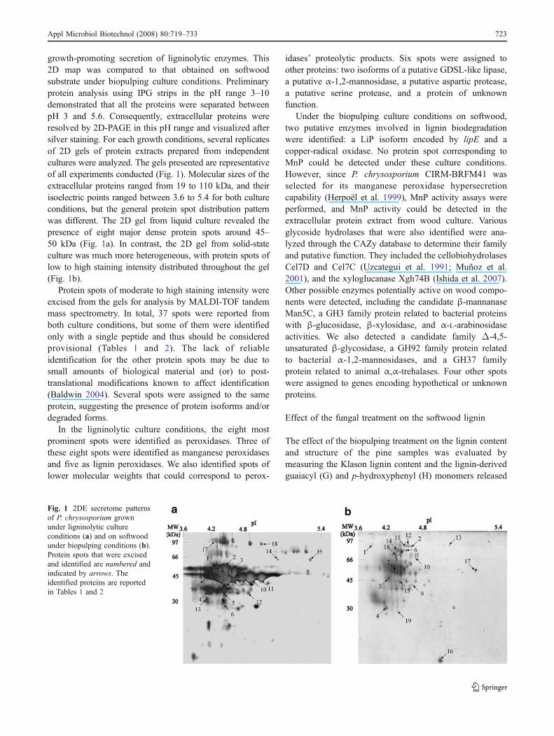
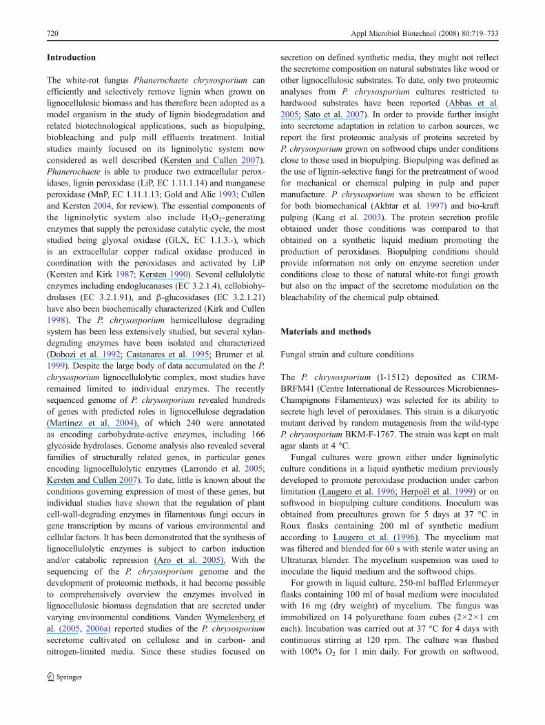
growth-promoting secretion of ligninolytic enzymes. This2D map was compared to that obtained on softwoodsubstrate under biopulping culture conditions. Preliminaryprotein analysis using IPG strips in the pH range 3–10demonstrated that all the proteins were separated betweenpH 3 and 5.6. Consequently, extracellular proteins wereresolved by 2D-PAGE in this pH range and visualized aftersilver staining. For each growth conditions, several replicatesof 2D gels of protein extracts prepared from independentcultures were analyzed. The gels presented are representativeof all experiments conducted (Fig. 1). Molecular sizes of theextracellular proteins ranged from 19 to 110 kDa, and theirisoelectric points ranged between 3.6 to 5.4 for both cultureconditions, but the general protein spot distribution patternwas different. The 2D gel from liquid culture revealed thepresence of eight major dense protein spots around 45–50 kDa (Fig. 1a). In contrast, the 2D gel from solid-stateculture was much more heterogeneous, with protein spots oflow to high staining intensity distributed throughout the gel(Fig. 1b).
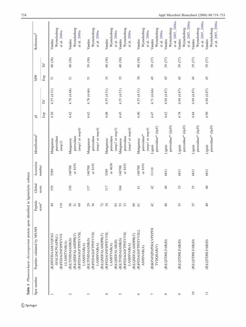
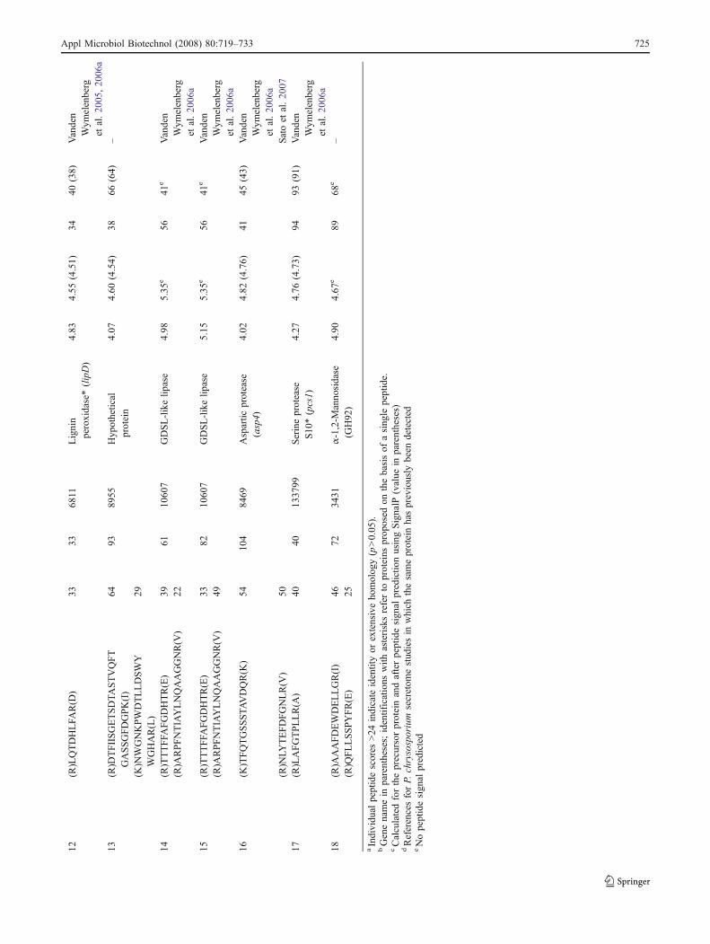
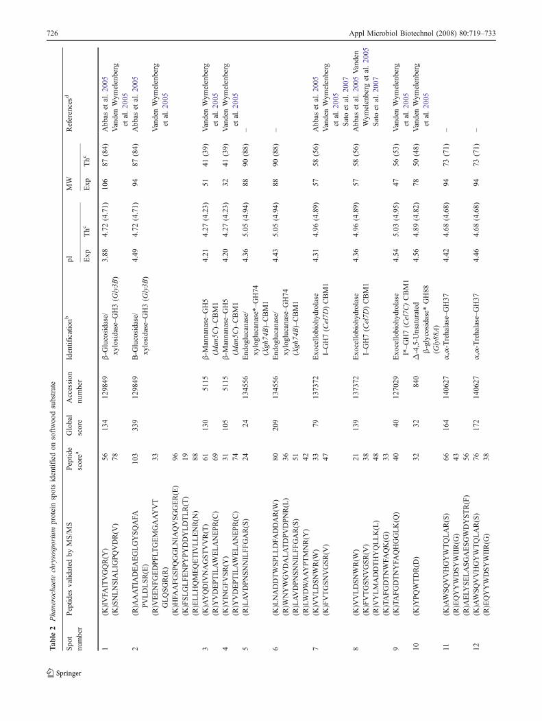
Protein spots of moderate to high staining intensity wereexcised from the gels for analysis by MALDI-TOF tandemmass spectrometry. In total, 37 spots were reported fromboth culture conditions, but some of them were identifiedonly with a single peptide and thus should be consideredprovisional (Tables 1 and 2). The lack of reliableidentification for the other protein spots may be due tosmall amounts of biological material and (or) to post-translational modifications known to affect identification(Baldwin 2004). Several spots were assigned to the sameprotein, suggesting the presence of protein isoforms and/ordegraded forms.
In the ligninolytic culture conditions, the eight mostprominent spots were identified as peroxidases. Three ofthese eight spots were identified as manganese peroxidasesand five as lignin peroxidases. We also identified spots oflower molecular weights that could correspond to perox-
idases’ proteolytic products. Six spots were assigned toother proteins: two isoforms of a putative GDSL-like lipase,a putative α-1,2-mannosidase, a putative aspartic protease,a putative serine protease, and a protein of unknownfunction.
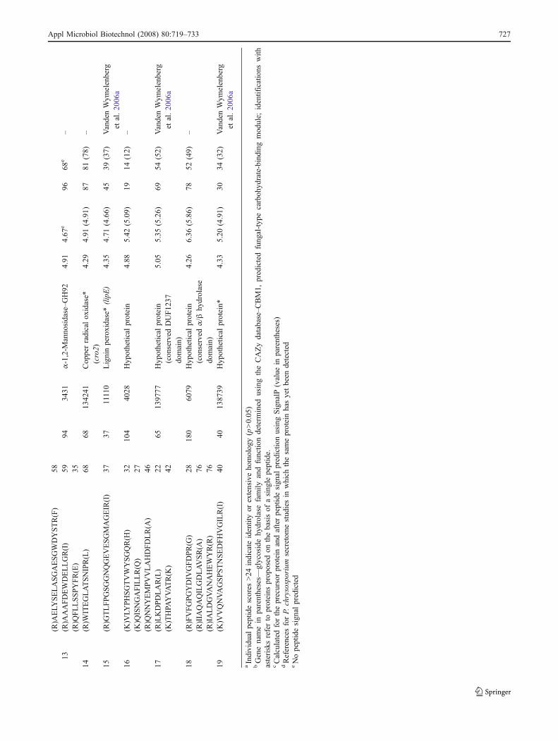
Under the biopulping culture conditions on softwood,two putative enzymes involved in lignin biodegradationwere identified: a LiP isoform encoded by lipE and acopper-radical oxidase. No protein spot corresponding toMnP could be detected under these culture conditions.However, since P. chrysosporium CIRM-BRFM41 wasselected for its manganese peroxidase hypersecretioncapability (Herpoël et al. 1999), MnP activity assays wereperformed, and MnP activity could be detected in theextracellular protein extract from wood culture. Variousglycoside hydrolases that were also identified were ana-lyzed through the CAZy database to determine their familyand putative function. They included the cellobiohydrolasesCel7D and Cel7C (Uzcategui et al. 1991; Muñoz et al.2001), and the xyloglucanase Xgh74B (Ishida et al. 2007).Other possible enzymes potentially active on wood compo-nents were detected, including the candidate β-mannanaseMan5C, a GH3 family protein related to bacterial proteinswith β-glucosidase, β-xylosidase, and α-L-arabinosidaseactivities. We also detected a candidate family Δ-4,5-unsaturated β-glycosidase, a GH92 family protein relatedto bacterial α-1,2-mannosidases, and a GH37 familyprotein related to animal α,α-trehalases. Four other spotswere assigned to genes encoding hypothetical or unknownproteins.
Effect of the fungal treatment on the softwood lignin
The effect of the biopulping treatment on the lignin contentand structure of the pine samples was evaluated bymeasuring the Klason lignin content and the lignin-derivedguaiacyl (G) and p-hydroxyphenyl (H) monomers released
Fig. 1 2DE secretome patternsof P. chrysosporium grownunder ligninolytic cultureconditions (a) and on softwoodunder biopulping conditions (b).Protein spots that were excisedand identified are numbered andindicated by arrows. Theidentified proteins are reportedin Tables 1 and 2
Appl Microbiol Biotechnol (2008) 80:719–733 723
Tab
le1
Pha
nerochaete
chrysosporium
proteinspotsidentifiedin
lignino
lytic
cultu
res
Spo
tnu
mber
Peptid
esvalid
ated
byMS/M
SPeptid
escorea
Global
score
Accession
number
Identificationb
pIMW
Referencesd
Exp
Thc
Exp
Thc
1(K
)HDTISAADLV
QFA
GAVALSNCPGAPR(L)
4915
935
89Manganese
peroxidase
(mnp
2)
4.30
4.55
(4.51)
5140
(38)
Vanden
Wym
elenberg
etal.20
06a
(R)FEDAGNFSPFEVVS
LLASHTVAR(A
)110
2(R)LTFHDAIA
ISR(S)
5015
014
0708
or81
91Manganese
peroxidase
(mnp
1or
mnp
4)
4.42
4.70
(4.68)
5140
(38)
Vanden
Wym
elenberg
etal.20
06a
(R)LQSDFA
LAHDPR(T)
31(R)FEDAGGFTPFEVVSL
LASHSVAR(A
)69
3(R)LTFHDAIA
ISR(S)
2913
714
0708
or81
91Manganese
peroxidase
(mnp
1or
mnp
4)
4.62
4.78
(4.66)
5139
(38)
Vanden
Wym
elenberg
etal.20
06a
(R)FEDAGGFTPFEVVSL
LASHSVAR(A
)36
(R)LQSDFA
LAHDPR(T)
724
(R)FEDAGNFSPFEVVSL
LASHTVAR(A
)34
117
3589 or46
36Manganese
peroxidase
(mnp
2or
mnp
5)
4.08
4.55
(4.51)
3540
(38)
Vanden
Wym
elenberg
etal.20
06a
(R)LQSDFA
LAR(D
)83
5(R)LTFHDAIA
ISR(S)
5318
414
0708
or81
91Manganese
peroxidase
(mnp
1or
mnp
4)
4.45
4.55
(4.51)
3540
(38)
Vanden
Wym
elenberg
etal.20
06a
(R)FEDAGGFTPFEVVSL
LASHSVAR(A
)33
(R)LQSDFA
LAHDPR(T)
996
(R)FEDAGGFTPFEVVSLL
ASHSVAR(A
)61
6114
0708
or81
91Manganese
peroxidase*
(mnp
1or
mnp
4)
4.40
4.55
(4.51)
3040
(38)
Vanden
Wym
elenberg
etal.20
06a
7(R)K
PATQPA
PDGLV
PEPFH
TVDQIIAR(V
)42
4211110
Lignin
peroxidase*(lipE)
4.47
4.71
(4.66)
4539
(37)
Vanden
Wym
elenberg
etal.20
06a
8(R)LQTDHLFA
R(D
)40
4068
11Lignin
peroxidase*(lipD)
4.62
4.94
(4.87)
4539
(37)
Vanden
Wym
elenberg
etal.20
05,20
06a
9(R)LQTDHLFA
R(D
)35
3568
11Lignin
peroxidase*(lipD)
4.78
4.94
(4.87)
4539
(37)
Vanden
Wym
elenberg
etal.20
05,20
06a
10(R)LQTDHLFA
R(D
)35
3568
11Lignin
peroxidase*(lipD)
4.84
4.94
(4.87)
4539
(37)
Vanden
Wym
elenberg
etal.20
05,20
06a
11(R)LQTDHLFA
R(D
)40
4068
11Lignin
peroxidase*(lipD)
4.90
4.94
(4.87)
4539
(37)
Vanden
Wym
elenberg
etal.20
05,20
06a
724 Appl Microbiol Biotechnol (2008) 80:719–733
Tab
le1
(con
tinued)
Spo
tnu
mber
Peptid
esvalid
ated
byMS/M
SPeptid
escorea
Global
score
Accession
number
Identificationb
pIMW
Referencesd
Exp
Thc
Exp
Thc
12(R)LQTDHLFA
R(D
)33
3368
11Lignin
peroxidase*(lipD)
4.83
4.55
(4.51)
3440
(38)
Vanden
Wym
elenberg
etal.20
05,20
06a
13(R)D
TFIISGETSDTA
STVQFT
GASSGFDGPK(I)
6493
8955
Hyp
othetical
protein
4.07
4.60
(4.54)
3866
(64)
–
(K)N
WGNKPWDTLLDSWY
WGHAR(L)
29
14(R)TTTFFA
FGDHTR(E)
3961
1060
7GDSL-likelip
ase
4.98
5.35
e56
41e
Vanden
Wym
elenberg
etal.20
06a
(R)A
RPFNTIAYLNQAAGGNR(V
)22
15(R)TTTFFA
FGDHTR(E)
3382
1060
7GDSL-likelip
ase
5.15
5.35
e56
41e
Vanden
Wym
elenberg
etal.20
06a
(R)A
RPFNTIAYLNQAAGGNR(V
)49
16(K
)TFQTGSSSTA
VDQR(K
)54
104
8469
Aspartic
protease
(asp4)
4.02
4.82
(4.76)
4145
(43)
Vanden
Wym
elenberg
etal.20
06a
(R)N
LYTEFDFGNLR(V
)50
Satoet
al.20
0717
(R)LAFGTPLLR(A
)40
4013
3799
Serineprotease
S10
*(pcs1)
4.27
4.76
(4.73)
9493
(91)
Vanden
Wym
elenberg
etal.20
06a
18(R)A
AAFDEWDELLGR(I)
4672
3431
α-1,2-M
anno
sidase
(GH92
)4.90
4.67
e89
68e
–(R)Q
FLLSSPYFR(E)
25
aIndividu
alpeptidescores
>24
indicate
identityor
extensiveho
molog
y(p>0.05
).bGenenamein
parentheses;identifications
with
asterisksreferto
proteins
prop
osed
onthebasisof
asing
lepeptide.
cCalculatedfortheprecursorproteinandafterpeptidesign
alpredictio
nusingSignalP
(value
inparentheses)
dReferencesforP.
chrysosporium
secretom
estud
iesin
which
thesameproteinhaspreviously
been
detected
eNopeptidesign
alpredicted
Appl Microbiol Biotechnol (2008) 80:719–733 725
Tab
le2
Pha
nerochaete
chrysosporium
proteinspotsidentifiedon
softwoo
dsubstrate
Spo
tnu
mber
Peptid
esvalid
ated
byMS/M
SPeptid
escorea
Global
score
Accession
number
Identificationb
pIMW
Referencesd
Exp
Thc
Exp
Thc
1(K
)IVFA
ITVGQR(Y
)56
134
1298
49β-G
lucosidase/
xylosidase–G
H3(G
ly3B
)3.88
4.72
(4.71)
106
87(84)
Abb
aset
al.20
05(K
)SNLNSIA
LIG
PQVDR(V
)78
VandenWym
elenberg
etal.20
052
(R)A
AATIA
DEAEGLGYSQAFA
PVLDLSR(E)
103
339
1298
49Β-G
lucosidase/
xylosidase–G
H3(G
ly3B
)4.49
4.72
(4.71)
9487
(84)
Abb
aset
al.20
05
(R)V
EENFGEDPFLT
GEMGAAYVT
GLQSGR(R)
33VandenWym
elenberg
etal.20
05(K
)HFA
AFGSPQGGLNIA
QVSGGER(E)
96(K
)FSLGLFENPYPYDDYLDTLR(T)
19(R)ELLHQMEQETIV
LLENR(N
)88
3(K
)AYQDIV
NAGSTVVR(T)
6113
05115
β-M
annanase–G
H5
(Man
5C)–CBM1
4.21
4.27
(4.23)
5141
(39)
VandenWym
elenberg
etal.20
05(R)Y
VDEPTILAWELANEPR(C)
694
(K)Y
INGFVSR(Y
)31
105
5115
β-M
annanase–G
H5
(Man
5C)–CBM1
4.20
4.27
(4.23)
3241
(39)
VandenWym
elenberg
etal.20
05(R)Y
VDEPTILAWELANEPR(C)
745
(R)LAVDPNSNNILFFGAR(S)
2424
1345
56End
oglucanase/
xyloglucanase*–G
H74
(Xgh
74B)–CBM1
4.36
5.05
(4.94)
8890
(88)
–
6(K
)LNADDTWSPLLDFA
DDAR(W
)80
209
1345
56End
oglucanase/
xyloglucanase–GH74
(Xgh
74B)–CBM1
4.43
5.05
(4.94)
8890
(88)
–(R)W
NYWGVDALATDPVDPNR(L)
36(R)LAVDPNSNNILFFGAR(S)
51(R)LWDWAAYPTMNR(Y
)42
7(K
)VVLDSNWR(W
)33
7913
7372
Exo
cello
bioh
ydrolase
I–GH7(Cel7D
)CBM1
4.31
4.96
(4.89)
5758
(56)
Abb
aset
al.20
05(K
)FVTGSNVGSR(V
)47
VandenWym
elenberg
etal.20
05Satoet
al.20
078
(K)V
VLDSNWR(W
)21
139
1373
72Exo
cello
bioh
ydrolase
I–GH7(Cel7D
)CBM1
4.36
4.96
(4.89)
5758
(56)
Abb
aset
al.20
05Vanden
Wym
elenberg
etal.20
05Satoet
al.20
07(K
)FVTGSNVGSR(V
)38
(R)V
YLMADDTHYQLLK(L)
48(K
)TAFGDTNWFA
QK(G
)33
9(K
)TAFGDTNYFA
QHGGLK(Q
)40
4012
7029
Exo
cello
bioh
ydrolase
I*–G
H7(Cel7C
)CBM1
4.54
5.03
(4.95)
4756
(53)
VandenWym
elenberg
etal.20
0510
(K)Y
PQWTDR(D
)32
3284
0Δ-4,5-U
nsaturated
β-glycosidase*GH88
(Gly88
A)
4.56
4.89
(4.82)
7850
(48)
VandenWym
elenberg
etal.20
05
11(K
)AWSQVVHGYWTQLAR(S)
6616
414
0627
α,α-Trehalase–G
H37
4.42
4.68
(4.68)
9473
(71)
–(R)EQYYWDSYWIIR(G
)43
(R)A
ELY
SELASGAESGWDYSTR(F)
5612
(K)AWSQVVHGYWTQLAR(S)
7617
214
0627
α,α-Trehalase–G
H37
4.46
4.68
(4.68)
9473
(71)
–(R)EQYYWDSYWIIR(G
)38
726 Appl Microbiol Biotechnol (2008) 80:719–733
Tab
le2
(con
tinued)
Spo
tnu
mber
Peptid
esvalid
ated
byMS/M
SPeptid
escorea
Global
score
Accession
number
Identificationb
pIMW
Referencesd
Exp
Thc
Exp
Thc
(R)A
ELY
SELASGAESGWDYSTR(F)
5813
(R)A
AAFDEWDELLGR(I)
5994
3431
α-1,2-M
anno
sidase–G
H92
4.91
4.67
e96
68e
–(R)Q
FLLSSPYFR(E)
3514
(R)W
ITEGLATSNIPR(L)
6868
1342
41Cop
perradicalox
idase*
(cro2)
4.29
4.91
(4.91)
8781
(78)
–
15(R)G
TLFPGSGGNQGEVESGMAGEIR(I)
3737
11110
Ligninperoxidase*(lipE)
4.35
4.71
(4.66)
4539
(37)
VandenWym
elenberg
etal.20
06a
16(K
)VLY
PHSGTVWYSGQR(H
)32
104
4028
Hyp
othetical
protein
4.88
5.42
(5.09)
1914
(12)
–(K
)QISNGAFILLR(Q
)27
(R)Q
NNYEMPVVLAHDFDLR(A
)46
17(R)LKDPDLAR(L)
2265
1397
77Hyp
othetical
protein
(con
served
DUF12
37do
main)
5.05
5.35
(5.26)
6954
(52)
VandenWym
elenberg
etal.20
06a
(K)THPA
YVATR(K
)42
18(R)FVFGPGYDIV
GFDPR(G
)28
180
6079
Hyp
othetical
protein
(con
served
α/β
hydrolase
domain)
4.26
6.36
(5.86)
7852
(49)
–(R)IIA
QAQILGDLAVSR(A
)76
(R)IALDGVANAHEWYR(R)
7619
(K)V
VQNVAGSPSTNSEDFHVGILR(I)
4040
1387
39Hyp
othetical
protein*
4.33
5.20
(4.91)
3034
(32)
VandenWym
elenberg
etal.20
06a
aIndividu
alpeptidescores
>24
indicate
identityor
extensiveho
molog
y(p>0.05
)bGenenamein
parentheses—
glycosidehy
drolasefamily
andfunctio
ndeterm
ined
usingtheCAZydatabase–C
BM1,
predictedfung
al-typ
ecarboh
ydrate-binding
mod
ule;
identifications
with
asterisksreferto
proteins
prop
osed
onthebasisof
asing
lepeptide.
cCalculatedfortheprecursorproteinandafterpeptidesign
alpredictio
nusingSignalP
(value
inparentheses)
dReferencesforP.
chrysosporium
secretom
estud
iesin
which
thesameproteinhasyetbeen
detected
eNopeptidesign
alpredicted
Appl Microbiol Biotechnol (2008) 80:719–733 727
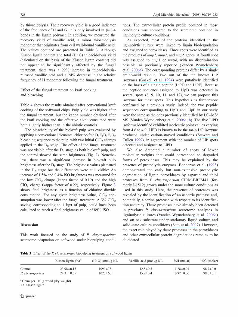
by thioacidolysis. Their recovery yield is a good indicatorof the frequency of H and G units only involved in β-O-4bonds in the lignin polymer. In addition, we measured therecovery yield of vanillic acid, a minor thioacidolysismonomer that originates from cell wall-bound vanillic acid.The values obtained are presented in Table 3. AlthoughKlason lignin content and total (H+G) thioacidolysis yield(calculated on the basis of the Klason lignin content) didnot appear to be significantly affected by the fungaltreatment, there was a 22% increase in thioacidolysis-released vanillic acid and a 24% decrease in the relativefrequency of H monomer following the fungal treatment.
Effect of the fungal treatment on kraft cookingand bleaching
Table 4 shows the results obtained after conventional kraftcooking of the softwood chips. Pulp yield was higher afterthe fungal treatment, but the kappa number obtained afterthe kraft cooking and the effective alkali consumed wereboth slightly higher than in the abiotic controls.
The bleachability of the biokraft pulp was evaluated byapplying a conventional elemental chlorine-free D0E1D1E2D2
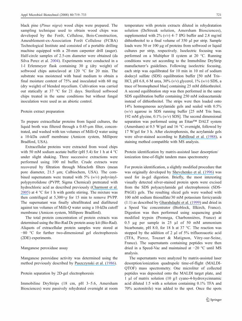
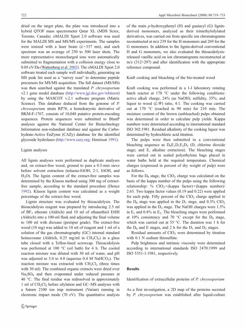
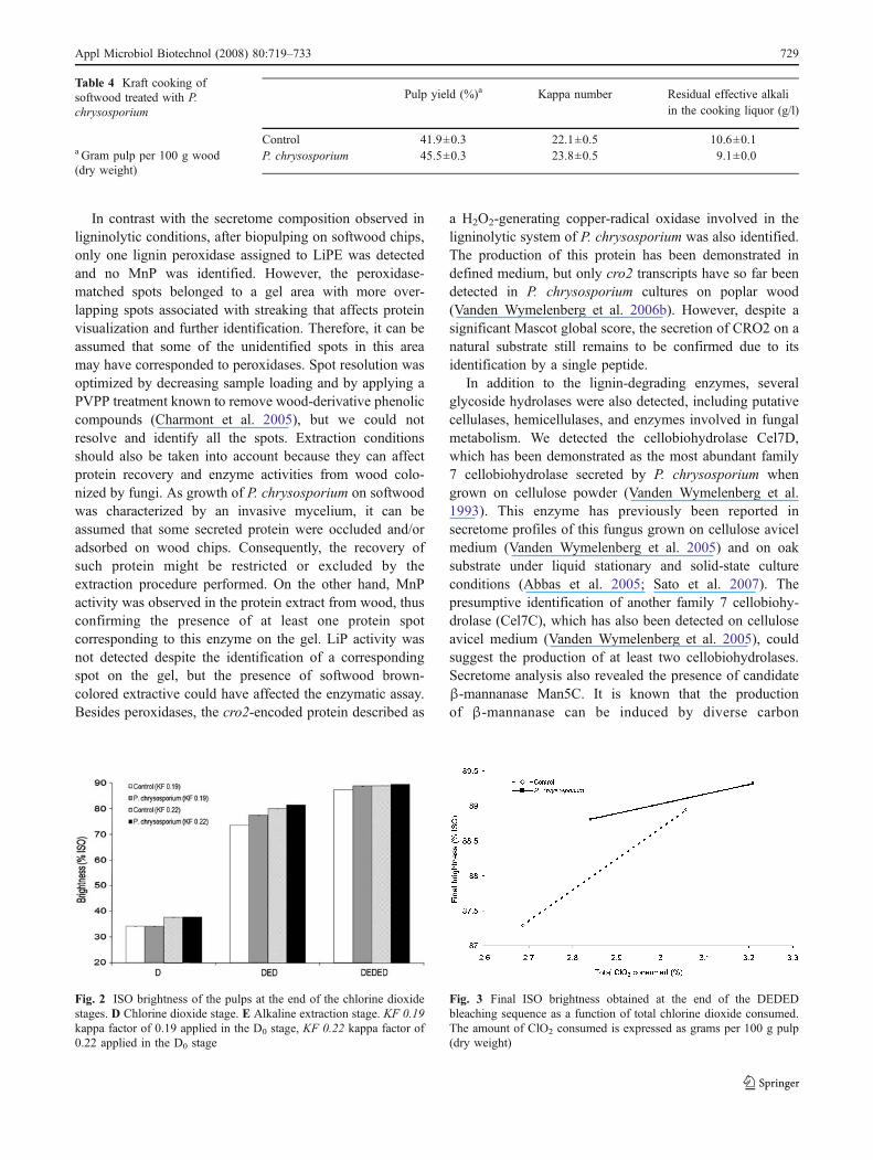
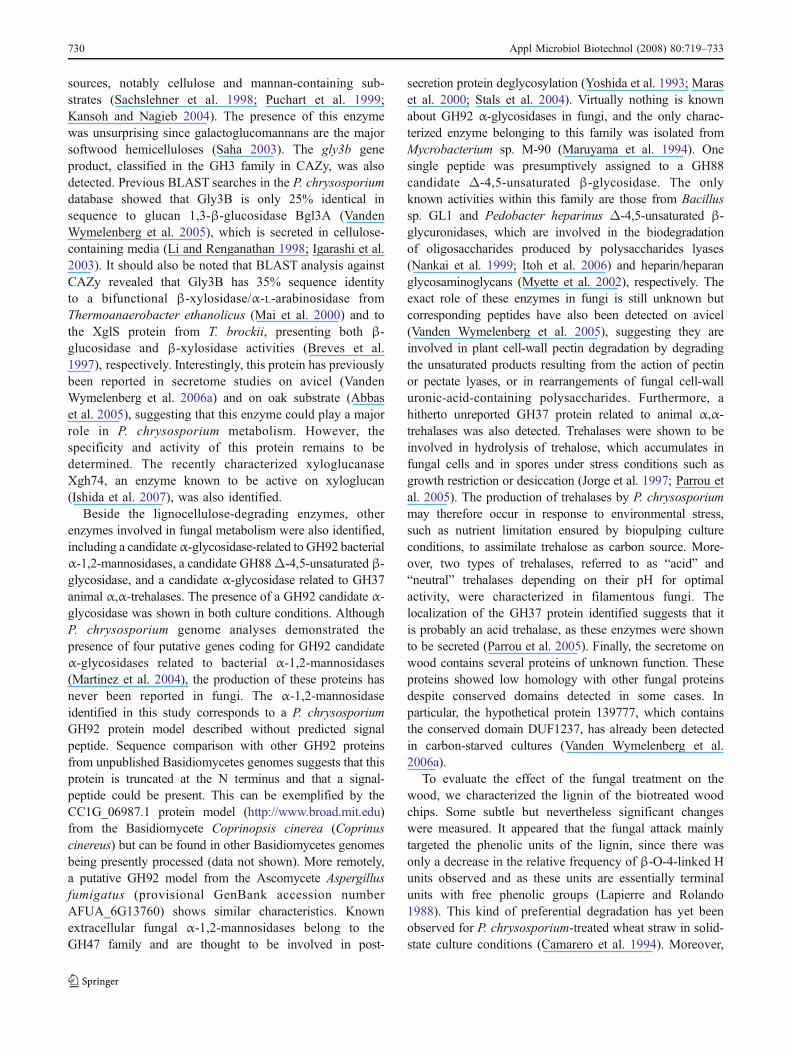
bleaching sequence to the pulp, with two initial ClO2 chargesapplied in the D0 stage. The effect of the fungal treatmentwas not visible after the D0 stage as both biokraft pulp, andthe control showed the same brightness (Fig. 2). Nonethe-less, there was a significant increase in biokraft pulpbrightness after the D1 stage. The brightness values plateauedin the D2 stage but the differences were still visible: Anincrease of 1.5% and 0.4% ISO brightness was measured forthe low ClO2 charge (kappa factor of 0.19) and the highClO2 charge (kappa factor of 0.22), respectively. Figure 3shows final brightness as a function of chlorine dioxideconsumption. For any given brightness value, ClO2 con-sumption was lower after the fungal treatment. A 3% ClO2
saving, corresponding to 1 kg/t of pulp, could have beencalculated to reach a final brightness value of 89% ISO.
Discussion
This work focused on the study of P. chrysosporiumsecretome adaptation on softwood under biopulping condi-
tions. The extracellular protein profile obtained in thoseconditions was compared to the secretome obtained inligninolytic culture conditions.
As expected, most of the proteins identified in theligninolytic culture were linked to lignin biodegradationand assigned to peroxidases. Three spots were identified asthe products of mnp1, mnp2, and mnp5 genes. A fourth spotwas assigned to mnp1 or mnp4, with no discriminationpossible, as previously reported (Vanden Wymelenberget al. 2006a). The corresponding proteins differ by a singleamino-acid residue. Two out of the ten known LiPisozymes (Gaskell et al. 1994) were putatively identifiedon the basis of a single peptide (LiPD and LiPE). Becausethe peptide sequence assigned to LipD was detected inseveral spots (8, 9, 10, 11, and 12), we can propose thisisozyme for these spots. This hypothesis is furthermoreconfirmed by a previous study. Indeed, the two peptidesequences corresponding to LipD and LipE in our studywere the same as the ones previously identified by LC–MS/MS (Vanden Wymelenberg et al. 2006a, b). The five LiPDisoforms identified exhibited isoelectric point values varyingfrom 4.6 to 4.9. LiPD is known to be the main LiP isozymeproduced under carbon-starved conditions (Stewart andCullen 1999), in agreement with the number of LiP spotsdetected and assigned to LiPD.
We also detected a number of spots of lowermolecular weights that could correspond to degradedforms of peroxidases. This may be explained by thepresence of proteolytic enzymes. Bonnarme et al. (1993)demonstrated the early but non-extensive proteolyticdegradation of lignin peroxidases by aspartic and thiolproteases from P. chrysosporium CIRM-BRFM41 (for-merly I-1512) grown under the same culture conditions asused in this study. Here, the presence of proteases wasrevealed by the identification of an aspartic protease and,potentially, a serine protease with respect to its identifica-tion accuracy. These proteases have already been detectedin previous P. chrysosporium secretome analyses inligninolytic cultures (Vanden Wymelenberg et al. 2006a)and on oak substrate under stationary liquid culture andsolid-state culture conditions (Sato et al. 2007). However,the exact role played by these proteases in the peroxidasesand other extracellular protein degradations remains to beelucidated.
Table 3 Effect of the P. chrysosporium biopulping treatment on softwood lignin
Klason lignin (%)a (H+G) µmol/g KL Vanillic acid µmol/g KL %H (molar) %G (molar)
Control 23.98±0.15 1099±73 12.5±0.5 1.28±0.01 98.7±0.0P. chrysosporium 24.31±0.05 1025±60 15.2±0.4 0.97±0.06 99.0±0.1
a Gram per 100 g wood (dry weight)KL Klason lignin
728 Appl Microbiol Biotechnol (2008) 80:719–733
In contrast with the secretome composition observed inligninolytic conditions, after biopulping on softwood chips,only one lignin peroxidase assigned to LiPE was detectedand no MnP was identified. However, the peroxidase-matched spots belonged to a gel area with more over-lapping spots associated with streaking that affects proteinvisualization and further identification. Therefore, it can beassumed that some of the unidentified spots in this areamay have corresponded to peroxidases. Spot resolution wasoptimized by decreasing sample loading and by applying aPVPP treatment known to remove wood-derivative phenoliccompounds (Charmont et al. 2005), but we could notresolve and identify all the spots. Extraction conditionsshould also be taken into account because they can affectprotein recovery and enzyme activities from wood colo-nized by fungi. As growth of P. chrysosporium on softwoodwas characterized by an invasive mycelium, it can beassumed that some secreted protein were occluded and/oradsorbed on wood chips. Consequently, the recovery ofsuch protein might be restricted or excluded by theextraction procedure performed. On the other hand, MnPactivity was observed in the protein extract from wood, thusconfirming the presence of at least one protein spotcorresponding to this enzyme on the gel. LiP activity wasnot detected despite the identification of a correspondingspot on the gel, but the presence of softwood brown-colored extractive could have affected the enzymatic assay.Besides peroxidases, the cro2-encoded protein described as
a H2O2-generating copper-radical oxidase involved in theligninolytic system of P. chrysosporium was also identified.The production of this protein has been demonstrated indefined medium, but only cro2 transcripts have so far beendetected in P. chrysosporium cultures on poplar wood(Vanden Wymelenberg et al. 2006b). However, despite asignificant Mascot global score, the secretion of CRO2 on anatural substrate still remains to be confirmed due to itsidentification by a single peptide.
In addition to the lignin-degrading enzymes, severalglycoside hydrolases were also detected, including putativecellulases, hemicellulases, and enzymes involved in fungalmetabolism. We detected the cellobiohydrolase Cel7D,which has been demonstrated as the most abundant family7 cellobiohydrolase secreted by P. chrysosporium whengrown on cellulose powder (Vanden Wymelenberg et al.1993). This enzyme has previously been reported insecretome profiles of this fungus grown on cellulose avicelmedium (Vanden Wymelenberg et al. 2005) and on oaksubstrate under liquid stationary and solid-state cultureconditions (Abbas et al. 2005; Sato et al. 2007). Thepresumptive identification of another family 7 cellobiohy-drolase (Cel7C), which has also been detected on celluloseavicel medium (Vanden Wymelenberg et al. 2005), couldsuggest the production of at least two cellobiohydrolases.Secretome analysis also revealed the presence of candidateβ-mannanase Man5C. It is known that the productionof β-mannanase can be induced by diverse carbon
Fig. 2 ISO brightness of the pulps at the end of the chlorine dioxidestages. D Chlorine dioxide stage. E Alkaline extraction stage. KF 0.19kappa factor of 0.19 applied in the D0 stage, KF 0.22 kappa factor of0.22 applied in the D0 stage
Fig. 3 Final ISO brightness obtained at the end of the DEDEDbleaching sequence as a function of total chlorine dioxide consumed.The amount of ClO2 consumed is expressed as grams per 100 g pulp(dry weight)
Table 4 Kraft cooking ofsoftwood treated with P.chrysosporium
a Gram pulp per 100 g wood(dry weight)
Pulp yield (%)a Kappa number Residual effective alkaliin the cooking liquor (g/l)
Control 41.9±0.3 22.1±0.5 10.6±0.1P. chrysosporium 45.5±0.3 23.8±0.5 9.1±0.0
Appl Microbiol Biotechnol (2008) 80:719–733 729
sources, notably cellulose and mannan-containing sub-strates (Sachslehner et al. 1998; Puchart et al. 1999;Kansoh and Nagieb 2004). The presence of this enzymewas unsurprising since galactoglucomannans are the majorsoftwood hemicelluloses (Saha 2003). The gly3b geneproduct, classified in the GH3 family in CAZy, was alsodetected. Previous BLAST searches in the P. chrysosporiumdatabase showed that Gly3B is only 25% identical insequence to glucan 1,3-β-glucosidase Bgl3A (VandenWymelenberg et al. 2005), which is secreted in cellulose-containing media (Li and Renganathan 1998; Igarashi et al.2003). It should also be noted that BLAST analysis againstCAZy revealed that Gly3B has 35% sequence identityto a bifunctional β-xylosidase/α-L-arabinosidase fromThermoanaerobacter ethanolicus (Mai et al. 2000) and tothe XglS protein from T. brockii, presenting both β-glucosidase and β-xylosidase activities (Breves et al.1997), respectively. Interestingly, this protein has previouslybeen reported in secretome studies on avicel (VandenWymelenberg et al. 2006a) and on oak substrate (Abbaset al. 2005), suggesting that this enzyme could play a majorrole in P. chrysosporium metabolism. However, thespecificity and activity of this protein remains to bedetermined. The recently characterized xyloglucanaseXgh74, an enzyme known to be active on xyloglucan(Ishida et al. 2007), was also identified.
Beside the lignocellulose-degrading enzymes, otherenzymes involved in fungal metabolism were also identified,including a candidateα-glycosidase-related to GH92 bacterialα-1,2-mannosidases, a candidate GH88Δ-4,5-unsaturated β-glycosidase, and a candidate α-glycosidase related to GH37animal α,α-trehalases. The presence of a GH92 candidate α-glycosidase was shown in both culture conditions. AlthoughP. chrysosporium genome analyses demonstrated thepresence of four putative genes coding for GH92 candidateα-glycosidases related to bacterial α-1,2-mannosidases(Martinez et al. 2004), the production of these proteins hasnever been reported in fungi. The α-1,2-mannosidaseidentified in this study corresponds to a P. chrysosporiumGH92 protein model described without predicted signalpeptide. Sequence comparison with other GH92 proteinsfrom unpublished Basidiomycetes genomes suggests that thisprotein is truncated at the N terminus and that a signal-peptide could be present. This can be exemplified by theCC1G_06987.1 protein model (http://www.broad.mit.edu)from the Basidiomycete Coprinopsis cinerea (Coprinuscinereus) but can be found in other Basidiomycetes genomesbeing presently processed (data not shown). More remotely,a putative GH92 model from the Ascomycete Aspergillusfumigatus (provisional GenBank accession numberAFUA_6G13760) shows similar characteristics. Knownextracellular fungal α-1,2-mannosidases belong to theGH47 family and are thought to be involved in post-
secretion protein deglycosylation (Yoshida et al. 1993; Maraset al. 2000; Stals et al. 2004). Virtually nothing is knownabout GH92 α-glycosidases in fungi, and the only charac-terized enzyme belonging to this family was isolated fromMycrobacterium sp. M-90 (Maruyama et al. 1994). Onesingle peptide was presumptively assigned to a GH88candidate Δ-4,5-unsaturated β-glycosidase. The onlyknown activities within this family are those from Bacillussp. GL1 and Pedobacter heparinus Δ-4,5-unsaturated β-glycuronidases, which are involved in the biodegradationof oligosaccharides produced by polysaccharides lyases(Nankai et al. 1999; Itoh et al. 2006) and heparin/heparanglycosaminoglycans (Myette et al. 2002), respectively. Theexact role of these enzymes in fungi is still unknown butcorresponding peptides have also been detected on avicel(Vanden Wymelenberg et al. 2005), suggesting they areinvolved in plant cell-wall pectin degradation by degradingthe unsaturated products resulting from the action of pectinor pectate lyases, or in rearrangements of fungal cell-walluronic-acid-containing polysaccharides. Furthermore, ahitherto unreported GH37 protein related to animal α,α-trehalases was also detected. Trehalases were shown to beinvolved in hydrolysis of trehalose, which accumulates infungal cells and in spores under stress conditions such asgrowth restriction or desiccation (Jorge et al. 1997; Parrou etal. 2005). The production of trehalases by P. chrysosporiummay therefore occur in response to environmental stress,such as nutrient limitation ensured by biopulping cultureconditions, to assimilate trehalose as carbon source. More-over, two types of trehalases, referred to as “acid” and“neutral” trehalases depending on their pH for optimalactivity, were characterized in filamentous fungi. Thelocalization of the GH37 protein identified suggests that itis probably an acid trehalase, as these enzymes were shownto be secreted (Parrou et al. 2005). Finally, the secretome onwood contains several proteins of unknown function. Theseproteins showed low homology with other fungal proteinsdespite conserved domains detected in some cases. Inparticular, the hypothetical protein 139777, which containsthe conserved domain DUF1237, has already been detectedin carbon-starved cultures (Vanden Wymelenberg et al.2006a).
To evaluate the effect of the fungal treatment on thewood, we characterized the lignin of the biotreated woodchips. Some subtle but nevertheless significant changeswere measured. It appeared that the fungal attack mainlytargeted the phenolic units of the lignin, since there wasonly a decrease in the relative frequency of β-O-4-linked Hunits observed and as these units are essentially terminalunits with free phenolic groups (Lapierre and Rolando1988). This kind of preferential degradation has yet beenobserved for P. chrysosporium-treated wheat straw in solid-state culture conditions (Camarero et al. 1994). Moreover,
730 Appl Microbiol Biotechnol (2008) 80:719–733
the sample subjected to fungal treatment released morevanillic acid upon thioacidolysis. This increase cannot beaccounted for by the formation of additional vanillic estersin the cell walls after the fungal treatment but was morelikely due to oxidative cleavage of certain lignin sidechains, a lignin degradation mechanism which has alsobeen shown for P. chrysosporium (Camarero et al. 1997).Interestingly, the results obtained are similar to thosereported for P. chrysosporium lignin-degrading enzymes,in particular for LiP (Toshiaki and Higuchi 1989). We alsoran conventional kraft pulping and chlorine dioxidechemical bleaching of the biotreated wood. Despite asignificant increase in pulp yield, the fungal treatment hada slight negative effect on the chemical consumption afterkraft cooking. The increase in chemical consumption maybe attributed to the presence of the fungal biomass, whichcan also be chemical-consuming (Atik et al. 2006). Thekappa number of the pulp was increased after the P.chrysosporium treatment, but kraft pulp bleachability wasnevertheless enhanced, as demonstrated by a decrease inchlorine dioxide consumption for the same range of finalbrightness values. This result can be partially explained byan increase in the amount of alkali-extractible compounds,which is known to be one of the effects of fungal treatmentsof wood and pulps (Reid 1998). The lignin-degradingenzymes and hemicellulases revealed by secretome analysisare expected to be involved in this bleachability improve-ment. It can be assumed that the P. chrysosporium-secretedenzymes may modify the wood compounds without aconcomitant significant release of biodegradation products.This hypothesis is consistent with the results obtained forthe lignin analyses, as the fungal treatment did not affectthe lignin content of the wood but nonetheless led tosignificant lignin modifications. It should be pointed outthat the same phenomenon has been observed during theearly stages of fungal treatment of wood for biomechanicalpulping applications (Akhtar et al. 1998).
In summary, the comparative secretome analysis dealingwith fungal culture on synthetic medium and on wooddemonstrates that P. chrysosporium readily adapts tochanges in environmental conditions. This study presentsthe first characterization of the P. chrysosporium secretomeon softwood under biopulping conditions. Identifying theproteins secreted on a natural substrate and in conditionsclose to the natural fungal growth conditions has provided abetter understanding of lignocellulose breakdown.
Acknowledgment This work was supported by the French NationalResearch Agency Program PNRB as part of the Stratégie dePrétraitements Physiques, Enzymatiques et Chimiques Appliquées àla Biomasse-Bio-Ethanol (SPECABBE) project. The authors thank F.Legée and L. Cézard for the lignin analyses.
References
Abbas A, Koc H, Liu F, Tien M (2005) Fungal degradation of wood:initial proteomic analysis of extracellular proteins of Phanerochaetechrysosporium grown on oak substrate. Curr Genet 47:49–56
Akhtar M, Blanchette RA, Kirk TK (1997) Fungal delignification andbiomechanical pulping of wood. In: Scheper T (ed) Advances inbiochemical engineering/biotechnology. Springer, Berlin, pp 159–195
Akhtar M, Blanchette RA, Myers G, Kirk TK (1998) An overview ofbiomechanical pulping research. In: Akhtar M, Young RA (eds)Environmentally friendly technologies for the pulp and paperindustry. Wiley, New York, pp 309–340
Aro N, Pakula T, Penttilä M (2005) Transcriptional regulation of plantcell wall degradation by filamentous fungi. FEMS Microbiol Rev29:719–739
Atik C, Imamoglu S, Bermek H (2006) Impact of xylanase pre-treatment on peroxide bleaching stage of biokraft pulp. IntBiodeterior Biodegrad 58:22–26
Baldwin MA (2004) Protein identification by mass spectrometry:issues to be considered. Mol Cell Proteomics 3:1–9
Bonnarme P, Asther M, Asther Ma (1993) Influence of primary andsecondary proteases produced by free and immobilized cells ofthe white-rot fungus Phanerochaete chrysosporium on ligninperoxidase activity. J Biotechnol 30:271–282
Breves R, Bronnenmeier K, Wild N, Lottspeich F, Staudenbauer WL,Hofemeister J (1997) Genes encoding two different β-glucosidasesof Thermoanaerobacter brockii are clustered in a common operon.Appl Environ Microbiol 63:3902–3910
Brumer 3rd H, Sims PF, Sinnott ML (1999) Lignocellulose degradationby Phanerochaete chrysosporium: purification and characterizationof the main α-galactosidase. Biochem J 339:43–53
Camarero S, Galletti GC, Martinez AT (1994) Preferential degradationof phenolic lignin units by two white rot fungi. Appl EnvironMicrobiol 60:4509–4516
Camarero S, Galletti GC, Martínez AT (1997) Demonstration of insitu oxidative degradation of lignin side chains by two white-rotfungi using analytical pyrolysis of methylated wheat straw. RapidCommun Mass Spectrom 11:331–334
Castanares A, Hay AJ, Gordon AH, McCrae SI, Wood TM (1995) D-Xylan-degrading enzyme system from the fungus Phanerochaetechrysosporium: isolation and partial characterisation of an α-(4-O-methyl)-D-glucuronidase. J Biotechnol 43:183–194
Charmont S, Jamet E, Pont-Lezica R, Canut H (2005) Proteomicanalysis of secreted proteins from Arabidopsis thaliana seedlings:improved recovery following removal of phenolic compounds.Phytochemistry 66:453–461
Cullen D, Kersten PJ (2004) Enzymology and molecular biology oflignin degradation. In: Brambl R, Marzulf GA (eds) The mycotaIII. Biochemistry and molecular biology. Springer, Berlin, pp 249–273
da Silva Perez D, Moreau J, Nougier P, Themelin A, Chantre G (2004)Effect of storage conditions on the wood and pulp quality ofwindthrow trees. Proceedings of the 8th European Workshop onLignocellulosics and Pulps, Latvian State Institute of WoodChemistry, Riga, Latvia, August 22–25, 2004, pp 295–298
Dence CW (1992) The determination of lignin. In: Lin SY, Dence CW(eds) Methods in lignin chemistry. Springer, Berlin, pp 33–61
Dobozi MS, Szakacs G, Bruschi CV (1992) Xylanase activity ofPhanerochaete chrysosporium. Appl Environ Microbiol 58:3466–3471
Gaskell J, Stewart P, Kersten PJ, Covert SF, Reiser J, Cullen D (1994)Establishment of genetic linkage by allele-specific polymerasechain reaction: application to the lignin peroxidase gene family ofPhanerochaete chrysosporium. Biotechnology 12:1372–1375
Appl Microbiol Biotechnol (2008) 80:719–733 731

Gharahdaghi F, Weinberg CR, Meagher DA, Imai BS, Mische SM(1999) Mass spectrometric identification of proteins from silver-stained polyacrylamide gel: a method for the removal of silver ionsto enhance sensitivity. Electrophoresis 20:601–605
Gold MH, Alic M (1993) Molecular biology of the lignin-degradingbasidiomycete Phanerochaete chrysosporium. Microbiol Rev57:605–622
Henrissat B (1991) A classification of glycosyl hydrolases based onamino-acid sequence similarities. Biochem J 280:309–316
Herpoël I, Asther M, Sigoillot JC (1999) Design and scale up of aprocess for manganese peroxidase production using the hyper-secretory strain Phanerochaete chrysosporium I-1512. BiotechnolBioeng 65:468–473
Igarashi K, Tani T, Rie K, Masahiro S (2003) Family 3 β-glucosidasefrom cellulose-degrading culture of the white-rot fungus Phaner-ochaete chrysosporium is a glucan 1,3-β-glucosidase. J BiosciBioeng 95:572–576
Ishida T, Yaoi K, Hiyoshi A, Igarashi K, Samejima M (2007) Substraterecognition by glycoside hydrolase family 74 xyloglucanase fromthe basidiomycete Phanerochaete chrysosporium. FEBS J274:5727–5736
Itoh T, Hashimoto W, Mikami B, Murata K (2006) Substraterecognition by unsaturated glucuronyl hydrolase from Bacillussp. GL1. Biochem Biophys Res Comm 344:253–262
Jorge JA, Polizeli ML, Thevelein JM, Terenzi HF (1997) Trehalasesand trehalose hydrolysis in fungi. FEMS Microbiol Lett 154:165–171
Kang KY, Jo BM, Oh JS, Mansfield SD (2003) Biopulping of hybridpoplar improves chemical and energy savings during kraftpulping. Wood Fiber Sci 35:594–600
Kansoh AL, Nagieb ZA (2004) Xylanase and mannanase enzymesfrom Streptomyces galbus NR and their use in biobleaching ofsoftwood kraft pulp. Antonie Van Leeuwenhoek 85:103–114
Kersten PJ (1990) Glyoxal oxidase of Phanerochaete chrysosporium:its characterization and activation by lignin peroxidase. Proc NatlAcad Sci U S A 87:2936–2940
Kersten PJ, Kirk TK (1987) Involvement of a new enzyme, glyoxaloxidase, in extracellular H2O2 production by Phanerochaetechrysosporium. J Bacteriol 169:2195–2201
Kersten P, Cullen D (2007) Extracellular oxidative systems of thelignin-degrading basidiomycete Phanerochaete chrysosporium.Fungal Genet Biol 44:77–87
Kirk TK, Cullen D (1998) Enzymology and molecular genetics ofwood degradation by white-rot fungi. In: Young RA, Masood A(eds) Environmentally friendly technologies for the pulp andpaper industry. Wiley, New York, pp 273–307
Lapierre C, Rolando C (1988) Thioacidolysis of pre-methylated ligninsamples from pine compression and poplar woods. Holzforschung42:1–4
Larrondo L, Vicuna R, Cullen D (2005) Phanerochaete chrysosporiumgenomics. In: Arora Berka DKR (ed) Applied mycology andbiotechnology. Elsevier, Amsterdam, pp 315–352
Laugero C, Sigoillot JC, Moukha S, Frasse P, Bellon-Fontaine M-N,Bonnarme P, Mougin C, Asther M (1996) Selective hyperproduc-tion of manganese peroxidases by Phanerochaete chrysosporiumI-1512 immobilized on nylon net in a bubble column reactor. ApplMicrobiol Biotechnol 44:717–723
Li B, Renganathan V (1998) Gene cloning and characterization of anovel cellulose-binding β-glucosidase from Phanerochaetechrysosporium. Appl Environ Microbiol 64:2748–2754
Mai V, Wiegel J, Lorenz WW (2000) Cloning, sequencing, andcharacterization of the bifunctional xylosidase-arabinosidasefrom the anaerobic thermophile Thermoanaerobacter ethanolicus.Gene 247:137–143
Maras M, Callewaert N, Piens K, Claeyssens M, Martinet W, DewaeleS, Contreras H, Dewerte I, Penttila M, Contreras R (2000)
Molecular cloning and enzymatic characterization of a Trichodermareesei 1,2-α-D-mannosidase. J Biotechnol 77:255–263
Martinez D, Larrondo LF, Putnam N, Gelpke MD, Huang K,Chapman J, Helfenbein KG, Ramaiya P, Detter JC, Larimer F,Coutinho PM, Henrissat B, Berka R, Cullen D, Rokhsar D(2004) Genome sequence of the lignocellulose degrading fungusPhanerochaete chrysosporium strain RP78. Nat Biotechnol22:695–700
Maruyama Y, Nakajima T, Ichishima E (1994) A 1,2-α-D-mannosidasefrom a Bacillus sp.: purification, characterization, and mode ofaction. Carbohydr Res 251:89–98
Muñoz IG, Ubhayasekera W, Henriksson H, Szabó I, Pettersson G,Johansson G, Mowbray SL, Ståhlberg J (2001) Family 7cellobiohydrolases from Phanerochaete chrysosporium: crystalstructure of the catalytic module of Cel7D (CBH58) at 1.32 Åresolution and homology models of the isozymes. J Mol Biol314:1097–1111
Myette JR, Shriver Z, Kiziltepe T, McLean MW, Venkataraman G,Sasisekharan R (2002) Molecular cloning of the heparin/heparansulfate delta 4,5 unsaturated glycuronidase from Flavobacteriumheparinum, its recombinant expression in Escherichia coli, andbiochemical determination of its unique substrate specificity.Biochemistry 41:7424–7434
Nankai H, Hashimoto W, Miki H, Kawai S, Murata K (1999)Microbial system for polysaccharide depolymerization: enzymaticroute for xanthan depolymerization by Bacillus sp. strain GL1.Appl Environ Microbiol 65:2520–2526
Parrou JL, Jules M, Beltran G, François J (2005) Acid trehalase inyeasts and filamentous fungi: localization, regulation andphysiological function. FEMS Yeast Res 5:503–511
Paszczynski A, Huynh VB, Crawford R (1986) Comparisonof ligninase-I and peroxidase-M2 from the white-rot fungusPhanerochaete chrysosporium. Arch Biochem Biophys 244:750–765
Puchart V, Katapodis P, Biely P, Kremnicky L, Christakopoulos P,Vrsanska M, Kekos D, Macris BJ, Bhat MK (1999) Productionof xylanases, mannanases, and pectinases by the thermophilicfungus Thermomyces lanuginosus. Enzyme Microb Technol24:355–361
Rabilloud T, Carpentier G, Tarroux P (1988) Improvement andsimplification of low-background silver staining of proteins byusing sodium dithionite. Electrophoresis 9:288–291
Reid ID (1998) Fate of residual lignin during delignification of kraftpulp by Trametes versicolor. Appl Environ Microbiol 64:2117–2125
Sachslehner A, Nidetzky B, Kulbe KD, Haltrich D (1998) Inductionof mannanase, xylanase, and endoglucanase activities in Sclerotiumrolfsii. Appl Environ Microbiol 64:594–600
Saha BC (2003) Hemicellulose bioconversion. J Ind MicrobiolBiotechnol 30:279–291
Sato S, Liu F, Koc H, Tien M (2007) Expression analysis ofextracellular proteins from Phanerochaete chrysosporium grownon different liquid and solid substrates. Microbiology 153:3023–3033
Shevchenko A, Wilm M, Vorm O, Mann M (1996) Mass spectrometrysequencing of proteins from silver-stained polyacrylamide gels.Anal Chem 68:850–858
Stals I, Sandra K, Geysens S, Contreras R, Van Beeumen J,Claeyssens M (2004) Factors influencing glycosylation ofTrichoderma reesei cellulases. I: Post-secretorial changes of theO- and N-glycosylation pattern of Cel7A. Glycobiology 14:713–724
Stewart P, Cullen D (1999) Organization and differential regulation of acluster of lignin peroxidase genes of Phanerochaete chrysosporium.J Bacteriol 181:3427–3432
Toshiaki U, Higuchi T (1989) Cleavages of aromatic ring and β-O-4bond of synthetic lignin (DHP) by lignin peroxidase. FEBS Lett242:325–329
732 Appl Microbiol Biotechnol (2008) 80:719–733
Uzcategui E, Ruiz A, Montesino R, Johansson G, Pettersson G (1991)The 1,4-β-D-glucan cellobiohydrolases from Phanerochaetechrysosporium. I. A system of synergistically acting enzymeshomologous to Trichoderma reesei. J Biotechnol 19:271–285
Vanden Wymelenberg A, Covert S, Cullen D (1993) Identification ofthe gene encoding the major cellobiohydrolase of the white rotfungus Phanerochaete chrysosporium. Appl Environ Microbiol59:3492–3494
Vanden Wymelenberg A, Sabat, G, Martinez, D, Rajangam AS, TeeriTT, Gaskell J, Kersten PJ, Cullen D (2005) The Phanerochaetechrysosporium secretome: database predictions and initial massspectrometry peptide identifications in cellulose-grown medium.J Biotechnol 118:17–34
Vanden Wymelenberg A, Minges P, Sabat G, Martinez D, Aerts A,Salamov A, Grigoriev I, Shapiro H, Putnam N, Belinky P,Dosoretz C, Gaskell J, Kersten P, Cullen D (2006a) Computa-tional analysis of the Phanerochaete chrysosporium v2.0 genome
database and mass spectrometry identification of peptides inligninolytic cultures reveal complex mixtures of secreted proteins.Fungal Genet Biol 43:343–356
Vanden Wymelenberg A, Sabat G, Mozuch M, Kersten PJ, Cullen D,Blanchette RA (2006b) Structure, organization, and transcriptionalregulation of a family of copper radical oxidase genes in the lignin-degrading basidiomycete Phanerochaete chrysosporium. ApplEnviron Microbiol 72:4871–4877
Wattenberg A, Organ AJ, Schneider K, Tyldesley R, Bordoli R,Bateman RH (2002) Sequence dependent fragmentation ofpeptides generated by MALDI quadrupole time-of-flight(MALDI Q-TOF) mass spectrometry and its implicationsfor protein identification. J Am Soc Mass Spectrom 13:772–783
Yoshida T, Inoue T, Ichishima E (1993) 1,2-α-D-Mannosidase fromPenicillium citrinum: molecular and enzymic properties of twoisoenzymes. Biochem J 290:349–354
Appl Microbiol Biotechnol (2008) 80:719–733 733