tlr7-dependent and fc r-independent production of type i interferon in experimental mouse lupus
TRANSCRIPT
The
Journ
al o
f Exp
erim
enta
l M
edic
ine
ARTICLE
The Rockefeller University Press $30.00
J. Exp. Med. Vol. 205 No. 13 2995-3006
www.jem.org/cgi/doi/10.1084/jem.20080462
2995
Systemic lupus erythematosus (SLE) is a chronic autoimmune disease characterized by the pro-duction of antibodies against an array of self-antigens such as double-stranded (ds) DNA and components of small nuclear ribonucleopro-teins (snRNPs), including the Sm/RNP anti-gens (U1, U2, U4-6, and U5 snRNPs), Ro/SS-A antigens (Y RNAs), and other antigens ( 1 ). Re-cent evidence strongly suggests that type I IFNs (IFN-Is), a family of antiviral cytokines, are in-tegral to the pathogenesis of SLE. Elevated serum levels of IFN-I and overexpression of IFN-stimulated genes (ISGs) in the peripheral blood of SLE patients have been demonstrated by several groups ( 2 – 4 ). This “ IFN signature ” is
associated with more active disease and the pres-ence of autoantibodies against dsDNA and the Sm/RNP and Ro/SS-A antigens ( 5, 6 ).
The etiology of excess IFN-I in SLE is in-completely understood. Research on innate im-munity has led to the identifi cation of several pathways mediating IFN-I production in mam-malian cells. Toll-like receptor (TLR) 3, a sen-sor for viral dsRNA, and TLR4, the receptor for LPS, both stimulate IFN-I secretion through
CORRESPONDENCE
Pui Y. Lee:
puilee05@ufl .edu
Abbreviations used: ago2, argo-
naute 2; ANA, antinuclear anti-
body; clo-lip, clodronate-
containing liposomes;
ds, double stranded; IC,
immune complex; IFN-I, type I
IFN; IPS-1, IFN-b promoter
stimulator 1; IRF, IFN regula-
tory factor; ISG, IFN-stimulated
gene; MCP, monocyte che-
moattractant protein; Mda5,
melanoma diff erentiation-associ-
ated gene 5; MFI, mean fl uores-
cence intensity; Mx1, myxoma
response protein 1; MyD88,
myeloid diff erentiation factor
88; PDC, plasmacytoid DC;
PEC, peritoneal exudate cell;
RIG-I, retinoic acid–inducible
gene I; RT-PCR, real-time
quantitative PCR; SLE, systemic
lupus erythematosus; snRNP,
small nuclear ribonucleoprotein;
ss, single stranded; TBK-1,
TANK-binding kinase 1; TLR,
Toll-like receptor; TMPD,
2,6,10,14-tetramethylpentadec-
ane; TRIF, Toll/IL-1 receptor
domain–containing adaptor
inducing IFN-b; Yaa, Y-linked
autoimmune accelerating.
TLR7-dependent and Fc � R-independent production of type I interferon in experimental mouse lupus
Pui Y. Lee , 1 Yutaro Kumagai , 3 Yi Li , 1 Osamu Takeuchi , 3 Hideo Yoshida , 1,4 Jason Weinstein , 1 Erinn S. Kellner , 1 Dina Nacionales , 1 Tolga Barker , 1 Kindra Kelly-Scumpia , 1 Nico van Rooijen , 5 Himanshu Kumar , 3 Taro Kawai , 3 Minoru Satoh , 1,2 Shizuo Akira , 3 and Westley H. Reeves 1,2
1 Division of Rheumatology and Clinical Immunology and Center for Autoimmune Disease and 2 Department of Pathology,
Immunology and Laboratory Medicine, University of Florida, Gainesville, FL 32610
3 Laboratory of Host Defense, World Premier International Research Center Immunology Frontier Research Center,
Osaka University, Suita, Osaka 565-0871, Japan
4 Division of Rheumatology and Infectious Diseases, Department of Internal Medicine, Fujita Health University, Toyoake,
Aichi-Ken 470-1192, Japan
5 Department of Molecular Cell Biology, Free University Medical Center, 1007MB Amsterdam, Netherlands
Increased type I interferon (IFN-I) production and IFN-stimulated gene (ISG) expression are
linked to the pathogenesis of systemic lupus erythematosus (SLE). Although the mecha-
nisms responsible for dysregulated IFN-I production in SLE remain unclear, autoantibody-
mediated uptake of endogenous nucleic acids is thought to play a role.
2,6,10,14-tetramethylpentadecane (TMPD; also known as pristane) induces a lupus-like
disease in mice characterized by immune complex nephritis with autoantibodies to DNA and
ribonucleoproteins. We recently reported that TMPD also causes increased ISG expression
and that the development of the lupus is completely dependent on IFN-I signaling (Nacio-
nales, D.C., K.M. Kelly-Scumpia, P.Y. Lee, J.S. Weinstein, R. Lyons, E. Sobel, M. Satoh, and
W.H. Reeves. 2007. Arthritis Rheum. 56:3770 – 3783). We show that TMPD elicits IFN-I
production, monocyte recruitment, and autoantibody production exclusively through a Toll-
like receptor (TLR) 7 – and myeloid differentiation factor 88 (MyD88) – dependent pathway.
In vitro studies revealed that TMPD augments the effect of TLR7 ligands but does not
directly activate TLR7 itself. The effects of TMPD were amplifi ed by the Y-linked autoim-
mune acceleration cluster, which carries a duplication of the TLR7 gene. In contrast, defi -
ciency of Fc � receptors (Fc � Rs) did not affect the production of IFN-I. Collectively, the
data demonstrate that TMPD-stimulated IFN-I production requires TLR7/MyD88 signaling
and is independent of autoantibody-mediated uptake of ribonucleoproteins by Fc � Rs.
© 2008 Lee et al. This article is distributed under the terms of an Attribu-tion–Noncommercial–Share Alike–No Mirror Sites license for the fi rst six months after the publication date (see http://www.jem.org/misc/terms.shtml). After six months it is available under a Creative Commons License (Attribution–Noncom-mercial–Share Alike 3.0 Unported license, as described at http://creativecommons.org/licenses/by-nc-sa/3.0/).
2996 MECHANISM OF INTERFERON PRODUCTION IN EXPERIMENTAL LUPUS | Lee et al.
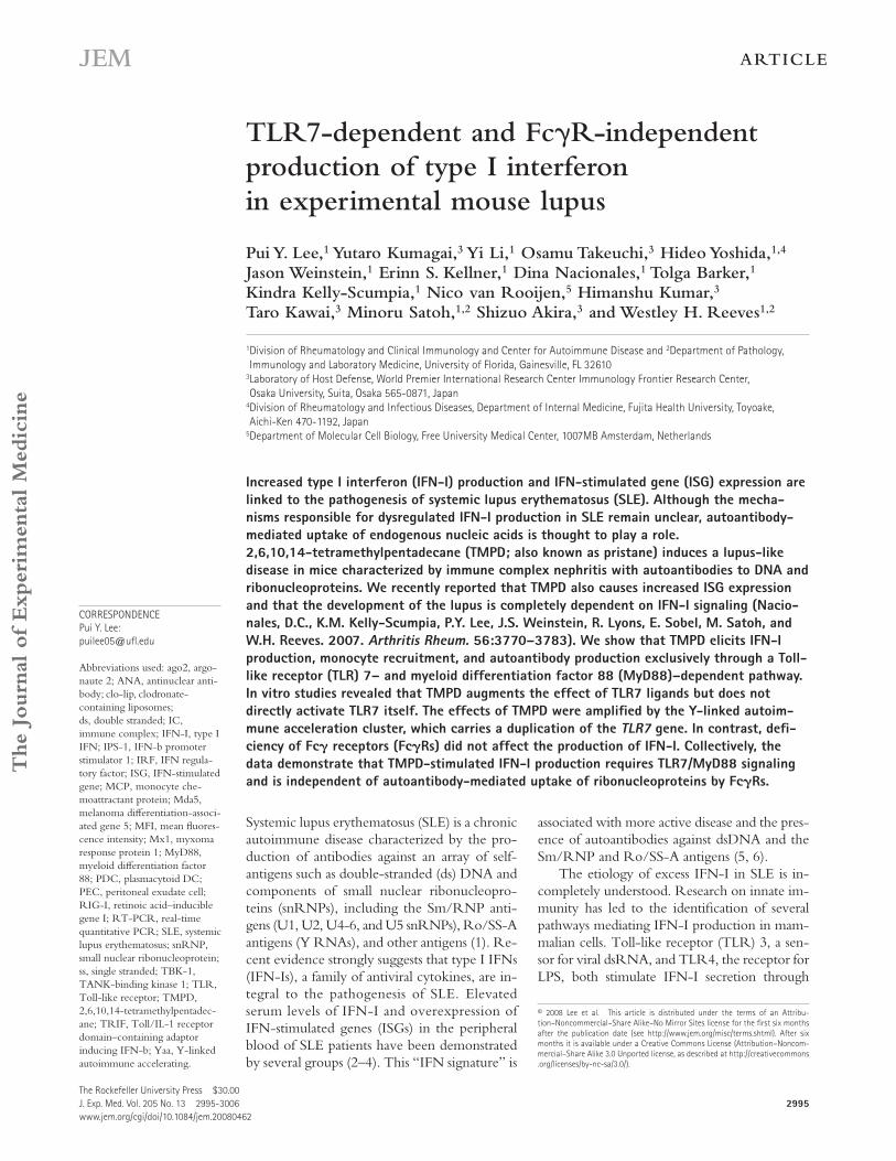
required to trigger IFN-I production by TLR3 and TLR4 ( 7 ), whereas MyD88 mediates TLR7/8 and TLR9 signaling ( 8 – 10 ). We have previously shown that within 2 wk of TMPD treatment, an accumulation of IFN-I – producing CD11b + Ly6C hi monocytes can be detected in the peritoneal cavity in wild-type mice concurrent with increased IFN-I production and ISG expression ( 27 ). Compared with wild-type mice, the to-tal number of peritoneal exudate cells (PECs) was signifi cantly reduced in MyD88 � / � mice after TMPD treatment ( Fig. 1 A ).
Toll/IL-1 receptor domain – containing adaptor inducing IFN- � (TRIF) ( 7 ). In contrast, TLR7/8 and TLR9 mediate IFN-I production via myeloid diff erentiation factor 88 (MyD88) in response to single-stranded (ss) RNA and unmethylated CpG DNA, respectively ( 8 – 10 ). In addition, cytoplasmic receptors that recognize intracellular nucleic acids and induce IFN-I have been described recently. Retinoic acid – inducible gene I (RIG-I) and melanoma diff erentiation-associated gene 5 (MDA5) recognize cytoplasmic RNA and trigger IFN-I by activating IFN- � promoter stimulator 1 (IPS-1; also known as MAVS, VISA, and CARDIF) and IFN regulatory factor (IRF) 3 ( 11 – 14 ). Cytoplasmic DNA binds to a newly described cyto-plasmic sensor and triggers IFN-I production via a pathway re-quiring TANK-binding kinase 1 (TBK-1) and IRF3 ( 15, 16 ).
It has been hypothesized that nucleic acids from dying cells may act as ligands for TLR7/8 and TLR9 to trigger IFN-I production in SLE. Immune complexes (ICs) formed by auto-antibodies to DNA and snRNPs help to transport these “ en-dogenous ligands ” to endosomes where TLR7, 8, and 9 are normally found ( 17 ). Activation of these TLRs then induces the production of IFN-I by plasmacytoid DCs (PDCs). This hypothesis is supported by numerous in vitro studies ( 18, 19 ). However, therapeutic administration of recombinant IFN- � can directly trigger the production of anti-dsDNA antibodies ( 20 ), and in several mouse model of lupus, IFN-I production is required for the induction of autoantibodies ( 21 – 23 ), suggest-ing that IFN-I dysregulation may occur upstream of autoanti-body development. Therefore, it remains controversial whether nucleic acid – containing ICs in SLE initiate IFN-I production or act to perpetuate a positive feedback loop of IFN produc-tion initiated by another factor, such as a viral infection.
Experimental lupus induced by the hydrocarbon oil 2,6,10,14-tetramethylpentadecane (TMPD; also known as pristane) displays many key immunological and clinical features of human SLE, including the presence of the IFN signature and lupus autoantibodies such as anti-dsDNA, -Sm, and -RNP ( 24 – 26 ). Importantly, IFN-I play an essential role in this model, as the development of glomerulonephritis and production of autoantibodies (anti-Sm/RNP, -dsDNA, and -Su) are abolished in IFN-I receptor – defi cient (IFNAR � / � ) mice ( 22 ). Unexpectedly, a population of Ly6C hi immature monocytes that accumulates in the peritoneal cavity after TMPD treat-ment, rather than DCs, is the major source of the excess IFN-I seen in this model ( 27 ). The persistent infl ux of Ly6C hi mono-cytes and production of IFN-I occur within 2 wk of TMPD treatment, long before the appearance of autoantibodies against snRNPs and dsDNA (3 – 5 mo), indicating that the initial wave of IFN-I production may be independent of the presence of RNA-containing ICs. In this study, we aimed to elucidate the mechanism of IFN-I production in TMPD-induced lupus.
RESULTS
TMPD-induced IFN-I production requires MyD88
To identify the mechanism of IFN-I induction by TMPD, we fi rst analyzed the eff ect of TMPD on mice with defi -ciency of the adaptor molecules TRIF or MyD88. TRIF is
Figure 1. TMPD-induced IFN-I production requires MyD88.
(A) Comparison of the number of total PECs, Ly6C hi monocytes, and granulo-
cytes 2 wk after TMPD treatment in wild-type ( n = 5), MyD88 � / � ( n = 6),
TRIF � / � ( n = 4), and IFNAR � / � mice ( n = 4). (B) Flow cytometry of perito-
neal cells (box indicates Ly6C hi monocytes and dashed oval indicates
granulocytes). (C) RT-PCR analysis of Mx1 and IRF7 expression in PECs
(normalized to peritoneal cells from an untreated wild-type mouse).
(D) ELISA quantifi cation of MCP-1 in the peritoneal lavage fl uid of TMPD-
treated mice. (E) Flow cytometry analysis of Sca-1 expression on periph-
eral blood mononuclear cells. Mean fl uorescence intensity (MFI) of Sca-1
on B220 + cells is shown. Each bar represents the mean, and error bars
indicate SE. Data are representative of two or more independent experi-
ments. *, P < 0.05 using the Student ’ s t test.
JEM VOL. 205, December 22, 2008
ARTICLE
2997
Both Ly6C hi monocytes and granulocytes (defi ned as CD11b + Ly6G + Ly6C mid ) were decreased by > 90% ( Fig. 1, A and B ). Importantly, we found that IFN-I induction by TMPD was completely dependent on MyD88, because elevated expres-sion of the ISGs myxoma response protein 1 ( Mx1 ) and IRF7 in PECs was abolished in MyD88 � / � mice, as also seen in IFNAR-defi cient mice ( Fig. 1 C ). The levels of the IFN-inducible chemokine monocyte chemoattractant protein 1 (MCP-1; also known as CCL2) in the peritoneal lavage fl uid were also reduced in the absence of MyD88 ( Fig. 1 D ). In contrast, TRIF defi ciency did not aff ect the accumulation of PEC populations or the increased expression of ISGs ( Fig. 1, A – D ). Although we were unable to detect signifi cant changes in serum IFN- � / � levels by ELISA, IFN-I secretion was re-quired for the response to TMPD, as the up-regulation of ISGs and recruitment of Ly6C hi monocytes were abolished in IFNAR � / � mice ( Fig. 1, A – D ). The absence of IFN-I signal-ing, however, did not aff ect the infl ux of granulocytes ( Fig. 1, A and B ).
The increase in IFN-I after TMPD treatment is not lim-ited to the peritoneal cavity, as the IFN signature is also de-tectable in the peripheral blood ( 22 ). We found that surface expression of the IFN-inducible gene Sca-1 (Ly6A/E) on B cells was dramatically up-regulated in wild-type mice treated with TMPD ( Fig. 1 E ). Although Sca-1 is naturally expressed by certain lymphocyte subsets and hematopoietic stem cells ( 28 ), TMPD induced Sca-1 expression in virtually all B cells in wild-type, but not IFNAR � / � , mice ( Fig. 1 E ). Increased Sca-1 expression was also evident on CD8 + and CD4 + T cells (unpublished data). Similar to the pattern of ISG expression in PECs, the up-regulation of Sca-1 was reduced in MyD88 � / � but not TRIF � / � mice ( Fig. 1 E ), further supporting an es-sential role of MyD88 in IFN-I production in this model.
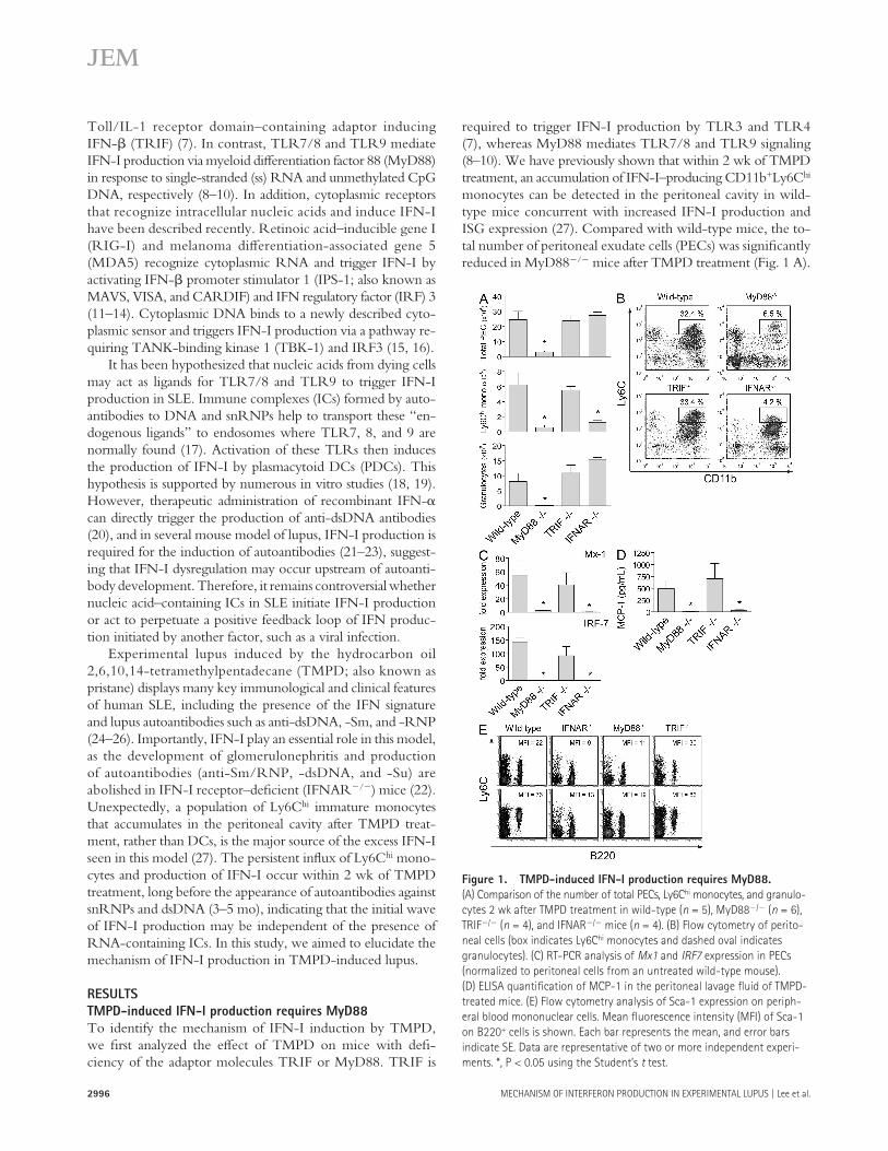
To address whether the cytoplasmic nucleic acid sensors also contribute to TMPD-induced IFN-I production, we tested the eff ect of TMPD on IPS-1 � / � and TNF � / � TBK1 � / � mice (TBK-1 � / � is embryonically lethal unless crossed with mice defi cient of TNF- � [ 29 ]). IPS-1 is a required adaptor for in-tracellular viral RNA detection via RIG-I and MDA5 ( 12 ), whereas TBK-1 is required for cytoplasmic DNA-induced IFN-I secretion ( 30 ). The expression of Mx1 and IRF7 in PECs was comparable in wild-type, IPS-1 � / � , TNF � / � TBK-1 � / � , and TNF � / � mice ( Fig. 2 ), suggesting that the intracellular nucleic acid – sensing pathways are not required for IFN-I production in this model. The patterns of peritoneal cell infl ux and Sca-1 expression on peripheral blood lympho-cytes were also similar among these strains (unpublished data). Collectively, our data indicate that TMPD-elicited IFN-I production was strictly MyD88 dependent.
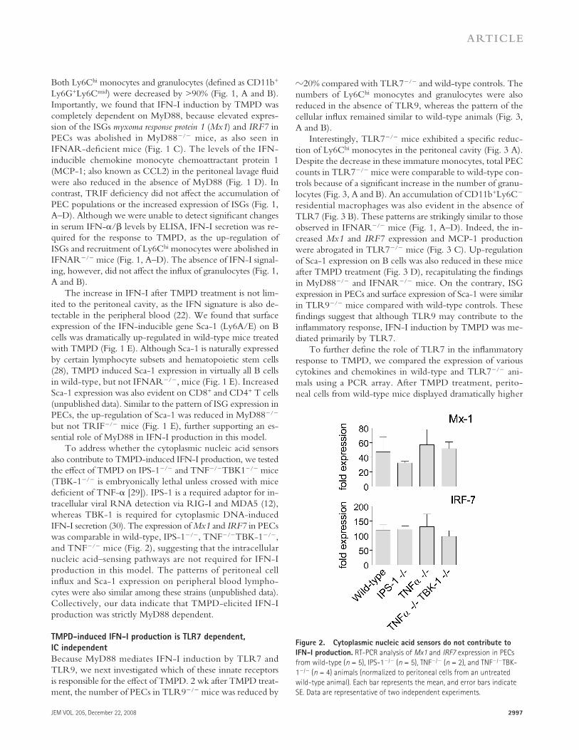
TMPD-induced IFN-I production is TLR7 dependent,
IC independent
Because MyD88 mediates IFN-I induction by TLR7 and TLR9, we next investigated which of these innate receptors is responsible for the eff ect of TMPD. 2 wk after TMPD treat-ment, the number of PECs in TLR9 � / � mice was reduced by
� 20% compared with TLR7 � / � and wild-type controls. The numbers of Ly6C hi monocytes and granulocytes were also reduced in the absence of TLR9, whereas the pattern of the cellular infl ux remained similar to wild-type animals ( Fig. 3, A and B ).
Interestingly, TLR7 � / � mice exhibited a specifi c reduc-tion of Ly6C hi monocytes in the peritoneal cavity ( Fig. 3 A ). Despite the decrease in these immature monocytes, total PEC counts in TLR7 � / � mice were comparable to wild-type con-trols because of a signifi cant increase in the number of granu-locytes ( Fig. 3, A and B ). An accumulation of CD11b + Ly6C � residential macrophages was also evident in the absence of TLR7 ( Fig. 3 B ). These patterns are strikingly similar to those observed in IFNAR � / � mice ( Fig. 1, A – D ). Indeed, the in-creased Mx1 and IRF7 expression and MCP-1 production were abrogated in TLR7 � / � mice ( Fig. 3 C ). Up-regulation of Sca-1 expression on B cells was also reduced in these mice after TMPD treatment ( Fig. 3 D ), recapitulating the fi ndings in MyD88 � / � and IFNAR � / � mice. On the contrary, ISG expression in PECs and surface expression of Sca-1 were similar in TLR9 � / � mice compared with wild-type controls. These fi ndings suggest that although TLR9 may contribute to the infl ammatory response, IFN-I induction by TMPD was me-diated primarily by TLR7.
To further defi ne the role of TLR7 in the infl ammatory response to TMPD, we compared the expression of various cytokines and chemokines in wild-type and TLR7 � / � ani-mals using a PCR array. After TMPD treatment, perito-neal cells from wild-type mice displayed dramatically higher
Figure 2. Cytoplasmic nucleic acid sensors do not contribute to
IFN-I production. RT-PCR analysis of Mx1 and IRF7 expression in PECs
from wild-type ( n = 5), IPS-1 � / � ( n = 5), TNF � / � ( n = 2), and TNF � / � TBK-
1 � / � ( n = 4) animals (normalized to peritoneal cells from an untreated
wild-type animal). Each bar represents the mean, and error bars indicate
SE. Data are representative of two independent experiments.
2998 MECHANISM OF INTERFERON PRODUCTION IN EXPERIMENTAL LUPUS | Lee et al.
production in the TMPD lupus model. Interestingly, consis-tent with the increased number of peritoneal granulocytes in TMPD-treated TLR7 � / � mice ( Fig. 3 A ), the neutrophil chemoattractant CXCL5 was up-regulated in the absence of TLR7, whereas the expression of other infl ammatory media-tors was comparable between the groups (Table S1).
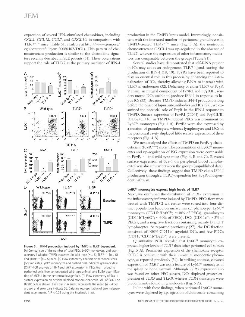
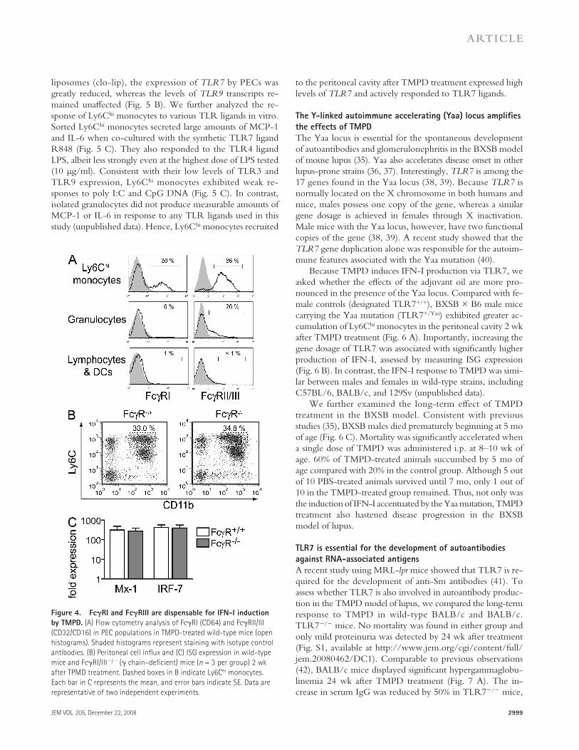
Several studies have demonstrated that self-RNA present in ICs may act as an endogenous TLR7 ligand causing the production of IFN-I ( 18, 19 ). Fc � Rs have been reported to play an essential role in this process by enhancing the inter-nalization of ICs, thereby allowing RNA to interact with TLR7 in endosomes ( 32 ). Defi ciency of either TLR7 or Fc � R � chain, an integral component of Fc � RI and Fc � RIII, ren-ders mouse DCs unable to produce IFN-I in response to lu-pus ICs ( 33 ). Because TMPD induces IFN-I production long before the onset of lupus autoantibodies and ICs ( 27 ), we ex-amined the potential role of Fc � R in the IFN-I response to TMPD. Surface expression of Fc � RI (CD64) and Fc � RII/III (CD32/CD16) in TMPD-induced PECs was prominent on Ly6C hi monocytes ( Fig. 4 A ). Fc � Rs were also expressed by a fraction of granulocytes, whereas lymphocytes and DCs in the peritoneal cavity displayed little surface expression of these receptors ( Fig. 4 A ).
We next analyzed the eff ects of TMPD on Fc � R � chain – defi cient (Fc � R � / � ) mice. The accumulation of Ly6C hi mono-cytes and up-regulation of ISG expression were comparable in Fc � R � / � and wild-type mice ( Fig. 4, B and C ). Elevated surface expression of Sca-1 on peripheral blood lympho-cytes was also similar between the groups (unpublished data). Collectively, these fi ndings suggest that TMPD elicits IFN-I production through a TLR7-dependent but Fc � R-indepen-dent pathway.
Ly6C hi monocytes express high levels of TLR7
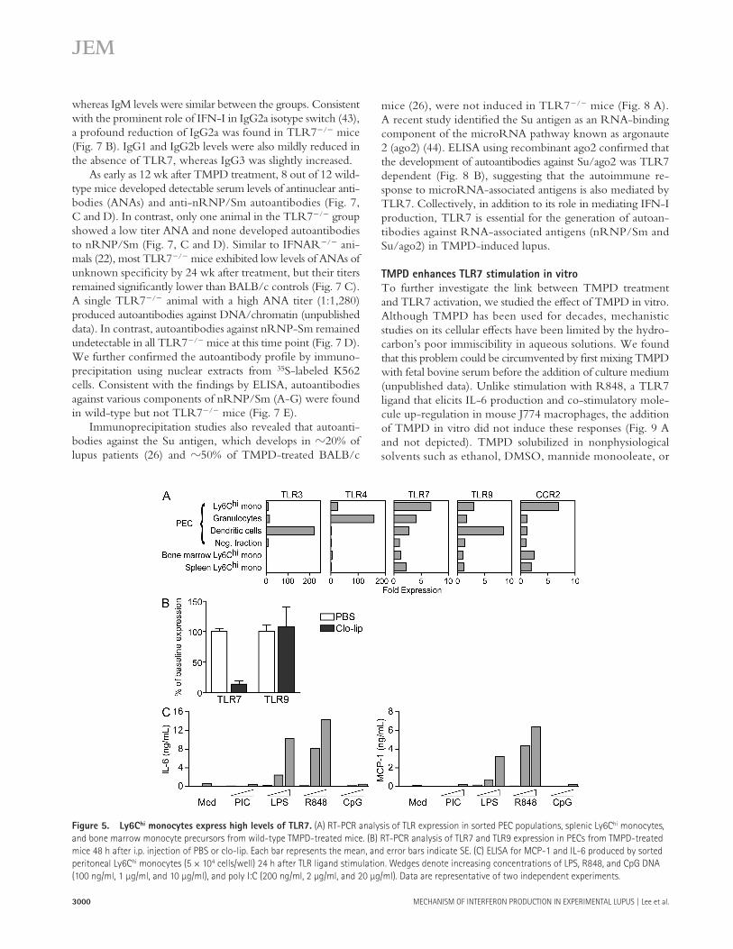
Next, we examined the distribution of TLR7 expression in the infl ammatory infi ltrate induced by TMPD. PECs from mice treated with TMPD 2 wk earlier were sorted into four dis-tinct populations based on surface marker phenotype: Ly6C hi monocytes (CD11b + Ly6C hi ; � 30% of PECs), granulocytes (CD11b + Ly6G + ; � 30% of PECs), DCs (CD11c + ; � 2% of PECs), and a negative fraction containing mainly B and T lymphocytes. As reported previously ( 27 ), the DC fraction consisted of > 80% CD11b + myeloid DCs, and few PDCs (CD11c + CD11b � B220 + ) were present.
Quantitative PCR revealed that Ly6C hi monocytes ex-pressed higher levels of TLR7 than other peritoneal cell subsets ( Fig. 5 A ). Prominent expression of the chemokine receptor CCR2 is consistent with their immature monocytic pheno-type, as reported previously ( 34 ). In striking contrast, elevated expression of TLR7 was not a feature of Ly6C hi monocytes in the spleen or bone marrow. Although TLR7 expression also was found on other PEC subsets, DCs displayed greater ex-pression of TLR3 and TLR9 , whereas TLR4 transcripts were predominantly found in granulocytes ( Fig. 5 A ).
In line with these fi ndings, when peritoneal Ly6C hi mono-cytes were depleted by i.p. injection of clodronate-containing
expression of several IFN-stimulated chemokines, including CCL2 , CCL12 , CCL7 , and CXCL10 , in comparison with TLR7 � / � mice (Table S1, available at http://www.jem.org/cgi/content/full/jem.20080462/DC1). This pattern of che-moattractant production is similar to the chemokine signa-ture recently described in SLE patients ( 31 ). These observations support the role of TLR7 as the primary mediator of IFN-I
Figure 3. IFN-I production induced by TMPD is TLR7 dependent.
(A) Comparison of the number of total PECs, Ly6C hi monocytes, and gran-
ulocytes 2 wk after TMPD treatment in wild-type ( n = 5), TLR7 � / � ( n = 5),
and TLR9 � / � ( n = 5) mice. (B) Flow cytometry analysis of peritoneal cells
(box indicates Ly6C hi monocytes and dashed oval indicates granulocytes).
(C) RT-PCR analysis of Mx1 and IRF7 expression in PECs (normalized to
peritoneal cells from an untreated wild-type animal) and ELISA quantifi ca-
tion of MCP-1 in the peritoneal lavage fl uid. (D) Flow cytometry of Sca-1
surface expression on peripheral blood mononuclear cells. MFI of Sca-1 on
B220 + cells is shown. Each bar in A and C represents the mean ( n > 4 per
group), and error bars indicate SE. Data are representative of two indepen-
dent experiments. *, P < 0.05 using the Student ’ s t test.
JEM VOL. 205, December 22, 2008
ARTICLE
2999
to the peritoneal cavity after TMPD treatment expressed high levels of TLR7 and actively responded to TLR7 ligands.
The Y-linked autoimmune accelerating (Yaa) locus amplifi es
the effects of TMPD
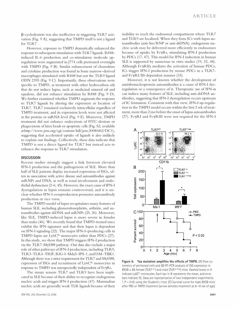
The Yaa locus is essential for the spontaneous development of autoantibodies and glomerulonephritis in the BXSB model of mouse lupus ( 35 ). Yaa also accelerates disease onset in other lupus-prone strains ( 36, 37 ). Interestingly, TLR7 is among the 17 genes found in the Yaa locus ( 38, 39 ). Because TLR7 is normally located on the X chromosome in both humans and mice, males possess one copy of the gene, whereas a similar gene dosage is achieved in females through X inactivation. Male mice with the Yaa locus, however, have two functional copies of the gene ( 38, 39 ). A recent study showed that the TLR7 gene duplication alone was responsible for the autoim-mune features associated with the Yaa mutation ( 40 ).
Because TMPD induces IFN-I production via TLR7, we asked whether the eff ects of the adjuvant oil are more pro-nounced in the presence of the Yaa locus. Compared with fe-male controls (designated TLR7 +/+ ), BXSB × B6 male mice carrying the Yaa mutation (TLR7 +/Yaa ) exhibited greater ac-cumulation of Ly6C hi monocytes in the peritoneal cavity 2 wk after TMPD treatment ( Fig. 6 A ). Importantly, increasing the gene dosage of TLR7 was associated with signifi cantly higher production of IFN-I, assessed by measuring ISG expression ( Fig. 6 B ). In contrast, the IFN-I response to TMPD was simi-lar between males and females in wild-type strains, including C57BL/6, BALB/c, and 129Sv (unpublished data).
We further examined the long-term eff ect of TMPD treatment in the BXSB model. Consistent with previous studies ( 35 ), BXSB males died prematurely beginning at 5 mo of age ( Fig. 6 C ). Mortality was signifi cantly accelerated when a single dose of TMPD was administered i.p. at 8 – 10 wk of age. 60% of TMPD-treated animals succumbed by 5 mo of age compared with 20% in the control group. Although 5 out of 10 PBS-treated animals survived until 7 mo, only 1 out of 10 in the TMPD-treated group remained. Thus, not only was the induction of IFN-I accentuated by the Yaa mutation, TMPD treatment also hastened disease progression in the BXSB model of lupus.
TLR7 is essential for the development of autoantibodies
against RNA-associated antigens
A recent study using MRL- lpr mice showed that TLR7 is re-quired for the development of anti-Sm antibodies ( 41 ). To assess whether TLR7 is also involved in autoantibody produc-tion in the TMPD model of lupus, we compared the long-term response to TMPD in wild-type BALB/c and BALB/c.TLR7 � / � mice. No mortality was found in either group and only mild proteinuria was detected by 24 wk after treatment (Fig. S1, available at http://www.jem.org/cgi/content/full/jem.20080462/DC1). Comparable to previous observations ( 42 ), BALB/c mice displayed signifi cant hypergammaglobu-linemia 24 wk after TMPD treatment ( Fig. 7 A ). The in-crease in serum IgG was reduced by 50% in TLR7 � / � mice,
liposomes (clo-lip), the expression of TLR7 by PECs was greatly reduced, whereas the levels of TLR9 transcripts re-mained unaff ected ( Fig. 5 B ). We further analyzed the re-sponse of Ly6C hi monocytes to various TLR ligands in vitro. Sorted Ly6C hi monocytes secreted large amounts of MCP-1 and IL-6 when co-cultured with the synthetic TLR7 ligand R848 ( Fig. 5 C ). They also responded to the TLR4 ligand LPS, albeit less strongly even at the highest dose of LPS tested (10 μ g/ml). Consistent with their low levels of TLR3 and TLR9 expression, Ly6C hi monocytes exhibited weak re-sponses to poly I:C and CpG DNA ( Fig. 5 C ). In contrast, isolated granulocytes did not produce measurable amounts of MCP-1 or IL-6 in response to any TLR ligands used in this study (unpublished data). Hence, Ly6C hi monocytes recruited
Figure 4. Fc � RI and Fc � RIII are dispensable for IFN-I induction
by TMPD. (A) Flow cytometry analysis of Fc � RI (CD64) and Fc � RII/III
(CD32/CD16) in PEC populations in TMPD-treated wild-type mice (open
histograms). Shaded histograms represent staining with isotype control
antibodies. (B) Peritoneal cell infl ux and (C) ISG expression in wild-type
mice and Fc � RI/III � / � ( � chain – defi cient) mice ( n = 3 per group) 2 wk
after TPMD treatment. Dashed boxes in B indicate Ly6C hi monocytes.
Each bar in C represents the mean, and error bars indicate SE. Data are
representative of two independent experiments.
3000 MECHANISM OF INTERFERON PRODUCTION IN EXPERIMENTAL LUPUS | Lee et al.
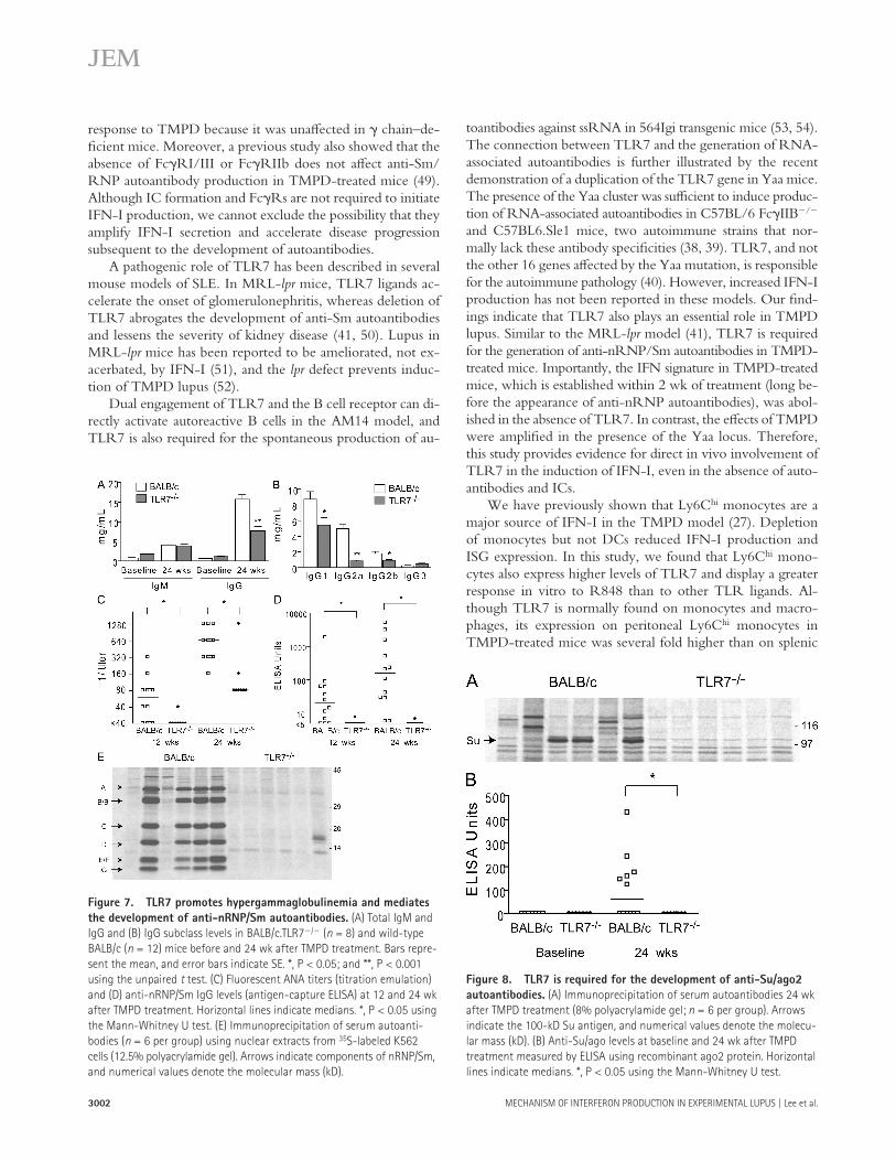
mice ( 26 ), were not induced in TLR7 � / � mice ( Fig. 8 A ). A recent study identifi ed the Su antigen as an RNA-binding component of the microRNA pathway known as argonaute 2 (ago2) ( 44 ). ELISA using recombinant ago2 confi rmed that the development of autoantibodies against Su/ago2 was TLR7 dependent ( Fig. 8 B ), suggesting that the autoimmune re-sponse to microRNA-associated antigens is also mediated by TLR7. Collectively, in addition to its role in mediating IFN-I production, TLR7 is essential for the generation of autoan-tibodies against RNA-associated antigens (nRNP/Sm and Su/ago2) in TMPD-induced lupus.
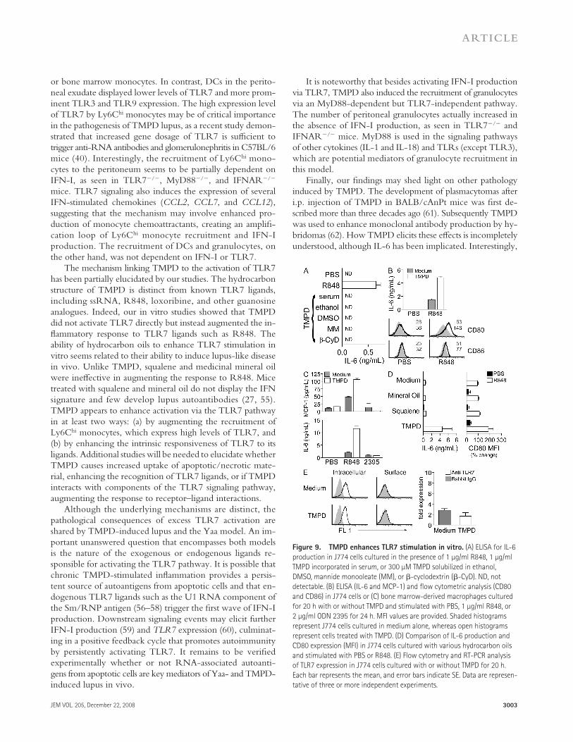
TMPD enhances TLR7 stimulation in vitro
To further investigate the link between TMPD treatment and TLR7 activation, we studied the eff ect of TMPD in vitro. Although TMPD has been used for decades, mechanistic studies on its cellular eff ects have been limited by the hydro-carbon ’ s poor immiscibility in aqueous solutions. We found that this problem could be circumvented by fi rst mixing TMPD with fetal bovine serum before the addition of culture medium (unpublished data). Unlike stimulation with R848, a TLR7 ligand that elicits IL-6 production and co-stimulatory mole-cule up-regulation in mouse J774 macrophages, the addition of TMPD in vitro did not induce these responses ( Fig. 9 A and not depicted). TMPD solubilized in nonphysiological solvents such as ethanol, DMSO, mannide monooleate, or
whereas IgM levels were similar between the groups. Consistent with the prominent role of IFN-I in IgG2a isotype switch ( 43 ), a profound reduction of IgG2a was found in TLR7 � / � mice ( Fig. 7 B ). IgG1 and IgG2b levels were also mildly reduced in the absence of TLR7, whereas IgG3 was slightly increased.
As early as 12 wk after TMPD treatment, 8 out of 12 wild-type mice developed detectable serum levels of antinuclear anti-bodies (ANAs) and anti-nRNP/Sm autoantibodies ( Fig. 7, C and D ). In contrast, only one animal in the TLR7 � / � group showed a low titer ANA and none developed autoantibodies to nRNP/Sm ( Fig. 7, C and D ). Similar to IFNAR � / � ani-mals ( 22 ), most TLR7 � / � mice exhibited low levels of ANAs of unknown specifi city by 24 wk after treatment, but their titers remained signifi cantly lower than BALB/c controls ( Fig. 7 C ). A single TLR7 � / � animal with a high ANA titer (1:1,280) produced autoantibodies against DNA/chromatin (unpublished data). In contrast, autoantibodies against nRNP-Sm remained undetectable in all TLR7 � / � mice at this time point ( Fig. 7 D ). We further confi rmed the autoantibody profi le by immuno-precipitation using nuclear extracts from 35 S-labeled K562 cells. Consistent with the fi ndings by ELISA, autoantibodies against various components of nRNP/Sm (A-G) were found in wild-type but not TLR7 � / � mice ( Fig. 7 E ).
Immunoprecipitation studies also revealed that autoanti-bodies against the Su antigen, which develops in � 20% of lupus patients ( 26 ) and � 50% of TMPD-treated BALB/c
Figure 5. Ly6C hi monocytes express high levels of TLR7. (A) RT-PCR analysis of TLR expression in sorted PEC populations, splenic Ly6C hi monocytes,
and bone marrow monocyte precursors from wild-type TMPD-treated mice. (B) RT-PCR analysis of TLR7 and TLR9 expression in PECs from TMPD-treated
mice 48 h after i.p. injection of PBS or clo-lip. Each bar represents the mean, and error bars indicate SE. (C) ELISA for MCP-1 and IL-6 produced by sorted
peritoneal Ly6C hi monocytes (5 × 10 4 cells/well) 24 h after TLR ligand stimulation. Wedges denote increasing concentrations of LPS, R848, and CpG DNA
(100 ng/ml, 1 μ g/ml, and 10 μ g/ml), and poly I:C (200 ng/ml, 2 μ g/ml, and 20 μ g/ml). Data are representative of two independent experiments.
JEM VOL. 205, December 22, 2008
ARTICLE
3001
inability to reach the endosomal compartment where TLR7 and TLR9 are localized. When they form ICs with lupus au-toantibodies (anti-Sm/RNP or anti-dsDNA), endogenous nu-cleic acids may be delivered more effi ciently to endosomes because of uptake by Fc � Rs, stimulating IFN-I production by PDCs ( 17, 47 ). This model for IFN-I induction in human SLE is supported by numerous in vitro studies ( 19, 32, 48 ). Although Fc � RIIa mediates the activation of human PDCs, ICs trigger IFN-I production by mouse PDCs in a TLR7- and Fc � RI/III-dependent manner ( 33 ).
However, it is not known whether the development of antiribonucleoprotein autoantibodies is a cause of IFN-I dys-regulation or a consequence of it. Therapeutic use of IFN- � can induce many features of SLE, including anti-dsDNA an-tibodies, suggesting that IFN-I dysregulation occurs upstream of IC formation. Consistent with that view, IFN-I up-regula-tion in the TMPD model occurs within the fi rst 2 wk of treat-ment, more than 2 mo before the onset of lupus autoantibodies ( 27 ). Fc � RI and Fc � RIII were not required for the IFN-I
� -cyclodextrin was also ineff ective in triggering TLR7 acti-vation ( Fig. 9 A ), suggesting that TMPD itself is not a ligand for TLR7.
However, exposure to TMPD dramatically enhanced the response to subsequent stimulation with TLR7 ligands. R848-induced IL-6 production and co-stimulatory molecule up-regulation were augmented in J774 cells pretreated overnight with TMPD ( Fig. 9 B ). Similar enhancement of chemokine and cytokine production was found in bone marrow – derived macrophages stimulated with R848 but not the TLR9 ligand ODN 2395 ( Fig. 9 C ). Importantly, these observations were specifi c to TMPD, as treatment with other hydrocarbon oils that do not induce lupus, such as medicinal mineral oil and squalene, did not enhance stimulation by R848 ( Fig. 9 D ). We further examined whether TMPD augments the response to TLR7 ligands by altering the expression or location of TLR7. TLR7 remained exclusively intracellular regardless of TMPD treatment, and its expression levels were not aff ected at the protein or mRNA level ( Fig. 9 E ). Moreover, TMPD treatment did not enhance endocytosis of FITC-dextran or phagocytosis of latex beads or apoptotic cells (Fig. S2, available at http://www.jem.org/cgi/content/full/jem.20080462/DC1), suggesting that accelerated uptake of ligands is also unlikely to explain our fi ndings. Collectively, these data indicate that TMPD is not a direct ligand for TLR7 but instead acts to enhance the response to TLR7 stimulation.
DISCUSSION
Recent studies strongly suggest a link between elevated IFN-I production and the pathogenesis of SLE. More than half of SLE patients display increased expression of ISGs, of-ten in association with active disease and autoantibodies against snRNPs and DNA, as well as renal involvement and endo-thelial dysfunction ( 2 – 6, 45 ). However, the exact cause of IFN-I dysregulation in lupus remains controversial, and it is un-clear whether IFN-I overproduction promotes autoantibody production or vice versa.
The TMPD model of lupus recapitulates many features of human SLE, including glomerulonephritis, arthritis, and au-toantibodies against dsDNA and snRNPs ( 25, 26 ). Moreover, like SLE, TMPD-induced lupus is more severe in females than males ( 46 ). We recently found that TMPD-treated mice exhibit the IFN signature and that their lupus is dependent on IFN-I signaling ( 22 ). The major IFN-I – producing cells in TMPD lupus are Ly6C hi monocytes rather than PDCs ( 27 ). In this study, we show that TMPD triggers IFN-I production via the TLR7 – MyD88 pathway. Our data also exclude a major role of other pathways of IFN-I production, including TLR9, TLR3 – TLR4 – TRIF, RIG-I – Mda5 – IPS-1, and DAI – TBK1. Although there was a strict requirement for TLR7 and MyD88, expression of ISGs and recruitment of Ly6C hi monocytes in response to TMPD was unexpectedly independent of Fc � Rs.
The innate sensors TLR7 and TLR9 have been impli-cated in SLE because of their ability to recognize endogenous nucleic acids and trigger IFN-I production ( 47 ). Mammalian nucleic acids are generally weak TLR ligands because of their
Figure 6. Yaa mutation amplifi es the effects of TMPD. (A) Flow cy-
tometry of peritoneal cells and (B) RT-PCR analysis of ISG expression in
BXSB × B6 female (TLR7 +/+ ) and male (TLR7 +/ Yaa ) mice. Dashed boxes in A
indicate Ly6C hi monocytes. Each bar in B represents the mean, and error
bars indicate SE. Data are representative of two independent experiments.
*, P < 0.05 using the Student ’ s t test. (C) Survival curve for male BXSB mice
after PBS or TMPD treatment (arrow denotes treatment at 8 – 10 wk of age).
3002 MECHANISM OF INTERFERON PRODUCTION IN EXPERIMENTAL LUPUS | Lee et al.
toantibodies against ssRNA in 564Igi transgenic mice ( 53, 54 ). The connection between TLR7 and the generation of RNA-associated autoantibodies is further illustrated by the recent demonstration of a duplication of the TLR7 gene in Yaa mice. The presence of the Yaa cluster was suffi cient to induce produc-tion of RNA-associated autoantibodies in C57BL/6 Fc � IIB � / � and C57BL6.Sle1 mice, two autoimmune strains that nor-mally lack these antibody specifi cities ( 38, 39 ). TLR7, and not the other 16 genes aff ected by the Yaa mutation, is responsible for the autoimmune pathology ( 40 ). However, increased IFN-I production has not been reported in these models. Our fi nd-ings indicate that TLR7 also plays an essential role in TMPD lupus. Similar to the MRL- lpr model ( 41 ), TLR7 is required for the generation of anti-nRNP/Sm autoantibodies in TMPD-treated mice. Importantly, the IFN signature in TMPD-treated mice, which is established within 2 wk of treatment (long be-fore the appearance of anti-nRNP autoantibodies), was abol-ished in the absence of TLR7. In contrast, the eff ects of TMPD were amplifi ed in the presence of the Yaa locus. Therefore, this study provides evidence for direct in vivo involvement of TLR7 in the induction of IFN-I, even in the absence of auto-antibodies and ICs.
We have previously shown that Ly6C hi monocytes are a major source of IFN-I in the TMPD model ( 27 ). Depletion of monocytes but not DCs reduced IFN-I production and ISG expression. In this study, we found that Ly6C hi mono-cytes also express higher levels of TLR7 and display a greater response in vitro to R848 than to other TLR ligands. Al-though TLR7 is normally found on monocytes and macro-phages, its expression on peritoneal Ly6C hi monocytes in TMPD-treated mice was several fold higher than on splenic
response to TMPD because it was unaff ected in � chain – de-fi cient mice. Moreover, a previous study also showed that the absence of Fc � RI/III or Fc � RIIb does not aff ect anti-Sm/RNP autoantibody production in TMPD-treated mice ( 49 ). Although IC formation and Fc � Rs are not required to initiate IFN-I production, we cannot exclude the possibility that they amplify IFN-I secretion and accelerate disease progression subsequent to the development of autoantibodies.
A pathogenic role of TLR7 has been described in several mouse models of SLE. In MRL- lpr mice, TLR7 ligands ac-celerate the onset of glomerulonephritis, whereas deletion of TLR7 abrogates the development of anti-Sm autoantibodies and lessens the severity of kidney disease ( 41, 50 ). Lupus in MRL- lpr mice has been reported to be ameliorated, not ex-acerbated, by IFN-I ( 51 ), and the lpr defect prevents induc-tion of TMPD lupus ( 52 ).
Dual engagement of TLR7 and the B cell receptor can di-rectly activate autoreactive B cells in the AM14 model, and TLR7 is also required for the spontaneous production of au-
Figure 8. TLR7 is required for the development of anti-Su/ago2
autoantibodies. (A) Immunoprecipitation of serum autoantibodies 24 wk
after TMPD treatment (8% polyacrylamide gel; n = 6 per group). Arrows
indicate the 100-kD Su antigen, and numerical values denote the molecu-
lar mass (kD). (B) Anti-Su/ago levels at baseline and 24 wk after TMPD
treatment measured by ELISA using recombinant ago2 protein. Horizontal
lines indicate medians. *, P < 0.05 using the Mann-Whitney U test.
Figure 7. TLR7 promotes hypergammaglobulinemia and mediates
the development of anti-nRNP/Sm autoantibodies. (A) Total IgM and
IgG and (B) IgG subclass levels in BALB/c.TLR7 � / � ( n = 8) and wild-type
BALB/c ( n = 12) mice before and 24 wk after TMPD treatment. Bars repre-
sent the mean, and error bars indicate SE. *, P < 0.05; and **, P < 0.001
using the unpaired t test. (C) Fluorescent ANA titers (titration emulation)
and (D) anti-nRNP/Sm IgG levels (antigen-capture ELISA) at 12 and 24 wk
after TMPD treatment. Horizontal lines indicate medians. *, P < 0.05 using
the Mann-Whitney U test. (E) Immunoprecipitation of serum autoanti-
bodies ( n = 6 per group) using nuclear extracts from 35 S-labeled K562
cells (12.5% polyacrylamide gel). Arrows indicate components of nRNP/Sm,
and numerical values denote the molecular mass (kD).
JEM VOL. 205, December 22, 2008
ARTICLE
3003
It is noteworthy that besides activating IFN-I production via TLR7, TMPD also induced the recruitment of granulocytes via an MyD88-dependent but TLR7-independent pathway. The number of peritoneal granulocytes actually increased in the absence of IFN-I production, as seen in TLR7 � / � and IFNAR � / � mice. MyD88 is used in the signaling pathways of other cytokines (IL-1 and IL-18) and TLRs (except TLR3), which are potential mediators of granulocyte recruitment in this model.
Finally, our fi ndings may shed light on other pathology induced by TMPD. The development of plasmacytomas after i.p. injection of TMPD in BALB/cAnPt mice was fi rst de-scribed more than three decades ago ( 61 ). Subsequently TMPD was used to enhance monoclonal antibody production by hy-bridomas ( 62 ). How TMPD elicits these eff ects is incompletely understood, although IL-6 has been implicated. Interestingly,
or bone marrow monocytes. In contrast, DCs in the perito-neal exudate displayed lower levels of TLR7 and more prom-inent TLR3 and TLR9 expression. The high expression level of TLR7 by Ly6C hi monocytes may be of critical importance in the pathogenesis of TMPD lupus, as a recent study demon-strated that increased gene dosage of TLR7 is suffi cient to trigger anti-RNA antibodies and glomerulonephritis in C57BL/6 mice ( 40 ). Interestingly, the recruitment of Ly6C hi mono-cytes to the peritoneum seems to be partially dependent on IFN-I, as seen in TLR7 � / � , MyD88 � / � , and IFNAR � / � mice. TLR7 signaling also induces the expression of several IFN-stimulated chemokines ( CCL2 , CCL7 , and CCL12 ), suggesting that the mechanism may involve enhanced pro-duction of monocyte chemoattractants, creating an amplifi -cation loop of Ly6C hi monocyte recruitment and IFN-I production. The recruitment of DCs and granulocytes, on the other hand, was not dependent on IFN-I or TLR7.
The mechanism linking TMPD to the activation of TLR7 has been partially elucidated by our studies. The hydrocarbon structure of TMPD is distinct from known TLR7 ligands, including ssRNA, R848, loxoribine, and other guanosine analogues. Indeed, our in vitro studies showed that TMPD did not activate TLR7 directly but instead augmented the in-fl ammatory response to TLR7 ligands such as R848. The ability of hydrocarbon oils to enhance TLR7 stimulation in vitro seems related to their ability to induce lupus-like disease in vivo. Unlike TMPD, squalene and medicinal mineral oil were ineff ective in augmenting the response to R848. Mice treated with squalene and mineral oil do not display the IFN signature and few develop lupus autoantibodies ( 27, 55 ). TMPD appears to enhance activation via the TLR7 pathway in at least two ways: (a) by augmenting the recruitment of Ly6C hi monocytes, which express high levels of TLR7, and (b) by enhancing the intrinsic responsiveness of TLR7 to its ligands. Additional studies will be needed to elucidate whether TMPD causes increased uptake of apoptotic/necrotic mate-rial, enhancing the recognition of TLR7 ligands, or if TMPD interacts with components of the TLR7 signaling pathway, augmenting the response to receptor – ligand interactions.
Although the underlying mechanisms are distinct, the pathological consequences of excess TLR7 activation are shared by TMPD-induced lupus and the Yaa model. An im-portant unanswered question that encompasses both models is the nature of the exogenous or endogenous ligands re-sponsible for activating the TLR7 pathway. It is possible that chronic TMPD-stimulated infl ammation provides a persis-tent source of autoantigens from apoptotic cells and that en-dogenous TLR7 ligands such as the U1 RNA component of the Sm/RNP antigen ( 56 – 58 ) trigger the fi rst wave of IFN-I production. Downstream signaling events may elicit further IFN-I production ( 59 ) and TLR7 expression ( 60 ), culminat-ing in a positive feedback cycle that promotes autoimmunity by persistently activating TLR7. It remains to be verifi ed experimentally whether or not RNA-associated autoanti-gens from apoptotic cells are key mediators of Yaa- and TMPD-induced lupus in vivo.
Figure 9. TMPD enhances TLR7 stimulation in vitro. (A) ELISA for IL-6
production in J774 cells cultured in the presence of 1 μ g/ml R848, 1 μ g/ml
TMPD incorporated in serum, or 300 μ M TMPD solubilized in ethanol,
DMSO, mannide monooleate (MM), or � -cyclodextrin ( � -CyD). ND, not
detectable. (B) ELISA (IL-6 and MCP-1) and fl ow cytometric analysis (CD80
and CD86) in J774 cells or (C) bone marrow-derived macrophages cultured
for 20 h with or without TMPD and stimulated with PBS, 1 μ g/ml R848, or
2 μ g/ml ODN 2395 for 24 h. MFI values are provided. Shaded histograms
represent J774 cells cultured in medium alone, whereas open histograms
represent cells treated with TMPD. (D) Comparison of IL-6 production and
CD80 expression (MFI) in J774 cells cultured with various hydrocarbon oils
and stimulated with PBS or R848. (E) Flow cytometry and RT-PCR analysis
of TLR7 expression in J774 cells cultured with or without TMPD for 20 h.
Each bar represents the mean, and error bars indicate SE. Data are represen-
tative of three or more independent experiments.
3004 MECHANISM OF INTERFERON PRODUCTION IN EXPERIMENTAL LUPUS | Lee et al.
plus 10 U/ml heparin) were seeded on 96-well cell-culture plates (5 × 10 4
cells/well). Cells were stimulated with the doses indicated in the fi gures of
peptidoglycan, poly I:C, R848, CpG ODN2395 (InvivoGen), or LPS (from
Salmonella typhimurium ; Sigma-Aldrich) and were incubated at 37 ° C in a 5%
CO 2 atmosphere for 24 h before collecting the supernatant. MCP-1 and IL-6
ELISAs (BD) were performed according to the manufacturer ’ s instructions.
Optical density was converted to concentration using standard curves based
on recombinant cytokines analyzed by a four-parameter logistic equation
(Softmax Pro 3.1 software; MDS Analytical Technologies).
Cell culture with TMPD. 1 ml TMPD, mineral oil or squalene was added
to 9 ml of fetal bovine serum in a 15-ml polypropylene tube and was rotated
for 48 h at 4 ° C. The surface layer of unincorporated hydrocarbon oil was re-
moved by aspiration at the end of the incubation. The amount of TMPD
incorporated using this method was � 1 μ g/ml, as determined by gas chroma-
tography/mass spectroscopy (not depicted; Analytical Toxicology Core, Uni-
versity of Florida). J774 cells or bone marrow – derived macrophages were
seeded on 24-well plates (5 × 10 5 cells/well) and cultured overnight in com-
plete DMEM containing 10% FCS with or without hydrocarbon oils. For sub-
sequent stimulation, cells were washed with PBS, and fresh complete medium
was added before the addition of TLR ligands. Incorporation of TMPD in
DMSO and � -cyclodextrin (Sigma-Aldrich) has been described previously
( 68 ). TMPD (10% vol/vol) also was added to ethanol or mannide monooleate
(5% in PBS; Sigma-Aldrich). Solvent alone was used as a control, and a range
of TMPD concentrations (3 – 300 μ M) was tested. Endocytosis was quantifi ed
by uptake of 5 μ g/ml FITC-dextran (Sigma-Aldrich), and phagocytosis by in-
ternalization of FITC-labeled microbeads (10:1 beads/cells ratio; Invitrogen) or
tetramethylindodicarbocyanine perchlorate (DiD) – labeled apoptotic BW5147
cells (10:1 apoptotic cell/ target cell ratio) after overnight incubation of J774
cells in complete medium with or without TMPD. Apoptosis of BW5147 cells
was induced by heat shock in a 45 ° C water bath for 10 min. After 4 h of incu-
bation at 37 ° C, apoptotic cells ( > 80% annexin V positive; not depicted) were
labeled with the fl uorescent dye DiD (Invitrogen). J774 Cells were washed and
incubated with the fl uorescent substrates for 30 min at 37 ° C (in PBS with 0.5%
BSA), washed three times, and analyzed by fl ow cytometry. ELISA, fl ow cy-
tometry, and RT-PCR were performed as described. Bone marrow – derived
macrophages were generated from BALB/c mice as previously described ( 8 ).
Autoantibody analysis. Serum ANAs in BALB/c.TLR7 � / � and wild-
type BALB/c mice were determined 12 and 24 wk after TMPD by indi-
rect immunofl uorescence using HEp-2 cells (Innova). Sera were diluted
1:40, and titers were determined using a titration emulation system (Image
Titer; Rhigene, Inc.). Immunoprecipitation and antigen-capture ELISA to
detect serum autoantibodies against nRNP/Sm were performed as previ-
ously described ( 26, 69 ). Determination of anti-Su/ago2 by ELISA has also
been previously described ( 44 ). Recombinant ago2 protein was a gift from
E. Chan and K. Ikeda (University of Florida, Gainesville, FL).
Statistical analysis. For quantitative variables, diff erences between groups
were analyzed by the unpaired Student ’ s t test. Survival curves were analyzed
using the log-rank test. ANA titers and autoantibody levels were compared
using the Mann-Whitney U test. Data are presented as means ± SD. All tests
were two-sided, and P < 0.05 was considered signifi cant. Statistical analyses
were performed using Prism 4.0 software (GraphPad Software, Inc.).
Online supplemental material. Table S1 provides PCR array analysis of
cytokine/chemokine expression in PECs from wild-type and TLR7 � / �
mice. Table S2 provides the sequence of all PCR primers used in this study.
Fig. S1 shows the levels of proteinuria in BALB/c and TLR7 � / � mice 24 wk
after TMPD treatment. Fig. S2 shows the eff ect of TMPD on endocytosis of
FITC-dextran and phagocytosis of FITC-coated latex beads or apoptotic
lymphocytes in J774 cells. Online supplemental material is available at
http://www.jem.org/cgi/content/full/jem.20080462/DC1.
We thank Dr. E. Sobel for helpful discussion, M. Xu for assistance with manuscript
preparation, Drs. E. Chan and K. Ikeda for providing recombinant ago2 protein, and
although TLR7 can trigger B cell activation and antibody production ( 53 ), IFN-I plays an important role in antibody class switching and promotes plasma cell diff erentiation in the presence of IL-6 ( 63 ). Whether TLR7 activation and IFN-I production are involved in the pathogenesis of plasmacyto-mas and enhancement of antibody production by hybridomas warrants further investigation.
MATERIALS AND METHODS Mice. MyD88 � / � , TRIF � / � , TLR7 � / � , TLR9 � / � , and IFNAR � / � mice
(backcrossed > 7 generations to the C57BL/6 background), and IPS-1 � / � ,
TNF � / � , TNF � / � TBK1 � / � mice (on a mixed 129Sv/B6 background) have
all been described previously ( 7, 8, 29, 64 – 66 ). Wild-type C57BL/6 and
heterozygous littermates were used as controls. BALB/c.TLR7 � / � (back-
crossed > 8 generations to the BALB/c background) and wild-type BALB/c
mice were used for long-terms studies of autoantibody production. Animals
were bred and maintained in a specifi c pathogen-free facility of the Re-
search Institute for Microbial Diseases, Osaka University. C57BL/6 wild-
type and Fc � RI/III � / � mice (Taconic) and BXSB mice (The Jackson
Laboratory) were maintained in a specifi c pathogen-free facility at the Uni-
versity of Florida. BXSB × B6 F1 mice were generated by breeding BXSB
males with C57BL/6 females. 12 – 16-wk-old animals received a single i.p.
injection of 0.5 ml TMPD (Sigma-Aldrich). Blood samples were obtained
before TMPD treatment and weekly thereafter. Peritoneal cells, spleen, and
blood were harvested 2 wk after treatment. Monocyte depletion was per-
formed by i.p. injection of 200 μ l clo-lip, as previously described ( 67 ).
These studies were approved by the University of Florida Institutional Ani-
mal Care and Use Committee and the Osaka University Animal Care and
Use Committee.
Real-time quantitative PCR (RT-PCR). RT-PCR was performed as
previously described ( 24 ). In brief, total RNA was extracted from 10 6 peri-
toneal cells using TRI zol reagent (Invitrogen), and cDNA was synthesized
using the Superscript II First-Strand Synthesis kit (Invitrogen) according to
the manufacturer ’ s protocol. SYBR green RT-PCR analysis was performed
using a thermocycler (Opticon II; MJ Research). Amplifi cation conditions
were as follows: 95 ° C for 10 min, followed by 45 cycles of 94 ° C for 15 s,
60 ° C for 25 s, and 72 ° C for 25 s. After the fi nal extension (72 ° C for 10 min),
a melting-curve analysis was performed to ensure specifi city of the products.
Primers used in this study are listed in Table S2 (available at http://www
.jem.org/cgi/content/full/jem.20080462/DC1). Cytokine/chemokine PCR
array (Superarray) analysis was performed using a sequence detector (ABI
7700; Applied Biosystems) according to the manufacturer ’ s protocols.
Flow cytometry and cell sorting. The following conjugated antibodies
were used: anti – CD11b-PE, anti – CD8-allophycocyanin (APC), anti – CD4-
FITC, anti – CD11c-PE, anti – B220-PerCPCy5.5, anti – Sca-1 – PE, anti – CD64-
PE, anti – CD32/16-PE (all from BD), anti – Ly6C-FITC, anti – Ly6C-biotin,
and avidin-APC (all from eBioscience). Before surface staining, peritoneal or
peripheral blood cells were incubated with anti – mouse CD16/32 (Fc block;
BD) for 10 min. Cells were then stained with an optimized amount of primary
antibody or the appropriate isotype control for 10 min at room temperature
before washing and resuspending in PBS supplemented with 0.1% BSA. Intra-
cellular staining for TLR7 was performed as previously described ( 50 ) using
rabbit anti – mouse TLR7 or rabbit IgG isotype control (eBioscience) and goat
anti – rabbit IgG-FITC (SouthernBiotech). 50,000 events per sample were ac-
quired using a FACSCalibur (BD) and analyzed with FCS Express 3 software
(De Novo Software). Cell sorting was performed using a fl ow cytometer
(FACSDiva; BD). Peritoneal, splenic, and bone marrow Ly6C hi monocytes
(CD11b + Ly6C hi ), peritoneal DCs (CD11c + ), and granulocytes (CD11b + Ly6G + )
were sorted to > 90% purity for cell culture or RNA isolation.
TLR stimulation. Sorted cells resuspended in complete DMEM (contain-
ing 10% FCS, 10 mmol/liter Hepes, glutamine, and penicillin/streptomycin
JEM VOL. 205, December 22, 2008
ARTICLE
3005
15 . Stockinger , S. , B. Reutterer , B. Schaljo , C. Schellack , S. Brunner , T. Materna , M. Yamamoto , S. Akira , T. Taniguchi , P.J. Murray , et al . 2004 . IFN regulatory factor 3-dependent induction of type I IFNs by intracellular bacteria is mediated by a TLR- and Nod2-independent mechanism. J. Immunol. 173 : 7416 – 7425 .
16 . Stetson , D.B. , and R. Medzhitov . 2006 . Recognition of cytosolic DNA activates an IRF3-dependent innate immune response. Immunity . 24 : 93 – 103 .
17 . Ronnblom , L. , M.L. Eloranta , and G.V. Alm . 2006 . The type I interferon system in systemic lupus erythematosus. Arthritis Rheum. 54 : 408 – 420 .
18 . Vallin , H. , A. Perers , G.V. Alm , and L. Ronnblom . 1999 . Anti-dou-ble-stranded DNA antibodies and immunostimulatory plasmid DNA in combination mimic the endogenous IFN-alpha inducer in systemic lupus erythematosus. J. Immunol. 163 : 6306 – 6313 .
19 . Barrat , F.J. , T. Meeker , J. Gregorio , J.H. Chan , S. Uematsu , S. Akira , B. Chang , O. Duramad , and R.L. Coff man . 2005 . Nucleic acids of mamma-lian origin can act as endogenous ligands for Toll-like receptors and may promote systemic lupus erythematosus. J. Exp. Med. 202 : 1131 – 1139 .
20 . Ronnblom , L.E. , G.V. Alm , and K.E. Oberg . 1991 . Autoimmunity af-ter alpha-interferon therapy for malignant carcinoid tumors. Ann. Intern. Med. 115 : 178 – 183 .
21 . Jorgensen , T.N. , E. Roper , J.M. Thurman , P. Marrack , and B.L. Kotzin . 2007 . Type I interferon signaling is involved in the sponta-neous development of lupus-like disease in B6.Nba2 and (B6.Nba2 × NZW)F(1) mice. Genes Immun. 8 : 653 – 662 .
22 . Nacionales , D.C. , K.M. Kelly-Scumpia , P.Y. Lee , J.S. Weinstein , R. Lyons , E. Sobel , M. Satoh , and W.H. Reeves . 2007 . Defi ciency of the type I interferon receptor protects mice from experimental lupus. Arthritis Rheum. 56 : 3770 – 3783 .
23 . Santiago-Raber , M.L. , R. Baccala , K.M. Haraldsson , D. Choubey , T.A. Stewart , D.H. Kono , and A.N. Theofi lopoulos . 2003 . Type-I in-terferon receptor defi ciency reduces lupus-like disease in NZB mice. J. Exp. Med. 197 : 777 – 788 .
24 . Nacionales , D.C. , K.M. Kelly , P.Y. Lee , H. Zhuang , Y. Li , J.S. Weinstein , E. Sobel , Y. Kuroda , J. Akaogi , M. Satoh , and W.H. Reeves . 2006 . Type I interferon production by tertiary lymphoid tissue developing in response to 2,6,10,14-tetramethyl-pentadecane (pristane). Am. J. Pathol. 168 : 1227 – 1240 .
25 . Satoh , M. , A. Kumar , Y.S. Kanwar , and W.H. Reeves . 1995 . Anti-nuclear antibody production and immune-complex glomerulonephri-tis in BALB/c mice treated with pristane. Proc. Natl. Acad. Sci. USA . 92 : 10934 – 10938 .
26 . Satoh , M. , and W.H. Reeves . 1994 . Induction of lupus-associated au-toantibodies in BALB/c mice by intraperitoneal injection of pristane. J. Exp. Med. 180 : 2341 – 2346 .
27 . Lee , P.Y. , J.S. Weinstein , D.C. Nacionales , P.O. Scumpia , Y. Li , E. Butfi loski , N. van Rooijen , L. Moldawer , M. Satoh , and W.H. Reeves . 2008 . A novel type I IFN-producing cell subset in murine lupus. J. Immunol. 180 : 5101 – 5108 .
28 . Spangrude , G.J. , Y. Aihara , I.L. Weissman , and J. Klein . 1988 . The stem cell antigens Sca-1 and Sca-2 subdivide thymic and peripheral T lympho-cytes into unique subsets. J. Immunol. 141 : 3697 – 3707 .
29 . Matsui , K. , Y. Kumagai , H. Kato , S. Sato , T. Kawagoe , S. Uematsu , O. Takeuchi , and S. Akira . 2006 . Cutting edge: Role of TANK-binding kinase 1 and inducible IkappaB kinase in IFN responses against viruses in innate immune cells. J. Immunol. 177 : 5785 – 5789 .
30 . Takaoka , A. , Z. Wang , M.K. Choi , H. Yanai , H. Negishi , T. Ban , Y. Lu , M. Miyagishi , T. Kodama , K. Honda , et al . 2007 . DAI (DLM-1/ZBP1) is a cytosolic DNA sensor and an activator of innate immune response. Nature . 448 : 501 – 505 .
31 . Bauer , J.W. , E.C. Baechler , M. Petri , F.M. Batliwalla , D. Crawford , W.A. Ortmann , K.J. Espe , W. Li , D.D. Patel , P.K. Gregersen , and T.W. Behrens . 2006 . Elevated serum levels of interferon-regulated chemokines are biomarkers for active human systemic lupus erythematosus. PLoS Med. 3 : e491 .
32 . Batteux , F. , P. Palmer , M. Daeron , B. Weill , and P. Lebon . 1999 . FCgammaRII (CD32)-dependent induction of interferon-alpha by serum from patients with lupus erythematosus. Eur. Cytokine Netw. 10 : 509 – 514 .
Dr. N.S. Szabo for gas chromatography/mass spectroscopy analysis of hydrocarbon
oil mixed with FCS. The work was supported with resources and the use of facilities
at the Malcolm Randall Veterans Administration Medical Center.
This work was supported by research grants AR44731, AR51766, and
AR050661 from the United States Public Health Service, and by generous gifts from
Lupus Link, Inc. and Mr. L.M. Schott to the University of Florida Center for
Autoimmune Disease. P.Y. Lee and J.S. Weinstein are National Institutes of Health
T32 trainees (DK07518 and AR007603). D.C. Nacionales is the recipient of an
Arthritis Foundation Postdoctoral Fellowship.
The authors have no confl icting fi nancial interests.
Submitted: 5 March 2008
Accepted: 3 November 2008
REFERENCES 1 . Reeves , W.H. , S. Narain , and M. Satoh . 2005 . Autoantibodies in systemic
lupus erythematosus. In Arthritis and Allied Conditions: A Textbook of Rheumatology. 15 th edition. W.J. Koopman and L.W. Moreland, editors. Lippincott Williams & Wilkins, Philadelphia. 1497 – 1521.
2 . Kirou , K.A. , C. Lee , S. George , K. Louca , M.G. Peterson , and M.K. Crow . 2005 . Activation of the interferon-alpha pathway identifi es a subgroup of systemic lupus erythematosus patients with distinct sero-logic features and active disease. Arthritis Rheum. 52 : 1491 – 1503 .
3 . Bennett , L. , A.K. Palucka , E. Arce , V. Cantrell , J. Borvak , J. Banchereau , and V. Pascual . 2003 . Interferon and granulopoiesis signatures in sys-temic lupus erythematosus blood. J. Exp. Med. 197 : 711 – 723 .
4 . Baechler , E.C. , F.M. Batliwalla , G. Karypis , P.M. Gaff ney , W.A. Ortmann , K.J. Espe , K.B. Shark , W.J. Grande , K.M. Hughes , V. Kapur , et al . 2003 . Interferon-inducible gene expression signature in peripheral blood cells of patients with severe lupus. Proc. Natl. Acad. Sci. USA . 100 : 2610 – 2615 .
5 . Zhuang , H. , S. Narain , E. Sobel , P.Y. Lee , D.C. Nacionales , K.M. Kelly , H.B. Richards , M. Segal , C. Stewart , M. Satoh , and W.H. Reeves . 2005 . Association of anti-nucleoprotein autoantibodies with upregulation of Type I interferon-inducible gene transcripts and dendritic cell maturation in systemic lupus erythematosus. Clin. Immunol. 117 : 238 – 250 .
6 . Kirou , K.A. , C. Lee , S. George , K. Louca , I.G. Papagiannis , M.G. Peterson , N. Ly , R.N. Woodward , K.E. Fry , A.Y. Lau , et al . 2004 . Coordinate overexpression of interferon-alpha-induced genes in sys-temic lupus erythematosus. Arthritis Rheum. 50 : 3958 – 3967 .
7 . Yamamoto , M. , S. Sato , H. Hemmi , K. Hoshino , T. Kaisho , H. Sanjo , O. Takeuchi , M. Sugiyama , M. Okabe , K. Takeda , and S. Akira . 2003 . Role of adaptor TRIF in the MyD88-independent toll-like receptor signaling pathway. Science . 301 : 640 – 643 .
8 . Hemmi , H. , T. Kaisho , O. Takeuchi , S. Sato , H. Sanjo , K. Hoshino , T. Horiuchi , H. Tomizawa , K. Takeda , and S. Akira . 2002 . Small anti-vi-ral compounds activate immune cells via the TLR7 MyD88-dependent signaling pathway. Nat. Immunol. 3 : 196 – 200 .
9 . Hemmi , H. , T. Kaisho , K. Takeda , and S. Akira . 2003 . The roles of Toll-like receptor 9, MyD88, and DNA-dependent protein kinase cata-lytic subunit in the eff ects of two distinct CpG DNAs on dendritic cell subsets. J. Immunol. 170 : 3059 – 3064 .
10 . Diebold , S.S. , T. Kaisho , H. Hemmi , S. Akira , and C. Reis e Sousa . 2004 . Innate antiviral responses by means of TLR7-mediated recogni-tion of single-stranded RNA. Science . 303 : 1529 – 1531 .
11 . Yoneyama , M. , M. Kikuchi , T. Natsukawa , N. Shinobu , T. Imaizumi , M. Miyagishi , K. Taira , S. Akira , and T. Fujita . 2004 . The RNA heli-case RIG-I has an essential function in double-stranded RNA-induced innate antiviral responses. Nat. Immunol. 5 : 730 – 737 .
12 . Kawai , T. , K. Takahashi , S. Sato , C. Coban , H. Kumar , H. Kato , K.J. Ishii , O. Takeuchi , and S. Akira . 2005 . IPS-1, an adaptor triggering RIG-I- and Mda5-mediated type I interferon induction. Nat. Immunol. 6 : 981 – 988 .
13 . Kato , H. , O. Takeuchi , S. Sato , M. Yoneyama , M. Yamamoto , K. Matsui , S. Uematsu , A. Jung , T. Kawai , K.J. Ishii , et al . 2006 . Diff erential roles of MDA5 and RIG-I helicases in the recognition of RNA viruses. Nature . 441 : 101 – 105 .
14 . Hornung , V. , J. Ellegast , S. Kim , K. Brzozka , A. Jung , H. Kato , H. Poeck , S. Akira , K.K. Conzelmann , M. Schlee , et al . 2006 . 5 � -Triphosphate RNA is the ligand for RIG-I. Science . 314 : 994 – 997 .
3006 MECHANISM OF INTERFERON PRODUCTION IN EXPERIMENTAL LUPUS | Lee et al.
52 . Satoh , M. , J.P. Weintraub , H. Yoshida , V.M. Shaheen , H.B. Richards , M. Shaw , and W.H. Reeves . 2000 . Fas and Fas ligand mutations in-hibit autoantibody production in pristane-induced lupus. J. Immunol. 165 : 1036 – 1043 .
53 . Lau , C.M. , C. Broughton , A.S. Tabor , S. Akira , R.A. Flavell , M.J. Mamula , S.R. Christensen , M.J. Shlomchik , G.A. Viglianti , I.R. Rifkin , and A. Marshak-Rothstein . 2005 . RNA-associated autoantigens activate B cells by combined B cell antigen receptor/Toll-like receptor 7 engagement. J. Exp. Med. 202 : 1171 – 1177 .
54 . Berland , R. , L. Fernandez , E. Kari , J.H. Han , I. Lomakin , S. Akira , H.H. Wortis , J.F. Kearney , A.A. Ucci , and T. Imanishi-Kari . 2006 . Toll-like receptor 7-dependent loss of B cell tolerance in pathogenic autoantibody knockin mice. Immunity . 25 : 429 – 440 .
55 . Kuroda , Y. , J. Akaogi , D.C. Nacionales , S.C. Wasdo , N.J. Szabo , W.H. Reeves , and M. Satoh . 2004 . Distinctive patterns of autoim-mune response induced by diff erent types of mineral oil. Toxicol. Sci. 78 : 222 – 228 .
56 . Kelly , K.M. , H. Zhuang , D.C. Nacionales , P.O. Scumpia , R. Lyons , J. Akaogi , P. Lee , B. Williams , M. Yamamoto , S. Akira , et al . 2006 . “ Endogenous adjuvant ” activity of the RNA components of lupus au-toantigens Sm/RNP and Ro 60. Arthritis Rheum. 54 : 1557 – 1567 .
57 . Savarese , E. , O.W. Chae , S. Trowitzsch , G. Weber , B. Kastner , S. Akira , H. Wagner , R.M. Schmid , S. Bauer , and A. Krug . 2006 . U1 small nuclear ribonucleoprotein immune complexes induce type I interferon in plasmacytoid dendritic cells through TLR7. Blood . 107 : 3229 – 3234 .
58 . Vollmer , J. , S. Tluk , C. Schmitz , S. Hamm , M. Jurk , A. Forsbach , S. Akira , K.M. Kelly , W.H. Reeves , S. Bauer , and A.M. Krieg . 2005 . Immune stimulation mediated by autoantigen binding sites within small nuclear RNAs involves Toll-like receptors 7 and 8. J. Exp. Med. 202 : 1575 – 1585 .
59 . Levy , D.E. , I. Marie , E. Smith , and A. Prakash . 2002 . Enhancement and diversifi cation of IFN induction by IRF-7-mediated positive feedback. J. Interferon Cytokine Res. 22 : 87 – 93 .
60 . Siren , J. , J. Pirhonen , I. Julkunen , and S. Matikainen . 2005 . IFN-alpha regulates TLR-dependent gene expression of IFN-alpha, IFN-beta, IL-28, and IL-29. J. Immunol. 174 : 1932 – 1937 .
61 . Anderson , P.N. , and M. Potter . 1969 . Induction of plasma cell tumours in BALB-c mice with 2,6,10,14-tetramethylpentadecane (pristane). Nature . 222 : 994 – 995 .
62 . Hoogenraad , N. , T. Helman , and J. Hoogenraad . 1983 . The eff ect of pre-injection of mice with pristane on ascites tumour formation and monoclonal antibody production. J. Immunol. Methods . 61 : 317 – 320 .
63 . Jego , G. , A.K. Palucka , J.P. Blanck , C. Chalouni , V. Pascual , and J. Banchereau . 2003 . Plasmacytoid dendritic cells induce plasma cell dif-ferentiation through type I interferon and interleukin 6. Immunity . 19 : 225 – 234 .
64 . Hemmi , H. , O. Takeuchi , T. Kawai , T. Kaisho , S. Sato , H. Sanjo , M. Matsumoto , K. Hoshino , H. Wagner , K. Takeda , and S. Akira . 2000 . A Toll-like receptor recognizes bacterial DNA. Nature . 408 : 740 – 745 .
65 . Kumar , H. , T. Kawai , H. Kato , S. Sato , K. Takahashi , C. Coban , M. Yamamoto , S. Uematsu , K.J. Ishii , O. Takeuchi , and S. Akira . 2006 . Essential role of IPS-1 in innate immune responses against RNA viruses. J. Exp. Med. 203 : 1795 – 1803 .
66 . Adachi , O. , T. Kawai , K. Takeda , M. Matsumoto , H. Tsutsui , M. Sakagami , K. Nakanishi , and S. Akira . 1998 . Targeted disruption of the MyD88 gene results in loss of IL-1- and IL-18-mediated function. Immunity . 9 : 143 – 150 .
67 . Van Rooijen , N. , and A. Sanders . 1994 . Liposome mediated depletion of macrophages: mechanism of action, preparation of liposomes and ap-plications. J. Immunol. Methods . 174 : 83 – 93 .
68 . Janz , S. , and E. Shacter . 1991 . A new method for delivering alkanes to mammalian cells: preparation and preliminary characterization of an inclusion complex between beta-cyclodextrin and pristane (2,6,10,14-tetramethylpentadecane). Toxicology . 69 : 301 – 315 .
69 . Richards , H.B. , M. Satoh , M. Shaw , C. Libert , V. Poli , and W.H. Reeves . 1998 . Interleukin 6 dependence of anti-DNA antibody pro-duction: evidence for two pathways of autoantibody formation in pris-tane-induced lupus. J. Exp. Med. 188 : 985 – 990 .
33 . Yasuda , K. , C. Richez , J.W. Maciaszek , N. Agrawal , S. Akira , A. Marshak-Rothstein , and I.R. Rifkin . 2007 . Murine dendritic cell type I IFN production induced by human IgG-RNA immune complexes is IFN regulatory factor (IRF)5 and IRF7 dependent and is required for IL-6 production. J. Immunol. 178 : 6876 – 6885 .
34 . Geissmann , F. , S. Jung , and D.R. Littman . 2003 . Blood monocytes con-sist of two principal subsets with distinct migratory properties. Immunity . 19 : 71 – 82 .
35 . Murphy , E.D. , and J.B. Roths . 1979 . A Y chromosome associated factor in strain BXSB producing accelerated autoimmunity and lymphoprolif-eration. Arthritis Rheum. 22 : 1188 – 1194 .
36 . Izui , S. , M. Higaki , D. Morrow , and R. Merino . 1988 . The Y chromo-some from autoimmune BXSB/MpJ mice induces a lupus-like syndrome in (NZW × C57BL/6)F1 male mice, but not in C57BL/6 male mice. Eur. J. Immunol. 18 : 911 – 915 .
37 . Bolland , S. , Y.S. Yim , K. Tus , E.K. Wakeland , and J.V. Ravetch . 2002 . Genetic modifi ers of systemic lupus erythematosus in Fc � RIIB � / � mice. J. Exp. Med. 195 : 1167 – 1174 .
38 . Subramanian , S. , K. Tus , Q.Z. Li , A. Wang , X.H. Tian , J. Zhou , C. Liang , G. Bartov , L.D. McDaniel , X.J. Zhou , et al . 2006 . A Tlr7 trans-location accelerates systemic autoimmunity in murine lupus. Proc. Natl. Acad. Sci. USA . 103 : 9970 – 9975 .
39 . Pisitkun , P. , J.A. Deane , M.J. Difi lippantonio , T. Tarasenko , A.B. Satterthwaite , and S. Bolland . 2006 . Autoreactive B cell responses to RNA-related antigens due to TLR7 gene duplication. Science . 312 : 1669 – 1672 .
40 . Deane , J.A. , P. Pisitkun , R.S. Barrett , L. Feigenbaum , T. Town , J.M. Ward , R.A. Flavell , and S. Bolland . 2007 . Control of toll-like receptor 7 expression is essential to restrict autoimmunity and dendritic cell prolifera-tion. Immunity . 27 : 801 – 810 .
41 . Christensen , S.R. , J. Shupe , K. Nickerson , M. Kashgarian , R.A. Flavell , and M.J. Shlomchik . 2006 . Toll-like receptor 7 and TLR9 dictate auto-antibody specifi city and have opposing infl ammatory and regulatory roles in a murine model of lupus. Immunity . 25 : 417 – 428 .
42 . Hamilton , K.J. , M. Satoh , J. Swartz , H.B. Richards , and W.H. Reeves . 1998 . Infl uence of microbial stimulation on hypergammaglobulinemia and autoantibody production in pristane-induced lupus. Clin. Immunol. Immunopathol. 86 : 271 – 279 .
43 . Finkelman , F.D. , A. Svetic , I. Gresser , C. Snapper , J. Holmes , P.P. Trotta , I.M. Katona , and W.C. Gause . 1991 . Regulation by interferon alpha of immunoglobulin isotype selection and lymphokine production in mice. J. Exp. Med. 174 : 1179 – 1188 .
44 . Jakymiw , A. , K. Ikeda , M.J. Fritzler , W.H. Reeves , M. Satoh , and E.K. Chan . 2006 . Autoimmune targeting of key components of RNA inter-ference. Arthritis Res. Ther. 8 : R87 .
45 . Lee , P.Y. , Y. Li , H.B. Richards , F.S. Chan , H. Zhuang , S. Narain , E.J. Butfi loski , E.S. Sobel , W.H. Reeves , and M.S. Segal . 2007 . Type I interferon as a novel risk factor for endothelial progenitor cell depletion and endothelial dysfunction in systemic lupus erythematosus. Arthritis Rheum. 56 : 3759 – 3769 .
46 . Smith , D.L. , X. Dong , S. Du , M. Oh , R.R. Singh , and R.R. Voskuhl . 2007 . A female preponderance for chemically induced lupus in SJL/J mice. Clin. Immunol. 122 : 101 – 107 .
47 . Marshak-Rothstein , A. 2006 . Toll-like receptors in systemic autoim-mune disease. Nat. Rev. Immunol. 6 : 823 – 835 .
48 . Bave , U. , M. Magnusson , M.L. Eloranta , A. Perers , G.V. Alm , and L. Ronnblom . 2003 . Fc gamma RIIa is expressed on natural IFN-al-pha-producing cells (plasmacytoid dendritic cells) and is required for the IFN-alpha production induced by apoptotic cells combined with lupus IgG. J. Immunol. 171 : 3296 – 3302 .
49 . Clynes , R. , N. Calvani , B.P. Croker , and H.B. Richards . 2005 . Modulation of the immune response in pristane-induced lupus by ex-pression of activation and inhibitory Fc receptors. Clin. Exp. Immunol. 141 : 230 – 237 .
50 . Pawar , R.D. , P.S. Patole , D. Zecher , S. Segerer , M. Kretzler , D. Schlondorff , and H.J. Anders . 2006 . Toll-like receptor-7 modulates im-mune complex glomerulonephritis. J. Am. Soc. Nephrol. 17 : 141 – 149 .
51 . Hron , J.D. , and S.L. Peng . 2004 . Type I IFN protects against murine lupus. J. Immunol. 173 : 2134 – 2142 .